Characterization and design of C2H2 zinc finger proteins as custom DNA binding domains
292
Graduate eses and Dissertations Iowa State University Capstones, eses and Dissertations 2008 Characterization and design of C2H2 zinc finger proteins as custom DNA binding domains Jeffry D. Sander Iowa State University Follow this and additional works at: hps://lib.dr.iastate.edu/etd Part of the Cell and Developmental Biology Commons , and the Genetics and Genomics Commons is Dissertation is brought to you for free and open access by the Iowa State University Capstones, eses and Dissertations at Iowa State University Digital Repository. It has been accepted for inclusion in Graduate eses and Dissertations by an authorized administrator of Iowa State University Digital Repository. For more information, please contact [email protected]. Recommended Citation Sander, Jeffry D., "Characterization and design of C2H2 zinc finger proteins as custom DNA binding domains" (2008). Graduate eses and Dissertations. 10871. hps://lib.dr.iastate.edu/etd/10871
Transcript of Characterization and design of C2H2 zinc finger proteins as custom DNA binding domains

Graduate Theses and Dissertations Iowa State University Capstones, Theses andDissertations
2008
Characterization and design of C2H2 zinc fingerproteins as custom DNA binding domainsJeffry D. SanderIowa State University
Follow this and additional works at: https://lib.dr.iastate.edu/etd
Part of the Cell and Developmental Biology Commons, and the Genetics and GenomicsCommons
This Dissertation is brought to you for free and open access by the Iowa State University Capstones, Theses and Dissertations at Iowa State UniversityDigital Repository. It has been accepted for inclusion in Graduate Theses and Dissertations by an authorized administrator of Iowa State UniversityDigital Repository. For more information, please contact [email protected].
Recommended CitationSander, Jeffry D., "Characterization and design of C2H2 zinc finger proteins as custom DNA binding domains" (2008). GraduateTheses and Dissertations. 10871.https://lib.dr.iastate.edu/etd/10871

Characterization and design of C2H2 zinc finger proteins as custom DNA binding
domains
by
Jeffry D. Sander
A thesis submitted to the graduate faculty
in partial fulfillment of the requirements for the degree of
DOCTOR OF PHILOSOPHY
Major: Bioinformatics and Computational Biology
Program of Study Committee:
Drena Dobbs, Co‐Major Professor Daniel Voytas, Co‐Major Professor
Les Miller Vasant Honavar Robyn Lutz
Iowa State University
Ames, Iowa
2008
Copyright © Jeffry D. Sander, 2008. All rights reserved.

ii
TABLE OF CONTENTS
LIST OF FIGURES ............................................................................................................................ vii
LIST OF TABLES ................................................................................................................................xi
ABSTRACT ....................................................................................................................................... xiii
CHAPTER 1. GENERAL INTRODUCTION..................................................................................1
INTRODUCTION..................................................................................................................................1
PROJECT GOALS..................................................................................................................................5
DISSERTATION ORGANIZATION ...................................................................................................6
REFERENCES ........................................................................................................................................9
CHAPTER 2. AN AFFINITY‐BASED SCORING SCHEME FOR PREDICTING DNA‐
BINDING ACTIVITIES OF MODULARLY ASSEMBLED ZINC FINGER PROTEINS .....13
ABSTRACT...........................................................................................................................................13
INTRODUCTION................................................................................................................................14
MATERIALS AND METHODS .........................................................................................................16
RESULTS ..............................................................................................................................................20
DISCUSSION .......................................................................................................................................31
ACKNOWLEDGEMENTS .................................................................................................................38
REFERENCES ......................................................................................................................................39
SUPPLEMENTARY DATA ................................................................................................................46
CHAPTER 3. INSIGHTS INTO FUNCTIONAL ZINC FINGER PROTEINS .......................49
REFERENCES ......................................................................................................................................54

iii
CHAPTER 4. PREDICTING OLIGOMERIZED POOL ENGINEERING (OPEN)
SUCCESS USING ZINC FINGER TARGET SEQUENCES .......................................................56
ABSTRACT...........................................................................................................................................56
BACKGROUND ..................................................................................................................................57
RESULTS ..............................................................................................................................................58
DISCUSSION .......................................................................................................................................62
CONCLUSION ....................................................................................................................................62
METHODS ...........................................................................................................................................63
ACKNOWLEDGEMENTS .................................................................................................................66
REFERENCES ......................................................................................................................................67
CHAPTER 5. ZINC FINGER TARGETER (ZIFIT): AN ENGINEERED ZINC
FINGER/TARGET SITE DESIGN TOOL ......................................................................................71
ABSTRACT...........................................................................................................................................71
INTRODUCTION................................................................................................................................72
MATERIALS AND METHODS .........................................................................................................75
FUTURE DIRECTIONS ......................................................................................................................84
ACKNOWLEDGMENTS ...................................................................................................................86
REFERENCES ......................................................................................................................................87
SUPPLEMENTARY DATA ................................................................................................................92
CHAPTER 6. GENERAL CONCLUSIONS AND FUTURE DIRECTIONS ...........................96
A HIGHLY ACCURATE SCORING SCHEME PREDICTS SUCCESS FOR MODULAR
ASSEMBLY...........................................................................................................................................96

iv
CHARACTERISTICS OF HIGHLY FUNCTIONAL ZINC FINGER ARRAYS............................97
PREDICTING OPEN SUCCESS BASED ON TARGET SITE FEATURES ....................................98
ZIFIT: A WEB‐BASED TOOL FOR ZINC FINGER DESIGN .........................................................99
GENOME‐WIDE IDENTIFICATION OF ZFP TARGETS WITHIN 1ST CODING EXONS
OF HUMAN AND ZEBRAFISH GENOMES.................................................................................100
FUTURE DIRECTIONS ....................................................................................................................100
REFERENCES ....................................................................................................................................102
APPENDIX A. STANDARDIZED REAGENTS AND PROTOCOLS FOR
ENGINEERING ZINC FINGER NUCLEASES BY MODULAR ASSEMBLY ......................105
ABSTRACT.........................................................................................................................................105
INTRODUCTION..............................................................................................................................106
MATERIALS ......................................................................................................................................114
PROCEDURE .....................................................................................................................................118
ANTICIPATED RESULTS................................................................................................................143
ACKNOWLEDGMENTS .................................................................................................................145
REFERENCES ....................................................................................................................................146
SUPPLEMENTAL INFORMATION ...............................................................................................154
APPENDIX B. UNEXPECTED FAILURE RATES FOR MODULAR ASSEMBLY OF
ENGINEERED ZINC FINGERS ....................................................................................................162
TO THE EDITOR ...............................................................................................................................162
ACKNOWLEDGMENTS .................................................................................................................166
REFERENCES ....................................................................................................................................166

v
SUPPLEMENTARY FIGURES & TABLES .....................................................................................168
SUPPLEMENTARY DISCUSSION .................................................................................................180
SUPPLEMENTARY METHODS .....................................................................................................184
SUMMARY ........................................................................................................................................190
INTRODUCTION..............................................................................................................................191
RESULTS ............................................................................................................................................193
DISCUSSION .....................................................................................................................................202
ACKNOWLEDGEMENTS ...............................................................................................................207
REFERENCES ....................................................................................................................................208
SUPPLEMENTARY MATERIALS ..................................................................................................214
SUPPLEMENTAL FIGURES AND TABLES..................................................................................228
APPENDIX D. ZINC FINGER DATABASE (ZIFDB): A REPOSITORY FOR
INFORMATION ON C2H2 ZINC FINGERS AND ENGINEERED ZINC FINGER
ARRAYS.............................................................................................................................................258
ABSTRACT.........................................................................................................................................258
INTRODUCTION..............................................................................................................................259
DATABASE CONTENTS .................................................................................................................261
DATABASE INTERFACE ................................................................................................................265
INTERFACE WITH PRE‐EXISTING ZIFIT SOFTWARE .............................................................267
IMPLEMENTATION ........................................................................................................................267
CONCLUSIONS ................................................................................................................................268
FUTURE PERSPECTIVE...................................................................................................................269
FUNDING ..........................................................................................................................................269

vi
REFERENCES ....................................................................................................................................269
ACKNOWLEDGEMENTS .............................................................................................................273
CURRICULUM VITAE ...................................................................................................................274
vii
LIST OF FIGURES
Figure 1.1 The zinc finger domain consists of two beta‐strands and an alpha helix,
coordinated by a zinc ion ...................................................................................... 3
Figure 1.2 The zinc finger nuclease consists of two individual zinc finger arrays,
each fused to a nuclease monomer. ..................................................................... 5
Figure 2.1 A three‐finger ZFP with its DNA target site .................................................... 15
Figure 2.2 Predicted energies are highly correlated with in vivo activity in a
bacterial two‐hybrid (B2H) assay....................................................................... 25
Figure 2.3 ZFDs contribute additively to B2H activity, independent of context
position. ................................................................................................................. 27
Figure 2.4 Determining binding affinity constants using fluorescence anisotropy ...... 31
Figure 2.5 In vitro affinity constants for ZFPs are highly correlated with
predictions............................................................................................................ .35
Figure 2.6 Predicted ZFP performance agrees with in vivo activity for an
independently‐generate set of ZFPs.................................................................. 37
Figure 2.S1 Energetic contributions of individual zinc finger modules can be
solved using a system of linear equations ........................................................ 46
Figure 2.S2 Fold activation in the B2H assay is logarithmically correlated with
binding affinity ..................................................................................................... 47

viii
Figure 2.S3 The affinity based scoring method out‐performs scoring schemes
implemented in Zinc Finger Tools..................................................................... 47
Figure 3.1 Failure of ZFP‐induced gene activation in a bacterial two‐hybrid (B2H)
functional assay can be predicted using a leave‐one‐out linear system....... 50
Figure 3.2 Comparison of WebLogos for OPEN ZF module recognition helices.......... 51
Figure 3.3 In OPEN‐generated ZFPs, non‐contact residues are dependant on
target site base composition................................................................................ 53
Figure 4.1 Receiver Operating Characteristic curves for Naïve Bayes and SVM
based classifiers .................................................................................................... 59
Figure 4.2 Target base composition varies between the active and inactive target
sites......................................................................................................................... 61
Figure 5.1 Zinc finger modules and binding sites.............................................................. 73
Figure 5.2 Comparison of single ZF array and dimeric ZF nuclease sites...................... 74
Figure 5.3 ZiFiT input windows........................................................................................... 80
Figure 5.4 Example of ZiFiT output for a dimeric ZF nuclease target site ..................... 83
Figure A.1 Schematic of a zinc finger nuclease and a zinc finger nuclease dimer
bound to its target cleavage site....................................................................... 108
Figure A.2 Schematic of the bacterial two‐hybrid reporter system................................ 111
Figure A.3 Overview of restriction digest‐based modular assembly............................. 113

ix
Figure A.4 Strategy for construction B2H expression vectors and transformation
of B2H reporter strains ...................................................................................... 126
Figure A.5 Strategy for construction B2H reporter vectors ............................................. 132
Figure A.6 Strategy for construction plant or human ZFN expression vectors............ 141
Figure B.1 Large‐scale evaluation of the modular assembly method for
engineering zinc‐finger arrays ......................................................................... 165
Figure B.S1 ZFNs include highly efficient gene targeting events .................................... 168
Figure B.S2 Schematic illustrating the “modular assembly” method of engineering
multi‐finger domains. ........................................................................................ 169
Figure B.S3 Schematic of the bacterial two‐hybrid (B2H) reporter used to assess
DNA‐binding activities of zinc finger arrays ................................................. 170
Figure B.S4 The B2H assay identifies zinc‐finger arrays that fail to show
significant activity as ZFNs in human cells.................................................... 171
Figure C.1 Open method for engineering zinc‐finger arrays.......................................... 194
Figure C.2 OPEN ZFNs engineered to cleave EGFP gene sequences ............................ 196
Figure C.3 Highly efficient mutagenesis of endogenous human genes by
OPEN ZFNs ........................................................................................................ 199
Figure C.4 Highly efficient gene targeting of endogenous human loci by
OPEN ZNFs ........................................................................................................ 201

x
Figure C.S1 ZiFiT 3.0 ‐‐ Web‐based software for identifying potential sites
targetable by OPEN selection........................................................................... 228
Figure C.S2 Schematic of target sites in the EGFP, VEGF‐A, HoxB13, and CFTR
genes..................................................................................................................... 230
Figure C.S3 DNA sequence analysis of endogenous human and plant genes
mutagenized by OPEN ZFNs........................................................................... 231
Figure C.S4 Schematics of donor templates and primers used for gene targeting
experiments at the human VEGF‐A and IL2R genes .................................... 232
Figure C.S5 Comparison of PCR‐based and southern blot methods for assaying
gene targeting efficiencies................................................................................. 233
Figure C.S6 DNA sequence analysis of endogenous human alleles that have
undergone gene targeting induced by ZFNs ................................................. 234
Figure C.S7 OPEN ZFNs induce stable multi‐allelic alterations of an endogenous
human gene......................................................................................................... 236
Figure D.1 Sample output from the zinc finger search page when GGG is
provided as the target and the positions and source are left blank ............ 265
Figure D.2 Sample output returned when searching for arrays with the binding
site 5ʹ‐GAAGGCGGC‐3ʹ .................................................................................... 266

xi
LIST OF TABLES
Table 2.1 ZFP variants that differ in the middle position bind targets with variable
affinities .................................................................................................................... 23
Table 2.2 Single module substitutions in ZFPs alter target affinity .................................. 28
Table 3.S1 Barbas ZF modules that recognize pyrimidine‐rich DNA target sites bind
with lower affinity than modules that recognize purine‐rich target sites....... 55
Table 4.1 Performance of the classifiers on OPEN targets ................................................. 58
Table 4.S1 Dataset of OPEN target sequences and labels..................................................... 70
Table 5.S1 Detailed information for ZF modules available from the Zinc Finger
Consortium............................................................................................................... 92
Table A.1 Troubleshooting table ........................................................................................... 142
Table A.S1 List of zinc finger modules cloned in the standardized pc3XB plasmid
vector framework. ................................................................................................. 154
Table B.S1 Small‐scale tests of modular assembly using various activity assays............ 172
Table B.S2 Modularly assembled zinc finger arrays, cognate target binding sites,
and their activities in the B2H assay................................................................... 175
Table C.S1 Sequences and B2H assays of zinc finger arrays selected by OPEN.............. 238
Table C.S2 ZFN targets results ............................................................................................... 253

xii
Table C.S3 Primers used in this study.................................................................................... 255

xiii
ABSTRACT
As the storage medium for the source code of life, DNA is fundamentally linked to
all cellular processes. Nature employs hundreds of sequence‐specific DNA binding proteins
as transcription factors and repressors to regulate the flow of genetic expression and
replication. By adapting these DNA‐binding domains to target desired genome locations,
they can be harnessed to treat diseases by regulating genes and repairing diseased gene
sequences.
The C2H2 zinc finger motif is perhaps the most promising and versatile DNA
binding framework. Each C2H2 zinc finger domain (module) is capable of recognizing
approximately three adjacent nucleotide bases in standard B form DNA. Through directed
mutagenesis, novel zinc finger modules (ZFMs) can be selected for most of the 64 possible
DNA triplets. By assembling multiple ZFMs with the appropriate linkers, zinc finger
proteins (ZFPs) can be generated to specifically bind extended DNA sequence motifs.
Several methods of varying complexity are currently available for ZFP engineering.
ZFPs generated from the relatively simple modular design method often fail to function
in vivo. Those generated using the most reliable module subsets, those recognizing triplets
with a 5ʹ guanine (GNN), only function successfully only an estimated 50% of the time,
while modularly assembled ZFPs comprising primarily non‐GNN modules rarely function
in vivo. These low success rates are extremely problematic for applications requiring
multiple ZFPs that target adjacent sequence motifs. More complex ZFP engineering

xiv
approaches provide enhanced success rates, as compared to modular design, with the
drawback that they are also more labor intensive and require additional biological expertise.
In this research we developed and engineered novel ZFPs, analyzed characteristics
of functional custom zinc finger proteins and their targets, formulated algorithms predictive
of ZFP success for both modular assembly and OPEN (Oligomerized Pool Engineering)
selection methods, and generated a web‐based server and software tools to aid others in the
successful application of this technology.

1
CHAPTER 1. GENERAL INTRODUCTION
This dissertation characterizes the C2H2 zinc finger motif as a framework for custom
DNA binding proteins. Although ZF proteins have been successfully exploited as
therapeutic tools in a number of studies, they have not yet achieved their full potential as
reagents for regulation of gene expression or site‐specific modification of genomes. The
primary reason is lack of reliability in ZF protein engineering. It has not been possible to
predict whether a designed ZF protein will efficiently interact with its intended target to
achieve the desired effect in vivo.
In this work, we have combined computational and experimental strategies to
identify protein characteristics and sequence features indicative of both in vitro and in vivo
functionality. In addition we have provided an online web‐server along with experimental
reagents to guide and aid others in implementating zinc finger technology.
INTRODUCTION
As the vehicle of genetic propagation, DNA is closely linked to every facet of cellular
function. The ability to specifically target functional protein domains to any desired DNA
sequence would not only enhance our capacity to regulate existing genomic material, but
also provide the opportunity for precise modification of the genome itself. Through
directed mutagenesis, the C2H2 zinc finger domain offers what is perhaps the most
promising and versatile DNA binding framework, with the potential to bind virtually any

2
desired DNA sequence(1‐3). By developing our capability to reliably engineer functional
zinc finger proteins, we can harness their power to study individual genes and cure genetic
diseases through direct modification of genomic DNA.
In looking at dsDNA, one could assume that sequence‐specific dsDNA binding
proteins have a relatively difficult job because all B form DNA lookspretty much the same.
The regularity of the double helix, with its uniformly negatively charged backbone, implies
that DNA would provide a relatively limited repertoire of binding substrates. Indeed DNA‐
protein recognition interfaces are less diverse than that of protein‐protein interfaces, which
can acquire highly specific recognition characteristics by co‐evolution of partners to attain
exquisite matches in interface shape, charge, and hydrophobicity.
As evidenced by our existence, however, there is sufficient variation within the
major and minor grooves of the DNA for proteins to reliably distinguish DNA sequences.
What may appear as a potential limitation, turns out to be a advantage: the constraints of
DNA‐protein complexes provide a simplified interaction framework, compared with
protein‐protein complexes. Due to the substrate similarity associated with the regularity of
the double helix, minor mutations in protein sequence to alter can target specificity.
Evolution has given rise to numerous families of DNA binding proteins with variable
specificities. One of the most intensively studied of these is zinc finger protein family,
exemplified by the C2H2 zinc finger proteins.

3
The C2H2 zinc finger protein array
C2H2 zinc finger proteins comprise multiple motif‐specific sub‐domains (modules),
each consisting of an alpha helix and two beta strands coordinated by a zinc ion (Figure
1.1A). Each zinc finger module (ZFM) makes base specific contacts by resting its alpha helix
in the major groove of the DNA. This specificity is primarily attributable to the amino acid
side chains that face the interior of the major groove (amino acids in positions ‐1, +3, and +6)
because they have the most direct opportunity to form and influence base‐specific contacts
(Figure 1.1B) (4). By utilizing an appropriate amino acid linker sequence to connect ZFMs,
zinc finger proteins arrays can be designed to bind adjacent nucleotide triplets with both
high specificity and affinity (Figure 1.1C)(5).
A) B) C)
Figure 1.1 A) The zinc finger domain consists of two beta-strands and an alpha helix, coordinated by
a zinc ion. B) Residues -1, +3, +6 (relative to the start of the alpha helix) form base specific contacts
with three adjacent DNA nucleotides. C) A tandem array of three zinc finger domains recognize an
extended 9 bp site.

4
The zinc finger nuclease
By fusing ZFPs to other functional protein domains such as transcriptional
activators, repressors, and nucleases, these domains can be targeted to specific and unique
regions of the genome. The zinc finger nuclease (ZFN), perhaps the most promising ZFP‐
fusion construct for clinical applications, creates site‐specific double‐stranded breaks in
DNA which can generate gene knockouts via error‐prone non‐homologous‐end‐joining
(NHEJ) or enhance site‐specific homologous recombination using exogenous template DNA
(6‐11) .
A zinc finger nuclease consists of two ZFPs, each fused to a single C‐terminal
monomeric domain of a dimeric dsDNA endonuclease. The ZFPs targets the nuclease
monomers to two recognition sequences located on opposite‐stands of the DNA and
separated by a spacer of 5‐7 bp, thereby promoting nuclease dimerization and inducing site‐
specific dsDNA cleavage (Figure 1.2) (12,13). Because nuclease monomers are virtually
inactive, cleavage is restricted to properly spaced (preferably unique) ZFN target sites Use
of asymmetric nuclease monomers can prevent formation of homo‐dimers and reduce the
potential for off‐target cleavage events (14,15).

5
Figure 1.2: A zinc finger nuclease consists of two individual zinc finger arrays, each fused to a
nuclease monomer. Specific binding by zinc finger helices (blue, red and yellow) to neighboring sites
on opposite strands of dsDNA (orange), separated by 5-7 bp, bring two nuclease monomers (green)
into the close proximity required to form a functional dimeric nuclease.
PROJECT GOALS
The goal of this research is to identify ZFP design characteristics, scoring algorithms, and
experimental methods to aid in successful engineering of the C2H2 zinc finger domain to
bind desired DNA targets with both high affinity and specificity. To achieve this goal we
have:
1. Developed scoring schemes to predict which modularly designed arrays are
most likely to function by generating ZFPs and testing their performance
both in vivo and in vitro. (Chapter 2, Appendix B)

6
2. Characterized which DNA sites can be most reliably targeted by C2H2 zinc
fingers and how ZFP protein sequences vary to bind their targets. (Chapter
2, Chapter 3, Chapter 4)
3. Generated machine learning classifiers that can predict the likely success of
Oligomerized Pool ENgineering of ZFPs, based on target composition
(Chapter 4)
4. Created a web-server and database to aid in design of custom zinc finger
proteins (Chapter 5, Chapter 6, Appendix D )
5. Generated experimental methods and biological reagents for design and
synthesis of zinc finger proteins. (Appendix A, Appendix C)
6. Explored OPEN modules as source modules for ZFP modular assembly
(Chapter 6)
7. Analyzed the human genome for optimal candidates for zinc finger nuclease
induced-knockout mutations (Chapter 6 )
DISSERTATION ORGANIZATION
This dissertation has six chapters and four appendices. Chapter 1 is a general
introduction to the composition, binding pattern and uses of C2H2 zinc finger proteins as
DNA binding domains. Chapter 2 is a paper submitted to Nucleic Acids Research, in which
custom zinc finger proteins were generated and tested, providing a module based scoring
function indicative of both in vivo and in vitro performance(16). My contributions included
project design, generation and testing of ZFP constructs, analysis of data, initial draft and

7
revision of the manuscript. Chapter 3 is a paper to be submitted to Nature Methods in which
we identified characteristics of nucleotide sequences that can be successfully targeted with
C2H2 zinc fingers and necessary features of the corresponding ZFPs. My contributions
include project design, data analysis, and preparing the manuscript. Chapter 4 is a paper to
be submitted to BMC Bioinformatics predicting success rates of OPEN zinc finger design,
based on target composition. My contributions include project design, data analysis, and the
initial draft and revisions to the manuscript. Chapter 5 is a paper published in Nucleic Acids
Research in 2007, describing the ZiFiT web‐server designed to aid in the modular design of
zinc finger proteins (17) My contributions include design and coding of the ZiFiT server
and preparation and revisions to manuscript. Chapter 6 contains general conclusions and
suggested directions for future work. It also includes preliminary results on a human
genome‐wide analysis of candidates for zinc finger nuclease‐induced gene knockouts,
likelihood of success scores, and similar off target sites as commonly observed OPEN
module solutions for use as modular design. Appendix A is a manuscript published in
Nature Protocols in 2006 by the Zinc Finger Consortium, providing a detailed description on
how to generate zinc finger proteins by modular design using reagents made available by
the consortium at the time of publication through Addgene a plasmid archive service (18).
The zinc finger modules provided in this paper come from the published work of Carlos
Barbas, SANGAMO Biosciences Inc, and Toolgen Inc (5,19‐22). My contributions were to
create the web‐server for identification of targetable DNA sequences and provide text for
the manuscript. Appendix B was published in Nature Methods in 2008 and is another Zinc

8
Finger Consortium effort led by the Joung lab (23). All authors generated and tested ZFPs
presented in this paper, with the majority tested by Cherie L. Ramirez and Jonathan E. Foley
of the Joung lab. I experimentally generated and functionally tested several ZFPs, in
addition to providing software for rapid analysis and sequence verification of engineered
ZFPs and their corresponding target sequences. Appendix C is Zinc Finger Consortium
manuscript, also spearheaded by the Joung lab and published in Molecular Cell in 2008 (24).
This paper describes Oligomerized Pool Engineering (OPEN), a new method for reliably
generating high affinity ZFPs using pools of modules for each triplet at each position. The
paper compares the results of the OPEN method against that of modular assembly in
several different organisms and makes the reagents available to the public via Addgene. I
added OPEN design and additional functionality to the online web‐server to aid in ZFPs
generated using this powerful new method. Appendix D is a paper recently accepted for
publication in Nucleic Acids Research Database Issue describing a database of three module
zinc fingers and their functionality. The database was designed by Fengli Fu and Les Miller.
I assisted in several aspects of the design and implementation of the database, in particular,
its interface with the ZiFiT online web‐server (17).

9
REFERENCES
1. Desjarlais, J.R. and Berg, J.M. (1993) Use of a zinc‐finger consensus sequence
framework and specificity rules to design specific DNA binding proteins. Proc Natl
Acad Sci U S A, 90, 2256‐2260.
2. Jamieson, A.C., Wang, H. and Kim, S.H. (1996) A zinc finger directory for high‐
affinity DNA recognition. Proc Natl Acad Sci U S A, 93, 12834‐12839.
3. Beerli, R.R., Segal, D.J., Dreier, B. and Barbas, C.F., 3rd. (1998) Toward controlling
gene expression at will: specific regulation of the erbB‐2/HER‐2 promoter by using
polydactyl zinc finger proteins constructed from modular building blocks. Proc Natl
Acad Sci U S A, 95, 14628‐14633.
4. Elrod‐Erickson, M., Rould, M.A., Nekludova, L. and Pabo, C.O. (1996) Zif268
protein‐DNA complex refined at 1.6 A: a model system for understanding zinc
finger‐DNA interactions. Structure, 4, 1171‐1180.
5. Bae, K.H., Kwon, Y.D., Shin, H.C., Hwang, M.S., Ryu, E.H., Park, K.S., Yang, H.Y.,
Lee, D.K., Lee, Y., Park, J. et al. (2003) Human zinc fingers as building blocks in the
construction of artificial transcription factors. Nat Biotechnol, 21, 275‐280.
6. Bibikova, M., Carroll, D., Segal, D.J., Trautman, J.K., Smith, J., Kim, Y.G. and
Chandrasegaran, S. (2001) Stimulation of homologous recombination through
targeted cleavage by chimeric nucleases. Mol Cell Biol, 21, 289‐297.
7. Klug, A. (2005) Towards therapeutic applications of engineered zinc finger proteins.
FEBS Lett, 579, 892‐894.

10
8. Porteus, M.H. and Carroll, D. (2005) Gene targeting using zinc finger nucleases. Nat
Biotechnol, 23, 967‐973.
9. Wu, J., Kandavelou, K. and Chandrasegaran, S. (2007) Custom‐designed zinc finger
nucleases: what is next? Cell Mol Life Sci, 64, 2933‐2944.
10. Cathomen, T. and Keith Joung, J. (2008) Zinc‐finger nucleases: the next generation
emerges. Mol Ther, 16, 1200‐1207.
11. Santiago, Y., Chan, E., Liu, P.Q., Orlando, S., Zhang, L., Urnov, F.D., Holmes, M.C.,
Guschin, D., Waite, A., Miller, J.C. et al. (2008) Targeted gene knockout in
mammalian cells by using engineered zinc‐finger nucleases. Proc Natl Acad Sci U S A,
105, 5809‐5814.
12. Kim, Y.G., Cha, J. and Chandrasegaran, S. (1996) Hybrid restriction enzymes: zinc
finger fusions to Fok I cleavage domain. Proc Natl Acad Sci U S A, 93, 1156‐1160.
13. Vanamee, E.S., Santagata, S. and Aggarwal, A.K. (2001) FokI requires two specific
DNA sites for cleavage. J Mol Biol, 309, 69‐78.
14. Szczepek, M., Brondani, V., Buchel, J., Serrano, L., Segal, D.J. and Cathomen, T.
(2007) Structure‐based redesign of the dimerization interface reduces the toxicity of
zinc‐finger nucleases. Nat Biotechnol, 25, 786‐793.
15. Miller, J.C., Holmes, M.C., Wang, J., Guschin, D.Y., Lee, Y.L., Rupniewski, I.,
Beausejour, C.M., Waite, A.J., Wang, N.S., Kim, K.A. et al. (2007) An improved zinc‐
finger nuclease architecture for highly specific genome editing. Nat Biotechnol, 25,
778‐785.

11
16. Sander, J.D., Zaback, P., Joung, J.K., Voytas, D.F. and Dobbs, D. (2008) An affinity‐
based scoring scheme accurately predicts DNA‐binding activity of modularly
assembled zinc finger proteins. NAR.
17. Sander, J.D., Zaback, P., Joung, J.K., Voytas, D.F. and Dobbs, D. (2007) Zinc Finger
Targeter (ZiFiT): an engineered zinc finger/target site design tool. Nucleic Acids Res,
35, W599‐605.
18. Wright, D.A., Thibodeau‐Beganny, S., Sander, J.D., Winfrey, R.J., Hirsh, A.S.,
Eichtinger, M., Fu, F., Porteus, M.H., Dobbs, D., Voytas, D.F. et al. (2006)
Standardized reagents and protocols for engineering zinc finger nucleases by
modular assembly. Nat Protoc, 1, 1637‐1652.
19. Segal, D.J., Dreier, B., Beerli, R.R. and Barbas, C.F., 3rd. (1999) Toward controlling
gene expression at will: selection and design of zinc finger domains recognizing each
of the 5ʹ‐GNN‐3ʹ DNA target sequences. Proc Natl Acad Sci U S A, 96, 2758‐2763.
20. Dreier, B., Beerli, R.R., Segal, D.J., Flippin, J.D. and Barbas, C.F., 3rd. (2001)
Development of zinc finger domains for recognition of the 5ʹ‐ANN‐3ʹ family of DNA
sequences and their use in the construction of artificial transcription factors. J Biol
Chem, 276, 29466‐29478.
21. Liu, Q., Xia, Z., Zhong, X. and Case, C.C. (2002) Validated zinc finger protein designs
for all 16 GNN DNA triplet targets. J Biol Chem, 277, 3850‐3856.
22. Dreier, B., Fuller, R.P., Segal, D.J., Lund, C.V., Blancafort, P., Huber, A., Koksch, B.
and Barbas, C.F., 3rd. (2005) Development of zinc finger domains for recognition of

12
the 5ʹ‐CNN‐3ʹ family DNA sequences and their use in the construction of artificial
transcription factors. J Biol Chem, 280, 35588‐35597.
23. Ramirez, C.L., Foley, J.E., Wright, D.A., Muller‐Lerch, F., Rahman, S.H., Cornu, T.I.,
Winfrey, R.J., Sander, J.D., Fu, F., Townsend, J.A. et al. (2008) Unexpected failure
rates for modular assembly of engineered zinc fingers. Nat Methods, 5, 374‐375.
24. Maeder, M.L., Thibodeau‐Beganny, S., Osiak, A., Wright, D.A., Anthony, R.M.,
Eichtinger, M., Jiang, T., Foley, J.E., Winfrey, R.J., Townsend, J.A. et al. (2008) Rapid
ʺopen‐sourceʺ engineering of customized zinc‐finger nucleases for highly efficient
gene modification. Mol Cell, 31, 294‐301.

13
CHAPTER 2. AN AFFINITY‐BASED SCORING SCHEME FOR
PREDICTING DNA‐BINDING ACTIVITIES OF MODULARLY
ASSEMBLED ZINC FINGER PROTEINS
A paper accepted by Nucleic Acids Research
Jeffry D. Sander*, Peter Zaback*, J. Keith Joung, Daniel F. Voytas & Drena Dobbs
*Co‐authors
ABSTRACT
Zinc finger proteins (ZFPs) have long been recognized for their potential to
manipulate genetic information because they can be engineered to bind novel DNA targets.
Individual zinc finger domains bind specific DNA triplet sequences; their apparent
modularity has led some groups to propose methods that allow virtually any desired DNA
motif to be targeted in vitro. In practice, however, ZFPs engineered using this “modular
assembly” approach do not always function well in vivo. Here we report a modular
assembly scoring strategy that both identifies combinations of modules least likely to
function efficiently in vivo and provides accurate estimates of their relative binding affinities
in vitro. Predicted binding affinities for 53 “three‐finger” ZFPs, computed based on energy
contributions of the constituent modules, were highly correlated (r = 0.80) with activity
levels measured in bacterial two‐hybrid assays. Moreover, Kd values for seven modularly

14
assembled ZFPs and their intended targets, measured using fluorescence anisotropy, were
also highly correlated with predictions (r = 0.91). We propose that success rates for ZFP
modular assembly can be significantly improved by exploiting the score‐based strategy
described here.
INTRODUCTION
The ability to reliably engineer DNA binding proteins that recognize any desired
DNA sequence would provide an unprecedented level of control over genetic information;
for example, by allowing the creation of site‐specific nucleases that specifically alter
genomic DNA(1‐5). The C2H2 zinc finger domain (ZFD) is arguably the best characterized
DNA binding motif and offers considerable promise for the rational engineering of site‐
specific DNA binding proteins (6‐11). Zinc finger proteins (ZFPs) consist of multiple
individual ZFDs, each of which typically recognizes adjacent sequence triplets in duplex
DNA (Figure 2.1). An individual ZFD comprises a pair of anti‐parallel beta strands and one
alpha helix, which coordinate a zinc ion through conserved pairs of cysteine and histidine
residues. In the canonical three‐finger domain of the Zif268 transcription factor, the amino
acid side chains at positions ‐1, +3, and +6 relative to the amino‐terminal end of the alpha
helix typically make base‐specific contacts with three adjacent nucleotides within the major
groove of double‐stranded DNA (12). An aspartic acid residue in the +2 position of the DNA
recognition helix can specify a fourth nucleotide, resulting in either target‐site overlap with

15
an adjacent module or specification of an additional nucleotide at the 3’ end of the target site
(13,14).
Figure 2.1 | A three-finger ZFP with its DNA target site. A ZFP consisting of 3 adjacent ZFDs binds its
target DNA through contacts between the amino acids of the DNA recognition helices and
consecutive nucleotides in the DNA. The protein chain is drawn in the N- to C-terminal direction and
the DNA target in the 3′ to 5′ direction. Note that an "unnatural″ extended array is shown to better
illustrate the critical amino acid/nucleotide contacts. Structure diagrams were generated using PyMol
(http://www.pymol.org).
Several research groups have characterized ZFDs that recognize many of the 64
possible DNA triplets (15‐20). Using a “modular assembly” approach, novel ZFPs that
recognize variant DNA sites are assembled by simply stringing together individual ZFDs. In
practice, however, ZFPs made by modular assembly display a wide range of binding
affinities and specificities (15,19,21‐23). Although modular assembly has proven useful for
some in vivo applications, such as artificial transcription factors, recent work suggests that
the success rate of creating artificial zinc finger nucleases (ZFNs ‐ fusions of engineered zinc

16
fingers to a non‐specific nuclease domain) by this method is considerably lower (24,25).
These low success rates, together with the inability to predict which ZFPs are likely to
function in vivo, have motivated our groups to improve the procedures and design criteria
for ZFP engineering (25,26).
The present study was motivated by our observation that among a small set of
modularly assembled ZFPs, those that fail to function in vivo are more likely to possess
modules previously shown to have relatively low affinity for target DNA. This observation
implies that insufficient affinity can contribute to poor function in vivo and also suggested
that it might be possible to predict the affinity of a modularly assembled ZFP using existing
affinity data for component modules. Here we test these hypotheses and demonstrate that
both the in vitro binding affinity and the lack of in vivo activity of a ZFP can be predicted
using the binding affinities of its component ZFDs. Our approach for predicting the binding
of ZFPs to desired target sequences should improve success rates of modular assembly by
guiding investigators away from target sites and ZFP combinations least likely to function
in vivo.
MATERIALS AND METHODS
Zinc Finger Modules and 3‐Finger Arrays (ZFPs)
All ZFDs used in these experiments have been described by the Barbas group (15)
and are referred to as “Barbas modules.” ZFPs containing desired 3‐finger (3‐module) arrays
were assembled by iterative ligation and cloning of restriction fragments encoding ZFDs

17
using reagents and protocols previously described by the Zinc Finger Consortium
(http://www.zincfingers.org/)(27). ZFP‐encoding fragments were then cloned into vectors
for expression as Gal11P‐hybrid proteins in the bacterial two‐hybrid (B2H) system as
previously described (27).
Bacterial Two‐Hybrid Assays
A series of bacterial two‐hybrid (B2H) reporter plasmids, each harboring a target
binding site for one of 27 different 3‐finger ZFPs, was constructed by cloning synthetic
target oligonucleotides into reporter plasmid pBAC‐lacZ as previously described (27).
Binding of a Gal11P‐ZFP hybrid protein to the target sequence on a B2H reporter plasmid
triggers transcriptional activation of a lacZ reporter gene encoding beta‐galactosidase.
In vivo ZFP performance was therefore assayed using a beta‐galactosidase assay in which
ZFP‐induced activation of lacZ expression was measured relative to control constructs
lacking the ZFP.
Zinc Finger‐MBP fusion protein construction, expression and purification
Zinc Finger‐Maltose Binding Protein (MBP) fusion protein constructs were generated
by transferring 3‐finger arrays, assembled as described above, into pHMTC (28). The MBP
fusion plasmids were transformed into BL21 E. coli cells (Invitrogen) using standard
chemical transformation procedures (29).

18
For protein expression, 5 ml cultures were grown for 16 hours at 30 ºC with agitation
in ZFE broth (Luria Broth (LB), 1.11 mM dextrose, 100 μg/ml ampicillin). 10 ml expansion
cultures were inoculated from these overnight cultures (1:100 dilution) and grown to an
OD600 of 0.5 before a 2 hour induction with isopropyl β‐D‐1‐thiogalactopyranoside (IPTG).
Cells were harvested by centrifugation for 10 min at 4000 g at 4 ºC and frozen overnight at
‐20 ºC. The following day, cells were resuspended in 4 ml WB1 (15 mM Hepes pH 7.8,
200 mM NaCl, 20 μM ZnSO4)/1 mM PMSF/0.1% Nonidet™ P40 (NP‐40) and refrozen at ‐
70 ºC. Cells were then thawed in ice water and centrifuged at 9000 g at 4 ºC for 20 min. To
remove remaining nucleic acids, the resulting supernatant was transferred to a new cold
tube and polyethyleneimine (PEI) was added to 0.1%. The supernatant was then incubated
for 30 min before a second centrifugation at 16000 g at 4 ºC for 30 min.
Amylose beads (NEB) were prepared in 50 μL aliquots in 1.5 ml micro‐centrifuge
tubes according to manufacturer’s instructions. Beads were washed (suspended, spun
down, and supernatant removed) 3 times in 1 ml WB1/0.1% NP‐40 at 4 ºC and resuspended
in 450 μl WB1. For affinity purification, 1 ml of clarified protein supernatant was added to
prepared beads, and incubated at 4 ºC for 30 min. The slurry was centrifuged and the
supernatant was removed. The proteins bound to beads were washed 2 times with 700 μl
WB1/NP‐40 and 2 times with zinc buffer A (ZBA; 10 mM Tris–HCl, pH 7.5, 90 mM KCl,
1 mM MgCl2, 90 μM ZnCl2)/NP‐40 (15). Purified proteins were then eluted in 200 μl
ZBA/NP‐40/40 mM maltose for 30 min at room temperature, with gentle agitation. After
elution, beads were centrifuged at 16,000 g. The supernatant was transferred to a new cold

19
tube and centrifuged again at 16,000 g. The supernatant was transferred to a new cold tube
and gently stirred to mix protein. Proteins were stored at ‐70 ºC in Axygen
MaxymumRecovery™ tubes. Protein concentrations were estimated using a Bradford assay
against a BSA standard in ZBA/0.1% NP‐40.
Binding Measurements Using Fluorescence Anisotropy
Binding reactions were performed in ZBA/0.1% NP‐40/0.1 mg/ml non‐acetylated
BSA (Sigma) for 30 min on ice with 5 nM target DNA. Target sites (shown in Figure 2.2a)
were formed using hairpin DNA oligonucleotides as described (15). HPLC purified, 3’‐6‐
FAM labeled oligonucleotides were ordered from Integrated DNA Technologies (Coralville,
IA). In each experiment, two serial dilutions of purified ZFP‐MBP fusion protein were
performed over a range of 1000 nM to 0.122 nM. Reported binding affinity values are based
on the average of 3 separate binding experiments, performed on different days, using
3 separate protein preparations. Fluorescence anisotropy measurements were made using a
Varian Cary Eclipse spectrophotometer in L‐format configuration. Each value was based on
5 measurements averaged over 5 seconds, using a 490 nm excitation wavelength (5 nm slit
width), and 530 nm emission wavelength (20 nm slit width) at 880 V. Background light
scattering for each protein sample dilution was measured and subtracted to correct for
protein concentration‐dependent variation in intensities. Kd values were determined by
nonlinear regression (30,31) using Prism (http://www.graphpad.com/prism/Prism.htm).

20
RESULTS
To test the hypothesis that binding affinities of individual ZFDs can be used to
predict the in vitro binding affinities and in vivo performance of extended ZFP arrays,
27 three‐module ZFPs were constructed by assembling GNN‐specific modules previously
characterized by the Barbas group (15). ZFP compositions were chosen to systematically
explore a wide range of predicted binding affinities and to test the influence of context on
module performance. As shown in Table 1, ZFDs were divided into three affinity classes
based on their reported affinity constants measured in a fixed context, namely as fingers in
the middle position of a three‐finger Zif268 variant (15). Modules comprising Zif268
variants with Kd values < 10 nM were categorized as “strong,” Kd = 10‐30 nM as “moderate,”
and Kd > 30 nM as “weak” (Table 1). Using three different modules to represent each
binding class, all possible combinations of strong, moderate, and weak affinity modules for
a three‐module ZFP were assembled. To allow direct comparisons among proteins that
differ by a single module, ZFPs were designed in subgroups in which only one finger
position was varied (Table 2).
Predicting Relative Binding Energies for Modularly‐Designed ZFPs
If one assumes that the binding energy of a three‐finger ZFP (ΔG˚ZFP) is equal to the
sum of the binding energies of its three component ZFDs (ΔG˚ZFD) (Equation 1), it follows
that the difference in binding energy between any two ZFPs is the sum over the positions of
the difference in binding energy between the modules at each position (Equation 2).

21
(Equation 1) ΔG˚ZFP = ΔG˚ZFD1 + ΔG˚ZFD2 + ΔG˚ZFD3
(Equation 2) ΔΔGZFP1‐ZFP2= (ΔG˚ZFP1‐F1 ‐ ΔG˚ZFP2‐F1) + (ΔG˚ZFP1‐F2 ‐ ΔG˚ZFP2‐F2) +(ΔG˚ZFP1‐F3‐ΔG˚ZFP2‐F3)
Because the ZFDs used in this study were evaluated in the middle (F2) position of a
three‐finger ZFP, and because the other fingers (F1 and F3) were constant in all these ZFPs,
the differences in measured binding constants among these constructs should be
attributable to the differences in binding energy between the F2 ZFDs. Thus, ΔΔG can be
calculated between any two ZFDs by using the identity relating Gibbs free energy to Kd
(Equation 3, RT = 0.58).
(Equation 3) ΔG˚ZFD1 ‐ ΔG˚ZFD2 = ‐RT ln(Kd ZFP1/ Kd ZFP2)
To compare binding affinity measurements with predicted values, the predicted
ΔΔG was calculated as the difference between each ZFP and a standard (STD) ZFP
composed entirely of the F2 domain of parental C7(15).
(Equation 4) ΔΔGZFPnew = ‐RT(ln(Kd ZFP1//Kd STD) + ln(Kd ZFP2/Kd STD) + ln(Kd ZFP3//Kd STD))
Thus, using Equation 4 and binding constants for ZFP variants published by the
Barbas group (15), we predicted ΔΔG values for 27 novel modularly assembled ZFPs
constructed using Barbas GNN modules (Figure 2.2). Predicted ΔΔG values ranged from 2.1

22
kcal/mol for ZFP #1, containing three strong modules to 8.2 kcal/mol for ZFP #27 containing
three weak modules.
In vivo Activities of ZFPs are Highly Correlated with Predicted Binding Energies
To evaluate in vivo binding of the 27 modularly assembled ZFPs to their cognate
DNA targets, we used a quantitative bacterial two‐hybrid (B2H) assay (32). In this assay,
binding of a ZFP to its target site activates transcription of a lacZ reporter positioned
downstream of an adjacent promoter. Thus, ZFP DNA‐binding activity can be assessed by
quantifying β‐galactosidase activity in ZFP‐expressing cells relative to control cells that do
not express the ZFP. We chose to use the B2H system as an assay because recently published
studies have shown that ZFP activity in this system is an excellent predictor for failure of
these proteins to function as ZFNs in human cells (24‐26). For 25 of the 27 ZFPs tested, the
level of lacZ activation observed was in excellent agreement with predicted energies
(Figure 2.2A). Expression of the two ZFPs with the strongest predicted binding energy was
toxic to cells, preventing analysis of these constructs. Several models describing the
relationship between predicted and measured activity were evaluated, with segmental
linear regression providing the best fit (r = 0.77; Figure 2.2B, dashed line). Inspection of the
data revealed that the GTA‐specific module (QSSSLVR) was present in most ZFPs that

23
Helix N ‐ to ‐ C
Target5′‐3′
Affinity(Kd, nM)
QSSNLVR GAA 0.5
RSDHLTT TGG 0.5
RSDNLVR GAG 1
QSGDLRR GCA 2
TSGNLVR GAT 3
DPGNLVR GAC 3
QRAHLER GGA 3
TSGSLVR GTT 5
RSDKLTR GGG 6
Strong
RSDDLVR GCG 9
TSGHLVR GGT 15
RSDELVR GTG 15 Moderate
QSSSLVR GTA 25
DPGHLVR GGC 40
DPGALVR GTC 40
TSGELVR GCT 65 Weak
DCRDLAR GCC 80
Table 2.1 | ZFP variants that differ in the middle (F2) position bind targets with variable affinities. The
data in this table were reported by Segal et al., 1999. Each ZFD in the F2 position was selected to
bind a particular target triplet. The F1 and F3 modules (derived from Zif268) were the same in each
construct. A binding affinity constant for each ZFP variant was determined using an EMSA. Here,
modules are classified based on the affinity of the ZFPs for their cognate target sites (Strong,
Moderate, Weak). Three modules from each class were chosen to construct 27 diverse three-module
ZFPs (Table 2).

24
exhibited significantly greater activation than predicted (Figure 2.2B, red
diamonds).Excluding ZFPs containing this module from the analysis increased the
correlation coefficient to 0.86 (Figure 2.2B, solid line).
The predictions described above relied on published in vitro binding affinities for
ZFPs in which modules were evaluated in a fixed context (15) to estimate binding
contributions of individual modules. In an alternate approach, we predicted ZFP
performance by solving individual module contributions as component variables of a
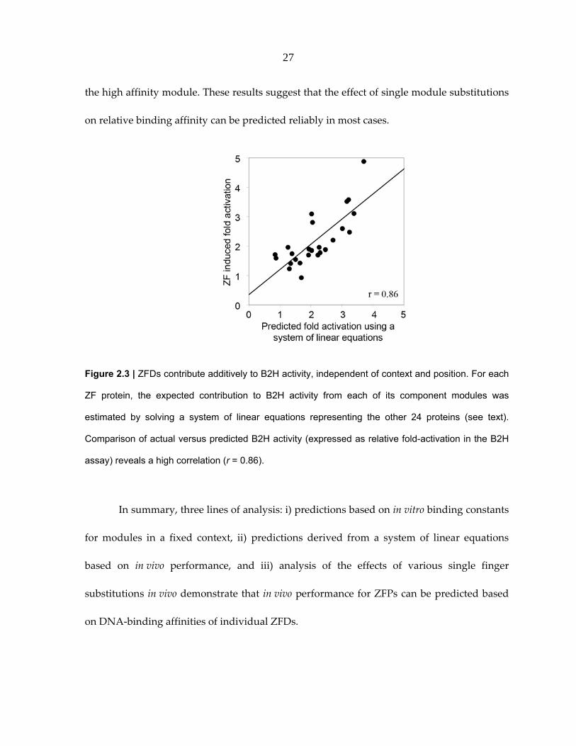
system of linear equations. Briefly, in constructing the 27 different three‐finger proteins,
nine ZFDs were used approximately 8‐10 times (~3 times at each of the possible 3 positions,
see Table 1). Assuming that affinities of individual ZFDs in a ZFP are additive, the B2H
activity of each ZFP was considered to result from its particular combination of modules
(Supplemental Figure 2.S1). Individual module contributions were calculated for each ZFP
using a leave‐one‐out linear system solution. Expected lacZ activation in the B2H assay for
each of the ZFPs was then predicted by summing individual module contributions. As
shown in Figure 2.3, expected levels of activation computed in this manner were highly
correlated with actual B2H activity measurements (r = 0.86).
The energy contributions computed using a system of linear equations to analyze
in vivo activity data from the B2H assay indicate that the GTA‐specific QSSSLVR module
binds with higher affinity than previous Kd estimates. This is consistent with our conclusion
based on inspection of energies computed from in vitro binding constants (Figure 2.2B). We
estimated a new value for this module by calculating the Kd that optimizes the correlation of
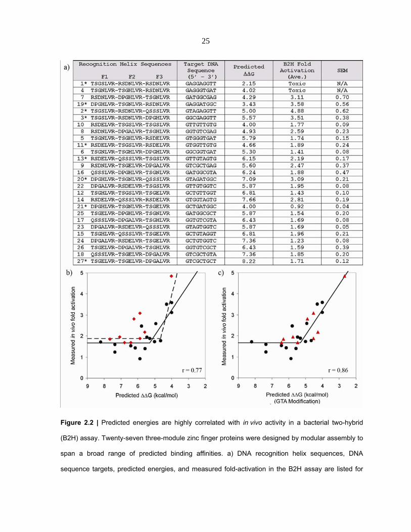
25
Figure 2.2 | Predicted energies are highly correlated with in vivo activity in a bacterial two-hybrid
(B2H) assay. Twenty-seven three-module zinc finger proteins were designed by modular assembly to
span a broad range of predicted binding affinities. a) DNA recognition helix sequences, DNA
sequence targets, predicted energies, and measured fold-activation in the B2H assay are listed for

26
Figure 2.2 | Continued each construct. Entries are sorted from highest to lowest predicted energy
Constructs marked with an asterisk were also tested in vitro (see Figure 2.4). b) ZFP activities in the
B2H assay are plotted versus predicted energies. The two constructs with greatest predicted affinity
were toxic to their host cells and therefore could not be plotted. Points shown as red diamonds
correspond to proteins containing the GTA-specific QSSSLVR module (see text). Best-fit lines from a
segmental linear regression model using all points (dashed line, r = 0.77), or excluding red points
(solid line, r = 0.86) are shown. (c) Same as (b), except that values indicated by red triangles were
adjusted assuming a binding affinity of 2.5 nM, rather than 25 nM, for the GTA-specific QSSSLVR
module; this increases the correlation coefficient to 0.86 (see text for details).
predicted energies with the B2H data. This approach resulted in an estimated Kd of 2.5 nM
for this module, 10‐fold lower than the previously reported value of 25 nM (15).
Incorporating this new estimate improved correlation between the in vitro energy model
and in vivo fold activation data (r = 0.86, Figure 2.2C).
To directly evaluate the effects of individual module affinities on in vivo
performance, sets of related ZFPs designed to vary at a single module position were
analyzed for differences in B2H activity (Table 2). For all 3 sets of ZFPs in which the F1
position was varied (while F2 and F3 were fixed), the greatest in vivo activity was observed
when the F1 position contained a high affinity module; the least activity was observed with
a low affinity module in this position. The same trend was observed for all 4 groups in
which the F3 position was varied while the F1 and F2 fingers were fixed. For sets in which
the F2 position was varied, only one strong module (TSGSLVR) and one moderate affinity
module (QSSSLVR) were tested. In these cases, the moderate affinity module outperformed
27
the high affinity module. These results suggest that the effect of single module substitutions
on relative binding affinity can be predicted reliably in most cases.
Figure 2.3 | ZFDs contribute additively to B2H activity, independent of context and position. For each
ZF protein, the expected contribution to B2H activity from each of its component modules was
estimated by solving a system of linear equations representing the other 24 proteins (see text).
Comparison of actual versus predicted B2H activity (expressed as relative fold-activation in the B2H
assay) reveals a high correlation (r = 0.86).
In summary, three lines of analysis: i) predictions based on in vitro binding constants
for modules in a fixed context, ii) predictions derived from a system of linear equations
based on in vivo performance, and iii) analysis of the effects of various single finger
substitutions in vivo demonstrate that in vivo performance for ZFPs can be predicted based
on DNA‐binding affinities of individual ZFDs.

28
Position Varied ID
F1 Helix
F2 Helix
F3 Helix
Fold Activation
7 RSDNLVR DPGHLVR TSGNLVR 3.11 16 QSSSLVR DPGHLVR TSGNLVR 1.88 25 TSGELVR DPGHLVR TSGNLVR 1.54 8 RSDNLVR DPGALVR TSGHLVR 2.59 17 QSSSLVR DPGALVR TSGHLVR 1.69 26 TSGELVR DPGALVR TSGHLVR 1.59 9 RSDNLVR TSGELVR DPGALVR 2.47 18 QSSSLVR TSGELVR DPGALVR 1.85
1
27 TSGELVR TSGELVR DPGALVR 1.71 10 RSDELVR TSGSLVR TSGSLVR 1.77 13 RSDELVR QSSSLVR TSGSLVR 2.19 11 RSDELVR TSGSLVR RSDELVR 1.89 14 RSDELVR QSSSLVR RSDELVR 2.81 12 TSGHLVR TSGSLVR TSGELVR 1.43
2
15 TSGHLVR QSSSLVR TSGELVR 1.96 1 TSGSLVR RSDNLVR RSDNLVR Toxic 2 TSGSLVR RSDNLVR QSSSLVR 4.88 3 TSGSLVR RSDNLVR DPGHLVR 3.51 4 TSGNLVR TSGHLVR RSDNLVR Toxic 5 TSGNLVR TSGHLVR RSDELVR 1.71 6 TSGNLVR TSGHLVR DPGHLVR 1.41 19 DPGHLVR TSGNLVR RSDNLVR 3.58 20 DPGHLVR TSGNLVR QSSSLVR 3.09 21 DPGHLVR TSGNLVR TSGELVR 0.92 22 DPGALVR RSDELVR TSGSLVR 1.95 23 DPGALVR RSDELVR QSSSLVR 1.69
3
24 DPGALVR RSDELVR TSGELVR 1.23
Table 2.2 | Single module substitutions in ZFPs alter target affinity. Individual ZFDs, represented by
their DNA recognition helix, are shaded to indicate their affinity class (Strong, Moderate, Weak, see
Table 1). For each subgroup (demarcated by horizontal lines), modules in two positions were held
constant while the position indicated in the leftmost column was varied. Fold activation denotes
performance of the ZFP in the bacterial two hybrid assay. Toxic refers to the poor growth of E. coli
cultures observed when cells expressed certain ZFPs.

29
In vitro DNA‐binding Affinities of ZFPs are Highly Correlated with Predicted Binding
Energies
Our success in estimating the activities of ZFPs in the B2H assay suggested that our
scoring scheme could be applied more generally to predict in vitro ZFP affinities. To test
whether activation measured in the B2H assay directly reflects DNA binding affinity for the
desired target site, nine of the twenty‐seven engineered proteins, along with a control Zif268
protein, were chosen for in vitro binding affinity measurements (Figure 2.4). Kd values were
determined using fluorescence anisotropy (FA), a rapid and reproducible solution‐based
DNA binding assay that allows computation of the bound fraction of a fluorescently‐labeled
ligand, based on the decrease in its rotational velocity due to binding (33,34).
As shown in Figure 2.5, binding affinity constants determined by FA were highly
correlated with predicted energies. The two ZF proteins with highest predicted affinities
were toxic to bacterial cells, prohibiting purification of sufficient quantities of protein for
in vitro analysis. Energies computed based on previously published in vitro affinity
measurements for modules in a fixed context (15) were proportional to the log of Kdʹs
measured in our experiments (r = 0.91) (Figure 2.5A). As before, assuming a Kd of 2.5 nM for
the QSSSLVR module significantly improved the correlation (r = 0.97). Predicted in vivo
activation levels generated by the leave‐one‐out, linear system method were also highly
correlated with experimentally determined binding constants (r = 0.93; Figure 2.5B). Thus,
results obtained using a rapid and reliable spectroscopic method suggest that ZFP binding
affinities measured in vitro generally correspond to results obtained in vivo using the B2H

30
system. This demonstrates that our rule‐based strategy can be used to predict ZFP DNA
binding affinity.
A Binding Energy Threshold for Zinc Finger Protein Function In Vivo
To evaluate the generality of this rule‐based approach, we calculated predicted
energies for another set of modularly assembled ZFPs that had been previously evaluated
using the B2H system (25). From 168 modularly assembled ZFPs, we selected all ZFPs
comprising GNN or TGG modules for which published in vitro DNA binding affinity
constants are available (measured in the F2 position of the standard Zif268 variant backbone
(15)). As shown in Figure 2.6A, based on a segmental linear regression model, binding
energies for 24 of these 26 ZFPs are highly correlated (r = 0.80) with reported B2H activity
measurements. These results are also in excellent agreement with the results described
above and shown in Figure 2.2B, although slightly higher activation levels were uniformly
observed in the latter experiments. Notably, both sets of experiments identify a ΔΔG of ~ 5
kcal/mol (corresponding to a Kd of ~100 nM) as the threshold for zinc finger function in vivo
(in bacterial cells). We also used the scoring function generated from the B2H experiments
performed by us (and shown in Figure 2.2C) to predict B2H activity for the 24 ZFPs
evaluated by Ramirez et al. (25). Again, the predicted and measured fold‐activation scores
were in close agreement, with a correlation coefficient of 0.79 (Figure 2.6B). Taken together,
these results suggest that the scoring function developed and evaluated may be generally
applicable to ZFPs assembled using the Barbas lab GNN modules.

31
Figure 2.4 | Determining binding affinity constants using fluorescence anisotropy (FA). B) A
representative in vitro binding isotherm obtained using FA. Data points for each ZFP were collected
using three separate purified protein preparations, each assayed for binding activity on a different
day. Curve fitting was performed using Prism. B) Kd values for seven modularly assembled ZFPs,
determined in FA experiments. Note that two ZFPs were toxic to host cells, preventing purification of
proteins in quantities required for in vitro analysis.
DISCUSSION
Using a rule‐based strategy which combines experimentally determined binding
energies of individual ZFDs, we were able to compute binding energies for ZFPs made from
a particular set of well‐characterized GNN‐modules (15). We also showed that these
predicted binding energies are in excellent agreement (r = 0.91; Figure 2.5A) with binding
affinity constants measured directly in vitro. Furthermore, we showed a strong correlation
between these computed binding energies and ZFP activities in a bacterial two‐hybrid

32
system for two different sets of modularly‐assembled three‐finger ZFPs. This is an
important advance because a ZFP which lacks activity in the B2H system will also have a
high probability of failing to function as a ZFN in human cells (24‐26). Thus, using only our
scoring method, researchers can now identify target sites which will have a high probability
of failing to yield functional zinc finger arrays by the method of modular assembly. Our
rule‐based strategy will thus allow researchers to focus their modular assembly efforts on a
smaller number of target sites with a higher probability of success.
We believe our results also provide one potential explanation for the discrepancy
between the overwhelming success rates for a previous in vitro report (35) and the low in
vivo success rates observed for ZFPs in the recent study of Ramirez et al.(25): many of the
modules used to perform modular assembly likely possess low affinities. Our data suggest,
in fact, that 30‐50% of potential three‐finger ZFPs made wholly from the Barbas GNN
modules will fail to function in the B2H system, a result in agreement with the recently
published results of Ramirez et al (25).
Although our results demonstrate that the affinities of individual ZFDs in a ZFP
array are additive, we also believe they lend additional support to the notion that context is
an important parameter that should be accounted for when engineering multi‐finger ZFPs
(i.e. that one single ZFD module will not always be optimal or adequate for recognition of
its cognate 3 bp subsite in different multi‐finger ZFP contexts). For example, our data show
that although a weak finger will sometimes be found in a non‐functional ZFP array (if it is
joined together with other weak affinity ZFDs), it will also sometimes be found in functional

33
arrays when paired with stronger affinity ZFDs. In addition, our data also show that
although strong fingers will sometimes be found in functional ZFPs, they can also be found
in non‐functional ZFPs. Furthermore, the use of three strong fingers in a ZFP can lead to
toxicity in E. coli cells. Although the precise mechanism of this toxicity is unclear, a
reasonable hypothesis is that excessively high affinity leads to binding to related but off‐
target sequences with sufficient affinity to cause biological consequences (essentially,
excessive affinity leading to problems of specificity). Thus, our data further re‐enforce the
ideas that individual ZFDs do not function completely independently and that the specific
attributes of neighboring fingers do matter in the context of engineering a multi‐finger ZFP.
The importance of context‐dependent effects also suggests that identification of
additional ZFDs with variable affinities for GNN triplets may be needed if the efficiency of
modular assembly is to be improved. If such ZFDs were available, it might be possible to
achieve higher success rates for modular assembly by using it to create multiple ZFPs for a
given target site so as to identify a combination that balances affinities (and presumably,
specificities) of its component ZFDs. However, it is also worth noting that the use of
additional modules would also increase the number of combinatorial possibilities and the
amount of labor required to perform modular assembly. A related point is that our findings
also suggest one possible reason why more complex selection‐based methods that account
for context‐dependent effects (e.g. the OPEN method recently described by Joung and
colleagues (26,32)) may be more successful than modular assembly: these methods are able
to balance the overall affinity and specificity of the final ZFP array by identifying optimal

34
combinations from various ZFDs with a range of affinities and specificities for their target 3
bp subsites.
The strong correlations among predicted binding energies, in vivo activities, and
in vitro binding affinity constants for the ZFPs analyzed in this work suggests that our rule‐
based approach might be extended to evaluate arrays assembled using other GNN modules
(17,19) and other non‐GNN modules. In this paper, we demonstrate two ways this could be
achieved: i) by directly measuring in vitro binding constants for modules in the F2 position
of a standardized ZFP framework; and ii) by computing individual module contributions to
ZFP binding as component variables of a system of linear equations that describe their
activities (measured in vivo in this work, but in vitro binding constants could also be used).
The energy scoring scheme proposed here will allow researchers to determine whether a
modular assembly strategy is likely to be feasible for specific targets of interest, based on
currently available well‐characterized modules, or whether an alternative selection‐based
engineering strategy should be considered.
A recent study on the use of zinc finger nucleases (ZFNs) for homologous
recombination cited lack of specificity as a primary determinant of ZFN‐mediated toxicity in
human cells (24). A likely mechanism for ZFN‐induced toxicity is through binding to
genomic sequences similar to the desired target sequence. As noted above, we observed
toxicity in bacterial cells for several ZFPs, even in the absence of a fused nuclease domain,
suggesting that ZFP binding to certain sites in genomic DNA can be toxic, particularly for
high affinity ZFPs. Although this is the first published report of such toxicity in bacterial

35
cells that we are aware of, it has been observed previously for several other sites (J.K. Joung,
unpublished observations). However, bacterial expression of ZFPs with affinities in the pM
range, with no toxic effects, has also been reported (19,36,37). High‐throughput chip or
microfluidics‐based DNA binding experiments (38‐41) could be used to obtain affinity and
specificity data for virtually every possible target site for a given ZFP, providing additional
insight into ZFP‐induced toxicity and into the fundamental rules that govern the affinity
and specificity of DNA recognition by zinc finger DNA binding proteins.
Figure 2.5 | In vitro affinity constants for ZFPs are highly correlated with predictions. Using affinity
constants for 7 ZFPs determined by fluorescence anisotropy (Figure 2.4B), log(Kd) is plotted against:
A) predicted energies (r = 0.91) and B) predicted B2H activity from a leave-one-out system of linear
equations analysis (r = 0.93).

36
A correlation between ZFP binding constants measured in vitro and functional
activity measured in vivo has also been observed by others using different reporter systems
(37). A similar degree of correlation was observed using the B2H system in our study
(Supplemental Figure 2.2). Our results further demonstrate that measurable ZFP activity in
an in vitro binding assay does not necessarily translate into adequate function in vivo, in
agreement with Beerli et al. (42). However, the energy threshold we determined for ZFP
activity in vivo, using B2H assays, corresponds to a Kd of ~ 100 nM, and thus differs from the
estimated threshold Kd of ~ 10 nM reported as the minimum affinity necessary for ZFP
function in mammalian cells (42). The significance of this difference between thresholds
determined in bacterial and mammalian cells is difficult to evaluate, given that functional
assays and Kd measurements were performed in different laboratories using different assays
and with ZFPs containing different numbers of fingers.
Stormo and colleagues have shown that the DNA‐binding specificity of ZFPs can be
effectively predicted from additive energy contributions of individual residues that make base‐
specific contacts with target site nucleotides (43‐47). Our results complement this idea by
demonstrating that the affinity of ZFPs also can be predicted, using affinity data for
component modules. Our application of the binding energy additivity concept differs
somewhat from that Stormo has used to predict specificity in that it assumes additivity of
affinities at the individual finger rather than individual residue level. Also, our approach
implicitly includes energetic contributions of residues that are not directly involved in base
contacts (e.g. phosphate contacts), as well as energetic contributions resulting from context‐

37
dependent effects that presumably occur among recognition helix residues within each
finger.
Figure 2.6. Predicted ZFP performance agrees with in vivo activity for an independently-generated
set of ZFPs. Data shown are for 24 of 26 ZFPs containing characterized GNN or TGG modules,
constructed and evaluated by Ramirez et al. 2008.(25) A) A segmental linear regression model
provides an excellent fit of in vivo ZF-induced fold activation measured in the B2H assay with
predicted binding energies (r = 0.80). B) ZF-induced fold activation values measured in the B2H
assay for 24 ZFPs from Ramirez et al. are also highly correlated (r = 0.79) with predicted fold-
activation levels calculated based on a scoring function derived from the segmental linear regression
model fit for the 25 ZFPs shown in Figure 2.2A (see text for details). Note: Predictions for 2 of 26
ZFPs containing characterized GNN or TGG modules from Ramirez et al. were considered outliers
(values were outside the range included in these graphs); they were not included in the regression
analysis.
The apparent simplicity of modular assembly has contributed to the current focus on

38
C2H2 ZFDs as the domains of choice for designing custom DNA binding proteins. Our
results make it possible, for the first time, to reliably identify prospective binding sites that
are unlikely to yield functional ZFDs by modular assembly using a set of GNN‐specific
finger modules. The rule‐based strategy presented here can provide accurate guidance for
both in vitro binding affinities and in vivo functionality for engineered zinc finger proteins
by computing energy contributions of individual ZFDs. We have updated the Zinc Finger
Targeter (ZiFiT) web server (http://bindr.gdcb.iastate.edu/ZiFiT) (48) so that it now provides
users with a list of potential ZFP‐target site pairs for a desired genomic sequence, scored
according to the procedures developed and validated in this work.
ACKNOWLEDGEMENTS
We thank members of our groups and colleagues, especially Fengli Fu, Deepak
Reyon, David Wright, Ronnie Winfrey, Ben Lewis, Bob Farnham, Abd Elhamid Azzaz, Les
Miller, Gaya Amarasinghe, and Vasant Honavar for their helpful suggestions and valuable
feedback. We also thank Guru Rao for the use of his spectrophotometer. This work was
supported in part by the following grants: NIH GM066387 (D.D.); NSF DBI0501678 (D.F.V.);
NIH GM069906 and GM078369 (J.K.J.); and graduate research assistantships provided by
USDA MGET 2001‐52100‐11506, NSF IGERT0504304, and ISU’s Center for Integrated
Animal Genomics (CIAG). Funding to pay the Open Access publication charges for this
article was provided by NFS DBI0501678.

39
Conflict of interest statement: None declared.
REFERENCES
1. Durai, S., Mani, M., Kandavelou, K., Wu, J., Porteus, M.H. and Chandrasegaran, S.
(2005) Zinc finger nucleases: custom‐designed molecular scissors for genome
engineering of plant and mammalian cells. Nucleic Acids Res, 33, 5978‐5990.
2. Klug, A. (2005) Towards therapeutic applications of engineered zinc finger proteins.
FEBS Lett, 579, 892‐894.
3. Porteus, M.H. and Carroll, D. (2005) Gene targeting using zinc finger nucleases. Nat
Biotechnol, 23, 967‐973.
4. Wu, J., Kandavelou, K. and Chandrasegaran, S. (2007) Custom‐designed zinc finger
nucleases: what is next? Cell Mol Life Sci, 64, 2933‐2944.
5. Cathomen, T. and Keith Joung, J. (2008) Zinc‐finger nucleases: the next generation
emerges. Mol Ther, 16, 1200‐1207.
6. Desjarlais, J.R. and Berg, J.M. (1993) Use of a zinc‐finger consensus sequence
framework and specificity rules to design specific DNA binding proteins. Proc Natl
Acad Sci U S A, 90, 2256‐2260.
7. Jamieson, A.C., Wang, H. and Kim, S.H. (1996) A zinc finger directory for high‐
affinity DNA recognition. Proc Natl Acad Sci U S A, 93, 12834‐12839.
8. Beerli, R.R., Segal, D.J., Dreier, B. and Barbas, C.F., 3rd. (1998) Toward controlling
gene expression at will: specific regulation of the erbB‐2/HER‐2 promoter by using

40
polydactyl zinc finger proteins constructed from modular building blocks. Proc Natl
Acad Sci U S A, 95, 14628‐14633.
9. Wolfe, S.A., Nekludova, L. and Pabo, C.O. (2000) DNA recognition by Cys2His2 zinc
finger proteins. Annu Rev Biophys Biomol Struct, 29, 183‐212.
10. Pabo, C.O., Peisach, E. and Grant, R.A. (2001) Design and selection of novel
Cys2His2 zinc finger proteins. Annu Rev Biochem, 70, 313‐340.
11. Segal, D.J. (2002) The use of zinc finger peptides to study the role of specific factor
binding sites in the chromatin environment. Methods, 26, 76‐83.
12. Pavletich, N.P. and Pabo, C.O. (1991) Zinc finger‐DNA recognition: crystal structure
of a Zif268‐DNA complex at 2.1 A. Science, 252, 809‐817.
13. Elrod‐Erickson, M., Rould, M.A., Nekludova, L. and Pabo, C.O. (1996) Zif268
protein‐DNA complex refined at 1.6 A: a model system for understanding zinc
finger‐DNA interactions. Structure, 4, 1171‐1180.
14. Miller, J.C. and Pabo, C.O. (2001) Rearrangement of side‐chains in a Zif268 mutant
highlights the complexities of zinc finger‐DNA recognition. J Mol Biol, 313, 309‐315.
15. Segal, D.J., Dreier, B., Beerli, R.R. and Barbas, C.F., 3rd. (1999) Toward controlling
gene expression at will: selection and design of zinc finger domains recognizing each
of the 5ʹ‐GNN‐3ʹ DNA target sequences. Proc Natl Acad Sci U S A, 96, 2758‐2763.
16. Wolfe, S.A., Greisman, H.A., Ramm, E.I. and Pabo, C.O. (1999) Analysis of zinc
fingers optimized via phage display: evaluating the utility of a recognition code. J
Mol Biol, 285, 1917‐1934.

41
17. Liu, Q., Xia, Z., Zhong, X. and Case, C.C. (2002) Validated zinc finger protein designs
for all 16 GNN DNA triplet targets. J Biol Chem, 277, 3850‐3856.
18. Dreier, B., Beerli, R.R., Segal, D.J., Flippin, J.D. and Barbas, C.F., 3rd. (2001)
Development of zinc finger domains for recognition of the 5ʹ‐ANN‐3ʹ family of DNA
sequences and their use in the construction of artificial transcription factors. J Biol
Chem, 276, 29466‐29478.
19. Bae, K.H., Kwon, Y.D., Shin, H.C., Hwang, M.S., Ryu, E.H., Park, K.S., Yang, H.Y.,
Lee, D.K., Lee, Y., Park, J. et al. (2003) Human zinc fingers as building blocks in the
construction of artificial transcription factors. Nat Biotechnol, 21, 275‐280.
20. Dreier, B., Fuller, R.P., Segal, D.J., Lund, C.V., Blancafort, P., Huber, A., Koksch, B.
and Barbas, C.F., 3rd. (2005) Development of zinc finger domains for recognition of
the 5ʹ‐CNN‐3ʹ family DNA sequences and their use in the construction of artificial
transcription factors. J Biol Chem, 280, 35588‐35597.
21. Alwin, S., Gere, M.B., Guhl, E., Effertz, K., Barbas, C.F., 3rd, Segal, D.J., Weitzman,
M.D. and Cathomen, T. (2005) Custom zinc‐finger nucleases for use in human cells.
Mol Ther, 12, 610‐617.
22. Beumer, K., Bhattacharyya, G., Bibikova, M., Trautman, J.K. and Carroll, D. (2006)
Efficient gene targeting in Drosophila with zinc‐finger nucleases. Genetics, 172, 2391‐
2403.

42
23. Segal, D.J., Crotty, J.W., Bhakta, M.S., Barbas, C.F., 3rd and Horton, N.C. (2006)
Structure of Aart, a designed six‐finger zinc finger peptide, bound to DNA. J Mol
Biol, 363, 405‐421.
24. Cornu, T.I., Thibodeau‐Beganny, S., Guhl, E., Alwin, S., Eichtinger, M., Joung, J. and
Cathomen, T. (2008) DNA‐binding Specificity Is a Major Determinant of the Activity
and Toxicity of Zinc‐finger Nucleases. Mol Ther, 16, 352‐358.
25. Ramirez, C.L., Foley, J.E., Wright, D.A., Muller‐Lerch, F., Rahman, S.H., Cornu, T.I.,
Winfrey, R.J., Sander, J.D., Fu, F., Townsend, J.A. et al. (2008) Unexpected failure
rates for modular assembly of engineered zinc fingers. Nat Methods, 5, 374‐375.
26. Maeder, M.L., Thibodeau‐Beganny, S., Osiak, A., Wright, D.A., Anthony, R.M.,
Eichtinger, M., Jiang, T., Foley, J.E., Winfrey, R.J., Townsend, J.A. et al. (2008) Rapid
ʺopen‐sourceʺ engineering of customized zinc‐finger nucleases for highly efficient
gene modification. Mol Cell, 31, 294‐301.
27. Wright, D.A., Thibodeau‐Beganny, S., Sander, J.D., Winfrey, R.J., Hirsh, A.S.,
Eichtinger, M., Fu, F., Porteus, M.H., Dobbs, D., Voytas, D.F. Joung, J.K. (2006)
Standardized reagents and protocols for engineering zinc finger nucleases by
modular assembly. Nat Protoc, 1, 1637‐1652.
28. Ryder, S.P., Frater, L.A., Abramovitz, D.L., Goodwin, E.B. and Williamson, J.R.
(2004) RNA target specificity of the STAR/GSG domain post‐transcriptional
regulatory protein GLD‐1. Nat Struct Mol Biol, 11, 20‐28.

43
29. Seidman, C.E., (1997) Transformation using calcium chloride In Ausubel, F.M. (ed.),
Current Protocols in Molecular Biology. John Wiley & Son, Inc., New York, Vol I
Unit 1.8
30. Lundblad, J.R., Laurance, M. and Goodman, R.H. (1996) Fluorescence polarization
analysis of protein‐DNA and protein‐protein interactions. Mol Endocrinol, 10, 607‐
612.
31. LiCata, V.J. and Wowor, A.J. (2008) Applications of fluorescence anisotropy to the
study of protein‐DNA interactions. Methods Cell Biol, 84, 243‐262.
32. Hurt, J.A., Thibodeau, S.A., Hirsh, A.S., Pabo, C.O. and Joung, J.K. (2003) Highly
specific zinc finger proteins obtained by directed domain shuffling and cell‐based
selection. Proc Natl Acad Sci U S A, 100, 12271‐12276.
33. Veprintsev, D.B. and Fersht, A.R. (2008) Algorithm for prediction of tumour
suppressor p53 affinity for binding sites in DNA. Nucleic Acids Res, 36, 1589‐1598.
34. Hayouka, Z., Rosenbluh, J., Levin, A., Maes, M., Loyter, A. and Friedler, A. (2008)
Peptides derived from HIV‐1 Rev inhibit HIV‐1 integrase in a shiftide mechanism.
Biopolymers.
35. Segal, D.J., Beerli, R.R., Blancafort, P., Dreier, B., Effertz, K., Huber, A., Koksch, B.,
Lund, C.V., Magnenat, L., Valente, D. et al. (2003) Evaluation of a modular strategy
for the construction of novel polydactyl zinc finger DNA‐binding proteins.
Biochemistry, 42, 2137‐2148.

44
36. Yang, W.P., Wu, H. and Barbas, C.F., 3rd. (1995) Surface plasmon resonance based
kinetic studies of zinc finger‐DNA interactions. J Immunol Methods, 183, 175‐182.
37. Kang, J.S. (2007) Correlation between functional and binding activities of designer
zinc‐finger proteins. Biochem J, 403, 177‐182.
38. Bulyk, M.L. (2006) DNA microarray technologies for measuring protein‐DNA
interactions. Curr Opin Biotechnol, 17, 422‐430.
39. Berger, M.F., Philippakis, A.A., Qureshi, A.M., He, F.S., Estep, P.W., 3rd and Bulyk,
M.L. (2006) Compact, universal DNA microarrays to comprehensively determine
transcription‐factor binding site specificities. Nat Biotechnol, 24, 1429‐1435.
40. Maerkl, S.J. and Quake, S.R. (2007) A systems approach to measuring the binding
energy landscapes of transcription factors. Science, 315, 233‐237.
41. Berger, M.F., Badis, G., Gehrke, A.R., Talukder, S., Philippakis, A.A., Pena‐Castillo,
L., Alleyne, T.M., Mnaimneh, S., Botvinnik, O.B., Chan, E.T. et al. (2008) Variation in
homeodomain DNA binding revealed by high‐resolution analysis of sequence
preferences. Cell, 133, 1266‐1276.
42. Beerli, R.R., Dreier, B. and Barbas, C.F., 3rd. (2000) Positive and negative regulation
of endogenous genes by designed transcription factors. Proc Natl Acad Sci U S A, 97,
1495‐1500.
43. Stormo, G.D. and Fields, D.S. (1998) Specificity, free energy and information content
in protein‐DNA interactions. Trends Biochem Sci, 23, 109‐113.

45
44. Benos, P.V., Bulyk, M.L. and Stormo, G.D. (2002) Additivity in protein‐DNA
interactions: how good an approximation is it? Nucleic Acids Res, 30, 4442‐4451.
45. Benos, P.V., Lapedes, A.S. and Stormo, G.D. (2002) Probabilistic code for DNA
recognition by proteins of the EGR family. J Mol Biol, 323, 701‐727.
46. Liu, J. and Stormo, G.D. (2005) Quantitative analysis of EGR proteins binding to
DNA: assessing additivity in both the binding site and the protein. BMC
Bioinformatics, 6, 176.
47. Liu, J. and Stormo, G.D. (2008) Context‐dependent DNA recognition code for C2H2
zinc‐finger transcription factors. Bioinformatics.
48. Sander, J.D., Zaback, P., Joung, J.K., Voytas, D.F. and Dobbs, D. (2007) Zinc Finger
Targeter (ZiFiT): an engineered zinc finger/target site design tool. Nucleic Acids Res,
35, W599‐605.

46
SUPPLEMENTARY DATA
Supplemental Figure 2.S1 | Energetic contributions of individual zinc finger modules can be solved
using a system of linear equations. If we assume that the measured B2H activity for each ZFP
construct is equal to the sum of its individual module contributions, each zinc finger module can be
represented as a variable (r-z) in a system of linear equations (e.g., r = RSDNLVR). Overlying box:
Each equation represents one specific three-module ZFP construct (e.g., 2), in which the coefficients
(0, 1 or 2) correspond to the number of times each module (variable) occurs within a specific three-
module ZFP construct analyzed in this study. Underlying table: Each row summarizes the number of
times each of 9 different modules (variables; columns r-z) is included in one of the 25 ZFP constructs
analyzed in this study. Values shown in the B2H activity column are the relative in vivo binding
activities, measured in bacterial 2-hybrid assays (B2H) performed in this study. Thus, for each ZFP
construct (equation), the energetic contribution of each individual module (value of each variable) can
be calculated by solving the system of linear equations representing all the remaining constructs (i.e.,
following a leave-one-out procedure), using the least squares method.

47
Supplemental Figure 2.S2 | Fold activation in the B2H assay is logarithmically correlated with
binding affinity. Seven ZFPs engineered in this study and Zif268 were assayed both for in vivo (B2H)
and in vitro (FA) activity. Results from the assays are highly correlated (r = 0.79).
Supplemental Figure 2.S3: | The affinity-based scoring method out-performs scoring schemes
implemented in Zinc Finger Tools (http://www.zincfingertools.org). Plots of observed ZFP-induced
activation in the B2H assay versus scores generated by the "Base" (a) and "XNN" (b) specificity
scoring functions reveal little correlation between predicted and actual ZFP function for the ZFPs
analyzed in this study. The corresponding plot for results obtained using the linear system affinity-
based scoring method developed in this work (Fig. 3) is reproduced here (c) to facilitate comparison.

48
Supplemental Figure 2.S3: | continued A much higher degree of correlation between predicted and
actual ZFP function is observed for this method. The affinity-based scoring method has been
implemented in the ZiFiT v3.0 server (http://bindr.gdcb.iastate.edu/ZiFiT/). (Note: Two additional
scoring functions implemented in Zinc Finger Tools, GNN and the GACT, generate the same score
for all GNN-only arrays and therefore have a correlation of 0.)

49
CHAPTER 3. INSIGHTS INTO FUNCTIONAL ZINC FINGER
PROTEINS
A paper to be submitted to Nature Methods
Jeffry D. Sander & Drena Dobbs
To the editor: Zinc finger proteins (ZFPs) can target linked functional domains to
specific sites within large genomes. Zinc finger nucleases, for example, have been
successfully used to enhance site‐specific homologous recombination by inducing double‐
stranded breaks in DNA(1). Individual zinc‐finger modules (ZFMs), the building blocks of
ZFPs, make base‐specific contacts with a DNA triplet by positioning an alpha helix in the
major groove of duplex DNA. By mutating residues in this ZFM recognition helix, ZFPs can
be engineered to bind specific DNA sequences(2).
A recent study in Nature Methods systematically evaluated in vivo ZFP performance
as a function of target site GNN triplet composition for modularly assembled ZFPs(3).
Analysis of the well‐characterized GNN module set of Barbas et al. (2)revealed that modules
targeting purine‐rich triplets generally bind with higher affinity than those targeting
pyrimidine‐rich triplets in in vitro binding assays(Supplemental Table 3.S1). These
individual module contributions have been identified as a critical component of in vitro and
in vivo function (4,5). Taken together, these observations have suggested that ZFP
performance in vivo is dependent on target site base composition. We have confirmed this

50
Figure 3.1 | Failure of ZFP-induced gene activation in a bacterial two-hybrid (B2H) functional assay
can be predicted using a leave-one-out linear system solution, based on DNA triplet base
composition. The graphs show measured increase in reporter gene activity (fold-activation) induced
by ZFP binding plotted versus predicted B2H activity.A) Target triplet composition distinguishes
sequences that are generally poor targets for ZFPs generated using modular assembly. B) ZFPs
assembled via OPEN typically function with high activity in vivo. However, a linear system solution
based on triplet composition does not accurately predict the level of activation.
hypothesis for modular assembly as illustrated in Figure 3.1A, by computing leave‐one‐out
linear system solutions for bacterial 2‐hybrid (B2H) fold activation using base counts by
triplet position for 193 modularly‐assembled ZFPs (3,4). As illustrated in Figure 3.1A,
elevated pyrimidine content, particularly a thymine in position one and cytosines in
positions two and three, in target sites was indicative of low in vivo activity, while more
favorable bases provided an upper bound for B2H activity. Compared with this set of

51
modularly assembled ZFPs, a set of ZFPs generated using the Oligomerized Pool
ENgineering (OPEN) method (6) were more active in vivo and capable of successfully
targeting sequences with less favorable base content(Figure 3.1B).
Figure 3.2 | Comparison of WebLogos for OPEN ZF module recognition helices (residues -1 to +6)
that recognize pyrimidine-rich versus purine-rich triplets in target DNA. Among ZFPs generated using
the OPEN procedure, DNA target sites enriched in pyrimidine bases (e.g., GCC, top row) result in an
increase in polar or positively-charged residues in typically “non-contacting” positions within the
recognition helix (e.g., S, R or K at position +1, S or R at +2, and K or R at +5), as compared with the
same positions in purine-rich helices (e.g., GAG, bottom row), which typically contain more favorable
positively-charged residues in the canonical base-contacting positions (-1, +3 and +6). F1, F2 and F3
indicate whether the observed sequences were selected in module in the 1st, 2nd or 3rd position within
a 3-finger ZFP.

52
Among zinc finger domains analyzed, those rich in cytosine contacts, typically
negatively charged aspartic acid and glutamic acid, were more likely to include residues
that favor backbone‐mediated contacts in the canonically “non‐base specific” helix positions
(+1, +2, +5) (Figure 3.2). As shown in Figure 3.3, analysis of 449 ZFPs generated by OPEN
(Supplemental Table 2)(6) reveals a steady increase in positive residues in non‐specific
contacting positions of the recognition helix as the number of purine specific amino acids
(HKRNQ) descreases at canonical contact positions (‐1, +3, +6) (Figure 3.3). This increase in
positively charged residues might provide favorable interactions with the negatively‐
charged DNA backbone, potentially required to achieve sufficient affinity for function
in vivo(4).
Our observations indicate that purine‐rich DNA sequences typically provide more
active target sites in vivo for modularly assembled ZFPs, given currently available reagents.
In addition, they suggest that the OPEN procedure may permit more efficient in vivo
targeting of sequences with higher cytosine content than modular assembly, in part by
selecting ZFPs with positively‐charged residues at positions in the recognition helix that are
not typically involved in base‐specific contacts. These lessons should facilitate ZFP target
site selection and the design of zinc finger domains that bind desired target sites with high
fidelity.

53
Figu
re 3
.3 |
In O
PE
N-g
ener
ated
ZFP
s, n
on-c
onta
ct re
sidu
es a
re s
elec
ted
base
d on
targ
et s
ite b
ase
com
posi
tion.
The
tabl
e sh
ows
the
aver
age
num
ber
of t
imes
eac
h am
ino
acid
s oc
curs
in t
he 9
can
onic
ally
non
-spe
cific
pos
ition
s (+
1, +
2, +
5) r
elat
ive
to t
he a
vera
ge
num
ber
of p
urin
e sp
ecifi
c re
sidu
es (
HK
RN
Q)
at p
ositi
ons
cano
nica
lly r
espo
nsib
le f
or b
ase
spec
ifici
ty (
-1,
+3,
+6).
Pos
ition
+4
is
typi
cally
Leu
cine
and
not
inc
lude
d.
As t
he n
umbe
r of
ZF
base
-con
tact
ing
resi
dues
com
mon
ly a
ssoc
iate
d w
ith p
urin
e sp
ecifi
catio
n
(HKR
NQ
) de
crea
ses
from
8 t
o 3,
the
tot
al n
umbe
r of
pos
itive
ly-c
harg
ed (
H,
K,
R)
resi
dues
in
the
non-
base
con
tact
ing
posi
tions
incr
ease
s. T
he n
umbe
r of
pos
itive
ly c
harg
es r
esid
ues
at n
on-b
ase
cont
actin
g po
sitio
ns (
+1,
+2,
+5)
incr
ease
s as
the
neg
ativ
ely
char
ged
resi
dues
incr
ease
s at
the
base
con
tact
ing
resi
dues
(-1
, +3,
+6)
. Th
is “
char
ge c
ompe
nsat
ion”
cou
ld p
rovi
de a
dditi
onal
DN
A-
bind
ing
affin
ity re
quire
d fo
r ZFP
func
tion
in v
ivo
(4) (
5)*
An
aspa
rtic
acid
in th
is p
ositi
on h
as b
een
show
n to
mak
e ba
se s
peci
fic c
onta
cts
on th
e op
posi
te s
trand
.

54
REFERENCES
1. Cathomen, T. and Keith Joung, J. (2008) Zinc-finger nucleases: the next generation
emerges. Mol Ther, 16, 1200-1207.
2. Segal, D.J., Dreier, B., Beerli, R.R. and Barbas, C.F., 3rd. (1999) Toward controlling
gene expression at will: selection and design of zinc finger domains recognizing each
of the 5'-GNN-3' DNA target sequences. Proc Natl Acad Sci U S A, 96, 2758-2763.
3. Ramirez, C.L., Foley, J.E., Wright, D.A., Muller-Lerch, F., Rahman, S.H., Cornu,
T.I., Winfrey, R.J., Sander, J.D., Fu, F., Townsend, J.A. et al. (2008) Unexpected
failure rates for modular assembly of engineered zinc fingers. Nat Methods, 5, 374-
375.
4. Sander, J.D., Zaback, P., Joung, J.K., Voytas, D.F. and Dobbs, D. (2008) An affinity-
based scoring scheme for predicting DNA-binding activities of modularly assembled
zinc finger proteins. NAR.
5. Beerli, R.R., Dreier, B. and Barbas, C.F., 3rd. (2000) Positive and negative regulation
of endogenous genes by designed transcription factors. Proc Natl Acad Sci U S A, 97,
1495-1500.
6. Maeder, M.L., Thibodeau-Beganny, S., Osiak, A., Wright, D.A., Anthony, R.M.,
Eichtinger, M., Jiang, T., Foley, J.E., Winfrey, R.J., Townsend, J.A. et al. (2008)
Rapid "open-source" engineering of customized zinc-finger nucleases for highly
efficient gene modification. Mol Cell, 31, 294-301.

55
Kd (nM) Triplet Helix 0.5 GAA QSSNLVR 1 GAG RSDNLVR 2 GCA QSGDLRR 3 GGA QRAHLER 3 GAT TSGNLVR 3 GAC DPGNLVR 5 GTT TSGSLVR 6 GGG RSDKLVR 9 GCG RSDDLVR 15 GGT TSGHLVR 15 GTG RSDELVR 25 GTA QSSSLVR 40 GGC DPGHLVR 40 GTC DPGALVR 65 GCT TSGELVR 80 GCC DCRDLAR
Supplemental Table 3.S1 | Barbas ZF modules that recognize pyrimidine-rich DNA target sites bind
with lower affinity than modules that recognize purine-rich target sites. ZF modules and Kd values
included in this table were generated by the Barbas group and published previously Segal et. al 1999.

56
CHAPTER 4. PREDICTING OLIGOMERIZED POOL
ENGINEERING (OPEN) SUCCESS USING ZINC FINGER TARGET
SEQUENCES
A paper to be submitted to BMC Bioinformatics
Jeffry D. Sander*, Deepak Reyon*, Maeder L. Maeder, Stacey Thibodeau‐Beganny,
J. Keith Joung & Drena Dobbs
*Co‐authors
ABSTRACT
Background: Oligomerized Pool ENgineering (OPEN) is a reliable method for
engineering C2H2 zinc finger proteins that bind custom DNA sequences with high activity
in vivo. To date, OPEN has successfully generated ZFPs with greater than three‐fold beta‐
galactosidase activation in a B2H assay for 98 of 134 nine base pair target sites comprised of
GNN and some TNN triplets. Because OPEN requires considerable time and expertise, it is
desirable to identify targets with the greatest chance of generating an active zinc finger
protein.
Results: Using alternate encodings for the 134 DNA target sites, Naïve Bayes
classifiers have achieved 84% overall accuracy with a correlation coefficient of 0.55, a

57
specificity of 0.83, sensitivity of 0.97 for our dataset evaluated using leaving‐one‐out cross‐
validation.
Conclusion: Classifiers trained to identify DNA sequences amenable for targeting
by OPEN‐generated zinc finger arrays can guide users to targets with elevated success rates.
These results suggest also a potential limit to the sequence space that can be targeted using
three module zinc finger arrays.
BACKGROUND
Custom zinc finger domains can be engineered to specifically bind DNA sequence
triplets through base specific contact in the major groove of double‐strand. Using the
modular assembly approach, extended sequences can be recognized by stringing multiple
domains together (1‐3). Three module arrays generated from a single solution for each
DNA triplet have been shown to specifically bind their 9 bp target in vitro(4). However,
other studies have demonstrated that many arrays fail to function in vivo (5,6). This has
been attributed to lack of affinity(6), and additional context dependencies(7), and chromatin
context(8). We have previously provided scoring schemes for proteins assembled by
modular assembly, which predict failure for most targets comprise of triplet subsites lacking
a 5ʹ guanine base (GNN triplets) (5,6).
Recently we have described zinc finger protein design using Oligomerized Pool
ENgineering (OPEN), which uses random assembly of up to 100 solutions for each
nucleotide triplet at each of three possible positions. This method typically generates ZFPs

58
that function well above basal levels in a Bacterial 2‐hybrid reporter system. However,
OPEN does not guarantee a ZFP with exemplary in vivo activity (greater than three‐fold
activation). Because OPEN requires considerable time and molecular biology expertise, it is
desirable to identify targets with a high probability of success. In this study we use target
sequence characteristics as input to Naïve Bayes and Support Vector Machine (SVM)
classifiers to predict which DNA sequences can be successfully target by OPEN ZFPs.
RESULTS
Identification of OPEN success based on target composition
Naïve Bayes, and SVM classifiers trained and tested on 134 validated nine base pair
target sequences, comprised of triplets with corresponding OPEN reagents, were used to
predict which sequences could be targeted by ZFP generated using OPEN with greater than
three fold activation in a B2H reporter assay (9). Classifiers include the following target
Table 4.1 | Performance measures of classifiers on OPEN targets.
Correlation Coefficient
Accuracy%
Specificity + %
Sensitivity + %
Window 0.48 81 83 94 Base Counts 0.58 84 83 99
Naïve
Bayes
Triplet Base Counts 0.55 84 83 97 Window 0.29 70 82 76
Base Counts 0.48 81 81 97
SVM
Triplet Base Counts 0.55 84 83 97

59
encodings a) 9 nucleotide target window b) nucleotide counts of the target site c) nucleotide
counts for each position within a triplet. Classifiers were evaluated on a set 134 target sites
(73% positive) using leave‐one‐out cross‐validation. As shown in Table 4.1, classifiers
optimized for correlation coefficient achieve correlation coefficients around of 0.5 with
overall accuracies slightly better than 80%.
When multiple target sites are available it may be desirable to identify those
with the greatest chance of succeeding, rather than optimizing the classifier based on
correlation coefficient. Receiver operating characteristic (ROC) curves in Figure 4.1
illustrate tradeoffs between sensitivity and the false alarm rate for various target encodings.
Figure 4.1 | Receiver Operating Characteristic (ROC) curves for Naïve Bayes and SVM based
classifiers.

60
To help understand how the classifiers use target composition to predict active
target sites we calculated average number of time each base occurs for active and inactive
targets (Figure 4.2). Average active sites comprise more guanines and fewer total thymines
than inactive targets. Because pools are available for GNN and TNN triplet subsites,
guanine and thymine counts are inflated as compared to adenine and cytosine counts. To
account for this bias and because most nucleotides specificity varies by position within
triplet subsites, average occurrences were calculated by nucleotide position within the
triplets (Figure 4.2B). The differences between the positive and negative classes identify
thymine composition at any triplet position as the difference between the positive and
negative class. Guanine, adenine, and cytosine appear equally more often in active sites,
offsetting the lower thymine composition. Target sites were also analyzed for base
propensities by absolute position.
Propensity (position specific)=
⎟⎟⎠
⎞⎜⎜⎝
⎛ N base arehat position tat that residues totalof %N base arehat position tat that residues interface of %2log
Residues propensities for thymine were negative, particularly the first and seventh
positions. All other residues typically showed positive, although slightly varied
propensities. Adenine and cytosine residues were not considered in positions 1, 4 and 7, as

61
the pools that recognize individual triplets are currently limited almost exclusive to GNN
and TNN triplets.
a) b)
0
1
2
3
4
5
G A C T
Active SitesInactive Sites
0
1
2
3
G1 G2 G3 A1 A2 A3 C1 C2 C3 T1 T2 T3
Active SitesInactive Sites
c)
-1.6
-1.2
-0.8
-0.4
0
0.4
1 2 3 4 5 6 7 8 9
G A C T
Figure 4.2 | Target base composition varies between the active and inactive target sites A) Variation
in the average occurrence of each bases differentiate classes independent of nucleotide position.
Availability of triplet pools is currently limited to GNN and TNN triplets which inflates the total
occurrence G and T relative to A and C. B) Residues counts grouped by position within target
triplets, indicates that thymine bases negatively impact binding at all three positions. C) Position
specific nucleotide propensities suggest composition affects zinc finger binding Nucleotide
propensities suggest Thymine typically makes a poor target. Other bases also appear to have
position specific affects, although to much smaller affects. Note: Because available pools target GNN
and TNN (one ANN) triplets, positions 1, 4, 7 are restricted T and G.

62
DISCUSSION
In this study, we have presented new experimental data and described methodology
for identifying DNA sequences most amenable to site specific targeting by three‐module
zinc finger arrays generated using OPEN. We implemented input encodings at various
levels of abstraction that perform comparably well on our dataset. All three methods
identify increased thymine composition as the primary impediment to significant in vivo
activity. Inputs, which included base position both absolute and within the triplet, suggest
positional context also plays a role. Although position independent base counts performs
comparably to more complex window and triplet position classifiers on the current dataset,
we expect that the position specific classifiers will perform better as additional ANN and
CNN pools become available(5).
CONCLUSION
We have shown that Target sequence can be used to identify which sequences have
an increased chance of success for ZFP targeting by OPEN. Optimized for correlation
coefficients, we were able to identify approximately half of the failed sites in this dataset
while incorrectly labeling a much smaller number of false negatives. These classifiers will
guide users of OPEN technology towards sites with an increased likelihood of success.

63
METHODS
Definition of active target
Active target sites are 9 bp DNA sequences for which OPEN has generated at least
one ZFP that induced production of beta‐galactosidase by greater 3‐fold as compared to
basal constructs (9,10) (11).
Data sets
A data set was generated from 134 DNA sequences targeted and selected for
antibiotic resistance by OPEN(11) and examined for activity in a Bacterial two‐hybrid assay
(Supplemental Table 4.S1) (9,10). Each 9 bp sequence comprised of three triplets for which
consortium generated pools are currently available(11).
Classifiers
1. Naïve Bayes is a probabilistic classifier that assumes the independence of each
attribute, this makes the model amenable to user interpretation without compromising
performance. Naïve Bayes classifiers commonly perform as well as more sophisticated
classifiers.(12). We used the implementation available in the WEKA package version
3.5.7(13). For each instance the classifier returns a classification of either “active” or
“inactive” based on the posterior probability. A label of “active” is assigned if:

64
( )( ) θ≥
====
xXInactiveCPxXActiveCP
||
Otherwise a label of “inactive” is assigned. The value of θ can be selected based on the
desired trade off between sensitivity and specificity. We tested the reliability of our classifier
using a leave‐one‐out cross validation on our dataset.
2. Support Vector Machines (SVM) find a hyper‐plane in high‐dimensional space
that maximizes the distance between the different classes of data in high dimensional space.
We implemented the SVM classifier using the wrapper class available for LIBSVM(14).
Because SVM classifiers are sensitive to the number of positive and negative instances in the
training set, and because our dataset is unbalanced (98 positive, 36 negative), we used a
variation of a leave‐one‐out cross validation technique. For each test case, we removed that
instance and generated 10 randomized balanced training sets, each consisting of 35 positive
and 35 negative instances. Probability estimates were averaged and assigned to each test
case.
Classifier inputs
The input for target window classifiers contains the 9 nucleotide target sequence in a
5ʹ ‐ 3ʹ direction. The input for classifiers using base counts consists of four numbers from 0
to 9, adding up to a total of 9, representing the number of times each of the four DNA bases

65
(G, A, C, T) was observed. The classifiers based on base counts by position within each
triplet consist of 12 numbers from 0 to 3, adding up to a total of 9, representing the number
of times each nucleotide was observed in each position of each triplet. For example, a target
consisting of GTT‐GAC‐GGC would have a 3 for the number of G’s occurring in the first
position of the triplet and a 0 for the number of G’s occurring in the third position of each
triplet.
Performance Measures
We report performance measures as combination of accuracy, correlation coefficient
(CC), specificity, and sensitivity as reported by Baldi et al (15)
Accuracy=FNTNFPTP
TNTP+++
+ CC=))()()((
**FNTNFPTNFPTPFNTP
FNFPTNTP++++
−
Specificity = FNTP
TP+
Sensitivity =FPTP
TP+
where True Positives (TP) is the number of targets correctly predicted as >3 fold activation;
False Positives (FP) is the number of targets incorrectly predicted as >3 fold activation; True
Negatives (TN) is the number of targets correctly predicted as <3 fold activation; False
Negatives (FN) is the number of targets incorrectly predicted as <3 fold activation.

66
Receiver Operating Characteristic (ROC) curve plots the tradeoff between hit rate versus the
false positive rate for different prediction thresholds(15).
Competing interests
The author(s) declare that they have no competing interests.
Authors’ contributions
JS was responsible for experimental design, analysis of results, initial draft of
manuscript, participated in discussions and manuscript revisions. DR parsed the data, ran
the machine learning algorithms, participated in discussions and manuscript revisions.
MM, and ST, and JF, and JK generated the experimental data and participated in manuscript
reviews. DD participated in discussions, analysis of results, and manuscript revision. All
authors read and approved the final manuscript.
ACKNOWLEDGEMENTS
This work was supported in part by the following grants: NIH GM066387 (D.D.);
NIH GM069906 and GM078369 (J.K.J.); and graduate research assistantships provided by
USDA MGET 2001‐52100‐11506, NSF IGERT0504304, and ISU’s Center for Integrated
Animal Genomics (CIAG). We thank members of our groups, particularly M. Terribilini for
his comments.

67
REFERENCES
1. Segal, D.J., Dreier, B., Beerli, R.R. and Barbas, C.F., 3rd. (1999) Toward controlling
gene expression at will: selection and design of zinc finger domains recognizing each
of the 5ʹ‐GNN‐3ʹ DNA target sequences. Proc Natl Acad Sci U S A, 96, 2758‐2763.
2. Liu, Q., Xia, Z., Zhong, X. and Case, C.C. (2002) Validated zinc finger protein designs
for all 16 GNN DNA triplet targets. J Biol Chem, 277, 3850‐3856.
3. Bae, K.H., Kwon, Y.D., Shin, H.C., Hwang, M.S., Ryu, E.H., Park, K.S., Yang, H.Y.,
Lee, D.K., Lee, Y., Park, J. et al. (2003) Human zinc fingers as building blocks in the
construction of artificial transcription factors. Nat Biotechnol, 21, 275‐280.
4. Segal, D.J., Beerli, R.R., Blancafort, P., Dreier, B., Effertz, K., Huber, A., Koksch, B.,
Lund, C.V., Magnenat, L., Valente, D. et al. (2003) Evaluation of a modular strategy
for the construction of novel polydactyl zinc finger DNA‐binding proteins.
Biochemistry, 42, 2137‐2148.
5. Ramirez, C.L., Foley, J.E., Wright, D.A., Muller‐Lerch, F., Rahman, S.H., Cornu, T.I.,
Winfrey, R.J., Sander, J.D., Fu, F., Townsend, J.A. et al. (2008) Unexpected failure
rates for modular assembly of engineered zinc fingers. Nat Methods, 5, 374‐375.
6. Sander, J.D., Zaback, P., Joung, J.K., Voytas, D.F. and Dobbs, D. (2008) An affinity‐
based scoring scheme for predicting DNA‐binding activities of modularly assembled
zinc finger proteins. NAR.
7. Liu, J. and Stormo, G.D. (2008) Context‐dependent DNA recognition code for C2H2
zinc‐finger transcription factors. Bioinformatics.

68
8. Beltran, A.S., Sun, X., Lizardi, P.M. and Blancafort, P. (2008) Reprogramming
epigenetic silencing: artificial transcription factors synergize with chromatin
remodeling drugs to reactivate the tumor suppressor mammary serine protease
inhibitor. Mol Cancer Ther, 7, 1080‐1090.
9. Hurt, J.A., Thibodeau, S.A., Hirsh, A.S., Pabo, C.O. and Joung, J.K. (2003) Highly
specific zinc finger proteins obtained by directed domain shuffling and cell‐based
selection. Proc Natl Acad Sci U S A, 100, 12271‐12276.
10. Joung, J.K., Ramm, E.I. and Pabo, C.O. (2000) A bacterial two‐hybrid selection
system for studying protein‐DNA and protein‐protein interactions. Proc Natl Acad
Sci U S A, 97, 7382‐7387.
11. Maeder, M.L., Thibodeau‐Beganny, S., Osiak, A., Wright, D.A., Anthony, R.M.,
Eichtinger, M., Jiang, T., Foley, J.E., Winfrey, R.J., Townsend, J.A. et al. (2008) Rapid
ʺopen‐sourceʺ engineering of customized zinc‐finger nucleases for highly efficient
gene modification. Mol Cell, 31, 294‐301.
12. Buntine, W. (1991), Proceedings of the seventh conference (1991) on Uncertainty in
artificial intelligence. Morgan Kaufmann Publishers Inc., Los Angeles, California,
United States.
13. Witten, I.H. and Frank, E. (2005) Data mining: practical machine learning tools and
techniques. 2nd ed. Morgan Kaufman, San Francisco.
14. Chang, C.C. and Lin, C.J. (2001) LIBSVM: a library for support vector machines.

69
15. Baldi, P., Brunak, S., Chauvin, Y., Andersen, C.A. and Nielsen, H. (2000) Assessing
the accuracy of prediction algorithms for classification: an overview. Bioinformatics,
16, 412‐424.

70
Supplemental Table 4.S1: Dataset of OPEN target sequences and labels
Target 5' - 3'
Label Target 5' - 3'
Label Target 5' - 3'
Label
GAGTGGTTA Inactive GGAGCTGTA Active GGCGACGAG Active GCGGTGGTT Inactive TTTGAAGGT Active GCTGAAGAA Active TAAGGGGAG Inactive GGGGGAGCT Active GAGGAGGTA Active GGGGCTGCG Inactive TGTGCCGTA Active GGAGATGTA Active GTATTTTGG Inactive GGAGGTGGA Active GACGGTGGA Active GTATGGTCT Inactive GTAGGGGTC Active GCGGACGTG Active GCTTGGGAC Inactive GCTTGGGGC Active GAGGGTTCT Active GAAGCTTTT Inactive GAAGCATAA Active GAGGCAGCA Active TGTGTGTTT Inactive GGCGGCGAG Active TTTGACGCT Active GAAGTATGG Inactive GGTGCTGTG Active GCTGGCGGT Active GAATTTGTG Inactive GAAGCAGCT Active GTTTGAGAT Active TTTGTCGGT Inactive GACGCTGGT Active GTGGACGAG Active GTTGTTTAA Inactive GCAGTGTGG Active GTTTGAGAG Active TTCGTGTGT Inactive GTGGGCGGA Active GGTGTGGGG Active GAGTTTGTT Inactive GTGGGGGGT Active GCAGCAGAA Active GTTTTTTGC Inactive TGTGTGGCA Active GGCTGAGTC Active TACGCCGGC Inactive GGCGACGAC Active GCCGTAGAT Active TTCGGTTCT Inactive GAGGAGGGG Active GTGTGGGGC Active TTCGTGTCT Inactive GGTGATGAA Active GACGCGGCG Active GAGGCTGCA Inactive GCAGGTGCC Active GGTGCTGCT Active TACGAAGGG Inactive GTTGATTGG Active GGATGAGAG Active GTGGTGTTT Inactive GGCGTTTTT Active GGTGGAGGA Active TTTTTTGCA Inactive GCAGAGGGT Active GACGTCGGG Active GATGAGTTT Inactive GCTGAAGAT Active GCAGCGGAT Active GCTTGAGCT Inactive GAGGAAGGG Active GGCGCTTCT Active GCCGCTGGA Inactive GATGAAGGG Active GCCGATGTC Active TTTGATGTT Inactive GGAGCCGAC Active GGAGGCGGC Active TGTGGCTGC Inactive GCTGCTGAC Active GAATGAGAC Active GACTGATGC Inactive GGTGGTGGA Active GACGAAGAT Active GGTGGATTA Inactive GGTGGAGCA Active GCTTGGGTC Active TTCGCATCT Inactive GTCGTGGAA Active GCTGGGGGA Active GATGCGGTG Inactive GGCGGTGCC Active GCGGGGGCG Active GCTGGCTCT Inactive GCGGCCGCG Active GCGGTGGCG Active GGGGCTGCC Inactive GCTGACGAA Active GCGTGGGCG Active GCTGCAGCC Inactive GGCTGAGGC Active GCGTTGGCG Active GCCGTCGCC Inactive GCCGGTTGG Active GAATTGGCG Active GTGGAATTA Active GGAGCGGCA Active GAGGGGGCG Active GCCGCTGCT Active GCTGAGGCT Active GATGAGGCG Active GGAGCTGCC Active GGCGCGGCA Active GACTGGGCG Active TCTGAGTGG Active GGGTAGGTG Active AACGCAGGC Active GGAGCAGCA Active GCCGATGCT Active AACGCCGAT Active GTCGCCGCT Active GCTGAAGCA Active GGCGGCGCC Active GCAGCAGGT Active GTGGGTGCG Active GCCTTTGAG Active GATGAAGCC Active GTCGACGGG Active GCCGACGTC Active GAGTTGTAA Active GGCGCCGCC Active
Active implies OPEN generated at least one zinc finger array that resulted in at least three fold
activation of beta-galactosidase.

71
CHAPTER 5. ZINC FINGER TARGETER (ZIFIT): AN ENGINEERED
ZINC FINGER/TARGET SITE DESIGN TOOL
A paper published in Nucleic Acids Research ‐ Web Server Issue
Jeffry D. Sander, Peter Zaback, J. Keith Joung, Daniel F. Voytas & Drena Dobbs
ABSTRACT
Zinc Finger Targeter (ZiFiT) is a simple and intuitive web‐based tool that facilitates
the design of zinc finger proteins (ZFPs) that can bind to specific DNA sequences. The
current version of ZiFiT is based on a widely employed method of ZFP design, the ʺmodular
assembly” approach, in which pre‐existing individual zinc fingers are linked together to
recognize desired target DNA sequences. Several research groups have described
experimentally characterized zinc finger modules that bind many of the 64 possible DNA
triplets. ZiFiT leverages the combined capabilities of three of the largest and best
characterized module archives by enabling users to select fingers from any of these sets.
ZiFiT searches a query DNA sequence for target sites for which a ZFP can be designed using
modules available in one or more of the three archives. In addition, ZiFiT output facilitates
identification of specific zinc finger modules that are publicly available from the Zinc Finger
Consortium. ZiFiT is freely available at http://bindr.gdcb.iastate.edu/ZiFiT/.

72
INTRODUCTION
Zinc fingers, the most abundant DNA‐binding motifs encoded in eukaryotic
genomes, offer perhaps one of the best understood protein‐DNA binding mechanisms (1‐4).
Engineered zinc finger proteins (ZFPs) have significant potential as tools for gene regulation
and genome modification because they can be used to target functional domains to virtually
any desired location in any genome. For example, engineered zinc fingers fused to a non‐
specific nuclease domain can be used to create double‐stranded DNA breaks for the purpose
of inducing high‐efficiency homologous recombination at specific genome loci (5‐10).
ZFPs provide a versatile framework for designing proteins with new DNA‐binding
specificities. One simple method for making ZFPs is to assemble pre‐existing single finger
ʺmodulesʺ (with known specificities) into multi‐finger arrays. Each ZF module recognizes
approximately three base pairs of DNA and, when appropriately joined together, the
resulting ZF arrays are capable of specifically recognizing longer DNA sequence motifs
(Figures 5.1 and 5.2). Three research groups have each described and characterized separate
archives of ZF modules for constructing multi‐finger arrays (11‐15). Recently, the Zinc
Finger Consortium (http://www.zincfingers.org) has incorporated all three of these archives
into a standardized framework that facilitates rapid assembly of multi‐finger arrays using a
simple restriction digest‐mediated cloning strategy (16).
In collaboration with the Zinc Finger Consortium, we have developed a web‐based
server, ZiFiT (Zinc Finger Targeter), which facilitates the design of zinc finger proteins that

73
recognize specific DNA sequences. ZiFiT has a simple interface through which users
provide the DNA sequence of the gene or region within which they wish to search for
Figure 5.1 | Zinc Finger Modules and Binding Sites. A) A canonical zinc finger module consists of
two anti-parallel beta strands and an alpha helix, with a zinc ion coordinated by the conserved
cysteine(C) and histidine(H) residues pairs. The numbered amino acid residues at positions -1, +2,
+3, and +6, relative to the amino-terminal end of the alpha helix, can form important base-specific
contacts in the major groove of the double-stranded target DNA (not shown). B) A ZFP consisting of
3 linked ZF modules binds its target DNA site with amino acids of the recognition alpha helices (in the
N- to C-terminal direction) contacting consecutive nucleotides in DNA running in the 3' to 5' direction.
This can lead to confusion because the DNA target site is typically referred to in the 5' to 3' direction.
Note that an “unnatural” extended array is shown to better illustrate the critical amino acid-nucleotide
contacts. Structure diagrams were generated using PyMol (http://www.pymol.org).
potential target ZFP binding sites. Target sites can be either those bound by a single ZFP
(Figure 5.2A) or by a dimeric zinc finger nuclease (in which ZFP arrays bind two “half‐sites”
separated by a fixed‐length ʺspacer,ʺ Figure 5.2B). ZiFiT output provides users with

74
potential ZFP target DNA sequence(s) within the region of interest, together with a
corresponding array of ZF modules needed to construct the desired ZFPs. The output
includes information specifying the source of each module and a reference number that
uniquely identifies each module within the standardized Zinc Finger Consortium modular
assembly archive (16).
Figure 5.2 | Comparison of Single ZF Array and Dimeric ZF Nuclease Sites. a) Zinc Finger Protein
Bound to a Single ZF Array Target Site: A single ZF array target site consists of three to eight
adjacent DNA triplets (9 - 24 nt) on the same strand of DNA. Each triplet of DNA is recognized by one
"finger" or "ZF module" (light blue ovals). The cartoon illustrates an array with three fingers (ZF1, ZF2,
ZF3) bound to a single ZF array target site, in this case, a 9 nt DNA sequence. b) Zinc Finger
Nuclease Bound to a Dimeric ZF Nuclease Target Site: A ZF nuclease target site consists of two
single ZF array sites on complementary DNA strands, separated by a spacer of 5 or 6 nt. In this
configuration, FokI endonuclease monomers (dark blue spheres), covalently linked to the C-terminal
end of each ZF array domain, can form an active dimeric nuclease and induce a double-stranded
break in the spacer DNA between two ZF array binding sites.

75
MATERIALS AND METHODS
Zinc Finger Module Structure
A canonical zinc finger module consists of two anti‐parallel beta strands and an
alpha helix coordinated with a zinc ion via cysteine and histidine contacts (1‐4). Amino
acids in positions ‐1 to 6 (numbered relative to the start of the alpha helix) recognize specific
DNA triplet sequences, primarily by forming base‐specific contacts in the major groove of
the double‐stranded target DNA (Figure 5.1A). ZF modules are often referred to according
to these ʺrecognitionʺ residues in the alpha helix, listed in N‐ to C‐terminal direction; we
refer to the other amino acids in the module as the finger backbone. As illustrated in Figure
5.1B, a ZFP binds its target DNA site with the amino acids of the recognition helices (from
N‐ to C‐terminus) contacting consecutive nucleotides in DNA running in the 3ʹ to 5ʹ
direction. This can lead to confusion because the DNA target site is typically referred to in
the 5ʹ to 3ʹ direction.
Zinc Finger Module Sets
ZiFiT was designed to take advantage of ZF module sets developed and
characterized by three independent research groups. Users can specify whether modules
should be chosen from only one, two, or all three of these module sets (designated as
Barbas, Sangamo, or ToolGen, based on the group that described them – see below). The
Zinc Finger Consortium has generated plasmids encoding modules from all three sets in a
standardized framework, allowing users to rapidly assemble desired ZF proteins using

76
individual modules from sets of their choice (16). ZiFiT was developed in conjunction with
the Zinc Finger Consortium and automatically provides reference numbers for requesting
these ready‐to‐assemble plasmid modules from Addgene, a non‐profit plasmid distribution
service (http://www.addgene.org/zfc/). It should be noted that the designers of each module
set did not necessarily intend for users to mix modules from different sets within a single
ZFP array. However, several studies have generated functional ZFPs by combining modules
from different sets (10,14).
Barbas Modules ‐ These ZF modules were developed using a combination of phage
display and rational design methods by the Barbas laboratory at The Scripps Research
Institute (11,12,15). Modules are available for recognition of all GNN triplets, most ANN
and CNN triplets, and a few TNN triplets. These Barbas modules were developed under the
assumption that individual ZF modules have virtually complete positional independence,
i.e., their recognition properties are not dramatically affected by their position within an
array or by the identities of neighboring zinc fingers. The current version of ZiFiT includes
49 distinct Barbas modules (see Supplementary Table 5.S1).
Sangamo Modules ‐ Sangamo ZF modules were designed at Sangamo BioSciences
Inc., and are currently available for all GNN triplets and a smaller number of “non‐GNN
triplets (13,17,18). Sangamo modules were developed under the assumption that the
position of a module within a three‐finger array can affect its recognition properties (e.g.

77
amino‐terminal finger compared with carboxy‐terminal finger). Each of the three positions
within a three‐finger array has a distinct ZF module developed for a given triplet at that
position. For this reason, if a user chooses to use Sangamo modules, ZiFiT restricts the user
to the design of three‐finger arrays in which positional context is preserved. The current
version of ZiFiT includes 57 position‐specific Sangamo modules (see Supplementary Table
5.S1).
ToolGen Modules ‐ ToolGen modules are naturally occurring human zinc fingers
that were identified and characterized by ToolGen Inc. and are available for a variety of
nucleotide triplets (14). The current version of ZiFiT includes 35 distinct ToolGen modules
(see Supplementary Table 5.S1).
ZiFiT Software: Designing ZFPs that Recognize Unique Target Sites in DNA
ZiFiT facilitates the design of ZFPs using the ʺmodular assemblyʺ approach in which
pre‐existing individual zinc finger modules are linked together to recognize desired target
sequences. ZiFiT is available at http://bindr.gdcb.iastate.edu/ZiFiT/ or can be accessed
under Software Tools from the Zinc Finger Consortium website at
http://www.zincfingers.org/software‐tools.htm). The ZiFiT website includes instructions
and examples as well as several links to other websites that provide background
information regarding zinc finger protein design. A FAQs page provides additional
guidance for new users.

78
Zinc Finger Target Sites
Single ZFP Binding Sites ‐ Individual zinc finger modules can be linked together to
form multi‐finger arrays that recognize specific sequences in double‐stranded genomic
DNA (Figure 5.2A). These multi‐finger arrays can be fused to other protein domains, such as
transcriptional activation or repression domains, in order to target them to specific locations
within large genomes (1‐4). Because a single ZF recognition helix typically binds three
contiguous nucleotides in DNA, most binding sites for single ZF proteins (which we
designate ʺsingle ZF array binding sitesʺ) have lengths that are multiples of three base pairs.
However, certain ZF modules containing aspartic acid in the +2 position of the DNA
recognition helix appear to recognize four nucleotides. This can result in ʺtarget site
overlapʺ between adjacent ZF modules or, if the Asp‐containing module occurs in the first
position of an array, the requirement for an additional 3ʹ nucleotide in the ZF array binding
site (19).
Dimeric Zinc Finger Nuclease Sites ‐ Zinc finger nucleases (ZFNs) consist of a zinc
finger array fused to a non‐specific dsDNA nuclease (e.g., the nuclease domain of the Type
IIS restriction enzyme FokI) (5,6,8,10). ZFNs made with FokI nuclease are catalytically active
only as dimers (20). Thus, a full ZFN target site consists of two ZF ʺhalf‐sitesʺ on
complementary DNA strands, separated by a ʺspacerʺ of five or six base pairs, as shown in
Figure 5.2B (6,21). In this report, we designate the two ʺhalf‐sitesʺ together with the spacer a
ʺdimeric ZF nuclease site.ʺ

79
Program Input
First‐time users must complete a quick and easy registration. After logging in, users
indicate which type of ZFP they wish to design, either single zinc finger arrays or dimeric
zinc finger nucleases (ZFNs) before proceeding to the main sequence input screen
(Figure 5.3). Check boxes near the top of the page allow users to select which ZF module
sets (Barbas, Sangamo, ToolGen) they wish to include in their search. The DNA sequence of
interest (i.e., the sequence of the region within which the user wishes to identify potential
ZFP target sites) can be submitted either in FASTA format or as a raw DNA sequence in
standard 5’ to 3’ orientation (both spaces and numbers are ignored). Users can specify the
number of DNA triplets to include in each ZF array target site using drop‐down menus
below the sequence input box.
Zinc finger proteins with less than three modules do not typically possess affinities
needed to bind their targets, while target sites of 18nt (6 modules) are typically enough to
ensure their uniqueness in eukaryotic genomes (22). ZiFiT restricts target site sizes to 3 ‐ 8
to triplets (corresponding to 3 ‐ 8 ZF modules) for standard single ZF arrays and target sizes
to 3 or 4 triplets (corresponding to 3 ‐ 4 ZF modules) for both the left and right arrays of a
dimeric ZF nuclease site. In dimeric ZFN target sites, the length of the spacer region, within
which the active dimeric nuclease cleaves, can be defined by the user as either five or six
DNA bases. The preferred distance is six bases (6,21).

80
Figure 5.3 | ZiFiT Input Window: In this example, the user has chosen to search for potential dimeric
zinc finger nuclease (ZFN) sites. The DNA sequence of interest can be supplied in either FASTA
format or as a raw DNA sequence, in standard 5’ to 3’ direction. Above the sequence input box,
check boxes allow users to specify which sets of characterized ZF modules should be considered in
designing a ZFN to recognize the desired target site. Below the sequence input box, drop down
menus allow users to specify how many DNA triplets should be included in each array site: a single
array site may contain from 3 to 8 triplets; a dimeric nuclease site may contain 3 or 4 triplets in each
array site (Left and Right), separated by a Spacer of either 5 or 6 nucleotides. Information that
appears below the "Submit" button in this screenshot corresponds to "Advanced Option" parameters
(see text). These have been revealed by selecting the "Advanced" link (to the right of the Submit
button) which toggles between "Basic" and "Advanced."

81
Advanced options can be accessed by selecting the ʺAdvancedʺ link in the lower
right corner of the input page (as shown in Figure 5.3; this box toggles to a “Basic” link,
which hides the Advanced Options). Advanced options allow the user to: i) define “Triplet
Composition” by specifying the minimum or maximum number of GNN, ANN, CNN, TNN
triplets to include in potential target sites; ii) choose whether to ʺignore Asp overlap,ʺ which
refers to the target site overlap that can occur with Asp in position +2 of the helix (see
Materials and Methods); iii) choose to search one or both strands of the input DNA
sequence for target sites, when searching for a single array site. By definition both strands
are considered in a ZFN site search, therefore this option is not available in the ZFN
window.
Guidance for using advanced options is provided on the ZiFiT Instructions and FAQ
pages. For example, users can adjust ʺTriplet Compositionʺ to enforce a bias for target sites
containing mostly GNN triplets. This can improve chances of successful ZFP design because
GNN‐specific ZF modules have been more thoroughly characterized than other modules.
The ʺIgnore Asp overlapʺ option is particularly useful for troubleshooting: for example Asp
in position +2 can cause unexpected results when the target site falls on the very 3ʹ end of a
submitted sequence. An Asp in position +2 of the first module specifies an additional 3ʹ
base. If this base is not available, a partial target site can be detected by ZiFiT and partial
matches are not returned to the user. Thus, this option should be used only by advanced
users or for troubleshooting why a suspected site is not returned by ZiFiT

82
Program Output
ZiFiT searches for potential ZFP target sites in the input DNA sequence, based on
the module sets chosen by the user (and other user‐specified restrictions described above).
For each “hit” within the query DNA sequence, ZiFiT output consists of two components: a
ʺDNA target siteʺ and a table of corresponding ʺZFP modulesʺ that could be assembled to
recognize that target site. As shown in Figure 5.4, the double‐stranded DNA sequence
(displayed at top) represents a potential dimeric ZFN target site identified by ZiFiT.
Recognition triplets in the target site are color‐coded to match corresponding entries in the
table of ZFP modules (displayed below the target site sequence). The table lists ZF modules
that have been experimentally shown to bind triplets in the displayed target site. If more
than one module is available for a given triplet position, all modules for that position are
displayed.
Each ZF module entry includes the recognition helix sequence of the module, the
color‐coded DNA sequence of its corresponding target triplet, a reference number for
requesting the ZF module, if desired, from the Addgene Zinc Finger Consortium
website (http://www.addgene.org/zfc/), and the original source of the module (Barbas,
Sangamo, or Toolgen). When ZiFiT identifies more than one potential ZFP/target site pair
within the query sequence, additional hits are also displayed and can be viewed by
scrolling. In the current implementation, the order in which ZiFiT returns potential target
sites is determined solely by their position within the query sequence. In some cases, ZiFiT

83
may return a large number of potential target sites and/or several corresponding potential
ZFP designs. Tips for choosing among these are provided on the ZiFiT FAQs page.
Figure 5.4 | Example of ZiFiT Output for a dimeric ZF nuclease target site. For each "hit" identified
by ZiFiT, the output consists of the DNA target site sequence and two tables that contain
corresponding ZF protein modules that could be assembled to recognize the displayed target site.
Each target site is displayed as a double-stranded DNA sequence, with triplets color-coded to
facilitate identification of the corresponding ZF binding modules in the accompanying table. Each row
in the ZF module table displays information for a single zinc finger module. Entries in the columns
indicate the Finger position (Finger) of this module, the amino acids at positions -1 through +6

84
corresponding to its recognition helix (HELIX), the color-coded DNA triplet it recognizes (TRIPLET), a
reference number to facilitate ordering the ZF module from the Zinc Finger Consortium
(REFERENCE NUMBER), and the module set from which the indicated ZF module was derived
(MODULE SOURCE). If more than one ZF module is known to recognize a given triplet sequence, all
Figure 5.4 | continued available modules for that position are displayed. The screenshot shown
here has been truncated to display complete output for only one of several potential dimeric ZF
nuclease target sites identified within the input query DNA sequence.
Assembling and Evaluating Engineered ZFPs
ZiFiT is designed to facilitate the experimental generation of ZFPs by providing
output that includes reference numbers for public‐domain, ready‐to‐assemble modules
generated by the Zinc Finger Consortium. These resources can greatly simplify the ZF
protein assembly process. All ZF modules used in ZiFiT can be requested through Addgene,
a non‐profit plasmid distribution service (see http://www.addgene.org/zfc for additional
details). A detailed protocol for assembling and evaluating zinc finger proteins using this
strategy has been published recently in Nature Protocols (16). Other research groups have
also described PCR‐based zinc finger assembly protocols (23,24). Additional general
information regarding zinc finger proteins and their applications is available at the Zinc
Finger Consortium website at http://www.zincfingers.org.
FUTURE DIRECTIONS
ZiFiT will be updated regularly to reflect growth in ZFP assembly resources (e.g.,
collections of zinc finger modules and arrays) and increasing availability of experimental

85
data regarding the in vivo efficacy of specific ZF proteins. While several studies have
demonstrated that ZFPs engineered using the modular assembly approach can function
successfully, other studies (both published and unpublished) have shown that designed
ZFPs function with variable degrees of success (14,25,26). As noted above, in the current
version of ZiFiT, the order in which potential ZF array target sites are displayed in the
output window corresponds to the order in which they occur within the query DNA
sequence ‐ no ranking is implied. A feature we plan to implement in the next version of
ZiFiT is a scoring function that will rank and provide information for each hit (i.e., for each
ZFP/target site pair) in the ZiFiT output. This scoring information and ranked listing of
experimentally validated ZF target sites will provide additional guidance to assist users in
designing ZF arrays that are most likely to function. In collaboration with the Zinc Finger
Consortium, members of our groups have designed and implemented a Zinc Finger
Experiment Database, with the goal of collecting and making accessible results from all
available zinc finger experiments. The next version of ZiFiT will be closely integrated with
this database. It will provide users with relevant results regarding ZF modules and potential
ZF target sites within a query sequence that are identical or highly similar to modules and
target sites that have been experimentally tested. We will update ZiFiT to support
additional ZF modules and/or methods as they are developed and made available by the
zinc finger research community.
Related Software

86
A server offering software similar to ZiFiT, named Zinc Finger Tools
(http://www.zincfingertools.org) has been developed by the Barbas lab at the Scripps
Institute, for use with their ZF module set (27). One significant advantage of ZiFiT is that it
offers users the ability to choose modules from three different module sets instead of only
the single Barbas set, and users may combine modules from different archives if desired.
When using the Barbas Zinc Finger Tools software, a user will only have one potential
protein to test for any given target site. Because modular assembly does not always yield
functional proteins (14,25) it is important to have multiple potential zinc finger arrays for
any given target site. Also, ZiFiT was intentionally designed to complement experimental
resources made publicly available by the Zinc Finger Consortium and distributed through
Addgene. Zinc Finger Tools offers a scoring function to assist users in choosing among
potential zinc finger target sites, but as noted on the Zinc Finger Tools website itself:
rigorous empirical validation of the scoring function awaits further experimental data.
ACKNOWLEDGMENTS
We respectfully acknowledge those who have shared their results with the zinc
finger research community. We also thank users and referees for providing many helpful
suggestions that have improved the ZiFiT server.
This work is supported in part by NIH grant GM066387 (D.D) & NSF grant DBI
0501678 (D.F.V.) and graduate research assistantships provided by USDA MGET grant 2001‐

87
52100‐11506 and ISU Center for Integrated Animal Genomics (CIAG). J.K.J is supported by
NIH grants GM0699006 and GM072621.
Funding to pay the Open Access publication charges for this article was provided by
NFS Grant DBI 0501678
Conflict of interest statement. None declared.
REFERENCES
1. Wolfe, S.A., Nekludova, L. and Pabo, C.O. (2000) DNA recognition by Cys2His2 zinc
finger proteins. Annu Rev Biophys Biomol Struct, 29, 183‐212.
2. Pabo, C.O., Peisach, E. and Grant, R.A. (2001) Design and selection of novel
Cys2His2 zinc finger proteins. Annu Rev Biochem, 70, 313‐340.
3. Moore, M. and Ullman, C. (2003) Recent developments in the engineering of zinc
finger proteins. Brief Funct Genomic Proteomic, 1, 342‐355.
4. Klug, A. (2005) Towards therapeutic applications of engineered zinc finger proteins.
FEBS Lett, 579, 892‐894.
5. Kim, Y.G., Cha, J. and Chandrasegaran, S. (1996) Hybrid restriction enzymes: zinc
finger fusions to Fok I cleavage domain. Proc Natl Acad Sci U S A, 93, 1156‐1160.
6. Bibikova, M., Carroll, D., Segal, D.J., Trautman, J.K., Smith, J., Kim, Y.G. and
Chandrasegaran, S. (2001) Stimulation of homologous recombination through
targeted cleavage by chimeric nucleases. Mol Cell Biol, 21, 289‐297.

88
7. Porteus, M.H. and Baltimore, D. (2003) Chimeric nucleases stimulate gene targeting
in human cells. Science, 300, 763.
8. Wright, D.A., Townsend, J.A., Winfrey, R.J., Jr., Irwin, P.A., Rajagopal, J., Lonosky,
P.M., Hall, B.D., Jondle, M.D. and Voytas, D.F. (2005) High‐frequency homologous
recombination in plants mediated by zinc‐finger nucleases. Plant J, 44, 693‐705.
9. Urnov, F.D., Miller, J.C., Lee, Y.L., Beausejour, C.M., Rock, J.M., Augustus, S.,
Jamieson, A.C., Porteus, M.H., Gregory, P.D. and Holmes, M.C. (2005) Highly
efficient endogenous human gene correction using designed zinc‐finger nucleases.
Nature, 435, 646‐651.
10. Beumer, K., Bhattacharyya, G., Bibikova, M., Trautman, J.K. and Carroll, D. (2006)
Efficient gene targeting in Drosophila with zinc‐finger nucleases. Genetics, 172, 2391‐
2403.
11. Segal, D.J., Dreier, B., Beerli, R.R. and Barbas, C.F., 3rd. (1999) Toward controlling
gene expression at will: selection and design of zinc finger domains recognizing each
of the 5ʹ‐GNN‐3ʹ DNA target sequences. Proc Natl Acad Sci U S A, 96, 2758‐2763.
12. Dreier, B., Beerli, R.R., Segal, D.J., Flippin, J.D. and Barbas, C.F., 3rd. (2001)
Development of zinc finger domains for recognition of the 5ʹ‐ANN‐3ʹ family of DNA
sequences and their use in the construction of artificial transcription factors. J Biol
Chem, 276, 29466‐29478.
13. Liu, Q., Xia, Z., Zhong, X. and Case, C.C. (2002) Validated zinc finger protein designs
for all 16 GNN DNA triplet targets. J Biol Chem, 277, 3850‐3856.

89
14. Bae, K.H., Kwon, Y.D., Shin, H.C., Hwang, M.S., Ryu, E.H., Park, K.S., Yang, H.Y.,
Lee, D.K., Lee, Y., Park, J. et al. (2003) Human zinc fingers as building blocks in the
construction of artificial transcription factors. Nat Biotechnol, 21, 275‐280.
15. Dreier, B., Fuller, R.P., Segal, D.J., Lund, C.V., Blancafort, P., Huber, A., Koksch, B.
and Barbas, C.F., 3rd. (2005) Development of zinc finger domains for recognition of
the 5ʹ‐CNN‐3ʹ family DNA sequences and their use in the construction of artificial
transcription factors. J Biol Chem, 280, 35588‐35597.
16. Wright, D.A., Thibodeau‐Beganny, S., Sander, J.D., Winfrey, R.J., Hirsh, A.S.,
Eichtinger, M., Fu, F., Porteus, M.H., Dobbs, D., Voytas, D.F., Joung, J.K. (2006)
Standardized reagents and protocols for engineering zinc finger nucleases by
modular assembly. Nat. Protocols, 1, 1637.
17. Zhang, L., Spratt, S.K., Liu, Q., Johnstone, B., Qi, H., Raschke, E.E., Jamieson, A.C.,
Rebar, E.J., Wolffe, A.P. and Case, C.C. (2000) Synthetic zinc finger transcription
factor action at an endogenous chromosomal site. Activation of the human
erythropoietin gene. J Biol Chem, 275, 33850‐33860.
18. Liu, P.Q., Rebar, E.J., Zhang, L., Liu, Q., Jamieson, A.C., Liang, Y., Qi, H., Li, P.X.,
Chen, B., Mendel, M.C. et al. (2001) Regulation of an endogenous locus using a panel
of designed zinc finger proteins targeted to accessible chromatin regions. Activation
of vascular endothelial growth factor A. J Biol Chem, 276, 11323‐11334.
19. Segal, D.J., Beerli, R.R., Blancafort, P., Dreier, B., Effertz, K., Huber, A., Koksch, B.,
Lund, C.V., Magnenat, L., Valente, D., Barbas, C.F., 3rd. (2003) Evaluation of a

90
modular strategy for the construction of novel polydactyl zinc finger DNA‐binding
proteins. Biochemistry, 42, 2137‐2148.
20. Mani, M., Smith, J., Kandavelou, K., Berg, J.M. and Chandrasegaran, S. (2005)
Binding of two zinc finger nuclease monomers to two specific sites is required for
effective double‐strand DNA cleavage. Biochem Biophys Res Commun, 334, 1191‐1197.
21. Smith, J., Bibikova, M., Whitby, F.G., Reddy, A.R., Chandrasegaran, S. and Carroll,
D. (2000) Requirements for double‐strand cleavage by chimeric restriction enzymes
with zinc finger DNA‐recognition domains. Nucleic Acids Res, 28, 3361‐3369.
22. Beerli, R.R. and Barbas, C.F., 3rd. (2002) Engineering polydactyl zinc‐finger
transcription factors. Nat Biotechnol, 20, 135‐141.
23. Segal, D.J. (2002) The use of zinc finger peptides to study the role of specific factor
binding sites in the chromatin environment. Methods, 26, 76‐83.
24. Carroll, D., Morton, J.J., Beumer, K.J. and Segal, D.J. (2006) Design, construction and
in vitro testing of zinc finger nucleases.
25. Alwin, S., Gere, M.B., Guhl, E., Effertz, K., Barbas, C.F., 3rd, Segal, D.J., Weitzman,
M.D. and Cathomen, T. (2005) Custom zinc‐finger nucleases for use in human cells.
Mol Ther, 12, 610‐617.
26. Segal, D.J., Crotty, J.W., Bhakta, M.S., Barbas, C.F., 3rd and Horton, N.C. (2006)
Structure of Aart, a designed six‐finger zinc finger peptide, bound to DNA. J Mol
Biol, 363, 405‐421.

91
27. Mandell, J.G. and Barbas, C.F., 3rd. (2006) Zinc Finger Tools: custom DNA‐binding
domains for transcription factors and nucleases. Nucleic Acids Res, 34, W516‐523.

92
SUPPLEMENTARY DATA
Supplementary Table 5.S1 | Detailed information for ZF modules available from the Zinc Finger
Consortium. This table includes all modules in the archive currently searched by ZiFiT (version
7/18/06). Each row in the ZF module table displays information for a single zinc finger module.
Entries in the columns indicate the amino acid sequence of its recognition helix (Helix), the color‐
coded DNA triplet it recognizes (Triplet), a reference number to facilitate ordering the ZF module
from the Zinc Finger Consortium (Reference #), and the module set from which the indicated ZF
module was derived (Module Source). Abbreviations: SGMO, Sangamo Biosciences, Inc.; Barbas,
Barbas Lab at Scripps Research Institute; ToolGen, ToolGen, Inc. Each SGMO module was selected
for use at a specific position within a three‐finger ZFP, denoted by *, **, or *** in the source column to
indicate first, second, and third position, respectively.
ZiFiT Zinc Finger Module Archive (1/31/07)
Helix Triplet Reference # Module Source QRSNLVR GAA ZF1 SGMO* DRSNLTR GAC ZF2 SGMO* RSDNLAR GAG ZF3 SGMO* QSSNLAR GAT ZF4 SGMO* QSGSLTR GCA ZF5 SGMO* ERGTLAR GCC ZF6 SGMO* RSDDLTR GCG ZF7 SGMO* QSSDLTR GCT ZF8 SGMO* QSGHLAR GGA ZF9 SGMO* DRSHLTR GGC ZF10 SGMO* RSDHLAR GGG ZF11 SGMO* QSSHLTR GGT ZF12 SGMO* QSGALTR GTA ZF13 SGMO* DRSALAR GTC ZF14 SGMO* RSDALTR GTG ZF15 SGMO* TTSALTR GTT ZF16 SGMO* KNWKLQA AGA ZF17 SGMO* QSGNLAR GAA ZF18 SGMO** DRSNLTR GAC ZF19 SGMO**

93
RSDNLAR GAG ZF20 SGMO** TSGNLVR GAT ZF21 SGMO** QSGDLTR GCA ZF22 SGMO** RSDDLQR GCG ZF23 SGMO** QSSDLTR GCT ZF24 SGMO** QSGHLQR GGA ZF25 SGMO** DRSHLAR GGC ZF26 SGMO** RSDHLTR GGG ZF27 SGMO** TSGHLVR GGT ZF28 SGMO** QSGALAR GTA ZF29 SGMO** DRSALAR GTC ZF30 SGMO** RSDALSR GTG ZF31 SGMO** TSGALTR GTT ZF32 SGMO** DRSDLTR GCC ZF33 SGMO** RSDHLTT TGG ZF34 SGMO** RADALMV TTG ZF35 SGMO** QSGNLAR GAA ZF36 SGMO*** DRSNLTR GAC ZF37 SGMO*** RSDNLTR GAG ZF38 SGMO*** TSANLSR GAT ZF39 SGMO*** QSGDLTR GCA ZF40 SGMO*** DRSDLTR GCC ZF41 SGMO*** RSDDLTR GCG ZF42 SGMO*** QSSDLQR GCT ZF43 SGMO*** QSGHLQR GGA ZF44 SGMO*** DRSHLAR GGC ZF45 SGMO*** TSGHLVR GGT ZF46 SGMO*** QRASLTR GTA ZF47 SGMO*** DRSALAR GTC ZF48 SGMO*** RSDALTR GTG ZF49 SGMO*** QSSALTR GTT ZF50 SGMO*** RSDHLSR GGG ZF51 SGMO*** RSDNLTQ AAG ZF52 SGMO*** RSDHLTQ AGG ZF53 SGMO*** RSDNLTE CGG ZF54 SGMO*** RSDALRE CTG ZF55 SGMO*** QSGHLTK TGA ZF56 SGMO*** RSDHLTT TGG ZF57 SGMO*** RSDKLVR GGG ZF58 Barbas QRAHLER GGA ZF59 Barbas TSGHLVR GGT ZF60 Barbas DPGHLVR GGC ZF61 Barbas RSDNLVR GAG ZF62 Barbas QSSNLVR GAA ZF63 Barbas TSGNLVR GAT ZF64 Barbas DPGNLVR GAC ZF65 Barbas RSDELVR GTG ZF66 Barbas QSSSLVR GTA ZF67 Barbas TSGSLVR GTT ZF68 Barbas

94
DPGALVR GTC ZF69 Barbas RSDDLVR GCG ZF70 Barbas QSGDLRR GCA ZF71 Barbas TSGELVR GCT ZF72 Barbas DCRDLAR GCC ZF73 Barbas QRANLRA AAA ZF74 Barbas DSGNLRV AAC ZF75 Barbas RKDNLKN AAG ZF76 Barbas TTGNLTV AAT ZF77 Barbas SPADLTR ACA ZF78 Barbas DKKDLTR ACC ZF79 Barbas RTDTLRD ACG ZF80 Barbas THLDLIR ACT ZF81 Barbas QLAHLRA AGA ZF82 Barbas ERSHLRE AGC ZF83 Barbas RSDHLTN AGG ZF84 Barbas HRTTLTN AGT ZF85 Barbas QKSSLIA ATA ZF86 Barbas RRDELNV ATG ZF87 Barbas HKNALQN ATT ZF88 Barbas QSGNLTE CAA ZF89 Barbas SKKALTE CAC ZF90 Barbas RADNLTE CAG ZF91 Barbas TSGNLTE CAT ZF92 Barbas TSHSLTE CCA ZF93 Barbas SKKHLAE CCC ZF94 Barbas RNDTLTE CCG ZF95 Barbas TKNSLTE CCT ZF96 Barbas QSGHLTE CGA ZF97 Barbas HTGHLLE CGC ZF98 Barbas RSDKLTE CGG ZF99 Barbas SRRTCRA CGT ZF100 Barbas QNSTLTE CTA ZF101 Barbas RNDALTE CTG ZF102 Barbas TTGALTE CTT ZF103 Barbas REDNLHT TAG ZF104 Barbas QAGHLAS TGA ZF105 Barbas RSDHLTT TGG ZF106 Barbas CPSNLRR GAC ZF107 ToolGen DKSCLNR GCC ZF108 ToolGen DRSALAR GTC ZF109 ToolGen HSSNFNK GAC ZF110 ToolGen HSSSLVR GTT ZF111 ToolGen ISSNLQR GAT ZF112 ToolGen KSSNLRR GAG ZF113 ToolGen QRAHLIR GGA ZF114 ToolGen QSFNLRR GAG ZF115 ToolGen QKGNLLR GAA ZF116 ToolGen QVSHLTR GGA ZF117 ToolGen

95
QHSHLVR GGA ZF118 ToolGen QSSHLTT AGA ZF119 ToolGen QSSHLNV CGA ZF120 ToolGen QKSNLTI CAA ZF121 ToolGen QKSNLII CAA ZF122 ToolGen QSSNLTK GAA ZF123 ToolGen QKSNLIR GAA ZF124 ToolGen QSSNLTR GAA ZF125 ToolGen QSSNLTV CAA ZF126 ToolGen QSSSLIR GTA ZF127 ToolGen QSSTLTR GTA ZF128 ToolGen QSTHLTQ CGA ZF129 ToolGen QSTHLTR GGA ZF130 ToolGen RSDELNR GCG ZF131 ToolGen RKDHLTR GGG ZF132 ToolGen RSDHLKT AGG ZF133 ToolGen RSDKLNR GGG ZF134 ToolGen RRSHLTR GGG ZF135 ToolGen RKSNLIR GAG ZF136 ToolGen SGSNFTR GAG ZF137 ToolGen VSSNLNV AAT ZF138 ToolGen VSSSLRR GTG ZF139 ToolGen VSSTLIR GCT ZF140 ToolGen WPSNLTR GGT ZF141 ToolGen

96
CHAPTER 6. GENERAL CONCLUSIONS AND FUTURE
DIRECTIONS
A HIGHLY ACCURATE SCORING SCHEME PREDICTS SUCCESS
FOR MODULAR ASSEMBLY
High failure rates associated with modular assembly in vivo (1) have driven the
development of more complex selection methods for generating zinc finger proteins
designed to target specific sites in genomic DNA (2,3). While more successful, these
selection methods require considerably more time and molecular biology expertise than
modular assembly. By generating and testing novel modularly‐assembled ZFPs in vivo and
in vitro, we identified low affinity as a leading problem for ZFPs assembled using currently
available modules. We have generated two score‐driven methods that accurately predict
not only which modularly assembled ZFPs are most likely to function in vivo, but also the
extent to which they are likely to function both in vivo and in vitro (4).
The first ZFP scoring method requires experimental characterization of modules as
the middle domain (finger 2) in a fixed three‐module context (fixed finger 1 and finger 3
domains). By attributing variations in Kd to the variant middle domain, we were able to
estimate energy contributions for individual modules, which we combined to predict ZFP
success rate for various domain combinations. In an alternate approach we calculated
individual module energies by solving for their contributions to the in vivo binding activity

97
of a three‐module ZFP, as component values in a system of linear equations. Both methods
accurately predicted in vivo and in vitro performance for a set of GNN‐based ZFPs
constructed by us. In addition, we validated our findings on an independently generated
dataset (1). Our accurate predictions of B2H performance for modularly‐assembled ZFPs
provides scientists with the tools to make informed decisions when choosing targets and
engineering methods.
CHARACTERISTICS OF HIGHLY FUNCTIONAL ZINC FINGER
ARRAYS
A recent study reported that DNA targets composed of exclusively GNN triplets
make the most reliable targets, suggesting that ZFP activity is dependent on the nucleotide
composition of the target site (1). Using linear system solutions based on either nucleotide
base counts (G A T C) or base counts by triplet position, we identified a range of DNA
sequences which cannot be targeted with high affinity using Barbas, Toolgen, and Sangamo
module sets. (1,4,5). However, a similar approach to identify optimal ZFP‐target site
combinations using available data for OPEN (Oligomerized Pool Engineering) was much
less informative (6).
ZFPs generated using OPEN generally function with much greater activity (and
higher binding affinity) than those generated using conventional modular assembly (3).
Analysis of WebLogos based on modules from highly active OPEN‐selected ZFPs revealed
98
more positively‐charged residues relative to those currently characterized for modular
design. This was particularly evident for modules that target cytosine‐rich triplets; OPEN
modules targeting purine‐rich triplets comprise few positively‐charged non‐contact
residues. In addition, in ZFPs selected via OPEN. the overall number of positively‐charged
amino acids increases as the purine content of the target site decreases, potentially
compensating for the loss of more energetically favorable purine contacts (6).
PREDICTING OPEN SUCCESS BASED ON TARGET SITE
FEATURES
Successful OPEN‐engineered zinc finger protein arrays typically function with
increased activity in the B2H assay compared with those generated by modular assembly
(1,3,8). However, because the OPEN protocol requires both considerable time and
molecular biology expertise, it is desirable to identify and avoid targets for which the
likelihood of obtaining a functional ZFP is low. Using alternative encodings for DNA target
sequences as input for Naïve Bayes and Support Vector Machine classifiers, we were able to
predict which sequences could be targeted by OPEN‐generated zinc fingers (with affinity
sufficient to provide greater than 3‐fold activation in the B3H assay). Our top classifiers
achieved 84% overall accuracy, with a correlation coefficient of 0.55, specificity of 0.83 and
sensitivity of 0.97, when trained and tested on a dataset of 126 ZFP target sites (98 positive;
36 negative) using a leave‐one‐out cross‐validation procedure. Given this level of

99
performance, these classifiers can help direct current users of OPEN towards DNA sites that
are most amenable to OPEN‐based zinc finger targeting. The results of this study also
support our suggestion that DNA target sequence composition is a limiting factor for
current applications of three‐module zinc finger proteins.
ZiFiT: A WEB‐BASED TOOL FOR ZINC FINGER DESIGN
Ideally, a zinc finger protein could be designed to target any desired DNA sequence
motif. However, current technology is limited, with validated reagents available for a only
fraction of theoretically possible target sequences. To promote the application of zinc finger
technology, we developed ZiFiT, a web‐based server to aid users in the application of ZFPs.
Initially developed to support modular assembly, ZiFiT can be used to identify targets for
either individual ZFP arrays or for ZF nuclease target sequences for which validated
consortium reagents are available (7,8).
ZiFiT has been continually updated to reflect advances in materials, methods and
understanding of zinc finger technology, including: 1) Scoring schemes that reflect expected
success rates (1,4); 2) Organism‐specific BLAST capability to identify potential off‐target
sites; 3) A seamless interface with ZiFDB to allow users to search for previously obtained
experimental results regarding specific ZFPs and target sites (9); 4) Support for OPEN – a
highly successful selection‐based method (and reagents) for generating ZFPs (3).

100
GENOME‐WIDE IDENTIFICATION OF ZFP TARGETS WITHIN 1ST
CODING EXONS OF HUMAN AND ZEBRAFISH GENOMES
Zinc finger nucleases (ZFNs) can be used to induce site‐specific double‐stranded
breaks in genomic DNA (10,11). Imperfect repair of these breaks by non‐homologous end
joining (NHEJ) often results in frame‐shift knockout mutations (12). We are currently
analyzing the human and zebrafish genomes to identify candidates for ZFN‐induced gene
knockout by scanning the 1st coding exon of every gene in each genome for potential ZFN
sites. We are evaluating each target for uniqueness by identifying all similar genomic sites
(differing in less than 4 positions) and classifying each as exon, coding exon, intron, or other.
This information will be made available via an online database. In addition, we will provide
a database that can be used to quickly identify all similar sites for any given target in either
the human or zebrafish genome. The high‐quality potential targets for ZFN knockouts
identified in this study should provide a valuable resource for the application of ZFNs to
study the effects of inactivating any desired gene in large vertebrate genomes.
FUTURE DIRECTIONS
Standardized experimental reagents and protocols, together with computational
tools and scoring methods that have identified ZFP characteristics requisite for function, are
transforming zinc finger methodology from an “art” based on unreliable theoretical
constructs and requiring both high levels of experimental expertise and specialized

101
knowledge ‐ possessed by only a handful of research groups ‐ into a reliable technology
accessible to mainstream molecular biologists. Currently the Zinc Finger Consortium
supports both modular assembly and OPEN (Oligomerized Pool ENgineering) for ZFP
design. As zinc finger technology continues to evolve, we plan to continue support for both
methods.
While relatively simple to implement, low success rates limit the use of modular
assembly based on currently available modules. In Chapter 2, we identified affinity as a
critical constraint for modular assembly and provided an affinity‐based scoring scheme to
predict activity of ZFPs assembled from Barbas GNN modules. An important future
direction will be the generation and experimental evaluation of ZFPs comprising new ANN,
TNN and CNN‐targeted modules, which will allow us to expand our scoring schemes to
include ZFPs that target sequences that are not exclusively composed of GNN triplets.
Specificity, in addition to sufficient affinity, is essential for many zinc finger
applications, including the minimization/elimination of off‐target cleavage by zinc finger
nucleases (13). We and others have performed preliminary microarray‐based DNA‐binding
chip experiments that offer encouragement for additional research into specificity‐based
scoring schemes. Scoring schemes based on a combination of affinity and specificity should
greatly advance the future application of ZFPs, especially in clinical settings.
Recent research has demonstrated that the OPEN method produces ZFPs with
superior activity (in B2H assays) compared to those currently produced by modular design.
In reaching this conclusion, however, we identified modules from OPEN selections that

102
function extremely well in numerous contexts. Our preliminary studies have shown that
ZFPs constructed from these new modules function with almost double the activity of ZFPs
constructed from current module sets. If an expanded study verifies our preliminary
results, we will release these OPEN‐derived modules in the standard ZF Consortium
framework for modular assembly.
Although very robust, the current OPEN protocol requires significant molecular
biology expertise. Current consortium efforts are underway to simplify the protocol. In
addition, module pools are available only for GNN and some TNN triplets. Generation of
additional pools will greatly enhance the number of sites which can be targeted using
OPEN.
REFERENCES
1. Ramirez, C.L., Foley, J.E., Wright, D.A., Muller‐Lerch, F., Rahman, S.H., Cornu, T.I.,
Winfrey, R.J., Sander, J.D., Fu, F., Townsend, J.A. et al. (2008) Unexpected failure
rates for modular assembly of engineered zinc fingers. Nat Methods, 5, 374‐375.
2. Hurt, J.A., Thibodeau, S.A., Hirsh, A.S., Pabo, C.O. and Joung, J.K. (2003) Highly
specific zinc finger proteins obtained by directed domain shuffling and cell‐based
selection. Proc Natl Acad Sci U S A, 100, 12271‐12276.
3. Maeder, M.L., Thibodeau‐Beganny, S., Osiak, A., Wright, D.A., Anthony, R.M.,
Eichtinger, M., Jiang, T., Foley, J.E., Winfrey, R.J., Townsend, J.A. et al. (2008) Rapid

103
ʺopen‐sourceʺ engineering of customized zinc‐finger nucleases for highly efficient
gene modification. Mol Cell, 31, 294‐301.
4. Sander, J.D., Zaback, P., Joung, J.K., Voytas, D.F. and Dobbs, D. (2008) An affinity‐
based scoring scheme accurately predicts DNA‐binding activity of modularly
assembled zinc finger proteins. NAR.
5. Bae, K.H., Kwon, Y.D., Shin, H.C., Hwang, M.S., Ryu, E.H., Park, K.S., Yang, H.Y.,
Lee, D.K., Lee, Y., Park, J. et al. (2003) Human zinc fingers as building blocks in the
construction of artificial transcription factors. Nat Biotechnol, 21, 275‐280.
6. Sander, J.D., Reyon, D. and Dobbs, D. (2009) Characteristics of highly functional zinc
finger proteins. To be submitted.
7. Wright, D.A., Thibodeau‐Beganny, S., Sander, J.D., Winfrey, R.J., Hirsh, A.S.,
Eichtinger, M., Fu, F., Porteus, M.H., Dobbs, D., Voytas, D.F. et al. (2006)
Standardized reagents and protocols for engineering zinc finger nucleases by
modular assembly. Nat Protoc, 1, 1637‐1652.
8. Sander, J.D., Zaback, P., Joung, J.K., Voytas, D.F. and Dobbs, D. (2007) Zinc Finger
Targeter (ZiFiT): an engineered zinc finger/target site design tool. Nucleic Acids Res,
35, W599‐605.
9. Fu, F., Sander, J.D., Maeder, M.L., Thibodeau‐Beganny, S., Joung, J.K., Dobbs, D.,
Miller, L. and Voytas, D.F. (2009) Zinc Finger Database (ZiFDB): a repository for
information on C2H2 zinc fingers and engineered zinc finger arrays. Nucleic Acids
Res, Database Issue, In Press.

104
10. Wu, J., Kandavelou, K. and Chandrasegaran, S. (2007) Custom‐designed zinc finger
nucleases: what is next? Cell Mol Life Sci, 64, 2933‐2944.
11. Cathomen, T. and Keith Joung, J. (2008) Zinc‐finger nucleases: the next generation
emerges. Mol Ther, 16, 1200‐1207.
12. Santiago, Y., Chan, E., Liu, P.Q., Orlando, S., Zhang, L., Urnov, F.D., Holmes, M.C.,
Guschin, D., Waite, A., Miller, J.C. et al. (2008) Targeted gene knockout in
mammalian cells by using engineered zinc‐finger nucleases. Proc Natl Acad Sci U S A,
105, 5809‐5814.
13. Cornu, T.I., Thibodeau‐Beganny, S., Guhl, E., Alwin, S., Eichtinger, M., Joung, J. and
Cathomen, T. (2008) DNA‐binding Specificity Is a Major Determinant of the Activity
and Toxicity of Zinc‐finger Nucleases. Mol Ther, 16, 352‐358.

105
APPENDIX A. STANDARDIZED REAGENTS AND PROTOCOLS
FOR ENGINEERING ZINC FINGER NUCLEASES BY MODULAR
ASSEMBLY
A paper published in Nature Protocols
David A. Wright, Stacey Thibodeau‐Beganny, Jeffry D. Sander, Ronnie J. Winfrey,
Andrew S. Hirsh, Magdalena Eichtinger, Fengli Fu, Matthew H. Porteus, Drena Dobbs,
Daniel F. Voytas, J. Keith Joung
ABSTRACT
Engineered zinc finger nucleases can stimulate gene targeting at specific genomic
loci in insect, plant and human cells. Although several platforms for constructing artificial
zinc finger arrays using ‘‘modular assembly’’ have been described, standardized reagents
and protocols that permit rapid, cross‐platform ‘‘mixing‐and‐matching’’ of the various zinc
finger modules are not available. Here we describe a comprehensive, publicly available
archive of plasmids encoding more than 140 well‐characterized zinc finger modules
together with complementary web‐based software (termed ZiFiT) for identifying potential
zinc finger target sites in a gene of interest. Our reagents have been standardized on a single
platform, enabling facile mixing‐and‐matching of modules and transfer of assembled arrays
to expression vectors without the need for specialized knowledge of zinc finger sequences

106
or complicated oligonucleotide design. We also describe a bacterial cell‐based reporter assay
for rapidly screening the DNA‐binding activities of assembled multi‐finger arrays. This
protocol can be completed in approximately 24–26 d.
INTRODUCTION
Zinc finger nucleases (ZFNs) can be used to introduce targeted double‐stranded
breaks (DSBs) into chromosomal DNA sequences, thereby stimulating homologous
recombination and gene targeting events at specific genomic loci in insect, plant and human
cells (1–7). ZFNs consist of an engineered Cys2His2 zinc finger DNA‐binding domain
(typically three (8–10) or four6 fingers) fused to a non‐specific nuclease domain from the
FokI restriction enzyme (Figure A.1). ZFNs bind as dimers to their target DNA sites, with
each monomer using its zinc finger domain to recognize a ʺhalf‐siteʺ (Figure A.1).
Dimerization of ZFNs is mediated by the FokI nuclease domain, which cleaves within a
spacer sequence (typically five or six base pairs in length) that separates the two inverted
half‐sites (5,10,11) (Figure A.1). Because each zinc finger recognizes a three base pair ‘sub‐
site,’ a three‐finger ZFN binds to a nine base pair half‐site and a four‐finger ZFN binds to a
twelve base pair half‐site. In principle, ZFN‐mediated gene targeting can be used to effect
correction, mutation, deletion or insertion at any endogenous gene, provided that
appropriate zinc finger domains can be engineered to target the locus of interest. Therefore,
although ZFN‐based technology has potentially important uses in biological research and
gene therapy, further application, development and refinement of this approach will require

107
a rapid and facile method for engineering the zinc finger arrays needed to target specific
DNA sequences of interest.
Several recent reports have demonstrated that ‘‘modular assembly’’ methods can
yield three‐finger arrays that can in turn be used to generate active ZFNs (1–3,7,12). These
modular assembly approaches involve the joining together of individual, pre‐characterized
finger modules to create multi‐finger arrays capable of recognizing extended DNA
sequences. Three different sets of modules, each with well‐characterized DNA‐binding
specificities, have been described in the literature:
1. Carlos Barbas and colleagues have constructed a set of modules that target all
GNN, most ANN and CNN and a few TNN triplet subsites (13–17). These modules were
selected by phage display and in some cases optimized by targeted mutagenesis. The DNA‐
binding specificities of these domains were each verified using an in vitro ELISA‐based
specificity assay. All of these modules are based on a consensus human zinc finger
framework18 and are designed to be used at any position (amino terminal, middle or
carboxy terminal) in the context of a multi‐finger domain. The Barbas group has described
web‐based software for using this set of modules (19).
2. Researchers at Sangamo Biosciences used results from phage display selections
together with targeted mutagenesis to construct a set of modules that target all GNN and a
small number of ANN, CNN and TNN triplet subsites (20–22). In vitro SELEX was used to
characterize the DNA‐binding specificities of most of these fingers (22). These modules were
generated by altering fingers from the three‐finger Sp1 zinc finger domain and have been

108
noted to show position‐dependence in their behavior (e.g., amino‐terminal fingers should
only be used in that position) (22).
Figure A.1 | Schematic of a) a zinc finger nuclease and b) a zinc finger nuclease dimer bound to its
target cleavage site. Individual zinc finger domains are depicted as colored spheres with F1
representing the amino-terminal finger, F2 the middle finger and F3 the carboxy-terminal finger in a
three-finger array. The FokI DNA cleavage domain is represented as a purple colored octagon. Note
that the spacer sequence between the two 9-bp ‘half-sites’ can be five or six base pairs.
3. Scientists at ToolGen, Inc. identified a set of naturally occurring human zinc
fingers that recognize a range of different triplet subsites (23). The DNA‐binding
specificities of these modules were determined using a yeast‐based one‐hybrid reporter
assay. Because these modules are derived from different human transcription factors, their
sequences show little similarity to one another outside of the well conserved zinc finger
motif residues. A number of studies have shown that many of these modules can function at
different positions within the context of a multi‐finger domain (23–29).

109
Although modular assembly has been used successfully to create zinc finger arrays
and ZFNs, published reports suggest that the process does not always yield functional
domains. Barbas and colleagues made a series of 80 different three‐finger domains using
their modules and found that all of these multi‐finger arrays were able to bind to their
intended target DNA site in an in vitro ELISA‐based DNA‐binding assay (30). In smaller‐
scale surveys, Cathomen and colleagues found that two out of four ZFNs they made using
the Barbas modules (7) and Porteus found that six out of six proteins made using the
Sangamo modules (12) could stimulate gene targeting in human cells. However, these
results may reflect a publication bias because unsuccessful attempts may not always be
reported. For example, our own collective experience has revealed that success rates for
modular assembly using various published modules may be lower than has been previously
described (D.A.W. & D.F.V., unpublished data; M.H.P., unpublished data; A.S.H., S.T.‐B.,
M.E. and J.K.J., unpublished data). We hypothesize that the most likely reason why modular
assembly does not always work is the failure of the method to account for context‐
dependent effects on DNA‐binding that can occur with zinc finger domains (31–35).
Although the overall success rate of modular assembly across a broad range of potential
binding sites remains to be determined, the relative ease of this method compared with
other more labor‐intensive selection‐based approaches makes it a reasonable potential
strategy for constructing ZFNs.
Given the imperfect success rate of modular assembly, multi‐finger domains
constructed using this approach should ideally be screened for DNA‐binding activity

110
before being tested as ZFNs in cells. In vitro tests (36) or mammalian cell‐based reporter
assays (7) have been used for this purpose, but these methods can be time consuming and
labor intensive. We prefer the use of a bacterial cell‐based two‐hybrid (B2H) reporter system
(31,37–39), which tests the ability of a zinc finger domain to bind a given target DNA
sequence. The B2H reporter assay is rapid, easy to perform and does not require protein
purification or mammalian cell culture or transfection. In the B2H reporter system, binding
of a multi‐finger domain to its target site leads to transcriptional activation of a readily
assayable lacZ reporter gene (Figure A.2). Previous work has shown that multi‐finger
domains that activate reporter gene expression efficiently in the B2H also possess excellent
DNA‐binding affinities and specificities for their target sites31. We have also found that
multi‐finger domains that work well in the B2H also yield ZFNs that mediate gene targeting
with high efficiency in human cells (S. Pruett, J.K.J. & M.H.P., unpublished data).
In this protocol, we describe standardized reagents, software tools and protocols for
constructing ZFNs by modular assembly that enable the user to leverage the potential
strengths of all three published module sets. We have constructed a set of plasmids
encoding a comprehensive archive of more than 140 zinc finger modules derived from the
Barbas, Sangamo and ToolGen module sets (Supplementary Table A.S1 online). We
designed these plasmids on a standardized framework, which permits any combination of
modules to be rapidly assembled into arrays using only standard restriction digest–based
subcloning techniques (Figure A.3). Although our assembly method requires two
subcloning steps to construct a three‐finger array, our strategy and reagents allow

111
researchers to avoid the need for specialized knowledge of zinc finger sequences and for
complicated oligonucleotide design or costly gene synthesis (17,19–21,40). In addition, we
describe a complementary web‐based ZiFiT (Zinc Finger Targeter) program, which
simplifies and automates the process of identifying target DNA sites that can potentially be
targeted by multi‐finger domains made using our archive of modules. Following assembly,
sequences encoding these multi‐finger arrays can then be transferred (again using only
restriction enzymes) to a B2H expression vector that permits rapid testing of DNA‐binding
activities in the B2H reporter system. Coding sequences for finger arrays that work well in
the B2H system can be rapidly transferred by simple restriction digest to compatible vectors
that permit their expression as ZFNs in plant or human cells. In addition, all of the reagents
described in our manuscript are available as a kit to scientists for noncommercial research
through Addgene, a non‐profit plasmid distribution service.
Figure A.2 | Schematic of the bacterial two-hybrid reporter system. Co-expression of the zinc finger-
Gal11P and alpha-Gal4 hybrid proteins in a B2H reporter strain leads to activated expression of a

112
Figure A.2 | continued lacZ reporter gene if the multi-finger domain interacts with a target DNA-
binding site located just upstream of the promoter39. This activation is mediated by recruitment of
RNA polymerase complexes (that have incorporated alpha-Gal4) to the promoter by promoter-bound
zinc finger-Gal11P proteins via interaction of Gal11P and Gal4. A three-finger array is depicted as
three colored spheres (as in Figure. 1) and Gal11P represents a fragment of the yeast Gal11P protein
(aa 263–352)39. The alpha-Gal4 hybrid protein consists of a portion of the E. coli RNA polymerase
alpha-subunit (aa 1–248, encompassing the amino-terminal domain and inter-domain linker) and a
fragment of yeast Gal4 (aa 58–97) (39).
We note that the zinc finger module reagents we describe should also be useful for
other applications of engineered zinc finger proteins (32,41–46). For example, many groups
have shown that multi‐finger domains built from these modules can be fused to
transcriptional regulatory domains (e.g., the NF‐KB p65 domain or the KRAB repression
domain) to create artificial transcription factors capable of regulating specific endogenous
genes (20,21,40,47–50). In addition, various sets of these modules have been linked together
in random combinations to generate libraries of artificial zinc finger transcription factors
(24,26,28,29,42,51). These libraries have been screened to identify members capable of
inducing specific cellular phenotypes, an approach that may be useful for functional
genomics studies (52). The reagents we describe here should greatly simplify the
construction of both specific individual and combinatorial libraries of zinc finger arrays for
these applications.
Our standardized framework has also been adopted by the international group of
scientists who participate in the Zinc Finger Consortium (http://www.zincfingers.org). All

113
Figure A.3 | Overview of restriction digest-based modular assembly. Plasmids pc3XB-F1, -F2 and -
F3 encode individual hypothetical finger modules from the archive cloned into plasmid pc3XB. Each
finger coding sequence is flanked on the 5’ end by unique XbaI and XmaI sites and on the 3’ end by
unique AgeI, BsgI and BamHI sites. The configuration of unique flanking restriction sites in all pc3XB-
based plasmids permits any two fingers (e.g., F1 and F2) to be joined together by ligating a finger F1-
encoding vector backbone (linearized by digestion with AgeI and BamHI) to a finger F2-encoding
fragment (released from the plasmid by digestion with XmaI and BamHI). The resulting plasmid
encodes a two-finger (F1 followed by F2) array which again is flanked on the 5’ end by XbaI and

114
Figure A.3 | continued XmaI and on the 3’ end by AgeI, BsgI and BamHI. (Note that ligation of
compatible XmaI and AgeI overhangs destroys both sites.) A third finger (F3) can be added to the
array by ligating an F1/F2-encoding AgeI/BamHI-digested vector backbone to a F3-encoding
XmaI/BamHI-digested fragment. Note that plasmid maps are not to scale.
members of the Consortium have committed to ensuring that future vectors developed by
their labs will be compatible with the plasmid frameworks described in this protocol. By
standardizing all of the steps, from modular assembly to functional testing, to expression in
the ultimate target cells, we believe our reagents and protocols will define the limitations
and strengths of modular assembly and spur the further development of engineered zinc
finger and ZFN technologies. We encourage scientists to report both positive and negative
attempts at designing zinc finger proteins using modular‐assembly (or other techniques) on
the Zinc Finger Consortium newsgroup (http://www.zincfingers.org/ listserv.htm).
MATERIALS
REAGENTS
• Plasmids and expression vectors (see REAGENT SETUP)
• Bacterial strain KJBAC1 (F‐ lacIq DhisB463 D(gpt‐proAB‐arg‐lac)XIII zaj::Tn10; strain
available through Addgene as part of the Zinc Finger Consortium Expression Vector
Kit v1.0)

115
• Bacterial strain Transformax EPI300 (F– mcrA D(mrr‐hsdRMS‐mcrBC)
F80dlacZDM15 DlacX74 recA1 endA1 araD139 D(ara, leu)7697 galU galK l– rpsL
nupG trfA dhfr; Epicentre)
• Bacterial strain XL1 Blue (recA1 endA1 gyrA96 thi‐1 hsdR17 supE44 relA1 lac [F´
proAB lacIq lacZDM15 Tn10 (TetR)]; Stratagene)
• Restriction enzymes (all from New England Biolabs): AgeI, BamHI, BsaI, BsgI,
EcoRI, HindIII, NotI, XbaI, XhoI, XmaI
• 10_ restriction enzyme buffers (New England Biolabs and Promega)
• 32 mM S‐adenosyl methionine (SAM) (New England Biolabs)
• T4 DNA ligase and associated reaction buffer (Promega)
• Calf intestinal phosphatase, CIP (Promega)
• Cloned Pfu polymerase and associated 10_ reaction buffer (Stratagene)
• 100% ethanol
• QIAquick gel extraction kit (Qiagen)
• QIAprep spin miniprep kit (Qiagen)
• LB medium and LB agar medium (Difco)
• SOB medium (Difco)
• SOC medium (SOB medium with 0.4% glucose)
• Sterile 10% glycerol and sterile 50% glycerol
• Ampicillin (100 mg ml–1 stock solution)
• Chloramphenicol (30 mg ml–1 stock solution in 100% ethanol)

116
• Kanamycin (30 mg ml–1 stock solution)
• Filter‐sterilized 1 M IPTG (isopropyl‐beta‐D‐thiogalactopyranoside; Sigma)
• Sterile 100 mM ZnSO4
• ONPG (o‐nitrophenyl‐beta‐D‐galactopyranoside; 4 mg ml–1)
• Z‐buffer and Z‐buffer with b‐mercaptoethanol (see REAGENT SETUP)
• Popculture reagent (Novagen, cat. no. 71092)
• R‐lysozyme (30,000 units/μl) and associated dilution buffer (Novagen, cat. no. 71110)
• Lysis master mix (10:1 mixture of Popculture reagent to diluted R‐lysozyme, 400
units/ml)
• Sterile 1 M MgCl2 .Solution A with 15% glycerol (10 mM MnCl2, 50 mMCaCl2, 10
mM MES, pH 6.3 (with KOH), 15% glycerol; filter sterilize and store at 4 °C)
• Sequencing primer T7‐1: 5¢AATACGACTCACTATAG3¢
• Sequencing primer OK163: 5¢CGCCAGGGTTTTCCCAGTCACGAC3¢
EQUIPMENT
• Electroporation cuvettes, 2 mm gap (BioRad)
• Electroporator (Micropulser, BioRad)
• Sterile 13 ml culture tubes (VWR, cat. no. 60818‐667)
• Standard orbital shaker or roller wheel for growing bacterial cultures
• 96‐well microtiter plates (Corning‐Costar, cat. no. 3596)
• Deep‐well 96‐well blocks (optional; Greiner, cat. no. 780271)

117
• Microtiter plate reader with temperature control option
• Sterile 250‐ml centrifuge bottles
REAGENT SETUP
Plasmids and expression vectors The following kits of plasmids (together with
detailed sequence and map information) are available through Addgene
(http://www.addgene.org): The ‘‘Zinc Finger Consortium Modular Assembly Kit v1.0’’
which consists of the 141 different zinc finger modules (pc3XB‐based plasmids, AMP®, high
copy Co1EI origin of replication). The ‘‘Zinc Finger Consortium Expression Vector Kit v1.0’’
contains the following plasmids: pGP‐FB expression plasmid: AMP®, low‐copy Co1EI origin
of replication. pGP‐FB‐origBA expression plasmid: positive control for B2H b‐galactosidase
assay; AMP®, ColEI origin of replication. pBAC‐lacZ reporter plasmid: CAM®, primary Fʹ
and secondary oriV origins of replication. pBAC‐BA‐lacZ reporter plasmid: positive control
reporter plasmid with binding site for the zinc finger array encoded by positive control
plasmid pGP‐FB‐origBA; CAM®, primary Fʹ and secondary oriV origins of replication. pAC‐
KAN‐alphaGal4 expression plasmid: KAN®, p15A origin of replication. pDW1775 plant
ZFN expression vector: AMP®, pST1374 ColEI origin of replication. High‐copy mammalian
ZFN expression vector: AMP®, high‐copy ColEI origin of replication.
Z‐buffer To prepare 1 l, use 16.1 g of Na2HPO4‐7H20, 5.5 g of NaH2PO4‐H20, 0.75 g
of KCl and 0.246 g ofMgSO4‐7H20 dissolved in ddH20; filter sterilize and store at 22–28 °C.

118
Z‐buffer with b‐mercaptoethanol Prepare fresh by adding 2.7 μl of b‐
mercaptoethanol to every 1 ml of Z‐buffer.
PROCEDURE
Identification of potential ZFN target sites using ZiFiT software ● TIMING <1d
1| Visit the ZiFiT software website at http://bindr.gdcb.iastate.edu/ZiFiT/ (the site can also
be accessed from the Zinc Finger Consortium website at http://www.zincfingers.org). From
the Introduction page, select ZiFiT from the menu and login. First time users will need to
create an account.
2| Select the option to design zinc finger nucleases. ZiFiT can also be used to design zinc
finger arrays for other applications (e.g., fusion to transcriptional regulatory domains).
3| Check boxes to select the desired module set(s) (Barbas, Sangamo and/or ToolGen). Each
module set recognizes different sets of nucleotide triplets. A more complete description of
the module sets is provided in the online instructions, which can be accessed from the menu
(see also Supplementary Table A.S1).
4| Paste the target nucleotide sequence to be analyzed into the text box labeled ‘sequence’.
Sequences may be entered as raw data or in FASTA format. Spaces and numbers are
ignored.
5| Select the number of fingers desired for each array from the Left Array and Right Array
drop down boxes. We recommend the use of three‐fingers in each array when using
modular assembly. Note that each finger recognizes three base pairs.

119
6| Select the number of nucleotides between the two arrays from the Spacer drop down box.
The double‐strand cleavage occurs in the spacer. For the ZFNs encoded by the expression
vectors described in this protocol, (6) spacers six base pairs in length work best, although
spacers five base pairs in length also work (5,6,10,53,54).
7| If you wish, choose the Advanced button to manually set limits on the number of GNN,
TNN, ANN and/or CNN triplets for a given site. Such limits may be desirable, for example,
because most success has been achieved with predominantly GNN fingers.
8| Click Submit. The output provides the nucleotide position and sequence of the target site
as well as a list of fingers for array assembly. Each finger is color‐coded to match the
nucleotide triplet it recognizes (note that along the primary strand of DNA [i.e.—the one
primarily contacted by the zinc finger array], the amino‐terminal finger binds the 3’ most
DNA triplet while the carboxy‐terminal finger binds the 5’ most triplet). If multiple fingers
exist for a given triplet, all potential fingers are listed. The identity of each finger is indicated
by a standard designation, which can be found in Supplementary Table A.S1.
? TROUBLESHOOTING Assembly of multi‐finger arrays ● TIMING 6–7 d 9| Linking together F1 and F2. Using the output from ZiFiT as a guide, assemble desired
modules into a three‐finger domain. (Note that for simplicity in the steps below, we use the
naming convention F1, F2, and F3 to specify finger positions within the context of a
hypothetical three‐finger domain where F1 is the amino‐terminal finger, F2 is the middle
finger and F3 is the carboxy‐terminal finger. This convention for finger position is used

120
throughout this protocol.) Digest 1 mg of plasmid encoding F1 (pc3XB‐F1) with AgeI and
BamHI (as tabulated below) at 37 °C for 3 h. once digestion is complete, add 0.5 units calf
intestinal phosphatase (CIP) and incubate at 37 °C for 5 min.
Component Amount F1 Plasmid DNA (pc3XB‐F1) 5.0 ml (1.0 mg) AgeI (5 U/ml) 0.5 ml BamHI (20 U/ml) 0.5 ml 10x Buffer(NEB 4) 2.0 ml Nuclease‐free Water 12.0 ml Total 20.0 ml
Also digest 2 mg of plasmid encoding F2 (pc3XB‐F2) with XmaI and BamHI (as tabulated
below) at 37 °C for 3 h (do not treat this digest with CIP).
Component Amount F2 Plasmid DNA (pc3XB‐F2) 5.0 ml (2.0 mg) XmaI (10 U/ml) 0.5 ml BamHI (20 U/ml) 0.5 ml 10xBuffer(NEB 4) 2.0 ml Nuclease‐free Water 12.0 ml Total 20.0 ml
■ PAUSE POINT Digests, once completed and before addition of CIP, can be stored at 4 °C
overnight or frozen indefinitely at –20 °C.
10| Purify the digested vector backbone from pc3XB‐F1 and the DNA fragment from pc3XB‐
F2 by electrophoresis on TAE 1.5%agarose gels. Purify the DNAs using a QIAquick Gel
Extraction kit.
■ PAUSE POINT Purified DNAs can be stored indefinitely at –20 °C.
11| Ligate purified pc3XB‐F1 vector and pc3XB‐F2 fragment together using T4 DNA ligase
and buffer supplied by the manufacturer, as described in the table below. A 2:1 insert:vector

121
ratio is typically used. As a control, we ligate pc3XB‐F1vector alone without any added
fragment. Incubate ligations at 15 °C overnight.
Component Volume Vector DNA (pc3XB‐F1) 5.5 ml Insert (F2 fragment) DNA 5.5 ml 10X Ligation Enzyme Buffer 2 ml Nuclease‐free Water 6 ml T4 DNA Ligase (1–3 Weiss units/ml) 1 ml Total 20 ml
■ PAUSE POINT Completed ligations can be stored indefinitely at –20 °C.
12| Transform 1 ml of each of the ligations into 100 ml cells by electroporation. Use all 100
ml of cells in a 2 mm gap electroporation cuvette at 2.5 kV, 200 Ohms, 25 mF. (See Box 1 for
preparation of electrocompetent cells.)
13| Add 1 ml of SOC medium immediately following electroporation and transfer
resuspended cells to a sterile 13 ml culture tube. Allow the cells to recover by growing them
at 37 °C for 20 min.
14| Transfer the 1 ml of culture to a sterile microfuge tube and centrifuge for 30 s at
16,000gin a microcentrifuge. Remove all but approximately 100‐200 ml of the media and
resuspend the cells. Plate the cells on an LB agar plate containing 100 μg ml–1 ampicillin.
Incubate overnight (~18 h) at 37 °C.
15| If the transformation of the actual ligation (i.e., the ligation with the fragment) yields at
least three‐fold more colonies than that of the control ligation, inoculate single colonies from
the actual ligation/transformation plate into 3 ml of LB containing 100 mgml–1 ampicillin and
grow overnight (14–18 h) at 37 °C for plasmid miniprep isolation. We typically inoculate at

122
least two colonies from each actual ligation plate.
? TROUBLESHOOTING
16| Isolate plasmid DNA from overnight cultures using a QIAprep Spin Miniprep Kit.
■ PAUSE POINT Purified DNAs can be stored indefinitely at 20 °C.
17| Digest candidate plasmid DNAs with XbaI and BamHI at 37 °C for 3 h, as detailed
below, and visualize digestion products on a 1.5% agarose gel. The desired plasmid should
yield a ~200 bp fragment. We will refer to this plasmid as pc3XB‐F1/F2 in the protocol steps
below.
Component Amount Candidate plasmid DNA XbaI(20 U/ml) BamHI (20 U/ml) 10xBuffer(NEB 2) Nuclease‐free water
5.0 μl (0.5‐2.0 μg) 0.5 μl 0.5 μl 2.0 μl 12.0 μl
Total 20.0 μl
18| Linking together F1/F2 and F3. Digest 1 μg of plasmid encoding the F1‐F2 array (pc3XB‐
F1/F2) with AgeI and BamHI at 37 °C for 3 h, as detailed below, and once digestion is
complete add 0.5 units CIP to the reaction and incubate at 37 °Cfor 5min.

123
Component Amount F1/F2 Plasmid DNA (pc3XB‐F1/F2) AgeI (5 U/μl) BamHI (20 U/μl) 10x Buffer (NEB 4) Nuclease‐free water
5.0 μl (1.0 μg) 0.5 μl 0.5 μl 2.0 μl 12.0 μl
Total 20.0 μl
Digest 2 mg of plasmid encoding F3 (pc3XB‐F3) with XmaI and BamHI at 37 °C for 3 h, as
detailed below.
Component Amount F3 Plasmid DNA (pc3XB‐F3) XmaI (10 U/μl) BamHI (20 U/μl) 10x Buffer (NEB 4) Nuclease‐free water
5.0 μl (1.0 μg) 0.5 μl 0.5 μl 2.0 μl 12.0 μl
Total 20.0 μl
■ PAUSE POINT Digests, once completed and before addition of CIP, can be stored at 4 °C
overnight or frozen indefinitely at –20 °C.
19| Purify the digested vector backbone from pc3XB‐F1/F2 and the DNA fragment from
pc3XB‐F3 by electrophoresis on TAE1.5% agarose gels. Purify the DNAs using a QIAquick
Gel Extraction kit.
■ PAUSE POINT Purified DNAs can be stored indefinitely at –20 °C.
20| Ligate purified pc3XB‐F1/F2 vector and pc3XB‐F3 fragment together using T4 DNA
ligase and buffer supplied by the manufacturer, as detailed below. A 2:1 insert:vector ratio is
typically used. As a control, ligate pc3XB‐F1/F2 vector alone without any added fragment.
Incubate ligations at 15 °C overnight.

124
Component Amount Vector DNA (pc3XB‐F1/F2) Insert (F3 fragment) DNA 10X Ligation Enzyme Buffer Nuclease‐free water T4 DNA Ligase (1‐3 Weiss units/μl)
5.5 μl (1.0 μg) 5.5 μl 2.0 μl 6.0 μl 1.0 μl
Total 20.0 μl
■ PAUSE POINT Completed ligations can be stored indefinitely at 20 °C.
21| Transform 1 μl of each of the ligations into 100 μl cells by electroporation and plate on
LB plates containing ampicillin as in Steps 12, 13 and 14 above. Incubate overnight (14‐18 h)
at 37 °C.
22| If the transformation of the actual ligation yields at least three‐fold more colonies than
that of the control ligation, use single colonies from the actual ligation/transformation plate
to inoculate two 3‐ml cultures of LB containing 100 μg ml–1 ampicillin and grow overnight
(14–18 h) at 37 °C for plasmid miniprep isolation.
? TROUBLESHOOTING
23| Isolate plasmid DNA from overnight cultures using a QIAprep Spin Miniprep Kit
■ PAUSE POINT Purified DNAs can be stored indefinitely at –20 °C.
24| Digest candidate plasmid DNAs with XbaI and BamHI at 37 °C for 3 h and visualize
digestion on a 1.5% agarose gel, as detailed below. The desired plasmid should yield a ~300
bp fragment upon digestion with XbaI and BamHI. Although no PCR is used to construct the
multi‐finger array plasmid, we recommend that it be sequenced to verify the presence of the
desired modules (particularly when constructing multiple arrays). Sequencing can be
performed using primer T7‐1 which is a sense‐strand primer that binds B34 base pairs
upstream of the XbaI site. We will refer to the sequence‐verified plasmid as pc3XB‐F1/F2/F3

125
in the protocol steps below.
Component Amount Candidate plasmad DNA XbaI (20 U/μl) BamHI (20 U/μl) 10x Buffer (NEB 2) Nuclease‐free water
5.0 μl (0.5‐2.0 μg) 0.5 μl 0.5 μl 2.0 μl 12.0 μl
Total 20.0 μl
■ PAUSE POINT Purified DNAs can be stored indefinitely at –20 °C.
Testing the DNA‐binding activity of a three‐finger domain using the B2H reporter assay
● TIMING 12–14 d
25| Constructing a low‐copy expression vector encoding a zinc finger‐Gal11P hybrid
protein. An overview of the strategy for making this expression plasmid is shown in Figure
A.4. Digest the pc3XB‐F1/F2/F3 plasmid constructed in Step 24 above with XbaI and BsgI, at
37 °C for 3 h, as detailed below. Purify the ~300 bp fragment by electrophoresis through a
1.5% agarose gel and isolate the DNA using a QIAquick Gel Extraction kit.
Component Amount F1/F2/F3 Plasmid DNA (pc3XB‐F1/F2/F3) 5.0 μl (0.5‐2.0 μg) XbaI(20 U/μl) 0.5 μl BsgI(3 U/μl) 0.5 μl 10xBuffer(NEB 4) 2.0 μl SAM (1 mM) 1.6 μl Nuclease‐free Water 10.4 μl Total 20.0 μl
A final concentration of 80 μM S‐adenosyl methionine (SAM) is a required buffer
component for the restriction endonuclease BsgI. 32 mM SAM is supplied with BsgI and a
sample should be diluted in water to 1 mM prior to use.

126
■ PAUSE POINT Purified DNA can be stored indefinitely at –20 °C.
Figure A.4 | Strategy for constructing B2H expression vectors and transformation of B2H reporter
strains. Zinc finger arrays assembled in the pc3XB plasmid can be cloned directly into vectors
designed to express zinc finger arrays as a Gal11P hybrid protein (for use in B2H assays) using the

127
Figure A.4 | continued unique XbaI and BsgI restriction sites. To perform B2H assays, the resulting
plasmid (pGP-FB-F1/F2/F3) is co-transformed with the pAC-KAN-alphaGal4 plasmid into a ‘‘B2H
reporter strain’’ harboring reporter plasmid pBAC-ZFBS-lacZ (ZFBS ¼ zinc finger binding site). Note
that the pGP-FB-F1/F2/F3, the pAC-KAN-alphaGal4 and the pBAC-ZFBS-lacZ plasmids confer
resistance to ampicillin, kanamycin and chloramphenicol, respectively. Note that plasmid maps are
not to scale.
Component Amount
Plasmid DNA (pGP‐FB) 5.0 μl (0.5‐2.0 μg)
XbaI (20 U/μl) 0.5 μl BsgI (3 U/μl) 0.5 μl 10xBuffer(NEB 4) 2.0 μl SAM (1 mM) 1.6 μl Nuclease‐free Water 10.4 μl Total 20.0 μl
A final concentration of 80 mM S‐adenosyl methionine (SAM) is a required buffer
component for the restriction endonuclease BsgI. 32 mM SAM is supplied with BsgI and a
sample should be diluted in water to 1 mM prior to use.
■ PAUSE POINT Purified DNA can be stored indefinitely at –20 °C.
27| Ligate the DNA fragments purified in Steps 25 and 26 together, as detailed below. As a
control, ligate pGP‐FB vector alone without any added fragment. Incubate ligations at 15 °C
overnight.
Component Amount Vector DNA (pGP‐FB) Insert (F3 fragment) DNA 10X Ligation Enzyme Buffer Nuclease‐free water T4 DNA Ligase (1‐3 Weiss units/μl)
5.5 μl 5.5 μl 2.0 μl 6.0 μl 1.0 μl
Total 20.0 μl

128
Transform E.coli strain XL1 Blue with the actual and control ligations and plate on LB plates
supplemented with 100 μg ml–1 ampicillin.
▲CRITICAL STEP Cells used for transformation in this step must express the lacIq allele of
lac repressor. This ensures repression of the strong lacUV5 promoter, which directs
expression of the zinc finger‐Gal11P hybrid protein encoded by plasmid pGP‐FB (Figure
A.4).
28| If the transformation of the actual ligation yields at least three‐fold more colonies than
that of the control ligation, use single colonies from the actual ligation/transformation plate
to inoculate two 10‐ml cultures of LB containing 100 μg ml–1 ampicillin and grow overnight
(14–18 h) at 37 °C for plasmid miniprep isolation.
? TROUBLESHOOTING
29| Isolate plasmid DNA from 10 ml of overnight culture using a QIAprep Spin Miniprep
Kit.
▲CRITICAL STEP Plasmid DNA should be isolated from a 10 ml culture because the pGP‐
FB plasmid is a low copy number plasmid.
■ PAUSE POINT Purified DNAs can be stored indefinitely at –20 °C.
30| Digest candidate plasmid DNAs with XbaI and BamHI at 37 °C for 3 h, as detailed
below.
Component Amount Candidate plasmid DNA 5.0 μl (0.5‐2.0 μg) XbaI (20 U/μl) 0.5 μl BsgI (3 U/μl) 0.5 μl 10xBuffer(NEB 4) 2.0 μl Nuclease‐free Water 12.0 μl

129
Total 20.0 μl
Visualize digestion on a 1.5% agarose gel. The desired plasmid should yield a ~300 bp
fragment. This plasmid expresses a zinc finger‐Gal11p fusion protein from a strong lacUV5
promoter that can be repressed by lac repressor. We will refer to this plasmid as pGP‐FB‐
F1/F2/F3 in the protocol steps below.
31| Construction of a single‐copy reporter vector bearing a target DNA‐binding site for a
multi‐finger domain positioned upstream of a lacZ reporter gene. Digest single‐copy vector
pBAC‐lacZ with BsaI, at 50 °C for 1 h, as detailed below.
Component Amount Plasmid DNA (pBAC‐lacZ) 5.0 μl (0.5‐2.0 μg) BsaI (10 U/μl) 0.5 μl 10xBuffer(NEB 3) 2.0 μl Nuclease‐free Water 12.5 μl Total 20.0 μl
Purify the ~11,100 bp vector backbone by electrophoresis through a 0.8% agarose gel and
isolate the DNA using a QIAquick Gel Extraction kit.
▲CRITICAL STEP BsaI digests should be incubated at 50 °C.
■ PAUSE POINT Purified DNA can be stored indefinitely at –20 °C.
32| Incubate purified, BsaI‐digested pBAC‐lacZ vector backbone with Pfu polymerase in the
presence of the dCTP only (i.e., omit dATP, dGTP and dTTP) at 72 °C for 10 min. The
combined 5ʹ to 3ʹ polymerase activity and 3ʹ to 5ʹ exonuclease activity of Pfu will generate
the extended overhangs illustrated in the bottom part of Figure A.5 (see also ref. 55)

130
Component Volume Vector DNA (BsaI‐digested vector) 10.0 μl 10 mM dCTP 2.0 μl 10� Pfu reaction buffer 2.0 μl Nuclease‐free water 4.8 μl Cloned Pfu polymerase 1.2 μl Total 20.0 μl
33| Design and synthesize a pair of oligonucleotides bearing the target DNA‐binding site
for a multi‐finger domain of interest (see Box 2 and top part of Figure A.5). Anneal 50 fmol
of each of these oligonucleotides together in 100 μl of annealing buffer (40 mM Tris, pH 8, 20
mM MgCl2, 50 mM NaCl) by heating to 95 °C for 2 min and then allowing the mixture to
cool slowly (over ~1 h) to 25 °C. Annealed oligonucleotides will form a short double‐
stranded DNA fragment with the overhangs shown in Figure A,5.
34| Ligate the pBAC‐lacZ vector backbone from Step 32 to the annealed oligonucleotides
from Step 33, as detailed below. As a control, perform a ligation with the pBAC‐lacZ vector
backbone only. Incubate ligation reactions at 15 °C overnight.
Component Volume Vector DNA (pBAC‐lacZ) 5.5 μl Insert DNA (annealed oligos) 5.5 μl 10X Ligation Enzyme Buffer 2.0 μl Nuclease‐free water 6.0 μl T4 DNA Ligase (1–3 Weiss units/μl) 1.0 μl Total 20.0 μl
Transform both ligations into E. coli strain Transformax EPI300 (Epicentre) using standard
chemical transformation. Plate transformations on LB plates supplemented with 12.5 μg ml–1
chloramphenicol and incubate overnight (14–18 h) at 37 °C.
▲CRITICAL STEP The pBAC‐lacZ plasmid is a mini‐F’ that can be replicated in standard
E.coli strains, but because it is maintained as a single copy episome, it gives low DNA

131
yields. pBAC‐lacZ also contains a second, higher copy number origin of replication (oriV)
that is only active in the presence of a trans‐acting factor encoded by the trfA gene.
Transformax EPI300 cells express trfA from an inducible promoter that we have found is
inducible with arabinose (S.T.B. and J.K.J., unpublished data). When the trfA gene is
induced in Transformax EPI300cells, the copy number of pBAC‐lacZ plasmid is increased in
the cells and reasonable yields of plasmid can be obtained using a standard miniprep
procedure.
35| If the transformation of the actual ligation yields at least three‐fold more colonies than
that of the control ligation, use single colonies from the actual ligation/transformation plate
to inoculate two 3 ml cultures of LB containing 12.5 μg ml–1 chloramphenicol and grow
overnight (14–18 h) at 37 °C.
? TROUBLESHOOTING
36| Inoculate 1 ml of each overnight culture into 9 ml of LB containing 14 μg ml–1
chloramphenicol and 1.1 mM arabinose. Grow cultures for 6 h at 37 °C and then harvest
cells and isolate plasmid DNA using a QIAprep Spin Miniprep Kit.
▲CRITICAL STEP Subculturing the cells containing the plasmids in arabinose is critical to
induce expression of the trfA gene product, which in turn increases the copy number of the
pBAC‐lacZ plasmid.’
■ PAUSE POINT Plasmids can be stored indefinitely at –20 °C.

132
Figure A.5 | Strategy for constructing B2H reporter vectors. The top half of the figure illustrates the
design and annealing of oligonucleotides comprising the zinc finger binding site as described in Box
2. The bottom half of the figure illustrates digestion of the plasmid pBAC-lacZ with BsaI and
generation of extended overhangs by treatment with Pfu polymerase in the presence of a single
deoxynucleotide (dCTP). Treatment with Pfu under these conditions will generate the specific
overhangs shown that are complementary to the overhangs of the annealed oligonucleotide cassette.

133
Figure A.5 | continued Ligation of the vector backbone to the annealed oligonucleotides will
generate the desired pBAC-ZFBS-lacZ reporter vector. Note that plasmid maps are not to scale.
37| Digest the candidate reporter plasmids (and the original pBAC‐lacZ plasmid as a
control) with EcoRI and HindIII at 37 °C for 3 h, as detailed below.
Component Amount Candidate plasmid DNA EcoRI (20 U/μl) HindIII (20 U/μl) 10xBuffer (NEB 2) Nuclease‐free water
5.0 μl (0.5–2.0 μg) 0.5 μl 0.5 μl 2.0 μl 12.0 μl
Total 20.0 μl
Visualize the digestions on a 2% agarose gel. Candidates that have taken up the annealed
oligonucleotides should yield fragments of sizes 159, 3,007 and 7,963 base pairs, whereas the
original pBAC‐lacZ plasmid should yield fragments of sizes 182, 3,007 and 7,963 base pairs.
38| Confirm the sequence of the target DNA binding site in the candidate reporter plasmid
by sequencing with primer OK163(this primer is an anti‐sense primer that anneals ~150 bp
downstream of the binding site). We will refer to the sequence‐confirmed reporter plasmid
as the pBAC‐ZFBS‐lacZ plasmid (ZFBS = Zinc Finger Binding Site) in subsequent steps of
the protocol.
39| Transformation of E. coli cells with B2H reporter and expression vectors. Transform
strain KJBAC1 (which constitutively expresses high levels of lac repressor) with the pBAC‐
ZFBS‐lacZ reporter plasmid. Also transform strain KJBAC1 with the positive control
reporter plasmid pBAC‐BA‐lacZ (containing a binding site for the zinc finger array encoded
by positive control expression plasmid pGP‐FB‐origBA). Plate transformations on LB plates

134
containing 12.5 μg ml–1 chloramphenicol and incubate overnight at 37 °C. We refer to the
resulting KJBAC1 transformants as the ʺZFBS B2H reporter strainʺ (Figure A.4) and the
positive control ʺBA B2H reporter strain.ʺ
40| Pick colonies from each of the transformation plates and inoculate each into a 3 ml LB
culture with 12.5 μg ml–1 chloramphenicol. Grow cultures overnight (12–16 h) with agitation
at 37 °C.
41| Make chemically competent cells from each of the B2H reporter strain overnight
cultures (see Box 3 for protocol).Also prepare glycerol stocks of each of the B2H reporter
strains for long‐term storage at –80 °C.
■ PAUSE POINT Competent B2H reporter strain cells can be stored and frozen indefinitely
at ‐80 °C.
42| Doubly transform the ZFBS B2H reporter strain with the pGP‐FB‐F1/F2/F3 expression
vector prepared in Step 30 above and the compatible pAC‐KAN‐alphaGal4 expression
plasmid encoding the alpha‐Gal4 hybrid protein (Figure A.4). As a positive control, doubly
transform the BA B2H reporter strain with the pGP‐FB‐origBA and pAC‐KAN‐alphaGal4
expression plasmids. As a negative control for assaying basal transcription of the lacZ
reporter gene, doubly transform both the ZFBS and BA B2H reporter strains with the pGP‐
FB (expressing the Gal11P fragment with no accompanying zinc finger domain) and pAC‐
KAN‐alphaGal4 plasmids. Plate all transformations on LB plates containing 100 μg ml‐1
ampicillin, 30 μg ml‐1 kanamycin and 12.5 μg ml‐1 chloramphenicol and incubate overnight
(14‐18h ) at 37 °C.

135
The pAC‐KAN‐ alphaGal4 expresses the alpha‐Gal4 hybrid protein from a strong tandem
lpp/lacUV5promoter (that is repressed by lac repressor) and confers resistance to kanamycin.
The p15A origin of replication present in plasmid pAC‐KAN‐alphaGal4 is compatible with
the BAC reporter and zinc finger‐Gal11P expression plasmids.
▲CRITICAL STEP The order in which KJBAC1 is transformed with plasmids is important.
Although one can readily obtain transformants of KJBAC1 that harbor pAC‐KAN‐
alphaGal4, we have sometimes encountered difficulties with introducing the reporter and
zinc finger‐Gal11P expression plasmids into these cells. However, the procedure works
consistently as described (i.e., transforming KJBAC1 first with the reporter plasmid and then
double‐transforming with the zinc finger‐Gal11P expression plasmid and pAC‐KAN‐
alphaGal4; Figure A.4).
■ PAUSE POINT Transformants of the B2H reporter strain can be stored at 4 °C but should
be assayed for β‐galactosidase activity within a few days. We find that transformants often
do not grow well, or at all, when left at 4 °C for more than a week.
? TROUBLESHOOTING
43| β‐galactosidase assay of doubly transformed B2H reporter strain cells. For each

136
transformant to be assayed, inoculate two overnight cultures (each from an independent
colony) in LB containing 100 μg ml–1 ampicillin, 30 μg ml–1 kanamycin, 12.5 μg ml–1
chloramphenicol, 10 mMZnSO4 and 500 mM IPTG. Grow overnight (14–18 h) with agitation
at 37 °C. Cultures can be grown either in standard sterile glass or plastic culture tubes or in
96‐well blocks. For growth in deep well blocks, we recommend shaking on a platform with
a sufficiently small throw radius to ensure uniform and adequate agitation of each well in
the block (e.g., the Microtitertron orbital shaker, Appropriate Technical Resources)56.
44| Subculture saturated overnight cultures from Step 43 by diluting them 1:40 into pre‐
warmed LB‐containing 100 μg ml–1 ampicillin, 30 μg ml–1 kanamycin, 12.5 μg ml–1
chloramphenicol, 10 mM ZnSO4 and 500 mM IPTG. Monitor the growth of cultures by
measuring OD600 (relative to a media only blank) on a spectrophotometer and harvest them
for lysis when they reach log phase (OD600 = 0.3‐0.8). Record the OD600 value at which
cultures are lysed.
? TROUBLESHOOTING
45| Lyse log‐phase subcultures from Step 44 in a 96‐well microtiter plate by adding 100 μl of
culture to 11 μl of Lysis MasterMix56 (already in the plate) and mixing well by pipetting up
and down. Allow lysis to proceed for a minimum of 15 min at22–28 °C.
■ PAUSE POINT The activity of β‐galactosidase is stable in the cell lysates for at least 18 h
when stored at room temperature56. Lysates should be covered to prevent evaporation.
46| Set up b‐galactosidase assay by adding 15 μl of lysate to a microtiter plate well
containing 135 μl of Z buffer with β‐mercaptoethanol and 30 μl of 4mg ml–1 ONPG and

137
mixing well. Perform duplicate assays for each lysate.
47| Place microtiter plate containing b‐galactosidase assay reactions in a microtiter plate
reader with temperature control(e.g., Biorad Model 680 Microplate Reader). Incubate
reactions at 28 °C and take timed serial measurements of absorbance at 420 nm. Calculate
the velocity of ONPG cleavage (v) by plotting A420 vs. time and calculating the slope of the
line.
▲CRITICAL STEP Many microtiter plate readers can be programmed to take absorbance
measurements at fixed intervals and to calculate the velocity of ONPG cleavage. We
typically take measurements every 10–30 s. Reactions should not be allowed to proceed for
more than 30 min as substrate can become limiting.
48| Calculate the units of b‐galactosidase for each assay using the following formula: V x 1,000 = OD600
We include the 1,000 multiplier to make all units larger than 1. The final value obtained for each B2H
reporter strain transformant is the average of duplicate cultures, which in turn are measured
in duplicate.
▲CRITICAL STEP If a microtiter plate reader is not available, the b‐galactosidase assay can
be performed in glass tubes as an endpoint assay using a standard spectrophotometer as
previously described57.

138
49| Calculate the fold‐activation mediated by each zinc finger protein tested by comparing
b‐galactosidase units obtained in the presence of the zinc finger‐Gal11P hybrid protein with
the units obtained in the presence of the Gal11P control. In our experience, zinc finger
domains that mediate three‐fold activation or higher will have a high probability of
generating active ZFNs in human cell‐based gene targeting assays. The zinc finger array
encoded by positive control plasmid pGP‐FB‐origBA should activate expression of the lacZ
reporter gene on plasmid pBAC‐BA‐lacZ approximately three‐fold.
Construction of ZFN expression vectors ● TIMING 3–5 d
50| As outlined in Figure A.6, DNA sequences encoding zinc finger arrays that show
significant activity in the B2H system can be transferred by standard restriction digest‐based
subcloning techniques to specially designed expression vectors that permit their expression
as zinc finger nucleases in plant or human cells. Digest the desired pGP‐FB‐F1/F2/F3
plasmid with XbaI and BamHI(these unique sites flank the zinc finger coding sequence) at 37
°C for 3 h, as detailed below.
Component Amount Plasmid DNA (pGP‐FB‐F1/F2/F3) 5.0 ml (0.5–2.0 mg) XbaI(20 U/ml) 0.5 ml BamHI (20 U/ml) 0.5 ml 10xBuffer(NEB 2) 2.0 ml Nuclease‐free water 12.0 ml Total 20.0 ml
Isolate the B300 bp fragment by electrophoresis on a 1.5% agarose gel followed by purification using a QIAquick Gel Extraction kit. 51| Digest the recipient ZFN expression vector with XbaI and BamHI at 37 °C for 3 h, as detailed below.

139
Component Amount Expression vector DNA 5.0 ml (0.5–2.0 mg) XbaI(20 U/ml) 0.5 ml BamHI (20 U/ml) 0.5 ml 10xBuffer(NEB 2) 2.0 ml Nuclease‐free water 12.0 ml Total 20.0 ml
Once digestion is complete, add 0.5 units CIP and incubate at 37 °C for 5 min. Isolate vector
backbone by electrophoresis on a 1.5% agarose gel. Purify the vector using a QIAquick Gel
Extraction kit. For expression in plants, use vector pDW1775 (see Figure A.6). For
expression in mammalian cells, use vector pST1374 (see Figure A.6).
52| Ligate the purified expression vector backbone from Step 51 to the purified zinc finger‐
encoding fragment from Step 50, as detailed below. As a control, also perform a ligation
with only the vector backbone. Incubate ligation reactions at 15 °C overnight.
Component Volume Expression vector DNA 5.5 ml Insert DNA (FB‐F1/F2/F3 fragment) 5.5 ml 10X ligation Enzyme Buffer 2.0 ml Nuclease‐free water 6.0 ml T4 DNA Ligase (1‐3 Weiss units/μl) 1.0 ml Total 20.0 ml
Transform both ligations into E.colistrain XL1 Blue and plate on LB plates supplemented
with 100 mgml–1 ampicillin.
53| If the transformation of the actual ligation yields at least three‐fold more colonies than
that of the control ligation, use single colonies from the actual ligation/transformation plate
to inoculate two 3 ml cultures of LB containing 100 μg ml–1 ampicillin and grow overnight
(14–18 h) at 37 °C.

140
? TROUBLESHOOTING
54| Isolate plasmid DNA from overnight cultures using a QIAprep Spin Miniprep Kit.
■ PAUSE POINT Plasmids can be stored indefinitely at –20 °C.
55| Digest the candidate expression plasmids with XhoI and BamHI (for the plant expression
vector) or with NotI and BamHI (for the mammalian expression vector).
Component Amount Candidate expression vector DNA (plant) 5.0 ml (0.5–2.0 mg) XhoI (20 U/ml) 0.5 ml BamHI (20 U/ml) 0.5 ml 10x Buffer (NEB 2) 2.0 ml Nuclease‐free water 12.0 ml Total 20.0 ml
Component Amount Candidate expression vector DNA (mammalian) 5.0 ml (0.5–2.0 mg) NotI (20 U/ml) 0.5 ml BamHI (20 U/ml) 0.5 ml 10x Buffer (Promega J) 2.0 ml Nuclease‐free water 12.0 ml Total 20.0 ml
Visualize the digestions on a 1.5% agarose gel. Candidates that have the zinc finger array
sequence successfully cloned in should yield a ~400 bp or a ~340 bp fragment for the plant
or mammalian expression vectors, respectively. In both of these vectors, the zinc finger
array is expressed as a ZFN (i.e., is fused to the amino‐terminal domain of the FokI nuclease
domain by a four amino acid linker of sequence LRGS) and harbors an SV40 nuclear
localization signal. An in‐frame AcV5 or FLAG epitope tag is also encoded by the plant or
mammalian expression vectors, respectively. Following insertion of the DNA fragment
encoding the assembled zinc finger arrays, each of these ZFN‐encoding plasmids is ready
for use in their respective cell type.

141
Figure A.6 | Strategy for constructing plant or human ZFN expression vectors. Zinc finger arrays
tested in the B2H system can be directly cloned from a pGP-FB-F1/F2/F3 expression vector using the
unique XbaI and BamHI sites into a vector designed to express ZFNs in either plant (pDW1775) or
human (pST1374) cells. The epitope tag (tag) in the plant ZFN expression vector is AcV5 whereas it
is FLAG in the human ZFN expression vector. The nuclear localization signal (NLS) in both vectors is
the SV40 NLS. Restriction sites referred to in the text are shown. Note that plasmid maps are not to
scale.
? TROUBLESHOOTING

142
See Table 1 for troubleshooting advice.
TABLE A.1 | Troubleshooting table. Step(s) Problem Possible reason Solution Step 8 No ZFN target sites
identified using ZiFiT Incomplete coverage of DNA triplets by module set
Consider using modules from all three collections instead of just one or consider expanding the DNA search space
Too many ZFN target sites identified using ZiFiT
Consider targeting sites that contain primarily GNN target sub‐sites
Steps 15, 22, 28, 35 and 53
No colonies obtained from ligation/transformation Too many colonies from vector backbone only control reaction Clones from transformations are missing restriction sites or have other mutations
Low quality competent cells Insufficient digestion of vector CIP can degrade the ends of DNA when left too long or when too much enzyme is used
Consider purchasing commercially available competent bacterial cells Re‐digest vector using higher concentrations of enzymes or screen additional colonies from the actual ligation/transformation plate Watch the CIP reaction time closely or reduce the amount of CIP enzyme used and run out the DNA on an agarose gel immediately after the CIP reaction is finished
Step 42 No colonies obtained with double transformation of the B2H reporter strain
Too little DNA used for transformation and/or low quality competent cells
Use larger amounts of DNA for transformation and plate a larger portion of the transformation
Step 44 Cells do not grow when subcultured
Toxic effects of Gal11P‐zinc finger hybrid protein
Consider using lower concentrations of IPTG in the medium (e.g., 0, 100, 250 μM). Also consider omitting IPTG from the overnight culture and only adding it to the subculture.
No activity for the ZF array in the B2H assay
Target site may not be suitable for modular design
Consider other targets, particularly those with primarily GNN sub‐sites; consider generating ZF arrays using a selection protocol that accounts for context dependent effects on DNA binding

143
● TIMING
Identification of potential ZFN target sites using ZiFiT software requires less than 1
d. Assembly of multi‐finger arrays requires 6–7 d. Testing the DNA‐binding activity of a
three‐finger domain using the B2H reporter assay requires 12–14 d. Construction of ZFN
expression vectors requires 3–5 d. Note that because our protocol has simplified all of the
required steps, the length of time needed to perform any of these steps does not change
appreciably even if one processes more than a single multi‐finger array.
ANTICIPATED RESULTS
As noted in the Introduction, modular assembly will not always yield a functional zinc
finger domain. Screening multi‐finger arrays for function is therefore critical before testing
these assembled domains as ZFNs in either plant or human cells. The B2H system provides
a rapid and effective method for identifying proteins that possess both good affinities and
specificities31. Although E.coli and human genomes differ in their sequence composition and
chromatin is present in human cells, our collective experience to date suggests that many of
the proteins that show at least three‐fold transcriptional activation in the B2H system will
also show activity in human cells. (Presumably, the B2H system also will serve as a reliable
screen for activity in plant cells as well although this remains to be determined.)
We believe that the B2H system provides a rapid and effective alternative to other assays
for assessing zinc finger activity. Various in vitro methods have been described for testing
binding of zinc finger arrays to target sites (e.g., gel shift assays (58,59)) or activity of ZFNs

144
(e.g., restriction digest assays with purified target DNA (36)). Although these assays provide
useful functional readouts, they only measure the affinity of a zinc finger array for its target
site but do not account for its specificity in the context of genomic DNA sequences. A
mammalian cell‐based reporter assay for zinc finger binding has also been described (7).
However, this assay requires transient transfections of human cells and is subject to
variability in results depending upon the absolute and relative concentrations of expression
and reporter plasmids introduced into the cells. By contrast, the B2H system only requires
transformations of bacterial cells and the reporter episome in this assay is strictly
maintained at single copy. Thus, the B2H system requires a zinc finger array to be able to
find its specific, single copy target sequence in the presence of competing chromosomal
E.coli DNA, thereby providing a stringent test for both DNA‐binding affinity and specificity.
Depending upon the specific application, certain ZFNs generated by modular
assembly may not provide sufficiently high rates of gene targeting and/or may exhibit toxic
effects. Both of these issues have been observed with ZFNs created by modular assembly
(7,12). Because four‐finger ZFNs have been reported to work well in human cells (6), one
possible alternative approach is to simply add an additional fourth finger to one or both of
the ZFNs. However, certain target sites may require zinc finger arrays with greater affinities
and/or specificities than can be achieved with modular assembly. In these cases, one may
need to consider the use of more labor‐intensive selection protocols that account for context‐
dependent effects on DNA‐binding (31,59,60). In our experience, multi‐finger domains
identified by such selection methods can exhibit higher efficiencies of gene targeting and

145
reduced toxicities relative to domains created by modular assembly (S. Pruett, J.K.J., &
M.H.P., unpublished data).
Another potential reason for the inability of modular assembled ZFNs to mediate
high efficiency gene targeting can be poor expression levels. The codons we used to encode
our zinc finger plasmids have not been fully optimized for plant or human expression. The
steady‐state levels of ZFN expression can be monitored using Western blots with an
antibody directed against an epitope tag encoded in all of our expression vectors (AcV5 or
FLAG for plants or mammalian cells, respectively). If the expression level of a ZFN is
problematic, one can consider recoding potentially promising zinc finger arrays using fully
optimized codon sets.
ACKNOWLEDGMENTS
We respectfully acknowledge the previously published work of Carlos Barbas’
group, Sangamo Biosciences and ToolGen, Inc., which described and characterized the
various zinc finger modules we used as the basis for our composite archive. We thank J.‐S.
Kim for providing the full amino acid sequences of the ToolGen human zinc finger
modules. We thank members of our groups, especially P. Zaback and J. Townsend for
helpful suggestions. J.D.S. is supported by USDA MGET 2001‐52100‐11506. A.S.H. was
supported by NIH T32 CA09216. M.H.P is supported by the NIH (R01 HL0792595 and R21
CA120681). D.F.V. is supported by NSF grant DBI 0501678. J.K.J. is supported by the NIH
(R01 GM069906 and R01 GM072621) and the MGH Department of Pathology. J.K.J.

146
dedicates this protocol to the memory of Robert L. Burghoff, a patient teacher and friend
who always knew how to make molecular biology experiments work. Note added in proof:
Caroll, Segal and colleagues have recently described PCR‐boned methods for assembling
zinc finger modules into arrays and methods for purifying ZFNs and testing their activities
in vitro(Caroll, D., Morton, J.J., Beumer, K.J. & Segal, D.J. Design, construction and in vitro
testing of zinc finger nucleones. Nat.Protocols3, 1329–1341).
REFERENCES
1. Beumer, K., Bhattacharyya, G., Bibikova, M., Trautman, J.K. & Carroll, D. (2006)
Efficient gene targeting in Drosophila with zinc‐finger nucleases. Genetics 172, 2391–
2403.
2. Bibikova, M., Beumer, K., Trautman, J.K. & Carroll, D. (2003) Enhancing gene
targeting with designed zinc finger nucleases. Science 300, 764.
3. Bibikova, M., Golic, M., Golic, K.G. & Carroll, D. (2002) Targeted chromosomal
cleavage and mutagenesis in Drosophila using zinc‐finger nucleases. Genetics 161,
1169–1175.
4. Wright, D.A. et al. (2005) High‐frequency homologous recombination in plants
mediated by zinc‐finger nucleases. Plant J. 44, 693–705.
5. Porteus, M.H. & Baltimore, D. (2003) Chimeric nucleases stimulate gene targeting in
human cells. Science 300, 763.

147
6. Urnov, F.D. et al. (2005) Highly efficient endogenous human gene correction using
designed zinc‐finger nucleases. Nature 435, 646–651.
7. Alwin, S. et al. (2005) Custom zinc‐finger nucleases for use in human cells. Mol.
Ther. 12, 610–617.
8. Kim, Y.G., Cha, J. & Chandrasegaran, S. (1996) Hybrid restriction enzymes: zinc
finger fusions to Fok I cleavage domain. Proc. Natl.Acad. Sci. USA93, 1156–1160.
9. Smith, J., Berg, J.M. & Chandrasegaran, S. (1999) A detailed study of the substrate
specificity of a chimeric restriction enzyme. Nucleic Acids Res. 27, 674–681.
10. Smith, J. et al. (2000) Requirements for double‐strand cleavage by chimeric restriction
enzymes with zinc finger DNA‐recognition domains. Nucleic Acids Res. 28, 3361–
3369.
11. Mani, M., Smith, J., Kandavelou, K., Berg, J.M. & Chandrasegaran, S. (2005) Binding
of two zinc finger nuclease monomers to two specific sites is required for effective
double‐strand DNA cleavage. Biochem. Biophys. Res. Commun. 334, 1191–1197.
12. Porteus, M.H. (2006) Mammalian gene targeting with designed zinc finger nucleases.
Mol. Ther. 13, 438–446.
13. Dreier, B., Beerli, R.R., Segal, D.J., Flippin, J.D. & Barbas, C.F. 3rd (2001)
Development of zinc finger domains for recognition of the 5’‐ANN‐3’ family of DNA
sequences and their use in the construction of artificial transcription factors. J. Biol.
Chem. 276, 29466–29478.

148
14. Dreier, B. et al. (2005) Development of zinc finger domains for recognition of the 5’‐
CNN‐3’ family DNA sequences and their use in the construction of artificial
transcription factors. J. Biol. Chem. 280, 35588–35597.
15. Dreier, B., Segal, D.J. & Barbas, C.F., 3rd (2000) Insights into the molecular
recognition of the 5’‐GNN‐3’ family of DNA sequences by zinc finger domains. J.
Mol. Biol. 303, 489–502.
16. Segal, D.J., Dreier, B., Beerli, R.R. & Barbas, C.F. 3rd (1999) Toward controlling gene
expression at will: selection and design of zinc finger domains recognizing each of
the 5’‐GNN‐3’ DNA target sequences. Proc. Natl. Acad. Sci. USA 96, 2758–2763.
17. Segal, D.J. (2002) The use of zinc finger peptides to study the role of specific factor
binding sites in the chromatin environment. Methods 26, 76–83.
18. Desjarlais, J.R. & Berg, J.M. (1993) Use of a zinc‐finger consensus sequence
framework and specificity rules to design specific DNA binding proteins. Proc. Natl.
Acad. Sci. USA 90, 2256–2260.
19. Mandell, J.G. & Barbas, C.F. 3rd (2006) Zinc Finger Tools: custom DNA‐binding
domains for transcription factors and nucleases. Nucleic Acids Res. 34, W516–523.
20. Liu, P.Q. et al. (2001) Regulation of an endogenous locus using a panel of designed
zinc finger proteins targeted to accessible chromatin regions. Activation of vascular
endothelial growth factor A. J. Biol. Chem. 276, 11323–11334.

149
21. Zhang, L. et al. (2000) Synthetic zinc finger transcription factor action at an
endogenous chromosomal site. Activation of the human erythropoietin gene. J. Biol.
Chem. 275, 33850–33860.
22. Liu, Q., Xia, Z., Zhong, X. & Case, C.C. (2002) Validated zinc finger protein designs
for all 16 GNN DNA triplet targets. J. Biol. Chem. 277, 3850–3856.
23. Bae, K.H. et al. (2003) Human zinc fingers as building blocks in the construction of
artificial transcription factors. Nat. Biotechnol. 21, 275–280.
24. Park, K.S. et al. (2003) Phenotypic alteration of eukaryotic cells using randomized
libraries of artificial transcription factors. Nat. Biotechnol. 21, 1208–1214.
25. Kwon, H.S., Shin, H.C. & Kim, J.S. (2005) Suppression of vascular endothelial growth
factor expression at the transcriptional and post‐transcriptional levels. Nucleic. Acids
Res. 33, e74.
26. Kwon, R.J. et al. (2006) Artificial transcription factors increase production of
recombinant antibodies in Chinese hamster ovary cells. Biotechnol. Lett. 28, 9–15.
27. Lee, D.K. et al. (2003) Toward a functional annotation of the human genome using
artificial transcription factors. Genome Res. 13, 2708–2716.
28. Park, K.S., Jang, Y.S., Lee, H. & Kim, J.S. (2005) Phenotypic alteration and target gene
identification using combinatorial libraries of zinc finger proteins in prokaryotic
cells. J. Bacteriol. 187, 5496–5499.

150
29. Park, K.S. et al. (2005) Identification and use of zinc finger transcription factors that
increase production of recombinant proteins in yeast and mammalian cells.
Biotechnol. Prog. 21, 664–670.
30. Segal, D.J. et al. (2003) Evaluation of a modular strategy for the construction of novel
polydactyl zinc finger DNA‐binding proteins. Biochemistry 42, 2137–2148.
31. Hurt, J.A., Thibodeau, S.A., Hirsh, A.S., Pabo, C.O. & Joung, J.K. (2003) Highly
specific zinc finger proteins obtained by directed domain shuffling and cell‐based
selection. Proc. Natl. Acad. Sci. USA 100, 12271–12276.
32. Pabo, C.O., Peisach, E. & Grant, R.A. (2001) Design and selection of novel Cys2His2
zinc finger proteins. Annu. Rev. Biochem. 70, 313–340.
33. Wolfe, S.A., Nekludova, L. & Pabo, C.O. (2000) DNA recognition by Cys2His2 zinc
finger proteins. Annu. Rev. Biophys. Biomol. Struct. 29, 183–212.
34. Isalan, M., Choo, Y. & Klug, (1997) A. Synergy between adjacent zinc fingers in
sequence specific DNA recognition. Proc. Natl. Acad. Sci. USA 94, 5617–5621.
35. Isalan, M., Klug, A. & Choo, Y. (1998) Comprehensive DNA recognition through
concerted interactions from adjacent zinc fingers. Biochemistry 37, 12026–12033.
36. Mani, M., Kandavelou, K., Dy, F.J., Durai, S. & Chandrasegaran, S. (2005) Design,
engineering, and characterization of zinc finger nucleases. Biochem. Biophys. Res.
Commun. 335, 447–457.
37. Dove, S.L., Joung, J.K. & Hochschild, A. (1997) Activation of prokaryotic
transcription through arbitrary protein‐protein contacts. Nature 386, 627–630.

151
38. Joung, J.K. (2001) Identifying and modifying protein‐DNA and protein‐protein
interactions using a bacterial two‐hybrid selection system. J. Cell. Biochem. Suppl 37:
53–57.
39. Joung, J.K., Ramm, E.I. & Pabo, C.O. (2000) A bacterial two‐hybrid selection system
for studying protein‐DNA and protein‐protein interactions. Proc. Natl. Acad. Sci.
USA 97, 7382–7387.
40. Beerli, R.R., Segal, D.J., Dreier, B. & Barbas, C.F. 3rd (1998) Toward controlling gene
expression at will: specific regulation of the erbB‐2/HER‐2 promoter by using
polydactyl zinc finger proteins constructed from modular building blocks. Proc.
Natl. Acad. Sci. USA 95, 14628–14633.
41. Beerli, R.R. & Barbas, C.F., 3rd (2002) Engineering polydactyl zinc‐finger
transcription factors. Nat. Biotechnol. 20, 135–141.
42. Blancafort, P. et al. (2005) Genetic reprogramming of tumor cells by zinc finger
transcription factors. Proc. Natl. Acad. Sci. USA 102, 11716–11721.
43. Jamieson, A.C., Miller, J.C. & Pabo, C.O. (2003) Drug discovery with engineered
zinc‐finger proteins. Nat. Rev. Drug Discov. 2, 361–368.
44. Klug, A. (2005) Towards therapeutic applications of engineered zinc finger proteins.
FEBS Lett 579, 892–894.
45. Lee, D.K., Seol, W. & Kim, J.S. (2003) Custom DNA‐binding proteins and artificial
transcription factors. Curr. Topics Med. Chem. 3, 645–657.

152
46. Falke, D. & Juliano, R.L. (2003) Selective gene regulation with designed transcription
factors: implications for therapy. Curr. Opin. Mol. Ther. 5, 161–166.
47. Beerli, R.R., Dreier, B. & Barbas, C.F., 3rd (2000) Positive and negative regulation of
endogenous genes by designed transcription factors. Proc. Natl. Acad. Sci. USA 97,
1495–1500.
48. Ren, D., Collingwood, T.N., Rebar, E.J., Wolffe, A.P. & Camp, H.S. (2002)
PPARgamma knockdown by engineered transcription factors: exogenous
PPARgamma2 but not PPARgamma1 reactivates adipogenesis. Genes Dev. 16, 27–
32.
49. Liang, Y. et al. (2002) Activation of vascular endothelial growth factor A
transcription in tumorigenic glioblastoma cell lines by an enhancer with cell type‐
specific DNase I accessibility. J. Biol. Chem. 277, 20087–20094.
50. Falke, D., Fisher, M., Ye, D. & Juliano, R.L. (2003) Design of artificial transcription
factors to selectively regulate the pro‐apoptotic bax gene. Nucleic Acids Res. 31, e10.
51. Blancafort, P., Magnenat, L. & Barbas, C.F. (2003) Scanning the human genome with
combinatorial transcription factor libraries. Nat. Biotechnol. 21, 269–274.
52. Blancafort, P., Segal, D.J. & Barbas, C.F. 3rd (2004) Designing transcription factor
architectures for drug discovery. Mol. Pharmacol. 66, 1361–1371.
53. Bibikova, M. et al. (2001) Stimulation of homologous recombination through targeted
cleavage by chimeric nucleases. Mol. Cell Biol. 21, 289–297.

153
54. Porteus, M.H. & Carroll, D. (2005) Gene targeting using zinc finger nucleases. Nat.
Biotechnol. 23, 967–973.
55. Aslanidis, C. & de Jong, P.J. (1990) Ligation‐independent cloning of PCR products
(LIC‐PCR). Nucleic Acids Res 18, 6069–6074.
56. Thibodeau, S.A., Fang, R. & Joung, J.K. (2004) High‐throughput beta‐galactosidase
assay for bacterial cell‐based reporter systems. Biotechniques 36, 410–415.
57. Miller, J.H. (1992) A Short Course in Bacterial Genetics. (Cold Spring Harbor
Laboratory Press, Cold Spring Harbor, New York.
58. Rebar, E.J. & Pabo, C.O. (1994) Zinc finger phage: affinity selection of fingers with
new DNA‐binding specificities. Science 263, 671–673.
59. Greisman, H.A. & Pabo, C.O. (1997) A general strategy for selecting high‐affinity
zinc finger proteins for diverse DNA target sites. Science 275, 657–661.
60. Isalan, M., Klug, A. & Choo, Y. (2001) A rapid, generally applicable method to
engineer zinc fingers illustrated by targeting the HIV–1 promoter. Nat. Biotechnol.
19, 656–660.

154
SUPPLEMENTAL INFORMATION
Supplemental Table A.S1 | List of zinc finger modules cloned in the standardized pc3XB plasmid
vector framework. All zinc finger modules shown have been cloned into plasmid pc3XB (described in
the text, also see Figure 3). Each module is assigned a unique ID (ID #) which is used by the ZiFiT
software in its outputs. Plasmids encoding each module are named according to the convention
pc3XB-“ID#” as described in the text (also see Figure 3). Original source and a reference are
provided for each module (Source/Reference). Published DNA-binding specificities (Target
Sequence) for each module are also shown (abbreviation: R=G or A). Finger position indicates
whether a module was designed to function at a particular position within the context of a three-finger
domain (F1 indicates the amino-terminal position, F2 the middle position, and F3 the carboxy-terminal
position). The full amino acid sequence for each module is shown (Finger Sequence) with the
recognition helix residues (-1, 1, 2, 3, 4, 5, and 6, numbered relative to the start of the finger alpha-
helix) highlighted in bold.
ID# Source/Reference Target
Sequence Finger Finger Sequence
ZF1 SGMO;ref.1 GAA F1 PGEKPHICHIQGCGKVYGQRSNLVRHLRWH
ZF2 SGMO;ref.1 GAC F1 PGEKPHICHIQGCGKVYGDRSNLTRHLRWH
ZF3 SGMO;ref.1 GAG F1 PGEKPHICHIQGCGKVYGRSDNLARHLRWH
ZF4 SGMO;ref.1 GAT F1 PGEKPHICHIQGCGKVYGQSSNLARHLRWH
ZF5 SGMO;ref.1 GCA F1 PGEKPHICHIQGCGKVYGQSGSLTRHLRWH
ZF6 SGMO;ref.1 GCC F1 PGEKPHICHIQGCGKVYGERGTLARHLRWH
ZF7 SGMO;ref.1 GCG F1 PGEKPHICHIQGCGKVYGRSDDLTRHLRWH
ZF8 SGMO;ref.1 GCT F1 PGEKPHICHIQGCGKVYGQSSDLTRHLRWH
ZF9 SGMO;ref.1 GGA F1 PGEKPHICHIQGCGKVYGQSGHLARHLRWH
ZF10 SGMO;ref.1 GGC F1 PGEKPHICHIQGCGKVYGDRSHLTRHLRWH
ZF11 SGMO;ref.1 GGG F1 PGEKPHICHIQGCGKVYGRSDHLARHLRWH
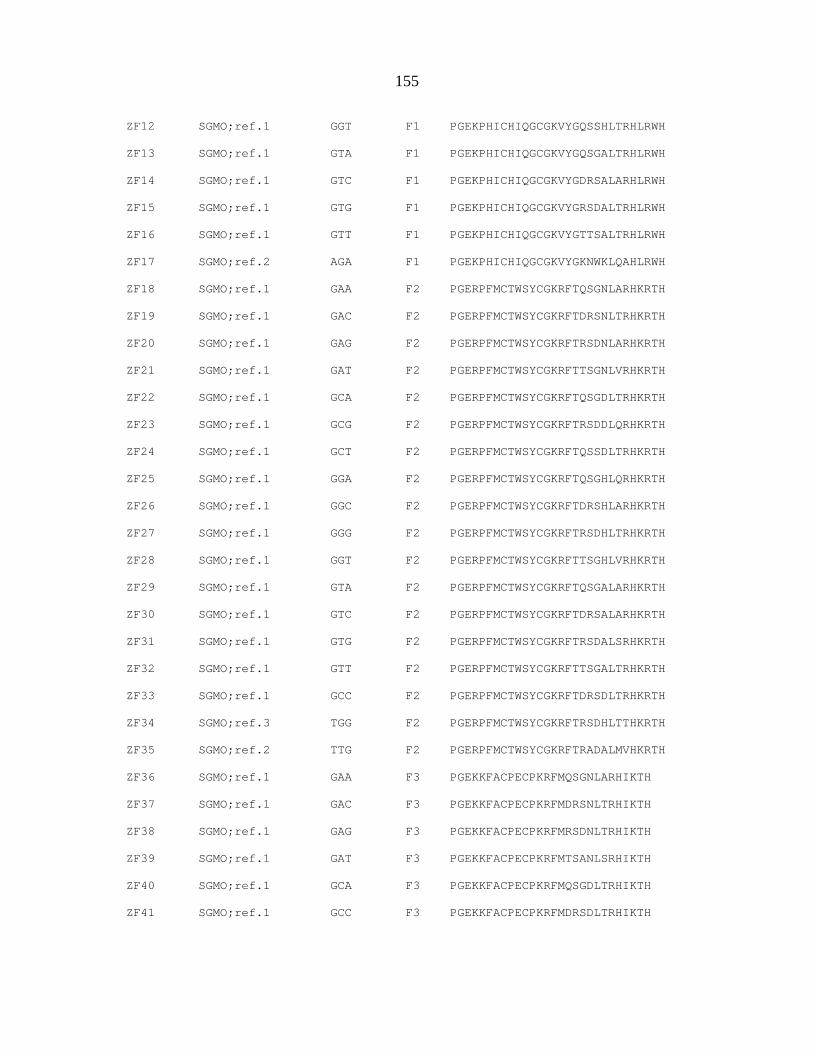
155
ZF12 SGMO;ref.1 GGT F1 PGEKPHICHIQGCGKVYGQSSHLTRHLRWH
ZF13 SGMO;ref.1 GTA F1 PGEKPHICHIQGCGKVYGQSGALTRHLRWH
ZF14 SGMO;ref.1 GTC F1 PGEKPHICHIQGCGKVYGDRSALARHLRWH
ZF15 SGMO;ref.1 GTG F1 PGEKPHICHIQGCGKVYGRSDALTRHLRWH
ZF16 SGMO;ref.1 GTT F1 PGEKPHICHIQGCGKVYGTTSALTRHLRWH
ZF17 SGMO;ref.2 AGA F1 PGEKPHICHIQGCGKVYGKNWKLQAHLRWH
ZF18 SGMO;ref.1 GAA F2 PGERPFMCTWSYCGKRFTQSGNLARHKRTH
ZF19 SGMO;ref.1 GAC F2 PGERPFMCTWSYCGKRFTDRSNLTRHKRTH
ZF20 SGMO;ref.1 GAG F2 PGERPFMCTWSYCGKRFTRSDNLARHKRTH
ZF21 SGMO;ref.1 GAT F2 PGERPFMCTWSYCGKRFTTSGNLVRHKRTH
ZF22 SGMO;ref.1 GCA F2 PGERPFMCTWSYCGKRFTQSGDLTRHKRTH
ZF23 SGMO;ref.1 GCG F2 PGERPFMCTWSYCGKRFTRSDDLQRHKRTH
ZF24 SGMO;ref.1 GCT F2 PGERPFMCTWSYCGKRFTQSSDLTRHKRTH
ZF25 SGMO;ref.1 GGA F2 PGERPFMCTWSYCGKRFTQSGHLQRHKRTH
ZF26 SGMO;ref.1 GGC F2 PGERPFMCTWSYCGKRFTDRSHLARHKRTH
ZF27 SGMO;ref.1 GGG F2 PGERPFMCTWSYCGKRFTRSDHLTRHKRTH
ZF28 SGMO;ref.1 GGT F2 PGERPFMCTWSYCGKRFTTSGHLVRHKRTH
ZF29 SGMO;ref.1 GTA F2 PGERPFMCTWSYCGKRFTQSGALARHKRTH
ZF30 SGMO;ref.1 GTC F2 PGERPFMCTWSYCGKRFTDRSALARHKRTH
ZF31 SGMO;ref.1 GTG F2 PGERPFMCTWSYCGKRFTRSDALSRHKRTH
ZF32 SGMO;ref.1 GTT F2 PGERPFMCTWSYCGKRFTTSGALTRHKRTH
ZF33 SGMO;ref.1 GCC F2 PGERPFMCTWSYCGKRFTDRSDLTRHKRTH
ZF34 SGMO;ref.3 TGG F2 PGERPFMCTWSYCGKRFTRSDHLTTHKRTH
ZF35 SGMO;ref.2 TTG F2 PGERPFMCTWSYCGKRFTRADALMVHKRTH
ZF36 SGMO;ref.1 GAA F3 PGEKKFACPECPKRFMQSGNLARHIKTH
ZF37 SGMO;ref.1 GAC F3 PGEKKFACPECPKRFMDRSNLTRHIKTH
ZF38 SGMO;ref.1 GAG F3 PGEKKFACPECPKRFMRSDNLTRHIKTH
ZF39 SGMO;ref.1 GAT F3 PGEKKFACPECPKRFMTSANLSRHIKTH
ZF40 SGMO;ref.1 GCA F3 PGEKKFACPECPKRFMQSGDLTRHIKTH
ZF41 SGMO;ref.1 GCC F3 PGEKKFACPECPKRFMDRSDLTRHIKTH

156
ZF42 SGMO;ref.1 GCG F3 PGEKKFACPECPKRFMRSDDLTRHIKTH
ZF43 SGMO;ref.1 GCT F3 PGEKKFACPECPKRFMQSSDLQRHIKTH
ZF44 SGMO;ref.1 GGA F3 PGEKKFACPECPKRFMQSGHLQRHIKTH
ZF45 SGMO;ref.1 GGC F3 PGEKKFACPECPKRFMDRSHLARHIKTH
ZF46 SGMO;ref.1 GGT F3 PGEKKFACPECPKRFMTSGHLVRHIKTH
ZF47 SGMO;ref.1 GTA F3 PGEKKFACPECPKRFMQRASLTRHIKTH
ZF48 SGMO;ref.1 GTC F3 PGEKKFACPECPKRFMDRSALARHIKTH
ZF49 SGMO;ref.1 GTG F3 PGEKKFACPECPKRFMRSDALTRHIKTH
ZF50 SGMO;ref.1 GTT F3 PGEKKFACPECPKRFMQSSALTRHIKTH
ZF51 SGMO;ref.1 GGG F3 PGEKKFACPECPKRFMRSDHLSRHIKTH
ZF52 SGMO;ref.3 AAG F3 PGEKKFACPECPKRFMRSDNLTQHIKTH
ZF53 SGMO;ref.4 AGG F3 PGEKKFACPECPKRFMRSDHLTQHIKTH
ZF54 SGMO;ref.5 CGG F3 PGEKKFACPECPKRFMRSDNLTEHIKTH
ZF55 SGMO;ref.2 CTG F3 PGEKKFACPECPKRFMRSDALREHIKTH
ZF56 SGMO;ref.6 TGA F3 PGEKKFACPECPKRFMQSGHLTKHIKTH
ZF57 SGMO;ref.7 TGG F3 PGEKKFACPECPKRFMRSDHLTTHIKTH
ZF58 Barbas;ref.8 GGG N/A PGEKPYKCPECGKSFSRSDKLVRHQRTH
ZF59 Barbas;ref.8 GGA N/A PGEKPYKCPECGKSFSQRAHLERHQRTH
ZF60 Barbas;ref.8 GGT N/A PGEKPYKCPECGKSFSTSGHLVRHQRTH
ZF61 Barbas;ref.8 GGC N/A PGEKPYKCPECGKSFSDPGHLVRHQRTH
ZF62 Barbas;ref.8 GAG N/A PGEKPYKCPECGKSFSRSDNLVRHQRTH
ZF63 Barbas;ref.8 GAA N/A PGEKPYKCPECGKSFSQSSNLVRHQRTH
ZF64 Barbas;ref.8 GAT N/A PGEKPYKCPECGKSFSTSGNLVRHQRTH
ZF65 Barbas;ref.8 GAC N/A PGEKPYKCPECGKSFSDPGNLVRHQRTH
ZF66 Barbas;ref.8 GTG N/A PGEKPYKCPECGKSFSRSDELVRHQRTH
ZF67 Barbas;ref.8 GTA N/A PGEKPYKCPECGKSFSQSSSLVRHQRTH
ZF68 Barbas;ref.8 GTT N/A PGEKPYKCPECGKSFSTSGSLVRHQRTH
ZF69 Barbas;ref.8 GTC N/A PGEKPYKCPECGKSFSDPGALVRHQRTH
ZF70 Barbas;ref.8 GCG N/A PGEKPYKCPECGKSFSRSDDLVRHQRTH
ZF71 Barbas;ref.8 GCA N/A PGEKPYKCPECGKSFSQSGDLRRHQRTH

157
ZF72 Barbas;ref.8 GCT N/A PGEKPYKCPECGKSFSTSGELVRHQRTH
ZF73 Barbas;ref.8 GCC N/A PGEKPYKCPECGKSFSDCRDLARHQRTH
ZF74 Barbas;ref.9 AAA N/A PGEKPYKCPECGKSFSQRANLRAHQRTH
ZF75 Barbas;ref.9 AAC N/A PGEKPYKCPECGKSFSDSGNLRVHQRTH
ZF76 Barbas;ref.9 AAG N/A PGEKPYKCPECGKSFSRKDNLKNHQRTH
ZF77 Barbas;ref.9 AAT N/A PGEKPYKCPECGKSFSTTGNLTVHQRTH
ZF78 Barbas;ref.9 ACA N/A PGEKPYKCPECGKSFSSPADLTRHQRTH
ZF79 Barbas;ref.9 ACC N/A PGEKPYKCPECGKSFSDKKDLTRHQRTH
ZF80 Barbas;ref.9 ACG N/A PGEKPYKCPECGKSFSRTDTLRDHQRTH
ZF81 Barbas;ref.9 ACT N/A PGEKPYKCPECGKSFSTHLDLIRHQRTH
ZF82 Barbas;ref.9 AGA N/A PGEKPYKCPECGKSFSQLAHLRAHQRTH
ZF83 Barbas;ref.9 AGC N/A PGEKPYKCPECGKSFSERSHLREHQRTH
ZF84 Barbas;ref.9 AGG N/A PGEKPYKCPECGKSFSRSDHLTNHQRTH
ZF85 Barbas;ref.9 AGT N/A PGEKPYKCPECGKSFSHRTTLTNHQRTH
ZF86 Barbas;ref.9 ATA N/A PGEKPYKCPECGKSFSQKSSLIAHQRTH
ZF87 Barbas;ref.9 ATG N/A PGEKPYKCPECGKSFSRRDELNVHQRTH
ZF88 Barbas;ref.9 ATT N/A PGEKPYKCPECGKSFSHKNALQNHQRTH
ZF89 Barbas;ref.10 CAA N/A PGEKPYKCPECGKSFSQSGNLTEHQRTH
ZF90 Barbas;ref.10 CAC N/A PGEKPYKCPECGKSFSSKKALTEHQRTH
ZF91 Barbas;ref.10 CAG N/A PGEKPYKCPECGKSFSRADNLTEHQRTH
ZF92 Barbas;ref.10 CAT N/A PGEKPYKCPECGKSFSTSGNLTEHQRTH
ZF93 Barbas;ref.10 CCA N/A PGEKPYKCPECGKSFSTSHSLTEHQRTH
ZF94 Barbas;ref.10 CCC N/A PGEKPYKCPECGKSFSSKKHLAEHQRTH
ZF95 Barbas;ref.10 CCG N/A PGEKPYKCPECGKSFSRNDTLTEHQRTH
ZF96 Barbas;ref.10 CCT N/A PGEKPYKCPECGKSFSTKNSLTEHQRTH
ZF97 Barbas;ref.10 CGA N/A PGEKPYKCPECGKSFSQSGHLTEHQRTH
ZF98 Barbas;ref.10 CGC N/A PGEKPYKCPECGKSFSHTGHLLEHQRTH
ZF99 Barbas;ref.10 CGG N/A PGEKPYKCPECGKSFSRSDKLTEHQRTH
ZF100 Barbas;ref.10 CGT N/A PGEKPYKCPECGKSFSSRRTCRAHQRTH
ZF101 Barbas;ref.10 CTA N/A PGEKPYKCPECGKSFSQNSTLTEHQRTH

158
ZF102 Barbas;ref.10 CTG N/A PGEKPYKCPECGKSFSRNDALTEHQRTH
ZF103 Barbas;ref.10 CTT N/A PGEKPYKCPECGKSFSTTGALTEHQRTH
ZF104 Barbas;ref.11 TAG N/A PGEKPYKCPECGKSFSREDNLHTHQRTH
ZF105 Barbas;ref.11 TGA N/A PGEKPYKCPECGKSFSQAGHLASHQRTH
ZF106 Barbas;ref.11 TGG N/A PGEKPYKCPECGKSFSRSDHLTTHQRTH
ZF107 ToolGen;ref.12 GAC N/A PGEKPYKCKQCGKAFGCPSNLRRHGRTH
ZF108 ToolGen;ref.12 GCC N/A PGEKPYTCSDCGKAFRDKSCLNRHRRTH
ZF109 ToolGen;ref.12 GTC N/A PGEKPFMCTWSYCGKRFTDRSALARHKRTH
ZF110 ToolGen;ref.12 GAC N/A PGEKPYKCKECGKAFNHSSNFNKHHRIH
ZF111 ToolGen;ref.12 GTT N/A PGEKPFKCPVCGKAFRHSSSLVRHQRTH
ZF112 ToolGen;ref.12 GAT N/A PGEKPYRCKYCDRSFSISSNLQRHVRNIH
ZF113 ToolGen;ref.12 GAG N/A PGEKPYGCHLCGKAFSKSSNLRRHEMIH
ZF114 ToolGen;ref.12 GGA N/A PGEKPYKCKECGQAFRQRAHLIRHHKLH
ZF115 ToolGen;ref.12 GAG N/A PGEKPYKCHQCGKAFIQSFNLRRHERTH
ZF116 ToolGen;ref.12 GAA N/A PGEKPFQCNQCGASFTQKGNLLRHIKLH
ZF117 ToolGen;ref.12 GGA N/A PGEKPYKCGQCGKFYSQVSHLTRHQKIH
ZF118 ToolGen;ref.12 GGA N/A PGEKPYVCRECGRGFRQHSHLVRHKRTH
ZF119 ToolGen;ref.12 AGA N/A PGEKPYKCEECGKAFRQSSHLTTHKIIH
ZF120 ToolGen;ref.12 CGA N/A PGEKPYECDHCGKSFSQSSHLNVHKRTH
ZF121 ToolGen;ref.12 CAA N/A PGEKPYMCSECGRGFSQKSNLIIHQRTH
ZF122 ToolGen;ref.12 GAA N/A PGEKPYKCEECGKAFTQSSNLTKHKKIH
ZF123 ToolGen;ref.12 GAA N/A PGEKPFECKDCGKAFIQKSNLIRHQRTH
ZF124 ToolGen;ref.12 GAA N/A PGEKPYECEKCGKAFNQSSNLTRHKKSH
ZF125 ToolGen;ref.12 CAA N/A PGEKPYVCSKCGKAFTQSSNLTVHQKIH
ZF126 ToolGen;ref.12 GTA N/A PGEKPYKCPDCGKSFSQSSSLIRHQRTH
ZF127 ToolGen;ref.12 GTA N/A PGEKPYKCEECGKAFNQSSTLTRHKIVH
ZF128 ToolGen;ref.12 CGA N/A PGEKPYECHDCGKSFRQSTHLTQHRRIH
ZF129 ToolGen;ref.12 GGA N/A PGEKPYECHDCGKSFRQSTHLTRHRRIH
ZF130 ToolGen;ref.12 GCG N/A PGEKPYVCDVEGCTWKFARSDELNRHKKRH
ZF131 ToolGen;ref.12 GGG N/A PGEKPFLCQYCAQRFGRKDHLTRHMKKSH
159
ZF132 ToolGen;ref.12 AGG N/A PGEKPFQCKTCQRKFSRSDHLKTHTRTH
ZF133 ToolGen;ref.12 GGG N/A PGEKPYVCDVEGCTWKFARSDKLNRHKKRH
ZF134 ToolGen;ref.12 GGG N/A PGEKPYKCMECGKAFNRRSHLTRHQRIH
ZF135 ToolGen;ref.12 GAG N/A PGEKPYICRKCGRGFSRKSNLIRHQRTH
ZF136 ToolGen;ref.12 GAG N/A PGEKPYECKECGKAFSSGSNFTRHQRIH
ZF137 ToolGen;ref.12 AAT N/A PGEKPYECDHCGKAFSVSSNLNVHRRIH
ZF138 ToolGen;ref.12 GTG N/A PGEKPYTCKQCGKAFSVSSSLRRHETTH
ZF139 ToolGen;ref.12 GCT N/A PGEKPYECNYCGKTFSVSSTLIRHQRIH
ZF140 ToolGen;ref.12 GGT N/A PGEKPYRCEECGKAFRWPSNLTRHKRIH
ZF141 ToolGen;ref.12 RGA N/A PGEKPYACHLCGKAFTQCSHLRRHEKTH
Supplemental References:
1. Liu, Q., Xia, Z., Zhong, X. & Case, C.C. (2002) Validated zinc finger protein designs
for all 16 GNN DNA triplet targets. J Biol Chem 277, 3850‐3856.
2. Ren, D., Collingwood, T.N., Rebar, E.J., Wolffe, A.P. & Camp, H.S. (2002)
PPARgamma knockdown by engineered transcription factors: exogenous
PPARgamma2 but not PPARgamma1 reactivates adipogenesis. Genes Dev 16, 27‐32.
3. Liu, P.Q., Rebar, E.J., Zhang, L., Liu, Q., Jamieson, A.C., Liang, Y., Qi, H., Li, P.X.,
Chen, B., Mendel, M.C., Zhong, X., Lee, Y.L., Eisenberg, S.P., Spratt, S.K., Case, C.C.
& Wolffe, A.P. (2001) Regulation of an endogenous locus using a panel of designed
zinc finger proteins targeted to accessible chromatin regions. Activation of vascular
endothelial growth factor A. J Biol Chem 276, 11323‐11334.

160
4. Jouvenot, Y., Ginjala, V., Zhang, L., Liu, P.Q., Oshimura, M., Feinberg, A.P., Wolffe,
A.P., Ohlsson, R., & Gregory, P.D., (2003)Targeted regulation of imprinted genes by
synthetic zinc‐finger transcription factors. Gene Ther 10, 513‐22.
5. Jamieson, A.C., Cradick, T.J., Guan, B., Xiao, H. & Carroll, P.M., (2004) Controlling
embryogenesis using engineered zinc fingers in Drosophila. Mol Ther 9 Supp. 1, S118.
6. Rebar, E.J., Huang, Y., Hickey, R., Nath, A.K., Meoli, D., Nath, S., Chen, B., Xu, L.,
Liang, Y., Jamieson, A.C., Zhang, L., Spratt, S.K., Case, C.C., Wolffe, A., Giordano,
F.J., (2002) Induction of angiogenesis in a mouse model using engineered
transcription factors. Nat Med. 8, 1427‐32.
7. Zhang, L., Spratt, S.K., Liu, Q., Johnstone, B., Qi, H., Raschke, E.E., Jamieson, A.C.,
Rebar, E.J., Wolffe, A.P. & Case, C.C. (2000) Synthetic zinc finger transcription factor
action at an endogenous chromosomal site. Activation of the human erythropoietin
gene. J Biol Chem 275, 33850‐33860.
8. Segal, D.J., Dreier, B., Beerli, R.R. & Barbas, C.F., 3rd (1999) Toward controlling gene
expression at will: selection and design of zinc finger domains recognizing each of
the 5ʹ‐GNN‐3ʹ DNA target sequences. Proc Natl Acad Sci U S A 96, 2758‐2763.
9. Dreier, B., Beerli, R.R., Segal, D.J., Flippin, J.D. & Barbas, C.F., 3rd (2001) Development
of zinc finger domains for recognition of the 5ʹ‐ANN‐3ʹ family of DNA sequences
and their use in the construction of artificial transcription factors. J Biol Chem 276,
29466‐29478.

161
10. Dreier, B., Fuller, R.P., Segal, D.J., Lund, C.V., Blancafort, P., Huber, A., Koksch, B. &
Barbas, C.F., 3rd (2005) Development of zinc finger domains for recognition of the 5ʹ‐
CNN‐3ʹ family DNA sequences and their use in the construction of artificial
transcription factors. J Biol Chem 280, 35588‐35597.
11. Blancafort, P., Magnenat, L. & Barbas, C.F. (2003) Scanning the human genome with
combinatorial transcription factor libraries. Nat Biotechnol 21, 269‐274.
12. Bae, K.H., Do Kwon, Y., Shin, H.C., Hwang, M.S., Ryu, E.H., Park, K.S., Yang, H.Y.,
Lee, D.K., Lee, Y., Park, J., Sun Kwon, H., Kim, H.W., Yeh, B.I., Lee, H.W., Hyung
Sohn, S., Yoon, J., Seol, W. & Kim, J.S. (2003) Human zinc fingers as building blocks
in the construction of artificial transcription factors. Nat Biotechnol 21, 275‐280.

162
APPENDIX B. UNEXPECTED FAILURE RATES FOR MODULAR
ASSEMBLY OF ENGINEERED ZINC FINGERS
A paper published in Nature Methods 2008
Cherie L. Ramirez, Jonathan E. Foley, David A. Wright, Felix Müller‐Lerch, Shamim
H. Rahman, Tatjana I. Cornu, Ronnie J. Winfrey, Jeffry D. Sander, Fengli Fu, Jeffrey
A. Townsend, Toni Cathomen, Daniel F. Voytas & J Keith Joung
TO THE EDITOR
Zinc‐finger nucleases (ZFNs) consist of an engineered zinc‐finger array fused
to a nuclease domain. Dimers of ZFNs can create targeted double‐strand DNA
breaks, which can stimulate highly efficient gene targeting in many cell types1
(Supplementary Figure B.S1 online). We found that the modular assembly method
of engineering zinc‐finger arrays has an unexpectedly higher failure rate than
previously reported. Modular assembly advocates linking individual zinc fingers,
each of which typically binds to a 3‐bp ‘subsite’ (Supplementary Figure B.S2 online).
Two large‐scale surveys have suggested that modular assembly is highly effective

163
(100% (2) and 60% (3) success rates) for making three‐finger arrays designed to bind
9‐bp target sites. The Zinc Finger Consortium recently assembled an archive of 141
previously published finger modules3–5 encoded on a standardized platform6. Our
initial experiences using these reagents suggested that modular assembly was
inefficient (Supplementary Discussion and Supplementary Table B.S1 online).
To perform a larger‐scale test, we assembled 168 zinc‐finger arrays designed
for 104 diverse target DNA sites (Supplementary Table B.S2 and Supplementary
Methods online). We tested these domains for DNA binding using a bacterial two‐
hybrid (B2H) assay (6), which accurately identifies arrays that lack activity as ZFNs
in human cells (Supplementary Discussion and Supplementary Figures. B.S3 and
B.S4 online). For 79 of the 104 target sites, we did not obtain a single three‐finger
array that scored positively in the B2H assay (overall failure rate of ~76%; Fig. 1a
and Supplementary Table B.S2). Notably, modular assembly was far less effective
for target sites composed of two, one, or no GXX subsites (where X is any base)
compared with those composed of three GXX subsites (Figure B.1A). Additionally,
because ZFNs function as dimers, we would expect failure rates for making a
functional ZFN pair to be even higher (Supplementary Discussion). Notably, these
values are all likely underestimates of actual failure rates because not all zinc‐finger

164
arrays that are positive in the B2H assay will be active as ZFNs in human cells
(Supplementary Figure B.S4).
One reason for the apparent discrepancy between previously published
studies2,3 and our results is that the former primarily used 9‐bp sites composed of
two or three GXX subsites, whereas we used sites with a more varied number of
GXX subsites (Figure B.1B); this difference will critically affect the observed failure
rate (Figure B.1A). Our study represents a more meaningful evaluation because
target sites containing one or no GXX subsites (underrepresented in previous
studies) encompass the majority (>75%) of the 107,011 potential 9‐bp sites that can be
targeted with existing modules (Figure B.1B).
Our results strongly suggest that potential users of modular assembly should
expect that this method will fail to yield a functional three–zinc finger array for the
majority of potentially targetable sites. To emphasize this we have modified content
on the Zinc Finger Consortium website (http://www.zincfigers.org; Supplementary
Methods). Highly effective but more labor‐intensive selection‐based methods for
engineering zinc finger arrays have been previously described (Supplementary
Discussion), and at present these are the only publicly available alternatives for
academic researchers interested in using ZFN technology.

165
Figure B.1 | Large-scale evaluation of the modular assembly method for engineering zinc-finger
arrays. A) Data for success and failure of modular assembly as judged by the B2H assay for all 104
target DNA sites (see Supplementary Discussion for definition of success threshold) and for
subsets of target sites containing three, two, one or no GXX subsites. ‘Successful’ sites are those for
which at least one functional zinc-finger array was identified; ‘failed’ sites are those for which we
failed to obtain a single successful array (see Supplementary Table 2 for details). Predicted failure
rates for each set of target sites are indicated above the bars. B) Distributions of target DNA sites
used in different modular assembly evaluation studies according to the number of GXX subsites
present. The distribution of all 107,011 potential 9-bp sites that can be targeted using 141 zinc-finger
modules from the Zinc Finger Consortium Modular Assembly Kit 1.0 (6) is also shown (far right bar).

166
ACKNOWLEDGMENTS
We thank D. Dobbs and P. Zaback for helpful discussions and E. Unger‐
Wallace (Iowa State University) for contributions to zinc‐finger array construction.
J.K.J. is supported by the US National Institutes of Health (R21 RR024189), the Cystic
Fibrosis Foundation (MCCRAY07G0) and the Massachusetts General Hospital
Department of Pathology; D.F.V. is supported by a US National Science Foundation
grant DBI 0501678; T.C. is supported by the German Research Foundation (SPP1230
CA311/2); C.L.R. is supported by a Ford Foundation Pre‐Doctoral Diversity
Fellowship and a National Science Foundation Graduate Research Fellowship; T.I.C.
is supported by a fellowship from the Swiss Foundation for Grants in Biology and
Medicine.
REFERENCES
1. Porteus, M.H. & Carroll, D. (2005) Gene targeting using zinc finger nucleases. Nat.
Biotechnol. 23, 967–973.
2. Segal, D.J. et al. (2003) Evaluation of a modular strategy for the construction of
novel polydactyl zinc finger DNA‐binding proteins. Biochemistry 42, 2137–2148.
3. Bae, K.H., Do Kwon, Y., Shin, H.C., Hwang, M.S., Ryu, E.H., Park, K.S., Yang,
H.Y., Lee, D.K., Lee, Y., Park, J., Sun Kwon, H., Kim, H.W., Yeh, B.I., Lee, H.W.,
Hyung Sohn, S., Yoon, J., Seol, W. & Kim, J.S. (2003) Human zinc fingers as building

167
blocks in the construction of artificial transcription factors. Nat Biotechnol 21, 275‐280.
4. Mandell, J.G. & Barbas, C.F. 3rd (2006) Zinc Finger Tools: custom DNA‐binding
domains for transcription factors and nucleases. Nucleic Acids Res. 34, W516–523.
5. Liu, Q., Xia, Z., Zhong, X. & Case, C.C. (2002) Validated zinc finger protein
designs for all 16 GNN DNA triplet targets. J. Biol. Chem. 277, 3850–3856.
6. Wright, D.A., Thibodeau‐Beganny, S., Sander, J.D., Winfrey, R.J., Hirsh, A.S.,
Eichtinger, M., Fu, F., Porteus, M.H., Dobbs, D., Voytas, D.F., Joung, J.K. (2006)
Standardized reagents and protocols for engineering zinc finger nucleases by
modular assembly. Nat. Protocols, 1, 1637.

168
SUPPLEMENTARY FIGURES & TABLES
Supplementary Figure B.S1 ZFNs induce highly efficient gene targeting events. (a) Schematic of a
ZFN monomer (left) and a pair of ZFNs cleaving DNA as a dimer (right). (b) Schematic representing
ZFN-enhanced recombination-based genome manipulation. ZFNs introduce site-specific double-
strand DNA breaks (DSBs) that can be harnessed to mediate gene targeting via homologous
recombination with an exogenously introduced “donor template.”

169
Supplementary Figure B.S2 Schematic illustrating the “modular assembly” method of engineering
multi-finger domains Fingers from archives of pre-characterized modules are joined together to create
three- finger domains capable of recognizing a 9 bp target DNA site. Single fingers (colored spheres)
recognize their cognate 3 bp “subsites” (colored rectangles).
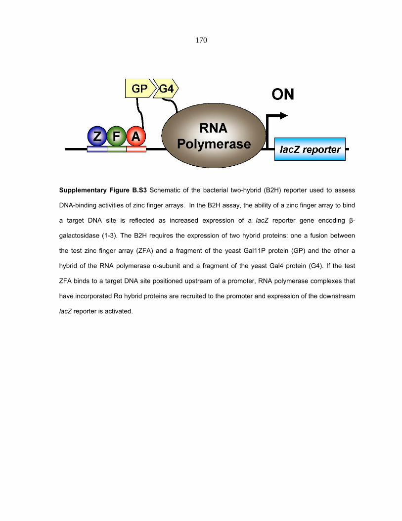
170
Supplementary Figure B.S3 Schematic of the bacterial two-hybrid (B2H) reporter used to assess
DNA-binding activities of zinc finger arrays. In the B2H assay, the ability of a zinc finger array to bind
a target DNA site is reflected as increased expression of a lacZ reporter gene encoding β-
galactosidase (1-3). The B2H requires the expression of two hybrid proteins: one a fusion between
the test zinc finger array (ZFA) and a fragment of the yeast Gal11P protein (GP) and the other a
hybrid of the RNA polymerase α-subunit and a fragment of the yeast Gal4 protein (G4). If the test
ZFA binds to a target DNA site positioned upstream of a promoter, RNA polymerase complexes that
have incorporated Rα hybrid proteins are recruited to the promoter and expression of the downstream
lacZ reporter is activated.

171
Supplementary Figure B.S4 The B2H assay identifies zinc-finger arrays that fail to show significant
activity as ZFNs in human cells. 23 different zinc-finger arrays (pink and light blue bars) which
activated transcription in the B2H assay to various levels (fold-activations are indicated in
parentheses on the x-axis) were tested as homodimeric ZFNs using a previously described human
cell-based episomal recombination (HR) assay (1). The names of these 23 zinc-finger arrays are
shown on the x-axis and details about their binding sites and the modules used to construct them can
be found in Supplementary Tables B.S1 and B.S2 online. ZFN activity is reported as a percentage of
EGFP-rescue relative to an I-SceI control. In addition, the episomal repair activities of control ZFNs
previously shown to lack or possess significant activity as ZFNs on chromosomal targets in human
cells (1) are shown for comparison (red and blue bars, respectively). Names of the control ZFNs are
shown on the x-axis and are as previously described (1). Means of at least three independent
experiments and standard deviations (error bars) are shown. ZFNs with mean episomal repair
activities that are significantly higher than that of the inactive EB0 ZFN are indicated with one
(p<0.05) or two (p<0.01) red asterisks. The expression levels of all ZFNs shown were verified by
Western blot using an antibody to a HA tag present on all proteins (data not shown).

172
Reference for Supplementary Figure 4:
1. Cornu, T. I. et al. (2008) DNA‐binding Specificity Is a Major Determinant of the
Activity and Toxicity of Zinc‐finger Nucleases. Mol Ther 16, 352‐8.
Supplementary Table B.S1 | Small-scale tests of modular assembly using various activity assays.
Thirty-six three-finger arrays (named “ZFA __” for zinc finger array) were assembled using modules
based on the Zinc Finger Consortium Modular Assembly Kit 1.0 (1), and the identities of the three
modules used to construct each three-finger array use the Consortium numbering scheme (F1, F2,
and F3 are the amino-terminal, middle, and carboxy-terminal fingers, respectively). All target binding
sites are written 5’ to 3’ (note that the F3 module binds to the 5’ most triplet subsite while F1 binds to
the 3’ most triplet subsite). The number of GXX subsites in each target site is also indicated. For nine
of the arrays, the amino acid sequence of the zinc finger backbone differs from the modules in the
archive. These arrays are noted in the far left column by a double asterisk, and their complete amino
acid sequences are available upon request.
Activity of 27 arrays was tested by electrophoretic mobility shift assays (EMSA). A plus sign
in the far right column indicates that the finger array caused a shift in mobility of a double-stranded
oligonucleotide corresponding to the intended target sequence on polyacrylamide gels. In all cases,
binding could be competed away with an excess of the target oligonucleotide. Western blots were
performed for all zinc finger arrays to ensure they were expressed.
Plant single-strand annealing (PSSA) assays (see Supplementary Methods online) were
used to assess function of seven arrays, five of which were also tested by EMSA. The PSSA assay
tests the ability of the zinc finger array to function as a ZFN. A minus sign in the table indicates that
activity was comparable to negative controls in which plant protoplasts were not transformed with the
ZFN construct.
An episomal recombination (HR) assay was used to assess the activity of nine ZFNs in
human 293T cells (see Supplementary Methods online). A plus sign indicates that ZFN activity in
stimulating HR was >40% as compared to the activity of a control I-SceI meganuclease on the same
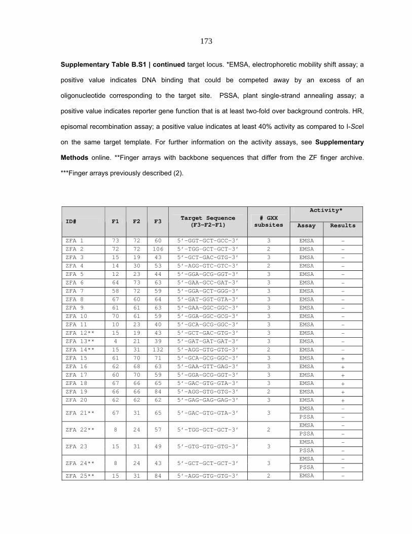
173
Supplementary Table B.S1 | continued target locus. *EMSA, electrophoretic mobility shift assay; a
positive value indicates DNA binding that could be competed away by an excess of an
oligonucleotide corresponding to the target site. PSSA, plant single-strand annealing assay; a
positive value indicates reporter gene function that is at least two-fold over background controls. HR,
episomal recombination assay; a positive value indicates at least 40% activity as compared to I-SceI
on the same target template. For further information on the activity assays, see Supplementary
Methods online. **Finger arrays with backbone sequences that differ from the ZF finger archive.
***Finger arrays previously described (2).
Activity*
ID# F1 F2 F3 Target Sequence (F3-F2-F1)
# GXX subsites Assay Results
ZFA 1 73 72 60 5’-GGT-GCT-GCC-3’ 3 EMSA - ZFA 2 72 72 106 5’-TGG-GCT-GCT-3’ 2 EMSA - ZFA 3 15 19 43 5’-GCT-GAC-GTG-3’ 3 EMSA - ZFA 4 14 30 53 5’-AGG-GTC-GTC-3’ 2 EMSA - ZFA 5 12 23 44 5’-GGA-GCG-GGT-3’ 3 EMSA - ZFA 6 64 73 63 5’-GAA-GCC-GAT-3’ 3 EMSA - ZFA 7 58 72 59 5’-GGA-GCT-GGG-3’ 3 EMSA - ZFA 8 67 60 64 5’-GAT-GGT-GTA-3’ 3 EMSA - ZFA 9 61 61 63 5’-GAA-GGC-GGC-3’ 3 EMSA - ZFA 10 70 61 59 5’-GGA-GGC-GCG-3’ 3 EMSA - ZFA 11 10 23 40 5’-GCA-GCG-GGC-3’ 3 EMSA - ZFA 12** 15 19 43 5’-GCT-GAC-GTG-3’ 3 EMSA - ZFA 13** 4 21 39 5’-GAT-GAT-GAT-3’ 3 EMSA - ZFA 14** 15 31 132 5’-AGG-GTG-GTG-3’ 2 EMSA - ZFA 15 61 70 71 5’-GCA-GCG-GGC-3’ 3 EMSA + ZFA 16 62 68 63 5’-GAA-GTT-GAG-3’ 3 EMSA + ZFA 17 60 70 59 5’-GGA-GCG-GGT-3’ 3 EMSA + ZFA 18 67 66 65 5’-GAC-GTG-GTA-3’ 3 EMSA + ZFA 19 66 66 84 5’-AGG-GTG-GTG-3’ 2 EMSA + ZFA 20 62 62 62 5’-GAG-GAG-GAG-3’ 3 EMSA +
EMSA - ZFA 21** 67 31 65 5’-GAC-GTG-GTA-3’ 3
PSSA - EMSA -
ZFA 22** 8 24 57 5’-TGG-GCT-GCT-3’ 2 PSSA - EMSA -
ZFA 23 15 31 49 5’-GTG-GTG-GTG-3’ 3 PSSA - EMSA -
ZFA 24** 8 24 43 5’-GCT-GCT-GCT-3’ 3 PSSA -
ZFA 25** 15 31 84 5’-AGG-GTG-GTG-3’ 2 EMSA -

174
PSSA - ZFA 26** 15 31 84 5’-AGG-GTG-GTG-3’ 2 PSSA - ZFA 27** 13 31 37 5’-GAC-GTG-GTA-3’ 3 PSSA -
HR - ZFA 28*** 61 65 65 5’-GAC-GAC-GGC-3’ 3
EMSA - ZFA 29 100 96 75 5’-AAC-CCT-CGT-3’ 0 HR - ZFA 30 61 80 61 5’-GGC-ACG-GGC-3’ 2 HR - ZFA 31 91 104 85 5’-AGT-TAG-CAG-3’ 0 HR - ZFA 32 83 103 104 5’-TAG-CTT-AGC-3’ 0 HR - ZFA 33 83 68 74 5’-AAA-GTT-AGC-3’ 1 HR - ZFA 34 100 104 72 5’-GCT-TAG-CGT-3’ 1 HR - ZFA 35 17 25 119 5’-AGA-GGA-AGA-3’ 1 HR -
HR + ZFA 36*** 60 64 63 5’-GAA-GAT-GGT-3’ 3
EMSA +
References for Supplementary Table 1:
1 Wright, D. A. et al. (2006) Standardized reagents and protocols for engineering zinc
finger nucleases by modular assembly. Nat Protoc 1, 1637‐52.
2 Alwin, S. et al. (2005) Custom zinc‐finger nucleases for use in human cells. Mol Ther
12, 610‐7.

175
Supplementary Table B.S2 | Modularly assembled zinc finger arrays, cognate target binding sites,
and their activities in the B2H assay. See legend to Supplementary Table B.S1 online for
descriptive detail pertaining to each of this table’s columns. In addition, the original source of the
modules used to construct each array (B=Barbas group (1), S=Sangamo Biosciences (2,3), and
T=Toolgen, Inc (4).) is indicated. Because the Zinc Finger Consortium Modular Assembly Kit 1.0
includes more than one module for certain subsites (3), we were able to construct 168 zinc finger
arrays that could potentially recognize the 104 different target sites. Additional details regarding the
construction of the B2H zinc finger expression plasmids and the B2H binding site reporter plasmids
are provided in Supplementary Methods online. Fold-activation of transcription in the B2H assay
was determined for each zinc finger array using β-galactosidase assays as previously described (5).
β-galactosidase assays for each zinc finger array were performed a minimum of four times and
means and standard errors of the mean are shown. The expression of all zinc finger arrays that
failed to activate transcription by more than 1.57-fold in the B2H assay were verified by Western blot
using a monoclonal antibody which detects a FLAG epitope present on all arrays (data not shown).
ID# F1 F2 F3 Module Source
Target Sequence (F3-F2-F1)
# GXX subsites
B2H fold activation (mean)
SEM
ZFA 37 75 75 74 B 5'-AAA-AAC-AAC-3' 0 1.152 0.030
ZFA 38 91 78 74 B 5'-AAA-ACA-CAG-3' 0 1.097 0.030 ZFA 39 73 91 74 B 5'-AAA-CAG-GCC-3' 1 1.325 0.095 ZFA 40 100 101 74 B 5'-AAA-CTA-CGT-3' 0 1.347 0.037
ZFA 41 83 68 74 B 5'-AAA-GTT-AGC-3' 1 1.166 0.106 ZFA 42 91 93 75 B 5'-AAC-CCA-CAG-3' 0 0.974 0.075 ZFA 43 68 93 75 B 5'-AAC-CCA-GTT-3' 1 0.980 0.010
ZFA 44 100 96 75 B 5'-AAC-CCT-CGT-3' 0 0.900 0.105 ZFA 45 64 86 77 B 5'-AAT-ATA-GAT-3' 1 1.606 0.037 ZFA 46 90 75 78 B 5'-ACA-AAC-CAC-3' 0 0.850 0.022 ZFA 47 85 75 79 B 5'-ACC-AAC-AGT-3' 0 1.001 0.028 ZFA 48 86 83 79 B 5'-ACC-AGC-ATA-3' 0 1.036 0.028 ZFA 49 83 100 79 B 5'-ACC-CGT-AGC-3' 0 1.235 0.016 ZFA 50 63 59 79 B 5'-ACC-GGA-GAA-3' 2 1.386 0.068 ZFA 51 91 83 80 B 5'-ACG-AGC-CAG-3' 0 0.955 0.027 ZFA 52 72 91 80 B 5'-ACG-CAG-GCT-3' 1 1.236 0.007 ZFA 53 70 95 80 B 5'-ACG-CCG-GCG-3' 1 1.266 0.040 ZFA 54 100 65 80 B 5'-ACG-GAC-CGT-3' 1 1.799 0.060 ZFA 55 71 73 80 B 5'-ACG-GCC-GCA-3' 2 1.295 0.031 ZFA 56 82 59 82 B 5'-AGA-GGA-AGA-3' 1 1.287 0.021 ZFA 57 119 117 119 T 5'-AGA-GGA-AGA-3' 1 0.911 0.012

176
ZFA 58 119 114 119 T 5'-AGA-GGA-AGA-3' 1 1.928 0.082 ZFA 59 119 129 119 T 5'-AGA-GGA-AGA-3' 1 1.974 0.066 ZFA 60 119 118 119 T 5'-AGA-GGA-AGA-3' 1 2.371 0.047 ZFA 61 64 69 83 B 5'-AGC-GTC-GAT-3' 2 1.206 0.019 ZFA 62 103 88 85 B 5'-AGT-ATT-CTT-3' 0 1.015 0.022 ZFA 64 91 104 85 B 5'-AGT-TAG-CAG-3' 0 0.879 0.092
ZFA 65 85 93 86 B 5'-ATA-CCA-AGT-3' 0 0.989 0.017 ZFA 66 78 81 87 B 5'-ATG-ACT-ACA-3' 0 0.956 0.043 ZFA 67 92 90 87 B 5'-ATG-CAC-CAT-3' 0 1.122 0.068
ZFA 68 78 96 88 B 5'-ATT-CCT-ACA-3' 0 1.064 0.135 ZFA 69 95 72 88 B 5'-ATT-GCT-CCG-3' 1 1.256 0.016 ZFA 70 63 98 90 B 5'-CAC-CGC-GAA-3' 1 1.099 0.011 ZFA 71 60 67 90 B 5'-CAC-GTA-GGT-3' 2 1.476 0.023 ZFA 72 86 98 92 B 5'-CAT-CGC-ATA-3' 0 0.985 0.027 ZFA 74 90 100 93 B 5'-CCA-CGT-CAC-3' 0 1.173 0.034 ZFA 75 73 64 93 B 5'-CCA-GAT-GCC-3' 2 1.085 0.006 ZFA 76 103 85 94 B 5'-CCC-AGT-CTT-3' 0 1.376 0.153 ZFA 77 66 100 94 B 5'-CCC-CGT-GTG-3' 1 1.431 0.069 ZFA 78 80 79 95 B 5'-CCG-ACC-ACG-3' 0 1.090 0.020 ZFA 79 74 91 95 B 5'-CCG-CAG-AAA-3' 0 1.123 0.009 ZFA 80 100 98 95 B 5'-CCG-CGC-CGT-3' 0 1.146 0.074 ZFA 81 86 63 95 B 5'-CCG-GAA-ATA-3' 1 0.907 0.048 ZFA 82 91 80 96 B 5'-CCT-ACG-CAG-3' 0 1.013 0.037
ZFA 83 90 81 96 B 5'-CCT-ACT-CAC-3' 0 1.113 0.042 ZFA 84 70 87 96 B 5'-CCT-ATG-GCG-3' 1 1.239 0.104 ZFA 85 58 67 96 B 5'-CCT-GTA-GGG-3' 2 1.504 0.009 ZFA 86 101 92 98 B 5'-CGC-CAT-CTA-3' 0 1.052 0.052 ZFA 87 88 81 101 B 5'-CTA-ACT-ATT-3' 0 0.884 0.013 ZFA 88 83 59 101 B 5'-CTA-GGA-AGC-3' 1 1.303 0.089 ZFA 89 80 83 103 B 5'-CTT-AGC-ACG-3' 0 1.037 0.086 ZFA 90 94 88 103 B 5'-CTT-ATT-CCC-3' 0 1.031 0.018 ZFA 91 91 60 103 B 5'-CTT-GGT-CAG-3' 1 1.044 0.032 ZFA 92 69 70 63 B 5'-GAA-GCG-GTC-3' 3 2.309 0.129 ZFA 93 14 23 36 S 5'-GAA-GCG-GTC-3' 3 1.620 0.059 ZFA 94 109 130 124 T 5'-GAA-GCG-GTC-3' 3 1.339 0.165
ZFA 95 109 130 122 T 5'-GAA-GCG-GTC-3' 3 2.279 0.123 ZFA 96 87 94 65 B 5'-GAC-CCC-ATG-3' 1 1.074 0.034 ZFA 97 72 61 65 B 5'-GAC-GGC-GCT-3' 3 1.118 0.045 ZFA 98 8 26 37 S 5'-GAC-GGC-GCT-3' 3 0.930 0.039 ZFA 101 4 24 38 S 5'-GAG-GCT-GAT-3' 3 1.327 0.126 ZFA 102 112 139 113 T 5'-GAG-GCT-GAT-3' 3 1.142 0.091
ZFA 103 112 139 136 T 5'-GAG-GCT-GAT-3' 3 1.185 0.087 ZFA 104 112 139 115 T 5'-GAG-GCT-GAT-3' 3 2.399 0.120 ZFA 105 66 72 62 B 5'-GAG-GCT-GTG-3' 3 4.553 0.295 ZFA 106 15 24 38 S 5'-GAG-GCT-GTG-3' 3 3.038 0.153 ZFA 107 138 139 136 T 5'-GAG-GCT-GTG-3' 3 0.959 0.026 ZFA 108 138 139 113 T 5'-GAG-GCT-GTG-3' 3 1.004 0.037 ZFA 109 138 139 115 T 5'-GAG-GCT-GTG-3' 3 3.277 0.153 ZFA 110 68 59 64 B 5'-GAT-GGA-GTT-3' 3 2.647 0.158 ZFA 111 16 25 39 S 5'-GAT-GGA-GTT-3' 3 1.571 0.111

177
ZFA 112 111 118 112 T 5'-GAT-GGA-GTT-3' 3 1.645 0.071 ZFA 113 111 129 112 T 5'-GAT-GGA-GTT-3' 3 2.013 0.083 ZFA 114 111 117 112 T 5'-GAT-GGA-GTT-3' 3 2.489 0.136 ZFA 115 111 114 112 T 5'-GAT-GGA-GTT-3' 3 3.840 0.308 ZFA 116 58 60 64 B 5'-GAT-GGT-GGG-3' 3 2.390 0.065
ZFA 117 11 28 39 S 5'-GAT-GGT-GGG-3' 3 0.959 0.098 ZFA 118 131 140 112 T 5'-GAT-GGT-GGG-3' 3 1.985 0.067 ZFA 119 133 140 112 T 5'-GAT-GGT-GGG-3' 3 2.444 0.103 ZFA 120 134 140 112 T 5'-GAT-GGT-GGG-3' 3 6.665 0.290 ZFA 121 60 78 71 B 5'-GCA-ACA-GGT-3' 2 1.388 0.014 ZFA 122 100 80 71 B 5'-GCA-ACG-CGT-3' 1 1.274 0.031
ZFA 123 81 90 71 B 5'-GCA-CAC-ACT-3' 1 0.851 0.023 ZFA 124 59 73 71 B 5'-GCA-GCC-GGA-3' 3 1.574 0.099 ZFA 125 9 33 40 S 5'-GCA-GCC-GGA-3' 3 0.953 0.111 ZFA 126 71 70 71 B 5'-GCA-GCG-GCA-3' 3 3.276 0.111 ZFA 127 5 23 40 S 5'-GCA-GCG-GCA-3' 3 0.887 0.068 ZFA 128 58 72 71 B 5'-GCA-GCT-GGG-3' 3 1.186 0.045 ZFA 129 11 24 40 S 5'-GCA-GCT-GGG-3' 3 1.157 0.039 ZFA 130 65 69 71 B 5'-GCA-GTC-GAC-3' 3 0.867 0.046 ZFA 131 2 30 40 S 5'-GCA-GTC-GAC-3' 3 0.897 0.052 ZFA 132 72 69 71 B 5'-GCA-GTC-GCT-3' 3 1.365 0.049 ZFA 133 8 30 40 S 5'-GCA-GTC-GCT-3' 3 1.116 0.092 ZFA 134 61 69 71 B 5'-GCA-GTC-GGC-3' 3 1.077 0.024 ZFA 135 10 30 40 S 5'-GCA-GTC-GGC-3' 3 0.999 0.039 ZFA 136 70 66 71 B 5'-GCA-GTG-GCG-3' 3 1.915 0.050 ZFA 137 7 31 40 S 5'-GCA-GTG-GCG-3' 3 0.819 0.051 ZFA 138 95 79 73 B 5'-GCC-ACC-CCG-3' 1 1.000 0.032 ZFA 139 59 87 73 B 5'-GCC-ATG-GGA-3' 2 0.974 0.043 ZFA 140 103 63 73 B 5'-GCC-GAA-CTT-3' 2 1.137 0.057 ZFA 141 66 71 73 B 5'-GCC-GCA-GTG-3' 3 1.061 0.036 ZFA 142 15 22 41 S 5'-GCC-GCA-GTG-3' 3 0.914 0.023 ZFA 143 59 61 73 B 5'-GCC-GGC-GGA-3' 3 1.182 0.049 ZFA 144 9 26 41 S 5'-GCC-GGC-GGA-3' 3 0.814 0.021 ZFA 145 132 116 130 T 5'-GCG-GAA-AGG-3' 2 1.342 0.134 ZFA 146 132 124 130 T 5'-GCG-GAA-AGG-3' 2 1.736 0.063 ZFA 147 132 122 130 T 5'-GCG-GAA-AGG-3' 2 1.759 0.054 ZFA 148 132 123 130 T 5'-GCG-GAA-AGG-3' 2 2.136 0.064 ZFA 149 102 59 70 B 5'-GCG-GGA-CTG-3' 2 1.522 0.088
ZFA 150 59 66 70 B 5'-GCG-GTG-GGA-3' 3 1.751 0.124 ZFA 151 9 31 42 S 5'-GCG-GTG-GGA-3' 3 2.774 0.116 ZFA 152 114 138 130 T 5'-GCG-GTG-GGA-3' 3 1.029 0.046 ZFA 153 129 138 130 T 5'-GCG-GTG-GGA-3' 3 1.272 0.039 ZFA 154 118 138 130 T 5'-GCG-GTG-GGA-3' 3 1.384 0.059 ZFA 155 117 138 130 T 5'-GCG-GTG-GGA-3' 3 1.953 0.050
ZFA 156 60 66 70 B 5'-GCG-GTG-GGT-3' 3 2.229 0.208 ZFA 157 12 31 42 S 5'-GCG-GTG-GGT-3' 3 3.298 0.266 ZFA 158 70 106 70 B 5'-GCG-TGG-GCG-3' 2 2.713 0.181 ZFA 159 7 34 42 S 5'-GCG-TGG-GCG-3' 2 0.856 0.034 ZFA 160 78 86 72 B 5'-GCT-ATA-ACA-3' 1 0.988 0.024 ZFA 161 62 100 72 B 5'-GCT-CGT-GAG-3' 2 1.703 0.012

178
ZFA 163 100 104 72 B 5'-GCT-TAG-CGT-3' 1 1.343 0.143 ZFA 164 61 74 59 B 5'-GGA-AAA-GGC-3' 2 1.392 0.084 ZFA 165 92 90 59 B 5'-GGA-CAC-CAT-3' 1 1.182 0.016 ZFA 166 80 101 59 B 5'-GGA-CTA-ACG-3' 1 1.085 0.034 ZFA 167 66 70 59 B 5'-GGA-GCG-GTG-3' 3 1.640 0.043 ZFA 168 15 23 44 S 5'-GGA-GCG-GTG-3' 3 2.599 0.092 ZFA 169 138 130 117 T 5'-GGA-GCG-GTG-3' 3 1.027 0.068 ZFA 170 138 130 118 T 5'-GGA-GCG-GTG-3' 3 1.732 0.041 ZFA 171 138 130 129 T 5'-GGA-GCG-GTG-3' 3 2.296 0.121 ZFA 172 138 130 114 T 5'-GGA-GCG-GTG-3' 3 2.316 0.046 ZFA 173 61 80 61 B 5'-GGC-ACG-GGC-3' 2 1.341 0.109
ZFA 174 63 95 61 B 5'-GGC-CCG-GAA-3' 2 1.171 0.030 ZFA 175 66 73 61 B 5'-GGC-GCC-GTG-3' 3 1.116 0.072 ZFA 176 15 33 45 S 5'-GGC-GCC-GTG-3' 3 0.868 0.017
ZFA 177 73 58 61 B 5'-GGC-GGG-GCC-3' 3 1.203 0.035 ZFA 178 6 27 45 S 5'-GGC-GGG-GCC-3' 3 1.258 0.053 ZFA 180 61 70 58 B 5'-GGG-GCG-GGC-3' 3 2.191 0.105 ZFA 181 10 23 51 S 5'-GGG-GCG-GGC-3' 3 6.162 0.147 ZFA 182 73 72 58 B 5'-GGG-GCT-GCC-3' 3 1.363 0.053 ZFA 183 6 24 51 S 5'-GGG-GCT-GCC-3' 3 1.494 0.054 ZFA 184 108 139 134 T 5'-GGG-GCT-GCC-3' 3 2.022 0.253 ZFA 185 79 88 60 B 5'-GGT-ATT-ACC-3' 1 1.078 0.016 ZFA 186 72 63 60 B 5'-GGT-GAA-GCT-3' 3 1.589 0.094 ZFA 187 8 18 46 S 5'-GGT-GAA-GCT-3' 3 0.944 0.081 ZFA 188 139 122 140 T 5'-GGT-GAA-GCT-3' 3 1.176 0.125 ZFA 189 139 124 140 T 5'-GGT-GAA-GCT-3' 3 1.800 0.124 ZFA 190 139 123 140 T 5'-GGT-GAA-GCT-3' 3 2.189 0.234 ZFA 191 139 116 140 T 5'-GGT-GAA-GCT-3' 3 2.431 0.090 ZFA 192 98 103 67 B 5'-GTA-CTT-CGC-3' 1 1.274 0.034 ZFA 193 61 68 67 B 5'-GTA-GTT-GGC-3' 3 1.056 0.098 ZFA 194 10 32 47 S 5'-GTA-GTT-GGC-3' 3 0.854 0.028
ZFA 195 91 101 69 B 5'-GTC-CTA-CAG-3' 1 1.132 0.035 ZFA 196 81 65 66 B 5'-GTG-GAC-ACT-3' 2 1.387 0.028 ZFA 197 73 61 66 B 5'-GTG-GGC-GCC-3' 3 1.104 0.036 ZFA 198 6 26 49 S 5'-GTG-GGC-GCC-3' 3 0.984 0.055 ZFA 199 66 82 68 B 5'-GTT-AGA-GTG-3' 2 1.683 0.046 ZFA 200 138 119 111 T 5'-GTT-AGA-GTG-3' 2 2.872 0.107 ZFA 201 12 20 50 S 5'-GTT-GAG-GGT-3' 3 0.671 0.015 ZFA 202 140 136 111 T 5'-GTT-GAG-GGT-3' 3 3.477 0.076 ZFA 203 140 113 111 T 5'-GTT-GAG-GGT-3' 3 4.735 0.489 ZFA 204 101 71 68 B 5'-GTT-GCA-CTA-3' 2 1.009 0.043 ZFA 205 73 70 68 B 5'-GTT-GCG-GCC-3' 3 5.065 0.290 ZFA 206 6 23 50 S 5'-GTT-GCG-GCC-3' 3 1.232 0.014 ZFA 207 83 103 104 B 5'-TAG-CTT-AGC-3' 0 0.849 0.100
ZFA 208 70 59 104 B 5'-TAG-GGA-GCG-3' 2 2.185 0.068 ZFA 209 3 20 56 S 5'-TGA-GAG-GAG-3' 2 1.704 0.068 ZFA 210 86 105 105 B 5'-TGA-TGA-ATA-3' 0 1.001 0.030

179
References for Supplementary Table 2:
1 Mandell, J.G. & Barbas, C.F., 3rd (2006) Zinc Finger Tools: custom DNA‐binding
domains for transcription factors and nucleases. Nucleic Acids Res 34, W516‐523.
2 Liu, Q., Xia, Z., Zhong, X. & Case, C.C. (2002) Validated zinc finger protein designs
for all 16 GNN DNA triplet targets. J Biol Chem 277, 3850‐3856.
3 Wright, D.A. et al. (2006) Standardized reagents and protocols for engineering zinc
finger nucleases by modular assembly. Nat Protoc 1, 1637‐1652.
4 Bae, K.H. et al. (2003) Human zinc fingers as building blocks in the construction of
artificial transcription factors. Nat Biotechnol 21, 275‐280.
5 Cornu, T. I. et al. (2008) DNA‐binding Specificity Is a Major Determinant of the
Activity and Toxicity of Zinc‐finger Nucleases. Mol Ther 16, 352‐8.

180
SUPPLEMENTARY DISCUSSION
Small‐scale surveys suggest a low success rate for modular assembly
Motivated by a desire to use ZFNs for genome modification, our labs constructed the
36 zinc finger arrays listed in Supplementary Table B.S1 online. These arrays are highly
biased for GXX subsites, and collectively include 22 3‐GXX arrays, 8 2‐GXX arrays, 2 1‐GXX
arrays and 4 arrays without GXX subsites. Using three different assays to test for function
(see legend to Supplementary Table B.S1 online), seven arrays were deemed functional. Six
of these sites were composed of three GXX subsites and one was composed of two GXX
subsites. Since six of the seven arrays were only tested by EMSA, it is difficult to extrapolate
how many of these arrays would function in vivo when challenged with the diverse
sequence targets present in a genome.
The B2H assay accurately identifies zinc finger arrays that fail to show ZFN function in
human cells
In previously published studies from our groups, we observed a general
correlation between failure to activate in the B2H assay and failure to show ZFN
function in human cells (1). To further assess this correlation, we tested the activities
of 23 ZFNs using a human cell‐based episomal homologous recombination (HR)
assay (Supplementary Methods online). The zinc finger arrays in these 23 ZFNs

181
each activated transcription to different levels in the B2H system ranging from 0.85‐
to 5.07‐fold. As shown in Supplementary Figure B.S4 online, we found that zinc
finger arrays which activated transcription by 1.57‐fold or less in the B2H system all
failed to show significant activity as ZFNs in human cells (nine out of nine ZFNs
tested; pink bars in Supplementary Figure B.S4 online). Conversely, many
(although not all) of the zinc finger arrays which activated transcription by >1.57‐
fold in the B2H system showed significant activity as ZFNs in the episomal repair
assay (light blue bars with red asterisks in Supplementary Figure B.S4 online). We
conclude that zinc finger arrays that activate transcription by 1.57‐fold or less in the
B2H system are unlikely to function as ZFNs in human cells and we used this
threshold level to interpret the results of our experiments.
Predicted failure rates for identifying zinc finger arrays needed to engineer a ZFN pair
Analysis of our results shows that modular assembly is far less effective for target
sites composed of two, one, or no GXX subsites (71%, 88% and 100% failure rates,
respectively; Figure B.1A) compared with those that contain three GXX subsites (41% failure
rate; Figure B.1A). Because ZFNs function as dimers, the failure rates for making a ZFN pair
will therefore be ~65%, ~92%, ~99%, and 100% for ZFN targets composed of pairs of 3‐GXX,
2‐GXX, 1‐GXX, and 0‐GXX 9 bp “half‐sites”, respectively. These failure rates are calculated
by multiplying estimated success rates for each monomer in the ZFN pair and subtracting

182
this percentage from 100%.
Alternative selection‐based strategies for engineering multi‐finger arrays
A fundamental flaw with the modular assembly method (and a likely cause of its
low success rate) is its assumption that zinc finger domains behave as independent modular
units. A number of studies have shown that zinc fingers can cross over and interact with
adjacent fingers and neighboring finger binding sites (2‐5). Various engineering strategies
have been described in the literature that account for these context‐dependent effects on
zinc finger DNA‐binding. Greisman and Pabo first described a sequential optimization
strategy in which combinations of fingers that work well together are identified using serial
selections from randomized libraries (6). Isalan, Klug, and Choo described a “bipartite”
optimization strategy in which “halves” of a three‐finger domain are first optimized by
randomization and selection and then joined together to create a final protein (7). Finally,
Joung and colleagues described a domain shuffling approach in which pools of fingers are
first identified for each subsite in parallel and then shuffled together to create a recombined
library for use in a final stringent selection (8). A limitation of all of these different
approaches is that they require specialized expertise in the construction of multiple very
large randomized libraries and in the interrogation of these libraries using selection
methods.

183
References for Supplementary Discussion:
1 Cornu, T. I. et al. (2008) DNA‐binding Specificity Is a Major Determinant of the
Activity and Toxicity of Zinc‐finger Nucleases. Mol Ther 16, 352‐8.
2 Isalan, M., Choo, Y. & Klug, A. (1997) Synergy between adjacent zinc fingers in
sequence‐specific DNA recognition. Proc Natl Acad Sci U S A 94, 5617‐21.
3 Isalan, M., Klug, A. & Choo, Y. (1998) Comprehensive DNA recognition through
concerted interactions from adjacent zinc fingers. Biochemistry 37, 12026‐33.
4 Elrod‐Erickson, M., Rould, M. A., Nekludova, L. & Pabo, C. O. (1996) Zif268 protein‐
DNA complex refined at 1.6 A: a model system for understanding zinc finger‐DNA
interactions. Structure 4, 1171‐80.
5 Wolfe, S. A., Grant, R. A., Elrod‐Erickson, M. & Pabo, C. O. (2001) Beyond the
ʺrecognition codeʺ: structures of two Cys2His2 zinc finger/TATA box complexes.
Structure (Camb) 9, 717‐23.
6 Greisman, H. A. & Pabo, C. O. (1997) A general strategy for selecting high‐affinity
zinc finger proteins for diverse DNA target sites. Science 275, 657‐61.
7 Isalan, M., Klug, A. & Choo, Y. (2001) A rapid, generally applicable method to
engineer zinc fingers illustrated by targeting the HIV‐1 promoter. Nat Biotechnol 19,
656‐60.
8 Hurt, J. A., Thibodeau, S. A., Hirsh, A. S., Pabo, C. O. & Joung, J. K. (2003) Highly
specific zinc finger proteins obtained by directed domain shuffling and cell‐based
selection. Proc Natl Acad Sci U S A 100, 12271‐6.

184
SUPPLEMENTARY METHODS
Construction of B2H zinc finger expression vectors and reporter strains
104 B2H reporter strains each harboring a single copy target site‐reporter plasmid
were constructed as previously described. (1) B2H expression vectors encoding different
three‐finger arrays were constructed essentially as previously described (1) using the Zinc
Finger Modular Assembly Kit 1.0 which includes 141 modules made by the Barbas group,
Sangamo Biosciences, and Toolgen, Inc. (1) For each target site, we made three‐finger arrays
using modules from the Barbas, Sangamo, and Toolgen collections but we did not mix and
match modules across these different sets. We chose not to make “crossplatform” arrays
because: 1: the Barbas website software does not advocate use of their modules with others
(2), 2: the Toolgen group discovered that their human zinc fingers worked best with one
another and not as well with other engineered modules, and 3: the Sangamo modules are
position‐specific and have linkers joining them that differ from the canonical TGEKP linker
used by the Barbas and Toolgen modules(4). 15 of the arrays we constructed were toxic
when expressed in the B2H assay and these proteins were not included in our analysis.
After setting aside these toxic proteins, we characterized the remaining 168 zinc finger
arrays. All expression and reporter plasmids were confirmed to be correct by DNA
sequencing prior to use in B2H assays.
B2H assays, verification of protein expression, and re‐verification of DNA sequences

185
The activities of three‐finger arrays were tested in the B2H assay by co‐transforming
a target site reporter strain with a B2H zinc finger array‐Gal11P expression vector and a
compatible plasmid encoding an E. coli RNA polymerase alpha‐Gal4 hybrid protein (1).
Fold‐activation was calculated as the ratio of the level of β‐galactosidase (lacZ) reporter
activity in the presence and absence of the zinc finger array as previously described (5). All
β‐galactosidase measurements were performed at least four independent times. The
expression of each zinc finger array that failed to activate transcription by more than 1.57‐
fold in the B2H assay was verified by performing Western blot on cell extracts from the β‐
galactosidase assays using a monoclonal antibody against the FLAG epitope present on all
zinc finger array‐Gal11P fusion proteins. For zinc finger arrays that exhibited less than 1.57‐
fold transcriptional activation in the B2H assay, we re‐sequenced the zinc finger coding
sequences and the reporter plasmid binding sites from the same cells used for β‐
galactosidase assays to re‐confirm the identities of the zinc finger recognition helices and
binding sites tested in these cells. We performed this additional re‐sequencing control on
>80% of our samples (94 of the 115 zinc finger array/binding site combinations) that were
negative in the B2H assay and found that all helices and binding sites were correct as
expected.
Plant single‐strand annealing (PSSA) assays
Zinc finger arrays were fused to the FokI nuclease domain and the resulting ZFNs
were transiently expressed in tobacco protoplasts. Plasmids encoding these ZFNs were

186
co‐transformed with a target plasmid carrying a non‐functional β‐glucuronidase (GUS)
gene. The GUS gene was rendered non‐functional by a direct duplication of part of its
coding sequence. Between the direct repeats, a recognition site was inserted for the given
ZFN being tested. After expression of the ZFN and cleavage of the GUS reporter, repair by
single strand annealing can restore GUS function. This can be measured by standard GUS
activity assays. We routinely observe GUS activity 25‐fold above background when the
assay is performed with a ZFN based on the transcription factor Zif268.
Episomal recombination (HR) assay in human cells
For the episomal HR assay, 293T cells in 24‐well plates were transfected by calcium
phosphate precipitation with 20 ng of the respective target plasmid, 1 μg of the donor
plasmid (pUC.Zgfp/REx), and 100 ng of a CMV‐driven endonuclease expression vector
encoding the ZFNs, I‐SceI (pRK5.LHA‐Sce1) or a control vector (pCMV.Luc). Two days after
transfection, 50,000 cells were analyzed by flow cytometry (FACSCalibur, BD Bioscience) to
determine the percentage of EGFP‐positive cells. The number of DsRedExpress (REx)‐
positive cells was used to normalize for transfection efficiency. All experiments were
repeated at least three times. ZFN activity is indicated as the fraction of EGFP‐positive cells
relative to the number of EGFP‐positive cells measured in the presence of I‐SceI.

187
Revisions to the Zinc Finger Consortium website
We have previously described ZiFiT, a web‐based software program that enables
users to rapidly identify potential zinc finger target sites within genes of interest (6). ZiFiT
identifies target sites for which zinc finger arrays might be made using modular assembly
and the Zinc Finger Consortium Modular Assembly Kit 1.0 (1). Given the results of this
current study, we have revised the ZiFiT program by adding the following section to its
instructions:
Scoring
To assess the effectiveness of modular assembly, we generated a large number of
zinc finger arrays and tested their activity using a bacterial cell‐based two‐hybrid (B2H)
assay (Ramirez et al., 2008). In the B2H assay, productive interaction of a zinc finger array
with its cognate binding site activates transcription of a linked reporter gene. A total of 168
three finger arrays were assembled that recognize 104 different target DNA sites that varied
in their GXX, AXX, CXX, and TXX subsite composition. Transcriptional activation of >1.57‐
fold over negative controls was determined to be indicative of target site recognition by a
given protein. After measuring the activity of the 168 zinc finger arrays, it was found that
the subsite composition of binding sites critically affected success rates. Modular assembly
was far less effective for target sites composed of one or no GXX subsites (12% and 0%
success rates, respectively) compared with those that contain three or two (59% and 29%
success rates, respectively). Based on these results, we provide users of ZiFiT with an

188
approximation of the likelihood that a given three finger protein will recognize its target.
For example, three finger proteins comprised of three GXX subsites will receive a score of
0.59, reflecting the success rate observed in our survey. Most users will be interested in the
likelihood that two arrays will function together as a ZFN. This can be approximated by
multiplying the success rate of individual arrays. For example, the likelihood of success that
a ZFN with all GXX subsites will recognize its target is 0.59 X 0.59 = 0.35 or 35%. Because
multiple modules are often available for given subsites, investigators may increase their
likelihood of success by making multiple different proteins against a single target. One note
of caution: the scoring function is a prediction for the activity of a given three finger array in
the B2H assay; the protein may behave differently when tested for activity in a eukaryotic
cell.
We have also revised the output that ZiFiT currently provides to users to include
guidance about the likelihood of success for different target DNA binding sites.
References for Supplementary Methods:
1 Wright, D. A. et al. (2006) Standardized reagents and protocols for engineering zinc
finger nucleases by modular assembly. Nat Protoc 1, 1637‐52.
2 Mandell, J. G. & Barbas, C. F., 3rd. (2006) Zinc Finger Tools: custom DNA‐binding
domains for transcription factors and nucleases. Nucleic Acids Res 34, W516‐23.
3 Bae, K. H. et al. (2003) Human zinc fingers as building blocks in the construction of
artificial transcription factors. Nat Biotechnol 21, 275‐80.

189
4 Liu, Q., Xia, Z., Zhong, X. & Case, C. C. (2002) Validated zinc finger protein designs
for all 16 GNN DNA triplet targets. J Biol Chem 277, 3850‐6.
5 Thibodeau, S. A., Fang, R. & Joung, J. K. (2004) High‐throughput beta‐galactosidase
assay for bacterial cell‐based reporter systems. Biotechniques 36, 410‐5.
6 Sander, J. D., Zaback, P., Joung, J. K., Voytas, D. F. & Dobbs, D. (2007) Zinc Finger
Targeter (ZiFiT): an engineered zinc finger/target site design tool. Nucleic Acids Res
35, W599‐605.

190
APPENDIX C: RAPID “OPEN‐SOURCE” ENGINEERING OF
CUSTOMIZED ZINC‐FINGER NUCLEASES FOR HIGHLY
EFFICIENT GENE MODIFICATION
A paper published in Molecular Cell 2008
Morgan L. Maeder, Stacey Thibodeau‐Beganny, Anna Osiak, David A. Wright,
Reshma M. Anthony, Magdalena Eichtinger, Tao Jiang, Jonathan E. Foley, Ronnie J.
Winfrey, Jeffrey A. Townsend, Erica Unger‐Wallace, Jeffry D. Sander, Felix Müller‐Lerch,
Fengli Fu, Joseph Pearlberg, Carl Göbel, Justin P. Dassie, Shondra M. Pruett‐Miller, Matthew
H. Porteus, Dennis C. Sgroi, A. John Iafrate, Drena Dobbs, Paul B. McCray, Jr., Toni
Cathomen, Daniel F. Voytas, & J. Keith Joung
SUMMARY
Custom‐made zinc‐finger nucleases (ZFNs) can induce targeted genome
modifications with high efficiency in cell types including Drosophila, C. elegans, plants, and
humans. A bottleneck in the application of ZFN technology has been the generation of
highly specific engineered zinc‐finger arrays. Here we describe OPEN (Oligomerized Pool
ENgineering), a rapid, publicly available strategy for constructing multi‐finger arrays,
which we show is more effective than the previously published modular assembly method.
191
We used OPEN to construct 37 highly active ZFN pairs which induced targeted alterations
with high efficiencies (1to 50%)at 11 different target sites located within three endogenous
human genes (VEGF‐A, HoxB13, CFTR), an endogenous plant gene (tobacco SuRA), and a
chromosomally‐integrated EGFP reporter gene. In summary, OPEN provides an “open‐
source” method for rapidly engineering highly active zinc‐finger arrays, thereby enabling
broader practice, development, and application of ZFN technology for biological research
and gene therapy.
INTRODUCTION
A reliable, publicly available method for creating targeted genome modifications
with high efficiency would be broadly useful for biological research and gene therapy (1).
Although strategies have been developed using viral vectors or short double‐or single‐
stranded DNA molecules to direct homologous recombination (HR), these methods
generally have low efficiencies even under optimal cell culture conditions (2). An alternative
and more efficient strategy involves introducing a double‐stranded break (DSB) into a target
locus of interest. Repair of this DSB by cellular mechanisms greatly increases rates of HR‐
mediated gene targeting with an exogenous DNA molecule (3) or of gene mutation by error‐
prone non‐homologous end‐joining (NHEJ) (4). However, this approach depends on the
capability to create “designer” nucleases targeted to specific genomic sequences of interest.
Engineered ZFNs are customized endonucleases that induce site‐specific DSBs and genome
modifications (as high as 50%) in Drosophila, somatic C. elegans, plant, and human cells (4‐

192
16). ZFNs function as dimers with each monomer composed of a non‐specific cleavage
domain from the FokI endonuclease fused to a zinc‐finger array engineered to bind a target
DNA sequence of interest (1,17,18). Because a single zinc‐finger domain binds a 3 bp subsite,
a ZFN dimer can recognize 18or 24‐bptargetsites, depending on the number of zinc‐fingers
in each ZFN monomer. Publicly available zinc‐finger engineering methods described in the
literature can be grouped into two general categories. “Modular assembly” involves joining
together single fingers with pre‐characterized specificities (19‐23). Although easy to
perform, modular assembly has an efficacy rate for making functional ZFN pairs that is less
than 6% (24) and can yield ZFNs with low activities and/or high toxicities (25,26).
Alternative approaches involve combinatorial selection‐based methods that yield multi‐
finger domains possessing high DNA‐binding affinities and specificities (27‐29) and high
activities and low toxicities when expressed as ZFNs in human cells (25,26). However,
selection‐based methods require construction and interrogation of large randomized
libraries (typically >108in size) and therefore remain intractable for all but a few labs that
possess the required expertise. Here we describe the development and validation of OPEN
(Oligomerized Pool ENgineering), a facile, robust, and publicly available platform for
engineering zinc‐finger arrays. OPEN is enabled by an archive of zinc‐finger pools
constructed by the Zinc Finger Consortium, a group of academic laboratories committed to
developing engineered zinc‐finger technology (http://www.zincfingers.org). The
Consortium used OPEN to rapidly engineer 37 ZFN pairs which induce modifications at 11
different sites located within three endogenous human genes (VEGF‐A, HoxB13, CFTR), an

193
endogenous plant gene (tobacco SuRA), and the EGFP reporter gene with efficiencies
ranging from 1 to 50%. The publicly available OPEN platform will enable routine practice
and further development of ZFN technology.
RESULTS
OPEN ‐ a rapid and robust strategy for engineering zinc‐finger arrays OPEN
selections require an archive of pre‐selected zinc‐finger pools, each containing a mixture of
fingers targeted to a different three base pair subsite at a defined position within a three‐
finger protein (Figure C.1A and Experimental Procedures). Fully enabling OPEN will
require 192 finger pools (64 potential three bp target subsites for each position in a three‐
finger protein). In this initial report, we created pools (each containing a maximum of 95
different fingers) targeted to 66 subsites (48 GXX subsites and 18 TXX subsites; Figure C.1B).
To perform an OPEN selection for a target site, appropriate finger pools from the archive are
recombined to create a small library of variants (953=8.6 x 10⁵members for a three‐finger
domain) which is interrogated using a bacterial two‐hybrid (B2H) selection system in which
binding of a zinc‐finger domain to its cognate site activates expression of selectable marker
genes (Figures C.1C & C.1D) (28,30). To simplify the identification of potential three‐finger
ZFN sites that can be targeted by OPEN, we created a new version of our web‐based ZiFiT
software (31), ZiFiT 3.0(Figure C.S1).

194
Figure C.1 | OPEN Method for Engineering Zinc-finger Arrays A) OPEN zinc-finger pool
construction. Zinc-finger domains are shown as spheres and associated 3 bp subsites as rectangles.
Randomized finger in the library is rainbow colored. Note that the figure illustrates how finger pools
for the middle position in a three-finger domain were made, but that pools for amino- or carboxy-
terminal fingers were also obtained by building libraries in which finger 1 or finger 3 were randomized,
respectively (Experimental Procedures). B) GXX and TXX target subsites for which finger pools have
been constructed (highlighted in grey). C) Schematic overview of OPEN selection for a target DNA
site. Zinc-fingers and associated subsites represented as in (A). Details in Supplemental
Experimental procedures. D) Schematic of the bacterial two-hybrid (B2H) system. ZFP = zinc-finger
protein. X and Y = arbitrary interacting proteins.

195
Comparing ZFNs made by modular assembly and OPEN
To compare the efficacy of OPEN with modular assembly, we used both strategies to
construct multi‐finger arrays for five sites in EGFP(ten “half‐sites”; Figure C.S2, Tables C.S1
and C.S2) and assessed the DNA‐binding activities of these proteins using a quantitative
B2H assay(Supplemental Experimental Procedures). Zinc‐finger arrays with high affinities
and specificities activate transcription by more than three‐fold in the B2H system (28). None
of the modularly assembled arrays we tested activated transcription by more than three‐fold
(Figure C.2A) even though Western blots showed that all proteins were expressed (data not
shown). By contrast, OPEN selections yielded at least one ‐‐in most cases many ‐‐zinc‐finger
protein that activated transcription by more than three‐fold for 9 of the 10 target half‐sites
(Figure C.2A and Table C.S1).For each of the five full EGFP target sites, various pairs of
modularly assembled and OPEN‐selected ZFNs (Table C.S2) were tested in human cells
using an assay in which repair of ZFN‐induced DSBs by error‐prone NHEJ leads to
insertions and deletions in a chromosomally integrated EGFP gene (Figure C.2B). Modular
assembly yielded ZFN pairs with activities above background for only one of the five sites
(EG502) (Figure C.2C). By contrast, OPEN yielded ZFN pairs which were active for four of
the five full ZFN target sites (EG223, EG292, EG382, and EG502). Although both methods
produced active ZFN pairs for the EG502 site, the pairs made by OPEN were more active
than those made by modular assembly (Figure C.2C). Western blots verified the expression
of all ZFNs tested (data not shown).

196
Figure C.2 | OPEN ZFNs engineered to cleave EGFP gene sequences A) Quantitative B2H assay of
modular assembly (MA; red bars) and OPEN (green bars) zinc finger arrays. Mean fold-activation
values (colored bars) and standard deviations (error bars) from three independent assays are shown.
B) EGFP-disruption assay for testing ZFN activities in human cells. C) Modularly assembled and
OPEN ZFNs assessed using the EGFP-disruption assay. Error bars represent standard deviations.
Single and double asterisks indicate p values <0.05 or <0.01, respectively.
OPEN selection of zinc‐finger arrays that bind to sequences in endogenous human and
plant genes
We used ZiFiT 3.0to identify 14 potential ZFN target sites in three endogenous
human genes (VEGF‐A, HoxB13, and CFTR) and one endogenous plant gene (tobacco SuRA)

197
(Table C.S2 and Figure C.S2). OPEN selections were performed for the 28 “half‐sites” within
these14 full ZFN target sites (Tables C.S1 and C.S2), and 24 of 28 were deemed successful
(Table C.S1). Using a subset of these finger arrays (Table C.S2), we constructed ZFN pairs
for five sites in VEGF‐A, four sites in HoxB13, one site in CFTR, and one site in the SuRA
gene.
OPEN ZFNs induce highly efficient mutation of endogenous human and plant genes
To test whether OPEN ZFNs induce mutations by NHEJ at endogenous human
genes, we employed a CEL I nuclease‐based mutation detection assay (Figure C.3A) (9,10).
We found that all four VF2468 and all four VF2471 ZFN pairs we tested generated
detectable mutations in the endogenous VEGF‐Agene in human 293 cells (Figure C.3B). In
addition, mutations could be detected at the endogenous HoxB13gene in 293 cells for all
ZFN pairs tested at the HX587, HX735 and HX761 sites (Figure C.3C). DNA sequencing of
HoxB13alleles amplified from cells modified by HX587 ZFN pair B (Supplemental
Experimental Procedures) revealed mutations at the expected location with an average
frequency of 9.6% (Figure C.S3A). ZFN pairs targeted to three sites in the VEGF‐Agene
(VF3537, VF3542, VF3552) and to one site in the HoxB13gene(HX508) failed to induce
detectable levels of mutation (Figure C.3B and C.3C). Due to polymorphisms at the CF877
site in CFTR alleles of 293 cells, we could not assess the 8CF877 ZFN pairs using the CEL I
assay (data not shown). However, DNA sequencing revealed ZFN‐induced insertions at the
CF877 cleavage site with an average frequency of 1.2% (Figure C.S3B). We also tested

198
whether OPEN ZFNs induced mutations in endogenous plant genes. Tobacco protoplasts
transformed with a construct encoding the SR2163 ZFN pair were regenerated into
individual plants (Experimental Procedures). The SR2163 site is present in both SuRA and
SuRB, and each plant was examined for evidence of cleavage at these loci using DNA
sequencing. Among 66 transgenic plants surveyed, three had mutations in SuRA, all of
which were deletions of a single base (Figure C.S3C). In one plant, both alleles of SuRA had
the same deletion. No mutated alleles of SuRB were detected. This frequency of mutagenesis
by NHEJ (~2% of potential target alleles) is comparable to what we observed for other
OPEN ZFNs in human cells.
OPEN ZFNs induce highly efficient gene targeting at an endogenous human gene
We next tested whether our OPEN ZFNs could induce high efficiency gene targeting
at the endogenous human VEGF‐A gene. For these experiments, we used VF2468 pair C and
VF2471 pair B (Table S2) and directly compared their activities with a previously published
ZFN pair (10) which cleaves the human IL2Rγ gene. Gene targeting frequencies using
VEGF‐A‐or IL2Rγ‐specific ZFNs and matched donor templates (Figure C.S4) were measured
in human K562 cells using a limited‐cycle PCR/restriction digest assay (Experimental
Procedures) (15): mean targeting efficiencies of 7.7%, 4.5% and 4.1% were observed with the
VF2468, VF2471, and IL2Rγ ZFNs, respectively (Figure C.4A). Efficient gene targeting
required both the donor construct and ZFN expression vectors (Figure C.4A). Control

199
Figure C.3 | Highly Efficient Mutagenesis of Endogenous Human Genes by OPEN ZFNs A)
Schematic of CEL I assay for assaying ZFN-induced mutations. (B-C) Mutation of the endogenous
human VEGF-A gene B) and HoxB13 gene C) by OPEN ZFNs. Colored arrows indicate expected
CEL I digestion products. Images shown are from representative experiments.
Southern blot assays of cells modified by VEGF‐A‐specific ZFNs confirm that, if anything,
the PCR‐based assay tended to underestimate gene targeting rates (Figures C.4B and
C.S5).Consistent with previous studies (15), we observed higher rates of gene targeting in
K562 cells which had been transiently arrested in G2 with vinblastine: 54%, 37%, and 44%
mean efficiencies with VF2468, VF2471, and IL2Rγ ZFNs, respectively (Figure C.4C).
Notably, vinblastine treatment greatly reduced the number of viable cells (data not shown).

200
Sequencing of VEGF‐A and IL2Rγ alleles revealed gene targeting events at the expected
locations and with the anticipated frequencies (Figure C.S6), but for all three target sites, we
also observed high frequencies of insertion and deletion events at the ZFN cleavage sites
(presumably caused by error‐prone NHEJ). Unexpectedly, we found that 1.8% and 9.0% of
alleles from cells treated with VF2468 and IL2Rγ ZFNs, respectively, contained evidence of
both HR‐mediated gene targeting and NHEJ‐mediated insertion events at a single allele.
Interestingly, analysis of single cell clones from vinblastine‐treated populations of VEGF‐
AZFN‐modified cells indicates that gene targeting events are stably maintained (even after
35 days) and strikingly can be induced in as many as four copies of VEGF‐A in polyploid
cells(Figure C.S7).
Toxicity profiles of OPEN ZFNs
We compared the relative toxicities of two pairs of three‐finger OPEN VEGF‐A ZFNs
and of a pair of four‐finger IL2Rγ ZFNs using cell survival assays in which K562 cells were
transfected with ZFN expression vectors, a donor plasmid, and a GFP‐expression plasmid.
Previous studies demonstrated that toxic ZFNs reduce both the percentage of GFP‐positive
cells (25,26) and cells that have undergone gene targeting (13)over time. All three ZFN pairs
we tested showed significant reductions in the relative number of GFP‐positive cells by
post‐transfection day 7 (Figure C.4D, green bars) and analogous decreases in the percentage
of gene targeting events (Figure C.4D, purple bars).We examined whether toxicity profiles
of the IL2Rγ and our OPEN VF2468 and VF2471 ZFNs could be improved by using obligate

201
Figure C.4 | Highly Efficient Gene Targeting of Endogenous Human Loci by OPEN ZFNs A) OPEN
VEGF-A ZFNs and previously described IL2Rγ ZFNs induce efficient gene targeting at endogenous
genes in human K562 cells. Top part shows representative gel images from limited-cycle
PCR/restriction digest assays and bottom part shows gene targeting frequency means (colored bars)
and standard errors (error bars) from multiple experiments. B) Gene targeting efficiencies of OPEN
VEGF-A ZFNs assessed nine days post transfection by limited-cycle PCR/restriction digest and
Southern blot assays. C) Vinblastine enhances gene targeting by OPEN VEGF-A and four-finger
IL2Rγ ZFNs. Assays performed four days post-transfection. Data presented as in (A). D) Toxicities of
OPEN VEGF-A and four-finger IL2Rγ ZFNs in human K562 cells. Means of GFP (green bars) and
gene targeting ratios (purple bars) are shown. Error bars represent standard deviations. Single and
double asterisks indicate p values <0.05 or <0.01, respectively. E) Gene targeting efficiencies of
OPEN VEGF-A and four-finger IL2Rγ ZFNs in toxicity experiments of (D). Means and standard
deviations (error bars) are shown of PCR-based assays performed four days post-transfection.

202
heterodimeric FokI nuclease domain variants which significantly reduce ZFN‐associated
toxicity (10,14). Both the variant IL2Rγ ZFN pair and the variant VF2471 ZFN pair showed
no significant toxicity as judged by the GFP toxicity assay (Figure C.4D, green bars). In
addition, the variant VF2468 ZFN pair revealed minimal toxicity (Figure C.4D) similar to
that of I‐SceI, a highly specific meganuclease used previously as a control (13). Comparable
effects were also observed in the relative percentage of gene targeting events (Figure C.4D,
purple bars). We note that all three variant ZFN pairs induce efficient gene targeting (Figure
C.4E), demonstrating that the absence of observable toxicity is not due to lack of ZFN
activity.
DISCUSSION
The OPEN reagents and methods we describe in this report provide a rapid, highly
effective, and publicly available platform for engineering zinc‐finger arrays. With OPEN, we
created 37 ZFN pairs which mediate highly efficient modification at four different sites
within a chromosomally integrated EGFP reporter gene, six different sites within three
endogenous human genes (VEGF‐A, HoxB13, and CFTR), and one site within an endogenous
plant gene (SuRA). The absolute rates of gene modification induced by our OPEN ZFNs
ranged from 1to 50%, and we were able to alter as many as two alleles in a single plant cell
and four alleles in a single polyploid human cell. We found OPEN to be more effective than
previously described modular assembly approaches for making zinc‐finger arrays. In our
studies targeting the EGFP gene, only two of 11 (~18%) modularly assembled ZFN pairs
tested showed activity in human cells, in contrast to 15 of 20 (75%) OPEN ZFN pairs. In

203
addition, at the one target site for which both methods were successful, OPEN ZFNs
showed significantly more activity than modularly assembled ZFNs. The low efficacy rate
observed with modular assembly is consistent with the results of a large‐scale assessment of
this method recently conducted by our groups (24). The higher success rate of OPEN is
likely attributable to its consideration of context‐dependent effects on DNA‐binding among
neighboring zinc‐fingers (32,33‐35) which are largely ignored by modular assembly.
With the set of zinc finger pools described in this report, we estimate that OPEN can
be used to engineer three‐finger proteins for ~4.1% ((23*21*22)/(64*64*64)) of all possible 9
bp target sites or ~0.16% (4.1%*4.1%) of all possible 18 bp ZFN sites. Because ZFNs can bind
to sites in which two “half‐sites” are separated by “spacer sequences” of five, six or seven
base pairs (13,36)[K. Wilson and M.H.P., unpublished data), one should be able to find
approximately five full ZFN sites in any given kb of random sequence (0.0016 *1000* 3).
Thus, with the pools described in this report, one should be able to target multiple ZFN sites
within a typical size gene. Important goals for future work will be to generate additional
finger pools that expand the targeting range of OPEN and to test whether the approach can
also be used to generate arrays composed of more than three‐fingers.
While using ZFNs to modify human genes, we observed two limitations that have
not been emphasized in previous reports. First, not all zinc‐finger arrays that possess
sequence‐specific DNA‐binding activities (as measured in the well‐established B2H method)
will function as ZFNs in human cells. ZFNs for one of the four sites targeted in the HoxB13
locus, for three of the five sites targeted in the VEGF‐A locus, and for one of the five sites

204
targeted in an integrated EGFP reporter gene failed to induce mutagenic NHEJ repair in
human cells. In addition, some ZFNs we made to the HoxB13 gene were active in 293 cells
(Figure C.3C) but not in K562 cells (data not shown). We speculate that the transcriptional
status and chromatin configuration of a target site may influence ZFN access to target sites:
HoxB13 is transcriptionally active in 293 cells (data not shown) but bears chromatin marks
consistent with a repressive state in K562 cells (B. Bernstein, personal communication).
Additional studies will be needed to determine whether lack of ZFN activity results from
chromatin effects on DNA accessibility or other reasons such as ZFN expression/stability or
target site methylation. Second, although the use of vinblastine increased the frequency of
gene targeting, DNA sequencing reveals that many alleles still underwent insertions or
deletions caused by error‐prone NHEJ and that some alleles underwent both a gene
targeting event and an insertion. These findings demonstrate limitations in relying solely on
PCR‐ or Southern blot‐based assays and suggest that DNA sequencing should always be
performed to verify ZFN‐induced gene targeting events.
Because OPEN is rapid, reliable, and publicly available, it will foster wider usage
and larger‐scale applications of engineered zinc‐finger technology. OPEN selections are
performed in E. coli and do not require specialized equipment. Although our ZFN
validation experiments were performed in plant and human cells, OPEN should also be
useful for generating ZFNs that function well in other organisms such as zebrafish,
mosquitoes, Drosophila, and C. elegans. The rapidity and effectiveness of OPEN should
enable genome‐scale ZFN projects (e.g. developing ZFNs for all human kinase genes or for

205
every zebrafish gene). We note that this report doubles the number of endogenous
mammalian genes described in the published literature ‐‐ from three (IL2Rγ, CCR5, and
DHFR) (15,37) to six ‐‐ that have been successfully modified using ZFNs. In conclusion, our
publicly available OPEN platform will enable scientists to perform the research and
development required to move ZFN technology forward for applications in biological
research and gene therapy.
Experimental Procedures
Additional details for all methods are provided in Supplemental Experimental
Procedures.
Construction of zinc‐finger pools
Randomized zinc‐finger libraries constructed by cassette mutagenesis were
introduced into “B2H selection strains” harboring a 9 bp target site as described (38) and
plated on histidine‐deficient selective media (NM media) containing 3‐AT, a competitive
inhibitor of the HIS3 enzyme. Zinc‐finger‐encoding phagemids were rescued from surviving
colonies, re‐introduced into fresh B2H selection cells, and plated on NM media containing 3‐
AT and streptomycin. 95 surviving colonies were inoculated into a 96‐well block for growth
and plasmids isolated to obtain the final finger pools.

206
OPEN selections
Each OPEN selection was performed in two steps: First, B2H selection strain cells
were infected with randomized zinc‐finger phage libraries consisting of three recombined
finger pools and then plated on NM media containing 3‐AT and streptomycin. Second, zinc
finger‐encoding phagemids were rescued from surviving colonies, re‐introduced into fresh
B2H selection strain cells, and plated on NM media containing a gradient of 3‐AT and
streptomycin. For a small number of the OPEN selections, we performed selections in a
single step (see Supplemental Experimental Procedures).
CEL I nuclease assay for NHEJ‐mediated mutation
Flp‐In T‐REx 293 cells (Invitrogen) were transfected with ZFN expression plasmids
and genomic DNA was isolated three days post‐transfection. Limited‐cycle PCR was
performed with radiolabeled nucleotides and VEGF‐A‐ or HoxB13‐specific primers. PCR
products were treated with CEL I nuclease and then separated on 10% polyacrylamide gels
and visualized using a phosphorimaging screen.
Gene targeting assays
Human K562 cells were transfected with ZFN expression plasmids and donor
constructs and genomic DNA harvested four days post‐transfection. Limited‐cycle PCR was
performed with radiolabeled nucleotides and VEGF‐A‐ or IL2Rγ‐specific primers. PCR
products digested with SalI (for VEGF‐A) or BsrBI (for IL2Rγ) were separated on 10%

207
polyacrylamide gels and visualized using a phosphorimaging screen. Additional details and
Southern blot assays are described in Supplemental Experimental Procedures.
Tobacco transformation and assay for mutations
The transformation of tobacco protoplasts by electroporation, selection for
kanamycin resistance, and regeneration into plantlets was carried out as previously
described (16). DNA was prepared from tissue harvested from individual plantlets and
SuRA and SuRB alleles were amplified by PCR, gel purified, and sequenced to identify
mutations.
ZFN toxicity assays
ZFN expression vectors, donor templates, and plasmid pmaxGFP (Amaxa) were
transfected into K562 cells. Cells were assayed for GFP expression at days 1 and 7 post
transfection using a FACScan cytometer and for gene targeting efficiencies at days 4 and 7
post‐transfection using the limited‐cycle PCR/restriction digest assay.
ACKNOWLEDGEMENTS
We thank Chong Jin Park for help with statistical calculations and Andrew Hirsh for
helpful discussions and comments. J.K.J. is supported by the NIH (R01GM069906,
R24GM078369, and R21RR024189), the Cystic Fibrosis Foundation (CFF) (MCCRAY07G0),
and the MGH Pathology Service. D.F.V. is supported by the NSF (DBI 0501678). T.C. is

208
supported by the European Commission’s 6th Framework Programme (037783 ZNIP).
P.B.M. is supported by the CFF (MCCRAY07G0) and the Roy J. Carver Charitable Trust.
A.J.I. is supported by the MGH Pathology Service. D.C.S. is supported by the NIH
(R01CA112021 and NCI SPORE in Breast Cancer at MGH). M.H.P. is supported by the NIH
(R01 HL079295). J.K.J. dedicates this paper to the memory of Robert L. Burghoff and his
belief in the power of methods.
REFERENCES
1. Cathomen, T. and Keith Joung, J. (2008) Zinc‐finger nucleases: the next generation
emerges. Mol Ther, 16, 1200‐1207.
2. Cathomen, T. (2004) AAV vectors for gene correction. Curr Opin Mol Ther, 6, 360‐366.
3. Jasin, M. (1996) Genetic manipulation of genomes with rare‐cutting endonucleases.
Trends Genet, 12, 224‐228.
4. Bibikova, M., Golic, M., Golic, K.G. and Carroll, D. (2002) Targeted chromosomal
cleavage and mutagenesis in Drosophila using zinc‐finger nucleases. Genetics, 161,
1169‐1175.
5. Alwin, S., Gere, M.B., Guhl, E., Effertz, K., Barbas, C.F., 3rd, Segal, D.J., Weitzman,
M.D. and Cathomen, T. (2005) Custom zinc‐finger nucleases for use in human cells.
Mol Ther, 12, 610‐617.

209
6. Beumer, K., Bhattacharyya, G., Bibikova, M., Trautman, J.K. and Carroll, D. (2006)
Efficient gene targeting in Drosophila with zinc‐finger nucleases. Genetics, 172, 2391‐
2403.
7. Bibikova, M., Beumer, K., Trautman, J.K. and Carroll, D. (2003) Enhancing gene
targeting with designed zinc finger nucleases. Science, 300, 764.
8. Lloyd, A., Plaisier, C.L., Carroll, D. and Drews, G.N. (2005) Targeted mutagenesis
using zinc‐finger nucleases in Arabidopsis. Proc Natl Acad Sci U S A, 102, 2232‐2237.
9. Lombardo, A., Genovese, P., Beausejour, C.M., Colleoni, S., Lee, Y.L., Kim, K.A.,
Ando, D., Urnov, F.D., Galli, C., Gregory, P.D. et al. (2007) Gene editing in human
stem cells using zinc finger nucleases and integrase‐defective lentiviral vector
delivery. Nat Biotechnol, 25, 1298‐1306.
10. Miller, J.C., Holmes, M.C., Wang, J., Guschin, D.Y., Lee, Y.L., Rupniewski, I.,
Beausejour, C.M., Waite, A.J., Wang, N.S., Kim, K.A. et al. (2007) An improved zinc‐
finger nuclease architecture for highly specific genome editing. Nat Biotechnol, 25,
778‐785.
11. Moehle, E.A., Rock, J.M., Lee, Y.L., Jouvenot, Y., DeKelver, R.C., Gregory, P.D.,
Urnov, F.D. and Holmes, M.C. (2007) Targeted gene addition into a specified
location in the human genome using designed zinc finger nucleases. Proc Natl Acad
Sci U S A, 104, 3055‐3060.

210
12. Morton, J., Davis, M.W., Jorgensen, E.M. and Carroll, D. (2006) Induction and repair
of zinc‐finger nuclease‐targeted double‐strand breaks in Caenorhabditis elegans
somatic cells. Proc Natl Acad Sci U S A, 103, 16370‐16375.
13. Porteus, M.H., Cathomen, T., Weitzman, M.D. and Baltimore, D. (2003) Efficient
gene targeting mediated by adeno‐associated virus and DNA double‐strand breaks.
Mol Cell Biol, 23, 3558‐3565.
14. Szczepek, M., Brondani, V., Buchel, J., Serrano, L., Segal, D.J. and Cathomen, T.
(2007) Structure‐based redesign of the dimerization interface reduces the toxicity of
zinc‐finger nucleases. Nat Biotechnol, 25, 786‐793.
15. Urnov, F.D., Miller, J.C., Lee, Y.L., Beausejour, C.M., Rock, J.M., Augustus, S.,
Jamieson, A.C., Porteus, M.H., Gregory, P.D. and Holmes, M.C. (2005) Highly
efficient endogenous human gene correction using designed zinc‐finger nucleases.
Nature, 435, 646‐651.
16. Wright, D.A., Townsend, J.A., Winfrey, R.J., Jr., Irwin, P.A., Rajagopal, J., Lonosky,
P.M., Hall, B.D., Jondle, M.D. and Voytas, D.F. (2005) High‐frequency homologous
recombination in plants mediated by zinc‐finger nucleases. Plant J, 44, 693‐705.
17. Durai, S., Mani, M., Kandavelou, K., Wu, J., Porteus, M.H. and Chandrasegaran, S.
(2005) Zinc finger nucleases: custom‐designed molecular scissors for genome
engineering of plant and mammalian cells. Nucleic Acids Res, 33, 5978‐5990.
18. Porteus, M.H. and Carroll, D. (2005) Gene targeting using zinc finger nucleases. Nat
Biotechnol, 23, 967‐973.

211
19. Bae, K.H., Kwon, Y.D., Shin, H.C., Hwang, M.S., Ryu, E.H., Park, K.S., Yang, H.Y.,
Lee, D.K., Lee, Y., Park, J. et al. (2003) Human zinc fingers as building blocks in the
construction of artificial transcription factors. Nat Biotechnol, 21, 275‐280.
20. Beerli, R.R. and Barbas, C.F., 3rd. (2002) Engineering polydactyl zinc‐finger
transcription factors. Nat Biotechnol, 20, 135‐141.
21. Liu, Q., Xia, Z., Zhong, X. and Case, C.C. (2002) Validated zinc finger protein designs
for all 16 GNN DNA triplet targets. J Biol Chem, 277, 3850‐3856.
22. Mandell, J.G. and Barbas, C.F., 3rd. (2006) Zinc Finger Tools: custom DNA‐binding
domains for transcription factors and nucleases. Nucleic Acids Res, 34, W516‐523.
23. Segal, D.J., Beerli, R.R., Blancafort, P., Dreier, B., Effertz, K., Huber, A., Koksch, B.,
Lund, C.V., Magnenat, L., Valente, D. et al. (2003) Evaluation of a modular strategy
for the construction of novel polydactyl zinc finger DNA‐binding proteins.
Biochemistry, 42, 2137‐2148.
24. Ramirez, C.L., Foley, J.E., Wright, D.A., Muller‐Lerch, F., Rahman, S.H., Cornu, T.I.,
Winfrey, R.J., Sander, J.D., Fu, F., Townsend, J.A. et al. (2008) Unexpected failure
rates for modular assembly of engineered zinc fingers. Nat Methods, 5, 374‐375.
25. Cornu, T.I., Thibodeau‐Beganny, S., Guhl, E., Alwin, S., Eichtinger, M., Joung, J. and
Cathomen, T. (2008) DNA‐binding Specificity Is a Major Determinant of the Activity
and Toxicity of Zinc‐finger Nucleases. Mol Ther, 16, 352‐358.

212
26. Pruett‐Miller, S.M., Connelly, J.P., Maeder, M.L., Joung, J.K. and Porteus, M.H. (2008)
Comparison of zinc finger nucleases for use in gene targeting in mammalian cells.
Mol Ther, 16, 707‐717.
27. Greisman, H.A. and Pabo, C.O. (1997) A general strategy for selecting high‐affinity
zinc finger proteins for diverse DNA target sites. Science, 275, 657‐661.
28. Hurt, J.A., Thibodeau, S.A., Hirsh, A.S., Pabo, C.O. and Joung, J.K. (2003) Highly
specific zinc finger proteins obtained by directed domain shuffling and cell‐based
selection. Proc Natl Acad Sci U S A, 100, 12271‐12276.
29. Isalan, M., Klug, A. and Choo, Y. (2001) A rapid, generally applicable method to
engineer zinc fingers illustrated by targeting the HIV‐1 promoter. Nat Biotechnol, 19,
656‐660.
30. Joung, J.K., Ramm, E.I. and Pabo, C.O. (2000) A bacterial two‐hybrid selection
system for studying protein‐DNA and protein‐protein interactions. Proc Natl Acad
Sci U S A, 97, 7382‐7387.
31. Sander, J.D., Zaback, P., Joung, J.K., Voytas, D.F. and Dobbs, D. (2007) Zinc Finger
Targeter (ZiFiT): an engineered zinc finger/target site design tool. Nucleic Acids Res,
35, W599‐605.
32. Elrod‐Erickson, M., Rould, M.A., Nekludova, L. and Pabo, C.O. (1996) Zif268
protein‐DNA complex refined at 1.6 A: a model system for understanding zinc
finger‐DNA interactions. Structure, 4, 1171‐1180.

213
33. Isalan, M., Choo, Y. and Klug, A. (1997) Synergy between adjacent zinc fingers in
sequence‐specific DNA recognition. Proc Natl Acad Sci U S A, 94, 5617‐5621.
34. Wolfe, S.A., Grant, R.A., Elrod‐Erickson, M. and Pabo, C.O. (2001) Beyond the
ʺrecognition codeʺ: structures of two Cys2His2 zinc finger/TATA box complexes.
Structure, 9, 717‐723.
35. Wolfe, S.A., Greisman, H.A., Ramm, E.I. and Pabo, C.O. (1999) Analysis of zinc
fingers optimized via phage display: evaluating the utility of a recognition code. J
Mol Biol, 285, 1917‐1934.
36. Bibikova, M., Carroll, D., Segal, D.J., Trautman, J.K., Smith, J., Kim, Y.G. and
Chandrasegaran, S. (2001) Stimulation of homologous recombination through
targeted cleavage by chimeric nucleases. Mol Cell Biol, 21, 289‐297.
37. Santiago, Y., Chan, E., Liu, P.Q., Orlando, S., Zhang, L., Urnov, F.D., Holmes, M.C.,
Guschin, D., Waite, A., Miller, J.C. et al. (2008) Targeted gene knockout in
mammalian cells by using engineered zinc‐finger nucleases. Proc Natl Acad Sci U S A,
105, 5809‐5814.
38. Thibodeau‐Beganny, S. and Joung, J.K. (2007) Engineering Cys2His2 zinc finger
domains using a bacterial cell‐based two‐hybrid selection system. Methods Mol Biol,
408, 317‐334.

214
SUPPLEMENTARY MATERIALS
Supplemental Experimental Procedures
B2H selection media
NM medium has been previously described (1). NM/CCK medium plates contain
100 μg/mL carbenicillin, 30 μg/mL chloramphenicol, 30 μg/mL kanamycin, and 1.5% Bacto‐
agar.
Construction of zinc finger pools
All master randomized zinc finger libraries were constructed in a standard
framework consisting of three tandem repeats of the middle finger of the murine
transcription factor Zif268 in which the recognition helix residues have been altered. For
each library, recognition helix residues –1, 1, 2, 3, 5, and 6 were randomized using 24 codons
(degenerate sequence 5’VNS3’) encoding 16 amino acids (excluding cysteine and the
aromatics). The theoretical complexity of each library is therefore 246= ~2 x 10⁸ members.
Each library was converted into infectious M13 phage particles as previously described (2).
B2H selection strains each harbor: (1) a single copy episome bearing a target site of
interest positioned upstream of a promoter which drives co‐cistronic expression of two
selectable markers (the yeast HIS3 gene and the bacterial aadA gene) and (2) a low copy
number plasmid expressing the RNA polymerase α‐subunit/yeast Gal4 hybrid protein (α‐
Gal4). Strains were constructed as previously described (1). The target binding site of each

215
strain was verified by DNA sequencing. Zinc finger pools were obtained using two selection
steps: In a first step, 109 ampicillintransducing‐ units (ATU) of randomized zinc finger
phage library were introduced into >3 x 10⁹ B2H selection strain cells harboring a target
subsite of interest. Transformed cells were plated on histidine‐deficient NM/CCK medium
plates containing 50 μM isopropyl β‐d‐thiogalactoside (IPTG) and 10 mM 3‐aminotriazole
(3‐AT), a competitive inhibitor of the HIS3 enzyme. After incubation for 24 hours at 37°C
followed by 18 hours at room temperature, surviving colonies were scraped from the plates
and infected with M13K07 helper phage to rescue the zinc finger‐encoding phagemids as
infectious phage. In a second step, this enriched phage library was then used to re‐infect
fresh B2H selection strain cells and the resulting transformants plated on NM/CCK medium
plates containing 50 uM IPTG, 10 mM 3‐AT, and 20 ug/ml streptomycin. After incubation at
37°C for 48 hours, we inoculated 95 surviving colonies of various sizes into individual wells
of a 96‐ well block containing 1ml of Terrific Broth and 50 μg/ml carbenicillin. These
cultures were grown overnight at 37°C and then a 96‐pin replicator was used to inoculate a
second block of identical cultures. Glycerol was added to a final concentration of 15% to all
wells in the first block and this was stored at ‐80°C. The second block was grown overnight
at 37°C and then the 95 cultures were pooled together. Plasmid DNA encoding the finger
pools was isolated from 10 ml of the pooled culture using a QIAgen miniprep kit and for
many pools a small number of random clones were sequenced with primer OK61. For finger
pools against nine subsites, sequencing revealed no strong consensus (i.e.—the sequences
were diverse and did not resemble one another), suggesting that selective pressure for those

216
sites was relatively weak under the initial selection conditions. For each of these nine
subsites, the second step of selection was repeated and plated on higher stringency plates to
obtain sequences that more closely resembled one another. The higher stringency plates
used were NM/CCK medium plates containing 50 μM IPTG, 20 mM 3‐AT, and 30 μg/ml
streptomycin (for seven subsites: F1 GAT, F2 GAC, F2 GAG, F2 GCG, F2 TGA, F2 TAG, and
F2 GTT) or 50 μM IPTG, 25 mM 3‐ AT, and 40 μg/ml streptomycin (for two subsites: F2
GAA, F2 TGG).
OPEN selections
To create libraries for use in OPEN selections, finger pools were amplified by PCR.
For each amplification, first five and then 20 cycles of PCR were performed using the
following primers and annealing temperatures (primer names; initial annealing temp; final
annealing temp): for finger 1: OK1424 and OK1425; 55°C; 59°C, for finger 2: OK1426 and
OK1427; 52°C; 57°C, for finger 3: OK1428 and OK1429; 41°C; 56°C. Amplified individual
finger pools were isolated from 10% polyacrylamide gels and fused together by PCR. To do
this, equal concentrations of the three finger pool fragments were fused using the following
PCR conditions: 94°C, 5 minutes; 10 cycles of 94°C, 30 sec; 50°C, 30 sec; 72°C, 2 min; final
extension 72°C, 7 min. This fusion product was purified using a QIAgen PCR purification
kit and then amplified by PCR using primers OK1430 and OK1432 with 10 initial cycles of
94°C, 30 sec; 56°C, 30 sec; 72°C, 1 min and 20 additional cycles of 94°C, 30 sec; 64°C, 30 sec;
72°C, 1 min; final extension 72°C, 7 min. The final PCR product (encoding a library of three‐

217
finger arrays) was isolated on a 5% polyacrylamide gel and treated with Pfu polymerase and
T4 polynucleotide kinase to create overhangs. This fragment was then ligated with BbsI
digested pBR‐UV5‐GP‐FD2 vector backbone which results in a plasmid that expresses the
zinc finger array as a FLAG‐tagged Gal11P fusion in the B2H system. This ligation was then
introduced into E. coli XL‐1 Blue cells by electroporation and each library was constructed
from >3 x 10⁶ independent transformants, ensuring at least three‐fold oversampling of the
theoretical library complexity of ~8.6 x 10⁵ (953). Libraries were then converted into
infectious M13 phage as previously described (1).
All but six (see below) of the OPEN selections were performed in two stages. In the
first stage, an OPEN three‐finger library was introduced by infection into a B2H selection
strain harboring the full target DNA sequence of interest. >2.2 x 10⁶ ATU of OPEN phage
library (a number which ensures adequate oversampling of the maximal theoretical library
diversity) were used to infect >2 x 10⁸ B2H selection strain cells and the resulting
transformants were plated on two different NM/CCK medium plates containing 50 μM
IPTG, 10 mM 3AT, and 20 μg/mL streptomycin or 50 μM IPTG, 25 mM 3AT, and 40 μg/mL
streptomycin. After 36‐48 hours of incubation, colonies were harvested from the highest
stringency plate yielding at least 1000 colonies. These cells were infected with M13K07
helper phage to rescue zinc finger‐encoding phagemids, thereby creating an enriched
library. In the second stage, 2.5 x 10⁶ ATU of this enriched library were used to infect >2 x
108 fresh B2H selection strain cells and the resulting transformants plated on a 245x245mm
NM/CCK medium plate containing parallel gradients of 3‐AT and streptomycin ranging

218
from 0 mM to 80 mM and 0 μg/mL to 100 μg/mL, respectively. Gradient plates were poured
using the method of Szybalski (3). After incubation at 37°C for a minimum of 48 and a
maximum of 96 hours, 12 surviving colonies were picked from the highest stringency edge
of the plate and ZFP‐encoding plasmids were isolated by plasmid miniprep and sequenced
with primer OK61. For six of the sites we targeted using OPEN, selections were (for
historical reasons) performed in a single step instead of two steps. For five sites (EG223L,
EG223R, EG292L, EG292R, and EG382L), >3.1 x 10⁶ ATU of OPEN phage library were used
to infect >4 x 10⁸ B2H selection strain cells and the resulting transformants were plated on a
series of NM/CCK medium plates containing 50 μM IPTG, 40 mM 3AT, and 60 μg/mL
streptomycin, 50 μM IPTG, 60 mM 3AT, and 80μg/mL streptomycin, 0 μM IPTG, 40 mM
3AT, and 60 μg/mL streptomycin or 0 μM IPTG, 60 mM 3AT, and 80 μg/mL streptomycin.
For each target site, we picked colonies from the highest stringency plates that yielded
colonies. For one site (EG382R), 4.25 x 10⁸ ATU of OPEN phage library were used to infect
>109 B2H selection strain cells and the resulting transformants were plated on a 245x245mm
NM/CCK medium plate containing parallel gradients of 3‐AT and streptomycin ranging
from 0 mM to 80 mM and 0 μg/mL to 100 μg/mL, respectively. Colonies were picked from
the highest stringency edge of the plate.
Construction of modularly assembled zinc finger arrays
Modularly assembled zinc finger arrays were assembled using the Zinc Finger
Consortium Modular Assembly Kit v1.0 as previously described (4). For each target half‐site

219
in the EGFP reporter gene, we assembled three‐finger arrays using modules from the
Barbas, Sangamo, and Toolgen archives but we did not mix and match modules across
platforms because: (1) the Barbas group does not suggest use of their modules with others
(2,5) the Toolgen group discovered that their human zinc fingers worked best with one
another but not as well with other engineered modules (6), and (3) the Sangamo modules
were designed to be finger‐position‐specific and have non‐canonical linkers joining them
that differ from the TGEKP linker used by the Barbas and Toolgen modules (7).
Quantitative bacterial two‐hybrid (B2H) assays
Zinc‐finger‐encoding plasmids identified from OPEN selections were co‐
transformed with an α‐Gal4 expression plasmid into a “B2H reporter strain” harboring a
single copy bacterial plasmid with a target binding site positioned upstream of a weak
promoter driving lacZ expression. B2H reporter strains were constructed as described (4). β‐
galactosidase assays were performed in triplicate as described (8).
ZFN expression vectors
All zinc finger arrays were expressed as ZFNs using the Zinc Finger Consortium
mammalian expression vector pST1374 (4). Zinc finger arrays were excised directly from
B2H expression vectors on an XbaI/BamHI fragment and cloned into pST1374. In this
configuration, zinc finger arrays are joined to the FokI nuclease domain by a four amino acid

220
linker of sequence LRGS. We constructed expression vectors encoding the four‐finger IL2Rγ
ZFNs using amino acid sequences obtained from a previously published study (9).
Human cell‐based EGFP‐disruption assay
Human 293.EGFP cells harbor an integrated retroviral construct which constitutively
expresses a β‐galactosidase‐EGFP fusion protein (Figure 2B). 293.EGFP cells were
transfected in triplicate in 24‐well plates using calcium phosphate precipitation as
previously described (10). Transfection cocktails included 300 ng each of a CMV promoter‐
controlled zinc finger nuclease expression vector, 100 ng pDS.RedExpress (Clontech,
Mountain view, CA) and pUC118 to 1.5 μg. 600 ng of pRK5.SceI plasmid (11), which
expresses the meganuclease I‐SceI, was used in place of GFP‐ZFN‐encoding plasmids for
negative controls. 50,000 cells were analyzed by flow cytometry two and five days post‐
transfection to determine the percentage of EGFP‐negative cells. The number of REx‐
positive cells at day 2 was used to normalize for transfection efficiency. Statistical
significance was determined using a two‐sided student’s t‐test with unequal variance.
CEL I nuclease assay for NHEJ‐mediated mutation
In this assay, limited‐cycle PCR is used to amplify a locus of interest from the
genomic DNA of a population of human cells transfected with ZFN expression vectors
(Figure 3A). The resulting PCR product is denatured and re‐annealed and heteroduplex
DNA will form if mutated alleles are present in the population. These DNA fragments can

221
be cleaved at the site of mismatch by the CEL I enzyme into smaller products of predictable
size. Specifically, 2 x 10⁶ human Flp‐In T‐REx 293 cells (Invitrogen) were transfected with
pairs of ZFN‐encoding plasmids (100 or 250 ng of each ZFN‐encoding plasmid) using
Lipofectamine 2000 (Invitrogen). Genomic DNA was isolated from nuclease‐treated cells at
3 days post‐transfection using the QIAgen Blood Mini kit. Limited‐cycle PCR (24 cycles) was
performed using Platinum PCR SuperMix Hi‐Fidelity (Invitrogen) or its equivalent
constituent components (Invitrogen) with 50 ng of genomic DNA as template, 8 μCi of each
[alpha‐32P]‐dATP and dCTP, 1 μM each of gene‐specific primers (primers OK1681 and
OK1682 for VEGF‐A sites VF2468/VF2471, primers OK1706 and OK1718 for VEGF‐A sites
VF3537/VF3542/VF3552, primers OK1733 and OK1734 for HoxB13 sites HX508/HX587 or
primers OK1736 and OK1738 for HoxB13 sites HX735/HX761) and 1.25 μl DMSO in a 25 μl
reaction volume. PCR products were cleaned up using Sephadex G‐50 columns (Roche) and
then melted/re‐annealed using the following conditions: 95°C for 10 min; 95°C to 85°C
cooling at a rate of ‐2°C/sec; 85°C to 25°C cooling at a rate of ‐0.1°C/sec; rapid cool to 4°C.
Re‐annealed PCR products were diluted 1:3.75 in a buffer of 20 mM Tris‐HCl (pH 8.8), 2
mM MgSO4, 60 mM KCl, 0.1% Triton X‐100 and treated with 1 μl CEL I enzyme (Surveyor
nuclease S; Transgenomic) and 1μl Surveyor Enhancer S (Transgenomic) in a 15 μl reaction
incubated at 42°C for 20 min. Products were visualized by electrophoresis on a 0.8mm thick,
10% 1X TBE polyacrylamide gel which was dried down and exposed overnight to a
phosphorimaging screen. All experiments were performed a minimum of two times.
222
Gene targeting assays
2 x 10⁶ Flp‐In T‐REx 293 cells were transfected with pairs of plasmids expressing
ZFNs (7.5 μg of each ZFN‐encoding plasmid) and 50 μg of donor plasmid using
nucleofection with solution V and program Q001 (Amaxa). 2 x 10⁶ K562 cells were
transfected with ZFN expression plasmid pairs (5 or 7.5 μg of each) and matched donor
construct (25 or 50 μg donor plasmid) using nucleofection with solution V and program T‐
16. Genomic DNA was harvested 3 or 4 days post‐transfection for 293 or K562 cells,
respectively, using a QIAgen Blood Mini kit. Transfection efficiencies were monitored by
including a GFP‐encoding plasmid in each transfection and determining the percentage of
GFP‐positive cells by flow cytometry one day post‐transfection. For experiments in which
cells were arrested in G2 phase, 0.2 μM vinblastine was added 24 hours post‐transfection
and then removed by washing three times with phosphate buffered saline 14‐18 hours later.
Limited‐cycle PCR assays (24 cycles) were performed using Platinum PCR SuperMix Hi‐
Fidelity (Invitrogen) or its equivalent constituent components (Invitrogen) with 4 ng
genomic DNA, 8 μCi of each [alpha‐32P]‐dATP and dCTP, 1 μM each of gene‐specific
primers (OK1776 and OK1777 for VEGF‐A or OK1845 and OK1846 for IL2Rγ) and 1.25 μl
DMSO in a 25 μl reaction. Purified PCR product was digested with 25 units SalI or 10 units
BsrBI restriction enzyme for 2 hours and the resulting products were visualized by
electrophoresis on a 10% 1X TBE polyacrylamide gel. This gel was dried down and exposed
overnight to a phosphorimaging screen. Quantification of bands was performed using
Quantity One software (Bio‐Rad).

223
For Southern blots, 15 μg of genomic DNA (15 μg) was digested with MscI and SalI
restriction enzymes for 20 hrs, electrophoresed in 0.8% tris‐acetate agarose gels (100 mM
Tris‐HCl, 10 mM EDTA, pH 8.0 with acetic acid), and transferred to Zeta‐probe nylon
membrane (BioRad) using 25 mM sodium phosphate (pH 6.5) according to the procedure of
Southern (12) as modified for use with the Turbo‐Blot downward transfer apparatus
(Schleicher & Schuell). The VEGF‐A DNA probe was generated by PCR amplification of a
cloned human VEGF‐A DNA template using primers OK1823 and OK1824 which was
subsequently labeled (25 ng) with [alpha‐32P]‐dCTP) using Rediprime II random priming
reagents (Amersham). Following hybridization (20 hrs) at 65°C in 5 mL ExpressHyb
solution (Clontech), the filters were washed with 0.1X SSC/0.1% SDS at 65°C (2 hrs), blotted
dry and exposed to a phosphorimager screen and/or film. The filters were scanned in the
Typhoon 8600 phosphoimager and relative band intensities were quantified by volume
analysis using ImageQuant software (GE Healthcare/Amersham).
Sequencing of modified genomic alleles
The region encompassing each potential ZFN cleavage site was amplified from
genomic DNA isolated from populations of human Flp‐In T‐REx 293 or K562 cells that had
been transfected with ZFN expression plasmids alone or with ZFN expression and donor
plasmids. PCR conditions for these amplifications were the same as those used for the CEL I
(for assessing NHEJ events) or limited‐cycle PCR/restriction digest (for assessing gene
targeting events) assays but with all components doubled to a final volume of 50μl. CF877

224
was amplified using primers OK1711 and 1713. PCR reactions were purified using the
QIAgen Minelute PCR Purification kit and eluted with 15 μl 0.1X EB buffer (QIAgen). PCR
fragments were cloned into the pCR4Blunt‐TOPO plasmid using the Zero Blunt TOPO PCR
Cloning Kit for Sequencing (Invitrogen). The TOPO cloning reaction used 4 μl purified PCR
product, 1 μl salt solution and 1 μl TOPO vector. 2 μl of TOPO reaction were transformed
into One Shot Mach1‐T1 chemically competent cells (Invitrogen) or chemically competent
Top10 cells (Invitrogen) and plated on LB plates containing 50 μg/ml kanamycin. Plasmid
DNAs from transformants were sequenced with a primer designed to bind internal to the
PCR product (OK1773 or T3 (Invitrogen) for VEGF‐A, OS216 for HoxB13, M13 primer
(Invitrogen) for CFTR or OK1838 for IL2Rγ).
Tobacco transformation and assay for mutations
The transformation of tobacco protoplasts by electroporation was carried out as
previously described (13). Plasmids introduced into protoplasts (10 μg each) included those
expressing ZFNs that recognize the left (pRW242) and right (pRW246) half sites of target
2163 in SuRA. Note that these constructs do not express the heterodimeric variants of FokI
endonuclease. Also transformed into protoplasts was a plasmid expressing neomycin
phosphotransferase (NPTII) (pDW998). The CaMV 35S promoter was used to drive
expression of both the ZFNs and NPTII. Plasmid DNAs were linearized with BglII prior to
transformation. Protoplasts were allowed to recover and then selected for kanamycin
resistance and regenerated into plantlets as previously described (13).

225
DNA was prepared from tissue harvested from individual plantlets using the
Epicentre MasterPure Plant Leaf DNA Purification Kit following the manufacturer’s
directions. An initial PCR screen for mutations at the site of ZFN cleavage was performed
using primers DVO4461 and DVO4462 to amplify a 445 bp fragment from both the SuRA
and SuRB loci. PCR was performed with 100 ng of genomic DNA and the following PCR
conditions: 94°C 2 min, followed by 34 cycles of 94°C 15 sec, 61°C 15 sec, 72°C 30 sec, and
then 72°C for 5 min. The reactions were run out on a 0.8% agarose gel, purified using a
QIAgen QIAquick Gel Extraction kit, and sequenced with DVO4462. The resulting
sequences were examined for double peaks, which not only indicate sequence differences
between SuRA and SuRB, but also identify potential insertion/deletion events at the ZFN
cleavage site in either or both loci.
DNA from candidate mutants was then PCR amplified using a set of nested, allele‐
specific primers in two consecutive PCR reactions to confirm the mutation and determine if
it occurred in SuRA or SuRB. The primary reaction amplified a 2.15 kb fragment using
approximately 100 ng of genomic DNA as template and primers DVO4565 and DVO4461
for SuRA and DVO4429 and DVO4461 for SuRB. The second PCR reaction amplified a 2 kb
fragment using 1 μl of the primary PCR reaction as template and primers DVO4444 and
DVO4461 for SuRA and DVO4445 and DVO4461 for SuRB. All PCR reactions were
performed using a Clontech Advantage cDNA Polymerase kit and the following PCR
conditions: 94°C 1 min, followed by 34 cycles of 94°C 30 sec, 66°C 30 sec, 68°C 3 min, and
226
then 68°C for 5 min. The reactions were run out on a 0.8% agarose gel, purified with a
QIAgen QIAquick Gel Extraction kit and sequenced with primer DVO4462.
ZFN toxicity assays
ZFN expression plasmids and donor templates were transfected into K562 cells
using nucleofection as described above. For these experiments, 5 μg of each ZFN expression
plasmid, 25 μg of donor, and 15 ng of pmaxGFP (encoding a GFP variant; Amaxa) were
included in each transfection. For controls, we transfected: (1) 10 μg of plasmid encoding I‐
SceI meganuclease (14) with 25 μg pUC118 and 15 ng of pmaxGFP, (2) 10 μg of plasmid
encoding CAD (caspase‐activated DNase) protein (15) with 25 μg pUC118 and 15 ng of
pmaxGFP, or (3) 35 μg of pUC118 and 15 ng of pmaxGFP. Cells were assayed for GFP
expression at post‐transfection days 1 and 7 with a FACScan cytometer. GFP ratios shown in
Figure 4D (green bars) were calculated using the formula:
(%GFP+ in ZFN‐transfected cells on day 7 / %GFP+ in ZFN‐transfected cells on day 1)
______________________________________________________________________
(%GFP+ in pUC‐transfected cells on day 7 / %GFP+ in pUC‐transfected cells on day 1)
In addition, genomic DNA was harvested using a QIAgen Blood Mini Prep kit on
post‐transfection days 4 and 7 and assayed for gene targeting using the limited‐cycle
PCR/restriction digest assay as described above. Gene targeting ratios shown in Figure 4D
(purple bars) were calculated by dividing the gene targeting rate on day 7 by the gene

227
targeting rate on day 4. All assays (both GFP and gene targeting) were performed on at least
three‐independent samples and t‐tests of significance were performed by comparing
experimentally determined ratios to a fixed ratio value of 1 (i.e.‐‐no change in value).
Variant heterodimer FokI domains were constructed by introducing the “+” and “‐“
mutations (previously described in Miller et al., 2007) into the wild‐type FokI nuclease
domain(9).
Fluorescence in situ hybridization (FISH)
Two‐color fluorescence in situ hybridization (FISH) was performed on 3:1
methanolacetic acid fixed cell lines using bacterial artificial chromosome clones RP11‐
710L16 (6p21.1; VEGF‐A) labeled in Spectrum Orange (Abbott‐Vysis, Downerʹs Grove, Il.),
or RP11‐142P4 (14q31.1; copy number control) labeled in Spectrum Green using standard
protocols. Images were captured using an Olympus BX61 fluorescent microscope equipped
with a CCD camera, and analysis was performed with Cytovision software (Applied
Imaging, San Jose, CA).

228
SUPPLEMENTAL FIGURES AND TABLES
Figure C.S1 | ZiFiT 3.0 -- Web-based software for identifying potential sites targetable by OPEN
selection A) Screen shot of ZiFiT 3.0 input page. We previously described ZiFiT, a web-based
software program that enables users to rapidly identify potential zinc finger nuclease target sites
within genes of interest (Sander et al., 2007; Wright et al., 2006). The original implementations of
ZiFiT (versions 1.0 and 2.0) were both geared toward making zinc finger nucleases by modular
assembly, and the output cross-referenced zinc finger modules available in the Zinc Finger
Consortium Modular Assembly Kit 1.0 (Wright et al., 2006). With the implementation of OPEN, we
have released ZiFiT Version 3.0 (http://www.zincfingers.org/software-tools.htm), which gives users
the option of searching for target sites for which OPEN pools are available. The user pastes a DNA
sequence (limited to 10,000 characters) into the text box at the top of the main window. Underneath
the main window are three boxes corresponding to finger positions in the zinc finger array. Each box
contains a list of the 64 possible nucleotide triplets. By default, triplets are checked for which OPEN
pools are currently available from the Zinc Finger Consortium; however, users can manually select or
deselect triplet pools to customize their search. Target site composition can be further restricted by
options available under the "Advanced/Basic" toggle near the bottom of the screen. By selecting the
"Submit" button, the sequence is searched for all target sites using the specified pools and an output
is returned. B) Screen shot of ZiFiT 3.0 output page. In addition to identifying zinc finger nuclease
target sites in the input sequence, it may be desirable to identify matching or highly similar sites within
a genome of interest. ZiFiT Version 3.0 enables the user to BLAST the genome sequence of a
desired organism to identify duplicate or related target sites. The BLAST search is implemented by a
button link and an organism selection menu provided for each target site returned in the output on the
results page. Requests are submitted to the NCBI BLAST servers to provide maximum reliability and
speed, and results are typically returned to ZiFiT within a minute. Note that BLAST scoring methods
do not identify all matches near the end of a hit if they are separated by mismatches that lower the

229
Figure C.S1 | | continued score of the local alignment. As a result, hits to highly similar target sites
lacking information at the ends may need to be analyzed manually.
A.
B.

230
Figure C.S2 | Schematic of target sites in the EGFP, VEGF-A, HoxB13, and CFTR genes Using
ZFNs made by OPEN selections, we targeted A) five full ZFN sites in the EGFP reporter gene
(EG223, EG292, EG382, EG502, and EG568), B) six sites in open chromatin regions of the human
VEGF-A gene (VF2468, VF2471, VF3537, VF3540, VF3542, and VF3552), C) five sites in the first
coding exon of the human HoxB13 gene (HX500, HX508, HX587, HX735, and HX761) and one site
in the last coding exon of the human HoxB13 gene (HX2119, not shown), and D) one site in exon 10
of the human CFTR gene (CF877) positioned within 100 base pairs of the ΔF508 deletion.

231
Figure C.S3 | DNA sequence analysis of endogenous human and plant genes mutagenized by
OPEN ZFNs (A-C) Sequences of A) HoxB13 alleles from human 293 cells transfected with HX587
ZFN pair B, B) CFTR alleles from human K562 cells transfected with CF877 ZFNs, and C) Figure

232
C.S3 | continued Sequences of SuRA alleles from tobacco plants transfected with SR2163 ZFNs
(matching sequence from the SuRB gene is also shown). Numbers of each allele identified are shown
in parentheses. ZFN recognition sites are in bold orange print.
Figure C.S4 | Schematics of donor templates and primers used for gene targeting experiments at the
human VEGF-A and IL2Rγ genes. Donor templates were constructed for OPEN ZFN pairs that
cleave at the VF2468 and VF2471 sites and for a previously described four-finger ZFN pair that
cleaves in the IL2Rγ gene (Urnov et al., 2005). Each donor template consists of 1.5 kb of genomic
DNA sequence centered on the cleavage site and introduces either an 11 bp insertion encoding a

233
Figure C.S4 | continued SalI restriction site at the center of the cleavage site (for VEGF-A targets) or
a translationally silent point mutation which creates a BsrBI restriction site adjacent to the ZFN
cleavage site (for IL2Rγ) (Urnov et al., 2005). Arrows indicate primers used for limited-cycle
PCR/restriction digest assay described in the text.
Figure C.S5 | Comparison of PCR-based and Southern blot methods for assaying gene targeting
efficiencies. Limited-cycle PCR and Southern blot were used to assess gene targeting efficiencies in
cells treated with different ZFNs under various conditions. Dotted red line represents where data
points would fall if the two methods were perfectly concordant.

234
Figure C.S6 | DNA sequence analysis of endogenous human alleles that have undergone gene
targeting induced by ZFNs (A-C) Sequences of alleles sequenced from human K562 cells transfected
with A) VF2468 ZFNs and donor, B) VF2471 ZFNs and donor, and C) IL2Rγ ZFNs and donor. Data
are presented as in Figure S3.

235
Figure C.S6 | continued

236
Figure C.S7 | OPEN ZFNs induce stable multi-allelic alterations of an endogenous human gene FISH
analysis indicated that K562 cells harbor four alleles of the VEGF-A gene (A, upper left panel) and we
therefore wished to determine how many of these alleles could be altered in a single cell. Limiting

237
Figure C.S7 | continued dilution cloning was used to isolate single cell clones from two vinblastine-
treated K562 cell populations which had undergone high frequencies of gene targeting: 45% and 26%
with the VF2468 and VF2471 ZFN pairs, respectively (B, upper panel). (The VF2468 ZFN-treated
cells also showed evidence of a 625 bp deletion (blue asterisks), a finding confirmed by sequencing
of alleles from these cells (data not shown).) Individual clones from VF2468 ZFN-treated cells
harbored no, one, three, or four alleles that had undergone a gene targeting event with some of the
clones also containing the 625 bp deletion (B, lower left panel). Genotype analysis of individual cell
clones from the VF2471 ZFN-treated cells revealed clones in which no, one, two, or three VEGF
alleles had undergone a gene targeting event (B, lower right panel). All individual cell clones were
genotyped more than thirty-five days post-transfection, demonstrating that gene targeting events we
observed are stably maintained. FISH analysis of three cell clones in which all four VEGF-A alleles
had undergone the VF2468 ZFN-induced gene targeting event confirmed the continued presence of
four copies of VEGF-A per cell (A, upper right, and lower left and right panels). FISH was performed
with a probe for VEGF-A (red) and a control probe for 14q which is present in two copies per cell
(green). Taken together, these results demonstrate that OPEN ZFNs can be used to induce
permanent alterations in as many as four alleles in a single human cell.

238
Table C.S1 | Sequences and B2H assays of zinc finger arrays selected by OPEN The subset of zinc
finger arrays we converted to ZFNs for testing in human or plant cells are highlighted in green. As
noted in the text, a small number of selections were performed in a single step on plates with fixed
concentrations of IPTG, 3-AT, and streptomycin. For these selections, IPTG concentration shown is
in μM, 3-AT concentration shown is in mM, and streptomycin concentration shown is in μg/ml. Fold-
activation of transcription in the B2H system was determined by comparing lacZ expression from B2H
reporter strains harboring a zinc finger array (fused to a fragment of the yeast Gal11P protein) to
matched strains harboring a control that does not express a zinc finger array. All determinations were
performed in triplicate and the means and standard deviations of these means are shown. Certain
target sites cause a higher basal level of lacZ expression in the quantitative B2H assay and the finger
arrays tested on these sites may exhibit lower apparent fold-activations due to this effect (affected
values are highlighted in yellow). Certain zinc finger arrays (marked by a “+” in the last column)
harbor additional mutations outside of the three randomized recognition helices that were most likely
introduced during library construction. The identities of these various mutations are available upon
request. Although half-site EG502L did not yield OPEN zinc-finger arrays which activated by more
than three-fold, we note that the basal level of transcription from the B2H reporter bearing this site
was high, a situation which can artifactually lower the apparent foldactivation observed. 25 of the 28
OPEN selections we performed for the human VEGF-A, human HoxB13, human CFTR, and tobacco
SuRA genes yielded zinc-finger arrays whose sequences closely resembled one another. However,
one of these 25 selections (for half-site VF3540R) yielded fingers with sequences that appeared to
bind to an alternative site (discussed below). For 22 of the other 24 selections, we obtained at least
one zinc-finger array which activated lacZ expression by more than three-fold in the quantitative B2H
assay. The two remaining selections were also deemed to be successful because their reporters
possessed a high basal level of transcription which can artifactually mask a higher true fold-activation.
For one selection (site VF3540R), examination of the recognition helix sequences of the arrays
obtained from OPEN selection suggests that these finger arrays do not bind to the intended nine base
pair site but rather to the site that is shifted 3’ in register by 1 bp (i.e.--these arrays bind to the site: 5’-

239
Table C.S1 | continued GCG GCG GAC-3’ instead of 5’-GGC GGC GGA-3’). For example, for finger
1, the Asp and Glu selected at position -1 and the Asn selected at position 3 of the recognition helix
would be expected to specify cytosine and adenine, respectively, and not the adenine and guanine
actually present in the subsite. Similarly, for finger 3, the Arg selected at position -1 and the Ser and
Asp selected at position 3 of the recognition helix would be expected to specify guanine and cytosine,
respectively, and not the cytosine and guanine actually present. Thus, this selection was deemed to
be unsuccessful.
ZFN
Name
Site
Name
Selection
conditions
(IPTG/3-
AT/Strep)
F1
subsite/
RH
sequence
F2
subsite/
RH
sequence
F3
subsite/
RH
sequence
Mean B2H
fold-
activation
S.D. of
B2H fold-
activation
Other
mutations
(of
unknown
significan
ce)
present?
GTA GGG GTC
OZ001 EG223L 50/40/60 QDSSLRR RQEHLVR DPTSLNR 4.24 0.17
OZ002 EG223L 50/40/60 QQSSLLR RQEHLVR DPTSLNR 3.50 0.13
OZ003 EG223L 50/40/60 QPSSLTR RKPHLVN QPTSLQL 0.79 0.03
OZ004 EG223L 50/40/60 QQSSLLR RGEHLTR DQTVLRR 3.50 0.33
OZ005 EG223L 50/40/60 QQSSLLR RGEHLTR EPTSLIR 3.17 2.10
OZ006 EG223L 50/40/60 QPSSLTR RREHLVS DRRPLPR 1.74 0.65
OZ007 EG223L 0/40/60 QGSALAR RREHLVR DPTSLNR 4.09 0.08
OZ008 EG223L 0/40/60 QSSSLTR RGEHLTR ESGALRR 2.57 1.15
OZ009 EG223L 0/40/60 QASALSR RREHLVR ESGALRR 5.23 0.94
OZ010 EG223L 0/40/60 QSQALVR RVEHLNN DRTSLPR 0.76 0.09
OZ011 EG223L 0/40/60 QQSSLLR RREHLVR DRTSLAR 3.65 0.23
OZ012 EG223L 0/40/60 QQSSLLR RREHLVR DPTSLNR 4.29 0.38
GCA GCA GAA
OZ013 EG223R 50/60/80 SQTQLVR QSTTLKR QRNNLGR 10.81 0.41
OZ014 EG223R 50/60/80 RRQELGR QGETLKR QHPNLTR 5.57 0.36
OZ015 EG223R 50/60/80 RRQELRR QGGTLNR QRNNLGR 10.36 1.40
OZ016 EG223R 50/60/80 RTQELKR QSGTLKR QGPNLGR 8.43 0.98
OZ017 EG223R 50/60/80 NNAQLTR QSGTLHR QRPNLGR 6.53 0.55

240
Table C.S1 (continued)
OZ018 EG223R 50/60/80 RRQELGR QGGTLKR QHPNLTR 9.05 0.42
OZ019 EG223R 0/60/80 RRQELGR QSGTLKR QRNNLGR 9.74 0.73
OZ020 EG223R 0/60/80 RGVELKR QSGTLHR QRPNLTR 11.48 1.63
OZ021 EG223R 0/60/80 RPQELAR QSGTLKR QHPNLTR 8.39 0.46
OZ022 EG223R 0/60/80 RRQELVR QSGTLKR QHPNLTR 9.38 0.62
OZ023 EG223R 0/60/80 HKGQLNR QSGTLKR QRNNLGR 8.74 0.41
OZ024 EG223R 0/60/80 RQQELDR QSGTLHR QHPNLTR 11.92 0.98
GGT GAT GAA
OZ025 EG292L 50/40/60 TSTRLMI LLHNLTR QRNNLGR 4.40 0.13
OZ026 EG292L 50/40/60 DKTKLNV VRHNLTR QDGNLGR 3.33 0.09
OZ027 EG292L 50/40/60 TNQKLVV VAHNLRR QHPNLTR 4.89 0.81
OZ028 EG292L 50/40/60 DRPTLRR QGGNLVR QGDNLGR 1.10 0.04
OZ029 EG292L 50/40/60 TRQRLTV VNHNLTR LGENLRR 3.69 0.59
OZ030 EG292L 50/40/60 TNQKLEV VRHNLQR QHPNLTR 5.43 0.40
OZ031 EG292L 0/40/60 TTTKLAI VRHNLTR LGENLRR 3.48 1.00
OZ032 EG292L 0/40/60 TKQRLEV VPHNLKR QSVNLRR 4.54 0.67
OZ033 EG292L 0/40/60 HMSPLRV QRETLKR DVGNLGR 1.05 0.22 +
OZ034 EG292L 0/40/60 TSQRLAV VAHNLRR QRNNLDR 6.25 1.16
OZ035 EG292L 0/40/60 RRLALNR LSQTLKR AGDNLGR 0.80 0.08
OZ036 EG292L 0/40/60 TRQRLTV VRHNLTR QRNNLGR 5.39 0.87
GGC GAC GAC
OZ037 EG292R 50/60/80 APSKLLR EQGNLLR DGGNLGR 5.65 0.65
OZ038 EG292R 50/60/80 APSKLDR LGENLRR DGGNLGR 4.74 0.05
OZ039 EG292R 50/60/80 APSKLDR LGENLRR DGGNLGR 5.57 1.03
OZ040 EG292R 50/60/80 APSKLQV EKQNLAR DPSNLRR 7.59 0.53
OZ041 EG292R 50/60/80 APSKLMR DKANLTR DQGNLIR 6.00 0.17
OZ042 EG292R 0/60/80 APSKLDR LSENLAR DPSNLRR 1.53 92.11
OZ043 EG292R 0/60/80 APSKLDR DDANLTR DPSNLRR 0.81 0.16
OZ044 EG292R 0/60/80 APSKLDR LGENLAR DPSNLRR 6.40 1.64
Z045 EG292R 0/60/80 APSKLDR LGENLRR DPSNLRR 1.38 0.17
Z046 EG292R 0/60/80 APSKLDR DSSNLRR DQGNLIR 8.40 2.07

241
Table C.S1 (continued) GCC GAT GTC
Z047 EG382L 50/40/60 KTTNLKR LSQTLKR HAHRLSD 1.06 0.40
Z048 EG382L 50/40/60 TNRDLGR LSENLKR DPTSLNR 3.39 0.15
Z049 EG382L 50/40/60 VRKDLVR LLHNLTR DRTPLNR 6.02 0.37
Z050 EG382L 50/40/60 HRRDLDR VAHNLTR DSSPLTR 5.50 0.28
Z051 EG382L 50/40/60 HQRDLMR VRHNLTR DPTSLNR 7.93 0.07
Z052 EG382L 0/40/60 DSPTLRR VRHNLTR DRTSLAR 16.44 1.39
Z053 EG382L 0/40/60 DSPTLRR VRHNLTR DRTSLAR 13.84 1.53
Z054 EG382L 0/40/60 MRRDLDR VAHNLRR DPTSLNR 6.90 0.57
Z055 EG382L 0/40/60 VRKDLDR IKENLMR DRSSLRR 21.18 2.30
GGC GAC GAG
Z056 EG382R 1-step gradient APSKLDR DVSNLAR RRDGLRR 10.15 0.24
Z057 EG382R 1-step gradient APSKLDR DRGNLTR RQDLLIR 5.83 0.28
Z058 EG382R 1-step gradient APSKLDR DSSNLRR VHWNLMR 9.06 0.25
Z059 EG382R 1-step gradient APSKLDR LVENLRR RVENLHR 5.84 1.27
Z060 EG382R 1-step gradient APSKLDR DGSNLRR REDNLGR 5.66 0.36
Z061 EG382R 1-step gradient APSKLDR DASNLAR VHWNLMR 9.21 0.47
Z062 EG382R 1-step gradient APSKLDR DRGNLQR RQDLLIR 6.39 1.12
Z063 EG382R 1-step gradient APSKLDR DKANLTR RHDQLTR 5.89 0.33
Z064 EG382R 1-step gradient VPSKLKR MRENLAR RIDNLGR 5.20 0.67
Z065 EG382R 1-step gradient APSKLDR DNANLKR RRDGLRR 5.37 1.01
Z066 EG382R 1-step gradient APSKLTV DGSNLAR RIDNLGR 0.79 0.08
GAT GCG GTG
Z067 EG502L 2-step gradient TKQHLAV RMDMLKR RPDALPR 1.62 0.02
Z068 EG502L 2-step gradient TRQNLDT RRDTLER RPDALPR 2.42 0.02
Z069 EG502L 2-step gradient TKQRLVV RTDTLAR RPDALPR 2.23 0.49
Z070 EG502L 2-step gradient TNQRLAV RTDTLAR RPDALPR 1.88 0.10
Z071 EG502L 2-step gradient TRQNLDT RRDTLRR RPDALPR 1.75 0.79
Z072 EG502L 2-step gradient EGNNLAR RRDDLQR RGDVLGK 1.33 0.10
Z073 EG502L 2-step gradient TKQRLDV RTDTLAR RRDRLGL 2.23 0.14
Z074 EG502L 2-step gradient TKQRLDV RTDTLAR RPDALPR 2.02 0.19
GGC GAC GAG
Z075 EG502R 2-step gradient APSKLMR DRANLTR RPDNLPR& 9.47 0.53
Z076 EG502R 2-step gradient SPSKLIR DGSNLAR RVDNLPR 10.74 0.53
Z077 EG502R 2-step gradient AQSKLAR DGSNLRR RQDGLGS 7.73 1.26
Z078 EG502R 2-step gradient SNSKLAR DMSNLSR RTENLTR 5.70 1.08
Z079 EG502R 2-step gradient APSKLDR DESNLRR RTSNLTR 10.28 1.61 +

242
Table C.S1 (continued) Z080 EG502R 2-step
gradient APSKLDR DGSNLRR RPDNLPR 8.72 0.51
Z081 EG502R 2-step gradient APSKLDR LQENLAR VHWNLMR 9.20 0.69
Z082 EG502R 2-step gradient APSKLDR DESNLRR RVDNLPR 9.98 1.62
GCC GTC GCC
Z083 EG568L 1-step gradient SNKDLTR DHSSLKR DPSNLRR 2.97 0.41
Z084 EG568L 1-step gradient DSPTLRR DPSVLKR EHRGLKR 1.31 0.30
Z085 EG568L 1-step gradient VRKDLDR DSAVLTR EHRGLKR 1.11 0.12
Z086 EG568L 1-step gradient DSPTLRR DREVLRR ERRALKR 3.99 1.47
Z087 EG568L 1-step gradient HQRDLMR DSAVLTR EKRRLAG 1.17 0.28
Z088 EG568L 1-step gradient HNRDLTR DISVLHR EKRRLAL 1.03 0.07
Z089 EG568L 1-step gradient VRKDLAR DREVLRR DHSNLSR 1.37 0.14
Z090 EG568L 1-step gradient TARALVR DHSVLKR ERRGLHR 1.54 0.12 +
Z091 EG568L 1-step gradient DRRDLGR DRGGLRR EHRGLNR 1.04 0.05
Z092 EG568L 2-step gradient DSPTLRR RTDGLRR DHSNLSR 5.49 0.74
Z093 EG568L 2-step gradient DSPTLRR RTDGLRR DHSNLSR 5.55 0.85
Z094 EG568L 2-step gradient DSPTLRR RTDGLRR DHSNLSR 5.20 1.08
Z095 EG568L 2-step gradient DSPTLRR RTDGLRR DHSNLSR 5.65 0.55
Z096 EG568L 2-step gradient DSPTLRR RTDGLRR DHSNLSR 5.13 0.15
Z097 EG568L 2-step gradient DSPTLRR RTDGLRR DHSNLSR 5.04 0.51
Z098 EG568L 2-step gradient HNTRLAV DREVLRR ERRGLAR 0.78 0.15
GCC GCT GCT
Z099 EG568R 2-step gradient TARALVR LSQTLKR LKHDLGR 10.04 2.23 +
Z100 EG568R 2-step gradient LKKDLLR LSQTLNR LRHDLHR 6.00 0.98
Z101 EG568R 2-step gradient DPSTLRR LRDSLKR LGHTLNR 7.01 1.81
Z102 EG568R 2-step gradient DPSTLRR LRDSLKR LGHTLNR 7.88 0.86
Z103 EG568R 2-step gradient DPSTLRR LRDSLKR LGHTLNR 7.32 1.49
Z104 EG568R 2-step gradient DPSTLRR LRDSLKR LGHTLNR 7.62 0.39
Z105 EG568R 2-step gradient HRRDLDR VAHSLKR LKHDLRR 2.25 1.63
Z106 EG568R 2-step gradient HRRDLDR LRDSLKR LGHTLNR 7.67 0.26
GTG GAA TTA
Z107 CF877L 2-step gradient RKHILDT QGGNLVR QQTGLAA 1.92 0.58 +
Z108 CF877L 2-step gradient RKSVLLV QGGNLVR QTTGLKS 2.31 0.51
Z109 CF877L 2-step gradient RTSSLKR RREHLTR QPTGLTA 2.83 0.20
Z110 CF877L 2-step gradient RNFILQR QGGNLVR QVNGLKA 1.61 0.15 +
Z111 CF877L 2-step gradient RKGVLRI QGGNLVR QQTGLNV 2.20 1.90 +
Z112 CF877L 2-step gradient RTSSLKR RREHLTR QPTGLTA 2.52 0.55

243
Table C.S1 (continued) Z113 CF877L 2-step
gradient RKSVLHN QGGNLVR QTTGLKS 1.61 0.20
Z114 CF877L 2-step gradient RNFILQR QGGNLVR QQTGLAA 2.95 0.18
Z115 CF877L 2-step gradient RNFILQR QGGNLVR QVNGLKA 2.71 0.12
Z116 CF877L 2-step gradient RRHVLER QGGNLVR QQTGLNV 2.53 0.38
Z117 CF877L 2-step gradient RKSVLLV QGGNLVR QQTGLAA 3.87 0.33
Z118 CF877L 2-step gradient RNFILQR QGGNLVR QQTGLNV 3.55 0.08
GAG TGG TTA
Z119 CF877R 2-step gradient RQSNLSR RKEHLDI QMTGLNA 2.78 0.35
Z120 CF877R 2-step gradient RQSNLAR RKEHLVG QASGLNS 2.53 0.01
Z121 CF877R 2-step gradient RQSNLSR RKEHLSI QRTGLTA 2.91 0.19
Z122 CF877R 2-step gradient TTHNLMR RADHLKV QGTGLRA 4.72 0.59
Z123 CF877R 2-step gradient TKHNLVR RREHLNI QTSGLTA 5.03 0.63
Z124 CF877R 2-step gradient TKHNLVR RREHLNI QTSGLTA 4.41 0.20
Z125 CF877R 2-step gradient TKHNLVR RQEHLNI QPTGLKV 4.00 0.85
Z126 CF877R 2-step gradient TAHNLMR RREHLTI QMTGLNA 2.57 0.28
Z127 CF877R 2-step gradient RMSNLDR RREHLTI QGTGLRA 1.60 0.02
Z128 CF877R 2-step gradient TTHNLMR RKEHLSI QMTGLNA 2.12 0.07
Z129 CF877R 2-step gradient RQSNLSR RKEHLDI QMTGLNA 2.55 0.07
Z130 CF877R 2-step gradient RPHNLLR RADHLKV QTTGLNA 2.47 0.22
Z131 CF877R 2-step gradient KHSNLTR RREHLTI QPTGLRA 2.08 0.12
Z132 CF877R 2-step gradient RQSNLSR RSEHLAI QRVGLHA 1.28 0.05
Z133 CF877R 2-step gradient KHSNLTR RADHLKV QNTGLHA 3.24 0.05
Z134 CF877R 2-step gradient RQSNLSR RNEHLVL QKTGLRV 3.71 0.19
Z135 CF877R 2-step gradient KHSNLTR RREHLTI QMTGLNA 2.25 0.07
Z136 CF877R 2-step gradient KHSNLTR RREHLTI QMTGLNA 2.16 0.06
Z137 CF877R 2-step gradient RHSNLTR RQEHLNI QMTGLNA 2.26 0.04
Z138 CF877R 2-step gradient KKTNLTR RREHLTI QQTGLNV 2.52 0.41
Z139 CF877R 2-step gradient KHSNLTR RKEHLSI QMTGLNA 2.74 0.03
Z140 CF877R 2-step gradient KHSNLTR RKEHLTI QRTGLSI 2.99 0.48
Z141 CF877R 2-step gradient KHGNLTR RREHLTI QQTGLNV 2.87 0.08
Z142 CF877R 2-step gradient KHSNLTR RKEHLDI QMTGLNA 2.73 0.16
GCT GAC GAA
Z143 VF2471L 2-step gradient QRTDLSR DKGNLSR QHPNLTR 5.71 0.14
Z144 VF2471L 2-step gradient TKQGLQR LQENLTR QHPNLTR 9.95 1.57
Z145 VF2471L 2-step gradient QRQALDR DNSNLAR QRNNLGR 6.33 0.14
Z146 VF2471L 2-step gradient GRQALDR DKANLTR QRNNLGR 6.85 0.97
Z147 VF2471L 2-step gradient QTADLRR DSGNLTR QGPNLSR 6.59 1.69 +

244
Table C.S1 (continued) Z148 VF2471L 2-step
gradient QRQALDR DNSNLAR QRNNLGR 5.04 1.01
Z149 VF2471L 2-step gradient TKQGLQR LQENLTR QHPNLTR 6.78 0.36
Z150 VF2471L 2-step gradient GRQALDR DKANLTR QRNNLGR 5.74 1.02
Z151 VF2471L 2-step gradient TKQGLQR LPENLTR QGPNLSR 6.08 1.02
Z152 VF2471L 2-step gradient TKQGLQR LGENLRR QRNNLGR 5.79 1.49
Z153 VF2471L 2-step gradient ARQGLNR DKANLTR QGPNLSR 6.00 0.34
Z154 VF2471L 2-step gradient TKQGLQR LRENLAR QHPNLTR 8.01 0.05
GTG GAC GAG
Z155 VF2471R 2-step gradient RHTVLRV DRANLLR RGDNLNR 8.42 0.60
Z156 VF2471R 2-step gradient RPFTLAR DRANLLR RADNLGR 9.19 0.80
Z157 VF2471R 2-step gradient RPFTLAR DRANLLR RADNLGR 9.36 0.57
Z158 VF2471R 2-step gradient RNFILQR DRSNLTR RHDQLTR 6.84 0.46
Z159 VF2471R 2-step gradient RPFTLAR DRANLLR RADNLGR 9.80 1.20
Z160 VF2471R 2-step gradient RSTLLRV DASNLAR RGDNLNR 9.22 3.83
Z161 VF2471R 2-step gradient RPFTLAR DRANLLR RADNLGR 9.02 0.33
Z162 VF2471R 2-step gradient RKTILTT DRANLLR RADNLGR 11.54 0.69
Z163 VF2471R 2-step gradient RNSVLQL DEGNLSR RIDNLGR 8.88 0.76
Z164 VF2471R 2-step gradient RASVLDI DRGNLTR REDNLGR 6.90 0.24
Z165 VF2471R 2-step gradient RNFILQR DPSNLAR RRDGLRR 5.40 0.09 +
Z166 VF2471R 2-step gradient RNFILQR DRANLRR RHDQLTR 9.67 1.17
GGT GGT GGA
Z167 VF3537L 2-step gradient LRHHLEA HNHHLVR QNSHLRR 5.07 0.60
Z168 VF3537L 2-step gradient LRHHLEA MKHHLTR QTTHLRR 4.56 2.10
Z169 VF3537L 2-step gradient LRHHLEA MSHHLIR QNSHLRR 5.59 0.12
Z170 VF3537L 2-step gradient LRHHLEA MKHHLTR QTTHLRR 4.97 0.18
Z171 VF3537L 2-step gradient TKQKLQV HNHHLVR QTDHLKR 5.77 0.44
Z172 VF3537L 2-step gradient MKHHLAR EAHHLSR QNSHLRR 5.46 0.96
Z173 VF3537L 2-step gradient LRHHLEA LPHHLQR QNSHLRR 5.00 0.79
Z174 VF3537L 2-step gradient RKQHLDA HNHHLVR QTTHLRR 7.21 0.53
Z175 VF3537L 2-step gradient LRHHLEA MKHHLGR QSQHLKR 3.37 0.07
Z176 VF3537L 2-step gradient MKHHLDA LPHHLQR QGGHLAR 5.38 0.09
Z177 VF3537L 2-step gradient LRHHLEA MSHHLIR QNSHLRR 6.39 0.32
Z178 VF3537L 2-step gradient LRHHLEA MSHHLIR QNSHLRR 4.58 0.15 +

245
Table C.S1 (continued) GGC GGC GCC
Z179 VF3537R 2-step gradient RGDHLDR LKEHLTR DPSNLRR 14.24 3.89
Z180 VF3537R 2-step gradient RGDHLDR LKEHLTR DPSNLRR 8.29 0.66
Z181 VF3537R 2-step gradient RGDHLDR LKEHLTR DPSNLRR 5.29 3.37
Z182 VF3537R 2-step gradient APSKLDR EASKLKR DPSNLRR 3.28 0.03
Z183 VF3537R 2-step gradient RGDHLDR LKEHLTR DPSNLRR 13.74 7.23
Z184 VF3537R 2-step gradient APSKLKR ERSHLKR DPSNLRR 1.03 0.11
Z185 VF3537R 2-step gradient APSKLKR EQSKLVR DPSNLRR 3.99 0.18
Z186 VF3537R 2-step gradient RGDHLDR LKEHLTR DPSNLRR 7.03 5.32
Z187 VF3537R 2-step gradient VPSKLKR ERSKLKR DPSNLRR 12.53 0.80
GGT GGA GGA
Z188 VF3540L 2-step gradient IPNHLAR QSAHLKR QNSHLRR 13.95 0.37
Z189 VF3540L 2-step gradient IPNHLAR QSAHLKR QNSHLRR 15.08 1.41
Z190 VF3540L 2-step gradient IPNHLAR QSAHLKR QNSHLRR 14.09 0.76
Z191 VF3540L 2-step gradient LRHHLEA QSPHLKR QGGHLKR 4.09 2.71
Z192 VF3540L 2-step gradient KHSNLTR RNENLAR RIDKLGP 4.45 0.32
Z193 VF3540L 2-step gradient IPNHLAR QSAHLKR QQAHLAR 12.73 1.36
Z194 VF3540L 2-step gradient IPNHLAR QSAHLKR QMSHLKR 15.85 5.11 +
Z195 VF3540L 2-step gradient IPNHLAR QSAHLKR QNSHLRR 14.00 1.17
Z196 VF3540L 2-step gradient IPNHLAR QSAHLKR QAGHLTR 13.06 1.05
Z197 VF3540L 2-step gradient IPNHLAR QSAHLKR QQAHLAR 10.80 0.44
Z198 VF3540L 2-step gradient IPNHLAR QSAHLKR QQAHLAR 14.31 1.08
Z199 VF3540L 2-step gradient IPNHLAR QSAHLKR QQAHLAR 14.58 0.60 +
GGA GGC GGC
Z200 VF3540R 2-step gradient EESNLRR RPDVLAR RVDDLGR 15.71 0.65
Z201 VF3540R 2-step gradient EESNLRR RPDVLAR RVDDLGR 16.79 1.38
Z202 VF3540R 2-step gradient DEANLRR RKDDLKR REDSLPR 8.64 0.32
Z203 VF3540R 2-step gradient DEANLRR RKDDLKR REDSLPR 7.92 1.49
Z204 VF3540R 2-step gradient DEANLRR RKDDLKR REDSLPR 7.15 0.79
Z205 VF3540R 2-step gradient EESNLRR RRESLVR REDSLPR 12.93 0.83
Z206 VF3540R 2-step gradient DEANLRR RKDDLKR REDSLPR 10.23 4.44
Z207 VF3540R 2-step gradient EESNLRR RRESLVR REDSLPR 11.05 2.29
Z208 VF3540R 2-step gradient DEANLRR RKDDLKR REDSLPR 8.34 0.09
Z209 VF3540R 2-step gradient DEANLRR RKDDLKR REDSLPR 7.95 0.09

246
Table C.S1 (continued) GCT GCT GAC
Z210 VF2468L 2-step gradient LRQTLAR VAHSLKR DPSNLRR 5.98 1.20
Z211 VF2468L 2-step gradient LRQTLAR VAHSLKR DPSNLRR 6.68 1.26
Z212 VF2468L 2-step gradient LRQTLAR VAHSLKR DPSNLRR 5.93 0.85
Z213 VF2468L 2-step gradient TKQVLDR VKHSLQR DPSNLRR 4.38 0.65
Z214 VF2468L 2-step gradient LRQTLAR VAHSLKR DPSNLRR 5.68 0.06
Z215 VF2468L 2-step gradient TKQVLDR VKHSLQR DPSNLRR 3.63 0.09
Z216 VF2468L 2-step gradient MKNTLTR QRSDLTR DPSNLRR 5.47 0.21
Z217 VF2468L 2-step gradient TGQILDR VAHSLKR DPSNLRR 7.19 2.15
Z218 VF2468L 2-step gradient LRQTLAR VAHSLKR DPSNLRR 6.84 0.43
Z219 VF2468L 2-step gradient GRLALLR VAHSLKR DPSNLRR 6.34 0.69
GGA TGA GAG
Z220 VF2468R 2-step gradient RQDRLDR QREHLVT RRDNLNR 9.50 0.19
Z221 VF2468R 2-step gradient RQDRLDR QKEHLAG RRDNLNR 9.65 1.51
Z222 VF2468R 2-step gradient RQDRLDR QKEHLAV RGDNLKR 10.22 1.00
Z223 VF2468R 2-step gradient RQDRLDR QNEHLKV RADNLRR 11.92 3.09
Z224 VF2468R 2-step gradient THAHLTR QREHLNG RADNLGR 11.96 1.07
Z225 VF2468R 2-step gradient THAHLTR QREHLTG RRDNLNR 11.54 0.52
Z226 VF2468R 2-step gradient RNDRLLR QKEHLTV RRDNLNR 11.55 0.53
Z227 VF2468R 2-step gradient RQDRLDR QKEHLAG RRDNLNR 11.59 1.15
Z228 VF2468R 2-step gradient THAHLTR QREHLVT RRDNLNR 11.69 1.17
Z229 VF2468R 2-step gradient THAHLTR QNEHLTG RGDNLAR 10.69 0.51
Z230 VF2468R 2-step gradient THAHLTR QREHLNG RGDNLKR 14.10 0.15
Z231 VF2468R 2-step gradient THAHLTR QREHLVT RIDNLGR 12.63 1.65
GAG GAG GGG
Z232 VF3542L 2-step gradient KLTNLTR REDNLDR RIDKLGG 4.80 0.39
Z233 VF3542L 2-step gradient RQMNLDR RQDNLGR RIDKLGG 5.88 1.39
Z234 VF3542L 2-step gradient KHHNLLR RDDNLQR RMEHLPR 5.22 0.47
Z235 VF3542L 2-step gradient KPSNLER RPDNLVR RIDKLGG 6.10 0.22
Z236 VF3542L 2-step gradient KHSNLTR RADNLGR RMEHLPR 4.44 0.41
Z237 VF3542L 2-step gradient KQSNLLR REDNLDR RRHGLGR 4.71 0.60
Z238 VF3542L 2-step gradient RMSNLDR REDNLGR RDDGLGR 3.13 0.12
Z239 VF3542L 2-step gradient RASNLTR REDNLDR RRWGLGR 4.38 0.40
Z240 VF3542L 2-step gradient KHSNLTR RDDNLQR RNDKLVP 5.18 0.45

247
Table C.S1 (continued) GAC GCG GCG
Z241 VF3542R 2-step gradient EEANLRR RRDDLTR REDVLGR 8.53 1.23
Z242 VF3542R 2-step gradient EEANLRR RSDDLRR RLDMLAR 8.72 1.11
Z243 VF3542R 2-step gradient EESNLRR RRDDLTR RLDMLAR 11.42 0.12
Z244 VF3542R 2-step gradient EESNLRR RRDDLTR RLDMLAR 13.12 1.53
Z245 VF3542R 2-step gradient EEANLRR RRDDLTR RLDMLAR 9.96 0.16
Z246 VF3542R 2-step gradient EESNLRR RRDDLTR RKDLLHR 10.16 0.55
Z247 VF3542R 2-step gradient EQSNLRR RRDDLTR RLDMLAR 8.42 1.54
Z248 VF3542R 2-step gradient EESNLRR RRDDLTR RLDMLAR 12.11 0.66
Z249 VF3542R 2-step gradient EESNLRR RRDDLLR RIDNLGR 12.25 0.70
Z250 VF3542R 2-step gradient EEANLRR RRDDLTR RTDLLGR 8.98 0.60
Z251 VF3542R 2-step gradient EEANLRR RRDDLTR RLDMLAR 10.25 1.46
Z252 VF3542R 2-step gradient EEANLRR RKEDLAR RLDMLAR 11.82 0.37
GGC GCC GCC
Z253 VF3552L 2-step gradient VPSKLLR DPSVLTR EHRGLKR 4.29 0.30
Z254 VF3552L 2-step gradient TPSKLLR DPSVLTR EHRGLKR 7.39 1.10
Z255 VF3552L 2-step gradient SNSKLDR DPSVLTR EHRGLKR 3.30 0.23
Z256 VF3552L 2-step gradient SPSKLVR DPSVLTR EGGTLRR 2.88 0.10 +
Z257 VF3552L 2-step gradient SPSKLVR DPSVLTR EHRGLKR 3.22 0.15
Z258 VF3552L 2-step gradient TPSKLLR DSSVLRR ERRGLAR 33.85 27.58
Z259 VF3552L 2-step gradient TPSKLLR DSSVLRR ERRGLAR 20.62 20.68
Z260 VF3552L 2-step gradient SPSKLVR DSSVLRR ERRGLAR 14.89 11.80
Z261 VF3552L 2-step gradient TPSKLLR DSSVLRR ERRGLAR 49.72 42.09
Z262 VF3552L 2-step gradient VPSKLLR DPSVLTR EHRGLKR 4.30 0.30
GCG GAC GTG
Z263 VF3552R 2-step gradient KHDTLHR DRGNLTR RGDALAR 36.42 20.49
Z264 VF3552R 2-step gradient KNDTLAR DRANLRR RPDALSR 8.12 0.82
Z265 VF3552R 2-step gradient KNDTLAR DRANLRR RPDALSR 8.03 1.21
Z266 VF3552R 2-step gradient KHDTLHR DRGNLTR RGDALAR 29.45 48.83
Z267 VF3552R 2-step gradient KKHTLVK LRENLAR RMDALMR 30.79 5.40
Z268 VF3552R 2-step gradient KNDTLAR DNSNLAR RGDALAR 3.93 0.17
Z269 VF3552R 2-step gradient KKHTLTK DRGNLQR RGDALAR 9.93 2.89
Z270 VF3552R 2-step gradient RNHTLAR DNSNLAR RGDALAR 4.67 4.12
Z271 VF3552R 2-step gradient KNDTLAR ESGNLAR RGDVLGK 8.83 0.45
Z272 VF3552R 2-step gradient RSDTLAR DQGNLRR RPDALPR 10.32 1.44
Z273 VF3552R 2-step gradient KRHTLTR DSGNLTR RLDVLGN 3.91 0.48

248
Table C.S1 (continued) GGG GCT GCC
Z274 HX508L 2-step gradient RSAHLQA LSQTLKR ERRGLAR 2.12 0.23
Z275 HX508L 2-step gradient RSAHLQA LSQTLKR EHRGLKR 3.43 0.23
Z276 HX508L 2-step gradient RSAHLQA LSQTLKR EHRGLKR 6.31 2.17 +
Z277 HX508L 2-step gradient RGEHLTN LSQTLKR EHRGLKR 3.61 0.72
Z278 HX508L 2-step gradient RSAHLQA LSQTLKR DPSNLRR 2.75 0.15
Z279 HX508L 2-step gradient RRLHLTN LSQTLKR DPSNLRR 1.89 0.04
Z280 HX508L 2-step gradient RRLHLTN LSQTLKR DPSNLRR 2.34 0.13
GCC GAT GCT
Z281 HX508R 2-step gradient AKRDLDR LMHNLTR LKHDLRR 5.74 0.65
Z282 HX508R 2-step gradient HRRDLDR LMHNLTR LKHDLRR 7.92 0.93
Z283 HX508R 2-step gradient AKRDLDR LLHNLTR LKHDLRR 4.79 0.37
Z284 HX508R 2-step gradient DGSTLNR VRHNLTR VQNSLTR 8.53 1.36
Z285 HX508R 2-step gradient DGSTLNR IRHNLTR LKHDLRR 6.19 0.18
Z286 HX508R 2-step gradient AKRDLDR LLHNLTR LKHDLRR 4.87 0.45
Z287 HX508R 2-step gradient LKKDLLR IKHNLAR LKHDLRR 14.03 4.08
Z288 HX508R 2-step gradient DSSTLAR VRHNLTR LKHDLRR 2.92 1.96
Z289 HX508R 2-step gradient DGSTLRR VRHNLTR VSNSLAR 5.88 0.19
Z290 HX508R 2-step gradient AKRDLDR VNHNLTR LKHDLRR 9.20 4.01
Z291 HX508R 2-step gradient DGSTLNR VRHNLTR LKHDLRR 15.46 14.60
GGG GCT GCG
Z292 HX761L 2-step gradient KQDHLTK LSQTLKR RLDMLAR 8.12 2.53
Z293 HX761L 2-step gradient KQDHLTK LSQTLKR RLDVLAR 5.32 2.71
Z294 HX761L 2-step gradient KQDHLRV LSQTLKR RPDGLAR 14.19 1.10
Z295 HX761L 2-step gradient KKDHLHR LSQTLKR RLDMLAR 13.99 1.72
Z296 HX761L 2-step gradient KQDHLRV LSQTLKR RPDGLAR 13.85 4.18
Z297 HX761L 2-step gradient KQDHLTK LSQTLKR RLDMLAR 10.47 2.07
Z298 HX761L 2-step gradient KQDHLTK LSQTLKR RLDMLAR 11.51 0.66
Z299 HX761L 2-step gradient KQDHLRV LSQTLKR RLDMLAR 9.86 0.62
Z300 HX761L 2-step gradient KKDHLHR LSQTLKR RLDMLAR 14.09 0.79
Z301 HX761L 2-step gradient KKDHLHR LSQTLNR RLDMLAR 11.23 0.76
Z302 HX761L 2-step gradient KKDHLHR LSQTLNR RLDMLAR 6.65 1.55

249
Table C.S1 (continued) GCC TTT GAG
Z303 HX761R 2-step gradient AKRDLDR SPNGLII VHWNLKR 4.07 0.06
Z304 HX761R 2-step gradient DRRGLVR SPNGLQV SASNLTR 3.31 0.58
Z305 HX761R 2-step gradient AKRDLDR SPNGLII VHWNLKR 4.01 0.20
Z306 HX761R 2-step gradient AKRDLDR SPNGLEI VHWNLMR 3.32 0.02
Z307 HX761R 2-step gradient AKRDLDR SPNGLII VHWNLKR 3.53 0.49
Z308 HX761R 2-step gradient LKKDLLR SQLGLQV SASNLHR 2.44 0.07 +
Z309 HX761R 2-step gradient MRRDLLR SQLGLSV SASNLTR 2.67 0.05 +
Z310 HX761R 2-step gradient MRRDLER SPNGLVI VHWNLMR 2.92 0.08
Z311 HX761R 2-step gradient MRRDLLR SQLGLSV SASNLTR 2.81 0.44 +
Z312 HX761R 2-step gradient AKRDLDR SPNGLVI VHWNLMR 3.63 0.17
Z313 HX761R 2-step gradient MRRDLLR SQLGLSV SASNLTR 2.55 0.38 +
Z314 HX761R 2-step gradient MRRDLLR SQLGLSV SASNLTR 2.94 0.14 +
GGT GCT GTG
Z315 HX500L 2-step gradient RQQKLDT LSQTLKR RGDALAR 2.76 0.13
Z316 HX500L 2-step gradient TTTKLAI LSQTLKR RGDVLGK 2.88 0.55
Z317 HX500L 2-step gradient TTTKLAI LSQTLKR RGDVLGK 3.48 0.71
Z318 HX500L 2-step gradient RRSRLDV LSQTLKR RGDALAR 3.15 0.06
Z319 HX500L 2-step gradient TTTKLAI LSQTLKR RGDVLGK 4.10 0.16
Z320 HX500L 2-step gradient TTTKLAI LSQTLKR RGDVLGK 3.86 0.19 +
Z321 HX500L 2-step gradient TNQKLQV LSQTLRR RGDALAR 3.11 0.11
Z322 HX500L 2-step gradient RQQKLDT LSQTLKR RGDVLGK 2.90 0.50
Z323 HX500L 2-step gradient TTTKLAI LSQTLKR RGDVLGK 4.09 0.22 +
Z324 HX500L 2-step gradient TTTKLAI LSQTLNR RGDVLGK 4.93 0.40
Z325 HX500L 2-step gradient TTTKLAI LSQTLNR RGDVLGK 5.65 1.02
TAC GCC GGC
Z326 HX500R 2-step gradient DPSNLIR DSAVLTR RKEPLGR 0.96 0.06
Z327 HX500R 2-step gradient PLARLEE DRSVLRN KRVSLQG 1.11 0.22
Z328 HX500R 2-step gradient VRSPLPD DVSVLRR KRVSLGA 0.96 0.04
Z329 HX500R 2-step gradient PLARLEE DRSVLKR KRVSLQG 1.40 0.17
Z330 HX500R 2-step gradient SRLGLVA DHSVLKR KNISLNH 1.01 0.27
Z331 HX500R 2-step gradient PLARLEE DHTVLRR KRVSLQG 0.93 0.04
Z332 HX500R 2-step gradient DRSNLRK DRTTLKR DGGHLSR 1.19 0.01 +

250
Table C.S1 (continued) GCA GTG TGG
Z333 HX587L 2-step gradient NKTDLGR RMDVLTR RSDHLSL 5.73 0.16 +
Z334 HX587L 2-step gradient NKTDLGR RMDVLTR RSDHLSL 4.87 0.54 +
Z335 HX587L 2-step gradient NKTDLGR RMDVLTR RSDHLSL 5.15 0.34 +
Z336 HX587L 2-step gradient NKTDLGR RMDVLTR RSDHLSL 5.40 0.35 +
Z337 HX587L 2-step gradient NKTDLGR RMDVLTR RSDHLSL 4.94 0.29 +
Z338 HX587L 2-step gradient NKTDLGR RMDVLTR RSDHLSL 5.00 0.52 +
Z339 HX587L 2-step gradient NKTDLGR RADSLGR RADHLPQ 2.51 0.33
Z340 HX587L 2-step gradient NKTDLGR RRDMLRR RMDHLAG 14.45 2.74
Z341 HX587L 2-step gradient NKTDLGR RADSLGR RADHLPQ 3.10 0.16
Z342 HX587L 2-step gradient TPTDLNR RRDTLRR RMDHLAG 5.49 0.17
Z343 HX587L 2-step gradient NKTDLGR RADSLGR RADHLPQ 3.34 0.22
Z344 HX587L 2-step gradient LKHGLLR RKDGLMR RSEHLGI 3.55 0.33
GCC GGT TGG
Z345 HX587R 2-step gradient DESTLRR MKHHLGR RSDHLSL 8.15 0.73
Z346 HX587R 2-step gradient AKKDLRR LRQHLVR RSDHLSL 2.32 0.32
Z347 HX587R 2-step gradient LKKDLLR LRQSLTR RSDHLSL 3.10 0.99
Z348 HX587R 2-step gradient VRKDLDR MKHHLGR RSDHLSL 4.05 0.12 +
Z349 HX587R 2-step gradient SKKSLTR EAHHLSR RSDHLSL 7.00 0.27 +
Z350 HX587R 2-step gradient DPSTLRR LPHHLQR RMDHLAG 4.66 0.64
Z351 HX587R 2-step gradient LAKDLVR HKHRLVT RSDHLSL 3.74 0.29
Z352 HX587R 2-step gradient SKKSLTR QAHILKR RSDHLSL 7.03 0.64
Z353 HX587R 2-step gradient HRRDLDR QRHHLVR RSDHLSL 4.60 0.16 +
Z354 HX587R 2-step gradient DPSTLRR LPHHLKR RSDHLSL 7.25 0.16 +
Z355 HX587R 2-step gradient MRRDLLR LGHHLVR RSDHLSL 3.94 0.27
Z356 HX587R 2-step gradient DPSTLRR QTHHLQR RSDHLSL 4.48 0.33
TCT GAG TGG
Z357 HX735L 2-step gradient SKPNLKM RQDNLGR RMDHLAG 3.58 0.69
Z358 HX735L 2-step gradient SKPNLKM RQDNLGR RSDRLAL 2.29 0.12 +
Z359 HX735L 2-step gradient SKPNLKM RADNLGR RSDRLAL 2.20 0.47 +
Z360 HX735L 2-step gradient SKPNLKM RADNLGR RSDRLAL 2.32 0.21
Z361 HX735L 2-step gradient TKPNLKI RGDNLVR RSDHLSL 2.84 0.36 +
Z362 HX735L 2-step gradient SKPNLKM RQDNLGR RRDRLRI 2.63 0.28
Z363 HX735L 2-step gradient SKPNLKM RGDNLGR RSDHLSL 3.01 0.12 +
Z364 HX735L 2-step gradient SNRNLKT RQDNLGR RKAGLHI 2.56 0.67
Z365 HX735L 2-step gradient TKPLLKI RQDNLGR RSDRLAL 1.70 0.09
Z366 HX735L 2-step gradient SNRNLRI RADNLGR RSDRLAL 1.96 0.14

251
Table C.S1 (continued) GAG GAA GGG
Z367 HX735R 2-step gradient KHSNLTR QGANLVR RNDKLVP 4.85 0.88 +
Z368 HX735R 2-step gradient KLTNLTR QKVNLAR RIDKLGG 4.72 1.15
Z369 HX735R 2-step gradient KHSNLTR QHTNLTR RIDKLGG 5.51 0.94 +
Z370 HX735R 2-step gradient KKTNLTR QMSNLDR RIDKLGG 6.88 1.22
Z371 HX735R 2-step gradient KLTNLTR QMSNLDR RGDKLGH 4.65 1.24
Z372 HX735R 2-step gradient KHSNLTR QRVNLAR RIDKLGG 4.13 0.92
Z373 HX735R 2-step gradient KHSNLTR QKVNLAR RGDKLGP 5.25 0.52
Z374 HX735R 2-step gradient KHSNLTR QLANLGR RIDKLGG 4.66 0.53 +
Z375 HX735R 2-step gradient KHSNLTR QKVNLAR RGDKLGP 4.53 0.96
Z376 HX735R 2-step gradient RQSNLSR QMSNLDR RIDKLGG 4.90 1.80 +
Z377 HX735R 2-step gradient KHSNLTR QQTNLAR RGDKLGP 5.59 0.65
GCT GGC TCT
Z378 HX2119L 2-step gradient HRPSLVR ERGHLTR QRNALAG 1.22 0.02
Z379 HX2119L 2-step gradient KHQTLQR ERGHLNR QRNALAG 1.26 0.37
Z380 HX2119L 2-step gradient ATGDLRR ENSGLKR QNNALTG 1.01 0.30
Z381 HX2119L 2-step gradient AKQGLRR LKEHLTR QRNTLQG 1.41 0.06 +
Z382 HX2119L 2-step gradient RSLTLLR EQSKLTR ARNLLRG 0.77 0.05
Z383 HX2119L 2-step gradient TKQVLDR ENSKLKR QRNALAG 1.46 0.38
Z384 HX2119L 2-step gradient NKQALDR LHENLKR QRNALAG 2.47 0.42
Z385 HX2119L 2-step gradient HKLTLVR ERGHLIR QRNTLKG 2.69 1.12
Z386 HX2119L 2-step gradient GMLALRR LHENLKR ARNALGG 0.95 0.08
TTC GGT TCT
Z387 HX2119R 2-step gradient RPSHLVT EAHHLGR ARHLLKG 1.28 0.21
Z388 HX2119R 2-step gradient LHKLLVI EPHHLMR QRNALAG 0.71 0.05
Z389 HX2119R 2-step gradient RENHLRI ERHQLVR QRNALGG 0.92 0.04 +
Z390 HX2119R 2-step gradient RHHHLDV EKHQLIR QRNTLKG 1.41 0.29
TTC GGA GAA
Z391 SR2163L 2-step gradient RPNHLAI QSPHLKR QSNNLTR 8.40 0.29
Z392 SR2163L 2-step gradient RPNHLTA QSAHLKR LGENLRR 5.74 0.34
Z393 SR2163L 2-step gradient RPNHLAI QSPHLKR QSNNLTR 8.10 0.05
Z394 SR2163L 2-step gradient RANHLTI QSAHLKR LGENLRR 6.41 0.87
GAT GGA GGC
Z395 SR2163R 2-step gradient SQQALGV QSAHLKR ESGHLKR 1.70 0.23
Z396 SR2163R 2-step gradient TKQHLAV QSAHLKR ESGHLRR 1.89 0.04 +
Z397 SR2163R 2-step gradient SKQALAV QSAHLKR ENSKLRR 1.89 0.13 +

252
Table C.S1 (continued) Z398 SR2163R 2-step
gradient TNQRLDV QNPHLTN KNVSLVG 0.89 0.02
Z399 SR2163R 2-step gradient SKQALAV QSAHLKR ENSKLRR 1.74 0.07 +
Z400 SR2163R 2-step gradient TKQHLAV QSAHLKR ESGHLRR 2.08 0.25 +
Z401 SR2163R 2-step gradient SKQALAV QSAHLKR ESGHLKR 1.75 0.15
high basal

253
Tabl
e C
.S2
*Thi
s si
te c
onta
ined
a d
am m
ethy
latio
n si
te (G
ATC
) and
ther
efor
e th
e 3’
mos
t C w
as m
utat
ed in
the
B2H
sel
ectio
n
stra
in a
nd B
2H r
epor
ter
stra
in.
**A
lthou
gh t
hese
arr
ays
faile
d to
act
ivat
e tra
nscr
iptio
n by
mor
e th
an t
hree
-fold
in
the
quan
titat
ive
B2H
ass
ay,
they
wer
e de
emed
to
be s
ucce
ssfu
l be
caus
e th
eir
repo
rters
pos
sess
ed a
hig
h ba
sal
leve
l of
trans
crip
tion
whi
ch c
an o
ften
mas
k a
high
er tr
ue fo
ld-a
ctiv
atio
n. **
*A P
CR
-indu
ced
mut
atio
n w
as in
trodu
ced
into
this
arr
ay

254
Tabl
e C
.S2
| co
ntin
ued

255
Table C.S3: Primers used in this study

256
Supplemental References
1. Thibodeau‐Beganny, S. and Joung, J.K. (2007) Engineering Cys2His2 zinc finger
domains using a bacterial cell‐based two‐hybrid selection system. Methods Mol Biol,
408, 317‐334.
2. Joung, J.K., Ramm, E.I. and Pabo, C.O. (2000) A bacterial two‐hybrid selection
system for studying protein‐DNA and protein‐protein interactions. Proc Natl Acad
Sci U S A, 97, 7382‐7387.
3. Bryson, V. and Szybalski, W. (1952) Microbial selection. Science, 115, 45‐51.
4. Wright, D.A., Thibodeau‐Beganny, S., Sander, J.D., Winfrey, R.J., Hirsh, A.S.,
Eichtinger, M., Fu, F., Porteus, M.H., Dobbs, D., Voytas, D.F. et al. (2006)
Standardized reagents and protocols for engineering zinc finger nucleases by
modular assembly. Nat Protoc, 1, 1637‐1652.
5. Mandell, J.G. and Barbas, C.F., 3rd. (2006) Zinc Finger Tools: custom DNA‐binding
domains for transcription factors and nucleases. Nucleic Acids Res, 34, W516‐523.
6. Bae, K.H., Kwon, Y.D., Shin, H.C., Hwang, M.S., Ryu, E.H., Park, K.S., Yang, H.Y.,
Lee, D.K., Lee, Y., Park, J. et al. (2003) Human zinc fingers as building blocks in the
construction of artificial transcription factors. Nat Biotechnol, 21, 275‐280.
7. Liu, Q., Xia, Z., Zhong, X. and Case, C.C. (2002) Validated zinc finger protein designs
for all 16 GNN DNA triplet targets. J Biol Chem, 277, 3850‐3856.
8. Thibodeau, S.A., Fang, R. and Joung, J.K. (2004) High‐throughput beta‐galactosidase
assay for bacterial cell‐based reporter systems. Biotechniques, 36, 410‐415.

257
9. Miller, J.C., Holmes, M.C., Wang, J., Guschin, D.Y., Lee, Y.L., Rupniewski, I.,
Beausejour, C.M., Waite, A.J., Wang, N.S., Kim, K.A. et al. (2007) An improved zinc‐
finger nuclease architecture for highly specific genome editing. Nat Biotechnol, 25,
778‐785.
10. Cathomen, T., Stracker, T.H., Gilbert, L.B. and Weitzman, M.D. (2001) A genetic
screen identifies a cellular regulator of adeno‐associated virus. Proc Natl Acad Sci U S
A, 98, 14991‐14996.
11. Alwin, S., Gere, M.B., Guhl, E., Effertz, K., Barbas, C.F., 3rd, Segal, D.J., Weitzman,
M.D. and Cathomen, T. (2005) Custom zinc‐finger nucleases for use in human cells.
Mol Ther, 12, 610‐617.
12. Southern, E.M. (1975) Detection of specific sequences among DNA fragments
separated by gel electrophoresis. J Mol Biol, 98, 503‐517.
13. Wright, D.A., Townsend, J.A., Winfrey, R.J., Jr., Irwin, P.A., Rajagopal, J., Lonosky,
P.M., Hall, B.D., Jondle, M.D. and Voytas, D.F. (2005) High‐frequency homologous
recombination in plants mediated by zinc‐finger nucleases. Plant J, 44, 693‐705.
14. Porteus, M.H., Cathomen, T., Weitzman, M.D. and Baltimore, D. (2003) Efficient
gene targeting mediated by adeno‐associated virus and DNA double‐strand breaks.
Mol Cell Biol, 23, 3558‐3565.
15. Pruett‐Miller, S.M., Connelly, J.P., Maeder, M.L., Joung, J.K. and Porteus, M.H. (2008)
Comparison of zinc finger nucleases for use in gene targeting in mammalian cells.
Mol Ther, 16, 707‐717.

258
APPENDIX D. ZINC FINGER DATABASE (ZIFDB): A REPOSITORY
FOR INFORMATION ON C2H2 ZINC FINGERS AND
ENGINEERED ZINC FINGER ARRAYS
A paper accepted in Nucleic Acids Research ‐ Database Issue 2009
Fengli Fu, Jeffry D. Sander, Morgan Maeder, Stacey Thibodeau‐Beganny, J. Keith Joung,
Drena Dobbs, Leslie Miller, Daniel F. Voytas
ABSTRACT
Zinc fingers are the most abundant DNA binding motif encoded by eukaryotic
genomes and one of the best understood DNA recognition domains. Each zinc finger
typically binds a three nucleotide target sequence, and it is possible to engineer zinc finger
arrays that recognize extended DNA sequences by linking together individual zinc fingers.
Engineered zinc finger proteins have proven to be valuable tools for gene regulation and
genome modification because they target specific sites in a genome. Here we describe ZiFDB
(Zinc Finger DataBase; http://bindr.gdcb.iastate.edu/ZiFDB), a web‐accessible resource that
compiles information on individual zinc fingers and engineered zinc finger arrays. To
enhance its utility, ZiFDB is linked to the output from ZiFiT – a software package that assists
biologists in finding sites within target genes for engineering zinc finger proteins. For many
molecular biologists, ZiFDB will be particularly valuable for determining if a given zinc

259
finger array (or portion thereof) has previously been constructed and whether or not it has
the requisite DNA binding activity for their experiments. ZiFDB will also be a valuable
resource for those scientists interested in better understanding how zinc finger proteins
recognize target DNA.
INTRODUCTION
The C2H2 zinc finger motif is approximately 30 amino acids in length and conforms
to the pattern (F/Y)‐X‐C‐X2‐5‐C‐X3‐(F/Y)‐X5‐ψ‐X2‐H‐X3‐5‐H, where X is an amino acid and
ψis a hydrophobic residue (1) The amino acids fold into a ββα structure, which is stabilized
by a zinc ion coordinated by the two conserved cysteine and histidine residues. In binding
DNA, each finger typically recognizes three adjacent nucleotides: amino acids at positions ‐
1, +3 and +6 (relative to the beginning of the α‐helix) contact nucleotides at the 3’, middle
and 5’ end of the target nucleotide triplet. Zinc fingers can function as monomers, making it
possible to link zinc fingers together in extended arrays that recognize unique DNA
sequences. Such artificial or engineered zinc finger arrays (ZFAs) are often fused to effector
domains (e.g. transcriptional activation domains or nucleases), and engineered zinc finger
fusion proteins are proving increasingly valuable as reagents for gene regulation and
genome modification (2)
A simple approach for engineering ZFAs is known as ‘modular assembly,’ wherein
individual fingers are joined together to create an array that recognizes a novel target
sequence. To facilitate modular assembly, efforts have been made to obtain zinc fingers that

260
recognize all 64 possible triplets. Approaches to create such a finger archive have included
parallel selection from random libraries (3‐7), rational design (8, 9) and characterization of
naturally occurring zinc finger proteins (10). Zinc fingers have thus far been identified that
recognize all 16 GNN triplets as well as most CNN, ANN and some TNN triplets. Modular
assembly assumes that individual zinc fingers are not influenced by neighboring fingers in
an array. However, systematic testing of a large number of ZFAs constructed with available
zinc finger modules resulted in success rates of 56%, 20%, 4% and 0% for nine bp target sites
composed of three, two, one or no GNN triplets, respectively (11). It is clear from this work
that zinc fingers are not always modular and that a better understanding of context‐
dependent effects is needed to increase the reliability of modular assembly.
To minimize the context dependence associated with modular design, sequential
selection (12), bipartite selection (13) and bacterial‐based selection methods have been
described (14, 15). These methods typically involve screening large libraries of ZFA variants.
The few arrays in the library that recognize a specific target with high affinity and specificity
are identified using phage display or a bacterial two‐hybrid system in which target binding
activates expression of a reporter gene. An advantage of these selection‐based strategies is
that context‐dependent effects are addressed through the process of selection. Selection‐
based approaches, however, require considerable molecular biological expertise and are
time‐consuming. Recently, a selection platform was described, called OPEN (Oligomerized
Pool ENgineering), that overcomes shortcomings of traditional selection‐based methods in
that it is simpler, faster and much more effective than modular assembly (11, 16). However,

261
even the simplified OPEN strategy remains more labor‐intensive and difficult to perform
than modular assembly.
Because many scientists are interested in generating zinc finger proteins for gene
regulation and genome modification, we implemented a zinc finger database (ZiFDB) – an
archive of information about zinc fingers and engineered ZFAs. The database is one of
several, integrated resources provided by the Zinc Finger Consortium
(http://www.zincfingers.org), a group of academic laboratories dedicated to improving
methods for engineering ZFAs and developing new applications for their use. The
Consortium has previously created a repository of zinc finger‐encoding plasmids which
simplify and facilitate modular assembly of ZFAs (17). Consortium labs also developed and
validated the OPEN platform for ZFA engineering (11, 16). Further, Consortium supported
software, called ZiFiT (Zinc Finger Targeter), allows users to scan DNA sequences to
identify potential sites for which ZFAs might be engineered by either modular assembly or
OPEN (18). A particularly important feature of ZiFDB is its linkage to ZiFiT, which allows
users to readily determine if particular ZFAs have been previously made by other
investigators that recognize a target sequence of interest.
DATABASE CONTENTS
Zinc fingers. ZiFDB currently contains 716 zinc fingers, each of which is assigned a unique
numerical designator. The database was initially populated with fingers generated
and characterized by four different research groups:

262
1. Sangamo BioSciences: Sangamo Biosciences designed zinc fingers that recognize
GNN triplets at each of the three positions in a three finger array (7). It is assumed
that these fingers function optimally in the position for which they were designed.
Sangamo has also generated several zinc fingers that recognize non‐GNN triplets,
and these are also included in the database (17, 19, 20).
2. Barbas laboratory: The Barbas laboratory at the Scripps Research Institute developed
a large number of zinc fingers that recognize diverse nucleotide triplets. The fingers
are presumed to be modular and function at any position within a ZFA (3, 4, 6, 21).
3. Toolgen: Toolgen Inc. assembled a collection of zinc fingers derived from naturally
occurring zinc finger proteins encoded in the human genome (10).
4. Joung laboratory: The Joung laboratory at Massachusetts General Hospital used the
OPEN platform to generate a large number of ZFAs (16). All of these ZFAs are three
finger domains that recognize a nine bp target. Unique zinc fingers comprising ZFAs
generated by OPEN are also included in the database.
In some cases, investigators have used the DNA recognition helices from the above fingers
within the context of a different zinc finger backbone. In ZiFDB, such fingers are given a
different numerical designator, since the backbone may affect zinc finger function.
The following information is provided for each zinc finger:
i. Triplet target: The sequence is provided for the cognate 3 bp DNA target of each
finger.

263
ii. Recognition helix: The sequence is provided for the seven amino acids that constitute
the recognition helix: positions ‐1 to +7 relative the start of the alpha helix that
contacts DNA in the finger.
iii. Position within a three‐finger array: As indicated above, some fingers were designed to
function optimally at certain positions within a three finger array. The fingers
described by Sangamo BioSciences, for example, were selected at different positions
within a three finger protein, and the recognition helices for the same triplet target at
different positions in a three finger protein usually differ (7). These differences are
likely due to position and context‐dependent effects. In addition, the fingers
generated by OPEN were selected to function in a specific position within a three
finger protein (16). The database records position information for the fingers
described by Sangamo BioSciences and generated by OPEN. By convention, finger 1
(F1) specifies the 3’ triplet in a nine bp target, F2 specifies the middle triplet, and F3
specifies the 5’ triplet.
iv. Source: The name of the group that generated the zinc finger is provided. The
groups at present include the Barbas and Joung laboratories, Sangamo BioSciences
and ToolGen.
v. Amino acid sequence: The amino acid sequence of the entire finger is provided.
vi. Article: A link to the relevant citation is provided so the user can get more detailed
information about a particular finger.

264
vii. Experiment: A link directs the user to information describing the method used to
generate the finger, relevant functional data, and other observations made by the
experimenter.
Zinc finger arrays. At present, ZiFDB houses 652 three finger arrays collected from the
published literature. Although engineered ZFAs of four or more fingers have been reported,
the engineering platforms for which the Zinc Finger Consortium has developed reagents are
only designed for constructing three finger arrays. Larger arrays, therefore, are currently not
supported by ZiFDB. Each three finger array in the database has a unique numerical
designator, and the following information is provided:
i. Identification numbers (IDs) of the component fingers: The IDs correspond to the
zinc finger designator and are hyperlinks that allow a user to look up detailed
information about individual fingers in an array.
ii. Binding subsites: These sequences correspond to the 3 bp nucleotide target of the
corresponding component finger.
iii. Recognition helix: For each finger in an array, the sequence is provided of the seven
amino acids that make up the recognition helix.
iv. Article: A link to the relevant citation is provided so the user can get more detailed
information about that array.
v. Experiment: A link is provided to information describing the method used to generate the
arrays and relevant functional data or other observations made by the experimenter.

265
Figure D.1 | Sample output from the finger search page when GGG is provided as the target and the
position and source are left blank.
DATABASE INTERFACE
To retrieve information about a particular zinc finger, any combination of three
search criteria can be used: triplet target, finger position and/or finger source. By providing
partial information, a list of all fingers matching the input is returned (Fig. 1). By clicking the
link in the article or experiment column, detailed information about related citations or
experiments is displayed.

266
When searching for information about an array, the user provides the nucleotide
triplets (subsites) recognized by each finger in the array. The database returns not only the
arrays recognizing the three inputted subsites, but also arrays matching any two of three
triplet subsites. This partial information may inform the design of novel ZFAs by providing
Figure D.2 | Sample output returned when searching for arrays with the binding site
5’-GAAGGCGGC-3’.
the user with a two finger protein that could be modified and tested for function. For arrays
that match only two of the inputted subsites, the matching subsites and their recognition

267
helices are colored in red for easy identification. The output also provides the amino acid
sequence of the array, IDs for each finger in the array, and IDs for related articles or
experiments. By clicking the relevant ID, the user is directed to information on specific
fingers, articles or experiments.
INTERFACE WITH PRE‐EXISTING ZIFIT SOFTWARE
To enhance its utility, ZiFDB is interfaced directly with ZiFiT – a web accessible
software package that assists biologists with zinc finger design
(http://bindr.gdcb.iastate.edu/ZiFiT/) (16, 18). ZiFiT provides users with a list of target sites
within their DNA sequence of interest as well as the corresponding Consortium developed
reagents available for engineering the ZFAs. Target site hyperlinks within the ZiFiT output
directly query ZiFDB to determine if any previously constructed arrays exist that bind to
completely or partially matched target sequences. In addition, ZiFiT users can query ZiFDB
for finger information for a specific triplet subsite by clicking on the triplet. Thus, ZiFiT and
ZiFDB work synergistically to aid in ZFA design.
IMPLEMENTATION
ZiFDB stores zinc finger information as a set of objects defined by Java classes. The
atomic information is contained in two main classes (Zinc Finger and Zinc Finger Array).
Both classes reference additional Article and Experiment classes. The Article class includes

268
the title, journal, volume, page, year and authors for the article being represented. The
Experiment class includes information on the submitter, assay, results and other comments
provided by the author or submitter. The authors of articles and experiments are organized
using an Author class. Each finger or array object can be associated with multiple Articles or
Experiments. The objects representing the Java classes are mapped to a MySQL version
4.0.15 database using Hibernate. Apache Tomcat version 5.5.15 is used as the servlet engine
for the web site. The web application uses Struts 1.2 to implement the model‐View‐
Controller (MVC) pattern. Java Server Pages (JSP) are used to implement the userʹs view.
The web application was developed using Eclipse 3.0.
CONCLUSIONS
ZiFDB is one of an increasing repertoire of tools for zinc finger protein engineering
provided by the Zinc Finger Consortium. ZiFDB houses information on individual zinc
fingers, including the 141 zinc finger modules currently made available as a clone library by
the Consortium. Importantly, the database also provides users with information on ZFAs
and their target sequences, and we believe this feature will be particularly valuable for the
rapidly growing number of molecular biologists interested in generating ZFAs for
modification or regulation of their genome locus of interest. Further, analysis of the data
housed in ZiFDB will also likely provide new insight into how zinc finger proteins
recognize their DNA targets, which, in turn, may lead to additional improvements in ZFA
engineering.

269
FUTURE PERSPECTIVE
`Future versions of ZiFDB will allow users to directly input information on novel
zinc fingers or zinc finger arrays. To ensure data quality, a shadow database will be created
that is associated with the persistent database. Users will be permitted to submit data to the
shadow database where it will be held pending approval by the curator and subsequent
loading into the persistent database. This feature will ensure that the database capitalizes
upon new information generated by the scientific community, including unpublished ZFAs,
and thereby should expand the utility of the database.
FUNDING
This work was supported by the National Science Foundation [0501678 and 209818
to D.V]; and the National Institutes of Health [GM069906 and GM078369 to K.J.].
REFERENCES
1. Wolfe,S., Nekludova,L. and Pabo,C. (2000) DNA recognition by Cys2His2 zinc finger
proteins. Annu. Rev. Biophys. Biomol. Struct., 29, 183‐212.
2. Cathomen,T. and Joung,J.K., (2008) Zinc‐finger nucleases: the next generation
emerges. Mol. Ther., 16, 1200‐1207.

270
3. Dreier,B., et al., (2001) Development of zinc finger domains for recognition of the 5ʹ‐
ANN‐3ʹ family of DNA sequences and their use in the construction of artificial
transcription factors. J. Biol. Chem., 276, 29466‐29478.
4. Dreier,B., et al., (2005) Development of zinc finger domains for recognition of the 5ʹ‐
CNN‐3ʹ family DNA sequences and their use in the construction of artificial
transcription factors. J. Biol. Chem., 280, 35588‐35597.
5. Dreier,B., Segal,D.J., and Barbas,C.F. (2000) Insights into the molecular recognition
of the 5ʹ‐GNN‐3ʹ family of DNA sequences by zinc finger domains. J. Mol. Biol., 303,
489‐502.
6. Segal,D.J., et al., (1999) Toward controlling gene expression at will: selection and
design of zinc finger domains recognizing each of the 5ʹ‐GNN‐3ʹ DNA target
sequences. Proc. Natl. Acad. Sci. USA, 96, 2758‐2763.
7. Liu,Q., et al., (2002) Validated zinc finger protein designs for all 16 GNN DNA triplet
targets. J. Biol. Chem., 277, 3850‐3856.
8. Desjarlais,J.R. and Berg,J.M. (1992) Toward rules relating zinc finger protein
sequences and DNA binding site preferences. Proc. Natl. Acad. Sci. USA, 89, 7345‐
7349.
9. Sera,T. and Uranga,C. (2002) Rational design of artificial zinc‐finger proteins using a
nondegenerate recognition code table. Biochemistry, 41, 7074‐7081.
10. Bae,K.H., et al., (2003) Human zinc fingers as building blocks in the construction of
artificial transcription factors. Nat. Biotechnol., 21, 275‐280.

271
11. Ramirez,C.L., et al., (2008) Unexpected failure rates for modular assembly of
engineered zinc fingers. Nat. Methods, 5, 374‐375.
12. Greisman,H.A. and Pabo,C.O., (1997) A general strategy for selecting high affinity
zinc finger proteins for diverse DNA target sites. Science, 275, 657‐661.
13. Isalan,M., Choo,Y., and Klug,A., (1997) Synergy between adjacent zinc fingers in
sequence‐specific DNA recognition. Proc. Natl. Acad. Sci. USA, 94, 5617‐5621.
14. Joung,J.K., Ramm,E.I., and Pabo,C.O., (2000) A bacterial two‐hybrid selection system
for studying protein‐DNA and protein‐protein interactions. Proc. Natl. Acad. Sci.
USA, 97, 7382‐7387.
15. Hurt,J.A., et al., (2003) Highly specific zinc finger proteins obtained by directed
domain shuffling and cell‐based selection. Proc. Natl. Acad. Sci. USA, 100, 12271‐
12276.
16. Maeder,M.L., et al., (2008) Rapid ʺopen‐sourceʺ engineering of customized zinc
finger nucleases for highly efficient gene modification. Mol. Cell, 31, 294‐301.
17. Wright,D.A., et al., (2006) Standardized reagents and protocols for engineering zinc
finger nucleases by modular assembly. Nature Protocols, 1, 1637‐1652.
18. Sander,J.D., et al., (2007) Zinc Finger Targeter (ZiFiT): an engineered zinc
finger/target site design tool. Nucleic Acids Res., 35, W599‐605.
19. Zhang,L., et al., (2000) Synthetic zinc finger transcription factor action at an
endogenous chromosomal site. Activation of the human erythropoietin gene. J. Biol.
Chem., 275, 33850‐33860.

272
20. Liu,P.Q., et al., (2001) Regulation of an endogenous locus using a panel of designed
zinc finger proteins targeted to accessible chromatin regions. Activation of vascular
endothelial growth factor A. J. Biol. Chem., 276, 11323‐11334.
21. Mandell,J.G. and Barbas,C.F. (2006) Zinc Finger Tools: custom DNA‐binding
domains for transcription factors and nucleases. Nucleic Acids Res., 34, W516‐523.

273
ACKNOWLEDGEMENTS
I would like to thank my major professor, Professor Drena Dobbs for her exceptional
guidance. She encouraged independent thought and fostered an environment for
independent learning. Her tireless patience, optimism, dedication, and solid belief in her
students made work feel like home. I would also like to extend my most earnest
appreciation to my co‐major professor, Professor Dan Voytas for welcoming me into his lab
as one of his own. His willingness to support my efforts and his spirit of collaboration are
as rare as they are exceptional. I would also like to express my gratitude to Professor Les
Miller, who has been a steady, selfless, and guiding influence since my undergraduate days.
I would also like to thank Professor Vasant Honavar and Professor Robyn Lutz for serving
on my committee and their advice and guidance through my graduate career.
I would like to thank the members of my lab Jae‐Hyung Lee, Michael Terribilini,
Peter Zaback, Deepak Reyon, and Ben Lewis for their collaborations, conversations, and
political banter. Iʹm not sure if thereʹs much we havenʹt talked about, and Iʹm even less sure
of what we have.
Iʹd like to thank all of my family members, for their encouragement, guidance, and
faith. Thank you Mom and Dad, I know it wasnʹt easy! Iʹd like express special thanks to my
grandparents for fostering and supporting a family‐wide thirst for education and
knowledge. Iʹd like to thank my four sisters by reference (youʹve spoiled me rotten in all the
important sisterly ways) and Suzanne by name; sheʹs earned it putting up with me in our
daily carpools to and from campus. Most of all, Iʹd like to thank my wife Julie, without
whom Iʹd surely be lost. You have been there for the good, the bad, and the painfully
frustrating. You have been my strongest supporter, my biggest believer, and my best friend.

274
CURRICULUM VITAE
JEFFRY D. SANDER
Dept. of Genetics, Development and Cell Biology Voice: 515 294 4991 (lab) 2114 Molecular Biology Building [email protected] Iowa State University http://bindr.gdcb.iastate.edu/Jeff/ Ames, IA 50010
EDUCATION
Iowa State University B.S. 2004 Computer Science Iowa State University Ph.D. 2008* Bioinformatics and Computational Biology
* Anticipated Graduation Date: Fall 2008 AWARDS AND HONORS
2004 USDA MGET Fellowship Award 2006 Sui Tong Chang Fung Travel Award, Iowa State University 2008 Pioneer Hi‐Bred International Fellowship Award
TEACHING ASSISTANTSHIPS
SEMESTER COURSE # TITLE CREDIT
HOURS # STUDENTS
Sum 2003 CS 103 Computer Literacy and Applications 3 60
Fall 2003 CS 103 Computer Literacy and Applications 3 60
Spr 2004 CS 309 Software Development 3 80
Fall 2006 BCB 444/544 Introduction to Bioinformatics 4 32
Fall 2007 BCB 444/544 Introduction to Bioinformatics 4 13
Publications 1. Wright, DA., Thibodeau‐Beganny, S., Sander, JD., Winfrey, RJ., Hirsh, AS., Eichtinger,
M., Fu, F., Porteus, MH., Dobbs, D., Voytas, DF., Joung, JK. (2006). Standardized reagents and protocols for engineering zinc finger nucleases by modular assembly. Nature Protocols, 1, 1637‐1652

275
2. Sander, JD., Zaback, P., Joung, JK., Voytas, DF., Dobbs, D. (2007) Zinc Finger Targeter(ZiFiT): An engineered zinc finger/target site design tool. Nucleic Acids Research ‐ 35, W599‐605
3. Terribilini, M., Sander, JD., Lee, JH., Zaback, P., Dobbs, D. (2007) RNABindR: A server for predicting RNA‐binding sites in proteins. Nucleic Acids Research, 35, W578‐584
4. Lee, JH., Hamilton, M., Gleeson, C., Caragea, C., Zaback, P., Sander, JD., Li, X., Wu, F., Terribilini, M., Honavar, V., Dobbs, D. (2008) Unexpected similarities in structure and nucleic acid binding sites of diverse telomerase proteins revealed by combining structure prediction PSB 2008 and machine learning Pac Symp Biocomput PSB08
5. Ramirez, CL., Foley, JE., Wright, DA., Muller‐Lerch, F., Rahman, SH., Cornu, TI., Winfrey, RJ., Sander, JD., Fu, F., Townsend, JA., Cathomen, T., Voytas, DF., Joung, JK. (2008) Unexpected failure rates for modular assembly of engineered zinc fingers Nature Methods, 5(5), 374‐375,
6. Maeder, ML., Thibodeau‐Beganny, S., Osiak, A., Wright, DA., Anthony, RM., Eichtinger,M., Jiang, T., Foley, JE., Winfrey, RJ., Townsend, JA., Unger‐Wallace, E., Sander, JD., Muller‐Lerch, F., Fu, F., Pearlberg, j., Gobel, C., Dassie, JP., Pruett‐Miller, SM., Porteus, MH., Sgroi, DC., Iafrate, JA., Dobbs, D., McCray, PB. Jr, Cathomen, T., Voytas, DF., Joung, JK. (2008) Rapid “open‐source” engineering of custom zinc‐finger nucleases for highly efficient gene modification Molecular Cell 31, 294‐301
7. Fu, F, Sander, JD., Maeder, ML., Thibodeau‐Beganny, S., Joung, JK., Dobbs, D., Miller, L., Voytas, DF. (2009) Zinc Finger Database (ZiFDB): a repository for information on C2H2 zinc fingers and engineered zinc finger arrays Nucleic Acids Research ‐ In Press
8. Sander, JD.*, Zaback, P.*, Joung, JK., Voytas, DF., Dobbs, D. (2008) An affinity‐based scoring scheme accurately predicts DNA‐binding activity of modularly‐designed zinc finger proteins. Nucleic Acids Research In Press
9. Sander, JD., Dobbs, D., (2008) Insights into effective zinc fingers. (In preparation) 10. Sander, JD.*, Reyon, D.*, Dobbs, D., (2008) Analysis of human and zebrafish 1st coding
exons for targets as zinc finger induced knockouts (In preparation) 11. Sander, JD.*, Reyon, D.*, Maeder, ML., Thibodeau‐Beganny, S., Foley, JE., Joung, JK.,
Dobbs, D., (2008) Predicting Oligomerized Pool ENgineering (OPEN) success using zinc finger target sequences (In preparation)
12. Terribilini, M., Caragea, C., Sander, JD., Honavar, V., Dobbs, D., (2008) Predicting RNA binding sites in proteins: use of structural and evolutionary information leads to improved performance (In preparation)
13. Maeder, ML., Thibodeau‐Beganny, S., Sander, JD., Eichtinger, M., Voytas, DF., Joung, JK., (2008) An “Open‐Source” protocol for engineering customized zinc finger nucleases (In preparation)
14. Taylor, A. Sr., Miller, L., Nilakanta, S., Sander, JD, Mitra, S., Sharda A., Chama. Bachar (2009). Using an Error Detection Strategy for Improving Web Accessibility for Older Adults. To appear in The Second International Conference on Advances in Computer Human Interactions (ACHI 2009). February 1‐7, 2009. Cancun, Mexico.
* Co‐first authors

276
Refereed web‐based software, databases & servers 1. ZiFiT: Software for engineering Zinc Finger proteins for specific Targets Sander, JD., Zaback, P., Joung, JK., Voytas, D., and Dobbs, D. http://bindr.gdcb.iastate.edu/ZiFiT 2. RNABindR: Software for prediction of RNA Binding Residues in proteins Terribilini, M., Sander, JD., Lee, JH., Zaback, P., Jernigan, R., Honavar, V., and Dobbs, D. http://bindr.gdcb.iastate.edu/RNABindR 3. ZiFDB: A repository for information on engineered C2H2 zinc finger arrays Fu, F,. Sander, JD., Maeder, M., Thibodeau‐Beganny, S., Joung, JK., Dobbs, D., Miller, L.,
Voytas, DF http://bindr.gdcb.iastate.edu/ZiFDB/ INVITED SEMINARS, ORAL PRESENTATIONS & POSTERS PRESENTED AT CONFERENCES • Sander, JD. , Fu, F., Townsend, J., Winfrey, R., Wright, D., Honavar, V., Voytas, D.,
Dobbs, D. (2005) Designing C2H2 zinc finger proteins to target specific sites in genomic DNA. Growth Factor and Signal Transduction, Ames, Iowa. Poster Presentation.
• Sander, JD., Terribilini, M., Lee, JH., Jernigan, LR., Honavar, V., Dobbs, D. (2006) A Computational Method to Identify RNA Binding Sites in Proteins. 4th Annual Rocky Mountain Bioinformatics Conference 12/2/2006, Snowmass, CO. Oral Presentation.
• Zaback, P., Sander, JD., Fu, F., Voytas, D., Dobbs, D. (2006) Designing C2H2 zinc finger proteins to target specific genomic sites. 4th Annual Rocky Mountain Bioinformatics Conference 12/1/2006, Snowmass, CO. Oral Presentation .
• Sander, JD. , Zaback, P.,, Fu, F., Townsend, J., Winfrey, R., Wright, D., Honavar, V., Voytas, D., Dobbs, D. (2006) Designing C2H2 zinc finger proteins to target specific sites in genomic DNA. Sixth Annual Joint Bioinformatic Symposium Ames,IA July 13‐14. Poster Presentation.
• Terribilini, M., Sander, JD. , Olson, B., Lee, J‐H., Jernigan, R., Honavar, V. and Dobbs, D. (2006) A computational method to identify amino acid residues involved in protein‐RNA interactions. Sixth Annual Joint Bioformatics Symposium Ames, IA July 13‐14 Poster Presentation.
• Minion, F. C., VanDyk, T., and Sander, JD., (2006) Screening a Mycoplasma hyopneumoniae Lambda Library Using Hyperimmune and Convalecent Antisera, and Lung Lavage Fluids. 16th Congress of the International Organization for Mycoplasmology, Cambridge, UK, July 9‐14. Poster Presentation.
• Sander, JD. , Zaback, P., Fu, F., Voytas, D., Dobbs, D. (2006) Designing C2H2 zinc finger proteins to target specific genomic sites. Intelligent Systems in Molecular Biology (ISMB 2006), Fortaleza, Brazil. Poster Presentation.

277
• Terribilini, M., Sander, JD. , Olson, B., Lee, J‐H., Jernigan, R., Honavar, V. and Dobbs, D. (2006) A computational method to identify amino acid residues involved in protein‐RNA interactions. Intelligent Systems in Molecular Biology (ISMB 2006), Fortaleza, Brazil. Poster Presentation.
• Sander, JD. , Terribilini, M., Fu, F., Townsend, J., Winfrey, R., Wright, D., Honavar, V., Voytas, D., Dobbs, D. (2006) Designing C2H2 zinc finger proteins to target specific sites in genomic DNA. Pacific Symposium on Biocomputing (PSB 2006), Honolulu, Hawaii. Poster Presentation.
• Sander, JD.*, Zaback, P., Fu, F., Joung, J.K., Voytas, D.F., Dobbs, D. (2007) Designing C2H2 Zinc Finger Proteins to Target Specific Genomic Sites. 11th Annual International Conference on Research in Computational Molecular Biology (RECOMB 07). San Francisco, CA. Poster Presentation.
• Sander, JD*., Zaback, PZ*, Fu, F., Voytas, DF., Dobbs, D., (2008) An affinity‐based scoring scheme accurately predicts DNA‐binding activity of modularly‐designed zinc finger proteins. Structural Bioinformatics and Computational Biophysics, an ISMB Satellite Meeting (3Dsig at ISMB 2008), Toronto, Canada. Oral and Poster Presentation.