Characteristics Stimulation H+ Transport Aldosterone in...
8
Characteristics of Stimulation of H+ Transport by Aldosterone in Turtle Urinary Bladder QAIS AL-AWQATI, LAURENCE H. NoRBY, ALLAN MUELLER, and PHILIP R. STEINMEIZ From the Department of Internal Medicine, University of Iowa College of Medicine, Iowa City, Iowa 52242 A B S T R A C T Aldosterone stimulates not only Na4 ab- sorption but also urinary acidification. In this investi- gation the effects of aldosterone on H4 transport are examined in vitro in turtle bladder, a urinary membrane in which several of the factors controlling H4 transport have been defined. H+ transport was increased in bladder halves exposed to aldosterone compared to control halves. Stimulation of H4 secretion was observed as early as 1 h after addition of aldosterone and occurred before that of Na4 transport. In bladders depleted of en- dogenous substrate addition of glucose increased H+ transport more in aldosterone-treated halves (10.0±1.3 nmol/min) than in control halves (6.8±2.3). Addition of pyruvate failed to increase H+ transport (- 0.3+0.7) in control halves but caused significant increments (2.4±0.5) in aldosterone-treated halves. In aldosterone- treated bladders glucose caused larger increments (16.5 ±2.7) in H+ transport than pyruvate (9.3±2.0) when halves of the same bladders were compared. Na+ trans- port, however, was equally increased by the two sub- strates. Despite the differences in time course and sub- strate requirements between the stimulation of H+ and Na+ transport, both increases were abolished by actino- mycin-D. To examine the effect of aldosterone on the force of the H+ pump, protonmotive force, the pH gradient that would nullify the transport rate was determined with and without aldosterone. Aldosterone did not alter pro- tonmotive force but significantly increased the slope of the H+ transport rate on the applied pH gradient. It This work was presented in parts before the Midwestern Section of the American Federation for Clinical Research on 1 November 1974, Chicago, Ill., and before the National Meeting of the American Federation for Clinical Research on 4 May, 1975, Atlantic City, N. J. Received for publication 29 September 1975 and in revised form 11 March 1976. is concluded that aldosterone stimulates H4 transport in- dependently of Na+ transport. It increases the respon- siveness of the transport rate to glucose and to a lesser extent pyruvate, an effect probably secondary to the in- creased transport rate. Equivalent circuit analysis indi- cates that aldosterone facilitates the flow of protons through the active transport pathway but does not in- crease the force of the pump. INTRODUCTION Several lines of evidence indicate that aldosterone not only stimulates the renal absorption of sodium but also the process of urinary acidification. To which extent the increased acidification is a function of accelerated so- dium absorption remains to be determined. At least some studies suggest that acidification may be stimulated in- dependently of sodium transport. Lifschitz et al. (1) provided some evidence in the dog that renal H4 secre- tion is stimulated by a pathway that does not involve DNA-dependent synthesis of RNA. Ludens and Fanestil (2) reported that aldosterone increased the rate of acidification in the urinary bladder of the Colombian toad, as judged from the reversed short-circuit current after inhibition of sodium transport; they did not at- tempt, however, to explore the transport step in acidifi- cation that was stimulated by the hormone. Since turtle urinary bladder responds to aldosterone (3) and is capable of urinary acidification under con- ditions that permit close examination of the transport components (4), this preparation was selected for a study of the mode of action of aldosterone on the process of acidification. The studies indicate that aldosterone stimulates H+ secretion directly and that this stimulation precedes that of Na+ transport. The stimulatory effect is markedly enhanced by the presence of substrates. Analysis of the stimulation of H+ transport by means of an equivalent The Journal of Clinical Investigation Volume 58 August 1976-351-358 351
Transcript of Characteristics Stimulation H+ Transport Aldosterone in...

Characteristics of Stimulation of H+ Transport
by Aldosterone in Turtle Urinary Bladder
QAIS AL-AWQATI, LAURENCEH. NoRBY, ALLAN MUELLER, andPHILIP R. STEINMEIZ
From the Department of Internal Medicine, University of Iowa College ofMedicine, Iowa City, Iowa 52242
A B S T R A C T Aldosterone stimulates not only Na4 ab-sorption but also urinary acidification. In this investi-gation the effects of aldosterone on H4 transport areexamined in vitro in turtle bladder, a urinary membranein which several of the factors controlling H4 transporthave been defined. H+ transport was increased in bladderhalves exposed to aldosterone compared to controlhalves. Stimulation of H4 secretion was observed asearly as 1 h after addition of aldosterone and occurredbefore that of Na4 transport. In bladders depleted of en-dogenous substrate addition of glucose increased H+transport more in aldosterone-treated halves (10.0±1.3nmol/min) than in control halves (6.8±2.3). Additionof pyruvate failed to increase H+ transport (- 0.3+0.7)in control halves but caused significant increments(2.4±0.5) in aldosterone-treated halves. In aldosterone-treated bladders glucose caused larger increments (16.5±2.7) in H+ transport than pyruvate (9.3±2.0) whenhalves of the same bladders were compared. Na+ trans-port, however, was equally increased by the two sub-strates. Despite the differences in time course and sub-strate requirements between the stimulation of H+ andNa+ transport, both increases were abolished by actino-mycin-D.
To examine the effect of aldosterone on the force ofthe H+ pump, protonmotive force, the pH gradient thatwould nullify the transport rate was determined withand without aldosterone. Aldosterone did not alter pro-tonmotive force but significantly increased the slope ofthe H+ transport rate on the applied pH gradient. It
This work was presented in parts before the MidwesternSection of the American Federation for Clinical Researchon 1 November 1974, Chicago, Ill., and before the NationalMeeting of the American Federation for Clinical Researchon 4 May, 1975, Atlantic City, N. J.
Received for publication 29 September 1975 and in revisedform 11 March 1976.
is concluded that aldosterone stimulates H4 transport in-dependently of Na+ transport. It increases the respon-siveness of the transport rate to glucose and to a lesserextent pyruvate, an effect probably secondary to the in-creased transport rate. Equivalent circuit analysis indi-cates that aldosterone facilitates the flow of protonsthrough the active transport pathway but does not in-crease the force of the pump.
INTRODUCTIONSeveral lines of evidence indicate that aldosterone notonly stimulates the renal absorption of sodium but alsothe process of urinary acidification. To which extent theincreased acidification is a function of accelerated so-dium absorption remains to be determined. At least somestudies suggest that acidification may be stimulated in-dependently of sodium transport. Lifschitz et al. (1)provided some evidence in the dog that renal H4 secre-tion is stimulated by a pathway that does not involveDNA-dependent synthesis of RNA. Ludens and Fanestil(2) reported that aldosterone increased the rate ofacidification in the urinary bladder of the Colombiantoad, as judged from the reversed short-circuit currentafter inhibition of sodium transport; they did not at-tempt, however, to explore the transport step in acidifi-cation that was stimulated by the hormone.
Since turtle urinary bladder responds to aldosterone(3) and is capable of urinary acidification under con-ditions that permit close examination of the transportcomponents (4), this preparation was selected for astudy of the mode of action of aldosterone on the processof acidification.
The studies indicate that aldosterone stimulates H+secretion directly and that this stimulation precedes thatof Na+ transport. The stimulatory effect is markedlyenhanced by the presence of substrates. Analysis of thestimulation of H+ transport by means of an equivalent
The Journal of Clinical Investigation Volume 58 August 1976-351-358 351

TABLE IStimulation of JH by Aldosterone
JH, nmol/min
0 2 h A+SE
FreshAldosterone 3.8 6.8 3.04±0.5Control 4.2 5.2 1.0±+0.5
DepletedAldosterone 4.9 5.8 +0.9i0.3Control 4.9 4.3 -0.6±40.2
Paired hemibladders were exposed to 0.5 AM aldosterone ordiluent (control), either after mounting (fresh; n = 7) or after18 h of incubation in the absence of exogenous substrate(depleted; n = 17). The gassing mixture was C02-free air.
circuit model indicates that aldosterone increases theconductance in the active transport pathway and notthe force of the pump. The increased conductance in thisanalysis would be consistent with the formation of newpump sites, an increase in the accessibility of existingpump sites or with other factors facilitating the flow ofprotons at a site close to the pump.
METHODSHemibladders of fresh water turtles, Pseudemys scripta,obtained from Mogul Ed, Oshkosh, Wis., were mounted inpairs of Lucite chambers so that one half served as controlfor the other which was exposed to aldosterone. Theturtles were soaked for 24-48 h in 0.6% NaCl to reduceendogenous mineralo-corticoid production. The experimentswere carried out in the short-circuited state, and the rateof H+ secretion was measured by the pH stat method asdescribed previously (5). The rate of Na+ transport wasestimated as the sum of the short-circuit current and therate of H+ transport. In one series of experiments (Table
1.2
1.0
JHJHO
08
0 1 2 3 4Time After Aldosterone (h)
5
FIGURE 1 Time course of stimulation of H+ secretion byaldosterone in seven substrate-depleted bladders.
VIII) carried out in bladders exposed to 0.5 mMouabain,the rate of H+ transport was considered to be equal to theshort-circuit current. The evidence for this equality hasrecently been reviewed (4). For the purposes of this studya series of eight experiments was carried out in the presenceof 19% CO2 and 0.5 mMouabain in the serosal solution;a plot of the pH stat rate against the simultaneously de-termined short-circuit current yielded a value of 1.01±0.01for the least-squares line.
The Ringer's solution used in these studies contained inmmol per liter: Na, 115: K, 3.5; Ca, 1.8; Cl, 119.7: HPO,,0.3; the pH was 7.4, and the gas phase was C02free airunless specified otherwise. r-aldosterone (Sigma ChemicalCo., St. Louis, Mo.) was added in a final concentration of0.5 ,uM to the serosal side only. Similarly, the substrates,glucose and pyruvate were added to the serosal side in afinal concentration of 5 mMin the substrate experiments.In all experiments two hemibladders from the same turtlewere used in ideintical chambers with an exposed mem-brane area of 8 cm2. Aldosterone was added in 50 IAI ofmethanol to the experimental halves, and 50 ,ul of methanolwas added to the control hemibladders. Statistical analysiswas carried out by the paired t-test, and differences wereconsidered significant at P <0.05. Results are expressed asmeans+SEM for an area of 8 cm2.
RESULTS
(a) Rate limiting factors for H+ secretion (Ju). Be-fore examining the effects of aldosterone on acidificationthe rate limiting factors for JH must be considered.They are the electrochemical gradient for H+ across theepithelium (6), the presence of C02 (7), and the avail-ability of metabolic fuels.
Metabolic fuel becomes limiting only in substrate-de-pleted bladders. In such bladders CO is no longer ratelimiting in the absence of exogenous substrate as isshown in the following experiments. In five bladdersthat had been depleted of substrate for at least 18 h JHwas 3.57 nmol/min when the gas phase was C02-freeair. After changing the gas phase to 1% C02 in air, JHwas 3.53 nmol/min; the difference of 0.04±0.94 nmol/min was not significant. Subsequent addition of 5 mMglucose to the serosal solution increased JH by 4.00±1.48 nmol/min (P < 0.05).
If, on the other hand, glucose is added before C02 iSmade available, JH is increased, and an additional incre-ment results when CO, is added (see control experi-ments of Tables II and VII).
In substrate-depleted bladders, therefore, the secre-tion rate of H+ is limited by the availability of metabolicfuel rather than C02. An opposing electrochemical gradi-ent, however, will reduce JH under all circumstances.
(b) Stimulation of H+ secretion by aldosterone. Theeffects of aldosterone on JH were examined in urinarybladders of water turtles that had been kept in dilutesaline to suppress the activity of endogenous mineralo-corticoid hormone. In Table I, JH is compared in hemi-bladders exposed to 0.5 AM aldosterone and controlhalves of the same bladders in two series of experi-
352 Q. Al-Awqati, L. H. Norby, A. Mueller, and P. R. Steinmetz

ments: in fresh bladders of turtles that had been quicklypithed to minimize the secretion of endogenous hor-mone and in bladders that had been depleted of substrateas well as endogenous hormone by overnight incubationin Ussing chambers. In both groups of experiments, JHwas increased 2 h after exposure to aldosterone, and theobserved differences between the aldosterone-treatedbladders and the control halves were significant. In thesubstrate-depleted bladders, however, the control ratesof H+ secretion and the magnitude of stimulation byaldosterone were variable. As will be shown later (Ta-ble II), the stimulation of JH by aldosterone is increasedby addition of substrate, especially glucose.
The time course of the effect of aldosterone in sub-strate-depleted bladders is given in Fig. 1. Initial JHwas 6.1±1.0 nmol/min in seven test bladders and 5.5+1.0 in the control halves. The ratios of JH at the 1st,2nd, 3rd, and 4th h over the initial rate, JH., are pre-sented. JH in the control halves (dashed line) declinedslowly with time. In contrast, JH increased in the al-dosterone-treated hemibladders. The ratio of JH overJHO was significantly different from that in the controlsas early as 1 h after aldosterone addition.
(c) The effect on JI of substrates and stimulation byaldosterone. In bladders depleted of endogenous sub-strate JH is increased by the addition of glucose. TableII shows that the addition of 5 mMglucose caused amarked increase in JH from 4.3 to 11.2 nmol/min. It isof interest (lower half of Table II) that pyruvate failedto stimulate JH in the absence of aldosterone. All mea-surements of JH were made in the absence of exogenousCO! by the pH stat method.
In aldosterone-treated bladders a synergistic effect isdemonstrated when glucose is added 4 h after the ad-dition of aldosterone. Aldosterone plus glucose causeda large increment in JI that was significantly greaterthan that caused by glucose alone. Pyruvate, which failedto stimulate JH in the control bladders, also caused a sig-nificant increment in the aldosterone-treated bladders.
To obtain a better comparison of the effects of glu-cose and pyruvate, paired hemibladders were incubatedovernight in media containing 5 X 10' M aldosteroneand no substrate. Since changes in sodium transportmay affect JH via changes in the availability of C02, theserosal gas phase contained 1% C02. The stimulation ofJH by glucose and pyruvate in halves of the same blad-ders is shown in Table III. Glucose caused an incre-ment of 16.5±2.7 nmol/min, whereas pyruvate causeda significantly smaller increment of 9.3±2.0 nmol/min.
The results of Tables II and III indicate that al-dosterone increases the responsiveness of the transportsystem for H4 to substrate and that under the experi-mental conditions used glucose stimulates Ji more thanequimolar quantities of pyruvate.
TABLE IIEffect of Aldosterone and Exogenous Substrates on JH
JH, nmol/min
No Glucose A Substratesubstrate
Aldosterone 5.8 15.8 10.041.3Control 4.3 11.2 6.8 42.3A (A-C) 3.240.8
No Pyruvate Substratesubstrate
Aldosterone 3.3 5.7 2.4 ±0.5Control 3.4 3.1 -0.3±0.7A (A-C) 2.7±0.8
Paired hemibladders depleted of endogenous substrate wereexposed to 0.5 ,uM aldosterone or diluent (control) for 4 h. Atthis time JH was measured (no substrate). Glucose (n = 7) orpyruvate (n = 5) was then added to the serosal side, and themaximal values for JH over the next 2 h were recorded. Thegassing mixture was C02-free air.
The effect of aldosterone on JH was also examined2-3 h after addition of either glucose or pyruvate tosubstrate-depleted bladders. Aldosterone caused a sus-tained increase in H+ secretion in the presence of glu-cose (n = 6), but failed to bring about a consistent in-crease in the presence of pyruvate (n = 6). In both setsof experiments JNa was markedly increased in the al-dosterone-treated bladder halves compared to the con-trol halves.
These results suggest that the stimulation of H+ se-cretion is best supported by glucose as the substrate.Although pyruvate consistently enhanced the stimulationof JNa, its effects on the stimulation of JR were variable.
(d) Comparison of stimulation of JH and JNa. Fig. 2shows the effect of aldosterone on JNa, estimated as thesum of the short-circuit current and the current equiva-lent of JH, in the seven substrate-depleted bladders ofFig. 1. In the hemibladders exposed to aldosterone, JNawas increased compared to the control halves. In con-
trast to the increase in JH (Fig. 1), which was signifi-
TABLE I IIComparison of the Effects of Glucose and Pyruvate on JH in
the Presence of Aldosterone in Paired Hemibladders
JH, nmol/min
Substrate 0 + A±SEGlucose 6.0 22.5 16.5 ±2.7Pyruvate 5.5 14.8 9.3 ±2.0
Six paired hemibladders were incubated for 18-20 h in mediacontaining 0.5 uM aldosterone but no exogenous substrate.JHwas then measured, and one substrate was added to eachhemibladder. The maximal values over the next 2 h were re-
corded. The serosal gas phase was 1%CO2 in air.
Stimulation of H+ Transport by Aldosterone 353
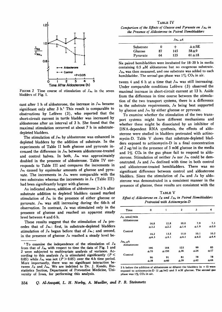
TABLE IVComparison of the Effects of Glucose and Pyruvate on JN. in
the Presence of Aldosterone in Paired Hemibladders
JNOJNao
0.8 1
1pI a I a I I
0 1 2 3 4 5Time After Aldosterone (h)
FIGURE 2 Time course of stimulation of JNa in the sevenbladders of Fig. 1.
cant after 1 h of aldosterone, the increase in JNs becamesignificant only after 3 h.' This result is comparable toobservations by Lefevre (3), who reported that theshort-circuit current in turtle bladder was increased byaldosterone after an interval of 3 h. She found that themaximal stimulation occurred at about 7 h in substrate-depleted bladders.
The stimulation of JN. by aldosterone was enhanced indepleted bladders by the addition of substrate. In theexperiments of Table II both glucose and pyruvate in-creased the difference in JN. between aldosterone-treatedand control halves. In both, JN. was approximatelydoubled in the presence of aldosterone. Table IV cor-responds to Table III and compares the increments inJN. caused by equimolar amounts of glucose and pyru-vate. The increments in JN. were comparable with thetwo substrates whereas the increments in JI (Table III)had been significantly larger with glucose.
As indicated above, addition of aldosterone 2-3 h aftersubstrate addition to depleted bladders caused markedstimulation of JNs in the presence of either glucose orpyruvate. JN. was still increasing during the 6th-h ofobservation. In contrast, JH was stimulated only in thepresence of glucose and reached an apparent steadylevel between 4 and 6 h.
These results suggest that the stimulation of JR pre-cedes that of JN.: first, in substrate-depleted bladdersstimulation of JH began before that of JN.; and second,in the presence of glucose JH reached a steady level be-
'To examine the independence of the stimulation of JHfrom that of JNs with respect to time the data of Fig. 1 and2 were subjected to multivariate analysis of variance. Ac-cording to this analysis JH is stimulated significantly (P <0.05) while JNa was not (P > 0.05) over the 4-h time period.More importantly, there was no significant interaction be-tween JI, and JNs. We are indebted to Dr. J. Kondo, Bio-statistics Section, Department of Preventive Medicine, Uni-versity of Iowa, for performing this analysis.
JNa, PA
Substrate 0 + A4SEGlucose 87 145 5849Pyruvate 64 125 61-4-18
Six paired hemibladders were incubated for 18-20 h in mediacontaining 0.5 ,uM aldosterone but no exogenous substrate.JNa was then measured, and one substrate was added to eachhemibladder. The serosal gas phase was 1% CO2 in air.
tween 4 and 6 h at a time that JN. was still increasing.Under comparable conditions Lefevre (3) observed themaximal increase in short-circuit current at 13 h. Asidefrom the difference in time course between the stimula-tion of the two transport systems, there is a differencein the substrate requirements, JH being best supportedby glucose and JNs by either glucose or pyruvate.
To examine whether the stimulation of the two trans-port systems might have different mechanisms andwhether these might be dissociated by an inhibitor ofDNA-dependent RNA synthesis, the effects of aldo-sterone were studied in bladders pretreated with actino-mycin-D. Table V shows that substrate-depleted blad-ders exposed to actinomycin-D in a final concentrationof 2 Mg/ml in the presence of 5 mMglucose in the mediaand 1% COs in the gas phase fail to respond to aldo-sterone. Stimulation of neither Jia nor JNa could be dem-onstrated. JH and JN. declined with time in both controland aldosterone-treated hemibladders. There was nosignificant difference between control and aldosteronebladders. Since the stimulation of JN. and JH by aldo-sterone was demonstrated in a consistent manner in thepresence of glucose, these results are consistent with the
TABLE VEffect of Aldosterone on JH and JN. in Paired Hemibladders
Pretreated with Actinomycin-D
Time 0 1 2 3 4
JH, nmol/minAldosterone
14.8 12.8 10.0 7.9 7.143.2 ±2.2 i±1.4 ±-1.9 ±2.0
Control14.2 13.5 11.0 10.1 10.2
±4.3 ±4.1 44.1 ±t4.5 ±4.4JN-, pA
Aldosterone196 164 123 99 85
±70 ±150 ±36 ±24 ±17Control
96 91 81 78 78438 ±40 ±39 ±42 ±44
2 h before the addition of aldosterone or diluent the bladders (n = 5) were
exposed to actinomycin-D (2 pg/ml) and 5 mMglucose. The serosal gasphase was 1% CO2 in air.
354 Q. Al-Awqati, L. H. Norby, A. MueUer, and P. R. Steinmetz
interpretation that both processes of stimulation dependon DNA-dependent RNA synthesis.
(e) Effect of substrate at two different levels of JH.The enhanced responsiveness of the transport rate tometabolic substrates in the presence of aldosterone couldbe a reflection of the increased transport rate per serather than of an effect of aldosterone on metabolism.To examine this point the effects of glucose on JHwere studied at two secretion rates in depleted bladdersin the absence of aldosterone. The rate of transport wasvaried by lowering luminal pH from 7.0 to 6.0. It hadbeen previously shown (6) that luminal acidificationdecreases the rate of active H+ transport. Table VIshows that serosal addition of glucose caused an incre-ment in JH that was almost three times as great at thehigher rate of transport (luminal pH = 7) as at the lowrate of transport (luminal pH = 6). The transport rateper se, therefore, appears to be a determinant of the ex-tent to which substrates will increase JH.
(f) Stimulation of JH by aldosterone is not mediatedby CO. Aldosterone could stimulate JH either directlyor by increasing the production of metabolic C02through a mechanism unrelated to transport. The latterexplanation is unlikely since the experiments of section(a) have shown that exogenous C02 fails to increaseJH in substrate-depleted bladders. Hence, even if aldo-sterone increased the C02 production by the tissue, theH+ transport system would not have responded. To dem-onstrate further that the increase in JH caused by al-dosterone was not mediated through increased C02 pro-duction, the effects of addition of exogenous CO2 wereexplored in aldosterone-treated and control hemibladders.Table VII shows that H+ secretion was greater in thealdosterone-treated hemibladders before addition of CO2.Addition of CO2 caused a larger increment in the aldo-sterone-treated hemibladders than in the control halves.Both rates were approximately doubled. These resultsindicate that aldosterone increases JH by some mecha-nism other than an increase in Pco2.
TABLE VIEffect of Glucose at Two Levels of JH
JH, nmol/min
No Glucose Asubstrate
Luminal pH = 6.0 12.8 17.2 4.4±1.6Luminal pH = 7.0 18.5 30.9 12.4±2.7A 8.0±2.2
JH was measured in six substrate-depleted bladders in theabsence of aldosterone at luminal pH = 6 and at pH = 7;the measurements were repeated after addition of glucose.Serosal pH was kept constant at 7.4. The gassing mixture was1% CO2.
TABLE VIIEffect of Exogenous CO2 on JH
JH1 nmol/min
C02-free 1% CO2 AAldosterone 6.5 12.5 6.0 4 1.9Control 3.9 8.0 4.1 4 1.0A (A-C) 1.940.8
JH was measured in four substrate-depleted bladders 4 h afteraddition of pyruvate and 0.5 ,M aldosterone or diluent(control). The serosal gas phase was then changed fromC02-free air to 1%CO2 in air.
(g) Effect of aldosterone on the force of the H+pump. To evaluate the site of action of aldosterone onH' transport, we will consider a general model of anenergy converter in which metabolic energy is linkedto a transport step. In such a model aldosterone couldeither facilitate the flow of protons through the trans-port system or alter the driving metabolic reaction insuch a way that the transfer of chemical energy is pro-moted. It has been shown previously (6) that applica-tion of an unfavorable pH gradient across the bladderresults in a decrease in JH.' Following Ussing andZerahn (8), it can be argued that the magnitude of thegradient sufficient to nullify the transport rate is equiva-lent to the force of the H+ pump (the protonmotiveforce, [PMF]).' Using a model of H+ transport (seeDiscussion for details), it can be shown that the slope ofJH on the applied pH gradient gives the parameter thatdescribes the ease with which protons flow through thesystem.' The value of the PMF is a composite functionwhich is determined, in part, by the free energy of thedriving metabolic reaction. To determine these twoparameters experiments were performed in paired hemi-bladders using the short-circuit current as the measureof H+ transport. The luminal pH was lowered in stepsin paired hemibladders in the presence of 1% C02 and 5mMglucose in the media. The serosal pH was kept at7.4.
2 Steinmetz and Lawson (6) showed that the passivetransepithelial H+ permeability is negligible over the rangeof spontaneous acidification. The decrease in JH with oppos-ing H+ gradient, therefore, reflects a decrease in the forwardflux of H+.
3 Abbresiation used in this paper: PMF, protonmotiveforce.
' For this analysis it is assumed that the conductance ofthe other ions is not affected by lowering of the luminal pH.Steinmetz and Lawson (6) observed that in ouabain-treatedbladders (as were used for the experiments of Table VIII)the Na+ conductance is not affected by luminal acidificationover the range used. Likewise, luminal acidification had noeffect on the permeabilities of C1-, Cs+, and K+ in short-circuited control bladders (6).
Stimulation of H+ Transport by Aldosterone 355

50rNO
ALDOSTERONE
9
50
4040-
30h
20 h AFTERALDOSTERONE
o--o Control
_ --o Experimentol
301-
201
10
20
10
7.0 6.0 5.0 7.0 6.0 5.0Mucosal pH Mucosal pH
FIGURE 3 Failure of aldosterone to alter the force of theH4 pump. In the left panel JH was reduced to zero bylowering of mucosal pH before addition of aldosterone.The panel on the right shows the results of the samemaneuver 20 h after addition of aldosterone or diluent(based on results of Table VIII).
Fig. 3 shows how JH decreased with decreasing lumi-nal pH. The left panel demonstrates that JH decreasedin similar manner in both hemibladders before additionof aldosterone. The panel on the right gives the results20 h after addition of aldosterone or diluent to thehemibladders. JH was increased in the tissues exposedto aldosterone, but the luminal pH at which JH becamezero (PMF) was the same for both groups of hemi-bladders. The slope of JH on the applied chemical gradi-ent, therefore, was increased by aldosterone. The valuesfor JH, PMF, and the slope OJH/OA pH are given inTable VIII for the 1st day on the left and after aldo-sterone on the right. Aldosterone caused no significantchange in PMF while JH and the slope were clearlyincreased.
The results suggest that aldosterone does not increasethe force of the pump directly but increases JH by in-creasing the conductance in series with the active trans-
port pathway or by a thermodynamically equivalentmechanism.
DISCUSSION
The present study indicates that aldosterone stimulatesH+ secretion as well as Na+ transport by the isolatedturtle bladder. It confirms the observation by LeFevre(3) that the response of sodium transport to aldosteroneis slow in turtle bladder, and it supports the results ob-tained by Ludens and Fanestil (2) on urinary acidifica-tion by the Colombian toad bladder. Several character-istics of the stimulation of H+ secretion in turtle bladderare defined. It is shown that H4 secretion is stimulatedindependently of the stimulation of Na4 transport andthat stimulation is demonstrable in the presence as wellas in the absence of the two known rate-limiting sub-strates for H4 secretion: exogenous COa and metabolicsubstrates. Finally, the mode of action of aldosterone isanalysed in an equivalent circuit model of H4 transport.
Several lines of evidence indicate that the stimulationof H4 secretion is independent of that of Na+ transport.First of all, the stimulation of H+ secretion precedes thatof Na4 transport by about 2 h. Secondly, in aldosterone-treated bladders the stimulation of H4 secretion has dif-ferent substrate requirements than the stimulation ofNa4 transport. H+ secretion was increased twice asmuch by addition of glucose as by addition of pyruvate,whereas Na+ transport was increased to the same extentby these two substrates. Thirdly, H4 secretion wasstimulated by aldosterone in bladders in which Na4transport was abolished by ouabain (Table VIII).Since aldosterone is thought to exert its effect on sodiumtransport through a process involving DNA-dependentRNA synthesis (9), it would be conceivable that theearly stimulation of H+ secretion would be mediated bysome other mechanism. This possibility, in fact, was sug-gested by Lifschitz et al. (1) who observed that actino-mycin-D inhibits the aldosterone-induced increase in
TABLE VIIIThe Force of the H+ Pumpbefore and after Addition of Aldosterone
Before addition 20 h after addition
JH PMF VJH/aApH JH PMF aJH1/aPHAldosterone 41.2 3.00 15.9 16.0 2.75 6.9Control 41.0 3.11 15.1 10.1 2.87 4.1
A (A-C) 5.9 -0.12 2.842.2 ±0.18 ±0.6
JH is expressed as nmol/min and PMFas pH units. The results of the left panelwere obtained when the bladder halves were fresh; the results of the right panelwere obtained either after 20 h of exposure to aldosterone or after depletion of en-dogenous hormone (control). All experiments were carried out in the presence of0.5 mMouabain, 5 mMglucose, and 1% CO2 in the gas phase.
356 Q. Al-Awqati, L. H. Norby, A. Mueller, and P. R. Steinmetz
.E_
E
F
?II I
I

sodium absorption but not in H+ secretion in the dog.In the isolated turtle bladder both the early stimulationof H+ secretion and the late stimulation of Na4 transportwere abolished by actinomycin-D. The separate stimula-tion of H+ secretion and Na4 absorption and their spe-cific substrate dependence could reflect the synthesis oftwo different proteins or merely inherent differences inthe characteristics of the two transport systems.
The results of Tables II and III clearly indicate thatthe presence of aldosterone increases the responsivenessof substrate-depleted bladders to the addition of glucoseor pyruvate. Both H+ secretion and sodium transportshowed this increased responsiveness, but for H+ secre-tion glucose was preferred over pyruvate as a substrate.This synergism between aldosterone and substrate iswell known for the sodium transport system in the toadbladder (9, 10). Its mechanism, however, remains onlypartially understood. Sharp and Leaf (10) have pro-vided evidence that the stimulation of metabolism issecondary to stimulation of Na+ transport, whereasFanestil et al. (9) have suggested that aldosterone hasa primary effect on the metabolic reactions coupled tosodium transport.
If the stimulation of H+ transport by metabolic sub-strates is secondary to a primary acceleration of thetransport step by aldosterone, then one should be able todemonstrate such a substrate effect following othermaneuvers that primarily accelerate transport. Changesin the pH gradient against which H+ secretion occursalter the transport rate instantaneously (6). It wasshown in Table VI that in depleted bladders not ex-posed to aldosterone the response of JH to glucose addi-tion was a function of the basal transport rate. The re-sponse was considerably greater at the higher basaltransport rate observed at a mucosal pH of 7 than at thereduced rate at pH 6. This apparent dependence of thesubstrate effect on the basal transport rate is consistentwith the interpretation that the stimulation of metabo-lism by aldosterone is probably secondary to its effecton the transport system.
The secretion of H+ by the turtle bladder may be out-lined by the following working description (4): protons,perhaps originating from splitting of water molecules,are translocated across the luminal border of the epi-thelium in a process that requires the input of metabolicenergy. The hydroxyl ions remaining are disposed ofwith the aid of cytoplasmic carbonic anhydrase by com-bining with C02; the HC0- so formed then diffusesdown an electrochemical gradient into the serosal side.By analogy with the sodium transport system (8, 11)this process can be represented by an equivalent circuitmade up of a battery representing the proton pump, aresistance in series with the pump and a shunt resistancein parallel with the pump. The force of the pump, the
PMF, can be evaluated following Ussing and Zerahn(8), as the transepithelial gradient necessary to bringnet transport to zero. It follows then that for a con-stant flow of current through the parallel limb, the seriesconductance is approximated by the slope of the H+transport rate on the applied gradient.' In evaluatingthese two parameters of the equivalent circuit, TableVIII shows that aldosterone does not change the PMFbut significantly increases the conductance in the activepathway.
In this representation, what do the elements of thecircuit represent? The PMF is considered to be a func-tion of the chemical reaction that leads to charge sepa-ration. It includes a term that is equivalent to the freeenergy (AG) of the reaction that is tightly linked totransport. In many transport systems it is believed thatATP hydrolysis is an important "energy donor"; hence,the free energy available from ATP hydrolysis wouldbe a determining factor in the value of the PMF. ThePMF, however, depends not only on the free energy ofthe chemical reaction but also on its relation to the flowand translocation of protons through the system (12).
The conductance in the active transport pathway re-flects the facility with which either protons or OH- ionsflow from the site of dissociation. This extensive func-tion also includes, in descriptive terms, the number ofpump sites available for the dissociation reaction. Theparallel conductance for H+ in turtle bladder is low asjudged from the studies of Steinmetz and Lawson (6)and from recent evidence (13).
Essig and Caplan (12) have given a formal descrip-tion of this kind of analysis in terms of linear irreversi-ble thermodynamics. The formalism requires that theflow of the transported ion species (JH) and the flow ofmetabolism (Jr) are coupled to the conjugate forces ofthe ion (XH), and of metabolism (A, the affinity of thedriving chemical reaction i.e. the AG noted above).
JH= Lia Xii + LHr A
Jr = LH XH+ L, A
where LH and Lr are "straight coefficients" relating theflow of one species to its driving force, and LHr and LrHare cross coefficients relating the flow of one species tothe driving force of the other species. Over the pHrange examined both JHi and J5S have been shown to bea linear function of the transepithelial pH gradient (6,13, 14). The two cross coefficients, following Onsager(15), are considered to be equal. The PMF in this
5Jr was measured continuously as the rate of "CO2 pro-duction from [U-`QC]glucose in ouabain-treated bladders(13, 14). Because of the linearity of Jr and XH, the affinitymay be considered constant during the period of experi-mental observation.
Stimulation of H+ Transport by Aldosterone 357

analysis is (XH)J, =o and is equal to
(XH)JHo = [LrH/LH]A
and
LuH =[aJH/XH]A
Since the affinity is probably constant during the periodof the experiment, LH is equivalent to the slope shown inFig. 3. The constancy of the PMF could reflect a con-stant affinity or, alternatively, an increased affinity bal-anced by a decrease in the fraction LV}H/LH. Recently,Saito et al. (16) have shown in the frog skin that theaffinity may indeed increase after prolonged incubationwith aldosterone.
It follows from this analysis that aldosterone increasesthe conductance of protons through the active pathwayand not the PMF in the system. As discussed, the in-creased active conductance could represent a number ofchanges in the overall transport operation of the sys-tem. Among the more attractive possibilities are theformation of new pump sites or a structural change inthe luminal membrane that would provide access to anincreased number of existing pump sites. These changeswould be mediated one way or another by the synthesisof new proteins (17). Alternatively, the increased con-ductance could be a manifestation of a change in thephysical state of the membrane that would increase themobility of protons through the active pathway. Re-cently, Goodman et al. (18) have provided evidence thataldosterone changes the lipid composition of the mem-brane in a way that would increase its "fluidity."
ACKNOWLEDGMENTSThis paper was supported by grant AM 17568 from theU. S. Public Health Service and, in part, by funds fromthe Iowa Heart Association.
REFERENCES1. Lifschitz, M. D., R. W. Schrier, and I. S. Edelman.
1973. Effect of actinomycin-D on aldosterone-mediatedchanges in electrolyte excretion. Am. J. Physiol. 224:376-380.
2. Ludens, J. H., and D. D. Fanestil. 1974. Aldosteronestimulation of acidification of urine by isolated urinarybladder of the Colombian toad. Am. J. Physiol. 226:1321-1326.
3. LeFevre, M. E. 1973. Effects of aldosterone on theisolated substrate-depleted turtle bladder. Am. J. Phys-iol. 225: 1252-1256.
4. Steinmetz, P. R. 1974. Cellular mechanisms of urinaryacidification. Physiol. Rev. 54: 890-956.
5. Steinmetz, P. R. 1967. Characteristics of hydrogen iontransport in urinary bladder of water turtle. J. Clin.Invest. 46: 1531-1540.
6. Steinmetz, P. R., and L. R. Lawson. 1971. Effect ofluminal pH on ion permeability and flows of Na+ and H+in turtle bladder. Am. J. Physiol. 220: 1573-1580.
7. Schwartz, J. H., and P. R. Steinmetz. 1971. CO2 re-quirements for H+ secretion in the isolated turtle blad-der. Am. J. Physiol. 220: 2051-2057.
8. Ussing, H. H., and K. Zerahn. 1951. Active transport ofsodium as the source of electric current in the short-circuited isolated frog skin. Acta. Physiol. Scand. 23:110-127.
9. Fanestil, D. D., T. S. Herman, G. M. Fimognari, andI. S. Edelman. 1968. Oxidative metabolism and aldo-sterone regulation of sodium transport. In: RegulatoryFunctions of Biological Membranes. J. Jarnefelt, editor.Elsevier, Amsterdam. pp. 177-194.
10. Sharp, G. W. G., and A. Leaf. 1968. Metabolic effectsassociated with the stimulation of sodium transport byaldosterone. In: Regulatory Function of Biological Mem-branes. J. Jarnefelt, editor. Elsevier, Amsterdam. pp.195-201.
11. Civan, M. M., 0. Kedem, and A. Leaf. 1966. Effect ofvasopressin on toad bladder under conditions of zeronet sodium transport. Am. J. Physiol. 211: 569-575.
12. Essig, A., and S. R. Caplan. 1968. Energetics of activetransport processes. Biophys. J. 8: 1434-1457.
13. Beauwens, R., and Q. Al-Awqati. 1975. Coupling be-tween H+ transport and metabolism in turtle urinarybladder. Abstract, 8th Ann. M. Am. Soc. Nephrol.,p. 71.
14. Al-Awqati, Q., R. Beauwens, A. Mueller, and P. R.Steinmetz. 1975. Urinary acidification against a gradient:Coupling of transport to metabolism. Clin. Res. 23:429A. (Abstr.)
15. Onsager, L. 1931. Reciprocal relations in irreversibleprocesses. Phys. Rev. 37: 405426.
16. Saito, T., A. Essig, and S. R. Caplan. 1973. The effectof aldosterone on the energetics of sodium transportin the frog skin. Biochim. Biophys. Acta. 318: 371-382.
17. Edelman, I. S., R. Bogoroch, and G. A. Porter. 1963.On the mechanism of action of aldosterone on sodiumtransport: The role of protein synthesis. Proc. Natl.Acad. Sci. U. S. A. 50: 1169-1177.
18. Goodman, D. B. P., J. E. Allen, and H. Rasmussen.1971. Studies on the mechanisms of action of aldoster-one: Hormone-induced changes in lipid metabolism.Biochemistry. 10: 3825-3831.
358 Q. Al-Awqati, L. H. Norby, A. Mueller, and P. R. Steinmetz