Jawbone Motion to Amend Lawsuit Against Fitbit Filed 3-14-16

CHAPTER 5
©Thompson Educational Publishing, Inc. 2003. All material is copyright protected. It is illegal to copy any of this material.
This material may be used only in a course of study in which Exercise Science: An Introduction to Health and Physical Education (Temertzoglou/Challen) is the

Diagnostic TestSkeletal Words Made Easy
Match the terms on the right with the terms on the left.
1. Breastbone
2. Shinbone
3. Skull
4. Kneecap
5. Shoulder blade
6. Lower jawbone
7. Upper jawbone
8. Thigh bone
9. Tail bone
10. Fingers
11. Backbones
12. Upper arm
13. Toes
14. collarbone
1. Clavicle
2. Coccyx
3. Cranium
4. Femur
5. Humerus
6. Mandible
7. Maxilla
8. Patella
9. Scapula
10. Phalanges
11. Vertebrae
12. Sternum
13. Tibia
14. *one word is used twice

Diagnostic TestAnswers
1. Breastbone
2. Shinbone
3. Skull
4. Kneecap
5. Shoulder blade
6. Lower jawbone
7. Upper jawbone
8. Thigh bone
9. Tail bone
10. Fingers
11. Backbones
12. Upper arm
13. Toes
14. collarbone
1. Sternum
2. Tibia
3. Cranium
4. Patella
5. Scapula
6. Mandible
7. Maxilla
8. Femur
9. Coccyx
10. phalanges
11. Vertebrae
12. Humerus
13. Phalanges
14. clavicle

Lesson 5.1
© 2015 Thompson Educational Publishing, Inc.
4
ANATOMICAL TERMINOLOGY
~ ~ ~
TOPICS COVERED IN THIS LESSON
• (a) The Anatomical Position, Planes, and Axes
• (b) Describing Movements at Joints
Pg.118

The Anatomical Position
The anatomical position is
the standard position
(standing straight, looking
forward, arms at your side,
and hands facing forward)
used to describe the
locations and relationships
of anatomical parts on your
body.
•It is used in similar way to
how the markings on a
compass are used to describe
locations in geography.

Characteristics of the AnatomicalPosition
© 2015 Thompson Educational Publishing, Inc.
6
The key features of the anatomical position are as follows:
•The person is in an upright,
standing position with his or
her head, eyes, and toes pointing forward.
•The feet are together and the
arms are slightly out to the
side.
•The forearms are fully
supinated; in other words, the palms of the hands are facing
forward.

Examples of Anatomical Relationships
© 2015 Thompson Educational Publishing, Inc.
7
• Anterior / PosteriorThe sternum is anterior to the heart (so
anterior also means “in front of”). The
heart also has an anterior surface.
• Superior / InferiorSuperior refers to upward surfaces, inferior
refers to downward surfaces.
• Medial / LateralMedial means towards the midline or
towards the median plane, whereas lateral means away from the midline or
away from the median plane.
• Proximal / DistalProximal means towards the point of
attachment of the limb to the body,
whereas distal means farther away from
the point of attachment.
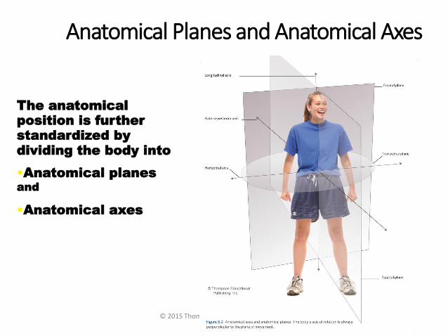
Anatomical Planes and Anatomical Axes
© 2015 Thompson Educational Publishing, Inc.
8
The anatomical position is further standardized by dividing the body into
•Anatomical planesand
•Anatomical axes
Pg. 120

Anatomical Planes
© 2015 Thompson Educational Publishing, Inc.
9
• Frontal planeThe frontal (coronal) plane is
vertical and extends from one side of the body to the other.
• Transverse plane The
transverse (horizontal) plane is
horizontal and divides the body into upper and lower segments.
• Sagittal planeThe sagittal (median) plane is
vertical and extends from the
front of the body to the back.

– A vertical plane that bisects the body into right and left halves.
Sagittal
Plane
Sagittal Plane
Flexion/extension

The Sagittal Plane

Sagittal plane– The plane dividing the body into right
and left portions

Frontal plane• A vertical plane that bisects the
body into front and back
• It is at right angles to the sagittal plane
Frontal
Plane

Frontal
Plane

Frontal plane– The plane dividing the body into front and back
portions
– Also called the Coronal plane
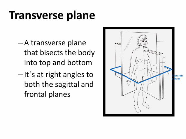
Transverse plane
– A transverse plane that bisects the body into top and bottom
– It’s at right angles to both the sagittal and frontal planes
Transvers
e Plane

Transverse
Plane

Transverse plane– The horizontal plane dividing the body into upper
and lower portions
– Also called the Horizontal plane

Anatomical Axes
© 2015 Thompson Educational Publishing, Inc.
19
• Horizontal axisThe horizontal axis extends from one side of the body to the other.
• Longitudinal axis The longitudinal axis (also known as the polar axis) is vertical, running from head to toe.
•Antero-posterior axisThe antero-posterioraxis extends from the front of the body to the back.

Horizontal Axis• Horizontal or (bilateral axis) is in an “east-west”
relationship to the anatomical position.

Antero-Posterior Axis• Antero-posterior axis is in a “front-to-back” relationship
to the anatomical position.

Longitudinal Axis
• Longitudinal or (polar axis) is in a “north-south” relationship to the anatomical position.

Planes and Corresponding Axes

Planes and Corresponding Axes

Describing a Body Movement:General Rule
© 2015 Thompson Educational Publishing, Inc.
10
A body movement can be described in terms of the anatomical plane through which it occurs and the anatomical axis around which it rotates.
THE GENERAL RULE FOR DESCRIBING A BODY
MOVEMENT:
The axis of rotation is always perpendicular to the plane of
movement.

Axis of Rotation Plane of Motion Example
Horizontal (Bilateral)
Longitudinal (Polar)
Antero-Posterior
Relationship between Planes and Axes

Relationship between Planes and Axes
Axis of Rotation Plane of Motion Example
Horizontal (Bilateral) Sagittal Flexion, extension
Longitudinal (Polar) Transverse Rotation of extremities, axial rotation
Antero-Posterior Frontal (Coronal) Abduction, adduction

Various Movements and Planes of Movement
Activity Axis PlaneStride Jump
Cartwheel
Elbow extension
Nodding yes
Tuck
Somersault
Twirling
Shaking head no

Various Movements and Planes of Movement
Activity Axis PlaneStride Jump Horizontal Sagittal
Cartwheel Antero-Posterior Frontal
Elbow extension Horizontal Sagittal
Nodding yes Horizontal Sagittal
Tuck Horizontal Sagittal
Somersault Horizontal Sagittal
Twirling Longitudinal Transverse
Shaking head no Longitudinal Transverse

#1. Guess the plane and corresponding axis.
Sagittal Plane/Horizontal Axis

#2. Guess the plane and corresponding axis.
Transverse Plane/Longitudinal Axis

#3. Guess the plane and corresponding axis.
Frontal Plane/Antero-posterior Axis

Terms Used to Describe Movement
© 2015 Thompson Educational Publishing, Inc.
33
• Flexion is the action of bending at a
joint such that the joint angle
decreases (e.g., when you bend your
elbow to bring your palm up towards
your face).
• Extension is the opposite of
flexion.
• Abduction (“ab” = “from”) occurs
when you move a body segment to
the side and away from your body
(e.g., moving your arm out to the side
and bringing it level with your
shoulder).
• Adduction (“ad” = “to”) is the
opposite of abduction.

Terms Used to Describe Movement
© 2015 Thompson Educational Publishing, Inc.
34
• Plantar flexion is specific to the ankle
joint. It occurs when you point your toes
(e.g., when you stand on your tip toes).
• Dorsiflexion occurs when you bend the
ankle to bring the top of your foot closer to
your shin.
• Supination is rotating the wrist such that
the palm of your hand is facing forward
(e.g., when youcatch a softball underhanded with one
hand).• Pronation occurs in the opposite
direction.

Terms Used to Describe Movement
© 2015 Thompson Educational Publishing, Inc.
35
• Inversion is associated with the ankle joint.
It is a result of standing on the outer edge of your foot (e.g., when you twist your ankle).
• Eversion is a result of standing on the inner
edge of your foot.
• External rotation results when you twist
or turn a body part outward from the midline (eg., turning your toes outward).
• Internal rotation results when you twist or
turn a body part inward towards the midline.

Terms Used to Describe Movement
© 2015 Thompson Educational Publishing, Inc.
36
• Elevation refers to movement in an upwards direction (e.g., hunching your shoulders).
• Depression is the opposite motion—movement in a downwards direction (e.g., slouching your shoulders).
• Circumduction is a combination of
flexion, extension, abduction, and
adduction. An example of this
movement occurs when asoftball pitcher throws a ball with a “windmill” action.

Supine Lying on the back
when performing a bench press
Prone Lying face down when preparing to
perform a push-up

Ankle Sprains
Inversion Ankle Sprain Eversion Ankle Sprain (Potts Fracture)

Guess the movements
Abduction

Guess the movements
Supination

Guess the movements
Dorsiflexion

Lesson 5.2
© 2015 Thompson Educational Publishing, Inc.
43
THE MUSCULOSKELETAL SYSTEM
~ ~ ~
TOPICS COVERED IN THIS LESSON
• (a) The Human Skeleton: An Overview
• (b) The Axial and Appendicular Skeleton
Pg.124

What Is the Human Skeleton?
The adult human skeleton is made up of 206 bones, accounting for about 14 percent of total body weight.
Humans start life with more bones than that—about 300 bones at birth. Over time, several bones fuse as growth takes place (such as in the skull and lower part of the vertebral column).
© 2015 Thompson Educational Publishing, Inc.
44
Pg.124

The Human Skeleton
© 2015 Thompson Educational Publishing, Inc.
20

The Human Skeletal System
Bones are made up of living tissue—bone cells, fat cells, and blood vessels.
Compared to other body systems, the human skeletal system is extremely hard and durable.
Bones themselves are composed primarily of the mineral calcium.
People whose diet is low in calcium may find their bones becoming increasingly brittle and breakable—a major concern for older people (osteoporosis).
© 2015 Thompson Educational Publishing, Inc.
46

Main Functions of the Skeletal System
© 2015 Thompson Educational Publishing, Inc.
47
The skeletal system provides structural support to the body, protects vital organs, serves as a growth centre for cells, acts as a reserve for minerals, and of course plays a major role in movement.

Classification of Bones in the Human Body
© 2015 Thompson Educational Publishing, Inc.
48
Bones are normally classified according to their
shape—long, short, flat, irregular, and a fifth type
(sesamoid) that is found within tendons.
Long bones are found in the arms and legs (e.g., the femur).
Short bones are most common in the wrists (e.g., the carpal
bones).
Flat bones are flat and thin and are found in the
roof of the skull.
Irregular bones include odd-looking bones such as the
bones in the vertebrae.
Sesamoid bones are unusual, small, flat bones wrapped
within tendons that move over bony surfaces (e.g., the patella).
Pg.125

© 2015 Thompson Educational Publishing, Inc.
49

Sesamoid Bones

©Thompson Educational Publishing, Inc. 2003. All material is copyright protected. It is illegal to copy any of this material.
This material may be used only in a course of study in which Exercise Science: An Introduction to Health and Physical Education (Temertzoglou/Challen) is the

©Thompson Educational Publishing, Inc. 2003. All material is copyright protected. It is illegal to copy any of this material.
This material may be used only in a course of study in which Exercise Science: An Introduction to Health and Physical Education (Temertzoglou/Challen) is the

Classify each bone.
1. 3. 4. 5.
6.
7.
2.
8.
See H.O.

Classify each bone.
1.FLAT3.LONG 4.SHORT 5.SESAMOID
6.LONG
7.IRREGULAR
2.SHORT
8. FLAT

The Structure of the Skeleton
© 2015 Thompson Educational Publishing, Inc.
55
The human skeletal system is generally divided into two main parts:
•The axial skeleton(shown in orange), and
•The appendicular skeleton
•(shown in green).
Pg.126

The Axial Skeleton—80 Bones
The axial skeleton is comprised mainly of the vertebral column (the spine), much of the skull, and the rib cage.
Most of the body’s core muscles originate from the axial skeleton.
These core muscles help stabilize and support the axial skeleton, thus providing proper posture and alignment.
© 2015 Thompson Educational Publishing, Inc.
56

The Appendicular Skeleton—126 Bones
© 2015 Thompson Educational Publishing, Inc.
57
The appendicular
skeleton includes the
movable limbs and their
supporting structures
(girdles), which play a
key role in allowing us to
move.
The appendicular
skeleton can be divided
into six major regions:
pectoral girdle; arms and
forearms; hands; pelvis;
thighs and legs; and feet
and ankles.

©Thompson Educational Publishing, Inc. 2003. All material is copyright protected. It is illegal to copy any of this material.
This material may be used only in a course of study in which Exercise Science: An Introduction to Health and Physical Education (Temertzoglou/Challen) is the required textbook.
Axial vs. Appendicular Skeleton
The axial skeleton consists of 80
bones:
26 vertebral column
1 hyoid
22 skull
6 auditory
25 ribs
The appendicular skeleton consists
of 126 bones:
64 upper extremity
62 lower extremity
Axial (80) + Appendicular (126) = 206 bones

Bone Landmarks
© 2015 Thompson Educational Publishing, Inc.
59
All the bones in the human skeleton have
features known as landmarks.
A landmark is a ridge, bump, groove,
depression, or prominence on the surface of
the bone that serves as a guide to the
locations of other body structures.
For example, the quadriceps muscles of the
front thigh ultimately wrap around the
patella (kneecap) and insert on the tibial
tuberosity (a landmark at the top of the
tibia).
Pg.126

The Anatomy of a Long Bone
© 2015 Thompson Educational Publishing, Inc.
60
See H.O.
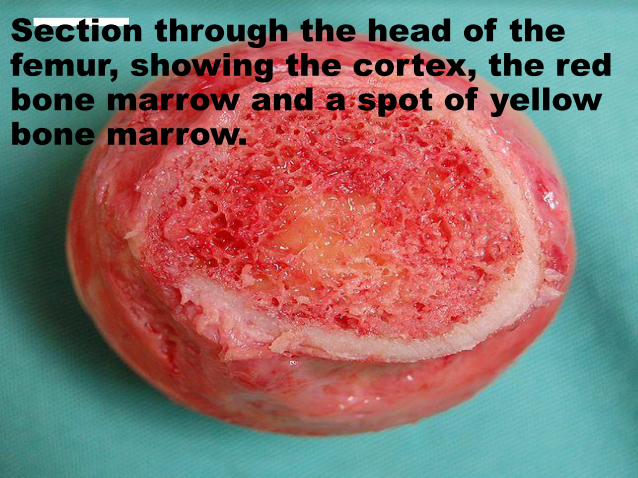
Section through the head of the femur, showing the cortex, the red bone marrow and a spot of yellow bone marrow.

©Thompson Educational Publishing, Inc. 2003. All material is copyright protected. It is illegal to copy any of this material.
This material may be used only in a course of study in which Exercise Science: An Introduction to Health and Physical Education (Temertzoglou/Challen) is the required textbook.
Epiphyseal Plates and Lines
Epiphyseal plates (growth
plates)
Occur at various
locations at the
epiphyses of long
bones
Growth possible
Epiphyseal lines
Occur when epiphyseal
plates have fused or
come together
Growth not possible
Epiphyseal
plate
Epiphyseal
line

Epiphyseal Plate or Line?
PlateLine
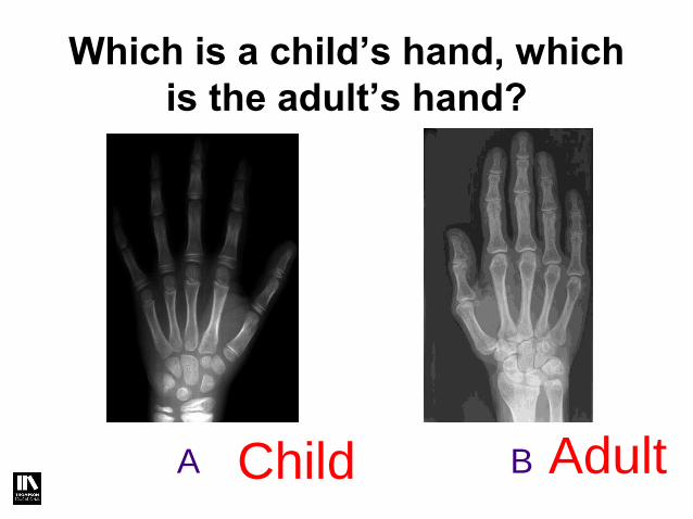
Which is a child’s hand, which
is the adult’s hand?
A BChild Adult

Osteoporosis
Low bone mass and deterioration of the bone tissue
The number of osteoblasts is decreased and the number of osteoclasts stay the same
Leads to bone fragility
Increased susceptibility to bone fractures
Preventative measures include:
Balanced diet rich in calcium and vitamin D, and a healthy lifestyle
Weight-bearing exercises
Bone density testing and medication when appropriate

Bone Injuries and Bone Disease
© 2015 Thompson Educational Publishing, Inc.
30
• Fractures—bone “breaks,” normally divided
into three types: simple, compound, and
comminuted.
• Stress fractures—tiny cracks caused by a
rapid increase in activity or when an athlete
switches training surfaces or wears footwear
with improper cushioning.
• Shin splints—a painful condition occurring on
the medial or lateral side of the tibia (shin
bone)—are another common sports injury.
• Tearing of the interosseous membrane
• If not treated, it may lead to a stress
fracture
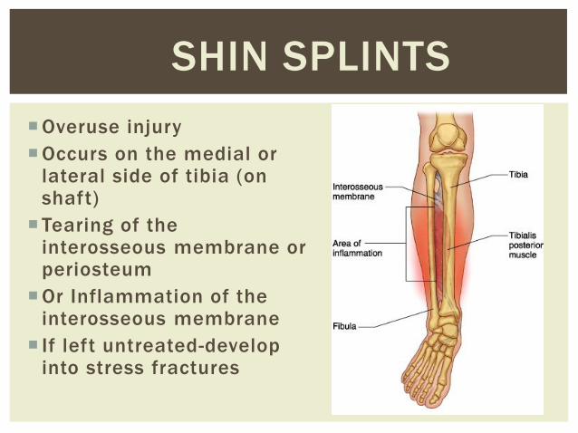
Overuse injury
Occurs on the medial or lateral side of tibia (on shaft)
Tearing of the interosseous membrane or periosteum
Or Inflammation of the interosseous membrane
If left untreated-develop into stress fractures
SHIN SPLINTS

Fractures
• Despite its mineral
strength, bone may
crack or even break if
subjected to extreme
loads, sudden
impacts, or stresses
from unusual
directions.
– The damage produced
constitutes a fracture.
©Thompson Educational Publishing, Inc. 2003. All material is copyright protected. It is illegal to copy any of this material.
This material may be used only in a course of study in which Exercise Science: An Introduction to Health and Physical Education (Temertzoglou/Challen) is the

©Thompson Educational Publishing, Inc. 2003. All material is copyright protected. It is illegal to copy any of this material.
This material may be used only in a course of study in which Exercise Science: An Introduction to Health and Physical Education (Temertzoglou/Challen) is the required textbook.
Types of Fractures
Stress fracture – most difficult to detect; tiny
crack in the bone; muscles too tired to
absorb shock; overuse injury
Simple (Closed) fracture – no separation;
bone ends don’t penetrate the skin.
Compound (Open) fracture – bone breaks
into separate pieces; bone ends penetrate
the skin.
Comminuted fracture – bone shatters into
many pieces. Common in the elderly (brittle
bones).
Greenstick fracture- bone breaks
incompletely. One side bent, one side
broken. Common in children.
Spiral-ragged break caused by excessive
twisting forces. Sports injury/Injury of abuse
Simple
fracture
Compound
fracture
Comminuted
fracture

Avulsion Fracture
• A closed fracture where a piece of bone is
broken off by a sudden, forceful contraction of a
muscle.

Fractures
Greenstick Fracture Spiral Fractures
©Thompson Educational Publishing, Inc. 2003. All material is copyright protected. It is illegal to copy any of this material.
This material may be used only in a course of study in which Exercise Science: An Introduction to Health and Physical Education (Temertzoglou/Challen) is the

Stress Fractures
©Thompson Educational Publishing, Inc. 2003. All material is copyright protected. It is illegal to copy any of this material.
This material may be used only in a course of study in which Exercise Science: An Introduction to Health and Physical Education (Temertzoglou/Challen) is the

©Thompson Educational Publishing, Inc. 2003. All material is copyright protected. It is illegal to copy any of this material.
This material may be used only in a course of study in which Exercise Science: An Introduction to Health and Physical Education (Temertzoglou/Challen) is the
What kind of fracture is this?Comminuted

©Thompson Educational Publishing, Inc. 2003. All material is copyright protected. It is illegal to copy any of this material.
This material may be used only in a course of study in which Exercise Science: An Introduction to Health and Physical Education (Temertzoglou/Challen) is the


Lesson 5.3
© 2015 Thompson Educational Publishing, Inc.
76
MAJOR BONES OF THE HUMAN BODY
~ ~ ~
TOPICS COVERED IN THIS LESSON
• (a) Bones of the Axial Skeleton
• (b) Bones of the Appendicular Skeleton
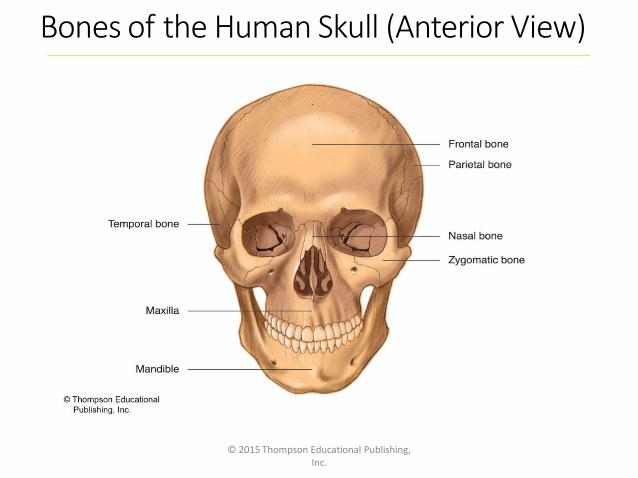
Bones of the Human Skull (Anterior View)
© 2015 Thompson Educational Publishing, Inc.
77

Bones of the Human Skull (Lateral View)
© 2015 Thompson Educational Publishing, Inc.
78
Coronal Suture
Sagittal Suture
Squamous Suture
Lambdoidal Suture
Occipital Suture

Skull of a Newborn

The Vertebral Column (Lateral View)
© 2015 Thompson Educational Publishing, Inc.
80

©Thompson Educational Publishing, Inc. 2003. All material is copyright protected. It is illegal to copy any of this material.
This material may be used only in a course of study in which Exercise Science: An Introduction to Health and Physical Education (Temertzoglou/Challen) is the

©Thompson Educational Publishing, Inc. 2003. All material is copyright protected. It is illegal to copy any of this material.
This material may be used only in a course of study in which Exercise Science: An Introduction to Health and Physical Education (Temertzoglou/Challen) is the

©Thompson Educational Publishing, Inc. 2003. All material is copyright protected. It is illegal to copy any of this material.
This material may be used only in a course of study in which Exercise Science: An Introduction to Health and Physical Education (Temertzoglou/Challen) is the

©Thompson Educational Publishing, Inc. 2003. All material is copyright protected. It is illegal to copy any of this material.
This material may be used only in a course of study in which Exercise Science: An Introduction to Health and Physical Education (Temertzoglou/Challen) is the

©Thompson Educational Publishing, Inc. 2003. All material is copyright protected. It is illegal to copy any of this material.
This material may be used only in a course of study in which Exercise Science: An Introduction to Health and Physical Education (Temertzoglou/Challen) is the

©Thompson Educational Publishing, Inc. 2003. All material is copyright protected. It is illegal to copy any of this material.
This material may be used only in a course of study in which Exercise Science: An Introduction to Health and Physical Education (Temertzoglou/Challen) is the

©Thompson Educational Publishing, Inc. 2003. All material is copyright protected. It is illegal to copy any of this material.
This material may be used only in a course of study in which Exercise Science: An Introduction to Health and Physical Education (Temertzoglou/Challen) is the

©Thompson Educational Publishing, Inc. 2003. All material is copyright protected. It is illegal to copy any of this material.
This material may be used only in a course of study in which Exercise Science: An Introduction to Health and Physical Education (Temertzoglou/Challen) is the

Cervical Vertebrae
Atlas –C1
-nodding
motion; “yes”
Axis –C2
-rotation;
“no”

Atlas
Axis

The Thoracic Cage (Anterior View)
© 2015 Thompson Educational Publishing, Inc.
91

Left Scapula (Anterior & Lateral Views)
© 2015 Thompson Educational Publishing, Inc.
92

Left Scapula (Posterior View)
© 2015 Thompson Educational Publishing, Inc.
40

Pelvis (Male, Anterior View)
© 2015 Thompson Educational Publishing, Inc.
94
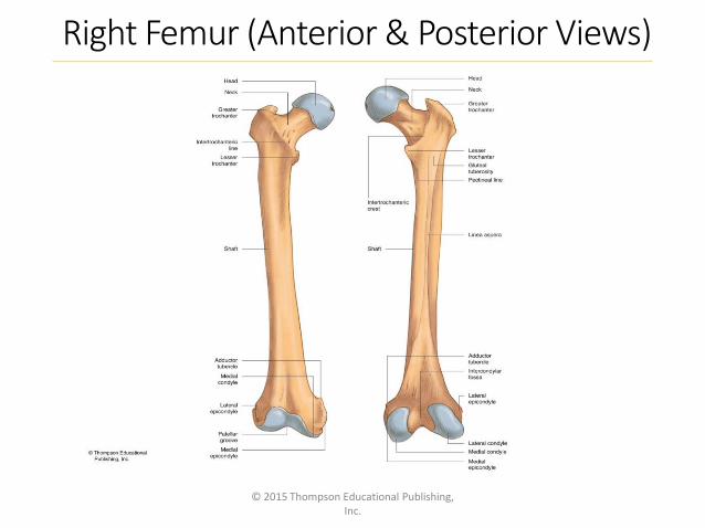
Right Femur (Anterior & Posterior Views)
© 2015 Thompson Educational Publishing, Inc.
95

Right Fibula (Anterior & Superior Views)
© 2015 Thompson Educational Publishing, Inc.
96

Right Foot (Superior View)
© 2015 Thompson Educational Publishing, Inc.
97

Left Humerus (Anterior & Posterior Views)
© 2015 Thompson Educational Publishing, Inc.
98

Left Ulna and Radius (Anterior View)
© 2015 Thompson Educational Publishing, Inc.
99

Left Hand (Anterior View)
© 2015 Thompson Educational Publishing, Inc.
100

Lesson 5.4
© 2015 Thompson Educational Publishing, Inc.
101
THE ARTICULAR SYSTEM
~ ~ ~
TOPICS COVERED IN THIS LESSON
•(a) The Different Types of Human Joints and Joint- Related Injuries
•(b) Shoulder Joint / Knee Joint / Ankle Joint

The Knee:• Femur, tibia and patella

The Shoulder: • Scapla, humerus and clavicle

The Ankle:
• Tibia, talus and fibula

Types of Joints
• With one exception (the hyoid bone), every bone in the body is connected (articulated) to or forms a joint with at least one other bone.
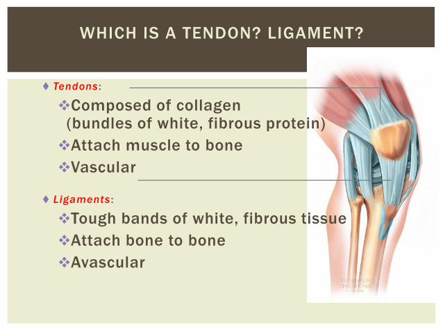
WHICH IS A TENDON? LIGAMENT?
Tendons :
Composed of collagen (bundles of white, fibrous protein)
Attach muscle to bone
Vascular
Ligaments :
Tough bands of white, fibrous tissue
Attach bone to bone
Avascular

Ligaments• Less rigid than bone• Do not stretch as much
as tendons• Tissue that attaches one
or more bones together• Made up of tough bands
of white, fibrous tissue • When they reach their
threshold-stretch minimally, usually tear

Tendons• Large bundles of white, fibrous protein (collagen) that
attaches muscle to bone• Greater stretching ability• Will tear with great force

1
0
9

Vascularity
• Amount of supplied blood a tissue has or requires
• Ligaments & cartilage are “avascular”-nutritional needs are not met through blood (take a long time to heal)
• The more vascular a tissue, the less time it takes to recover from an injury
• Bone and muscle are vascular and will take less time to heal than a ligament

Classification of Joints
© 2015 Thompson Educational Publishing, Inc.
111
Joints are classified according to their structure
(what they are made of) or their function (the type
and extent of movement they permit).
The structural classification recognizes three main types of joints:
•Fibrous joints,
•Cartilaginous joints, and
•Synovial joints

Structurally joints are classified as:(Their structural classification is based on the nature of the material
comprising them.)
1. Fibrous joints • joints held together by fibrous connective tissue, allow no movement, lack
joint cavity
• eg. sutures
2. Cartilaginous joints • (held together by cartilage, lacking a joint cavity, slight movement is possible)
• eg. intervertebral discs of the vertebral column
3. Synovial joints
• allow the most movement
• the joint contains a synovial cavity
• eg. Knee, shoulder, and the ankle

1.FIBROUS JOINTS
In fibrous joints, the bones are united by dense connective tissue consisting of collagen fibres which run between the bones.
There is NO JOINT CAVITY.

FIBROUS JOINTS (con’t)• The degree of movement permitted depends on the length of
the collagen fibers, and on the shape and extent of the bone surface at the joint.
Examples:
•Sutures-connecting fibres are short
•Syndesmosis- tib/fib jt.-a fibrous membrane connects the shafts of two long bones (connecting fibres are long)
•Gomphosis-peg-in-socket fibrous joint, where a tooth joins its bony socket

Gomphosis

FIBROUS JOINTS
SutureSyndesmosis

CARTILAGINOUS JOINTS
The bones are united with each other by cartilage.
NO JOINT CAVITY.
Ex. -the cartilaginous epiphyseal plate which separates the epiphysis from the diaphysis in long bones during growth. Obliterated by bone in adults
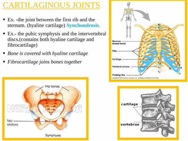
CARTILAGINOUS JOINTS
Ex. -the joint between the first rib and the sternum. (hyaline cartilage) Synchondrosis.
Ex.- the pubic symphysis and the intervertebral discs.(contains both hyaline cartilage and fibrocartilage)
Bone is covered with hyaline cartilage
Fibrocartilage joins bones together

CARTILAGINOUS
JOINTS

CARTILAGINOUS
JOINTS

The Characteristics of Synovial Joints
© 2015 Thompson Educational Publishing, Inc.
121
Synovial joints permit movement between bones
and are distinguished by the following:
-Articular cartilage is located on the ends of bones that
come in contact with one another.
-The joint capsule consists of the synovial
membrane and fibrous capsule.
-The joint cavity is filled with synovial fluid, which
acts as a lubricant for the joint.
-The bursae are the small fluid sacs found at the
friction points (“bursa” is the singular).
-Intrinsic ligaments are thick bands of fibrous connective
tissue that help thicken and reinforce the joint capsule.
-Extrinsic ligaments separate from the joint capsule and
help to reinforce the joint.

The SynovialJoint
© 2015 Thompson Educational Publishing, Inc.
122

Ball & Socket and Gliding Joints
© 2015 Thompson Educational Publishing, Inc.
123
• Ball-and-socket (spheroidal) joints. The “ball” at one bone fits
into the “socket” of another, allowing movement around three axes
(e.g., the humerus rests in the glenoid cavity).
• Gliding (or plane or arthrodial) joints. This type connects flat
or slightly curved bone surfaces that glide against one another (e.g.,
between the tarsals and among the carpals).
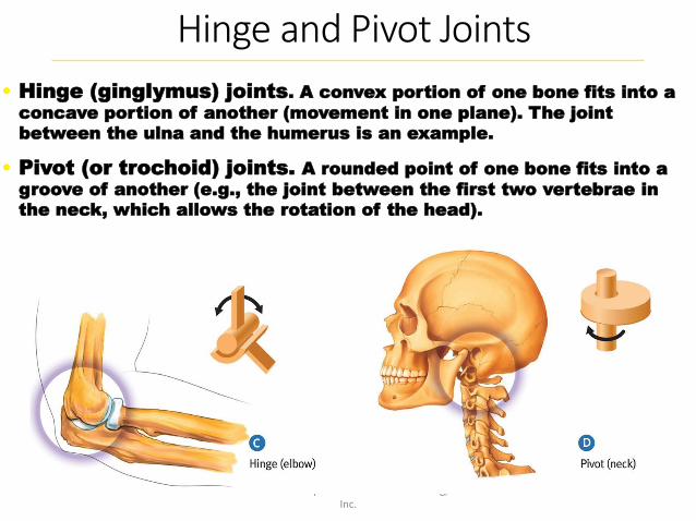
Hinge and Pivot Joints
© 2015 Thompson Educational Publishing, Inc.
124
• Hinge (ginglymus) joints. A convex portion of one bone fits into a
concave portion of another (movement in one plane). The joint
between the ulna and the humerus is an example.
• Pivot (or trochoid) joints. A rounded point of one bone fits into a
groove of another (e.g., the joint between the first two vertebrae in
the neck, which allows the rotation of the head).

Saddle and Ellipsoid Joints
© 2015 Thompson Educational Publishing, Inc.
125
• Saddle joints. Saddle joints allow movement in two
planes (but not rotation like a ball-and-socket joint). A key
saddle joint is found at the carpo- metacarpal articulation
of the thumb.
• Elipsoid joints. This type of synovial joint also allows
movement in two planes. The wrist is an example of an
ellipsoid joint.

Joint-Related Injuries and Disease
© 2015 Thompson Educational Publishing, Inc.
126
•Dislocations. A dislocation occurs when a bone is
displaced from its joint. Dislocations are often caused by
collisions or falls, and are common in finger and shoulder
joints.
•Separations. A separation is more serious than a
dislocation. In a shoulder separation, the
ligaments attaching the collarbone (clavicle) and shoulder
blade (scapula) are disrupted.
•Osteoarthritis is a condition involving loss of cartilage
at joints. Osteoarthritis (a joint disease) is often confused
with osteoporosis, which is a disease characterized by low
bone mass and bone deterioration.


SEPARATIONS
occurs when
bones held
together by
fibrous
ligaments tear
and separate
from each other
130

Also called the glenohumeral joint
Extremely versatile
Ball and socket joint
Made up of the scapula, humerusand indirectly with the clavicle
131
THE SHOULDER JOINT

Rotator Cuff Tears
© 2015 Thompson Educational Publishing, Inc.
132
Rotator cuff tears usually
involve one or all four muscles
that make up the rotator cuff
at the shoulder joint:
supraspinatus, infraspinatus,
teres minor, and subscapularis.
•These muscles share a
common tendinous
insertion on the greater
tubercle of the humerus. Thus,
when a part of the tendon is
torn, all three muscles around
the joint are affected.
•The severity of a rotator cuff
tear must be diagnosed by a
doctor.

Left Shoulder Joint(Anterior View)
© 2015 Thompson Educational Publishing, Inc.
133


-Modified hinge joint (some rotation)
-Consists of articulation b/w: femur, tibia, and patella
-Largest joint in body
TH
E K
NE
E

Right Knee (Anterior & Anterior Deep)
© 2015 Thompson Educational Publishing, Inc.
60

Anterior Cruciate Ligament (ACL)
Comprised of 2 (sometimes 3) bands
Prevents anterior translation of tibia on femur and resists internal rotation of the tibia
THE KNEE
137

Extensive research
Mechanism of
injury
Deceleration of knee
rotation (cutting)
Hyperextension of
knee
Direct blow to knee
More often in non-
contact situations
ACL INJURY

Signs and symptoms
Hear or feel a “pop”
Rapid swelling (within hours)
“giving way” (feeling of
instability)
Locking (portion of ACL lig
sometimes get caught in the
joint)
Decrease ROM (due to
swelling)
Pain at time of impact
ACL INJURY

Right Knee (Posterior) & Left Knee (Deep)140

modified hinge joint that comprises the distal ends of the tibia and fibula resting on the talus to form the ankle joint
the joint is responsible for plantar flexion and dorsiflexion
Ankle sprain-Most common injuries seen in sports medicine
Common in sports involving jumping and changes in direction
141
THE ANKLE JOINT

Right Ankle Joint(Medial View)
© 2015 Thompson Educational Publishing, Inc.
142

Right Ankle Joint(Lateral View)
© 2015 Thompson Educational Publishing, Inc.
143

144
INVERSION ANKLE
SPRAINS
Approx. 85%

Inversion Sprains
80-85% of all ankle sprains are
to the lateral ligaments --
inversion sprains
Commonly referred to as
“rolling over your ankle” or
“twisted ankle”
Can affect one or all of the
lateral ligaments
Less bony stability on medial
side of ankle
145
ANKLE SPRAINS

Eversion Sprains (<15% of all ankle sprains)
Eversion sprains, while less frequent, are often severe.
Mech. Of Injury-comb. of eversion, dorsiflexion, & foot abduction
Rare because of the strength of the deltoid ligament
The deltoid ligament attaches the medial malleolus to three bones of the foot and is so strong that, instead of tearing, it tears off the tip of the medial malleolus
Pott’s Fracture-break in the medial malleolus and a break in the fibula (15 % of cases are avulsion fracture)
146
ANKLE SPRAINS