
Chapter 8 Injuries to the face, nose, and ears · applied to the nose with anterior and posterior...
112
General points in facial trauma 236 Maxillofacial (trauma) emergencies 238 Facial trauma—general considerations 242 Useful clinical signs and their significance 244 Mandibular fractures 254 Dislocated condyle 266 Alveolar fractures 268 Dental trauma 270 Zygomatic and orbital fractures—applied anatomy 272 Zygomatic (malar) fractures 274 Orbital fractures (isolated) 280 Mid-facial (‘middle-third’) fractures—applied anatomy 282 Le Fort mid-face fractures 284 Nasal fractures 292 Naso-ethmoid (naso-orbital-ethmoid) fractures 296 Pan-facial fractures 298 Special considerations for children and the elderly atrophic mandible 310 Wound healing 312 Types of wound healing 314 Wound assessment 318 Wound cleansing 320 Soft tissue injuries 322 Soft tissue injuries of the head and neck 328 The closure of wounds 330 Suturing and suture removal 332 Soft tissue injuries—tissue loss 334 Surgical drains 340 Scars 342 Paediatric facial trauma 344 Injuries to the face, nose, and ears 235 Chapter 8 Hene_Ch08.qxd 8/30/04 6:24 AM Page 235
Transcript of Chapter 8 Injuries to the face, nose, and ears · applied to the nose with anterior and posterior...

General points in facial trauma 236Maxillofacial (trauma) emergencies 238Facial trauma—general considerations 242Useful clinical signs and their significance 244Mandibular fractures 254Dislocated condyle 266Alveolar fractures 268Dental trauma 270Zygomatic and orbital fractures—applied anatomy 272Zygomatic (malar) fractures 274Orbital fractures (isolated) 280Mid-facial (‘middle-third’) fractures—applied anatomy 282Le Fort mid-face fractures 284Nasal fractures 292Naso-ethmoid (naso-orbital-ethmoid) fractures 296Pan-facial fractures 298Special considerations for children and the elderly
atrophic mandible 310Wound healing 312Types of wound healing 314Wound assessment 318Wound cleansing 320Soft tissue injuries 322Soft tissue injuries of the head and neck 328The closure of wounds 330Suturing and suture removal 332Soft tissue injuries—tissue loss 334Surgical drains 340Scars 342Paediatric facial trauma 344
Injuries to the face,
nose, and ears
235Chapter 8
Hene_Ch08.qxd 8/30/04 6:24 AM Page 235

4General points in facial trauma
In trauma care time is of the essence. A useful working approach is
needed, in order to rapidly identify those patients that need immediate
care, from those that can wait.
Facial ‘emergencies’ following maxillofacial trauma can be regarded as
those that are life- and sight-threatening. These may occur following
isolated injuries, or they may be associated with significant injuries else-
where. They may even present following what would usually be regarded
as a minor injury (highlighting the need to maintain a high index of suspi-
cion), or after some delay. In the context of emergencies following facial
trauma, the objectives are therefore to safeguard life first and vision
second.
Assessment therefore needs to be systematic and repeated,
with the establishment of clear priorities in the patient’s overall care.
In many respects parallels can be drawn with orthopaedic surgery.
Management of facial trauma can arguably be regarded as ‘facial
orthopaedics’, as both specialties share common surgical principles in
trauma care.
What is an ‘emergency’?
In this text, any clinical problem requiring immediate identification and/or
management, constitutes an emergency. Many conditions may be consid-
ered ‘urgent’ (i.e. contaminated, mucky wounds, open fractures), but these
can generally be left until the patient is fully stabilized, with little or no
increase in mortality or morbidity. In the face, emergency care effectively
means airway control, control of profuse bleeding, and the management of
vision-threatening injuries (VTI). Failure to rapidly recognize and manage
these conditions can result in loss of life or sight.
Head and neck injuries resulting in life-threatening
conditions
- Facial injuries resulting in airway compromise (e.g. pan-facial fractures
with gross mobility or swelling, comminuted #s of the mandible,
gun-shot, profuse bleeding, foreign bodies, burns, etc.).
- Anterior neck injuries resulting in airway compromise (e.g. penetrating
injuries, circumferential burns, laryngeal injuries).
- Injuries resulting in profuse blood loss (e.g. penetrating neck,
pan-facial fractures).
- Intracranial injuries.
ATLS and the maxillofacial region
In over 29 countries, worldwide management of trauma is now based on
Advanced Trauma Life Support (ATLS) principles. This is now generally
accepted as the ‘gold standard’ of trauma care, and many of its principles
are equally important in the management of facial injuries. However, it is
important to remember that every case is unique and on occasions, strict
adherence to guidelines may result in some difficulties. It is therefore
important to be mindful of potential complications when dealing with
severe facial injuries and be aware of early warning signs. If in doubt,
re-assess the patient from the start.
236 CHAPTER 8 Injuries to the face, nose, and ears
Hene_Ch08.qxd 8/30/04 6:24 AM Page 236

237
All clinicians involved in trauma care need to be able to assess and
maintain airway problems and control obvious bleeding during the primary
survey. Be alert also to possible vision-threatening injuries during the
primary survey, i.e. retrobulbar haemorrhage. Although the aim of
the primary survey is to identify and treat life-threatening problems, the
early identification of a sight-threatening condition may be possible during
‘D’ when the pupils are assessed. This enables early referral to an
‘appropriate’ specialist. When dealing with acute sight-threatening condi-
tions, ‘appropriate’ may refer to an on-site speciality with expertise in
peri-orbital trauma care.
Airway, bleeding, and sight-threatening conditions may initially be subtle
and may not become apparent until the secondary survey is under way.
This reminds us of the two well-known principles in trauma care:
- the need for a high index of suspicion;
- the need for frequent re-revaluation.
Preliminary assessment of multiple injured
patients—the primary survey
The sequence in which the primary survey should proceed is related to
those conditions that would lead to loss of life quickest:
- Airway (whilst protecting the cervical spine to prevent any
neurological damage).
- Breathing (all trauma patients should be given 100% oxygen to
breathe on arrival).
- Circulation with control of haemorrhage.
- Disability (brain function).
- Exposure—this must be complete so a full examination, front and back,
can be undertaken. However, the patient needs to be covered to
prevent hypothermia.
Any problem discovered is corrected at the time of identification.
This is preferably done by a team approach where different team
members carry out the above simultaneously. In such cases, a ‘team
leader’ ensures that everything is done. If team numbers are not sufficient
for this, the above sequence is followed. It is also vital to enlist the help of
any speciality not on the team early, if a problem or potential problem is
identified that requires their input, e.g. neurosurgery
GENERAL POINTS IN FACIAL TRAUMA
Hene_Ch08.qxd 8/30/04 6:24 AM Page 237

1Maxillofacial (trauma) emergencies
(See also Assessment.)
Airway
- Obstruction can be caused by dentures/teeth or severely displaced
fractures of the mandible or mid-face. The commonest cause is
bleeding and/or saliva, notably when the patient is intoxicated or
supine.
- Mid-face fractures may displace downwards and backwards along
the skull base, impinging on the posterior pharyngeal wall, resulting
in obstruction. Bilateral anterior (‘bucket handle’) or comminuted
mandibular fractures can similarly displace backwards allowing the
base of the tongue to fall back. Both of these are much more likely
when patients are supine and there is alteration in the conscious level.
Both can be dealt with by pulling the fractured part forward to relieve
the obstruction. This provides only temporary relief and a definitive
airway will probably be required.
- Saliva and blood should be cleared by suction. If the bleeding is
ongoing from an identifiable source, which can be stopped, it should
be. However, it is usually generalized from multiple sites. Displaced
fractures should be manually reduced as this often helps slow the
bleeding. Nasal packs may be necessary (remember the possibility
of skull base #). If bleeding continues the airway should be
protected with a definitive airway.
- Direct trauma to the airway will probably require a definitive
airway to be placed.
Beware the patient who keeps trying to sit up—they may be
trying to clear their airway.
Bleeding
- Bleeding from maxillofacial injury is common but not usually
life-threatening. If the patient is in shock, look for another
cause. Actively consider facial bleeding, as supine patients will
be swallowing blood, which will go unnoticed.
- If obvious and significant, bleeding is controlled in the primary survey
by pressure. Bleeding from lacerations can usually be controlled by
pressure applied either with a swab (care with scalps if risk of skull
fracture) or by placement of sutures. These are used to apply
pressure and not intended as definitive closure.
- Mid-face bleeding can be troublesome as the bleeding is from multiple
sites from comminuted bones and torn mucosa. Pressure can be
applied to the nose with anterior and posterior nasal packs. Bleeding
from displaced/mobile mid-face fractures should be reduced.
Gentle pressure can be applied antero-superiorly on the maxilla
and maintained by placing mouth props bilaterally against an intact
mandible. Surprisingly, this is not as painful as one might think. In
selected cases, use of external fixators from skull to maxilla, may
be necessary but this requires transfer to an operating theatre
and considerably more time.
238 CHAPTER 8 Injuries to the face, nose, and ears
Hene_Ch08.qxd 8/30/04 6:24 AM Page 238

239
- Bleeding from a ‘hole’ (e.g. following a gun-shot) can sometimes
be stemmed by placing a Foley catheter in the hole and inflating
it. Obviously be careful and think what may be in the depths of
the hole!
- If local pressure is not sufficient to stop haemorrhage from either
soft or hard tissue injury, the use of angiography and
embolization or ligation of external carotids should
be considered. This is rare.
Vision-threatening injuries
(See also The Eye.)
Following trauma, vision can be threatened anywhere along
the visual pathway from globe to cortex. The main (potentially
treatable) causes to consider are:
- direct globe injury;
- retrobulbar haemorrhage;
- optic nerve compression;
- loss of eyelids;
- direct injury to the globe requires urgent ophthalmic referral.
Retrobulbar haemorrhage
Bleeding behind the globe is a form of compartment syndrome. This is
a surgical emergency. It can lead to an increase in pressure that results
eventually in irreversible ischaemia of the retina and optic nerve. Key
symptoms are:
- severe pain;
- progressive loss of vision;
- the eye becomes proptosed with ophthalmoplegia;
- development of a fixed dilated pupil as the vision deteriorates.
Treatment requires immediate relief of pressure. In the emergency
department the following should be given intravenously:
- acetazolamide;
- mannitol;
- steroids;
during which arrangements are made for surgery. Under LA, a lateral
canthotomy may be possible, but these measures really only buy time while
preparation for surgery is made. Definitive treatment involves drainage of
the haematoma. Decompression should lead to an improvement in visual
acuity if undertaken early enough.
Traumatic optic neuropathy
Traumatic optic neuropathy occurs when there is disruption around the
optic canal resulting in either compression of the optic nerve, shearing
forces to the nerve as it passes through the canal, or haematoma formation
within the nerve itself. Untreated it can render the patient blind; the dia-
gnosis needs to be made early to allow the best chance of visual recovery.
The signs that suggest an optic nerve injury include poorly reactive pupil,
afferent papillary defect, and decreased colour vision, decreased
visual acuity with relatively normal ocular examination. This is an
ophthalmic emergency and should be referred accordingly.
MAXILLOFACIAL (TRAUMA) EMERGENCIES
Hene_Ch08.qxd 8/30/04 6:24 AM Page 239

Treatment of optic nerve compression is controversial and again may
be either medical or surgical. The options include observation, IV cortico-
steroids, and optic nerve decompression.The latter option is carried out
via either a craniotomy approach or lateral facial approach.
One steroid regime is:
- methylprednisolone 30mg/kg STAT;
- followed by methylprednisolone 15mg/kg every 6 h.
Of course others exist—always check local policy.
Time is of the essence, best results are obtained if steroids are
given within 8 h of the injury.
240 CHAPTER 8 Injuries to the face, nose, and ears
Fig. 8.1 CT scan showing compression of the right optic nerve at the optic foramen
following blunt trauma. This required urgent decompression.
Hene_Ch08.qxd 8/30/04 6:24 AM Page 240

241MAXILLOFACIAL (TRAUMA) EMERGENCIES
Hene_Ch08.qxd 8/30/04 6:24 AM Page 241
4Facial trauma—general
considerations
Minor injuries to the maxillofacial region are very common in the UK,
around 80% occur in children. Major facial disruption is less frequently
seen and motor vehicle crashes (MVC) now accounts for about 5% of
facial trauma. This is partly due to seat belt and drink-driving legislation.
However, whereas patients would have previously died at the scene as a
result of their head injuries, many are now surviving having sustained
major facial injuries. Sporting injuries and assaults account for most of the
remainder.
In assessing the multiple-injured patient, the first step is to determine
whether any life-threatening injuries exist and to deal with them first. The
advanced trauma life-support system—ATLS—is one approach to assess-
ment and initial management of the multiply injured patient. It recognizes
that identification of life-threatening injuries needs to be prioritized
(primary survey) and treated at the time of identification (resusitation).
Most maxillofacial injuries can wait treatment, and in the multiply injured
patient, the first step is keeping the patient alive.
Once the primary survey and resuscitation have been carried out, atten-
tion can then be focused on the maxillofacial region.
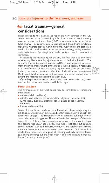
Facial skeleton
The arrangement of the facial bones may be considered as comprising
three areas:
- upper-third (frontal bone);
- middle-third, between the supra-orbital ridges and the upper teeth
(2 maxillae, 2 zygomas, 2 lacrimal bones, 2 nasal bones, 1 vomer, 1
ethmoid);
- lower-third (mandible).
Some of these bones, such as the ethmoid and those comprising the
orbital roof, are extremely delicate and so thin that on a dry skull light can
easily pass through. The remainder vary in thickness but often remain
quite delicate (nasal, zygoma). The mandible is the strongest of the facial
bones. It is a U-shaped bone comprised of an outer dense cortical layer
and delicate trabecular bone inside. The face is not solid but contains
several ‘cavities’, such as the sinuses, orbits, oral and nasal cavities. Around
these the bones form a series of vertical struts known as ‘buttresses’ As a
result, these bones are very good at resisting vertically directed forces
(e.g. during chewing) but are weak when it comes to resisting horizontal
forces (i.e. during most injuries).
242 CHAPTER 8 Injuries to the face, nose, and ears
AQ: Please
check the
spelling of
resusitation.
Hene_Ch08.qxd 8/30/04 6:24 AM Page 242

243FACIAL TRAUMA—GENERAL CONSIDERATIONS
Fig. 8.2 Transilluminated skull demonstrating the thinness of many of its bones.
Fig. 8.3 Diagrammatic representation of the ‘butresses’ of the face.
Hene_Ch08.qxd 8/30/04 6:24 AM Page 243

4Useful clinical signs and their
significance
The usefulness of clinical signs can vary from those that only suggest an
underlying pathology (*) to those that are almost pathognomonic (***).
Their interpretation must be taken in conjunction with the history and
likelihood of the condition being present.
General
Facial burns***
When associated with soot in the nose and mouth, singeing of the nasal
vibrissae, and sooty sputum, this represents a potential airway problem.
There is also the risk of and inhalation of carbon monoxide and other
toxins.
Facial nerve palsy**
Following head injury—fractured base of skull.
Horse voice/bovine cough**
Following a direct blow to the anterior neck, may indicate disruption of
the larynx. A bovine cough is where the vocal cords do not meet in the
mid-line prior to the explosive expulsion of air. As a result the cough is
relatively weak and ineffectual.
Wry neck*
Following trauma is due to muscle spasm. May occasionally be associated
with dislocation of the posterior facet joints.
The face
Intercanthal distance**
Separation of the eyes. If greater than 30–32mm (female) or 32–34mm
(male) the patient may have detached canthi secondary to an underlying
naso-ethmoidal fracture. As well as an increased intercanthal distance, the
medial canthus loses its pointed shape becoming rounded. This can occur
uni- or bilaterally. If unilateral the distance from mid-line to canthus will be
greater on one side. The interpupillary distance should be within normal
limits. There may also be depression at the root of the nose.
Anterior open bite and elongated face**
If not pre existing, is suggestive of posterior and inferior displacement of
the maxilla following a Le Fort fracture. This results in posterior gagging
of the molar teeth. If the maxilla is stable, then the teeth should be
percussed. If the note is dull (‘cracked cup’ sound), the maxilla is likely to
be fractured and impacted—hence the stability. If the percussion note is
normal, the anterior open bite may be due to bilateral fractured condyles.
Septal haematoma*
Seen as a blue/reddened swelling on the septum on direct examination.
Needs drainage, as failure to do so can result in septal perforation,
abscesses, and intra-cranial infection.
Numbness of the cheek*
Suggests a cheek or blow-out fracture.
244 CHAPTER 8 Injuries to the face, nose, and ears
Hene_Ch08.qxd 8/30/04 6:24 AM Page 244

245
Numbness of the lower lip*
Suggests a mandibular fracture.
Anosmia*
Loss of smell due to tearing of olfactory nerves secondary to an underlying
anterior cranial fossa floor fracture. Not reliably detectable in acute phase
of injury.
USEFUL CLINICAL SIGNS AND THEIR SIGNIFICANCE
Fig. 8.4 Normal intercanthal distance.
Fig. 8.5 Increased intercanthal distance.
AQ:
Please
provide
figure 8.4.
Hene_Ch08.qxd 8/30/04 6:24 AM Page 245
‘Bow-string’ test***
Assess for medial canthal detachment in nasothmoid injuries—the lateral
canthus is pulled laterally, if there is detachment medially this will also
move away.
Within the mouth
Dysphagia*
Many causes. When related to submandibular, pharyngeal, or other poster-
ior oral swellings, it is a significant finding often requiring admission. Is
often painful (odynophagia).
Inability to protrude the tongue**
When related to submandibular, sublingual, or other oral swellings, it is a
significant finding often requiring admission.
Trismus**
Limitation of mouth opening due to muscle spasm (usually masseter or
medial pterygoid). May be seen following a direct blow. When related to
submandibular, pharyngeal, or other posterior oral swellings, it is a signific-
ant finding often requiring admission.
Guerins sign***
Palatal bruising of the hard palate—underlying fracture involving palatine
foramen.
Upper buccal sulcus bruising**
Fractured zygoma or unilateral Le Fort I or II.
Sublingual haematoma***
Bruising/bleeding under the tongue—fractured mandible (body/symphysis/
parasymphysis).
246 CHAPTER 8 Injuries to the face, nose, and ears
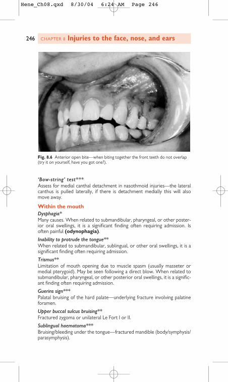
Fig. 8.6 Anterior open bite—when biting together the front teeth do not overlap
(try it on yourself, have you got one?).
Hene_Ch08.qxd 8/30/04 6:24 AM Page 246

247
Peri-odontal bleeding*
May indicate an associated fracture of the tooth or bone.
Change in the patients bite***
In either the mandible or maxilla may indicate a fracture.
‘Cracked cup’ note*
When percussing the maxillary teeth, suggests a fracture.
Malocclusion*
If the occlusion has changed it is likely that there is an underlying fracture
of maxilla, mandible, or alveolar bone. Subluxation of teeth may also
produce a malocclusion, although usually much more minor. If non-of
the above are present a malocclusion may be a result of a TMJ effusion/
haemarthrosis.
The eyes
Visual acuity
This is the single most sensitive indicator of visual impairment. It must
be recorded in all patients with mid-facial trauma. In patients who wear
spectacles to correct short sight, the recording must be done with the
spectacles on or through a pinhole. Acuity is expressed as a distance in
meters from which a chart being read is usually 6/X where X is the line on
the chart the patient can read, usually 6. If less than 6, e.g.12 or 60, this
means the patient can read at 6m from the chart that which a person with
normal vision could read at 12 or 60 m from the chart. A visual acuity of
6/18 or worse should be referred for an ophthalmological opinion.
If the patient cannot read the chart at all at 6m, the distance at which
they can read the top line is recorded as Y/60. If they are not able to do
this, whether they can count fingers, see them moving, or perceive light,
are recorded as the visual acuity, in that order.
If the patient is unconscious, pupillary responses to light must be checked.
All patients must have a documented visual acuity. Any decrease in
visual acuity requires an ophthalmic opinion.
Pupillary responses**
Check direct response to light and consensual response. Responses
should be equal on both sides and to direct and consensual stimulation.
A pupil that reacts poorly to direct stimulation but briskly to consensual
has an afferent pupillary defect.
Swinging flashlight test***
Detects subtle defects to the optic nerve. Light is shone in one eye, then
swung to the other, and back and forth. If the right eye has a problem, on
shining the light in the right eye, both pupils will constrict, as the light
moves to the left they will constrict further. As the light is brought back to
the right, the pupils will not respond or dilate a little.
Peri-orbital haematoma***
When well-defined this represents a fracture involving the orbit. Usually
this means a fractured zygoma but can also include a blow-out fracture,
fractured base of skull (anterior cranial fossa), unilateral naso-ethmoidal,
or nasal bones.
USEFUL CLINICAL SIGNS AND THEIR SIGNIFICANCE
AQ: Please
check the
spelling of
sentence
“ If non-of...
should it
be “none”
or “non”.
Hene_Ch08.qxd 8/30/04 6:24 AM Page 247

Racoon (panda) eyes***
Bilateral well-defined ‘black eyes’—fractured base of skull (anterior cranial
fossa), Le Fort III or naso-ethmoid fracture.
Lateral subconjunctival haemorrhage**.
This indicates a fracture involving the orbit usually the cheek or naso-
ethmoid region. There is no posterior limit.
248 CHAPTER 8 Injuries to the face, nose, and ears
Fig. 8.7 RAPD. When the eye is opened the pupil does not react. When the
contralateral eye is opened it does—this patient is blind.
Fig. 8.8 RAPD. When the eye is opened the pupil does not react. When the
contralateral eye is opened it does—this patient is blind.
AQ: Please
check the
figure and
figure
caption
for figures
7 and 8.
Hene_Ch08.qxd 8/30/04 6:24 AM Page 248

249
Chemosis*
Swelling of conjunctiva often seen in significant trauma, looks a bit like
frog’s spawn. If no tear of conjunctiva present, it will resolve. If tear
present, then you need to rule out globe injury.
Hyphema**
Blood in anterior chamber seen as fluid level when patient is
standing. Needs ophthalmic assessment probably require admission and
observation.
Iridodialysis*
The iris is detached from its root leading to a distorted pupil shape.
Dilated pupil (traumatic mydriasis)*
Spasm of the dilator pupillae. Can be seen following a direct blow to
the eye. Not to be confused with a third nerve palsy.
USEFUL CLINICAL SIGNS AND THEIR SIGNIFICANCE
Fig. 8.9 Well-defined ‘black’ and ‘red’ eye.
Fig. 8.10 ‘Panda’ or ‘Racoon’ eyes. Often not as obvious as this example.
Hene_Ch08.qxd 8/30/04 6:24 AM Page 249

Diplopia*
Double vision may be neurogenic, myogenic, or bony in origin. It may be
temporary or permanent and should be reviewed. Depending on possible
cause, refer to ophthalmics or maxillofacial.
Unilateral restricted upward gaze**
Often a sign of a ‘blow-out’ fracture, occasionally due to injury to ocular
muscle or its nerve. Painful diplopia from a blow-out may require
urgent release.
250 CHAPTER 8 Injuries to the face, nose, and ears
Fig. 8.11 Chemosis.
Fig. 8.12 Unilateral restricted upward gaze.
Hene_Ch08.qxd 8/30/04 6:24 AM Page 250

251
Retraction sign***
When looking from the side of the patient, as they look up, the globe is seen
to move posteriorly. This is a good sign for a blow-out fracture. Entrapment
of the fat and restriction of the inferior rectus muscle results in a shift of the
axis of rotation of the globe from its centre to the point of entrapment. Thus
the pull of the superior rectus results in a backward rotation of the globe.
Hypoglobus*
Inferior displacement of the globe seen in cheek complex fractures, where
the bone and Lockwood’s ligament drop down. May also be seen in large
blow-out fractures.
Enophthalmos*
Posterior displacement of the globe due to increased orbital volume. Seen
in blow-out fractures of the orbit and cheek fractures. Globe appears
‘sunken in’ with a deep supra tarsal groove.
Third nerve palsy***
Dilated pupil, the eye looks down and out, and ptosis. In severe head
injuries this represents third nerve compression from an expanding intra-
cranial lesion. The patient has a reduced GCS.
Aqueous leakage***
A penetrating injury of the cornea.
Superior orbital fissure (SOF) syndrome**
Ophthalmoplegia, fixed dilated pupil, and ipsilateral forehead numbness—
fracture extending into the SOF, or possible carotid anuerism. This is
usually part of a significant injury.
Orbital apex syndrome**
As in the SOF but here the patient has reduced visual acuity.
Peri-orbital oedema*
When infective in origin represents significant spread of infection. If the
eye is closing the patient may probably need admission.
The ears
Heamotympanum**
Blood visualized behind the eardrum. Indicative of a fracture of the middle
cranial fossa.
USEFUL CLINICAL SIGNS AND THEIR SIGNIFICANCE
Fig. 8.13 Enophthalmos.
AQ: Please
check the
Change of
“obrbital”
to “orbital”
is okay
AQ: Please
check the
spelling
“Heamo-
tympanum”
Hene_Ch08.qxd 8/30/04 6:24 AM Page 251

Battles sign***
Bruising around the mastoid region—fractured base of skull (middle
cranial fossa).
CSF rhinorrhea/otorrhea***
‘Tramlining’—fractured base of skull. Blood mixes with CSF and leaks out.
Along the edges the blood clots while centrally the CSF leak washes it
away to form two parallel lines (like tramlines).
Bleeding from the ear**
Fractured base of skull or mandibular condyle. If the tympanic membrane
is intact the bleeding is local to the EAM, usually the anterior wall, often
secondary to an underlying condylar fracture. If the tympanic membrane is
perforated the blood may be from a middle cranial fossa fracture.
252 CHAPTER 8 Injuries to the face, nose, and ears
Fig. 8.14 CSF otorrhea.
Hene_Ch08.qxd 8/30/04 6:25 AM Page 252

253USEFUL CLINICAL SIGNS AND THEIR SIGNIFICANCE
Hene_Ch08.qxd 8/30/04 6:25 AM Page 253

3Mandibular fractures
Applied anatomy
The mandible forms the lower third of the face and facial skeleton. It is the
only mobile bone of the face (excluding the ossicles) and has numerous
muscle insertions. It plays an important role in:
- speech;
- mastication (chewing);
- deglutition (swallowing);
- maintaining the airway.
Patients with mandibular injuries, therefore, have difficulty in talking, and are
unable to eat and drink easily. In severe injuries the airway may be at risk.
Osteology
Morphologically the mandible is a U-shaped ‘long bone’ (e.g. femur, radius,
etc.). It can be divided anatomically into:
- symphysis
- parasymphysis
- body
- angle
- ramus.
The almost vertical ramus carries two processes, the condyle, which
articulates with the glenoid fossa of the temporal bone to form the
temporo-mandibular joint, and the coronoid process, which receives the
insertion of temporalis. The condyle is supported on a slender condylar
neck, a frequent site for fracture.
On the medial or inner aspect of the ramus the inferior alveolar
neurovascular bundle enters the bone via the mandibular foramen and runs
through a bony canal within the mandible providing nutrition and sensory
innervation to the lower teeth. An important branch of this bundle, the
mental nerve, leaves the bone via the mental foramen in the premolar
region and provides sensation to the lower lip and anterior gums
Muscle attachments
The muscles of mastication (temporalis, masseter, medial and lateral
pterygoid), together with the suprahyoid muscles (digastric, geniohyoid,
and mylohyoid) are the principle movers of the mandible. In addition the
mandible receives the insertion of genioglossus, which forms the main bulk
254 CHAPTER 8 Injuries to the face, nose, and ears
Fig. 8.15 (a) and (b) Outer and inner view of the mandible showing muscle
attachments.
Hene_Ch08.qxd 8/30/04 6:25 AM Page 254

255
of the tongue. Note that genioglossus and geniohyoid are attached to the
mid-line genial tubercles. A fracture in this region may, therefore, lead to
loss of anterior support for the tongue, leading to posterior displacement
and airway compromise.
Dentition
The full complement of adult teeth in a mandible is 16 comprising of:
- 4 incisor teeth;
- 2 canines;
- 4 premolars;
- 6 molars.
The canine teeth have long roots and the third molar (wisdom)
teeth are often partially erupted, these factors tend to weaken
the bone locally and account for the frequency of fractures in
these regions.
Age-related changes
In the child, the dentition will be at various stages of development and
developing tooth germs are present within the bone. While these lead to a
structural weakening of the bone, this is compensated for by increased
elasticity and pliability of the young mandible, compared with mature bone.
As a result, relatively higher forces are required to fracture the bone in
children. Furthermore, the facial skeleton is less prominent in children and
hence mandibular fractures are somewhat rare in this age group. In the
edentulous elderly jaw, loss of the ‘alveolar bone’, which would normally
support the teeth, leads to a gradual reduction in bone height. This feature,
together with age-related conditions such as osteoporosis, weaken its
structure and, as a result, the bone is more vulnerable to fracture.
Common fracture patterns
The periosteum is an important structure in determining the stability of
a mandibular fracture. In young patients it is generally a strong unyielding
membrane. Gross displacement of fragments cannot occur if it remains
intact and attached to the bone. However, once the periosteum has been
breeched (by injury or surgical exposure), displacement of the bones
can occur under the influence of the attached muscles.
Common fracture patterns include the following.
Angle fractures
Fractures of the angle are affected by the medial pterygoid and masseteric
muscles. Fractures in this region have been classified as vertically and
horizontally favourable or unfavourable. The medial pterygoid muscle
may pull the posterior fragment lingually or together with the masseter in an
upward direction. This is only important when the periosteum has been
ruptured or stripped from the bone allowing displacement to occur.
Fractures at the symphysis and parasymphysis
The mylohyoid muscle has been described as the ‘diaphragm’ of the mouth,
passing between the hyoid bone and the inner aspect of the mandible.
With mid-line fractures of the symphysis, the mylohyoid and geniohyoid
muscles can act as a stabilizing force. However, oblique fractures will tend
to overlap due to the pull of these muscles. With bilateral parasymphyseal
fractures (which result from considerable force), the periosteum is often
MANDIBULAR FRACTURES
Hene_Ch08.qxd 8/30/04 6:25 AM Page 255

torn and the fragments can displace posteriorly under the influence of the
genioglossus—so called ‘bucket handle’ fractures.
Condylar fractures
The hinge joint of the mandible is a common site of fracture and often
occurs in association with fractures elsewhere in the jaw. The classical
history is a blow or fall onto the point of the chin, where one or both
condyles are fractured, often associated with a symphyseal or parasym-
physeal fracture (so called ‘gaurdsmans’ fracture). Beware the lacera-
tion over the chin following a fall—check the condyles. On mouth
opening, the jaw deviates towards the site of injury.
Condylar fractures in adults tend to occur outside the joint, although
transmitted forces can still damage the joint and cause long-term prob-
lems. Effusion or bleeding into the joint space can occur in the absence of
a fracture, the space is distended and the patient complains of an
abnormal bite. Intra-capsular fractures in children are more common and
can result in growth disturbances in the condyle later on.
Injuries to related structures
- The inferior alveolar nerve is often damaged in fractures of the
body and angle causing paraesthesia of the lower lip on the same side.
- Medial displacement of the condyle can compress the trigeminal
nerve leading to loss of facial sensation.
- The facial nerve may be damaged by a direct blow over the ramus
and needs to be assessed especially if a laceration is present. Rarely,
the mandibular condyle may impact upon and fracture the temporal
bone through which the facial nerve runs. Facial nerve injury leads
to variable paralysis or weakness of the facial muscles.
- Injury to major blood vessels is unusual, although the facial artery
and vein may be damaged where they cross the lower border of the
mandible.
- Temperomandibular joint. Condylar fractures in adults tend to
occur outside the joint, although transmitted forces can still damage
the joint and cause long-term problems. Effusion into the joint space
can occur in the absence of a fracture, the space is distended and the
patient complains of an abnormal bite. Intra-capsular fractures in
children are more common and can result in growth disturbances
in the condyle later on.
Assessment
Secondary survey
History
Most patients will give a history of blunt injury to the face, the
most common mechanism being interpersonal violence. Sports, falls, and
accidents are other common causes. Common symptoms include:
- pain;
- swelling;
- altered bite;
- numbness of the lower lip;
- difficulty in opening and closing of the jaw;
- pain on swallowing with drooling.
256 CHAPTER 8 Injuries to the face, nose, and ears
Hene_Ch08.qxd 8/30/04 6:25 AM Page 256

257
The hallmark of a mandible fracture is a change in the bite
(occlusion); however, normal occlusion does not rule out a
mandible fracture.
Other physical findings include:
- loosened teeth;
- facial deformity;
- mobility of fractured segment;
- bleeding (= tear) in gingival tissue;
- sublingual haematoma;
- trismus;
- numbness of the lower lip (ID nerve).
Examination
This must be undertaken in good light, having cleansed the face of blood
and debris. Suction must be immediately available. Look for:
- facial swelling;
- asymmetry;
- bruising;
- palpate the lower border for any step deformity;
- feel the condyles and assess their movements—this is done best by
placing a gloved finger in each auditory meatus;
- document the dentition, any occlusal derangement, or steps in the
occlusal plane;
- gently manipulate across the suspected fracture site feeling for
abnormal movement or crepitus;
- look for gingival lacerations; and
- mucosal bruising, in particular, for a sublingual haematoma.
Remember
- Mental parasthesia is both an important diagnostic sign and an
important medico-legal observation prior to treatment.
MANDIBULAR FRACTURES
Fig. 8.16 A sublingual haematoma in a toothless mandible.
Hene_Ch08.qxd 8/30/04 6:25 AM Page 257

- Missing teeth must be accounted for. If unable, get a chest and
soft tissue view of the neck. Similarly, if associated with a lip laceration,
a soft tissue radiograph of the lips is essential.
- A sublingual haematoma is pathognmonic of a mandibular
fracture and may also compromise the airway.
- Bleeding from the external auditory meatus may be a result of
tearing of its anterior wall by a condylar fracture. However, it may also
be a sign of a fractured skull base—be careful in your assessment
Radiographs
The principle of radiology for any fracture is to take two views at
90 degrees to each other. This minimizes the risk of missing a fracture that
may be minimally displaced. For the mandible, this is most simply achieved
by ordering:
- ortho-pantomagram (OPG, OPT, DPT), or lateral obliques, together
with a PA view of the mandible.
It is essential that both views include the entire bone from lower border
to the condylar processes. For anterior fractures, if facilities exist, a true
lower occlusal view is also very helpful. These will often provide sufficient
information, but supplementary views include:
- reverse townes for the condylar neck;
- CT scans are useful for further evaluation of condylar fracture/
dislocations but are not necessary in A&E.
258 CHAPTER 8 Injuries to the face, nose, and ears
Fig. 8.17 A displaced fracture resulting in a gap. This was misinterpreted as a
missing tooth—count the teeth and assess for fracture mobility.
Hene_Ch08.qxd 8/30/04 6:25 AM Page 258
259
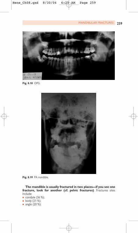
The mandible is usually fractured in two places—if you see one
fracture, look for another (cf. pelvic fractures). Fractures sites
include:
- condyle (36 %);
- body (21 %);
- angle (20 %);
MANDIBULAR FRACTURES
Fig. 8.18 OPG.
Fig. 8.19 PA mandible.
Hene_Ch08.qxd 8/30/04 6:25 AM Page 259

260 CHAPTER 8 Injuries to the face, nose, and ears
Fig. 8.20 Lateral oblique.
Fig. 8.21 Lower occlusal view anterior mandible.
Hene_Ch08.qxd 8/30/04 6:25 AM Page 260
261
- parasymphyseal (14%);
- ramus (3 %);
- alveolar (3%);
- coronoid (2%);
- mid-line symphysis (1 %).
Treatment
First-aid measures
A mandibular fracture is often painful and if it occurs through the tooth
socket, it is by definition a compound or open fracture. Pain relief is a
priority and may be simply achieved by infiltration of local anaesthesia or,
if possible, by an inferior dental nerve block. It is important to minimize
movement across the fracture site, not only for pain relief (as for all
long-bone fractures) but also to reduce bleeding and contamination from
oral bacteria. A simple method is to apply a soft neck collar but this
should only be done after the cervical spine has been formally cleared.
A common procedure is to apply a bridal wire across the fracture site.
This is a loop of stainless steel wire encircling the teeth either side of the
fracture. Clearly this is only relevant if the fracture is in a tooth-bearing
segment and care must be taken not to avulse the teeth by applying too
much force. Any loose dento-alveolar fractures should be splinted.
Treatment principles
In general, fracture treatment has traditionally been classified as conservat-
ive or operative. Today the preferred terminology is closed or open to
avoid ambiguity.
Indications for closed treatment (soft diet, antibiotics, review)- No or minimal displacement of stable fracture.
- No or minimal mobility across fracture line.
- No impairment of function.
- Ability to obtain pre-trauma occlusion with or without analgesia and
instructions.
- Absence of evidence of infection.
- Absence of haemorrhage requiring immobilization to control.
- Good patient co-operation and follow-up.
Indications for open treatmentWhen conservative treatment is inappropriate or has failed.
Closed treatment
In its simplest form, closed treatment would be:
- analgesia;
- antibiotics in open fractures (1 week);
- soft diet until a firm callus forms (usually around 4–6 weeks).
This is often suitable for a firm, minimally or un-displaced fracture
with a normal bite.
Intermaxillary fixation (IMF)For painful or more displaced fractures this may be applied. Here the
upper and lower teeth are fixed together using wires or elastics. This uses
the upper teeth as both a splint and a mandibular positioning device,
ensuring a normal bite and immobilization of the fracture for healing.
MANDIBULAR FRACTURES
Hene_Ch08.qxd 8/30/04 6:25 AM Page 261

However, reduction is not anatomical. Various devices are available
to achieve intermaxillary fixation including eyelet wires, Leonard buttons,
arch bars, and splints. These are ligated or cemented to the teeth thereby
enabling the two jaws to be fixed to each other. If the patient has no teeth
(edentulous patients), modified dentures (Gunning splints) can be ligated
to the jaws to achieve a similar goal.
A further variation is to use elastic bands instead of wires between the
jaws to allow some degree of movement.
Closed treatment is known to work and has remained a main method of
treatment. However, it is not risk free. Disadvantages include:
- airway risk (asthmatics, COAD, epileptics, reduced conscious level);
- loss of weight;
- patients need to tolerate it (beware alcoholics and mentally ill);
- injury to tooth structure, restorations, and the periodontal tissues.
Relative contra-indications to IMF are:
- inadequate post-operative monitoring of airway during recovery
from general anaesthesia;
- need for prolonged oral/nasal airway post-operatively;
- possibility of convulsions;
- head injury (GCS 8 or less).
Open treatment
Closed treatment does not produce anatomical reduction of the fracture
(hence the term closed treatment as opposed to closed reduction). With
262 CHAPTER 8 Injuries to the face, nose, and ears
Fig. 8.22 Wire IMF—rarely used now (note no gloves).
Hene_Ch08.qxd 8/30/04 6:25 AM Page 262

263
open treatment, surgical exposure of the fracture site and (hopefully)
anatomical reduction is carried out. Exposure is most commonly carried
out through the mouth, but occasionally may be done through a skin
incision or an existing laceration. Once exposed, the fracture is accurately
reduced and fixed. Common fixation techniques use titanium ‘mini’ plates
or screws. This is now the preferred approach to most mandibular
fractures resulting in faster recovery and rehabilitation. However, there is
potentially more morbidity, especially injury to the inferior alveolar nerve
and tooth roots. The patient still requires a soft diet for the same period
of time.
Treatment of condylar fractures
Particular concerns with these fractures are long-term joint dysfunction,
ankylosis, and, in children, abnormal growth of mandible
Undisplaced condylar fractures, where the occlusion is normal, are
generally managed with rest, soft diet, and simple analgesics. These are not
compound fractures and so antibiotics are not necessary. Regular review
is essential in the early stages of healing to ensure that the fracture does
not ‘slip’ and derange the bite. Fractures that are displaced with an
associated malocclusion, however, need to be reduced. This is often
carried out using rigid or elastic IMF, the latter encouraging dynamic
realignment of the fragments. IMF is applied for 7–14 days to avoid ankyl-
osis. Following IMF early mobilization and physiotherapy are required.
Alternatively, and where IMF is contra-indicated, fractures may be
openly reduced and fixed using mini plates, interosseous wires, screws.
These are approached via a skin incision just behind the angle of the
jaw below the ear. There is a small risk of facial nerve injury, although
with careful surgical technique, this can be kept to a minimum. Bilateral
MANDIBULAR FRACTURES
Fig. 8.23 ‘ORIF’ parasymphyseal fracture.
Hene_Ch08.qxd 8/30/04 6:25 AM Page 263

condylar fractures may be managed similarly. However, these fractures
must be kept under close review until healed. ‘Telescoping’ of the
condyles with loss of jaw height posteriorly can lead to the occlusion
being propped open at the front—an anterior open bite. This would
require surgical correction at a later date.
Rarely, the condylar head may dislocate out of the articular fossa. This
usually requires open reduction
Special considerations
Tooth in the fracture site
For angle, body, parasymphyseal and symphyseal fractures, usually a tooth
is present in the fracture site. This is especially common in angle fractures,
where an unerupted third molar may weaken the bone locally. While it
may be tempting to remove the tooth (especially in open treatment),
this may make reduction of the fracture more difficult and unstable. For
anterior fractures, the loss of a tooth has obvious aesthetic consequences.
Consequently, unless the tooth itself is fractured, grossly decayed, has
associated periapical infection, or is interfering with fracture reduction, it
may be preferable to leave it in situ. Opinions may differ.
Analgesia/antibiotics/tetanus
Mandibular fractures are painful. The use of local anaesthesia (LA) and
bridal wires has been described in the first-aid section. LA can also be
repeated with a long-acting local anaesthetic, such as bupivicaine. Analgesia
requirements often dramatically reduce following fracture reduction and
fixation; useful agents include NSAIDS or paracetamol-based compounds.
Antibiotic therapy is generally required for compound (open) fractures
and should give appropriate cover against anaerobic organisms and strep-
tococci. Many policies exist and may include metronidazole with benzyl
penicillin or a compound agent such as co-amoxyclav. Antibiotic treat-
ment may be continued for 5 days.
Tetanus status should be checked and the patient given tetanus toxoid,
if appropriate.
Fractures in children
Similar principles apply and early mobilization is desirable, as there is an
increased risk of ankylosis. Consideration must be given to the unerupted
teeth, if plating is necessary. Long-term follow-up during growth is
recommended, as some fractures (especially condylar) are associated with
disturbances of growth.
Post-operative care
Closed treatment without intermaxillary fixation is usually treated on an
out-patient basis with regular review to ensure the occlusion remains
normal. Post-operative radiographs are necessary to confirm a correct
position of the bone fragments. Hospital dieticians are a valuable resource
and will give nutritional advice and provide supplements.
Closed treatment with intermaxillary wire fixation requires very close
observation in the immediate post-operative period. Prior to discharge,
the patient must be provided with wire cutters, which must be with
him/her24 h a day, and they must be instructed on their use. More
recently there has been a swing in favour of using elastics rather that wire.
264 CHAPTER 8 Injuries to the face, nose, and ears
Hene_Ch08.qxd 8/30/04 6:25 AM Page 264

265
This enables quicker release and a degree of flexibility in the immobiliza-
tion enabling ‘micro-movement’.
Following open treatment, the occlusion should be assessed the fol-
lowing day. It is not uncommon for this to be slightly deranged due to
muscle spasm or a joint effusion; this should settle with appropriate anal-
gesia or light elastic IMF. The position of the fragments should be con-
firmed with post-operative radiographs (OPT and PA mandible) and
arrangements made to review the patient weekly. A soft diet is needed for
the next 4–6 weeks.
MANDIBULAR FRACTURES
Hene_Ch08.qxd 8/30/04 6:25 AM Page 265

3Dislocated condyle
The temporomandibular joint (TMJ) is the joint between the mandible and
the skull base, located in the pre-auricular region. It is a synovial ‘ball and
socket’ type joint, the ‘ball’ portion being the mandibular condyle, and the
‘socket’ the glenoid fossa of the temporal bone. A fibrous sleeve encapsul-
ates the joint. Between the condyle and fossa there is a fibro-cartilaginous
disc, or ‘meniscus’, which is attached peripherally to the deep surface of
the joint capsule. The four muscles of mastication (the masseter, the tem-
poralis, and the medial and lateral pterygoids) act directly across the TMJ
to effect mandibular movements. Of note, the insertion of the lateral
pterygoid is into both the condylar neck, and through the fibrous capsule,
into the anterior aspect of the articular disc.
Movement across the joint is complex. On opening the mouth from the
closed position, initially a hinge-type movement occurs for the first 1 cm.
After that a forward translation is added in which the condyle moves
forward and downwards along the slope of the fossa. Very little move-
ment occurs side-to-side. Appreciation of TMJ anatomy and function helps
understand the clinical picture associated with fractures of the condyle.
Displacement of the mandibular condyle from the glenoid fossa is
termed dislocation of the condyle. This may be unilateral or bilateral,
solitary or recurrent. Following significant trauma, it can be associated
with a fracture of the condylar process (fracture dislocation).
Dislocation is usually in an anterior direction so that the condyle comes
to lie in front of the articular eminence. Following major trauma it may
occur in other directions, including intra-cranially.
Patients present with a sudden inability to close the mouth, which is
painful and held wide open. This is in distinction to ‘open lock’, where the
mouth is held open only a few millimeters due to meniscal entrapment.
Reduction of a dislocation can often be accomplished with simple
analgesia alone or in combination with local anaesthetic. LA can be
injected into lateral pterygoid and masseter muscles and, if necessary, into
the periarticular tissues (possible temporary VII palsy). This weakens those
muscles that keep the condyle dislocated and reduces capsular pain, which
stimulates spasm. Oral or intravenous sedation may also be necessary.
Entonox has the advantage of being rapidly excreted afterwards, so the
patient can go home. General anaesthesia may be required if the previous
methods fail. Manual reduction of an anteriorly displaced condyle
requires downward pressure on the retromolar (wisdom tooth) region
and simultaneous upward pressure on the chin. This can be accomplished
either by standing in front or behind the patient. (Protect your thumbs
from being bitten if reduction is successful!) In long-standing cases,
prolonged traction on the mandibular ramus under general anaesthesia or
open reduction may be necessary.
Management of fracture-dislocation is either closed (intermaxillary
fixation) or open (surgical repositioning of the displaced condyle with
internal fixation)
Recurrent dislocation may also be treated conservatively with
a barrel bandage, injection of sclerosant solutions into the capsule
(to produce tightening), or by a variety of surgical procedures (capsular
plication, eminectomy, or articular augmentation).
266 CHAPTER 8 Injuries to the face, nose, and ears
Hene_Ch08.qxd 8/30/04 6:25 AM Page 266

267DISLOCATED CONDYLE
Fig. 8.25 Dislocated condyle—the mouth is wide open and the condyle can easily
be seen displaced..
Fig. 8.24 Dislocated condyle.
Hene_Ch08.qxd 8/30/04 6:25 AM Page 267

3Alveolar fractures
This term relates to fractures of the tooth-bearing part of the
mandible and maxilla. Where there is minimal displacement and
minimal mobility, and the patient can obtain a pre-injury occlusion, these
tend to be managed conservatively, as for mandibular fractures.
Indications for treatment
- Disruption of the bite.
- Physical evidence of fracture.
- Evidence of fracture from imaging—plain radiographs.
Closed, non-surgical treatment is required when there is:
- no or minimal displacement of fracture;
- no or minimal mobility of fracture line;
- ability to bite normally;
- absence of infection;
- no or minimal soft tissue loss from the bone segment;
- absence of bleeding requiring immobilization;
- good patient co-operation and follow-up.
Closed treatment
- Soft diet, analgesia, antibiotics, and close monitoring.
- Indirect fixation using arch bars or splints.
- Intermaxillary fixation may be required for large segments.
Operative treatment
- Removal of bone segment—indicated where segment is severely
comminuted or significant soft tissue loss (loss of blood supply to
the fragment).
- Open reduction and fixation with wires or plates. However this may
compromise the blood supply.
If the teeth on the fragment are damaged, these need appropriate
attention. If they need to be removed, this is sometimes delayed until the
alveolar fracture has healed to maximize the blood supply to the segment.
268 CHAPTER 8 Injuries to the face, nose, and ears
Hene_Ch08.qxd 8/30/04 6:25 AM Page 268

269ALVEOLAR FRACTURES
Hene_Ch08.qxd 8/30/04 6:25 AM Page 269

3Dental trauma
(See also the Mouth, lips, and teeth.)
This includes injuries to the teeth or their supporting structures.
Despite continuing improvements in oral health, one in four children in
Britain suffer some form of injury to their front teeth. Injuries range from
chips off the enamel to crown/root fractures involving the tooth pulp.
The periodontal ligament and surrounding ‘gum’ support the teeth in
their sockets and cushion the forces of mastication. These structures may
be concussed (periodontal injury without loosening). Alternatively, intact
teeth may be intruded (driven into the bone), displaced, or avulsed (pulled
out the socket). Fractures of the teeth and/or supporting alveolar bone
may also occur.
Treatment
This is required when there is:
- pain (pulp exposure);
- sensitivity to hot and cold (dentine exposure);
- recent avulsion;
- mobility (fracture or displacement);
- alveolar bone fracture;
- retention of part of the tooth;
- cosmetic deformity.
Principles of treatment
These include:
- pain relief;
- control of bleeding;
- dressing of exposed dentine or pulp;
- repositioning/replantation of the tooth—this is then splinted for a vari-
able time depending on the nature of the injury;
- reduction and fixation of alveolar bone;
- cosmetic repair.
The avulsed tooth
Immediate action is required when one or more adult teeth are knocked
out of the mouth. If these teeth are put back into the socket, there is a
good chance they will take. The primary aim of treatment is to
replant an avulsed adult tooth as soon as possible. This does not
apply to baby teeth, if one is knocked out, it should not be pushed back
into the socket because the underlying developing adult tooth may be
damaged. The chances of success depend on how long it has been out of
the mouth. Ideally, encourage an adult at the scene of the accident to
replant it. Unfortunately, many lay people (and a few doctors) are worried
about pushing a tooth back into a socket and getting it the right way
round. If unable to do this, store the tooth in an appropriate solution and
refer as quickly as possible. The main reason why avulsed teeth do not
re-attach is that the periodontal cells on the root dry out. The tooth
must be kept moist. Milk is good storage medium. Provided that the
tooth is kept moist in milk, it can be replanted up to 24h later.
270 CHAPTER 8 Injuries to the face, nose, and ears
Hene_Ch08.qxd 8/30/04 6:25 AM Page 270

271
You therefore have two options when confronted by a patient
with a tooth in his hand:
- re-implant the tooth back into its socket and refer the child immediately
to a dentist for splinting;
- make sure the tooth is safe (in milk or physiological saline) and arrange
for immediate dental treatment.
Consider antibiotics and, if appropriate, a tetanus booster. Treatment is
often prolonged, usually requiring endodontic treatment (root filling) at a
later stage.
Premature loss of an adult tooth can be particularly disfiguring, as the
bone around the margins of the socket becomes resorbed. This comprom-
ises the success of replacement bridge work.
DENTAL TRAUMA
Hene_Ch08.qxd 8/30/04 6:25 AM Page 271

4Zygomatic and orbital fractures—
applied anatomy
Fractures of the orbit and cheek often go hand in hand. Hence both will be
considered together.
The cheek is predominently formed by the zygomatic bone. More
correctly, it is made up of a complex of bones known as the zygomatico-
maxillary complex or ‘malar’ bone.
The zygomatic bone links with the frontal bone at the fronto-zygomatic
suture under the eyebrow; the maxilla medially and the temporal bone
posteriorly. The body of the zygoma provides the aesthetic prom-
inence of the cheek and forms part of the inferior and lateral rim of the
orbit. The orbital rim supports the lower eyelid. The face is constructed of
a series of buttresses that allow the transmission of force from the maxilla
to the skull base (during chewing). The zygoma forms part of the lateral
buttress and protects the eye by forming part of the orbital rim. It also
projects a more delicate bony bar posteriorly towards the temporal bone
forming the zygomatic arch.
The temporalis muscle passes beneath the zygomatic arch to insert into
the coronoid process of the mandible. Fracture of the zygomatic arch can
therefore produce limitation of mouth opening by interfering with the
coronoid process or temporalis muscle. The temporalis muscle is invested
in temporal fascia, which arises from the superior temporal line of the
skull and passes down to insert into the zygomatic arch. This is an import-
ant surgical landmark (Gillies approach).
The orbit—applied anatomy
In essence, each orbit is a pyramidal-shaped structure. Its primary function
is to contain, protect, and support the globe of the eye enabling binocular
3D vision. The walls of the pyramid are made up of a number of different
bones and are of varying thickness and strengths. The orbital floor and
medial orbital wall are particularly delicate and are prone to damage
(blow-out fractures). The orbital floor carries the infra-orbital nerve,
which supplies sensation to the majority of the cheek and one half of the
nose and upper lip. Under the floor lies the maxillary sinus. The medial
wall is predominately made up of the ethmoid bones and the ethmoidal air
cells. The ethmoidal vessels pass through the orbit, into the nose, and may
bleed profusely following trauma.
Numbness of the cheek and upper lip is an important sign that
should generate a high index of suspicion for orbital or cheek
bone fracture.
Peri-orbital fat fills the gaps in orbit and contains a radiating pattern of
fine but strong septa that are the principal method of support for the eye.
Damage to the bony orbit can result in herniation of peri-orbital fat and
associated trapping of the septa. This prevents the co-ordinated action of
the ocular muscles and results in restricted eye movements and double
vision (diplopia).
Co-ordinated movements of the eye are achieved by the extra-ocular
muscles—four recti and two oblique. The recti muscles have a common
point of attachment at the tendinious ring at the orbital apex. They form a
272 CHAPTER 8 Injuries to the face, nose, and ears
Hene_Ch08.qxd 8/30/04 6:25 AM Page 272

273
muscular cone before inserting into the sclera. This cone is important in
that it can act as a closed compartment containing blood following surgery
or trauma (see Retrobulbar haemorrhage).
Terminology
Many terms are used including zygomatic, zygomatic complex (ZC),
zygomatico-maxillary complex, zygomatico-orbital, tripod and malar frac-
tures. They all mean the same thing.
Epidemiology
- ZC fractures are the second most common facial fracture after nasal
bone fractures.
- Most occur in males in their 2nd to 3rd decades.
- Inter-personal violence is the commonest cause.
- When fracture is caused by assault, the left side is affected more often
than the right.
- Most are unilateral fractures.
ZYGOMATIC AND ORBITAL FRACTURES—APPLIED ANATOMY
Fig. 8.26 Fissures and foramina of the orbit.
Hene_Ch08.qxd 8/30/04 6:25 AM Page 273

3Zygomatic (malar) fractures
Assessment
Fractures involving the ‘zygomatic complex’ (ZC) involve the zygoma and
adjacent bones (maxilla, frontal, and temporal bones). They are, therefore,
not just zygomatic fractures. Strictly speaking they should be termed
‘zygomatico-maxillary complex’ fractures.
Practically, fractures can be considered as:
- isolated:
• zygomatic arch;
• infra-orbital rim (uncommon);
- minimally displaced malar;
- displaced fractured malar;
- comminuted fractured malar;
- fractured malar with associated mid-facial or orbital floor/wall injury.
All zygomatico-maxillary fractures, by definition, have a fracture
line running through the orbit. Patients should therefore be assessed
for ocular injury, diplopia, and entrapment. The eye takes priority.
Associated ocular problems include:
- globe/muscle injury;
- retrobulbar haemorrhage;
- superior orbital fissure syndrome;
- orbital apex syndrome.
Symptoms
- Pain.
- Swelling.
- Depressed cheek bone.
- Altered sensation of cheek/upper lip.
- Double vision.
- Restricted jaw movements.
Examination
Signs of ZC fractures may include:
- ocular injury (check visual acuity);
- peri-orbital bruising and swelling;
- subconjunctival haemorrhage;
- surgical emphysema;
- numb cheek;
- flattening of malar prominence (often masked by swelling
immediately after injury);
- palpable infra-orbital step;
- antimongaloid slant;
- unilateral epistaxis (due to bleeding into maxillary sinus);
- limitation of eye movements with diplopia;
- enophthalmos;
- exophthalmos;
- hypoglobus (vertical ocular dystopia);
- restricted mouth opening;
- malocclusion (premature contact on molar teeth).
274 CHAPTER 8 Injuries to the face, nose, and ears
Hene_Ch08.qxd 8/30/04 6:25 AM Page 274

275
Radiographs and other useful investigations
- Visual acuity.
- Orthoptic assessment (Hess chart).
- Plain radiographs—occipitomental (OM), lateral face and submental-
vertex (SMV)—look carefully, sometimes the only clue is a fluid level in
the antrum.
- CT scan-axial and coronal.
- Ultrasound scan.
- Maxillary sinus endoscopy for orbital floor fractures.
Indications for CT scan
- Suspected orbital floor fracture.
- Comminuted or severely displaced fractures.
- Injuries associated with other facial fractures.
Fractures
These are classified according to:
- degree of comminution;
- whether or not they are compound (open);
- the site of fractures;
- the direction and degree of displacement.
(At present no universally accepted classification system exists.)
ZYGOMATIC (MALAR) FRACTURES
Fig. 8.27 Vertical displacement of the zygoma can drag the lateral canthus and
lateral attachment of the globe with it. This can result in diplopia, hypoglobus, and an
anti-mongoloid slant to the eye.
Hene_Ch08.qxd 8/30/04 6:25 AM Page 275

276 CHAPTER 8 Injuries to the face, nose, and ears
Fig. 8.28 OM view of the mid-face, showing a fractured zygoma.
Fig. 8.29 ‘Campbell’s’ lines aid identification of midface fractures.
Hene_Ch08.qxd 8/30/04 6:25 AM Page 276

277
Treatment
First aid and warnings
These fractures do not require urgent intervention and can be assessed
as an out-patient. The eye takes priority—a high percentage of injuries
to the orbit are also associated with injuries to the eye itself. Check
visual acuity and consider ophthalmic opinion if abnormal. Also
refer to maxillofacial team on-call. Advise the patient not to blow their
nose. If they do this, it can result in peri-orbital surgical emphysema.
Pressurized air passes through the nose and antrum into the orbit through
the fracture. If it gets infected this can result in orbital cellulitis, which
can be devastating. Many hospitals advise prophylactic antibiotics. Tell
the patient to return if they have increasing swelling, pain, or change in
visual acuity.
Remember—a high percentage of injuries to the bony orbit are also
associated with injuries to the eye itself. Always check visual acuity to
identify rare sight-threatening emergencies that can occur.
Indications for operative treatment
- Clinical signs consistent with displaced zygomatic fracture.
- Radiological evidence of displaced fracture.
- Sensory nerve deficit.
- Limitation of mandibular opening.
- Ocular dysfunction.
- Facial deformity.
The timing of surgical intervention depends on the degree of swelling and
general medical/surgical considerations—in particular, head or ocular
injury. Surgery is usually carried out either immediately or about 5–6 days
following injury. Acceptable results can still be obtained at 3 weeks, but
reduction becomes more difficult.
Surgical reduction
This depends on the degree and nature of displacement of the fracture
sites.
Isolated arch or simple fractures, which are incomplete at the
fronto-zygomatic suture, are those most suitable for simple eleva-
tion by the Gillies approach. Closed reduction techniques
include:
- Temporal approach (Gillies).
- Percutaneous hook.
- Eyebrow approach—zygomatic elevator.
- Carroll–Girard screw.
- Intra-oral approaches (upper buccal sulcus).
Most fractures are now treated by open reduction and internal fixa-
tion (ORIF) with titanium miniplates. Surgical access for reduction and
fixation is commonly through the mouth to avoid facial scars. Access to
the fronto-zygomatic suture and infra-orbital rim may be necessary to
assist reduction and fixation. The more displaced or comminuted the frac-
ture the more fracture sites need to be exposed.
ZYGOMATIC (MALAR) FRACTURES
Hene_Ch08.qxd 8/30/04 6:25 AM Page 277

Gillies’ approachThis consists of a temporal hairline incision, which is deepened to expose
the temporalis fascia overlying the temporalis muscle. This is incised and
an elevator is slid beneath the fascia over the underlying muscle. The fascia
acts as conduit to guide the elevator under the body of the zygoma. The
zygoma can then be elevated into its correct position with care taken not
to use the temporal bone as a fulcrum. (Iatrogenic temporal skull fracture
is an embarrassing complication!)
278 CHAPTER 8 Injuries to the face, nose, and ears
Fig. 8.30 Gillies approach.
Hene_Ch08.qxd 8/30/04 6:25 AM Page 278

279ZYGOMATIC (MALAR) FRACTURES
Hene_Ch08.qxd 8/30/04 6:25 AM Page 279

3Orbital fractures (isolated)
This refers to fractures affecting the bony orbital walls or orbital
margin but not involving the surrounding complexes, e.g. naso-ethmoid,
zygomatic, anterior cranial fossa.
Blow-out fractures
The classical isolated injury to the orbit is the orbital floor ‘blow-out’ frac-
ture, which occurs as result of a direct blow to the globe (e.g. squash ball
to the eye). This increases pressure within the bony orbit. It may also
follow a blow to the prominence of the cheek, where the bone ‘buckles’,
resulting in fracture propagation. The floor of the orbit is relatively weak
and is fractured with herniation of orbital contents into the maxillary sinus.
The injury can also occur on the medial wall in association with other
localized facial fracture (fractured malar or Le Fort fracture) or in any
combination.
Clinical features
Early assessment of the eye is essential, as management takes priority over
the fracture itself. Very often the eyelids become closed due to painful
swelling. However, gently pressing on the eyelids (not the globe) for a
few minutes reduces this sufficiently to assess visual acuity, pupillary size,
and reaction and visualize the anterior chamber for hyphema. Contact
lenses and superficial foreign bodies can be removed. If a penet-
rating injury to the eye is suspected, pressure should be avoided.
A dilated pupil may often be due to traumatic midryasis but its significance
in relation to head injuries must be remembered.
Look for:
- peri-orbital bruising;
- subconjunctival haemorrhage;
- numb cheek;
- restricted eye movements (usually upwards);
- retraction sign;
- diplopia;
- enopthalmous (may not be apparent immediately unless severe
injury—may occur late, >3 months).
Consider also the following:
- naso-lacrimal dysfunction;
- presence of foreign bodies;
- motor and sensory nerve deficit;
- other peri-orbital soft tissue injuries.
Remember—a high percentage of injuries to the bony orbit are also
associated with injuries to the eye itself. Always check visual acuity to
identify rare sight-threatening emergencies that can occur.
Associated ocular problems include:
- globe/muscle injury;
- retrobulbar haemorrhage;
- superior orbital fissure syndrome;
- orbital apex syndrome.
280 CHAPTER 8 Injuries to the face, nose, and ears
Hene_Ch08.qxd 8/30/04 6:25 AM Page 280

281
Assessment and initial investigations
- Visual acuity.
- Examination of globe.
- Plain radiographs If possible get occipitomental (OM), and lateral facial
views (for associated malar or mid-facial injury). Look for a ‘hanging
drop’: this represents the herniation of orbital contents into the
maxillary sinus. It may not be easily seen. A fluid level in the sinus
suggests a fracture somewhere.
- Coronal/axial CT of orbits.
- Orthoptic assessment—Hess chart, measurement of globe projection
and fields of binocular vision (to assess restriction of ocular
movement).
Treatment
Where an injury to the globe or associated nerves is suspected an
ophthalmic opinion should be sought. In all cases advise the patient not to
blow their nose. If they do this, it can result in peri-orbital surgical
emphysema. Pressurized air passes through the nose and antrum into the
orbit via the fracture. If it gets infected this can result in orbital cellulitis
which can be devastating. Many hospitals advise prophylactic antibiotics.
Tell the patient to return if they have increasing swelling, pain or change in
visual acuity. Chloramphenicol ointment may be applied to any conjunc-
tival injury.
Further management
This is controversial. Some fractures can be treated conservatively.
Indications for surgery include significant diplopia, a retraction sign,
dystopia (displacement of globe), enopthalmous or a ‘large’ blow-out on
CT—said to predispose to the late development of enopthalmous. The aim
is to release entrapped soft tissues and restore orbital volume. This should
release any restriction of eye movement and restore globe position.
Surgical treatment
The timing of surgery is controversial and dependent on multiple factors. If
there is minimal displacement and no evidence of enophthalmos or signific-
ant orbital volume change (which might result in late enophthalmos), it is
common practice to delay surgery for up to 10–14 days post-injury. This
allows any swelling to settle and gives an idea of any deformity. If there is
evidence of ischaemic incarceration of the orbital soft tissues or
muscles immediate intervention may prevent scar contraction.
There are a number of cosmetically acceptable local approaches to the
orbit. Extensive injuries, particularly of the medial wall, may need a
bicoronal approach. The orbit can be reconstructed with a number of allo-
genic or autogenous materials. Bone can be used and may be harvested
from the maxillary antral wall, cranium, rib, or iliac crest. Allogenic mater-
ials are numerous and include specially formed titanium plates or mesh,
polymers and newer resorbable materials.
ORBITAL FRACTURES (ISOLATED)
Hene_Ch08.qxd 8/30/04 6:25 AM Page 281

4Mid-facial (‘middle-third ’ )
fractures—applied anatomy
The bones of the mid-face consist of:
- 2 maxillae;
- 2 zygomas;
- 2 zygomatic processes of the temporal bones;
- 2 palatine bones;
- 2 nasal bones;
- 2 lacrimal bones;
- 2 inferior conchae;
- sphenoid pterygoid plates;
- ethmoid;
- vomer.
The arrangement of these bones, together with the presence of the air-
filled sinuses, essentially converts the mid-face into a series of vertical
bony struts, or buttresses, passing upwards from the teeth to the skull
base. The lateral buttresses are formed by the lateral orbital margins and
the zygomatic complex. The medial butresses are formed by the frontal
processes and medial body of the maxillae.
Anatomy of the buttresses
The mid-face has three paired buttresses that are the ‘pillars’
that take the load of any vertically applied force. These are
- anterior, which runs up from the piriform fossa lateral to the nose,
up to the fronto-nasal process, which is made up by the articulation
of the maxilla with the frontal bone;
- middle, which is formed by the buttress of the zygoma articulating
with the maxilla anteriorly and inferiorly, and the frontal bone above;
- posterior, which is made up by the pterygoid plates attaching the
maxilla to the base of skull.
The mandible can be thought of as being indirectly buttressed through the
maxilla by the above buttresses when the teeth are in occlusion.
As a result of these butresses, the face is very good at resisting vertically
directed forces (i.e. chewing). However, there are very few strong,
horizontally directed butresses and consequently the face is not good
at resisting horizontally directed forces (i.e. a significant vector in most
trauma). It has been argued that the sinuses effectively convert the
face into a ‘crumple zone’, thereby absorbing kinetic energy and
protecting the brain from injury.
Because the mid-facial skeleton sits on an inclined skull base at
45 degrees to the horizontal plane, mid-face fractures can result
in the middle-third being sheered off the cranial base, and forced
downwards and backwards along this inclined plane. Clinically
this results in an elongated face and deranged bite (anterior
open bite).
282 CHAPTER 8 Injuries to the face, nose, and ears
Hene_Ch08.qxd 8/30/04 6:25 AM Page 282
283MID-FACIAL (‘MIDDLE-THIRD’) FRACTURES—APPLIED ANATOMY
Hene_Ch08.qxd 8/30/04 6:25 AM Page 283
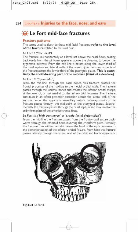
4Le Fort mid-face fractures
Fracture patterns
The terms used to describe these mid-facial fractures, refer to the level
of the fracture related to the skull base.
Le Fort I (‘low level’)
The fracture lies horizontally at a level just above the nasal floor, passing
backwards from the piriform aperture, above the alveolus, to below the
zygomatic buttress. From the mid-line it passes along the lower-third of
the nasal septum and lateral walls of the nose to join the lateral aspects of
the fracture across the lower third of the pterygoid plates. This is essen-
tially the tooth-bearing part of the mid-face (think of a denture).
Le Fort II (‘pyramidal’)
From the mid-line, through the nasal bones, this fracture crosses the
frontal processes of the maxillae to the medial orbital walls. The fracture
passes through the lacrimal bones and crosses the inferior orbital margin
at the level of, or just medial to, the infra-orbital foramen. The fracture
continues in an infero-posterior extension across the lateral wall of the
antrum below the zygomatico-maxillary suture. Infero-posteriorly the
fracture passes through the mid-point of the pterygoid plates. Supero-
medially the fracture passes through the nasal septum and may involve the
cribriform plate of the anterior cranial fossa.
Le Fort III (‘high transverse’ or ‘cranio-facial dysjunction’)
From the mid-line the fracture passes from the fronto-nasal suture back-
wards through the ethmoid bone involving the cribriform plate. Laterally
the fracture runs within the orbit below the level of the optic foramen to
the posterior aspect of the inferior orbital fissure. From here the fracture
passes laterally through the lateral wall of the orbit and fronto-zygomatic
284 CHAPTER 8 Injuries to the face, nose, and ears
Fig. 8.31 Le Fort I.
Hene_Ch08.qxd 8/30/04 6:25 AM Page 284

285
process and posteriorly it crosses the pterygo-maxillary fissure to sep-
arate the base of the pterygoids. This separates the entire facial
skeleton from the skull base.
Le Fort II and III both
- Involve the orbit (risk to the eyes).
- Potentially involve the anterior cranial fossa (associated head
injury/CSF leakage).
Often these fractures occur in various combinations ipsilaterally
and bilaterally.
LE FORT MID-FACE FRACTURES
Fig. 8.32 Le Fort II.
Fig. 8.33 Le Fort III.
Hene_Ch08.qxd 8/30/04 6:25 AM Page 285
The type of Le Fort fracture can usually be determined by
examination. This consists of holding the head stable and rocking on the
upper front teeth (or the alveolar ridge, if the patient is edentulous).
- If the teeth and palate move but the nasal bones are stable, it is a Le
Fort I.
- If the teeth, palate and nasal bones move but the inferior orbital rims
are stable it is a Le Fort II.
- If the whole mid-face feels unstable, it is probably a Le Fort III.
Assessment
(See also Pan-facial fractures below.)
Consider serious facial injuries in anyone with gross swelling.
Clinical features and associated problems
Clinical features may be a direct result of the force of trauma, or as a
consequence of the effects of this force on disruption and displacement of
the mid-facial skeleton.
General features
- Airway compromise (uncommon).
- Haemorrhage—associated with mucosal tears in the naso-pharynx or
facial wounds/lacerations. Rarely torrential, although may require nasal
packing or fracture reduction to stem the flow. If the patient is
shocked, look for another cause.
- CSF rhinorrhoea—CSF leak from the nose (anterior cranial fossa #).
- CSF otorrhoea—CSF leak from the ear (middle cranial fossa #).
- ‘Tramlining’ may be seen when blood mixes with CSF and leaks
from the nose or ear. Along the edges of the flow the blood clots
while the CSF washes it away centrally forming two parallel lines:
hence ‘tramlining’.
- Complications of CSF leaks (meningitis, aerocele or fistula
formation with significant CSF loss).
- Facial swelling.
- Massive facial oedema.
- Abnormal mobility of the mid-face—checked by holding the
anterior maxillary alveolus and gently attempting to mobilize the
maxilla; the other hand is held at sites of suspected fractures,
e.g. the nasal bridge or the inferior orbital margins.
Eye signs
- Bilateral peri-orbital ecchymoses (‘panda faces’ or ‘racoon
eyes ’ ). These are often associated with the gross degree of facial
oedema as seen in the Le Fort II and III fractures.
- Bilateral subconjunctival ecchymoses (bright red). This is bruising
within the conjunctiva adjacent to fractures that pass through the
orbit (Le Fort II and III).
- Enophthalmus—sinking in of the eyes—may initially be masked by
oedema.
- Diplopia—caused by swelling , orbital displacement, or tethering of
ocular muscles within fracture lines and hence lack of stereoscopic vision.
- Traumatic mydriasis (dilated pupil)—spasm of dilator pupillae
secondary to a direct blow.
286 CHAPTER 8 Injuries to the face, nose, and ears
Hene_Ch08.qxd 8/30/04 6:25 AM Page 286

287
Features related to the displacement of the mid-face skeleton
The mid-face may be forced downwards and backwards along the skull
base at 45 degrees to the maxillary occlussal plane. This can result in:
- obstructed airway—this results from the soft palate resting on the
dorsum of the tongue and the posterior oro-pharyngeal wall;
- anterior open bite;
- apparent trismus caused by gagging of the occlusion due to prema-
ture contact in the molar region;
- lengthening of the mid-face;
- ‘dish-faced’ deformity—comminution of the mid-facial bones
(naso-ethmoid complex/anterior maxilla) may result in a central
collapse rather than a posterior displacement of the whole mid-face:
this will produce the classic dish-face deformity;
- pain and crepitus;
• over the nasal bridge and palpable fracture sites; step deformities
may also be elicited;
• inferior orbital rims (Le Fort II );
• upper buccal sulcus (Le Fort I and II);
- upper buccal sulcus bruising;
- anaesthesia—commonly in the distribution of the infra-orbital
nerve—may be as a result of direct trauma, or the widespread
compressive effects of facial oedema.
Radiographs and other useful investigations
Except for the CXR and Head CT these can wait.
- Chest radiograph. Remember the possibility of inhaled foreign
bodies, e.g. from dental trauma.
- Occipito-mental. One or two projections (15 and 30 degrees)
together with a good clinical examination will usually show most
Le Fort fractures adequately. Campbell’s ‘search’ lines provide
a system of assessing these views.
- True lateral projection. This may demonstrate the postero-inferior
displacement of the maxilla responsible for gagging on the posterior
dentition and an anterior open bite. It is also useful for assessment of
the frontal sinuses (fluid levels, posterior wall fractures) and visualizing
the pterygoid plates (an indication of the level of the fracture).
- Peri-apical and upper occlusal views. These are useful for
assessment of dento-alveolar fractures and identification of
a palatal mid-line split.
- Panoral tomography (OPG). Very useful if a mandibular fracture is
suspected. It may also show low level Le Fort I fractures, dento-alveolar
fractures, and dental injuries.
- Computed tomography (CT scans). These are particularly
useful in associated head injuries and in assessing fractures of the
naso-ethmoidal complex, frontal sinus and orbit.
- Magnetic resonance imaging. May be of value in the diagnosis
of CSF leaks and in the assessment of soft tissue injury, particularly
the orbit.
LE FORT MID-FACE FRACTURES
Hene_Ch08.qxd 8/30/04 6:25 AM Page 287

Treatment
(See also Pan-facial fractures below.)
Maxillofacial trauma should be integrated into Advanced Trauma
Life Support—ATLS. ATLS dictates:
…maxillofacial trauma, not associated with airway obstruction or major bleeding,
should be treated only after the patient is stabilized completely and life threatening
injuries have been managed.
Consider first:
- airway with c spine;
- breathing;
- circulation;
- head injuries;
- ocular injuries.
Beware the patient who keeps trying to sit up—they may be
trying to clear their airway. If there is no spinal injury let them sit up, if
unsure log roll or tip the table head down. This is at variance with ATLS
guidelines but facial bleeding will continue unrecognized in the supine
position until the patient vomits and possibly aspirates.
First aid
Le Fort fractures can present with prolonged epistaxis (nose bleed).
Definitive treatment may involve reduction of the fracture or cautery/
ligation of appropriate vessels. As a first-aid measure nasal packs may
tamponade the flow. If posterior nasal packing is required, urinary (Foley)
catheters may be inserted beyond the soft palate prior to inflating the
balloon and then withdrawn until the balloons wedge in the posterior
naso-pharynx; anterior nasal packing with ribbon gauze may also be
required. Custom devices are available (Epistat™) with dual balloons that
will occlude the entire nasal airway and hence tamponade haemorrhage.
Wounds and haemorrhage—‘assess, arrest, and replace’
Remove any dressings to fully assess wounds, remembering that they may
have been placed as pressure dressings to stem initial blood loss. Ideally
careful exploration under LA should be carried out before closure to
clean and remove any debris or foreign bodies, and to identify any under-
lying fractures. If the patient obviously requires to go to theatre, then
formal wound closure should be delayed (although ideally no longer than
24h) but judicious placement of a few sutures along with simple dressings
should help to stem a steady venous ooze.
Replace lost fluids—any patient with Le Fort fractures must have ade-
quate intravenous (IV) access and appropriate fluid resuscitation.
Assessment must be made of any losses, as well as ongoing needs.
Prevent infection
Le Fort fractures are almost always compound injuries via communica-
tions with the sinuses or facial wounds.
- Antibiotics—intravenous and broad spectrum. Many regimes exist
and include augmentin, or benzyl penecillin + metronidazole.
- Tetanus—ensure tetanus prophylaxis is up to date.
Analgesia
Remember, pain is a potent stimulus for the release of catecholamines.
However, facial fractures are often not as painful as one may think.
288 CHAPTER 8 Injuries to the face, nose, and ears
Hene_Ch08.qxd 8/30/04 6:25 AM Page 288

289
- Local—regional nerve blocks: bilateral infra-orbital blocks may be
particularly useful in the Le Fort I and II fractures. Consider
supra-orbital blocks for the Le Fort III pattern.
- Systemic—avoid opiates until the patient is cleared of a head injury:
• IM (intramuscular), a possibility but avoid in the shocked patient;
• IV (intravenous) rapid onset, and easy to titrate. Ideally, if the
patient’s conscious level is appropriate, establish a PCA (patient
controlled analgesia) in liaison with anaesthetic colleagues.
Definitive management—‘reduction and fixation’
(See also Pan-facial fractures below.)
Indications for treatment
These include:
- disruption of the occlusion of the teeth;
- displacement of the maxilla;
- fractured or displaced teeth;
- cerebrospinal fluid leak (controversial);
- abnormal eye movement or restriction of eye movement;
- occlusion of the naso-lacrimal duct;
- sensory or motor nerve deficit.
Reduction
Anatomical reduction is the key to successful treatment of mid-face
fractures. However, mobilization of the fractures varies greatly and is
dependent on fracture pattern, comminution, and impaction. Surgical
reduction and fixation of the maxilla is usually required in the majority of
cases. Where the maxilla is undisplaced and stable or if the patient if unfit
for an operation non-operative treatment may be appropriate.
Manual reduction involves reduction by digital pressure. Sometimes
disimpaction forceps (Rowe’s) are required. These are two forceps with
upper blades, which pass into the nostril to grip the nasal floor above, and
lower blades to grip the intra-orally against the palate. Open reduction
involves visualization of the fracture sites via incisions for mobilization.
Fixation
A variety of fixation techniques may be used either by an open or closed
approach. In some cases no fixation is necessary (undisplaced, immobile
fractures).
Internal fixation is achieved by the application of plates to the
main bone buttresses of the mid-face via intra-oral incisions. The teeth are
usually wired together (IMF) during the operation to help guide the fragments
into position, although this is usually removed at the end of the procedure. In
the edentulous patient, Gunning splints may be appropriate. Fixation uses
semi-rigid mini-plates and mono-cortical screws. Under direct vision the frac-
tures are reduced and plated. For complete fixation of Le Fort II and III frac-
tures access may be gained via peri-orbital incisions or a coronal flap.
Indirect fixation and external fixation may be indicated for rapid
immobilization of Le Fort fractures. In such cases the bite is used to align
and stabilize the fracture or the fracture is immobilized by fixing it to the
stable cranium/frontal bone. External fixation is generally carried out
using supra-orbital pins or a halo frame connected to the maxilla with
a bar. However, this method has largely been superseded by internal
LE FORT MID-FACE FRACTURES
Hene_Ch08.qxd 8/30/04 6:25 AM Page 289

fixation using plates. It may still be useful when rapid fixation is required,
there are infected wounds (rare), in extensive comminution, or in
children. Associated skull or frontal bone fractures is a relative contra-
indication to external fixation.
Internal suspension involves suspending the maxillary fracture from
the skull with wires and placing the patient in IMF.
Surgical approaches
Exposure of fracture sites is the key to accurate reduction and
fixation. This can be:
- via wounds;
- intra-orally;
- peri-orbital incisions:
• lateral brow;
• supra-tarsal fold (‘upper blepharoplasty’);
• mid-tarsal;
• subciliary (‘lower blepharoplasty’);
• trans-conjunctival +/– lateral canthotomy;
- through the skin in the fronto-nasal area:
• ‘H’ (converse);
• mid-line vertical;
• ‘W’;
• bilateral medial canthal incisions;
- Coronal flap (‘bicoronal fla p ’ )—an incision is placed in the
coronal plane across the scalp from left to right preauricular regions.
The scalp is reflected forwards to give excellent exposure of the upper
cranio-facial skeleton, including the zygomatic complexes and the
fronto-naso-ethmoidal region. This also enables harvesting of
cranial bone grafts if required.
290 CHAPTER 8 Injuries to the face, nose, and ears
Fig. 8.34 ‘ORIF’ of a butress in Le Fort I fracture.
Hene_Ch08.qxd 8/30/04 6:25 AM Page 290

291LE FORT MID-FACE FRACTURES
Fig. 8.35 External fixation.
Fig. 8.36 Internal suspension and IMF in Le Fort 1 fracture.
Hene_Ch08.qxd 8/30/04 6:25 AM Page 291
3Nasal fractures
This refers to disruption of the normal bony and cartilaginous skeleton
of the nose. The nose is the most commonly fractured bone in the face.
Diagnosis is clinical not radiological. The pattern of injury depends
on the magnitude and direction of the force applied (lateral or frontal
direction).
Three levels of frontal directed injury can be distinguished:
- plane one injuries do not extend beyond a line joining the nasal bones
and the anterior nasal spine and therefore involve the cartilaginous
nasal skeleton only;
- plane two injuries are limited to the external nose and do not
transgress the orbital rims;
- plane three injuries are more serious and involve orbit walls and
possibly the cranium—this corresponds to a naso-ethmoidal
fracture.
Indications for treatment
- Nasal deformity.
- Obstructed nasal airway.
- Epistaxis.
292 CHAPTER 8 Injuries to the face, nose, and ears
Fig. 8.37 Classification of nasal fractures.
Hene_Ch08.qxd 8/30/04 6:25 AM Page 292

293NASAL FRACTURES
Fig. 8.38 Nasal fracture (predominantly the nasal bones).
Fig. 8.39 Nasal fracture (predominantly the cartilagenous septum—compare tip
position).
Hene_Ch08.qxd 8/30/04 6:25 AM Page 293

- Septal haematoma—this forms between the septal cartilage and its
perichondrium. It must be ruled out in every patient, and appears as a
dark swelling on the septum with narrowing of the nasal airway. This
requires urgent incision and drainage. If missed it can lead to a septal
abscess with intra-cranial complications or a delayed ‘saddle nose’
following necrosis and cartilage loss.
Conservative management
This is indicated if there is no significant deformity, airway obstruction, or
haemorrhage.
Septal haematoma may be aspirated but may require open drainage
under local or general anaesthesia.
Anterior nasal packing may be required if there is any nasal bleeding.
For more serious bleeding, post-nasal packs may be required. For torren-
tial haemorrhage, uncontrollable by packing, surgery or embolization may
be necessary.
Closed manipulation (MUA) of nasal bones with nasal packing and
external splinting (POP) is usually carried out to correct simple deformity.
If undertaken very early (on the sports field), this often requires no anaes-
thesia. Cases must be selected carefully, as manipulation may start
bleeding. Nasal manipulation and packing otherwise requires general
anaesthesia. If the septum is significantly damaged, closed manipulation
with SMR or septoplasty may be undertaken.
Open reduction
Naso-ethmoidal fractures require open surgery to correct fronto-nasal
dysjunction and associated frontal and orbital fractures. Correction of
traumatic telecanthus may be necessary.
Beware nasal fractures with associated black eyes—they may
be naso-ethmoid injuries.
294 CHAPTER 8 Injuries to the face, nose, and ears
Hene_Ch08.qxd 8/30/04 6:25 AM Page 294

295NASAL FRACTURES
Hene_Ch08.qxd 8/30/04 6:25 AM Page 295

3Naso-ethmoid (naso-orbital-ethmoid)
fractures
(See also Pan-facial fractures below.)
Consider serious facial injuries in anyone with gross swelling.
This refers to injuries to the nose, orbits, and ethmoid sinuses
(NOE). Naso-orbital-ethmoid fractures are among the most challenging
facial injuries to diagnose and treat. Fractures of this region are often
complex and comminuted. When assessing these fractures considera-
tion of the frontal sinus should also be made. This is because the
drainage apparatus of the sinus may be blocked as it passes through the
ethmoid region or it may be communicating with the CSF.
Clinical features
NOE fractures occur following a direct blow to the bridge of the nose.
The ethmoid sinuses act as a crumple zone absorbing the impact. This
results in a ‘pushed-in’ look to bridge of the nose—a ‘Miss Piggy nose’.
Clinically there can be:
- severely comminuted nasal fracture;
- detachment of the medial canthal tendons resulting in
pseudotelecanthus;
- fracture of the anterior cranial fossa;
- fracture of the frontal sinus;
- CSF leak.
The ‘bow-string’ test assesses for canthal detachment—the lateral
canthus is pulled laterally, if there is detachment medially the medial
canthus will also move laterally.
Consider also concomitant injury to:
- the head/neck;
- the eye;
- frontal sinus;
- canthal ligaments;
- naso-lacrimal apparatus.
The severity of the injury may vary considerably in this region. The degree
of bone displacement and comminution is difficult to visualize on plain
radiographs, and computerized tomography is necessary. Usually there is
extensive comminution of the involved bones with associated soft tissue
injuries. This makes reduction and fixation difficult, particularly when
there are other facial bone fractures.
Indications for treatment
- Cerebrospinal fluid leakage (controversial).
- Traumatic telecanthus.
296 CHAPTER 8 Injuries to the face, nose, and ears
Fig. 8.40 (a) and (b) Nasoethmoid (naso-orbital-ethmoid ) fractures.
Hene_Ch08.qxd 8/30/04 6:25 AM Page 296

297
- Orbital dystopia.
- Abnormalities of eye movement.
- Naso-lacrimal duct obstruction.
- Deformity of the nose.
- Functional abnormality of the nose including nasal airway obstruction.
- Deviation of the nasal septum, and septal haematoma.
Reduction is indicated in the majority of cases. Non-operative treatment
may be appropriate where the fracture is undisplaced or when the general
condition of the patient makes surgery inadvisable. Simple closed reduc-
tion of the nasal bones may be carried out under a day case general
anaesthetic or even local anaesthesia. More complex fractures may
require open reduction and internal fixation. Accurate repositioning
of the canthal ligaments is essential for a good cosmetic result. A number
of surgical techniques may be employed using either an open or closed
approach. An open approach (usually via a bicoronal flap) is indicated for
complex fractures or when frontal sinus treatment is required.
In general, the best cosmetic result is obtained when a definitive
anatomical reduction of the bone fragments and soft tissue reconstruction
is carried out at the primary operative procedure.
NASO-ETHMOID (NASO-ORBITAL-ETHMOID) FRACTURES
Fig. 8.41 Flat face and ‘Miss Piggy nose’ in NOE fracture.
Hene_Ch08.qxd 8/30/04 6:25 AM Page 297

3Pan-facial fractures
(See also Le Fort fractures above.)
The term ‘pan-facial’ is used when multiple fracture patterns are seen in
the patient. Commonly this means a mid-face and mandibular fracture, but
can include any of the previously described fracture patterns. Fractures of
the face can be considered as those involving the upper-third, mid-face,
and lower face. Some of these bones, such as the ethmoid and those
comprising the orbital roof, are extremely delicate and so thin that on a
dry skull light can easily pass through. The remainder vary in thickness but
often remain quite delicate (nasal, zygoma). The mandible is the strongest
of the facial bones.
The face is not solid but contains several ‘cavities’, such as the sinuses,
orbits, oral, and nasal cavities. Around these the bones form a series of
vertical struts known as ‘buttresses’. As a result, these bones are very
good at resisting vertically directed forces (e.g. during chewing) but
are weak when it comes to resisting horizontal forces (i.e. during most
injuries).
Upper facial fractures
These involve the frontal bone, fronto-naso-ethmoidal region, orbits- and
associated sinuses. The important points to note are whether there is a
displaced fracture and/or any fracture of the posterior wall of the frontal
sinus, fronto-naso-ethmoidal region- or orbit. These will be seen following
CT scans.
Mid-face fractures
These are classically those described by Le Fort as I, II, and III.
- Le Fort I runs above the apices of the teeth from the nasal aperture
back to and across the lower end of the pterygoid plates, it also runs
down the lateral nasal wall; these two are connected by a fracture
running across the posterior wall of the maxilla.
- Le Fort II runs from the nasal bridge down across medial orbital wall
and floor, and across the anterior maxilla under the zygomatic buttress.
From there it passes across the lateral wall of the maxilla to the
pterygomaxillary plates. It also runs down the lateral wall of nose
and across the posterior wall of the maxilla.
- Le Fort III runs across the bridge of the nose into the orbit, down the
medial orbital wall, across the orbital apex, and up the lateral orbital
wall. There is also a fracture along the arch of the zygoma. This
results in disruption at the fronto-nasal zygomatico-frontal and
zygomatico-temporal sutures leading to cranio-facial disruption.
- Le Fort II and III both:
• involve the orbit (risk to the eyes);
• potentially involve the anterior cranial fossa (associated
head injury/CSF leakage).
Fractures may be unilateral or bilateral—more commonly the latter. If
it is unilateral and is displaced, there must be an associated palatal fracture.
It is not uncommon to find one or more of these fracture patterns
present. The bones are often comminuted and the fracture lines rarely run
as neatly as described from one point to another.
298 CHAPTER 8 Injuries to the face, nose, and ears
Hene_Ch08.qxd 8/30/04 6:25 AM Page 298

299
Lower facial fractures
Mandibular fractures in pan-facial trauma are of the same pattern as isol-
ated mandibular fractures but are more often multiple and comminuted.
Associated problems
- Associated problems from the pan-facial trauma relate to the airway.
These are usually secondary to swelling and haemorrhage but can
occasionally be secondary to posterior displacement of the facial
bones, obstructing the airway.
- Although blood loss from facial wounds can look dramatic, in a
shocked patient with facial injuries it is wise to look for other causes of
the shock, as often there is more significant blood loss elsewhere (chest,
abdomen, pelvis, or long bones, etc.) that is responsible for the shock.
- All pan-facial trauma patients have suffered a significant degree of
trauma and have a high risk of injury elsewhere. In particular, they
are at risk of having sustained a head injury and/or cervical spine injury.
A careful and repeated evaluation of the neurological status must be
made. Particular care must be taken, in those patients who have
ingested alcohol prior to the injury, not to assume that drowsiness
or poor co-operation is secondary to the effects of the alcohol.
Assessment
Consider serious facial injuries in anyone with gross swelling.
These should be elicited after completion of the primary survey. If the
patient is intubated, some aspects cannot be undertaken at this stage but
must be recorded in the notes as requiring completion later.
Inspection
All patients with pan-facial fractures will have considerable facial
swelling, unless seen almost immediately after injury or several days later.
The face should be cleaned of blood to enable a clear inspection.
Look for:
- tramline or clear fluid suggestive of CSF leak;
- facial swelling;
- presence of lacerations;
- obvious asymmetry despite swelling;
- check visual acuity;
- diplopia;
- inter-canthal distance;
- shape of medial canthus (does it come to a sharp point angled
slightly downward at the insertion or is it rounded?);
- inter-pupillary distance;
- pupillary levels;
- nasal deviation/air entry and septal haematoma;
- how far can the patient open their mouth? is there any pain or
deviation on opening any reduction of lateral movement?;
- auroscopy—looking for tears in external auditory meatus and blood
behind the tympanic membrane;
- examine VII nerve function;
- missing teeth, crowns, dentures, etc. (if any are missing where
are they?);
- any evidence of palatal or sublingual haematoma.
PAN-FACIAL FRACTURES
Hene_Ch08.qxd 8/30/04 6:25 AM Page 299

Palpation
Be systematic start at the top and work down (or bottom and work up).
Feel for:
- Examine lacerations for depth and underlying fractures. If the
laceration overlies the course of the parotid duct, look for saliva in the
wound. Remember the lacrimal system, facial nerve, and canthal
attachments of the eye.
- Palpated bony margin for tenderness or steps.
- Palpate nasal bones for mobility.
- Assess sensation in if the patient is awake.
- Assess the medial canthus by displacing the lateral canthus
posteriorly. If the medial canthus is detached it will move laterally;
if not, the point of the medial canthus will sharpen.
- Assess maxilla for mobility. If there is mobility assess the level.
When the maxilla moves, is there any movement of the bone at the
infra-orbital and or zygomatico-frontal level? Mobility can also be due
to isolated dentoalveolar fractures.
- Assess the palate for a split.
- Palpate the mandible for mobility and assess the occlusion if possible.
Investigations
- Baseline biochemical and haematological investigations should be
undertaken, and patients grouped and saved or cross-matched,
depending on blood loss. Other blood investigations should be
determined by specific features of the medical history, e.g. diabetes.
- Imaging is the most useful investigation to ascertain the presence and
location of bony fractures. Generally these will either be CT scan or
plain radiographs. Patients with pan-facial fractures frequently have
an associated head injury and will have a brain CT. If it does not com-
promise the patient’s overall well-being, scan the facial bones at the
same time; if possible, generating axial of the facial skeleton and coronal
views of the orbits. Occasionally angiography may be required to
indicate the source of persistent or major haemorrhage.
- If it is not possible to obtain images as part of a head series of scans,
imaging is likely to be required prior to definitive treatment.
Alternatively, good quality plain radiographs are valuable. An OPG, PA
mandible, OMs, and lateral facial bones should be obtained. If
good plain views are not obtainable, or there are condylar, orbital,
frontal sinus, or NOE fractures present, coronal and axial CTs should
be obtained.
- Determining the position of foreign bodies relative to vital structures
will require two images at 90 degrees to one another. Occasionally
angiography may be needed to establish the position of a foreign body
to major vessels. If foreign bodies may be present that are radiolucent,
e.g. wood, ultra sound scans can be useful to demonstrate their
presence.
- Impressions for study models can often be invaluable in planning to
establish pre-operatively the desired occlusion and enable an occlusal
wafer to be constructed pre operatively
- Ask relatives to bring in any pre-injury photographs of the patient.
300 CHAPTER 8 Injuries to the face, nose, and ears
Hene_Ch08.qxd 8/30/04 6:25 AM Page 300

301
Treatment
Maxillofacial trauma should be integrated into Advanced Trauma Life
Support—ATLS. ATLS dictates:
…maxillofacial trauma, not associated with airway obstruction or major
bleeding, should be treated only after the patient is stabilized completely
and life threatening injuries have been managed.
Consider first:
- airway with c spine;
- breathing;
- circulation;
- head injuries;
- ocular injuries.
Beware the patient who keeps trying to sit up—they may be
trying to clear their airway. If there is no spinal injury, let them sit up; if
unsure, either let them lay on their side or tip the table head down. This is
at variance with ATLS guidelines but facial bleeding will continue unrecog-
nized in the supine position until the patient vomits and possibly aspirates.
First aid
Haemorrhage is usually due to displacement of fractured bones or lacera-
tions. Displaced mandibular fractures can be reduced and if teeth are
present on either side of the fracture, a bridle wire placed This provides
some stability to the fracture thereby reducing bleeding and making the
patient more comfortable. Ideally the bridle wire should include two teeth
on each fractured segment.
Associated Le Fort fractures can present with prolonged epistaxis
(nose bleed). Definitive treatment may involve reduction of the fracture
PAN-FACIAL FRACTURES
Fig. 8.42 CT scan (and 3D reformatting) has revolutionized assessment in facial
trauma.
Hene_Ch08.qxd 8/30/04 6:25 AM Page 301
or cautery/ligation of appropriate vessels. As a first-aid measure, nasal
packs may tamponade the flow. If posterior nasal packing is required
urinary (Foley) catheters may be inserted beyond the soft palate prior to
inflating the balloon and then withdrawn until the balloons wedge in the
posterior naso-pharynx; anterior nasal packing with ribbon gauze may
also be required. Custom devices are available (Epistat™) with dual
balloons that will occlude the entire nasal airway and hence tamponade
haemorrhage.
Replace lost fluids—any patient with Le Fort fractures must have ade-
quate intravenous (IV) access and appropriate fluid resuscitation.
Assessment must be made of any losses as well as ongoing needs.
Dento-alveolar fractures can be reduced and stabilized with splints.
If teeth have been avulsed or subluxed, they are best re-implanted or
repositioned as soon as possible and splinted.
Lacerations should be inspected for contamination and foreign
bodies (do not do this with your fingers if glass could be involved). This is
particularly important to prevent tattooing of the skin, which can be
difficult to eradicate. Bites should be thoroughly irrigated to minimize
the risk of infection. Assess the viability of the soft tissues. Due to the
extremely good blood supply of the face, loss of viability is unusual, so
tissue should not be discarded unless it is certain that it is not viable.
Assess facial nerve function. If this is disrupted, repair should be
undertaken prior to definitive closure, which should be done as soon as
possible. However, if a large laceration is present, and other more urgent
problems need attention, tack the wound with a few large sutures, and
close it definitively later. If a laceration gives access to an underlying frac-
ture, which may not be addressed for several days, it is probably best to
close the wound definitively and accept that it may be re-opened later,
than leave definitive closure until fixation of any fractures.
Prevent infection
- Le Fort fractures are almost always compound injuries via the sinuses
or facial wounds.
- Antibiotics—intravenous and broad spectrum. Many regimes exist
and include augmentin, or benzyl penecillin+metronidazole.
- Tetanus—ensure tetanus prophylaxis is up to date.
Analgesia
Remember, pain is a potent stimulus for the release of catecholamines.
However, facial fractures are often not as painful as one may think.
- Local—regional nerve blocks: bilateral infra-orbital blocks may be
particularly useful in the Le Fort I and II fractures. Consider
supra-orbital blocks for the Le Fort III pattern.
- Systemic—avoid opiates until the patient is cleared of a head injury.
- IM (intramuscular), a possibility but avoid in the shocked patient.
- IV (intravenous)—rapid onset, and easy to titrate. Ideally, if the
patient’s conscious level is appropriate, establish a PCA
(patient-controlled analgesia) in liaison with anaesthetic colleagues.
Definitive treatment
Optimal results requires careful pre-operative assessment and planning.
This should result in a comprehensive list of the problems and objectives
302 CHAPTER 8 Injuries to the face, nose, and ears
Hene_Ch08.qxd 8/30/04 6:25 AM Page 302

303
of treatment. This can be summarized as restoration of normal form and
function without producing complications. Broadly speaking definitive
management can be broken down into non-operative or operative
management. The benefits of each needs to be weighed against the risks,
taking into account all aspects of the patient, particularly other injuries and
pre-existing medical conditions.
Planning should be undertaken in collaboration with other specialities
that may be involved, e.g. anaesthesia, neurosurgery, or ophthalmology.
Consideration should be given to whether there is any need for primary
bone-grafting of orbital walls or expected bony defects
AnaestheticBecause IMF is usually required, avoid an oratracheal tube if possible.
Given also that patients also require nasal reduction, thought may need to
be given to either a tracheostomy or submental tube.
AccessThis can be via wide exposure of the facial skeleton via a bicoronal
approach or through localized incisions. In the presence of naso-ethmoid,
frontal sinus, anterior cranial fossa fractures, or Le Fort III fractures, wide
exposure is usually required to get the best results. This allows
direct visualization of fractures, while hiding scars in the scalp. Localized
incisions in the infra-orbital area may still be requires as will intra oral
incisions.
Surgery aims to:
- exposure and sealing of cranial floor fractures;
- restore:
• facial width;
• orbital volume;
• orbital rim;
• occlusion;
• nasal projection;
- repair of VII nerve;
- precise neat closure of lacerations.
If the mandible has bilateral condylar fractures, then, if possible, at least one
condyle should be fixed to restore mandibular height. Mandibular
width must not be increased. This can occur with parasymphyseal fractures.
Often all fractures are exposed before fixation. Fixation starts peripher-
ally, at the junction with uninjured bone. In the mid-face this usually means
either starting laterally and working medially, restoring the orbital rims
and arch. Restoration of orbital volume may require grafting. Nasal projec-
tion can then be addressed. If cranial floor repair is required this should be
done after reduction of fractures to prevent disruption of repair.
When mid-face and mandibular fractures are present, the mandible
should be fixed and the occlusion established with IMF prior to the Le
Fort fractures being fixed.
Any soft tissue repair must be undertaken last. Facial sutures should be
removed at 4–5 days, scalp sutures/clips at 7–10 days.
PAN-FACIAL FRACTURES
Hene_Ch08.qxd 8/30/04 6:25 AM Page 303

General principles in management and terminology
- Debridement—thoroughly clean any open wounds.
- Reduction—open/closed.
- Fixation—IMF/internal/external.
- Care of soft tissues.
- Functional rehabilitation.
Debridement
All wounds need to be thoroughly cleaned as soon as possible, as often
there may be some delay in surgery. Skin wounds need to be gently
cleaned and kept from drying out with saline or antiseptic-soaked swabs.
Oral hygiene is particularly important with fractures of the mandible and
maxilla, as these often communicate with the oral cavity, which, due to
pain, is hard for the patient to keep scrupurously clean. In such cases
regular use of antiseptic mouthwashes (e.g. corsodyl) and hot salt-water
mouthwashes is recommended. Remember the possible need for tetanus
prophylaxis. Once in theatre, dead tissue is excised but this is kept to
a minimum. Wounds are thoroughly irrigated and if grit is ingrained,
scrubbed.
Open reduction and internal fixation (ORIF)
This involves exposing and reducing the fracture(s) under direct vision.
Whenever possible (e.g. mandible, maxilla, and some zygomatic fractures)
this is done via the mouth so as to avoid incisions on the face and scars.
However, if there are associated lacerations these may be used instead.
Although the mouth is heavily colonized with numerous organisms, infec-
tion is surprisingly uncommon. This is in part due to the excellent blood
304 CHAPTER 8 Injuries to the face, nose, and ears
Fig. 8.43 The ‘Coronal’ or ‘Bicoronal’ flap provides excellent access with minimal
morbidity.
Hene_Ch08.qxd 8/30/04 6:25 AM Page 304

305
supply to the face and its capacity to fight infection. By directly visualizing
the fracture, more precise anatomical reduction is possible when
compared to indirect methods (e.g. IMF). Methods of fixation include tran-
sosseous wiring, intra-medullary pins, and rigid/semi-rigid plates and screws.
Plates used for internal fixation may be adaptional plates (semi-rigid),
compression plates (rigid), or mesh. For some overlapping fragments a ‘lag
screw’ technique may be possible.
Rigid internal fixation
This form of ‘ORIF’ uses heavy metal plates and screws to rigidly immobi-
lize fractures. This facilitates precise anatomical reduction and increased
mechanical stability, enabling early return of function. Intermaxillary
fixation is not needed and the patient can eat soon afterwards. However,
this technique risks damaging the inferior dental nerve or teeth. Plates may
become infected and the wound may dehisce.
Intermaxillary fixation (IMF)
This form of immobilization uses the patient’s bite to reduce and stabilize
fractures and therefore can only be used for those fragments firmly
attached to healthy teeth (usually the mandible or dentoalveolar frac-
tures). It is also commonly used in the treatment of condylar fractures.
Fracture reduction is not directly visualized and can result in non-precise
alignment. Examples include eyelet wiring, arch bars, cast cap silver splints,
and gunning splints (edentulous fractures). Caution with post-op airway
problems, COAD, head injury, epileptics, patients likely to vomit, psychi-
atrically unwell. IMF needs to be in place for two to three weeks and
patients can lose weight.
External pin fixation
This involves inserting rigid pins into the bone fragments via the skin,
which are then joined by universal joints and connecting rods or splints.
This method is particularly useful where tissue has been lost (‘continuity
defects’, e.g. gun-shot injuries), infected, severely comminuted, and patho-
logical fractures. Fixation can be applied rapidly. However, reduction is
‘blind’—the pins may damage the inferior dental nerve or teeth, patient
activity is restricted, and the pin sites may become infected and scarred.
Timing of surgery
Early repair (within 5–10 days) gives the best results; immediate surgery
is only required for life-threatening injuries, or sometimes if the patient is
going to theatre for some other reason. Most maxillofacial injuries can
wait, although late repair (once the fractures have united) is difficult to
carry out well. By delaying surgery for several days:
- observation for head or ‘missed’ injuries is possible;
- facial swelling will settle, enabling better assessment;
- quality radiographs can be obtained (face, teeth);
- further investigations can be carried out, e.g. CT scan, study models,
vitality testing;
- further details including pre injury photographs can be obtained
from relatives, GP, etc.;
- aids to surgery can be fabricated, e.g. custom-made arch bars,
gunnings splint.
PAN-FACIAL FRACTURES
Hene_Ch08.qxd 8/30/04 6:25 AM Page 305

Antibiotics, steroids, and tetanus prophylaxis
Protocols may vary between different units. Antibiotics are usually given
for fractures that are compound (open) into the mouth or through
the skin (e.g. mandible). Oral bacteria of a mixed anaerobic type, and a
combination of a penicillin and metronidazole, is one suitable choice.
Prophylactic antibiotics when there is CFS leakage is controversial and the
opinion of a neurosurgeon should be sought. Tetanus prophylaxis should
be considered, especially in mucky wounds, which should be thoroughly
cleaned as soon as possible. Steroids, e.g. dexamethasone, may be given to
reduce facial swelling.
Reduction of and fixation of fractures
If untreated, displaced facial fractures may result in significant disfigure-
ment and functional problems, such as diplopia and malocclusion.
Inadequate treatment can also result in poor aesthetics or function
requiring further surgery to correct these. However, not all displaced
facial fractures need anatomical reduction. Poor results may occur for
several of reasons, for example:
- coexisting medical problems (e.g. head injuries, multi-organ failure,
sepsis- etc.) may delay definitive repair making it difficult to get good
reduction;
- inadequate work up, particularly with radiographs, may miss bony
injuries resulting in inadequate repair;
- fractures may be treated by inadequately trained surgeons;
- patients refuse or fail to attend for treatment.
Reduction—open vs. closed
Terminology can be confusing. ‘Closed’ treatment is preferable to ‘conser-
vative’ treatment. Intermaxillary fixation (IMF) for instance is often
referred to as ‘conservative’, but still has reported morbidity and even
mortality.
Facial fractures 30 years ago were commonly managed by closed tech-
niques, since it was thought that open reduction would lead to infection
and severe osteomyelitis. Open reduction was reserved for compound
fractures or certain unstable mandibular fractures.
Nowadays, many fractures are treated by open reduction, allowing
direct access for fixation. This involves exposure of the fracture, either
through the skin or mucosa. It can then be anatomically reduced and
directly fixed through the incision. Closed reduction, however, is a
‘blind’ procedure relying on the fragments ‘locking’ together. Fixation norm-
ally occurs without direct visualization of the fragments in their final posi-
tion. Examples of closed reduction include nasal manipulation (MUA),
fixing the teeth in occlusion (IMF), or using an arch bar to support dento-
alveolar fractures. IMF relies on the teeth to guide the bones, and then
fixing them to provide adequate stabilization.
Closed reduction—advantages
In the UK, particularly in the Second World War, this technique was
widely used. Custom-made silver splints were constructed, and sectioned
at the fracture site. These were then cemented on to the teeth and inter-
maxillary fixation slowly applied via the splints. This enabled gradual
reduction followed by fixation, often without the need for anaesthetics.
306 CHAPTER 8 Injuries to the face, nose, and ears
Hene_Ch08.qxd 8/30/04 6:25 AM Page 306

307
Closed reduction worked well in missile injuries, stabilizing continuity
defects. Today in Third World countries with inadequate resources or
surgical skills, these simple techniques still have a place. Cast silver splints
have been replaced by arch bars, which are a lot easier and cheaper to
manufacture.
Closed treatment may be an acceptable compromise in a patient with
medical problems precluding a general anaesthetic. IMF may be used to treat
simple, minimally displaced, ‘crack’ fractures relatively easily. However,
those very conditions that prevent a general anaesthetic may also contra-
indicate IMF (e.g. uncontrolled epilepsy, chronic respiratory disease).
Closed reduction—disadvantages
Since the fragments are not directly visualized, ‘closed’ techniques mean
that only the teeth, palpation, or X-rays are used as a guide to the accur-
acy of the reduction. The commonest problem with closed reduction,
therefore, is poor fracture alignment. Errors in reduction are tolerated
much less in the face than in long-bone fractures.
IMF relies on positioning the teeth correctly, assuming that this will
correctly orientate the bony fractures. However, the teeth per se have
only limited control over the positioning of the bones. The attached
muscles may also displace fragments, despite firm location of the teeth.
Control significantly diminishes with fractures further from the occlusion.
This becomes compounded in patients with pre-existing malocclusions or
missing teeth.
The choice between ‘open’ and ‘closed’ reduction may be clear in some
cases, but there are many in which both are equally acceptable.
Nowadays, with good outcomes from surgery and anaesthesia, open
reduction offers the chance of better reduction and fixation. In most cases
this means a safer post-operative recovery and earlier return to normal
function.
Timing of reduction
Timing of surgery is not clearly defined in the literature. For compound
(open) fractures, it is generally assumed that the longer the delay the
more likely the wound will become contaminated. However, it is not clear
what is considered to be an excessive delay. Some studies have failed to
show any differences in complications whether treated within or after 24h
following injury.
However, after long delays (around 14 days), initial healing is well-
established and this makes mobilization and reduction difficult. Soft tissues
become adherent to the displaced bones and this makes reduction even
harder to achieve. This is particularly important in the upper mid-face,
especially the canthal region of the eye. Once displacement is established,
the canthal ligament will never settle into its correct position. This is espe-
cially difficult to correct at a later date.
Life-threatening conditions often take precedent, and in patients with
associated head injuries, fears over long reconstructions leads to delay in
definitive surgery. However, some studies have shown that early interven-
tion is possible without any significant morbidity. Close working relations
with neurosugeons is essential.
PAN-FACIAL FRACTURES
Hene_Ch08.qxd 8/30/04 6:25 AM Page 307

Dynamic compression osteosynthesis
Compression plates have had an enormous impact in orthopaedics and
have become established practice in most trauma units. This has not been
the case in maxillofacial surgery. There are a number of reasons for this:
- only mandibular fractures can be treated in this way;
- the complex curvature of the mandible creates difficulties in plate
placement;
- the teeth and the inferior alveolar nerve are at risk with bicortical
screws;
- precision of reduction is much more than in orthopaedic surgery;
- skin incisions are required (scarring and a small risk of damage to the
mandibular branch of the facial nerve);
- dynamic compression plating is an unforgiving and a difficult technique;
- fears exist that the plates are so rigid that stress shielding delayed
healing strength;
- there is normally a need for a second, often extra oral, operation to
remove these thick bulky plates.
Miniplates
Animal models have shown that some micro movement, using semi-rigid
fixation, encourages prompt healing. Rigid fixation is, therefore, not essential
to fracture healing. Non-compression, smaller sized plates (‘miniplates’)
have now become the standard method of internal fixation in many units.
Furthermore, these plates can be placed orally with much less morbidity.
The principle of this technique is to identify a ‘zone of tension’ within
the mandible at the site of the fracture. The plate is then applied across
the fracture along this line. In a way analogous to suspension bridges, huge
loads can be controlled by relatively small structures, relying on the tensile
strength of the materials. Relatively large loads can be controlled by small
plates—‘miniplates’. In additon, the plate can be fixed by monocortical
screws and, as a result, can be placed where it is biomechanically desirable.
Monocortical screws can be safely placed over dental roots and the
inferior alveolar nerve.
Miniplates have reduced the risks of malocclusions, compared with the
very rigid compression plates. Fine-tuning of the bite is possible with
elastic IMF. The technique is carried out entirely through the mouth, with
no need for skin incisions. The plates are small and can be left in situ if
desired.
Other methods of fixation
External fixation was, prior to plating, an important method of fixation. It
is used in two main ways.
In mid-face fractures, it can be used to immobilize upper and/or lower
arch bars by fixing the frame usually to the cranium. This involves the use
of a halo frame around the skull. This is mechanically very stable and was
initially developed for traction of unstable cervical injuries.
Alternatively, it can be used as a direct fixator across a fracture, com-
monly the mandible or zygoma. Fixators placed across fractures have
several attractions: they can be quickly applied, there is minimal exposure,
and stripping of the periosteum around the fracture. Stability can be
adjusted and modified during healing (dynamization), thereby reducing the
308 CHAPTER 8 Injuries to the face, nose, and ears
Hene_Ch08.qxd 8/30/04 6:25 AM Page 308

309
potential effects of stress shielding. The position of the fractures can be
adjusted if post-operative radiology shows inadequate reduction.
Unfortunately the bulky apparatus is disliked by patients, and care is
required not to injure themselves. Placement normally involves skin
punctures and these can leave unsightly scars.
Nowadays, its main role is to provide ‘first-aid’ stabilization in the
multiply injured patient or where there are limited facilities prior to
transfer to a definitive care centre. In those areas where gun-shot wounds
are common, this method of fixation provides good ‘long-term’ temporary
fixation, until the contaminated wounds have healed. The external fixator
is also particularly useful in maintaining space and orientation in continuity
defects.
PAN-FACIAL FRACTURES
Hene_Ch08.qxd 8/30/04 6:25 AM Page 309

3Special considerations for children
and the elderly atrophic mandible
Both these groups respond to treatments differently.
Fractures in children are marked by rapid healing and rapid remodelling.
In most fractures, reduction and fixation is not necessary. However, there
is a risk of ankylosis in intra-capsular mandibular joint fractures and early
mobilization is necessary. If fixation is required, microplating systems norm-
ally are adequate.
Edentulous mandibular fractures
In the atrophic mandible, the picture is the opposite. The severely
atrophic edentulous mandible often has a poor outcome, especially those
in which the radiographic height of the mandible is 10mm or less. They are
characterized by poor blood supply and slow reparative efforts. In addi-
tion, the older population are frequently complicated by poor general
health, sometimes precluding prolonged general anaesthesia. Patients are
commonly female with osteoportic bones, making screw-fixation difficult
and unreliable. The poor blood supply within the mandible places greater
demands on having an intact periosteal blood supply, making use of large
plates undesirable. Traditional methods of fixation are severely comprom-
ised by the lack of teeth.
Fractures of the edentulous mandible may be treated using the patients
dentures (if a good fit) or customized ‘gunning splints’. These are essen-
tially modified dentures that are wired to the maxilla and mandible and
then fixed together while healing takes place. In selected cases, open
reduction and internal fixation using miniplates applied extra-periostally
(to preserve the blood supply) may be carried out. This is preferable in
the presence of respiratory disease to avoid IMF.
310 CHAPTER 8 Injuries to the face, nose, and ears
Hene_Ch08.qxd 8/30/04 6:25 AM Page 310

311SPECIAL CONSIDERATIONS FOR CHILDREN
Hene_Ch08.qxd 8/30/04 6:25 AM Page 311

4Wound healing
General considerations
The head and neck has a very rich blood supply, which helps fight infection
and improves healing. Intact skin and mucosa also prevents infection of
deeper structures, particulary bone. Skin is also essential in maintaining
body fluids, as seen following extensive burns.
Despite the high bacterial count in the mouth, infected wounds are
uncommon. Saliva and exudates from around the gums contain antibodies
and ‘growth factors’, which stimulate rapid wound healing and prevent
infections (which is why dogs lick their wounds). Skin infections may,
however, occur secondary to commensals (normal inhabitants of the skin),
or from contact with another source (e.g. MRSA).
In the head and neck, unrepaired wounds may produce functional
problems with important structures, such as the eyelids, resulting in signi-
ficant morbidity. Poor repair and aftercare may also result in unacceptable
scarring with distortion of the surrounding structures.
Following trauma, partially avulsed skin, even if attached by a small
pedicle, may still have a good enough blood supply to enable it to heal
if replaced. This is highlighted in the irradiated patient in whom wound
breakdown and infection is more common following surgery or injury.
If in doubt save tissue—never excise widely.
Wound closure is a most effective form of analgesia
The wound-healing environment
The wound needs the correct environment to heal. Many intrinsic and
extrinsic factors (e.g. nutrition, poor dressing, infection, slough, and
necrosis) can delay wound healing.
The ideal dressing should:
- be impermeable to bacteria;
- be free of particles and toxic wound contaminates;
- maintain high humidity at the wound/dressing interface;
- remove excess exudate;
- allow gaseous exchange;
- provide thermal insulation;
- allow removal without causing trauma to the wound;
- be comfortable for the patient whilst in situ and not cause distress or
pain on removal.
Wounds, therefore, heal more effectively in a moist, warm environment,
which new-generation dressings now provide. Epithelial cells can migrate
easily, decreasing healing time. Pain from the wound is reduced as
exposed nerve endings are not allowed to dry out. The body’s natural
autolytic response to deslough is increased.
Wounds heal best at a constant temperature of 37ºC. It can take
several hours for normal cell activity to return following a dressing change.
It is, therefore, important to reduce the number of dressing changes.
Historically, some dressings have been changed up to three to four times
a day. This means that wound healing could potentially be delayed. Too
frequent dressing changes also increase the risk of infection and cross-
infection to other patients.
312 CHAPTER 8 Injuries to the face, nose, and ears
Hene_Ch08.qxd 8/30/04 6:25 AM Page 312

313WOUND HEALING
Hene_Ch08.qxd 8/30/04 6:25 AM Page 313

4Types of wound healing
This can be considered as:
• Primary intention or primary wound healing. The wound is closed as
soon as possible using sutures, clips or ‘steristrips’. There are no gaps in
the wound.
• Secondary intention (granulation) or secondary wound healing. The
skin edges are not closed but left if there is infection or tissue loss. The
wound granulates from its base. This is seen where tissue is lost. It is
unpredictable and can be lengthy. Scarring and deformity are significant
• Exuberant granulation—overgrowth of granulation tissue prevents
in-growth of surrounding epithelium.
Phases of wound healing
This involves a complex interaction between epidermis, dermis, extra-
cellular matrix, angiogenesis, and plasma proteins, all coordinated by
cytokines and growth factors. It is divided into several phases, which
overlap—inflammation, proliferation, and remodelling. The initial stimulus
is thrombus formation. Cells within the clot, notably platelets, trigger an
inflammatory response by releasing vasodilators and chemo-attractants.
Four phases are described here, although there is often a large degree
of overlap between them.
Inflammatory phase
Inflammation is the body’s immediate response to trauma (including
surgery), tissue damage, or invasion of bacteria. Initial vessel constriction
occurs and a loose fibrin clot forms a ‘plug’ loosely uniting the skin edges. If
sutured, wound strength at this stage depends on the suture and any break-
down is the result of poor closure. Mast cells release histamine, resulting in
vasodilation, increased capillary permeability, and swelling, and neutrophils
are attracted by kinins. As the capillaries dilate, tissue fluids rich in plasma
proteins, antibodies, red cells, white cells, and platelets ‘leak’ into the
tissues. Platelets release growth factors and fibrnectin, which promote cell
migration and wound healing. Macrophages are attracted and these remove
any wound debris, thereby beginning the process of repair. Signs of inflam-
mation are heat, swelling, redness, and pain.
Reconstructive phase
Macrophages stimulate the formation of fibroblasts, which migrate along
fibrin threads and produce collagen. Fibroblasts are important in the
production of the extra-cellular matrix (granulation tissue). They produce
collagen, fibronectin, and proteoglycans, such as hyaluronic acid. Fibrin is
produced from the second day and forms a weak framework for healing.
Immature blood vessels develop (angiogenesis) providing oxygen and
nutrients to the wound. It therefore becomes filled with capillaries
and collagen fibres—‘granulation tissue’, which appears deep red in colour
and bleeds easily. Granulation tissue is abundant in secondary intention
healing, although not seen as much following primary closure. It is import-
ant in scar formation. In addition, the wound undergoes contraction. This
may be responsible for 40–80% of wound closure.
314 CHAPTER 8 Injuries to the face, nose, and ears
Hene_Ch08.qxd 8/30/04 6:25 AM Page 314
315
Epithelialization or proliferative phase
Re-epithelializtion is usually evident within 24h. Cells at the wound edges
migrate across the surface–this can only take place over a healthy moist
wound. If infection or debris are present, the inflammatory phase is
prolonged and migration will not take place. ‘Scabs’ are separated from the
wound by proteolytic enzymes. Collagen continues to be produced,
resulting in a red, raised, and often unsightly scars. Wound strength
increases rapidly but is still relatively weak and can stretch if not protected.
Maturation phase
Remodelling takes places over several years, with a reduction of both cell
content and blood flow. The vascularity of the wound decreases and the
scar changes colour, fading to a silvery white appearance. Collagen fibres
re-align themselves, tending to lie at right angles to the wound, increasing
the strength of the scar. At one year, 70% or more of the original tissue
may be regained, but scar tissue is never as strong as unwounded tissue.
Secondary intention healing
Wounds may gape following trauma or infection, coupled with the elastic
pull of the dermis on each side. This defect initially fills with blood clot,
which dries to form a scab. In small, uninfected wounds, re-epithelialization
begins from the wound edges, passing under the scab. The scab is gradually
lifted at its edges until it falls off. The wound heals from below upwards.
Capillary loops and fibroblasts form granulation tissue giving a velvet
appearance to the wound. Myofibroblasts in the wound contract and
reduce the volume of the defect.
The difference between primary and secondary intention
healing is quantitative only.
Healing chronic wounds
Because of the rich blood supply to the head and neck, most wounds heal
quickly. However, when chronic wounds occur, they can be difficult to
treat and, depending on their site, quite disfiguring.
In any patient with a non-healing, chronic wound, ask yourself
why has it not healed?. Consider the possibility of underlying:
- foreign body;
- infection;
- malignancy;
- immunosuppression.
Management strategies
Consider any predisposing conditions (local or systemic).
DressingsIn recent years there has been the development of dressings that promote
a moist environment to assist healing. In animals, re-epithelialization of
partial thickness, acute wounds occurs more rapidly if it is occluded. In
patients they reduce pain. As yet there is still not a dressing that can alter
the healing cascade, although dressings containing hyaluronic acid seem to
promote healing.
TYPES OF WOUND HEALING
Hene_Ch08.qxd 8/30/04 6:25 AM Page 315

Topical growth factorsClinical results from application of growth factors to chronic wounds have
not been as good as initially hoped. To date, only platelet derived growth
factor has been licensed for use, but only for treating non-infected foot
ulcers in diabetic patients (becaplermin, regranex). Other factors showing
some promise include granulocyte colony stimulating factor, fibroblast
growth factor, and epidermal growth factor.
Skin graftsSplit thickness and pinch skin-grafting techniques are very useful, so long as
there is no infection and a health bed on which to place the graft.
Synthetic ‘skin’Previously, cultured patient’s epidermal cells, in which a skin biopsy was
cultured to produce epidermal sheets, were used following in burns.
These have had only limited success due to graft fragility, difficulty in applica-
tion, poor rate of uptake, and the frequent occurrence of infection.
More recently synthetic skin equivalents have been developed, in which
donor tissue with reduced immunogenicity is used.
- Alloderm is a dermal matrix without immunogenic cells.
- Integra is a combination of dermal fibroblasts and bovine collagen.
- Dermagraft consists of non-immunogenic neonatal fibroblast cultured
on a polyglactin mesh.
- Apligraf contains both epidermal and dermal components.
All of these products have been used to treat burns. They are absorbed
into the wound bed and are thought to alter the profile of cytokines
Future developmentsVascular endothelial growth factor and proteinase inhibitors may have a
role in improving healing of chronic wounds. Gene therapy may allow
genes important in healing to be delivered directly into a wound. Another
potential treatment lies in embryonic stem cells.
Risk factors in wound breakdown
General- Advanced age.
- Malnourished (protein, vitamins, trace elements).
- Anaemic.
- Uraemic.
- Jaundice.
- Malignancy (cachexia).
- Chronic steroid therapy.
- Diabetes mellitis.
- Nutritional deficiencies (vitamin C, vitamin K, hypoproteinaemia, zinc).
- Chemotherapy and radiotherapy.
Local- Tension.
- Infection.
- Crushed tissues (forceps, tight sutures).
- Poor tissue vascularity eg after irradiation.
- Foreign body, e.g. dirt, gravel, glass.
316 CHAPTER 8 Injuries to the face, nose, and ears
Hene_Ch08.qxd 8/30/04 6:25 AM Page 316

317
- Necrotic tissue (crushed, excessive use of diathermy).
- Haematoma.
Non-healingMany factors can impair healing.
Local factors- Foreign bodies.
- Tissue maceration.
- Ischaemia.
- Infection.
- Malignancy.
- Systemic factors
- Advanced age.
- Malnutrition.
- Diabetes.
- Renal disease.
- Steroids.
- Immunosupressive drugs.
At a cellular level, tissue growth factors are reduced and an imbalance
between proteolytic enzymes and their inhibitors occurs. Chronic ulcers
seem to have reduced levels of platelet derived growth factor, fibroblast
growth factor, epidermal growth factor, and transforming growth factor
compared with acute wounds. Excessive proteinase activity (notably
matrix metalloproteins) results in abnormal degradation of the extra-
cellular matrix and fibroblasts have impaired responsiveness to growth
hormone.
Research into newer treatments is directed at altering the imbalance by
the topical application of proteinase inhibitors, or by combining proteinase
inhibitors with growth factors.
Classification of early wounds
Wounds may be classified as ‘clean’ or ‘untidy’. Clean wounds have the
greatest chance of healing with minimal scar formation.
Clean
- Sharp incision.
- Uncontaminated.
- Less than 6h old.
- Low energy trauma.
Untidy
- Ragged edge.
- Contaminated.
- More than 12h old.
- High energy trauma.
- Crushed tissue.
- Tissue loss.
- Burns.
TYPES OF WOUND HEALING
Hene_Ch08.qxd 8/30/04 6:25 AM Page 317

4Wound assessment
There are many ways to assess a wound. Five types are described:
- black or necrotic;
- infected;
- sloughy;
- granulating;
- epithelializing.
Black or necrotic wounds
These are covered with a dry hard eschar, which can vary in colour (off
white/yellow to brown/black). This may increase in size if not debrided
(surgically, chemically, or biochemically). Dressings based on hydrocolloid
or hydrogel or an enzymatic desloughing agent can be used. If a cavity
wound is necrotic it can produce a heavy exudate and become very
offensive. Alginate dressings may be used to control the exudate.
Infected wounds
All wounds have bacteria present but many do not have clinical evidence
of infection, i.e. pus. Commensal organisms do not necessarily delay
healing and cause infection. Signs of infection include:
- cellulitis/abscess formation;
- discharge
- localized heat;
- localized pain;
- oedema;
- pocketing at the base of the wound;
- delayed healing;
- wound breakdown
- offensive smell.
A wound swab should be taken. Antibiotics are not always required, some
wounds can be treated topically. Flammazine is useful when Pseudomonasis present and iodine-based dressings have been shown to be effective
against MRSA (methycillin- resistant Staphylococcus aureus). Hydrocolloids
and hydrogels are also useful, but not when there is excessive exudate. An
alginate may be appropriate in this instance but will require changing daily.
Sloughy wounds
These consist of dead neutrophils in exudate. The wound has a white /yellow
appearance. Sometimes there is excessive exudate. Hydrocolloid,
hydrogel, or alginate is useful in these cases.
Granulating wounds
These are red and granular in appearance. They bleed easily as the capil-
lary loops are friable. Dressings such as hydrocolloids, hydrogels, foams,
and alginates are all useful in low-exudating granulating wounds. In a more
heavily exudating wound, an alginate or foam dressing can be used. Some
granulating wounds develop excess granulations, silver nitrate sticks can
be used to cauterize the excess.
318 CHAPTER 8 Injuries to the face, nose, and ears
Hene_Ch08.qxd 8/30/04 6:25 AM Page 318
319
Epithelializing wounds
These have pink/white tissue present as the epithelial cells spread over the
granulating tissue. A moist, warm environment needs to be maintained, as
well as one that is protective. Dressings such as hydrocolioids and foams
are, therefore, appropriate.
WOUND ASSESSMENT
Hene_Ch08.qxd 8/30/04 6:25 AM Page 319

4Wound cleansing
The general aim of wound cleansing is to remove any organic and
inorganic debris from the wound before applying a dressing. Debris can
delay the healing process and result in infection.
A number of antiseptics are available but it is now thought that they can
delay healing and harm the tissues. Sterile saline solution or water are
not harmful to the wound and are recommended. However, swabbing
a wound can damage granulating tissue and may leave small fibres in the
wound, which can be a focus for infection. It is much better to irrigate
wounds, where possible. A 35ml syringe and a 19-gauge needle can be
used, which should not exert too much pressure on the wound. Tap
water is increasingly used to cleanse wounds. Ideally warm irrigant should
be used to optimize healing.
Methicillin-resistant staphylococcus aureus (MRSA)
MRSA is a human pathogen that is resistant to a number of antibiotics, not
only methicillin. Some strains show the potential to become epidemic
(EMRSA). To most fit and healthy people, MRSA is not a threat. However,
to those undergoing major or even minor maxillofacial surgery, it can
cause serious harm.
The pathogens can colonize a particular site, e.g. nose or throat,
without necessarily producing any symptoms. Staphylococci are Gram-
positive organisms that grow on the surface of the skin, in the nostrils,
mouth, umbillicus, and perineal areas. Since the introduction of antibiotics,
Staphylococcus aureus has shown a history of resistance, firstly with peni-
cillin then methicillin. Vancomycin is the antibiotic commonly used to treat
patients with MRSA, but now there are concerns regarding resistance to it
(VRSA).
Patients at risk include:
- immuno-compromised patients;
- the elderly or neonates;
- patients with wounds or skin lesions;
- patients who have had several hospital admissions in the past year;
- patients transferred from other areas where MRSA is prevalent;
- patients admitted from abroad.
The principle method of cross infection is hand carriage, although it can
also be airborne. Other sources include clothing and bedding.
All patients with a positive result should be nursed in isolation. They
should also commence a decontamination regime, which should be avail-
able in every Trust. Decontamination takes around one week followed by
re-swabbing two days later. If the swabs are again positive, than the decon-
tamination regime is repeated until the results are negative. If the swabs
are negative, the screening process is repeated until there are three clear
screens. Even then it is not advisable to nurse the patients in the same area
as those patients deemed to be at risk.
320 CHAPTER 8 Injuries to the face, nose, and ears
Hene_Ch08.qxd 8/30/04 6:25 AM Page 320

321WOUND CLEANSING
Hene_Ch08.qxd 8/30/04 6:25 AM Page 321

3Soft tissue injuries
General points
- Consider injuries to underlying structures (bones/eye/lacrimal
gland/parotid duct/canthal attachments).
- Consider the possibility of tattooing and scarring—can this be
managed in A&E or should I refer?
- Soft tissue injuries are best evaluated after the wound has been
cleaned of dry blood and debris. A local anesthetic may be necessary
to clean the wound so that a thorough examination can be made.
- Where applicable assess the facial nerve prior to anesthetic use.
- Accurately document the injuries, ideally photographically.
- Consider tetanus prophylaxis.
- Ideally repair should be performed within 12h.
- Because of the rich vascular supply of the head and neck, only
minimal debridement is necessary. Preservation of tissue is the
rule—if in doubt leave.
- When suturing wounds, alignment of key anatomic boundaries
(vermilion border, gray line of the eyelid) and alignment of the
eyebrow, should be performed first. With lip lacerations, it is important
to align the vermilion border for good comesis. Place the first stitch to
re-approximate the vermilion border.
- Close in layers (mucosa to mucosa, muscle to muscle, and skin to
skin). Deep sutures are used to close dead space and remove tension
from the skin to prevent hematoma and scarring.
- With deep lacerations of the cheek, the wound should be explored for
injury to the parotid duct and, if possible, the duct is repaired over a
stent.
- If facial nerve weakness results from a laceration medial to a line
dropped perpendicular to the lateral canthus, the nerve branches are
not repaired. (The nerves are generally too fine.) If, however, paralysis
results from a laceration posterior to this line, the facial nerve should
be repaired. Ideally, repair should occur as soon as possible, but no
later than 72h unless the wound is heavily contaminated (e.g. gun shot).
In this situation, the nerve endings are tagged and repair is performed
when the wound is clean.
- With significant tissue loss, similar tissues should be re-approxi-
mated: mucosa to mucosa, muscle to muscle, cartilage to cartilage,
and skin to skin.
- Consider drains.
Following trauma, partially avulsed skin, even if attached by only a small
pedicle, may still have a good enough blood supply to enable it to heal, if
replaced. If in doubt, save tissue—never excise widely.
Wound closure is a most effective form of analgesia.
In addition to the obvious skin and mucosa, assesment of soft tissue
injuries must also include associated specialized structures. These
include:
- fractures;
- salivary gland and/or duct (e.g. parotid);
- lacrimal apparatus;
322 CHAPTER 8 Injuries to the face, nose, and ears
Hene_Ch08.qxd 8/30/04 6:25 AM Page 322

323SOFT TISSUE INJURIES
Fig. 8.44 Wound assessment—always separate the edges and assess depth before
attempting to sutute.
Fig. 8.45 Wound assessment—consider the possibility of underlying fractures.
- nerve injury (facial, accesory, supra-orbital, supra-trochlear,
infra-orbital, mental)—see Microsurgical nerve repair below.
- major vascular injury (especially in neck lacerations);
- loss of function of eyelids;
- loss of function of lips.
In penetrating injuries and lacerations, foreign bodies and contamina-
tion must be considered. If grit is not removed early tattooing can result,
which is extremely difficult to remove later. Underlying fractures must
also be considered and treated before any wounds are closed definitively.
The patient’s tetanus status should be assessed and managed appropriately.
Hene_Ch08.qxd 8/30/04 6:25 AM Page 323

324 CHAPTER 8 Injuries to the face, nose, and ears
Fig. 8.46 Wound assessment—consider the possibility of underlying fractures.
Fig. 8.47 Wound assessment—consider the possibility of foreign bodies.
Hene_Ch08.qxd 8/30/04 6:25 AM Page 324

325
Immediate primary closure with complete haemostasis and accurate
restoration of anatomy should be the aim whenever possible. In most
cases, head and neck wounds can be closed primarily with acceptable
results. The blood supply to the tissues is a major factor in this and it is
essential that the surgery itself does not damage wound edges. Provided
the tissues are alive, even wounds heavily colonized with bacteria can be
closed, primarily using appropriate antiseptic and antibiotics. This is seen
particularly with intra-oral wounds, which are all heavily contaminated
with oral bacteria.
Bites
Whether animal or human in origin these must be considered serious and
managed quickly. Both can rapidly become infected if left too long. Check
for tissue loss and tetanus status (follow local protocols).
Compared to elsewhere in the body, bites on the face can be closed prim-
arily due to its rich blood supply. However, they must be thoroughly
cleaned and irrigated, and sutures placed sparingly to allow any collections
to drain. Broad spectrum antibiotics should be prescribed and, ideally, take
a wound swab for culture. Warn the patient about scarring.
Abrasions
These need to be thoroughly, but gently cleaned to remove all traces of
grit and other foreign bodies. This is the best opportunity to manage these
well. Tattooing results if this is neglected, which is very difficult to deal
with secondarily (excision/dermabrasion/lasers).
Specialised tissues
Eyelid injuries
This requires specialist care. In the first instance, protect and assess the
underlying globe. Assess visual acuity as soon as is practical. Consider
foreign bodies and perforating injuries and therefore avoid any pressure
on the wounds. As a temporary measure, liberally apply chlormycetin
ointment (not drops) to the wound.
Parotid injuries
Lacerations along the side of the face must be carefully assessed to
exclude injuries to the parotid gland, parotid duct, and, most import-
antly, facial nerve. Injuries to the duct and nerve must be repaired using
microsurgical techniques before the skin is closed. Failure to repair the
duct may result in the formation of a ‘sialocele’, which will eventually drain
through the wound, resulting in a salivary fistula. Failure to repair the
nerve may result in various degrees of facial weakness.
Nerve injuries that may benefit from exploration and repair
These include:
- Facial nerve—this may be damaged following trauma (or surgery) to
the parotid region. If necessary, immediate repair can be delayed and
the nerve endings marked with sutures.
- Accessory nerve—injury to this nerve may occur following trauma
(also following lymph node biopsies or neck dissection). Loss of
function of the trapezius muscle results in limitations of shoulder
motions.
SOFT TISSUE INJURIES
Hene_Ch08.qxd 8/30/04 6:25 AM Page 325

- Inferior alveolar nerve—injury may occur as a result of a mandibular
fracture. It can also occur following wisdom tooth removal, jaw surgery,
tumour resection, fracture fixation, and placement of implants. This can
result in numbness or ‘dysaethesia’ in the lower teeth, the lower lip,
and the chin. This should be reconstructed within 3–4 weeks after
injury.
Principles of initial wound care
Decontamination and debridement
All wounds should be thoroughly cleaned and foreign bodies such as dirt
and glass should be removed. However, over-vigorous scrubbing can
cause further damage. All dead tissue should be excised and, if wound
edges are ragged, trimming to form a straight line may be useful.
Wound closure
Suturing is the commonest method of wound closure. Metal clips, adhes-
ive tapes, and glues are also available, they are more difficult to use but
can be quickly applied. Accurate skin apposition is, however, difficult to
achieve and, consequently, they tend to be reserved for lacerations
involving the scalp. The epidermis and underlying tissues are accurately
realigned to eliminate ‘dead’ space beneath the surface. A well-opposed
everted wound edge is the aim to compensate for flattening during wound
contracture.
326 CHAPTER 8 Injuries to the face, nose, and ears
Fig. 8.48 Deep laceration over the parotid—what structures are at risk?
Hene_Ch08.qxd 8/30/04 6:26 AM Page 326

327SOFT TISSUE INJURIES
Hene_Ch08.qxd 8/30/04 6:26 AM Page 327

3Soft tissue injuries of the
head and neck
Checklist—indications for treatment
Presence of abrasions, lacerations and/or avulsions
- Evidence of contamination/foreign bodies.
- Penetrating wounds.
- Burns.
- Motor and/or sensory nerve deficit (5th and 7th cranial nerves).
- Vascular injury.
- Injury to salivary gland and/or duct.
- Injury to lacrimal apparatus.
- Other loss of function of eyelids and/or lips.
Factors affecting the risk
- Association with multiple injuries.
- Impairment of the airway.
- Presence of uncontrolled haemorrhage.
- Presence or absence of contamination/infection.
- Site and extent of lacerations/abrasions.
- Presence or absence of skin/mucosal loss.
- Degree of burn.
- Involvement of specialized structures.
- Presence of severe oedema/emphysema.
- Time lapse between injury and treatment.
- Co-operation of the patient.
- Propensity to keloid/hypertrophic scar formation.
- Systemic disease or treatment that will affect healing.
- Medical/surgical status.
328 CHAPTER 8 Injuries to the face, nose, and ears
Hene_Ch08.qxd 8/30/04 6:26 AM Page 328

329SOFT TISSUE INJURIES OF THE HEAD AND NECK
Hene_Ch08.qxd 8/30/04 6:26 AM Page 329

4The closure of wounds
This may be classified as ‘primary’, ‘delayed primary’ or ‘secondary’.
Primary closure
Clean wounds are closed as soon as possible with meticulous care,
haemostasis, and accurate repositioning of the tissues. Deep wounds
are closed in layers, using resorbable sutures. In some cases, where tissue
has been damaged, trimming of the edges may convert an untidy to a
clean wound, which can then be closed primarily. However, compared to
the elsewhere this is kept to a minimum. If doubt exists about viability, tissue
is often left in situ. To widely excise tissues on the face may lead to difficult-
ies in closing the defect cosmetically, particularly near the eyes, nose, and
mouth, which may become distorted. There should be no tension across
the wound. In cases where tension exists as a result of tissue loss, under-
mining of the skin, local flap closure, or skin grafts may be used.
Delayed primary closure
When doubt exists about the status of a wound, it can be maintained with
moist dressings, antiseptics, and antibiotics for several days. This is useful
in heavily contaminated wounds. Dead tissue will then ‘declare itself’
and can be excised, allowing primary closure. Following excision of obvi-
ously necrotic tissue, a non-adherent dressing is placed or the wound
lightly packed. It is then be inspected under sterile conditions a few days
later. If there is no evidence of further necrosis or infection, it can then be
closed.
Secondary closure
This is the same as secondary wound healing. It is generally avoided if the
face and neck because of contractures and cosmesis. If unavoidable, skin
grafts are often placed on the wound bed to minimise these.
330 CHAPTER 8 Injuries to the face, nose, and ears
Hene_Ch08.qxd 8/30/04 6:26 AM Page 330

331THE CLOSURE OF WOUNDS
Hene_Ch08.qxd 8/30/04 6:26 AM Page 331

4Suturing and suture removal
When carried out correctly, this provides excellent cosmetic results.
Many different sutures are now available, but are generally classified as:
- absorbable—polyglactin (vicryl) and polyglycolic acid (dexon);
- non-absorbable- silk, nylon, and prolene.
They may also be classified according to structure—monofilament,
twisted, or braided. Monofilament sutures minimize infection, produce
less tissue reaction, and are the suture of choice for skin. Braided sutures,
e.g. silk, have plated strands, which provide secure knots but may entrap
material providing a focus for infection. Traditionally, silk has been used
for routine suturing in the mouth, as it is easy to handle, strong, and the
ends are comfortable for the patient. However, vicryl is also popular,
particularly since it resorbs and does not need to be removed. Which of
these is used, often depends on the preference of the surgeon.
Sutures placed in the face and neck tend to be removed at around five
days, or earlier in delicate tissues. ‘Cross-hatching’ of a scar occurs as a
result of closing the wound under tension. Ischaemia of the deeper tissues
damages the skin and stimulates excess collagen formation. Such scars are
difficult to improve and may require further excision and primary closure.
Early removal and continued support with steristrips reduces the chances
of scarring as a result of the sutures themselves. Subcuticular sutures may
be kept in place for longer, as scarring is less likely. With neck lacerations,
sutures are often retained for longer (7–10 days). Scalp sutures are
similarly left for 7–10 days or absorbable ones used instead. In patients
with poor wound healing, e.g. on steroids, with malignancy, infection, or
cachexia, the sutures may need longer. Absorbable sutures are useful for
deep stitches, either as part of a layered closure or for subcuticular skin
closure.
‘Steristrips’ and ‘glue’
These are particularly useful in children and those who will not
co-operate. However, care is required in patient and wound selection.
Only superficial and scalp wounds should be considered. The final
cosmetic results are less predictable than with carefully placed sutures, as
deep wounds require deep sutures. Although simpler to do, gluing can still
be quite tricky—be careful not to glue your glove to the patient—
although it can be removed, it is very embarrassing!
332 CHAPTER 8 Injuries to the face, nose, and ears
Hene_Ch08.qxd 8/30/04 6:26 AM Page 332

333SUTURING AND SUTURE REMOVAL
Hene_Ch08.qxd 8/30/04 6:26 AM Page 333

4Soft tissue injuries—tissue loss
This is relatively uncommon but may be seen, for example, following gun-
shots (attempted suicides), road traffic accidents, or industrial accidents.
Significant tissue loss may also occur following relatively minor injuries
(animal/human bites to the ears, lips, and nose). Particularly following
trauma, tissues may be displaced, rather than lost, but nevertheless result
in significant problems.
With such injuries there is often significant associated bone, brain,
or ocular injuries, and these may need to be dealt with prior to soft
tissue reconstruction. However, occasionally only the soft tissues are
involved, for example, following scalp avulsions. With the use of microsur-
gical techniques many defects can now be either reconstructed using ‘free
flaps’ or, in selected cases (scalp avulsions), the tissues replaced and the
circulation restored by vessel anastamosis.
Repair or reconstruction depends on many variables. Associated injuries
may take priority. Early repair is ideal especially with avulsed tissue.
However, if there is gross swelling or contamination, a delayed
approach may be necessary. Following initial debridement the wound
needs to be protected and dressed until reconstructed. Local flaps are
often useful where tissue loss is not extensive and has the advantage that
the tissue used is similar to that lost (e.g. Abbe flap for lip reconstruction).
For elective reconstructions, tissue expansion can be of use. However,
where the defect is extensive, larger flaps may be necessary. These can be
either pedicled or free flaps depending on the defect and general condi-
tion of the patient. Where necessary, bone can also be raised with the flap
in cases where bony defects are present (e.g. mandible).
Major tissue loss and gun-shot wounds
This is uncommon but may arise following assaults, industrial injuries
(scalp avulsions), or pedestrian–motor vehicle collisions. The initial
approach is no different to other injuries, i.e. ATLS. However, bear in
mind that there can be significant airway problems, blood loss, and
a high risk of infection. Once stabilized, gently clean the wounds and
try to re-align the tissues. This prevents kinking of the veseels, which can
necrose distal tissues.
Definitive management
Recent advances in biomaterials, and our understanding in microvascular
reconstruction, now means that just about any defect in the cranio-facial
region can be reconstructed. Unfortunately, although in most cases func-tional requirements can be met, in many patients, particularly when skin or
specialized structures are involved, some stigmata of their disease persists.
The eye, for instance, can not be replaced and invisible scars do not exist.
However, results are considerably better than 20 years ago. It is an
exciting time in deformity care.
The main choice is between using tissues from the patient (autogenous
transplants) or synthetic materials. Each has its own advantages and disad-
vantages (risks of infection, donor site morbidity, costs, etc.) and each has
its own advocates. With the recent problems of prion transmission,
animal-derived products are rarely used now. The ability to grow tissues,
334 CHAPTER 8 Injuries to the face, nose, and ears
Hene_Ch08.qxd 8/30/04 6:26 AM Page 334

335
either in the lab or on the patient, is the most recent of innovations and
we are moving from an era of ‘tissue transplantation’ into one of ‘tissue
induction’. Bone morphogenic protein, distraction osseogenesis, bone
transportation, and prefabricated ‘free flaps’ are such developments.
Unfortunately, many of these techniques are still relatively unproven in the
long term and are quite expensive.
SOFT TISSUE INJURIES—TISSUE LOSS
Fig. 8.49 Loss of tissue following dog bite.
Fig. 8.50 Local flap in lip reconstruction.
Hene_Ch08.qxd 8/30/04 6:26 AM Page 335
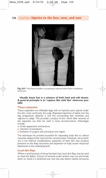
Usually tissue loss is a mixture of both hard and soft tissues.
A general principle is to ‘replace like with like’ wherever pos-
sible.
Tissue expansion
Tissue expanders are inflatable bags with an injection port, placed under
the skin, most commonly the scalp. Repeated injections of saline into the
bag progressivly distends it and the surrounding skin stretches and
regrows to adapt. This provides a surplus of skin, which after removal of
the expander can then be used in local reconstructions. Advantages
include:
- similar appearance and texture;
- retention of sensations;
- limitation of surgery and scarring to one region.
This technique has proved successful for expanding scalp skin to reduce
traumatic alopecia (hair loss) and ear reconstruction. However, the proced-
ure is not without complications; expansion of neck skin may produce
pressure on the deep structures and expansion of scalp causes reciprocal
depression in the underlying skull.
Local skin flaps
Where a small amount of tissue has been lost, local skin flaps may be used
to close the defect. Closure of wounds under tension may not only break
down or result in a stretched scar, but may also distort nearby structures
336 CHAPTER 8 Injuries to the face, nose, and ears
Fig. 8.51 Free tissue transfer is occasionaly required when there is extensive
tissue loss.
Hene_Ch08.qxd 8/30/04 6:26 AM Page 336
337
such as the lips, nose, etc. Many flap designs are described. These make
use of the fact that there is often excess skin on the face, which is highly
vascular, elastic to a degree, and can therefore be undermined and used to
close nearby defects. Well-defined ‘axial flaps’, e.g. glabellar flap, naso-
labial flap, may be raised, based on a small pedicle through which feeding
vessels pass. So long as these are preserved and not kinked during rotation
of the flap, large areas of skin can be used to facilitate tissue closure.
Random pattern flaps, however, require a broad attachment at the base, if
necrosis is to be avoided. In contrast with the remainder of the body, the
skin of the head and neck is very well vascularized and the success rate of
local flaps is generally very high. These techniques are particularly useful in
the reconstruction of nasal tips, eyelids, and lips.
Parotid injuries
Lacerations along the side of the face must be carefully assessed to
exclude injuries to the parotid gland, parotid duct, and, most importantly,
facial nerve. Injuries to the duct and nerve must be repaired, often using
microsurgical techniques, before the skin is closed. Failure to repair the
duct may result in the formation of a ‘sialocele’, which will eventually drain
through the wound resulting in a salivary fistula. Failure to repair the nerve
may result in various degrees of facial weakness.
Microsurgical nerve repair
Repair of damaged or severed nerves is now possible with the use of the
operating microscope. Both direct suturing of the cut ends or ‘interposi-
tional’ nerve grafting may be carried out, depending on the type of inury.
Good results have been obtained following reconstruction or repair of the
accessory and facial nerves. However, reconstruction of sensory nerves
has a lower success rate, but is still often of benefit to the patient. Ideally,
early repair (within 24h) is preferable and should be carried out if there is
no infection or significant associated soft tissue trauma. With the facial
nerve, successful late repair is limited by wasting of the facial muscles,
which occurs around 6–12 months after injury. In long-standing cases of
facial palsy with atrophy of the facial muscles, free flaps of muscle with
their associated nerves and vessels have been used.
Examples of nerve injuries which may benefit from exploration
and repair include:
- Facial nerve—this may be damaged following trauma or surgery to
the parotid region. If necessary, immediate repair can be delayed and, at
the time of wound closure, the nerve endings marked with sutures.
Reconstruction may also be undertaken in those cases of malignant
tumours of the parotid gland, which require sacrifice of the facial nerve.
- Accessory nerve—injury to this nerve may occur following trauma,
lymph node biopsies, or following neck dissection. Loss of function of
the trapezius muscle results in limitation of shoulder motions.
- Lingual nerve—this supplies sensation and taste to the anterior
two-thirds of the tonguue. It may be damaged during surgical removal
or a lower third molar. Repair, ideally, is undertaken within 6 months
following injury.
- Inferior alveolar nerve—injury may occur following wisdom tooth
removal, orthognathic surgery, tumour resection, fracture fixation,
SOFT TISSUE INJURIES—TISSUE LOSS
Hene_Ch08.qxd 8/30/04 6:26 AM Page 337

and placement of implants. This can result in numbness of ‘dysaethesia’
in the lower teeth, the lower lip, and the chin. This should be
reconstructed within 3–4 weeks after injury.
Since the facial and accessory nerve are purely motor nerves, and the
lingual and inferior alveolar nerves mostly sensory nerves, repair can be
simply carried out by alignment of the nerve stumps or by nerve grafting.
Manipulation of the nerves must be kept to a minimum, as this stimulates
scar tissue at the suture site, which can prevent nerve growth. It is essen-
tial that there is no tension across the repair. The least number of sutures
consistent with accurate alignment are used. Where grafting is required,
the great auricular and the sural nerve are often used.
338 CHAPTER 8 Injuries to the face, nose, and ears
Hene_Ch08.qxd 8/30/04 6:26 AM Page 338

339SOFT TISSUE INJURIES—TISSUE LOSS
Hene_Ch08.qxd 8/30/04 6:26 AM Page 339

4Surgical drains
Drains in the head and neck are generally used:
- to provide drainage of infected material (abscess);
- to prevent the collection of blood or fluid between two large raw sur-
faces (following neck dissection, coronal flap);
- where a large potential space exists for blood to collect, which cannot
be closed (following removal of submandibular gland, thyroid).
Drains should not be used as a substitute for poor surgical technique and
haemostasis should be established before wounds are closed. However,
‘reactive’ haemorrhage can occur, i.e. as the patient recovers from the
anaesthetic, their blood pressure rises and previously closed vessels may
open and bleeding restart. This probably happens to a minor degree in all
patients and draining the small amount of blood that is released prevents
the formation of haematoma, which may become infected.
Drains may be left protruding from the skin and simply covered with an
absorbent dressing, e.g. following abscess drainage. Fluids drain under
gravity. Alternatively, they may be connected to a vacuum container. If the
wound is not airtight, or if there is persistent accumulation of fluid, the
drains may be placed on continuous low pressure suction.
‘Shortening a drain’ means withdrawing it bit by bit to allow the cavity to
seal up gradually, while still allowing fluids to drain. This is rarely necessary
following head and neck surgery, as most drains are usually quite small. It
may be of use, however, following incision and drainage of an extensive
abscess, e.g. Ludwig’s angina, to prevent further collection of pus.
340 CHAPTER 8 Injuries to the face, nose, and ears
Hene_Ch08.qxd 8/30/04 6:26 AM Page 340

341SURGICAL DRAINS
Hene_Ch08.qxd 8/30/04 6:26 AM Page 341
4Scars
All wounds extending deeper than the epidermis heal with
scarring. The aim is to minimize this to cosmetically and functionally
acceptable levels. In uninjured skin, collagen synthesis and breakdown is
balanced. However, during healing there is a marked rise in collagen
production. This reaches a maximum at around one month, then falls
slowly to pre-injury levels. Early on the scar is red, hard, and sometimes
itches. Temptation to try and improve the scar before maturation must be
resisted. However, improvements in its appearance can be achieved by
regular wound massage with a moisturising cream, which encourages
breakdown of collagen. It takes about 9–12 months for a scar to mature,
during which significant improvements in appearance can be achieved. The
wound eventually becomes less vascular, softens, and flattens. Scarring is
due to an excess of collagen in the proximity of the wound.
Factors that promote poor scars:
- healing by secondary intention;
- healing of wounds under tension;
- continuing tension on a maturing scar;
- poor vascularity, e.g. diabetes,irradiation;
- dead tissue, e.g. crushed;
- linear wounds running perpendicular to crease lines or linear lines.
Hypertrophic scars and keloids
In some cases scar tissue continues to thicken. A hypertrophic scar
remains confined to the wound closure and may soften and flatten after
several years. A keloid, however, continues to overgrow and may involve
previously undamaged tissue. They can become very bulky. The difference
between the two is one of extent rather than structure. Keloids occur
more commonly among blacks than whites and are often seen after burns.
In the early stages, massage with a moisturiser maintains the suppleness of
a scar. Regular intra-lesional steroid injection over 3–4 months may be
useful for rapidly growing keloids. Alternatively, steroids may be applied
topically (Helan tape). Sustained pressure on the scar (some silicone
dressings) has been shown to improve flattening and softening. However,
this must be continuous and maintained for 6 months to be of any use.
Face masks have been designed and are of most use in burns patients.
Surgical excision may be necessary for very large keloids, however, there
is a high incidence of recurrence. Radiotherapy was used in the past but is
no longer used because of the risk of radiation-induced cancer.
Management of established scars
This depends on the particular characteristics of the scar.
- Shortened scars—scars contract during maturation, and may distort
adjacent structures, particularly around the mouth, base of nose, and
eyes. Lengthening of the scar may be carried out by rearrangement
using a technique of ‘Z-plasty’.
- Shelf scars—semi-circular scars contract unequally along each side
and the inner part often bulges over the outer (pin-cushioning). This is
difficult to correct, but may be improved by increasing its length using
multiple Z-plasties.
342 CHAPTER 8 Injuries to the face, nose, and ears
Hene_Ch08.qxd 8/30/04 6:26 AM Page 342

343
- Widened or stretched scars—tension across the wound or repeated
movement can lead to this. These can be excised and resutured.
Supporting the wound with adhesive tape (e.g. steristrips or subcuticu-
lar suture) during the first month will minimize recurrence.
- Tattooing—this occurs following inadequate removal of grit, etc., at the
time of primary closure. Dermabrasion or excision and primary closure
is the best way of removing tattooed tissue.
- Badly aligned scars—scars running in natural skin creases are less
noticable than those running across them. Some may therefore be
realigned using a Z-plasty changing the direction of the scar by
90 degrees.
- Uneven scars—these can be improved by multiple interdigitating flaps
(‘W’ plasty, ‘geometric broken line closure’) following excision of the
scar. By creating irregularities in the new scar, this breaks up the
straight line appearance.
SCARS
Hene_Ch08.qxd 8/30/04 6:26 AM Page 343

4Paediatric facial trauma
General considerations
Paediatric facial injuries commonly occur following falls, bicycle accidents,
road traffic accidents, and sporting accidents. Emergency admission to
hospital can be a very stressful, frightening, and anxiety-provoking experi-
ence for the child and family. The injured child will be unprepared for what
is about to happen and will need constant explanations. If possible, the
parents should be allowed to stay with their child during all investigations
and treatments. They too will require support and information.
During an emergency admission, children are often in pain while being
subjected to a strange and unfamiliar surrounding. They need a calm,
relaxed, and unhurried admission process. Children are compliant if they
are comfortable and pain-free, if they understand what is happening to
them, and if they have their parents in attendance with them. Most paedi-
atric emergency admissions are not life-threatening and time can therefore
be spent explaining and preparing the child and parent for their surgery
and hospital stay.
Unfortunately there is a tendency with emergency admissions to
perform procedures and treatments quickly, and sometimes the child’s
needs can be overlooked. Any emergency admission is made worse by any
need for surgery. Remember, this may be the first time the child has been
admitted to hospital, let alone have to undergo a surgical procedure. All
explanations given to the child must be age-appropriate, allowing them to
understand the treatment they will receive. Children need to participate in
their care in order to maintain a sense of control and an understanding of
all events.
Parental participation should be welcomed, the child/parent relationship
is unique and can help the child cope with the special tests, procedures,
and surgery they may need to undergo. Parents know their children better
than anyone and if they are supported and kept informed of their child’s
condition, this will benefit the child.
Facial lacerations
Childhood accidents resulting in abrasions, scratches, cuts, and bruises are
generally normal events in a child’s lifetime. However, do not forget
the possibility that injuries may be non-accidental.
Facial lacerations need to be assessed thoroughly. Facial lacerations can
bleed profusely and this adds to what already is a stressful and anxious
time. For some children, assessment and management may require a short
general anaesthetic, in others it may be possible to use local anaesthetic.
This avoids general anaesthesia, but must only be undertaken with the full
consent and co-operation of the child and parent.
Initial treatment should be directed towards controlling blood loss and
cleaning up the wound. It should be irrigated gently with normal saline
and a non-adhesive dressing applied. Analgesia or sedation (if no contra-
indications) may be given prior to this if necessary. Consider also:
- X-ray for any foreign bodies;
- was the child was knocked out;
- the child’s tetanus status.
344 CHAPTER 8 Injuries to the face, nose, and ears
Hene_Ch08.qxd 8/30/04 6:26 AM Page 344

345
If there are no complicating factors, the child should be taken to theatre
as soon as possible. Children should only be fasted for the minimum of
time required for a safe general anaesthetic. Protocols vary slightly
between units but this is approximately 4h for food and 2h for clear
fluids. The child and parents should be warned of the possibility of post-
operative bruising and swelling, and reassured that this will settle quickly.
Post-operatively, suture lines can be left exposed to the air or sup-
ported by steristrips. With traumatized wounds, some surgeons prescribe
an antibiotic course, especially if the wound was dirty. The child’s stay in
hospital should be short and they can be considered for discharge when
fit. At home the suture line will require daily cleaning with normal saline
and the parents can be taught this prior to discharge. Crust formation on
the wound can prevent optimum healing, resulting in a larger scar.
Sutures are generally removed 4–5 days following surgery. Depending
on the number of sutures and the area of injury, the child may need to
return to the ward or wound care clinic, or attend the family’s GP practice
for removal. If the wound has a large number of fine sutures and is in an
awkward place, then an experienced nurse will need to remove them.
Some children may require a light oral sedation prior to suture removal.
Following removal, the repaired laceration can be supported with
steristrips.
The parents should be advised to keep the steristrips in place until they
fall off. After 2–3 months they should be encouraged to massage the scar
with a moisturising cream to prevent thickening and shrinkage of wounds.
Facial fractures
Do not forget the possibility that injuries may be non-accidental.
Children’s bones are elastic, i.e. they tend to bend or buckle
rather than fracture. Therefore, any fracture in a child is a
marker of a serious injury and the underlying soft tissues should
be considered injured also (e.g. rib fractures and lung contusion).
Children who have sustained facial fractures require immediate assess-
ment of their injuries. A full examination should be performed, as other
injuries are often associated. Facial fractures are often associated with a
head injury and the neurological status must be investigated and treated
accordingly. Facial fractures can also involve the orbit—assess visual
acuity. Depending on the age, use a Snellon chart or pictures for younger
children.
Clinically there may be pain or tenderness, swelling, bruising, and non-
occlusion of their jaw and teeth. Some fractures will be stable and require
no further treatment, others may require surgical reduction once the facial
swelling has reduced. If possible, nurse upright to reduce the facial
swelling. Consider intravenous fluids if the child has difficulty taking fluids
orally—they can rapidly become dehydrated.
Post-operatively the child should be nursed in an upright position, as
soon as anaesthetic recovery allows. This will help to reduce facial
swelling. Ice packs can also be used to reduce swelling, if tolerated. Fluids
and a soft diet can be introduced, as tolerated, and any intravenous fluids
discontinued. The child should start oral hygiene as soon as possible and
must understand the importance of doing so. Mouthwashes should be
PAEDIATRIC FACIAL TRAUMA
Hene_Ch08.qxd 8/30/04 6:26 AM Page 345

used regularly, especially following diet, and the child should be encour-
aged to continue normal teeth-brushing using a soft toothbrush.
Paediatric maxillofacial trauma—non-accidental injury
Any child presenting with an injury to the head and neck that does not
appear consistent with the history warrants further investigation. Injuries
such as facial fractures, torn frenulum, bites, puncture wounds, and ocular
trauma should arouse suspicion. Where there is suspicion of abuse, advice
should be sought from a more experienced individual such as a paediatri-
cian or child protection advisor.
Diagnosis can be difficult at times but a number of clues exist:
- there may be a delay in seeking medical attention;
- the history given is vague and lacking in detail, the account may vary
with every telling, and the accounts may differ from person to
person;
- the explanation and account of the accident is not consistent with
the injury observed;
- parent shows inappropriate reaction to severity of injury;
- the parent’s behaviour gives cause for concern: obvious signs of
irritability, and hostility, seldom touches or speaks to the child;
- the child appears fearful, withdrawn, sad, and avoids physical contact
with parents;
- when there is disclosure of an abusive act.
346 CHAPTER 8 Injuries to the face, nose, and ears
Hene_Ch08.qxd 8/30/04 6:26 AM Page 346


![PROTECTING PEOPLE DISPLACED BY WEATHER RELATED …vjel.vermontlaw.edu/files/2014/06/Thomas_forprint.pdf2014] Protecting People Displaced by Weather-related Disasters 805+ face enormous](https://static.fdocuments.net/doc/165x107/5e7214a18b4de178ac4e68c2/protecting-people-displaced-by-weather-related-vjel-2014-protecting-people-displaced.jpg)
