Chapter 7 Hominins from the Upper Laetolil and Upper ...
48
141 Abstract Renewed investigations at Laetoli in northern Tanzania have led to the recovery of a number of new fossil hominins. A lower canine and a mandibular fragment from the Upper Laetolil Beds (3.63–3.85 Ma) are referred to Australopithecus afarensis, and an edentulous maxilla and a proximal tibia from the Upper Ndolanya Beds (2.66 Ma) are attributed to Paranthropus aethiopicus and Hominini gen. et sp. indet., respectively. Additional hominin specimens from earlier collections are described here for the first time, including three specimens of A. afarensis, probably from the Upper Laetolil Beds, and a possible cranial fragment of an infant from the Upper Ndolanya Beds. The chronology and prove- nance of the Laetoli hominins are reconsidered. The species afarensis is provisionally retained in Australopithecus to reflect its anatomical and paleobiological similarities to the other species of Australopithecus sensu lato, but a reasonable case could be made on phylogenetic grounds to transfer it to Praeanthropus. It has been argued that the Laetoli sample of A. afarensis is morphologically and temporally intermediate between A. anamensis and the Hadar sample of A. afarensis, and that A. anamensis and A. afarensis represent a single anagenetically evolving lineage. However, the new specimens from the Upper Laetolil Beds help to close the gap between the Laetoli and Hadar samples, and a critical assessment of the morphological variation in the two samples indicates that there are few consistent differences separating them. Rather than being intermediate in morphology, the Laetoli sam- ple appears to represents an earlier population of A. afarensis, with almost the full complement of derived features that char- acterizes the Hadar sample, but still retaining a few primitive traits. The morphological features that distinguish A. ana- mensis from A. afarensis are much more extensive, and these provide adequate justification for the recognition of a species distinction. The evidence best fits an evolutionary model involving a cladogenetic event rather than a simple anage- netic transformation of a single unbranched anamensis-afa- rensis lineage through time. The Paranthopus aethiopicus specimen from the Upper Ndolanya Beds represents the oldest securely dated specimen definitively attributable to this taxon and the first definitive record outside of the Turkana Basin. The Paranthropus clade probably immigrated into eastern Africa before 2.7 Ma, and became widely distributed throughout the region soon thereafter. The timing and biogeo- graphic patterning of the occurrence of Paranthropus and Homo suggest that their respective dispersals into eastern Africa were not coincident or synchronous. Homo appeared somewhat later than Paranthropus across most of eastern Africa, except in the Awash region of Ethiopia where Homo makes its first appearance in the absence of Paranthropus. These differences in the timing and distribution suggest that Paranthropus and Homo may have had different biogeo- graphic histories, and that their ancestral species may have had different ecological requirements at the time of their ini- tial influx into eastern Africa. Keywords Australopithecus afarensis Paranthropus aethiopicus Introduction Laetoli in northern Tanzania has yielded a relatively small but important collection of early hominins from the mid- Pliocene Upper Laetolil Beds, dated from ~3.63–3.83 Ma (Weinert 1950; White 1977, 1980a, 1981; Leakey 1987a, b; Kyauka and Ndessokia 1990). Although there has been debate about the number of taxa represented at Laetoli, and what these taxa should be named (Tobias 1980a, b; Johanson 1980a; Day et al. 1980a; White et al. 1981; Olson 1981, 1985; Logan et al. 1983; Ferguson 1986, 1987, 1988; Falk et al. 1995; Groves 1996; Senut 1996; Kimbel et al. 2004; Grine et al. 2006), there is current consensus that the remains can all be attributed to a single species, Australopithecus afaren- sis (or Praeanthropus afarensis) (see Table 7.1). The Laetoli sample of A. afarensis (n = 33) is not as large as the collec- tions from Hadar (330 specimens; Kimbel and Delezene 2009), and the specimens tend to be isolated elements and T. Harrison ( ) Center for the Study of Human Origins, Department of Anthropology New York University, 25 Waverly Place, New York, NY 10003, USA e-mail: [email protected] Chapter 7 Hominins from the Upper Laetolil and Upper Ndolanya Beds, Laetoli Terry Harrison T. Harrison (ed.), Paleontology and Geology of Laetoli: Human Evolution in Context. Volume 2: Fossil Hominins and the Associated Fauna, Vertebrate Paleobiology and Paleoanthropology, DOI 10.1007/978-90-481-9962-4_7, © Springer Science+Business Media B.V. 2011
Transcript of Chapter 7 Hominins from the Upper Laetolil and Upper ...

141
Abstract Renewed investigations at Laetoli in northern Tanzania have led to the recovery of a number of new fossil hominins. A lower canine and a mandibular fragment from the Upper Laetolil Beds (3.63–3.85 Ma) are referred to Australopithecus afarensis, and an edentulous maxilla and a proximal tibia from the Upper Ndolanya Beds (2.66 Ma) are attributed to Paranthropus aethiopicus and Hominini gen. et sp. indet., respectively. Additional hominin specimens from earlier collections are described here for the first time, including three specimens of A. afarensis, probably from the Upper Laetolil Beds, and a possible cranial fragment of an infant from the Upper Ndolanya Beds. The chronology and prove-nance of the Laetoli hominins are reconsidered. The species afarensis is provisionally retained in Australopithecus to reflect its anatomical and paleobiological similarities to the other species of Australopithecus sensu lato, but a reasonable case could be made on phylogenetic grounds to transfer it to Praeanthropus. It has been argued that the Laetoli sample of A. afarensis is morphologically and temporally intermediate between A. anamensis and the Hadar sample of A. afarensis, and that A. anamensis and A. afarensis represent a single anagenetically evolving lineage. However, the new specimens from the Upper Laetolil Beds help to close the gap between the Laetoli and Hadar samples, and a critical assessment of the morphological variation in the two samples indicates that there are few consistent differences separating them. Rather than being intermediate in morphology, the Laetoli sam-ple appears to represents an earlier population of A. afarensis, with almost the full complement of derived features that char-acterizes the Hadar sample, but still retaining a few primitive traits. The morphological features that distinguish A. ana-mensis from A. afarensis are much more extensive, and these provide adequate justification for the recognition of a species distinction. The evidence best fits an evolutionary model involving a cladogenetic event rather than a simple anage-netic transformation of a single unbranched anamensis-afa-rensis lineage through time. The Paranthopus aethiopicus
specimen from the Upper Ndolanya Beds represents the oldest securely dated specimen definitively attributable to this taxon and the first definitive record outside of the Turkana Basin. The Paranthropus clade probably immigrated into eastern Africa before 2.7 Ma, and became widely distributed throughout the region soon thereafter. The timing and biogeo-graphic patterning of the occurrence of Paranthropus and Homo suggest that their respective dispersals into eastern Africa were not coincident or synchronous. Homo appeared somewhat later than Paranthropus across most of eastern Africa, except in the Awash region of Ethiopia where Homo makes its first appearance in the absence of Paranthropus. These differences in the timing and distribution suggest that Paranthropus and Homo may have had different biogeo-graphic histories, and that their ancestral species may have had different ecological requirements at the time of their ini-tial influx into eastern Africa.
Keywords Australopithecus afarensis Paranthropus aethiopicus
Introduction
Laetoli in northern Tanzania has yielded a relatively small but important collection of early hominins from the mid-Pliocene Upper Laetolil Beds, dated from ~3.63–3.83 Ma (Weinert 1950; White 1977, 1980a, 1981; Leakey 1987a, b; Kyauka and Ndessokia 1990). Although there has been debate about the number of taxa represented at Laetoli, and what these taxa should be named (Tobias 1980a, b; Johanson 1980a; Day et al. 1980a; White et al. 1981; Olson 1981, 1985; Logan et al. 1983; Ferguson 1986, 1987, 1988; Falk et al. 1995; Groves 1996; Senut 1996; Kimbel et al. 2004; Grine et al. 2006), there is current consensus that the remains can all be attributed to a single species, Australopithecus afaren-sis (or Praeanthropus afarensis) (see Table 7.1). The Laetoli sample of A. afarensis (n = 33) is not as large as the collec-tions from Hadar (330 specimens; Kimbel and Delezene 2009), and the specimens tend to be isolated elements and
T. Harrison ( ) Center for the Study of Human Origins, Department of Anthropology New York University, 25 Waverly Place, New York, NY 10003, USA e-mail: [email protected]
Chapter 7Hominins from the Upper Laetolil and Upper Ndolanya Beds, Laetoli
Terry Harrison
T. Harrison (ed.), Paleontology and Geology of Laetoli: Human Evolution in Context. Volume 2: Fossil Hominins and the Associated Fauna, Vertebrate Paleobiology and Paleoanthropology, DOI 10.1007/978-90-481-9962-4_7, © Springer Science+Business Media B.V. 2011

142 T. Harrison
more fragmentary. However, the sample does represent the second largest sample of A. afarensis, and perhaps more significantly derives from an earlier time period (the homi-nins from Hadar date from ~3.0–3.4 Ma; Kimbel et al. 2004; Campisano and Feibel 2008). The fossil hominins from Woranso-Mille (~3.57–3.8 Ma) in Ethiopia, with 30 speci-mens recovered to date, including a partial skeleton (Haile-Selassie et al. 2007, 2010), have not yet been formally taxonomically assigned, but if they later prove to belong to A. afarensis they would provide another important sample of this taxon contemporary with Laetoli.
The site of Laetoli is unique for the remarkable preservation of trails of footprints, presumably of A. afarensis (see Leakey
and Hay 1979; Clarke 1979; Day and Wickens 1980; White 1980b; Charteris et al. 1981, 1982; Hay and Leakey 1982; Leakey 1978, 1979, 1981, 1987c; White and Suwa 1987; Tuttle 1985, 1987, 1990, 1994, 2008; Tuttle et al. 1990, 1991a, b, 1992; Feibel et al. 1996; Agnew and Demas 1998; Meldrum 2004; Sellers et al. 2005; Berge et al. 2006; Raichlen et al. 2008). This evidence has been used to confirm earlier inferences based on functional morphology of the skeletal remains that bipedal-ism was an important component of the terrestrial locomotor behavior of mid-Pliocene hominins (e.g., Johanson et al. 1982; Stern and Susman 1983; Susman et al. 1984, 1985; Latimer et al. 1987; Latimer 1991; Susman and Stern 1991; McHenry 1986, 1991, 1994; Stern 2000; Ward 2002).
Fossil hominins have not yet been recovered from the Lower Laetolil Beds (~4.4–3.85 Ma; Deino 2011), and are rare in the younger stratigraphic units that overlie the Upper Laetolil Beds, although indirect evidence of their presence is provided by the occurrence of stone tools in the Olpiro Beds (~2.0 Ma; Deino 2011) and Ngaloba Beds (Late Pleistocene) (Harris and Harris 1981; Leakey 1987a; Hay 1987; Ndessokia 1990). Mary Leakey’s expeditions did recover a relatively complete cranium of Homo sapiens from the Late Pleistocene Upper Ngaloba Beds. Most recently, Harrison (2002) reported specimens attributable to Paranthropus aethiopicus from the Upper Ndolanya Beds, dated to 2.66 Ma (Deino 2011) (see Fig. 7.1). The history of discovery of A. afarensis and the other hominin finds from Laetoli is briefly reviewed below.
Table 7.1 Taxonomy and synonymy list of Australopithecus afarensis
Superfamily: Hominoidea Gray, 1825Family: Hominidae Gray, 1825Subfamily: Homininae Gray, 1825Tribe: Hominini Gray, 1825Genus: Australopithecus Dart, 1925Species: A. afarensis Johanson, 1978
Synonymy1948 – Praeanthropus Hennig, 1948 – Hennig (1948) [nomen nudum,
no fixation of type species]1950 – Meganthropus africanus Weinert, 1950 – Weinert (1950)1954 – Australopithecus africanus transvaalensis Broom, 1936 –
Robinson (1954) [partim]1955 – Praeanthropus africanus (Weinert 1950) – Senyürek (1955)1978 – Australopithecus afarensis Johanson, 1978 – Hinrichsen
(1978)1978 – Australopithecus afarensis Johanson, White and Coppens,
1978 – Johanson et al. (1978)1980 – Australopithecus africanus afarensis Johanson, 1978 –
Tobias (1980b)1980 – Australopithecus africanus aethiopicus Tobias, 1980 –
Tobias (1980b) [nomen nudum, conditionally proposed]1980 – Australopithecus africanus tanzaniensis Tobias, 1980 –
Tobias (1980b) [nomen nudum, conditionally proposed]1981 – Paranthropus africanus (Weinert 1950) – Olson (1981)1981 – Homo (Australopithecus) sp. indet. – Olson (1981)1983 – Dryopithecus (Sivapithecus) sivalensis (Lydekker, 1879) –
Ferguson (1983)1984 – Homo antiquus Ferguson, 1984 – Ferguson (1984) [junior
homonym, name preoccupied by Homo antiquus Adloff, 1908]1985 – Homo (Australopithecus) aethiopicus (Tobias 1980b) – Olson
(1985) [junior homonym, name preoccupied by Homo aethiopicus Bory de Saint-Vincent, 1825]
1987 – Australopithecus africanus miodentatus Ferguson, 1987 – Ferguson (1987)
1996 – Australopithecus antiquus (Ferguson 1984) – Senut (1996)1996 – Australopithecus bahrelghazali Brunet et al., 1996 – Brunet
et al. (1996)1999 – africanus Weinert, 1950 – name suppressed by the
International Commission on Zoological Nomenclature for the purposes of the Principle of Priority but not for those of the Principle of Homonymy, Opinion 1941
2000 – Praeanthropus afarensis Johanson, 1978 – Wood and Richmond (2000)
Fig. 7.1 Stratigraphic column and radiometric dating of the lower part of the sequence at Laetoli (After Hay 1987; Drake and Curtis 1987; Ndessokia 1990; Manega 1993; Mollel et al. 2011; Deino 2011)

1437 Hominins from Laetoli
The fossil collections made by Louis and Mary Leakey at Laetoli in 1935 included a hominin lower canine (M.42323, formerly M.18773), which is housed in the Natural History Museum in London. This was the first Pliocene hominin to be recovered from East Africa, although the specimen was not identified as such until some decades later (White 1981). In 1939 Kohl-Larsen’s expedition to Garusi (= Laetoli) included a hominin maxilla with P3 and P4 (Garusi I) and an isolated M3 (Garusi II) (Weinert 1950; Remane 1950, 1954; Robinson 1953, 1955; Protsch 1976, 1981). Both of these specimens are housed in the Institut für Ur- und Frühgeschichte und Archäologie des Mittelalters, Tübingen. An undescribed occipital fragment (Garusi III), apparently of a fossil hominin from Pleistocene sediments, has been lost (Protsch 1976, 1981; Ullrich 2001). In the late 1970s, Eric Delson identified a previously unrecognized hominin lower incisor among the fossil cercopithecids in Berlin that had been collected by Kohl-Larsen (White 1981; Delson, personal communication), and this specimen has been briefly described (Ullrich 2001).
The most extensive collection of hominins from the Upper Laetoli Beds (n = 25) was recovered by expeditions led by Mary Leakey from 1974 to 1979 (Leakey 1987b). These comprise 14 isolated teeth, 10 cranial/jaw fragments or asso-ciated dentitions, and a partial skeleton of an immature indi-vidual. Of these, 23 have been described previously (White 1977, 1980a; Leakey 1987b), and two are identified here as belonging to A. afarensis for the first time. These include a weathered and heavily rolled mandibular fragment (L.H. 29), initially referred to Homo cf. H. erectus, and a weathered isolated upper canine (LAET 79-5447), which were found the same field season at Loc. 8. At the time of their discovery these surface finds were presumed to be derived from the deflated Pleistocene sediments, because they have the black and orange staining typical of the fossils from these beds. However, specimens that erode out of the Upper Laetolil Beds and are reworked into the superficial lag deposits can often develop similar preservational characteristics. Since their morphology is entirely consistent with material from the Upper Laetolil Beds, these two specimens are reassigned here to A. afarensis. A further undescribed specimen (LAET 75-3817) of a possible hominin was excavated by Mary Leakey’s team at Loc. 7E from the Upper Ndolanya Beds. This is a zygomatic process of a right frontal of an infant, recorded in the catalogue as a cercopithecid. The only other hominin recovered by Mary Leakey’s expeditions is a homi-nin cranium (L.H. 18) from the Late Pleistocene Upper Ngaloba Beds, referrable to Homo sapiens (Day et al. 1980b; Magori and Day 1983).
Renewed investigations at Laetoli by the Institute of Human Origins, directed by D.C. Johanson from 1985 to 1988, succeeded in recovering a single hominin specimen,
an isolated right M3 (L.H. 31) (Ndessokia 1990; Kyauka and Ndessokia 1990). The specimen was recovered from the Upper Laetolil Beds at Loc. 10 in 1987, but the precise strati-graphic provenance is unknown. Unfortunately, the author has not been an able to relocate the specimen.
The resumption of full-scale paleontological and geo-logical research at Laetoli and at other sites on the Eyasi Plateau in 1998, under the direction of the author, led to the recovery of four additional hominins. Two specimens, an isolated lower canine (EP 162/00) and a mandibular frag-ment with P3-M1 (EP 2400/00), were recovered from the Upper Laetolil Beds at Loc. 16 (see Harrison and Kweka 2011; Fig. 7.2). Both specimens are referable to A. afarensis. In addition, two hominins were recovered from the Upper Ndolanya Beds – a proximal tibia (EP 1000/98) and an eden-tulous maxilla (EP 1500/01). These are the first hominins to be recovered from this stratigraphic unit, and have proven to be of exceptional interest. EP 1500/01 was found in 2001 at the newly recorded locality of Silal Artum, just to the north of the main fossiliferous outcrops at Laetoli (see Harrison and Kweka 2011; Fig. 7.2). As is discussed below, the maxilla can be confidently attributed to Paranthropus aethi-opicus. EP 1000/98 was found during the first season of renewed fieldwork at Laetoli in 1998, at yet another new locality, Loc. 22S, this time at the southern edge of the main Laetoli outcrops (see Harrison and Kweka 2011; Fig. 7.2). Attribution of isolated postcranial specimens to early hominin taxa is obviously problematic, but given the occurrence of P. aethiopicus as the only hominin known from the Upper Ndolanya Beds it is likely that the proximal tibia belongs to this species. However, since there is no direct association and the hominins from the Upper Ndolanya Beds are few, the proximal tibia is conservatively identified here as Hominini gen. et sp. indet. Further discussion concerning the affinities of EP 1000/98 is presented below.
The aim of this contribution is to present a descriptive account of the morphology of the newly collected hominin specimens from the Upper Laetolil and Upper Ndolanya Beds, as well as of the previously undescribed specimens from the Kohl-Larsen and Mary Leakey collections. The study also provides an opportunity to clarify aspects of the chronology and provenance of the Laetoli hominins, and to discuss their implications for understanding the evolutionary history of Australopithecus afarensis and Paranthropus aethiopicus.
Material
A list of hominins recovered from Laetoli between 1935 and 1979 was presented in Leakey (1987b: 116–117), and the individual specimens have been figured and described in

144 T. Harrison
some detail (Leakey et al. 1976; White 1977, 1980a, 1981; Day et al. 1980b; Magori and Day 1983; Leakey 1987b). An updated list is presented in Table 7.2, which includes a number of emendations and corrections to the original list, as well as the addition of new specimens recovered or identified since 1987. All of the hominins recovered from the Upper Laetolil Beds can be referred to Australopithecus afarensis. The only taxon so far identified in the Upper Ndolanya Beds is Paranthropus aethiopicus.
The Laetoli hominins are housed in the Natural History Museum in London (NHM.M; 1935 Leakey collection), Humboldt-Universität Museum für Naturkunde in Berlin (MB Ma.; 1938–1939 Kohl-Larsen collection), Institut für Ur- und Frühgeschichte und Archäologie des Mittelalters, Tübingen (Garusi hominins; 1938–1939 Kohl-Larsen collec-tion), Kenya National Museum in Nairobi (LAET; 1974–1979 Leakey collections on loan from Tanzania), and National Museum of Tanzania (EP, Eyasi Plateau expedition;
1998–2005 Harrison collection). Comparisons with original specimens and casts of Australopithecus afarensis, A. ana-mensis, Paranthropus boisei, P. robustus, and P. aethiopicus were carried out at the Kenya National Museum (KNM), National Museum of Tanzania (TNM), American Museum of Natural History (AMNH), and Natural History Museum in London (NHM).
Provenance
Most of the hominins from Laetoli, including the new speci-mens described here, are surface finds that had already eroded out of their original stratigraphic context at the time of their discovery. In most cases, based on the position of the find spot, the local topographic and stratigraphic context, and the occur-rence of associated fossils, it is possible to determine the
Fig. 7.2 Map of the Laetoli area showing the main outcrops of the Upper Laetolil and Upper Ndolanya Beds and the paleontological collecting localities

1457 Hominins from Laetoli
Tab
le 7
.2
Cat
alog
ue o
f fos
sil h
omin
in s
peci
men
s re
cove
red
from
Lae
toli
Spec
imen
Yea
rC
olle
ctor
Loc
ality
Stra
tigra
phic
pos
ition
Ana
tom
ical
par
tTa
xon
Com
men
ts
M.4
2323
1935
L.S
.B. L
eake
y E
xped
ition
Unk
now
nU
pper
Lae
tolil
Bed
sL
t C1
A. a
fare
nsis
Publ
ishe
d as
M.1
8773
(Whi
te 1
980a
, 19
81)
Gar
usi I
1939
Koh
l-L
arse
n E
xped
ition
Unk
now
nU
pper
Lae
tolil
Bed
sR
t max
illa
frag
. with
P3 -
P4A
. afa
rens
isH
olot
ype
of P
raea
nthr
opus
(see
Ta
ble
7.1)
Gar
usi I
I19
39K
ohl-
Lar
sen
Exp
editi
onU
nkno
wn
Upp
er L
aeto
lil B
eds
Lt M
3A
. afa
rens
is
MB
Ma.
829
419
39K
ohl-
Lar
sen
Exp
editi
onU
nkno
wn
Upp
er L
aeto
lil B
eds
Lt I
1A
. afa
rens
is(G
arus
i 4)
L.H
. 119
74M
. Mw
oka
Loc
. 1~1
.2 m
abo
ve T
uff 7
Rt P
4 fra
g.A
. afa
rens
isFo
und
by M
. Muo
ka in
Lea
key
et a
l. (1
976)
L.H
. 219
74M
. Mul
uila
Loc
. 3~0
.5 m
abo
ve T
uff 7
Man
dibl
e (i
nfan
t) w
ith rt
and
lt d
P 4, un
erup
ted
crow
ns o
f rt a
nd lt
I 1, C
, P3;
dam
aged
cr
owns
of r
t and
lt d
C, d
P 3 and
M1
A. a
fare
nsis
L.H
. 319
74–7
5M
. Mw
oka
Loc
. 7B
etw
een
Tuff
s 7
and
8, ju
st a
bove
xe
nolit
h ho
rizo
n
Part
ial u
pper
and
low
er d
entit
ion:
(a) r
t dP4 ,
(b) l
t I1 ,
(c) r
t I2 ,
(d) l
t I2 ,
(e) l
t C1 ,
(f) l
t P3 ,
(g) l
t P4 ,
(h) r
t M1 ,
(i) l
t M1 f
rag.
, (j)
lt d
I 2, (k
) rt d
C1,
(l) r
t dP 3,
(m) r
t I1,
(n) r
t C1,
(o) l
t C1,
(p) r
t P3,
(q) r
t P4 (
r) lt
P4,
(s) r
t M1
frag
., (t
) lt M
1
A. a
fare
nsis
L.H
. 419
74M
. Mul
uila
Loc
. 71.
2 m
bel
ow T
uff 7
Man
dibl
e w
ith rt
C-M
3, lt
P 4-M
2, ro
ots
lt C
-P3,
alve
oli r
t and
lt in
ciso
rsA
. afa
rens
isL
ecto
type
of A
. afa
rens
is (s
ee T
able
7.1
)
L.H
. 519
74M
. Mul
uila
Loc
. 8~1
.8 m
bel
ow T
uff 7
Rt m
axill
a fr
ag. w
ith I2 -
M1
A. a
fare
nsis
Sam
e in
divi
dual
as
L.H
. 27
and
28L
.H. 6
1974
–75
M. M
wok
aL
oc. 7
~0.5
m a
bove
Tuf
f 7A
ssoc
iate
d up
per t
eeth
: (a)
rt I2 ,
(b) r
t C1 ,
(c) r
t P3 ,
(d
) rt d
P4 and
une
rupt
ed P
4 , (e
) rt M
1A
. afa
rens
is
L.H
. 719
75M
. Mw
oka
Loc
. 5~0
.6 m
abo
ve T
uff 5
Rt M
3A
. afa
rens
isL
.H. 8
1975
E. K
andi
ndi
Loc
. 11
~0.9
m a
bove
Tuf
f 7R
t M2 a
nd M
3A
. afa
rens
isSa
me
indi
vidu
al a
s L
.H. 2
2L
.H. 1
019
75E
. Kan
dind
iL
oc. 1
0W5.
5 m
bel
ow T
uff 3
Lt m
andi
bula
r fra
g., e
dent
ulou
s w
ith b
roke
n ro
ots
C-M
1
A. a
fare
nsis
Inco
rrec
tly s
tate
d as
5.5
m a
bove
Tuf
f 3
in W
hite
(198
0a)
L.H
. 11
1975
E. K
andi
ndi
Loc
. 10W
7.3
m b
elow
Tuf
f 3L
t M2
A. a
fare
nsis
L.H
. 12
1975
E. K
andi
ndi
Loc
. 51.
8 m
bel
ow T
uff 4
Lt M
3A
. afa
rens
isL
.H. 1
319
75M
. Jac
kes
Loc
. 8~3
m b
elow
Tuf
f 7R
t man
dibu
lar f
rag.
, ede
ntul
ous
with
root
s of
M
1-M
3
A. a
fare
nsis
L.H
. 14
1975
–76
E. K
andi
ndi
Loc
. 19
~0.3
m a
bove
Tuf
f 5A
ssoc
iate
d lo
wer
den
titio
n: (a
) rt I
1, (b
) lt I
1, (c
) rt I
2, (d
) lt I
2, (e
) rt C
1, (f
) lt C
1, (g
) lt P
4, (h
) rt M
2, (i
) rt P
3, (j
) lt P
3, (k
) lt M
1
A. a
fare
nsis
Inco
rrec
tly s
tate
d as
3 m
abo
ve T
uff 5
in
Lea
key
(198
7a);
see
Whi
te (1
980a
: 50
3)L
.H. 1
519
76M
rs. L
uce
Loc
. 10.
9 m
abo
ve T
uff 8
Lt M
2A
. afa
rens
isIn
corr
ectly
sta
ted
as 9
m a
bove
Tuf
f 8
in L
eake
y (1
987a
); s
ee W
hite
(198
0a:
503)
. Pre
viou
sly
iden
tified
as
M3
(Whi
te 1
980a
)L
.H. 1
619
76L
. Kan
gira
nL
oc. 6
Just
bel
ow T
uff 6
Rt M
1A
. afa
rens
isL
t M1 i
n L
eake
y (1
987a
)L
.H. 1
719
76A
. Mw
onge
laL
oc. 9
Bet
wee
n Tu
ffs
5 an
d 8
Lt M
1A
. afa
rens
isL
.H. 1
819
76E
. Kan
dind
iL
oc. 2
Upp
er N
galo
ba B
eds
Cra
nium
Hom
o cf
. H
. sap
iens
Inco
rrec
tly li
sted
as
Loc
. 25
in L
eake
y (1
987a
)
(con
tinue
d)

146 T. Harrison
Tab
le 7
.2
(con
tinue
d)Sp
ecim
enY
ear
Col
lect
orL
ocal
itySt
ratig
raph
ic p
ositi
onA
nato
mic
al p
art
Taxo
nC
omm
ents
L.H
. 19
1976
M. M
wok
aL
oc. 8
Bet
wee
n Tu
ffs
5 an
d 6
Lt M
2A
. afa
rens
isL
.H. 2
019
76C
. Kam
auL
oc. 1
Bet
wee
n Tu
ffs
7 an
d 8
Lt I
1N
on-h
omin
in,
Initi
ally
iden
tified
as
a ho
min
in, b
ut
subs
eque
ntly
reco
gniz
ed a
s a
cerc
opith
ecid
Rhi
noco
lobu
s
L.H
. 21
1976
M. M
wok
aL
oc. 1
2EB
etw
een
Tuff
s 6
and
7Pa
rtia
l ske
leto
n (j
uven
ile):
(a) r
t max
illa,
(b) l
t m
axill
a fr
ag.,
(c) l
t max
illa
frag
, (d)
lt d
P4 , (e
) rt
zygo
mat
ic, (
f) lt
fron
tal f
rag.
, (g)
fron
tal f
rag.
, (h
) par
ieta
l fra
g., (
i) rt
par
ieta
l fra
g., (
j) p
arie
tal
frag
., (k
) rt t
empo
ral f
rag.
, (l)
tem
pora
l fra
g.,
(m) r
t occ
ipita
l fra
g., (
n) lt
occ
ipita
l fra
g., (
o) rt
oc
cipi
tal f
rag.
, (p)
rt c
lavi
cle
frag
., (q
) rib
st
erna
l end
, (r)
lt p
roxi
mal
uln
a, (s
) uln
a fr
ag.,
(t) d
ista
l uln
a fr
ag.,
(u) r
t fem
ur s
haft
and
nec
k,
(v) l
t fem
ur s
haft
frag
., (w
) int
erm
edia
te
phal
anx,
(x) p
roxi
mal
pha
lanx
, (y)
pro
xim
al
phal
anx,
(z) m
etac
arpa
l II f
rag.
, (a-
1) m
etap
o-di
al h
ead,
(a-2
) pha
lanx
epi
phys
is
A. a
fare
nsis
Inco
rrec
tly s
tate
d as
from
Loc
. 12
(Whi
te 1
980a
)
L.H
. 22
1977
E. K
andi
ndi
Loc
. 11
~0.9
m a
bove
Tuf
f 7L
t P4 a
nd M
1A
. afa
rens
isSa
me
indi
vidu
al a
s L
.H. 8
L.H
. 23
1978
M. M
wok
aL
oc. 8
~1.3
m b
elow
Tuf
f 7L
t M2
A. a
fare
nsis
2 m
bel
ow T
uff 7
acc
ordi
ng to
the
field
ca
talo
gue
entr
yL
.H. 2
419
78E
. Kan
dind
iL
oc. 1
0E~2
.1 m
bel
ow T
uff 7
Lt P
3A
. afa
rens
isL
.H. 2
519
78M
. Mw
oka
Loc
. 215
cm
abo
ve T
uff 6
Rt P
3A
. afa
rens
isFr
om L
oc. 1
4 ac
cord
ing
to W
hite
(1
980a
). In
corr
ectly
list
ed a
s R
t P3 i
n L
eake
y (1
987a
)L
.H. 2
619
78J.
Mas
ovo
Loc
. 6~3
.7 m
bel
ow T
uff 7
Rt M
2A
. afa
rens
isFi
nder
is J
. Mas
obo
acco
rdin
g to
Whi
te
(198
0a)
L.H
. 27
1979
N. M
buik
aL
oc. 8
~2 m
bel
ow T
uff 7
Rt M
3A
. afa
rens
isSa
me
indi
vidu
al a
s L
.H. 5
and
28
L.H
. 28
1979
P. S
ilaL
oc. 8
~2 m
bel
ow T
uff 7
Rt M
2A
. afa
rens
isSa
me
indi
vidu
al a
s L
.H. 5
and
27
L.H
. 29
1979
M. M
wok
aL
oc. 8
Unk
now
nL
t man
dibu
lar f
rag.
with
M1-
M3
A. a
fare
nsis
Publ
ishe
d as
Hom
o cf
. H. e
rect
us
(Lea
key
1987
a)L
.H. 3
019
75M
.D. L
eake
y E
xped
ition
Loc
. 7Pr
obab
ly b
etw
een
Tu
ffs
6 an
d 8
Lt d
C1
A. a
fare
nsis
Syno
nym
ous
with
L.H
. 3/6
(c)
L.H
. 31
1987
L. D
otha
Loc
. 10
Unk
now
nR
t M3
A. a
fare
nsis
Spec
imen
lost
LA
ET
75–
3817
1975
M.D
. Lea
key
Exp
editi
onL
oc. 7
EU
pper
Ndo
lany
a B
eds
Rt z
ygom
atic
pro
cess
of f
ront
al; t
wo
asso
ciat
ed
bone
frag
s. (i
nfan
t)cf
. Hom
inin
i in
det.
Prev
ious
ly id
entifi
ed a
s a
cerc
opith
ecid
(str
ip 8
)L
AE
T 7
9–54
4719
79M
.D. L
eake
y E
xped
ition
Loc
. 8U
nkno
wn
Rt C
1A
. afa
rens
isPr
evio
usly
und
escr
ibed
EP
1000
/98
1998
C. R
obin
son
Loc
. 22S
Upp
er N
dola
nya
Bed
sL
t pro
xim
al ti
bia
Hom
inin
i gen
. et
sp.
inde
t.E
P 16
2/00
2000
A. K
wek
aL
oc. 1
6B
etw
een
Tuff
s 7
and
8L
t C1
A. a
fare
nsis
EP
2400
/00
2000
M. M
bago
Loc
. 16
51 c
m a
bove
Tuf
f 8R
t man
dibu
lar f
rag.
with
P3-
M1
A. a
fare
nsis
EP
1500
/01
2001
T. H
arri
son
Sila
l Art
umU
pper
Ndo
lany
a B
eds
Rt m
axill
ary
frag
., ed
entu
lous
P. a
ethi
opic
usSo
urce
s: W
hite
(19
77, 1
980a
, 198
1); D
ay e
t al.
(198
0b);
Mag
ori a
nd D
ay (
1983
); L
eake
y (1
987a
); K
yauk
a an
d N
dess
okia
(19
90);
Kya
uka
(199
4); U
llric
h (2
001)
; Har
riso
n (2
002,
unp
ublis
hed
data
)

1477 Hominins from Laetoli
original stratigraphic unit from which the hominin fossil was derived (usually narrowly constrained between two sequential marker tuffs, such as between Tuffs 7 and 8 in the case of EP 2400/00). However, except for those rare instances of hominins being found in situ (i.e., L.H. 2, L.H. 3 and L.H. 6), it is impor-tant to make a distinction between the finding spot and the pre-sumed original stratigraphic placement of the specimens. For example, Leakey (1987b) recorded precise information on the stratigraphic location of the hominins recovered by her team, but this represents the stratigraphic level of the surface on which specimens were found rather than that of the level from which they eroded. Although long distance transportation can be largely discounted at Laetoli, the displacement of surface finds by livestock and game animals and by seasonal run-off over short distances is certainly conceivable, and can be shown to have occurred for some associated dental remains (Leakey 1987b). The main point is that all surface finds, including those recovered by Mary Leakey for which precise stratigraphic locations have been published, are at best derived from narrow stratigraphic horizons between marker tuffs.
The three hominin specimens recovered by Kohl-Larsen are certainly derived from the Upper Laetolil Beds based on their preservation, but otherwise the published and archival information does not permit a more precise geographic or stratigraphic provenance (Kohl-Larsen 1943; Protsch 1981). Garusi Hominid I and II were apparently recovered from the same locality, 16 days apart. Protsch (1981: 12) has described the location as being “the most northwesterly corner of the Garusi River”, but Kohl-Larsen’s sketch map (published in Protsch 1981: 4) marks the find spot to the northeast of the
head of the Garusi River, between the Garusi and Gadjingero river valleys. Protsch (1981: 12) further indicates that “the finds were located about 500 m west of Kohl-Larsen’s camp-site at a tributary of the Garusi River, at the foot of [a] …sandstone plateau” (see also Kohl-Larsen 1943: 386). From the sketch map published by Kohl-Larsen (1943), we know that his camp (Lagerplätze) was situated in the vicinity of Loc. 4 on the southern side of the Garusi valley. All of the evidence, which is admittedly rather scanty, appears to be consistent with the Garusi hominins having come from Loc. 16. Protsch (1981: 10–11) published photographs from the Kohl-Larsen expedition that identify the location of the Garusi I and II finds. Unfortunately, I have not been able to relocate the exact spots where these photographs were taken because there are no distinguishable landmarks, but the photos are not inconsistent with them having been taken at Loc. 16.
Another issue pertaining to the provenance of the hominin fossils collected by Mary Leakey is the stratigraphic place-ment of the marker pedestals at Laetoli. The hominin find spots were marked by stones embedded in a concrete block with the L.H. number inscribed on top (Leakey 1987b). Most of these pedestals are still traceable today, but the original structures have been damaged to varying degrees, and in some cases they have been repaired or replaced by subse-quent workers (Mabulla 2000). The problem is that the loca-tions of the pedestals do not always match the recorded stratigraphic position of the hominin find. According to new observations in the field (see Ditchfield and Harrison 2011) there is a discrepancy in the stratigraphic location of at least seven pedestals (Table 7.3). There are two possible
Table 7.3 Discrepancies between the published stratigraphic position of hominins at Laetoli and the placement of the marker pedestals
Specimen Locality
Recorded stratigraphic position of hominin (Leakey 1987b)
Stratigraphic position of marker pedestal Additional comments
L.H. 3/6 Loc. 7 Between Tuffs 7 and 8 On top of Tuff 6 See Ditchfield and Harrison (2011)L.H. 4 Loc. 7 1.2 m below Tuff 7 1.25 m below Tuff 6 See Ditchfield and Harrison (2011)L.H. 7 Loc. 5 ~0.6 m above Tuff 5 On top of Tuff 3 See Ditchfield and Harrison (2011). The main fossil-
bearing horizon at Loc. 5 is between Tuffs 3 and 5L.H. 10 Loc. 10W 5.5 m below Tuff 3 Not located Incorrectly stated as 5.5 m above Tuff 3 in White (1980a).
Based on the section in Hay (1987) the hominin is from between Tuffs 1 and 2. This is the main fossil-bearing horizon at Loc. 10W
L.H. 11 Loc. 10W 7.3 m below Tuff 3 Not located Based on the section in Hay (1987) the hominin is from between Tuffs 1 and 2. This is the main fossil-bearing horizon at Loc. 10W
L.H. 12 Loc. 5 1.8 m below Tuff 4 Just below Tuff 3 The main fossil-bearing horizon at Loc. 5 is between Tuffs 3 and 5
L.H. 14 Loc. 19 ~0.3 m above Tuff 5 ~0.3 m above Tuff 5 Incorrectly stated as 3 m above Tuff 5 in Leakey (1987a); see White (1980a: 503)
L.H. 15 Loc. 1 9 m above Tuff 8 ~0.9 m above Tuff 8 Typographic error in Leakey (1987a); see White (1980a: 503)L.H. 16 Loc. 6 Just below Tuff 6 1.5 m below Tuff 6L.H. 21 Loc. 12E Between Tuffs 6 and 7 7.6 m above Tuff 7 Incorrectly stated as from Loc. 12 (White 1980a)L.H. 23 Loc. 8 ~1.3 m below Tuff 7 Between Tuffs 6 and 7 2 m below Tuff 7 according to the catalogue entryL.H. 25 Loc. 2 15 cm above Tuff 6 Not located From Loc. 14 according to White (1980a)L.H. 26 Loc. 6 ~3.7 m below Tuff 7 5 m below Tuff 6 See Ditchfield and Harrison (2011)

148 T. Harrison
explanations for these inconsistencies: (1) the stratigraphic placement of the hominin is incorrectly recorded; or (2) the pedestals were placed in the wrong positions. Without evidence to the contrary, I am inclined to accept that the recorded position of the hominins is accurate, and that the pedestals are incorrectly placed.
Given these considerations, as well as what is known about the occurrence of fossiliferous horizons in the Upper Laetolil Beds (see Harrison and Kweka 2011), most of the fossil hominins can be placed into their appropriate stati-graphic context (see Table 7.4). It can be seen that A. afaren-sis specimens occur throughout the Upper Laetolil Beds, with dates ranging from 3.63 Ma to 3.83 Ma. When their stratigraphic placement is taken into consideration, it can be seen that most of the hominins from the Upper Laetolil Beds (n = 23; 88.5%) are derived from above Tuff 5, and there are relatively few specimens from the lower part of the sequence. However, this is largely a reflection of the number of expo-sures and the frequency of occurrence of fossil vertebrates throughout the sequence. The percentage of fossil mammals recovered from above Tuff 5, between Tuffs 3 and 5, and below Tuff 3 are 80.6%, 5.5% and 13.9% respectively. By comparison the corresponding percentages of hominin finds are 88.5%, 3.9% and 7.7%, which implies that their fre-quency of occurrence is quite close to that expected given variations in paleontological productivity throughout the sequence. However, the relative rarity of hominins obtained from the productive localities that sample the sequence below Tuff 3 (i.e., Locs. 10, 10W and 9S) may eventually prove to be of some significance.
New Hominins from the Upper Laetolil Beds
Since 1998, two additional specimens of A. afarensis have been recovered, both derived from the upper part of the Upper Laetolil Beds at Loc. 16: EP 2400/00, a right mandibular fragment with P3-M1, and EP 162/00, a left lower canine. In addition, two specimens recovered by Mary Leakey’s expedi-tions are described here for the first time: L.H. 29, a left man-dibular fragment with M1-M3, and LAET 79-5447, an isolated upper canine. An isolated lower incisor, MB Ma. 8294 (Garusi 4), in the Kohl-Larsen collections of the Museum für Naturkunde in Berlin, briefly discussed by Ullrich (2001), is also formally described here for the first time.
EP 2400/00
Right mandibular fragment with P3-M1 (Fig. 7.3).
Location and Stratigraphic Provenance
This specimen was discovered by Michael Mbago at Loc. 16 on August 7, 2000. The specimen was found on the surface in a shallow drainage channel on the western flank of the main gully, 51 cm above Tuff 8 (see Harrison and Kweka 2011). Although EP 2400/00 had been displaced from it original location at the time of discovery, its original strati-graphic provenance can be interpreted to be 0.5–1.3 m above Tuff 8. The absolute age of specimen can be constrained by the bracketing radiometric dates for Tuff 8 (3.631 ± 0.018 Ma) and the overlying Yellow Marker Tuff (3.627 ± 0.018 Ma) (Deino 2011), and can inferred to have an age of ~3.63 Ma (see Fig. 7.1). It is one of the youngest specimens of A. afa-rensis known from Laetoli.
Preservation
The mandibular corpus is partially preserved below the cheek teeth; the inferior portion of the corpus is missing below the level of the mental foramen. Anteriorly, the posterior margin of the alveolus for the lower canine is preserved. Posteriorly there is a pair of indentations that represents the anterior margin of the alveolus for the mesial root of M2. Laterally, the bone is quite weathered and the surface is marked by numerous fine cracks oriented anteroposteriorly in line with the bone fibers. Thin flakes of bone have been lost from the surface immediately over the mesial roots of P3 and P4, and to a lesser extent on the M1 anterior root. As a consequence, the roots are exposed to a greater degree than they would
Table 7.4 Stratigraphic distribution of hominins in the Upper Laetolil BedsMarker Tuff Age (Ma)a In situ findsb Surface findsb
Yellow Marker Tuff 3.63L.H. 15, EP 2400/00
Tuff 8 3.63L.H. 2, 3/6 L.H. 1, 8, 22,
EP 162/00Tuff 7 3.66
L.H. 4, 5, 13, 21, 23, 24, 25, 26, 27, 28
Tuff 6 3.70L.H. 7, 14, 16, 19
Tuff 5 3.79Tuff 4 3.79
L.H. 12Tuff 3 3.80Tuff 2 3.81
L.H. 10, 11Tuff 1 3.83Lower Laetolil Beds 3.85–4.36a Data from Deino (2011)b Data from Leakey (1987a) and Harrison (unpublished)

1497 Hominins from Laetoli
have in life. Posteriorly, a large triangular flake of bone has been removed by abrasion from the lateral side of the corpus from just below M1, at the point where the corpus is begin-ning to expand for the anterior root of the ramus. The medial side of the corpus also shows fine longitudinal cracking of the surface bone, with a relatively sharp break anteriorly, and a feathered surface posteriorly below and behind M1, which may be the result of pre-fossilization weathering. None of the broken edges are fresh, and it seems likely that the
mandible was fragmented prior to fossilization, although the sharp breaks inferiorly and anteriorly suggest that additional breakage occurred after it was fossilized and exposed on the surface. There are no indications of carnivore or rodent gnawing.
The teeth are very worn occlusally. P3 has lost a small flake of enamel from its distolingual margin. P4 has lost the enamel from its entire buccal face, and the marginal enamel is chipped and flaked. No enamel remains on the occlusal surface of M1. In addition, enamel has been chipped from the mesiobuccal corner of the crown, as well as from the mesial and lingual faces. However, preservation is adequate to allow accurate measurements of the original dimensions of the teeth.
Morphology
The mandibular fragment consists of the alveolar portion of the corpus below P3-M1. The inferior portion of the corpus is missing, with only a maximum depth of 18.6 mm remaining below P4-M1. The corpus appears to be relatively thick in relation to the size of the teeth. The thickness of the alveolar portion of the corpus is 18.3 mm at mid-P3, 18.9 mm at mid-P4, and at least 22.2 mm at mid-M1.
The lateral surface of the corpus below P3 is relatively flat. A small accessory mental foramen, with a diameter of 1.7 mm, is located vertically 15.2 mm below mid-P3. There is a shallow groove posterior to the aperture indicating that a branch of the mental nerve exited in this direction. The corpus is broken at the level of the main mental foramen, but its superior and posterior margins of the mental foramen are preserved. It was quite large and elliptical in shape, with a minimum anteroposterior diameter of 4.8 mm. The preserved margins are sharply defined. The dorsal margin of the fora-men is located 19.4 mm below the cemento-enamel junction (and 17.5 mm below the alveolar margin) of the mesial margin of P4. The accessory foramen is situated superior and anterior to the main foramen, and separated from it by a distance of 6.3 mm. Directly superior to and slightly poste-rior to the main foramen is a distinct, but shallow depression, which occurs just inferior to the root apices of P4. Superior to this the lateral aspect of the corpus is slightly convex infero-superiorly. Below M1 the lateral wall is more markedly convex, where the lateral prominence of the anterior root of the ramus originates. In inferior view, the broken surface shows that the bony wall of the corpus was thick, up to 3.6 mm thick both medially and laterally.
Medially the surface of the corpus is evenly convex inf-ero-superiorly; more markedly so below P3 than M1. Posteriorly the alveolus for the mesial root of M2 indicates that it was a relatively broad tooth buccolingually. The minimum breadth of the alveolus is 13.7 mm, which compares with the equiva-lent dimension in M1 of only 12.1 mm. The alveolus for the
Fig. 7.3 EP 2400/00, right mandibular fragment with P3-M1 of Australopithecus afarensis. (a) lateral view; (b) occlusal view; (c) medial view

150 T. Harrison
lower canine is partially preserved. It has a minimum diameter of 8.0 mm, but judging from its contour it would have exceeded 9 mm in diameter. It was clearly a sizeable root, although it did not exceed the span of the roots for P3. The inferosuperior length of the canine alveolus is incomplete, but at its point of breakage, 12.3 mm below the superior margin of the alveolus, it still accommodated a stout root. Judging from the relatively small size of the alveolus for the canine root EP 2400/00 likely belonged to a female individual. The minimum distance between the alveoli of the canine and the mesial root of P3 is only 1.6 mm, so there was effectively no diastema. In occlusal view, the preserved cheek tooth row exhibits a very slight lateral convexity. Judging from the preserved portion of the canine alveolus, the canine crown would have been positioned slightly medial to the long-axis of the postcanine tooth row, as is typical of A. afarensis compared to the more primitive lat-eral position in A. anamensis (Ward et al. 2001).
The P3 is larger in overall size than P4. It is obliquely ori-ented, with its long-axis oriented at 44° to the line of P3-M1. The maximum length of the crown is 11.9 mm and its perpen-dicular breadth is 9.0 mm. The mesiodistal and buccolingual dimensions are given in Table 7.6. In occlusal view, the crown is trapezoidal in shape, with the greatest breadth mesially and narrower distally. The occlusal surface of the crown is worn nearly flat, with much of the enamel worn away and an exten-sive area of dentine exposed (more than 50% of the occlusal surface). Apart from the enamel around the margin of the crown, there is a small area retained at the base of the crown mesiobuccally and a small circular area (3.5 × 3.2 mm) of very thin enamel retained in the talonid basin. The protoconid is worn flat, and is represented by a large area of exposed dentine, but was almost centrally positioned on the crown. The mesiobuccal face of the crown bulges outwards beyond the lat-eral surface of the corpus, but then tapers apically. The inferior enamel junction on the mesiobuccal face extends slightly more inferiorly onto the root than it does in the rest of the crown. The worn height of the mesiobuccal face is 6.1 mm, compared with only 3.3 mm lingually. The small size and morphology of the P3 confirms the observation from the size of the alveolus for the canine root that EP 2400/00 belongs to a female individual.
Occlusal contact between the upper canine and the mesiobuccal face of P3 was evidently concentrated on the apex of the protoconid, and did not extend far down onto the mesiobuccal face of the crown. An obliquely oriented furrow in the enamel at the base of the crown mesially represents a trace of the buccal cingulum. A vestige of cingulum is also present along the buccal margin of the crown, and these two sections are linked by a shallow and irregular indentation in the enamel surface that arcs mesiodistally around the mesiobuccal face, about 4 mm up from the base of the crown. The buccal face of the crown is evenly convex, except for a slight angulation midway along its length, presumably repre-senting the point where the postprotocristid would have con-verged with the distal marginal ridge. Mesiolingually, the
crown has a prominent protuberant beak, which represents the mesial termination of the preprotocristid. The mesiolingual margin of the crown is slightly concave. Distolingually there is a small triangular area of dentine exposure, continuous with that produced by the worn protoconid, located between the island of enamel preserved in the talonid basin and a small fold of enamel along the mesial margin that represents a rem-nant of enamel between the preprotocristid and hypoprotocris-tid. This area of dentine exposure represents the location of the metaconid, which, in the unworn state, would have been very small judging from the size of the dentine exposure. The talonid heel was narrow. Distally, where the enamel wall is exposed, the enamel is 1.1 mm thick. The tooth is two-rooted, with the mesial and distal roots subequal in size. The mesial root is placed more laterally than the distal root, and it is cylin-drical in shape rather than mesiodistally compressed. The exposed portion of the mesial root has a length of 10.2 mm, but it cannot have been longer than 15 mm, otherwise it would be visible inferiorly where the corpus is broken.
The P4 is oval in occlusal outline, with a long-axis directed at 72° to the line of P3-M1. The crown is broader than long. The tooth is heavily worn, with dentine exposure over more than 50% of the occlusal surface. The protoconid and metaconid have been worn flat and are represented by con-tiguous areas of dentine exposure. The two cusps were appar-ently subequal in size. Two thin layers of enamel are retained on the flattened occlusal surface. A large rectangular remnant is located centrally and distally, corresponding to the floor of the talonid basin, and a much smaller oval-shaped remnant occurs along the mesiolingual margin of the crown, corre-sponding to the floor of the mesial fovea. The mesiobuccal face of the crown bulges laterally, and its cemento-enamel junction extends inferiorly below that of P3 and M1. Even in its very worn state, the buccal face of the crown is much higher than the lingual face (7.4 mm as opposed to 2.4 mm). The tooth has two subequal roots, both transversely aligned.
M1 is very heavily worn, with no enamel remaining on the occlusal surface. The specimen evidently belonged to an aged individual. The dentine surface is smoothly concave, with no residual topography of the cusps, surrounded by an elevated rim of enamel. The broken enamel suggests that the sides of the teeth were coated with relatively thin enamel, although no measurements can be taken. The crown is rela-tively broad and rectangular in shape, with a slight degree of buccolingual waisting midway along its length (slightly more pronounced on the buccal side). There are no observable traces of cingulum, but if originally present they likely would have been removed by the excessive wear.
Comparisons
The corpus of EP 2400/00 is similar in contour and size to that of L.H. 4, and of the larger mandibles from Hadar, such as A.L.

1517 Hominins from Laetoli
400-1 and A.L. 266-1, and more robust than the smaller Hadar mandibles, such as A.L. 128-23 and A.L. 198-1. The mean mediolateral breadth of the corpus below M1 in the Hadar sam-ple (n = 22) is 19.8 mm, with a range of 15.8 mm to 24.7 mm (Lockwood et al. 2000; Kimbel et al. 2004) (Table 7.5). EP 2400/00 with a minimum breadth at M1 of 22.2 mm places the Laetoli specimen in the upper end of the range for A. afarensis and A. anamensis, and close to the mean value for A. africanus (Tobias 1991; Ward et al. 2001; Kimbel et al. 2004).
Judging from the contour of the broken anterior margin of the medial surface of the corpus, the mandible would have had an anteroposteriorly elongated subincisive planum, as in other spec-imens of A. afarensis, such as A.L. 400-1 and A.L. 198-1, and more pronounced than in A.L. 266-1 and A.L. 288-1. However, it was clearly not as posteriorly inclined as in A. anamensis (Ward et al. 2001). The medial surface of the corpus below M1 is strongly convex, indicating that it was heavily buttressed medi-ally as in A.L. 400-1. The root of the ramus on the lateral side of the corpus is situated below M1, as in L.H. 4, MAK-VP-1/12,
and most of the mandibular specimens from Hadar (White et al. 2000; Kimbel et al. 2004), but it does occur more posteriorly in A.L. 400-1 (mesial M2) and A.L. 198-1 (distal M2).
EP 2400/00 has a small elliptical depression located below mid-P4, just superior and posterior to the mental foramen. A similar small depression occurs in A.L. 207-13, A.L. 288-1i, A.L. 333w-60, A.L. 400-1a, A.L. 437-2, A.L. 438-1 g, A.L. 444-2, L.H. 4 and MAK-VP-1/12, and the general area is concave in A.L. 198-1, but otherwise mandibles of A. afarensis are uniformly convex in this area. The mental foramen is positioned below mesial P4. The modal position in A. afarensis and A. africanus is below P4, although it varies in location from below distal P3 to P4/M1 (Ward et al. 1982; Tobias 1991). A similar pattern characterizes the small sample of mandibular specimens of A. anamensis (Ward et al. 2001). The occurrence of a main foramen and a smaller accessory foramen in EP 2400/00 is commonly observed among A. afa-rensis. Robinson (2003) recorded multiple mental foramina in 36.0% of the specimens from Hadar. Paired foramina also
Table 7.5 Comparison of dimensions (mm) of the mandibular corpus in EP 2400/00 with other specimens of A. afarensis from Laetoli, Hadar and Maka
Locality Specimen Breadth at P4 Breadth at M1
Height mental foramen to alveolar margin
Laetoli EP 2400/00 18.9 22.2(–) 17.5L.H. 4 18.5 19.7 21.4
Hadar A.L. 128-23 16.6 18.0 17.4A.L. 145-35 18.9 21.1 18.0A.L. 198-1 15.8 15.8 18.4A.L. 207-13 17.4 18.1 21.0A.L. 228-2 16.0 16.3 20.1A.L. 266-1 19.9 21.7 18.4A.L. 277-1 17.8 17.9 23.0A.L. 288-1i 16.6 17.1 20.0A.L. 311-1 22.0 – 26.3A.L. 315-22 17.3 19.2 21.1A.L. 330-5 18.5 20.9 19.7A.L. 333w-12 16.8 17.4 19.0A.L. 333w-1a,b 18.9 19.4 18.7A.L. 333w-32 + 60 22.0 23.6 24.1A.L. 400-1a 18.5 18.7 20.1A.L. 417-1a 18.4 18.0 21.5A.L. 433-1a 20.3 20.2 17.0A.L. 437-1 21.2 20.0 25.0A.L. 437-2 22.2 22.2 23.2A.L. 438-1 g 25.0 24.7 20.5A.L. 444-2 21.1 23.0 21.6A.L. 582-1 22.6 21.4 21.1A.L. 620-1 19.5 20.5 23.5
Maka MAK-VP 1/12 17.7 18.7 18.8Dimensions: Maximum mediolateral breadth of corpus at mid-P4; maximum mediolateral breadth of corpus at mid-M1; vertical inferosuperior height of the corpus between the alveolar margin and the mental foramenData: Laetoli (Harrison, unpublished); Maka (White et al. 2000); Hadar (Kimbel et al. 2004). Where both sides are measurable, the value is the average of the right and left sides.

152 T. Harrison
occur in the MAK-VP-1/12 mandible from Maka and in L.H. 4 from Laetoli. In EP 2400/00 the mental foramen is located 17.5 mm below the alveolar margin (Table 7.5). This falls at the low end of the range for the A. afarensis sample from Hadar (17.0–26.3 mm, n = 23), which has a mean value of 20.8 mm (Kimbel et al. 2004). The distance in L.H. 4 is 20.4 mm and 22.4 mm on the left and right sides of the corpus respectively.
Although incomplete, the canine alveolus in EP 2400/00 is consistent in size with the canine root in EP 162/00, which has a mesiodistal length of 9.3 mm and breadth of 6.3 mm. The small size of the alveolus for the canine root would suggest that EP 2400/00 belonged to a female individual. This is supported by the size and morphology of P3.
P3 in EP 2400/00 is the smallest known example of this tooth from Laetoli (Table 7.6). It is slightly smaller than that in L.H. 4 and L.H. 2, which have occlusal areas 1.6% and 2.0% larger respectively, but it is much smaller (18.8% smaller in area) than the very large P3 in L.H. 3. The morphology of the crown in EP 2400/00 is quite similar to that in L.H. 4 and L.H. 24, but it does differ in a number of respects: more pronounced mesiolingual beak, narrower distal basin, smaller metaconid, more pronounced buccal cingulum, and greater extension of the cemento-enamel junction mesiobuccally onto the base of the mesial root. EP 2400/00 is similar in occlusal outline to L.H. 3, but it is much smaller, and has a more prominent mesiolingual beak, a less convex distal margin, and probably a much smaller metaconid. However, the latter is similar in having distinct traces of the buccal cingulum mesially and distally. Also, the P3 in L.H. 4 is oriented less obliquely to the long axis of the cheek tooth row (28°) than in EP 2400/00 (44°), which is close to the mean value (43°, range = 32–52°) for the Hadar sample. Judging from the orientation of the distal contact facet in L.H. 24, the crown was positioned more obliquely in the tooth row than in EP 2400/00. The configuration of the roots appears to match that in L.H. 4 (and the majority of speci-mens from Hadar), with a large cylindrical mesial root and a mesiodistally compressed distal root. However, as noted by White et al. (2000) and Kimbel and Delezene (2009) there is variation in P3 root number and structure at Laetoli and Hadar, ranging from a pair of roots as in EP 2400/00, to a Tome’s root (e.g., L.H. 14, A.L. 145-35, A.L. 288-1, A.L. 400-1a), and divided distal root (e.g., L.H. 24)
The P3 in EP 2400/00 is quite similar in size and shape to the smaller examples of P3 from Hadar, such as A.L. 128-23 and A.L. 288-1, which presumably belonged to female individuals. It differs from A.L. 288-1 in being slightly larger in size, mesi-odistally longer, and with a more distinct lingual cingulum, especially mesially. EP 2400/00 is similar in shape, proportions and general morphology to A.L. 128-23, but it is slightly larger in size. The specimens are a good match in the development of the mesiobuccal beak and the apparent small size of the
metaconid. A.L. 128-23 differs in having a more protuberant distal tubercle and a less well-developed buccal cingulum mesi-ally. EP 2400/00 is also larger than the P3 in A.L. 207-13, but similar in proportions. It differs in having a more pronounced mesiolingual beak, a less protuberant distal tubercle, a more distinct buccal cingulum mesially, and probably a smaller metaconid. A.L. 400-1 is similar in overall size to EP 2400/00, but it had a larger metaconid, a less-well-developed buccal cingulum, and it lacks the mesiolingual beak. A.L. 277-1 is slightly larger, with a smaller mesiolingual beak, a somewhat larger metaconid, and a less shelf-like buccal cingulum. EP 2400/00 is similar in size to A.L. 266-1, but it has a narrower and longer crown, a more prominent mesiolingual beak, a less protuberant distal tubercle, and a slightly smaller metaconid.
As noted by White (1985), the full range of metaconid expression, from absent to well developed, is present in the sample from Hadar. A weak or absent metaconid is found in 40.0% of P3s from Hadar. In the previously collected homi-nins from Laetoli, a large metaconid is present in L.H. 2, L.H. 3, L.H. 4, and L.H. 14, whereas it is weakly developed in L.H. 24 (20.0% of the sample). EP 2400/00 adds a second example of P3 from Laetoli with a weakly expressed metaconid, bringing the incidence to 33.3%. It may be that a lower proportion of P3s with a well-developed metaconid does occur at Hadar compared with the specimens from Laetoli, but the samples are still too small to adequately test the significance of the difference. Overall, the P3 of EP 2400/00 does not appear to have any morphological features that can be used to consistently discriminate it from the sample from Hadar, except that the crown is relatively longer.
Lockwood et al. (2000) and Kimbel et al. (2006) have demonstrated that the length of P3 in the Laetoli sample is significantly greater than that in the Hadar sample, and that this is part of a temporal trend in A. afarensis. The more primitive condition, in which the P3s are relatively longer than in A. afarensis from Laetoli, is seen in Australopithecus anamensis (Ward et al. 2001). With the recovery of EP 2400/00, a relatively small P3, which is more similar in over-all size to examples from Hadar, the magnitude of the temporal trend is somewhat diminished. Nevertheless, the mesiodistal length and the maximum length of the P3 crown are still significantly greater in the Laetoli sample than in the sample from Hadar (see Discussion).
The P4 in EP 2400/00 is consistent in length and breadth dimensions to previously described specimens of A. afarensis (Table 7.6). In terms of its occlusal area (mesiodistal length
buccolingual breadth; 103.7 mm2) EP 2400/00 falls in the lower end of the range for the Hadar sample (mean = 106.9 mm2; range = 77.0–134.5 mm2; Kimbel et al. 2004), being most comparable in size to A.L. 228-2, A.L. 266-1 and A.L. 400-1a. EP 2400/00 is also smaller than the P4 in L.H. 3 and L.H. 14. It is similar in size those of L.H. 4, but the crown is slightly shorter. The long-axis of the P4 in EP 2400/00 relative

1537 Hominins from Laetoli
to the line of the cheek teeth (72°) is more obliquely directed than the majority of P4s from Hadar, which have a mean ori-entation of 61°, but it does fall in the upper end of the range (43°–85°). At noted by White (1985), and confirmed by fur-ther comparisons of EP 2400/00, there appear to be no con-sistent differences in the morphology of P4 in the Hadar and Laetoli samples.
M1 in EP 2400/00 is heavily worn and prevents detailed comparison of the occlusal morphology. In terms of its mesi-odistal and buccolingual dimensions it represents the small-est example from Laetoli, and falls in the low end of the range of the series of first lower molars from Hadar (Table 7.6). The crown is relatively narrow, with a breadth-length index of 92.7, which again falls at the low end of the range for the sample from Hadar (mean = 95.8, range = 88.5–103.1; Kimbel et al. 2004).
EP 162/00
Left lower canine (Fig. 7.4).
Location and Stratigraphic Provenance
This specimen was found by Amandus Kweka at Loc. 16 on January 17, 2000. It was recovered as a surface find between Tuffs 7 and 8 (see Harrison and Kweka 2011). The absolute age of EP 162/00 can be constrained by the new radiometric dates for Tuff 7 (3.67 ± 0.04 Ma) and Tuff 8 (3.631 ± 0.018 Ma) (Deino 2011), giving the specimen an inferred age of ~3.63–3.67 Ma (see Fig. 7.1).
Preservation
The crown is damaged by abrasion and slightly worn. A section of enamel 3.7 mm wide has been lost from the
Fig. 7.4 EP 162/00, left lower canine of Australopithecus afarensis. (a) buccal view; (b) lingual view
Table 7.6 Dimensions (mm) of EP 162/00 and EP 2400/01 compared with other A. afarensis teeth from Laetoli, Hadar, Dikika and Maka
Tooth Dimension
Laetolib Hadar and Dikikac Makad
EPa N Mean Range N Mean Range N Mean Range
C1 MD 8.5 2 10.5 9.3–11.7 10 8.6 7.5–9.5 1 9.5 9.5BL 8.0 3 10.3 10.1–10.4 11 10.6 8.8–12.4 1 10.2 10.2
P3 MD 10.8 6 10.9 9.8–12.2 19 9.2 7.9–11.4 2 9.5 9.3–9.7BL 10.0 6 10.9 9.8–12.3 19 10.4 8.9–12.6 2 11.3 11.2–11.3
P4 MD 9.1 5 10.4 9.6–11.1 20 9.7 7.7–11.4 2 9.4 9.0–9.7BL 11.4 5 11.5 10.8–12.1 17 11.0 9.8–12.8 2 10.4 9.9–10.8
M1 MD 12.3 5 13.4 12.2–14.2 23 13.0 10.1–14.8 3 13.2 13.0–13.6BL 11.4 5 13.1 12.5–13.5 17 12.5 11.0–13.5 3 12.2 12.1–12.4
MD mesiodistal length, BL buccolingual breadtha New specimens collected from Laetoli by the Eyasi Plateau expedition (1998–2005): EP 162/00 (lower canine) and EP 2400/00 (mandible with P3-M1). Canine measurements follow method used by White (1977), but discrepancies reflect differences in tooth orientationb Specimens from Laetoli collected by Louis Leakey (1935) and Mary Leakey (1974–1979). Canine data from White (1977, 1980a); all other measurements by the authorc Data from Kimbel et al. (2004) and Alemseged et al. (2006)d Data from White et al. (2000). The data include estimated and maximum values, as well as teeth from both sides of the MAK-VP-1/12 mandible

154 T. Harrison
distal margin around the base of the crown, so that the distal tubercle is incompletely preserved. In addition, a large flake of enamel has been lost from the mesial and mesiobuccal face of the crown from the tip of the apex to half way down the crown. The lingual face is moderately pitted by weathering, and an apico-basally directed crack originates at the base of the crown distolingually and con-tinues for much of the length of the root. The mesial and buccal faces of the crown are lightly pitted by weathering, with a series of fine cracks running apico-basally around the base of the crown. Mesiolingually there is a prominent crack that extends almost to the apex of the crown and runs almost the full length of the root. The root is complete, but the distal margin of the apex shows clear signs of having been gnawed by a small rodent. It is evident from the weathering and rodent gnawing that the isolated canine was exposed on the Pliocene land surface prior to being buried. The loss of enamel from the crown appears to be relatively fresh, and was likely caused by the trampling of livestock.
Morphology
The crown is relatively short and distally recurved. The height of the crown is estimated to be 11.7 mm (with the chipped apex it has a minimum height of 11.4 mm). The mesiodistal length and buccolingual breadth of the crown are given in Table 7.6, along with comparative data on other specimens of A. afarensis from Laetoli and Hadar. It is oval in occlusal outline, and markedly buccolingually com-pressed. The breadth-length index is estimated to have been 80.9. The apex is situated distal to the midline axis of the root in the mesiodistal plane. The lingual face of the crown is apico-basally slightly concave and mesiodistally convex. It is bordered basally by a low, rounded and ill-defined lin-gual cingulum, which is best developed mesially. The distal margin, and most of the distal tubercle on the distobuccal margin have been lost through abrasion, but it is evident that the latter was quite prominent. A low rounded distal crest descends from the apex to terminate at the distal tubercle. Just lingual to the distal crest, and slightly better developed, is a low distolingual crest that descends from the apex to the base of the crown. The two crests are sepa-rated by a shallow crescent-shaped groove, which is deep-est and broadest basally. Both crests have been flattened by slight wear along their apical aspects. Mesially the lingual cingulum curves apically. The enamel at the mesial junc-tion of the lingual cingulum is damaged, but presumably it would have become continuous mesially with the relatively short mesial ridge. The mesial ridge itself is not preserved. The enamel in this region has been sheared away to expose a narrow strip of dentine running obliquely across the crown
from the apex to the mesiolingual margin of the crown. The exposed enamel has a maximum thickness of 0.7 mm. The buccal surface is mesiodistally convex and curves distolin-gually towards the apex. It is generally smooth and feature-less, except for a shallow groove around the base of the mesiobuccal face, representing a vestige of the buccal cin-gulum. In addition, skirting the base of the buccal face of the crown from the mesial margin to the distal tubercle is a hypoplasia, represented by a distinct band of thin enamel, 0.8 mm wide and originating about 2.4 mm up from the base of the crown.
The root is apico-basally much taller than the crown (the length of the root buccally is 22.1), and relatively stout. It is elliptical in cross-section, with a slightly con-cave mesiolingual face. In lateral view the distal margin is relatively straight, while the mesial margin is convex and curves distally. In distal view the root is relatively straight, although the apex shows a slight curvature towards the lin-gual side.
Comparisons
EP 162/00 is comparable in size and morphology to the smaller lower canines from Hadar, which presumably belonged to female individuals (Table 7.6). EP 162/00 is most similar to A.L. 198-1, but the latter is slightly larger (although the crown height is comparable), and has a less strongly bilaterally compressed crown and a thicker and more rounded lingual pillar. The specimen also matches quite well with the incomplete crown in A.L. 128-23, which is among the smallest lower canines of A. afarensis. EP 162/00 is slightly larger and has a somewhat broader distal face, but is otherwise similar in proportions and overall dimensions. They also share a similarly placed hypoplastic feature on the buccal aspect of the crown. The larger canines from Hadar, such as A.L. 333w-58, A.L. 333-90 and A.L. 277-1, presumably from male individuals, differ in being relatively higher crowned and less bilaterally compressed, with more profound mesial and distal grooves, a more strongly developed and rounded lingual pillar, a better-defined lingual cingulum mesially, and a more prominent distal tubercle. EP 162/00 is much smaller and relatively lower-crowned than the five examples of lower canines pre-viously recovered from Laetoli (L.H. 2, L.H. 3n, L.H. 14e, L.H. 14f and M.42323) (Table 7.6). Compared with the range of size variation and morphology seen in the larger sample from Hadar, the latter canines from Laetoli are all likely to have belonged to male individuals. EP 162/00 represents, therefore, the first canine from Laetoli that can be assigned to a female individual, and shows that the size range of A. afarensis canines from Laetoli is comparable to that at Hadar.

1557 Hominins from Laetoli
MB Ma. 8294
Unerupted left I1 (Fig. 7.5).
Location and Stratigraphic Provenance
The specimen was first identified as that of a hominin in the late 1970s by Eric Delson, while studying the Laetoli cercopithecids from the Kohl-Larsen collection in Berlin (White 1981). The root bears an original field abbreviation “gar.”, which signifies that it came from the Garusi Valley (= Laetoli). Preservation of the specimen is consistent with it being derived from the Upper Laetolil Beds, but otherwise its geographical and stratigraphic provenance is unknown.
Preservation
The crown is well preserved, with no indication of wear. The enamel surface is slightly pitted due to weathering, and there are a number of fine cracks, some of which extend onto the base of the root. There are no mesial or distal interproxi-mal contact facets. It is evident that the tooth was unerupted. The tip of the root is missing, with loss of small flakes from the distal apect of the root apex, but the mesial aspect of the root is relatively complete. Judging from the preserved por-tion of the root and the size of the root canal, the root was still open, but the tooth was otherwise close to being com-pletely formed. Fine striations on the buccal, mesiobuccal and distobuccal faces of the root appear to have been pro-duced by gnawing by a small rodent. This indicates that the tooth was isolated and exposed on the Pliocene landsurface prior to burial and fossilization.
Morphology
The tooth has been briefly described by Ullrich (2001), who refers to the specimen as Garusi 4, although use of the museum accession number is preferable. The specimen is described as a lateral incisor, but the narrowness and symmetry of the crown makes it a better match with the lower central incisors from Hadar.
As noted above, the specimen is well preserved and unworn. The crown is tall and relatively narrow (see Table 7.7 for dimen-sions). The apical margin is mesiodistally narrow, with a fine incisive edge that becomes buccolingually slightly thicker where it meets the mesial and distal margins. There are two small swellings on the distolingual aspect of the apical margin, which presumably represent traces of mammelons. The crown is mesiolingually broadest at the apex and narrows basally. The mesial and distal margins are both slightly convex, producing a relatively symmetrical crown bilaterally. The mesial margin is slightly longer than the distal margin, so the incisive apex slopes inferiorly as it passes distally. The lingual face is apicobasally strongly concave, but slightly convex mesiodistally. A low and indistinct swelling, a trace of the lingual pillar, descends the lingual face obliquely from just below the apex in the midline to blend in with the general surface of the lingual aspect of the crown about two-thirds down and to the distal side of the mid-line. The lingual pillar is separated from the rounded mesial and distal marginal rims by shallow grooves. There is no lingual cingulum around the base of the lingual face of the crown.
In mesial view, the crown tapers apically with a concave lingual face and a slightly convex buccal face. The base of the crown meets the root at an inverted V-shaped cemento-enamel junction. The root is lingually recurved, such that when the incisive apex is oriented as in tip-to-tip occlusion with its upper counterpart, the apicobasal long axis of the root would have been directed lingually at an angle of 21°. This angling of the root reflects the posterior inclination of the mandibular symphysis in A. afarensis.
The buccal face of the crown is generally featureless. It is biconvex apico-basally and mesiodistally. The mesial and distal margins taper slightly in the apical half of the crown, then more abruptly in the basal half. This gives the mesial and distal margins their convexity. Overall, the crown is
Fig. 7.5 MB Ma. 8294, left I1 of Australopithecus afarensis. From left to right: lingual view; buccal view; distal view; mesial view
Table 7.7 Dimensions (mm) of I1 of Australopithecus afarensis
Dimensions MB Ma. 8294
Laetolia Hadar and Dikikab
N N Mean Range
MD 6.3 1 7.8 6 6.3 5.6–7.1BL 7.1 1 7.8 5 7.3 6.9–7.6BHT 11.7(e) 1 13.0MD mesiodistal length, BL buccolingual breadth, BHT buccal height of crown, (e) estimated valueaData collected by authorbData from Kimbel et al. (2004) and Alemseged et al. (2006)

156 T. Harrison
bilaterally almost symmetrical, apart from the slightly shorter distal margin and the slightly distally receding apex. There are no evident anomalies in the surface of the enamel associ-ated with hypoplasias or other developmental disturbances.
The root is mostly complete, except for the tip, which has lost some flakes from the mesial face. The root canal was apparently still open, with a dumbbell-shaped lumen, 4.3 mm wide in the buccolingual plane. The root is mesiodistally compressed, with shallow grooves on the mesial and distal faces. The buccal margin is slightly broader than the lingual face. The length of the root (11.4 mm) is subequal to the height of the crown, but when complete would have been slightly longer.
Comparisons
MB Ma. 8294 is morphologically similar to the I1 germ in L.H. 2, the only other complete lower central incisor from Laetoli, but it is much smaller in its overall dimensions (16.9% smaller on average in its linear dimensions) (see Table 7.7). Moreover, L.H. 2 differs in being relatively broader, having a distinct median groove on the lingual face, and a greater number of mammelons on the incisive apex. L.H. 3(m) consists of the distal half of I1 only. It appears to be similar in size and morphology to L.H. 2, but slightly higher crowned, and somewhat larger in overall size than MB Ma. 8294. The I1s from Hadar are consistent in morphol-ogy and dimensions to MB Ma. 8298 (see Table 7.7) (Johanson et al. 1982; Kimbel et al. 2004).
L.H. 29
Left mandibular corpus with M1-M3 (Fig. 7.6).
Location and Stratigraphic Provenance
The specimen was found on July 21, 1979 by Mwongela Mwoka at Loc. 8 (field number LAET 79-5487). According to Leakey (1987b) the heavily weathered and rolled speci-men was recovered from the surface of the lower part of the exposures near the Garusi River. It was assumed at the time to be derived from the Pleistocene Lower Ngaloba Beds (Leakey 1987b: 108), because of the dark staining and pati-nation. As a consequence, the specimen was attributed to Homo cf. H. erectus (Leakey 1987b: Table 5.1). However, the specimen is heavily mineralized, unlike the Pleistocene fossils from Laetoli, which tend to be more lightly mineral-ized. It is much more likely that the specimen was originally derived from the fossil-rich Upper Laetolil Beds, and that it
developed a similar coloration to that typical of Pleistocene fossils at Laetoli after eroding out onto the surface and becoming secondarily associated with the superficial sediments. Moreover, the morphology of L.H. 29 is entirely consistent with that of A. afarensis, and it seems most plau-sible to assume that the specimen was originally derived from the Upper Laetolil Beds. Regardless of its provenance, the morphological evidence alone supports reassignment of L.H. 29 to Australopithecus afarensis.
Preservation
The specimen preserves the mandibular corpus below the lower molars, but it is quite heavily weathered and smoothed by rolling, especially at the broken edges. A deep longitudinal crack in the corpus runs anteroposteriorly along the inferior one-third of the medial face. The lower molars are damaged, eroded and moderately to heavily worn. M1 has been almost entirely obliterated by weathering and rolling, leaving only a well-rounded dentine core and a small fragment of enamel, 4 mm long, along the distal margin of the crown. M2 has a rounded mesiolingual face that has been eroded and rolled.
Fig. 7.6 L.H. 29, left mandibular corpus with M1-M3 of Australopithecus afarensis. (a) occlusal view; (b) medial view

1577 Hominins from Laetoli
A large crack passes transversely across the crown at an oblique angle, passing through the hypoconid and entoconid. The enamel rim from the buccal margin of the protoconid has been chipped away. The M3 is complete, but several cracks have caused slight expansion of the dimensions of the crown. Mesially there is a transverse fracture that passes across the mesial aspect of the protoconid and metaconid to skirt the distal margin of the mesial fovea. A second fracture originates from the midpoint of the transverse crack and passes distally in the midline into the center of the talonid basin, where it deviates buccally to pass through the distal margin of the hypoconid. A third fracture passes mesiolin-gually across the mesiobuccal face of the hypoconulid at an oblique angle to join the midline fracture. A finer crack passes between the hypoconulid and entoconid.
The bone of the mandibular corpus is yellow-grey in color-ation with a black tar-like staining, the enamel of the teeth is dark grey with orange staining, and the exposed dentine is black. This resembles the coloration of fossils from late Pleistocene sediments, but given the degree of weathering and rolling, the specimen clearly spent a long period of time on the surface prior to being recovered, and it is very likely that the fossil developed a coloration and patination associated with the rare fossil material derived from the surface lag.
Morphology
The mandibular corpus is poorly preserved. It is robust, being thickest in the alveolar region and narrowing inferiorly, and increases in thickness posteriorly. The corpus maintains a relatively constant depth below the molars or deepens slightly posteriorly. The lateral face of the corpus is slightly convex inferosuperiorly. The root of the ramus originates opposite M3. The medial face is relatively flat.
The M1 is poorly preserved; it comprises a rounded stump of dentine, with a small segment of the enamel rim preserved along the distal margin. The occlusal surface of the enamel remnant is worn flat, indicating that the molar was probably very worn with
much of the enamel cap missing. The dimensions of M1 cannot be measured or accurately estimated, but preservation of the enamel distally and the mesial root does indicate that the tooth was at least 10 mm in length.
The M2 is mostly complete, but the mesiolingual corner and much of the lingual face have been damaged and smoothed as a result of erosion and rolling. The length of the crown can be measured with precision, but only a minimum and estimated buccolingual breadth can be obtained (see Table 7.8). The crown appears to have been mesiodistally longer than broad and rectangular in shape with rounded corners. The enamel cap is worn flat and large areas of dentine are exposed on the protoconid and hypoconid and a smaller area is exposed on the hypoconulid. Damage to the lingual side of the crown does not permit determination of the extent of dentine exposure on the metaconid and ento-conid. The crown is too worn and damaged to provide any information on the detailed morphology of the occlusal surface. In terms of its size, proportions, cusp distribution, and enamel thickness it appears to be consistent in morphology with other M2s of A. afarensis (see below).
The M3 is complete, but the crown is cracked and moder-ately worn. The crown is longer than broad and ovoid in shape, with well-rounded corners. The mesial margin of the crown has a broad and concave interproximal facet for contact with M2. The length of the tooth is currently 14.1 mm, but its original length can be estimated to have been 14.8 mm (Table 7.8). M3 was clearly larger in area than M2. Based on the estimated dimensions of the molars, the occlusal area (mesiodistal length buccolingual breadth) of M3 is 9.8% larger than that of M2.
The protoconid is larger than the hypoconid, which is situated immediately distally. The hypoconulid is a small triangular cusp situated just to the buccal side of the midline of the crown. The three buccal cusps are closely approxi-mated, being separated by fine fissures that radiate out from the talonid basin. The metaconid is smaller in area than the protoconid. A short groove running obliquely along the mesiobuccal margin of the metaconid represents the remnant
Table 7.8 Dimensions (mm) of lower molars in L.H. 29 compared with other A. afarensis teeth from Laetoli, Hadar, and Maka
Tooth Dimension L.H. 29a
Laetolib Hadarc Makad
N Mean Range N Mean Range N Mean Range
M2 MD 13.2 (13.8) 4 14.3 14.0–14.9 25 14.2 12.1–16.5 3 14.4 14.0–15.0BL (12.9) 4 13.4 12.7–14.1 21 13.4 11.1–15.2 3 13.2 13.0–13.3
M3 MD 14.1 (14.8) 1 16.2 16.2 19 15.1 13.4–17.4 4 15.6 15.2–16.2BL 13.4 1 13.9 13.9 16 13.4 11.3–15.3 4 13.4 13.0–13.8
MD mesiodistal length, BL buccolingual breadtha Estimated values are in parentheses. Data collected by the authorb Specimens collected by Mary Leakey (1974–1979), excluding L.H. 29. Data collected by the authorc Data from Kimbel et al. (2004)d Data from White et al. (2000). The data include estimated and maximum values, as well as teeth from both sides of the MAK-VP-1/12 mandible

158 T. Harrison
of the mesial fovea. The entoconid is similar in size to the metaconid. Interposed between the metaconid and entoconid is a small wedge-shaped metastylid, separated from the main buccal cusps by deep fissures. A V-shaped distal basin sepa-rates the entoconid from the hypoconulid. A small area of dentine is exposed on the protoconid, and the other buccal cusps are worn flat but without exposed dentine. The metaconid and entoconid exhibit a moderate degree of wear, but they retain greater topographic relief than the buccal cusps. Bordering the buccal face of the protoconid and the mesiobuccal face of the hypoconid is a rounded, but well-developed buccal cingulum (or protostylid, expression state 6 of Hlusko 2004), as is typically found in the lower molars of A. afarensis from Laetoli. The cingulum is better devel-oped than on L.H. 4, but is comparable to that seen on M1 and M2 of L.H. 14h.
Comparisons
The height and breadth dimensions of the mandibular corpus below mid-M2 can be compared with data on A. afarensis mandibles from Hadar (see Table 7.9). The breadth of the corpus in L.H. 29 is 22.8 mm, compared with a mean value of 21.8 mm in the sample from Hadar (range = 18.1-30.5 mm; n = 24) (Kimbel et al. 2004). The height of the corpus at M2 is 38.4, which exceeds the deepest mandible known from Hadar (A.L. 444-2). The mean corpus height at M2 in the Hadar sample is 31.7 mm (range = 25.3–37.6; n = 19) (Kimbel et al. 2004). L.H. 29 is similar in corpus breadth to L.H. 4, but is somewhat deeper (Table 7.7). The size of the corpus indicates that L.H. 29 was probably a male individual. It also indicates that there was a good deal of variation in corpus dimensions in the Laetoli sample, comparable to that from Hadar. If L.H. 29 does derive from the Upper Laetolil Beds, then it may have important implications for inferences about evolutionary change in A. afarensis. Lockwood et al. (2000) have demonstrated a shift toward larger corpus size in the youngest A. afarensis mandibles from Hadar, but the recov-ery of an individual with a relatively large mandibular corpus from Laetoli, earlier in time than the Hadar sample, may call into question whether this was a temporal trend in the species as a whole or a more localized phenomenon uniquely charac-teristic of the Hadar sample.
The M1 and M2 are too worn and damaged to allow detailed comparisons of the occlusal morphology. In its shape, proportions, cusp size and distribution, and enamel thickness, the M2 in L.H. 29 matches well with the corre-sponding tooth in L.H. 4, but is slightly smaller. In terms of its estimated occlusal area (178.2 mm2) it represents the smallest M2 so far recovered from Laetoli (previous finds range in occlusal area from 180.3 to 197.4 mm2). However, the estimated occlusal area does fall well within the range of
variation for the Hadar sample (n = 21, range = 137.6–234.1 mm2, mean = 189.0; Kimbel et al. 2004). The M2 crown is relatively narrow with an estimated breadth-length index of 93.5, which falls within the range of variation of the previ-ously collected specimens from Laetoli (n = 4, range = 89.4–100.7, mean = 93.6) and the Hadar sample (n = 21, range = 84.6–107.7, mean = 95.6; Kimbel et al. 2004).
The M3 in L.H. 29 is similar to L.H. 4, the only other M3 previously recovered from Laetoli (L.H. 15 is considered here to be an M2 rather than M3, contra White 1980a), but it differs in being smaller in size (the occlusal area in L.H. 29 is 198.3 mm2 versus 225.2 mm2 in L.H. 4) and relatively broader (the breadth-length index in L.H. 29 is 90.5 versus 85.8 in L.H. 4), with more convex buccal and lingual margins, a larger hypoconulid, a larger distal fovea, a more pronounced metastylid, and a better developed buccal cingulum. The occlusal morphology of M3 in L.H. 29 corresponds well with examples from Hadar. The occlusal area and breadth-length index fall close to the mean values for the Hadar sample (occlusal area: n = 15, range = 151.4–266.2 mm2, mean = 200.0; breadth-length index: n = 15, range = 82.4–100.7, mean = 89.0; Kimbel et al. 2004).
Table 7.9 Comparison of dimensions (mm) of the mandibular corpus in L.H. 29 with other specimens of A. afarensis
Locality Specimen Ht at M2 Br at M2
Laetoli L.H. 29 38.4 22.8L.H. 4 30.3 22.6L.H. 13 – 23.6
Hadar A.L. 128-23 – 22.9A.L. 145-35 – 24.8A.L. 188-1 34.3 18.8A.L. 198-1 30.8 18.1A.L. 198-22 34.0 20.9A.L. 207-13 25.3 18.4A.L. 225-8 28.1 21.4A.L. 266-1 27.6 24.2A.L. 315-22 28.0 20.0A.L. 330-5 28.3 19.5A.L. 333w-1a, b 32.4 23.0A.L. 333w-32 + 60 35.4 23.6A.L. 417-1a 32.8 18.4A.L. 432-1 – 20.3A.L. 433-1a, b – 20.8A.L. 436-1 26.0 19.6A.L. 437-1 – 19.6A.L. 437-2 37.0 24.2A.L. 438-1 g 37.1 28.1A.L. 444-2 37.6 30.5A.L. 620-1 34.5 22.6
Maka MAK 1/12 29.6 20.6MAK 1/2 32.6 21.4
aDimensions: Inferosuperior height of corpus at mid-M2; mediolateral breadth of corpus at mid-M2bData: Laetoli (Harrison, unpublished); Maka (White et al. 2000); Hadar (Kimbel et al. 2004). Where both sides are measurable, the value is the average of the right and left sides

1597 Hominins from Laetoli
In L.H. 29 M3 is slightly larger (11.4% larger) in its occlusal area than M2. A similar relationship occurs in L.H. 4 (17.4% larger than M2) and the Hadar sample (the occlusal area of M3 exceeds that of M2 in associated teeth by an aver-age of 9.4%), but there is a good deal of variation in the latter sample, with some specimens having M3 slightly smaller than M2 (up to 9.0% smaller) or much larger (up to 50.4% larger) (Kimbel et al. 2004).
LAET 79-5447
Right upper canine (Fig. 7.7).
Location and Stratigraphic Provenance
According to Mary Leakey’s field catalogue this specimen was found on July 6, 1979 at Loc. 8, but details of its strati-graphic provenance and the identity of the collector of the specimen are not recorded. Although identified as a “hom-inid” in the catalogue, it presumably remained undescribed and unpublished because of the weathering and dark stain-ing, similar to L.H.29, which gives it a superficial resem-blance to the preservation typical of late Pleistocene fossils at Laetoli (see below).
Preservation
The specimen preserves a complete crown and most of the root, except for the tip. It is quite heavily weathered. The crown has a prominent crack that runs from the apex along the mesial ridge and margin and continues along the mesial face of the root to terminate at the broken root tip. A similar
crack runs perpendicular, passing from the apex onto the buccal and lingual faces where it terminates at mid-crown. A series of smaller hairline cracks disrupt the enamel around the base of the crown, especially on the buccal aspect. The buccal face of the crown is eroded and pitted by weathering. The lingual face is more heavily etched, and much of the enamel from the base of the lingual aspect of the crown has been lost. A small chip of enamel is missing from the mesial marginal ridge on the lingual face.
The enamel in the unweathered state was grey-brown, as is common among fossil teeth from the Upper Laetolil Beds, but most of the enamel surface has developed a white patination, with orange staining and smaller areas of black patination. The root was originally grey-brown, but is now stained black and orange with light grey patches. This black and orange mottling is typical of Late Pleistocene fossils, and this probably accounts for why the specimen has previ-ously been overlooked. However, as noted above for L.H. 29, specimens that erode out of the Upper Laetolil Beds and become reworked with the superficial sediments often secondarily develop a patination and stain resembling Late Pleistocene fossils. The original coloration of the fossils does indicate that it was derived from the Upper Laetolil Beds, and this is supported by the anatomy and size of the tooth, which is identical to that of upper canines of A. afarensis (see Table 7.10).
Morphology
The entire crown and most of the root are preserved. The crown is relatively tall, with an angular apex. The apico-mesial margin runs for 6.5 mm from the apex to form a long and relatively sharp ridge. It is angled at 23° to the apicobasal axis of the crown. Light wear is evident along the mesiolin-gual aspect of the apex, where a small area of dentine is exposed, and it continues mesially as a flattened facet along the apicomesial margin of the crown. This wear facet is pro-duced by occlusion with the apex of the lower canine. The apicomesial margin meets the mesial margin at an obtuse angle of 126°, and forms the mesial shoulder situated about two-thirds down from the base of the crown. From the mesial
Fig. 7.7 LAET 79-5447, right upper canine of Australopithecus afaren-sis. (a) distal view; (b) mesial view; (c) buccal view; (d) lingual view
Table 7.10 Dimensions (mm) of upper canines of Australopithecus afarensis
Dimensions LAET 79-5447
Laetolia Hadar and Dikikab
N Mean Range N Mean Range
MD 10.3 3 10.4 9.5–11.7 12 9.7 8.8–10.4BL 9.3 3 10.0 9.1–11.5 11 10.9 9.3–12.4BHT 14.2 2 14.0 13.9–14.0MD mesiodistal length, BL buccolingual breadth, BHT buccal height of crowna Data collected by authorb Data from Kimbel et al. (2004) and Alemseged et al. (2006)

160 T. Harrison
shoulder, the mesial margin ascends at a slightly oblique angle towards the base of the crown. The distal margin forms a relatively straight and sharp ridge that meets the apicome-sial margin at an angle of 82°. Basally, the distal margin ridge terminates at a small distal tubercle, forming the distal shoulder, situated about one-third down from the base of the crown.
The buccal face of the crown is apicobasally and mesi-odistally strongly convex and generally featureless. The distal tubercle is bordered mesially by a shallow apicoba-sally oriented groove. A very shallow groove is also evident bordering the mesial margin. Despite the weathered enamel there is no evidence of any developmental disturbances, such as hypoplasias.
The mesial face of the crown forms a flattened V-shaped surface. The enamel junction at base of the mesial aspect of the crown is U-shaped. Although the mesial face is weath-ered and eroded, traces of the original enamel surface confirm that the base of the crown had a large interproximal contact facet for I2 and that there was no maxillary diastema (absent in 50% of A. afarensis specimens according to Kimbel et al. 2004).
In lingual view, the mesial shoulder is much more api-cally placed than the distal shoulder, producing a rhomboidal shaped crown with a strong mesiodistal asymmetry. The lin-gual face is mesiodistally slightly convex. An eroded remnant of a narrow and sharp lingual pillar is preserved in the middle one-third of the crown. It presumably ran from the base of the crown to the apex in its original state. A small subsidiary ridge originates from the base of the crown, close to the origin of the lingual pillar, and passes obliquely mesially across the lingual face of the crown to join the distal ridge midway along its length. Mesial to the lingual pillar, the lingual face is gen-erally quite flat, although some fine crenulations in the weath-ered enamel surface may indicate that there was originally some secondary wrinkling. A shallow groove and a raised rim border the mesial margin of the lingual face. A narrow and shallow groove separates the lingual pillar from the distal margin. The base of the crown of the buccal face is damaged, so the morphological details are not discernable.
The root is almost complete except for the missing apex. It is quite short. The preserved portion is 14.8 mm long, but it was probably 18.5 mm long when complete. The root is mesiodistally compressed, and its maximum cross-sectional dimensions are 10.3 mm by 6.9 mm. The mesial face of the root is flattened, with a shallow groove running along its length. The buccal face is convex and thicker than the lingual face, giving the root a triangular cross-section.
Comparisons
LAET 79-5447 is very similar in overall size and crown height to the unerupted upper canine, L.H. 6b from Laetoli,
which is inferred to have belonged to a male individual. L.H. 6b does differ, however, in a few features, including: (a) a steeper and more convex apicomesial ridge that grades smoothly into the mesial margin, without the sharp angulation seen in LAET 79-5447; (b) a slightly more prominent lingual pillar; (c) a less pronounced distal tubercle; and (d) the mesial margin of the lingual face lacking the distinct marginal rim. The largest of the upper canines from Laetoli, LH 3e, is quite a bit larger than LAET 79-5447. The mesiodistal length and buccolin-gual breadth of the crown in LAET 79-5447 are 13.6% and 23.7% smaller than in L.H. 3e, respectively, but the crown height was slightly higher. LAET 79-5447 also differs from L.H. 3e in the following features: (a) the apicomesial ridge and the mesial margin are angled as in LAET 79-5447, but the angulation occurs at mid-crown height, rather than one-third up from the apex as in LAET 79-5447; (b) the lingual pillar is better developed and positioned closer to the mesial margin; (c) the distal margin is shorter and less steep, giving the crown in L.H. 3e greater mesiodistal symmetry in lingual view; and (d) there is more strongly developed secondary wrinkling on the buccal face. LAET 79-5447 is larger and relatively higher crowned than the worn upper canine in L.H. 5, inferred to have belonged to a female individual, although it is generally similar in morphology. The root is longer and more slender in L.H. 5.
The sample of upper canines of A. afarensis from Hadar (n = 11; Kimbel et al. 2004) exhibits metrical variation that can be interpreted as sexual dimorphism in overall size (i.e., cross-sectional area and crown height). Metrically, LAET 79-5447 falls in the lower end of the size range of canines inferred to belong to male individuals (being most similar in dimensions and morphology to A.L. 200-1) (Table 7.10). LAET 79-5447 is morphologically similar to A.L. 333x-3 in the shape and degree of symmetry of the crown, but some-what smaller in its overall dimensions (see Fig. 7.8). The latter specimen is the largest of the upper canines from Hadar, and is reasonably interpreted as belonging to that of a male indi-vidual, but it is still smaller than L.H. 3e, which is the largest upper canine attributed to A. afarensis. A.L. 333x-3 also differs from LAET 79-5447 in the following features: (a) the apicomesial ridge is slightly steeper; (b) the lingual pillar is similar in development, but slightly more mesially placed; and (c) the distal tubercle is less prominent. These relatively minor differences are outweighed by the striking similarities. Kimbel et al. (2004: 207) suggest that the small sample of upper canines from Laetoli can be distinguished from those from Hadar in having more symmetrical crowns with a more cervically positioned mesial shoulder, being more similar in height to the distal shoulder. This is the case for L.H. 3e and possibly also for L.H. 5, but not for L.H. 6b or LAET 79-5447, which have the pattern typically found in the upper canines from Hadar. This can be considered a variable feature in the A. afarensis sample from Laetoli, and claims about the

1617 Hominins from Laetoli
purported difference between the two samples have been influenced by the extreme morphology represented by the very large canine, L.H. 3e, from Laetoli. Nevertheless, it is true that the configuration seen in L.H. 3e and to some extent L.H. 5 is not matched by any specimens in the large sample of upper canines from Hadar, thereby implying that a discernable difference may exist in the relative frequency of different upper canine morphs between the samples from Laetoli and Hadar.
New Hominins from the Upper Ndolanya Beds
Since the resumption of systematic fieldwork at Laetoli in 1998 fossil hominins have been recovered for the first time from the Upper Ndolanya Beds, which are dated to 2.66 Ma. In 1998 a proximal tibia (EP 1000/98) was discovered at Nenguruk Hill at Loc. 22S. At the time, its taxonomic affinities could not be determined because hominin craniodental specimens were not known from the Upper Ndolanya Beds, and several potential
candidate taxa in eastern Africa were penecontemporaneous (i.e., Paranthropus aethiopicus, Australopithecus garhi, and Homo sp.). In 2001 a maxillary fragment was recovered from the Upper Ndolanya Beds at Silal Artum that was identifiable as Paranthropus aethiopicus. This is the only specimen defini-tively attributed to this species from outside the Turkana Basin of northern Kenya and southern Ethiopia (see Table 7.11 for a summary of the taxonomy and nomenclature of this species).
With this discovery, it becomes more likely that the proximal tibia from Loc. 22S belongs to the same species, but the possibility that it represents a second hominin taxon that is not yet identified in the Upper Ndolanya Beds cannot be entirely ruled out. As a consequence, it is left unattrib-uted. In addition, a possible hominin, previously undescribed, was recovered by Mary Leakey’s expeditions in 1975 during excavations in the Upper Ndolanya Beds at Loc. 7E. The specimen consists of a small fragment of the right zygomatic process of the frontal bone of an infant, associated with two other indeterminate cranial fragments. The specimen was originally identified as a cercopithecid, but the size and morphology are consistent with it being a hominin. The spec-imen is provisionally identified here as cf. Hominini indet.
EP 1500/01
Edentulous right maxilla with the roots for I2-M2 (Fig. 7.9).
Location and Stratigraphic Provenance
The author discovered the specimen on July 28, 2001 at the locality of Silal Artum. The specimen was a surface find, partially reburied, but, along with the associated fauna, was unquestionably derived from the Upper Ndolanya Beds.
Fig. 7.8 Comparison of LAET 79-5447 with upper canine A.L. 333x-3 from Hadar. (a) A.L. 333x-3 (cast), lingual view, left upper canine (reversed image). (b) LAET 79-5447, lingual view, right upper canine
Table 7.11 Taxonomy and synonymy list of Paranthropus aethiopicusSuperfamily: Hominoidea Gray, 1825Family: Hominidae Gray, 1825Subfamily: Homininae Gray, 1825Tribe: Hominini Gray, 1825Genus: Paranthropus Broom, 1938Species: P. aethiopicus (Arambourg and Coppens 1968)Synonymy1967 – “Paraustralopithecus aethiopicus” – Arambourg and Coppens
(1967) [unavailable, conditionally proposed]1968 – Paraustralopithecus aethiopicus Arambourg and Coppens,
1968 – Arambourg and Coppens (1968)1978 – Australopithecus africanus Dart, 1925 – Howell (1978)
[partim]1986 – Australopithecus aethiopicus (Arambourg and Coppens 1968)
– Walker et al. (1986)1988 – Paranthropus aethiopicus (Arambourg and Coppens 1968)
– Clarke (1988)1989 – Australopithecus walkeri Ferguson, 1989 – Ferguson (1989)
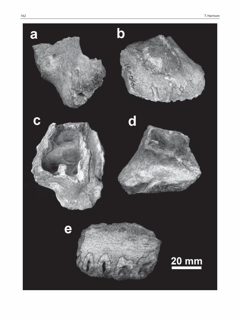
162 T. Harrison
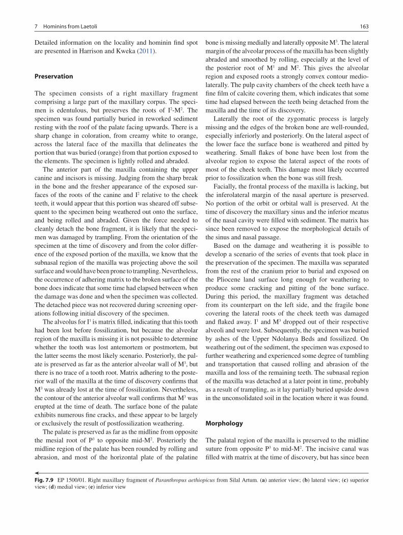
1637 Hominins from Laetoli
Detailed information on the locality and hominin find spot are presented in Harrison and Kweka (2011).
Preservation
The specimen consists of a right maxillary fragment comprising a large part of the maxillary corpus. The speci-men is edentulous, but preserves the roots of I2-M2. The specimen was found partially buried in reworked sediment resting with the roof of the palate facing upwards. There is a sharp change in coloration, from creamy white to orange, across the lateral face of the maxilla that delineates the portion that was buried (orange) from that portion exposed to the elements. The specimen is lightly rolled and abraded.
The anterior part of the maxilla containing the upper canine and incisors is missing. Judging from the sharp break in the bone and the fresher appearance of the exposed sur-faces of the roots of the canine and I2 relative to the cheek teeth, it would appear that this portion was sheared off subse-quent to the specimen being weathered out onto the surface, and being rolled and abraded. Given the force needed to cleanly detach the bone fragment, it is likely that the speci-men was damaged by trampling. From the orientation of the specimen at the time of discovery and from the color differ-ence of the exposed portion of the maxilla, we know that the subnasal region of the maxilla was projecting above the soil surface and would have been prone to trampling. Nevertheless, the occurrence of adhering matrix to the broken surface of the bone does indicate that some time had elapsed between when the damage was done and when the specimen was collected. The detached piece was not recovered during screening oper-ations following initial discovery of the specimen.
The alveolus for I1 is matrix filled, indicating that this tooth had been lost before fossilization, but because the alveolar region of the maxilla is missing it is not possible to determine whether the tooth was lost antemortem or postmortem, but the latter seems the most likely scenario. Posteriorly, the pal-ate is preserved as far as the anterior alveolar wall of M3, but there is no trace of a tooth root. Matrix adhering to the poste-rior wall of the maxilla at the time of discovery confirms that M3 was already lost at the time of fossilization. Nevertheless, the contour of the anterior alveolar wall confirms that M3 was erupted at the time of death. The surface bone of the palate exhibits numerous fine cracks, and these appear to be largely or exclusively the result of postfossilization weathering.
The palate is preserved as far as the midline from opposite the mesial root of P3 to opposite mid-M2. Posteriorly the midline region of the palate has been rounded by rolling and abrasion, and most of the horizontal plate of the palatine
bone is missing medially and laterally opposite M3. The lateral margin of the alveolar process of the maxilla has been slightly abraded and smoothed by rolling, especially at the level of the posterior root of M1 and M2. This gives the alveolar region and exposed roots a strongly convex contour medio-laterally. The pulp cavity chambers of the cheek teeth have a fine film of calcite covering them, which indicates that some time had elapsed between the teeth being detached from the maxilla and the time of its discovery.
Laterally the root of the zygomatic process is largely missing and the edges of the broken bone are well-rounded, especially inferiorly and posteriorly. On the lateral aspect of the lower face the surface bone is weathered and pitted by weathering. Small flakes of bone have been lost from the alveolar region to expose the lateral aspect of the roots of most of the cheek teeth. This damage most likely occurred prior to fossilization when the bone was still fresh.
Facially, the frontal process of the maxilla is lacking, but the inferolateral margin of the nasal aperture is preserved. No portion of the orbit or orbital wall is preserved. At the time of discovery the maxillary sinus and the inferior meatus of the nasal cavity were filled with sediment. The matrix has since been removed to expose the morphological details of the sinus and nasal passage.
Based on the damage and weathering it is possible to develop a scenario of the series of events that took place in the preservation of the specimen. The maxilla was separated from the rest of the cranium prior to burial and exposed on the Pliocene land surface long enough for weathering to produce some cracking and pitting of the bone surface. During this period, the maxillary fragment was detached from its counterpart on the left side, and the fragile bone covering the lateral roots of the cheek teeth was damaged and flaked away. I1 and M3 dropped out of their respective alveoli and were lost. Subsequently, the specimen was buried by ashes of the Upper Ndolanya Beds and fossilized. On weathering out of the sediment, the specimen was exposed to further weathering and experienced some degree of tumbling and transportation that caused rolling and abrasion of the maxilla and loss of the remaining teeth. The subnasal region of the maxilla was detached at a later point in time, probably as a result of trampling, as it lay partially buried upside down in the unconsolidated soil in the location where it was found.
Morphology
The palatal region of the maxilla is preserved to the midline suture from opposite P3 to mid-M2. The incisive canal was filled with matrix at the time of discovery, but has since been
Fig. 7.9 EP 1500/01. Right maxillary fragment of Paranthropus aethiopicus from Silal Artum. (a) anterior view; (b) lateral view; (c) superior view; (d) medial view; (e) inferior view

164 T. Harrison
cleaned. Just posterior to the anterior broken edge of the maxilla, opposite the posterior margin of the canine root, is a shallow groove that represents the vestibule of the incisive foramen. This groove passes posteriorly for a distance of 11.2 mm, where it disappears beneath the roof of the palate to enter the incisive canal transversely opposite the mesial margin of P4. The groove deepens as it passes posteriorly, and is angled slightly (~6°) towards the midline. The canal itself forms a bilaterally paired structure, partially separated by a raised keel that passes along the midline of the floor of the nasal canal and the midline of the floor of the inferior surface of the clivus. The dorsal surface or roof of the canal has a low and fine keel that runs posteriorly midway along its length. In section, the palatal aperture of the incisive canal is elliptical in shape with a dorsoventral height of 3.1 mm and a breadth to the midline of 4.1 mm. The vestibule and canal combined have a minimum anteroposterior length of 21.4 mm, measured along the midline of the clivus. In supe-rior view, the canal is funnel shaped, becoming wider as it passes posteriorly, and it eventually opens into a broad and shallow groove running along the floor of the nasal passage. The incisive canal is directed posteriorly at a 30° angle rela-tive to the roof of the palate. In medial view the nasoalveolar clivus overlaps the hard palate (see McCollum 1997).
The palate is relatively wide, with a slight increase in breadth posteriorly. The estimated breadth of the palate at mid-P3, mea-sured externally to include the alveolar process, is 66.2 mm, whereas at mid-M2 it is 81.4 mm. The palatal breadth between the roots can be estimated to have been 32.6 mm between the mid-P3s and 44.6 mm the between mid-M2s. The depth of the palate is difficult to measure with precision because it is not a simple task to orient the maxilla in the correct mediolateral plane. However, by aligning the alveolar process and the mid-line axis with the parasagittal plane, it is possible to approxi-mate the correct anatomical position. The palate is flat and relatively shallow, with a depth of only 15 mm in the midline at P3/P4 and appears to retain a constant depth posteriorly. The palate is bordered laterally by the sloping internal wall of the alveolar process, which increases in steepness posteriorly.
Beginning anteriorly opposite mid-M1 is a shallow groove for the greater palatine vessels. Posteriorly opposite M2 this develops into a pair of grooves, 3.8 mm wide, separated by a low keel. The keel terminates at mid-M2 to become a shallow single groove after which it begins to ascend more steeply as it approaches the entrance of the greater palatine canal. No portion of the greater palatine foramen is preserved, but it was presumably located opposite M3. A section of the pala-tomaxillary suture is visible on the palate originating ~11 mm lateral to the midline opposite anterior M2. It passes laterally and slightly posteriorly to terminate at the lateral margin of the groove for the greater palatine vessels. The point at which the palatomaxillary suture intercepts the intermaxillary suture in the midline is not preserved.
The sheared-off subnasal clivus exposes the alveolus for I1 and roots of the right I2 and upper canine. The alveolus for I1 is filled with matrix, implying that the tooth was lost peri-mortem prior to fossilization. Since the clivus and the roots of the canine and incisors were broken obliquely, the cross-sectional area of the roots are greater than they would be if they were broken transversely. Nevertheless, it is possible to compare the size of the roots of the two incisors and to esti-mate the original diameter of the canine root at the point of breakage. The size of the incisor roots/alveoli indicates that I1 and I2 were subequal in size, and relatively small compared to the size of the roots of the cheek teeth. The apico-basal orientation of the roots and the configuration of the subnasal region indicate that the incisors were procumbently implanted in the premaxilla. The intercanine distance is estimated to be ~29 mm, while the external breadth of the palate at the level of the canine roots can be estimated to have been ~50 mm. The I1 alveolus shows that the root was ovoid in cross-section with a buccolingual diameter much greater than the mesi-odistal diameter. A portion of the I2 root is retained, but it appears to be broken close to the apex of the root. The I2 and canine root are separated by a distance of only 1.6 mm, indi-cating that there was no diastema.
The canine root is pyriform in section, narrowing mesiobuccally, and with a concavity on the mesiolingual side. The dimensions of the broken canine root are 11.8 mm (mesiodistal length) and 14.1 (buccolingual breadth), but these are maximal dimensions because of the obliquity of the break. By taking the measurements orthogonal to the long-axis of the root it is possible to estimate that the root was ~10 mm long and ~13 mm wide. This indicates a large canine root, and presumably a large crown, in absolute terms, but one that is relatively small in terms of the overall size of the palate and the roots of the cheek teeth. The lateral margin of the canine root is set medial to the lateral margin of P3, so the latter tooth forms the anterolateral corner of the rectangular palate. The root of the canine is procumbently implanted at an angle of about 45° to the anteroposterior plane of the pal-ate. The root appears to be relatively straight, with only a slight degree of apicobasal curvature.
None of the crowns of the cheek teeth is preserved, but the roots of P3-M2 are exposed in the alveolar process. The roots indicate that the cheek teeth were aligned in relatively straight line that diverged slightly posteriorly. The length of the P3-M2 chord, based on the roots alone, is 49.3 mm, although with the reconstructed length of the crowns it can be esti-mated to have been ~51 mm. This implies that the cheek teeth were absolutely very large.
The premolar roots are extremely broad suggesting that the anterior cheek teeth were massive. The section of the roots for P4 is slightly larger than that for P3. The bases of the roots are ovoid in outline with single pulp chambers, but these divide into buccal and lingual canals that enter separate

1657 Hominins from Laetoli
roots. The buccal root of P3 is prominent, causing the lateral wall of the maxilla to bow outwards slightly, and it curves slightly medially to converge with the canine root. The buccal root in P4 is partially visible on the lateral side of the maxilla where a small flake of bone has been lost from the lower face. The exposed root is angled slightly anteriorly relative to the midline transverse axis of the pulp cavity. From the external morphology it is likely that the buccal roots of P3 and P4 are paired, but this will need to be confirmed with x-ray or CT scanning methods.
The pulp cavities of M1 and M2 are relatively large. Each exposes three canals, which lead to separate roots. The lateral roots are partially exposed. The mesiobuccal root penetrates the alveolar process of the maxilla almost vertically, while the distobuccal root is angled posteriorly with a slight distal curvature. The lingual root is single, and judging from the base of the root, the medial face is strongly grooved to produce a bilobate cross-section. Posterior to M2 is a short section of the lamina of bone that separates it from the alveo-lus of M3. The anterior face of the M3 alveolus has a sharp inferior border, indicating that the last molar was erupted and in place at the time of death. Apart from concluding that the specimen belonged to an adult individual, no other particu-lars of its age can be given. Judging from the size of the canine root and from the overall robusticity of the maxillary fragment it is possible to infer that the specimen belonged to a male individual.
Anteroposteriorly, the lateral alveolar wall forms a straight line, rather than bowing laterally around the molar row, and diverges slightly posteriorly (5°) relative to the midline axis of the maxilla. This indicates that the palate was rectangular, rather than forming a laterally bowing palate as seen in A. afarensis. The lateral surface of the maxilla in the alveolar region of the molars is relatively vertical. There is a slight protuberance of the alveolar region anteriorly to accommo-date the buccal root of the P3. Posterior to this is a small, shallow fossa located above and posterior to the region of P4. Anterior to the premolars, the alveolar region of the maxilla curves sharply around the root of the canine. There is no dis-tinct anterior pillar or canine jugum associated with the canine root, just a low rounded eminence. There is a very shallow canine fossa (i.e., the maxillary fossula) between the area of the maxilla filled by the canine root and the zygoma-ticoalveolar crest (Rak 1983, 1985). The curvature of the maxilla around the canine root implies a sharp demarcation between the lateral and anterior regions of the face, and a relatively abbreviated muzzle anteriorly.
The facial region anterior to the zygomatic process and bordering the nasal aperture is very slightly concave medio-laterally, to produce a dished mid-facial region (see Rak 1983, 1985). The lower face slopes steeply down from the infraorbital region, at an angle of 46° to the palatal plane (i.e., the nasocanine angle), to the horizontal level of the
middle of the nasal aperture, after which it descends to the incisal region slightly less steeply, at an angle of 38° (i.e., the nasoalveolar angle). Although much of the region inferior to the nasal aperture is not preserved, the configuration of the face lateral to the nasal aperture suggests that subnasal prog-nathism was pronounced. The minimum dorsoventral thick-ness of the broken surface of the subnasal alveolar region at I1 is 12.2 mm in EP 1500/01. A similar thickness occurs at a distance of 10.6 mm posterior to prosthion in KNM-WT 17000, so we can estimate that at least 10 mm is missing from the front of the palate in EP 1500/01. If this is the case, then the anteroposterior length of the subnasal clivus (pros-thion-nasospinale) can be estimated to have been ~37 mm.
The root of the zygomatic process is very thick anteropos-teriorly, with a broad and smoothly rounded inferior margin. The anterior margin of the zygomatic process is positioned above mid-P4 and the mid-point of the process is positioned above mid-M1. It has a minimum height above the alveolar margin of ~15 mm. The anterior face of the zygomatic pro-cess is relatively flat to slightly concave mediolaterally, and flares laterally at an angle of 80° to the long-axis of the lateral margin of the alveolar process in inferior view. The low rounded zygomaticoalveolar crest descends the lower face obliquely from the anteroinferior margin of the zygomatic process to terminate just posterior to the P3 root. This forms the anterior margin of the shallow depression in the maxilla above P4, and demarcates the lateral face of the maxilla from the anterior face. In anterior view, the zygomaticoalveolar crest is weakly arched.
Superior to the infraorbital foramen the maxilla is slightly mediolaterally concave, but this transitions to a slightly con-vex surface as it approaches the nasal aperture. This convex surface between the infraorbital foramen and the nasal aper-ture extends inferiorly to become continuous with the eminence for the canine root, but it does not form a distinct anterior pillar. The center of the infraorbital foramen is located 31.5 mm from the midline in the mediolateral plane and 17.6 mm from the margin of the nasal aperture, opposite the aperture’s greatest width. The inferior margin of the fora-men is horizontally in line with the anterior nasal spine. It is positioned dorsoventrally above P4 at a height of 35.9 mm above the alveolar margin. The distance between the center of the foramen and the inferior margin of the zygomatic pro-cess is 17.2 mm. The foramen is situated quite low on the face, midway between the nasal aperture and zygomatic process. The infraorbital foramen is circular in outline, with a mediolateral breadth of 4.3 mm. The foramen opens inferi-orly and slightly laterally into a distinct teardrop shaped depression.
Unfortunately, no portion of the inferior orbital margin is preserved, so the lower facial height cannot be measured or estimated, but based on the preserved anatomy the lower face was evidently relatively deep. The minimum height of the

166 T. Harrison
lower face above P3 is 48.8 mm to the point where the maxilla is broken superiorly.
Although only a portion of the nasal aperture is preserved, it was clearly pyriform in outline, being broadest inferiorly and narrowing superiorly. The lateral margin of the nasal aperture is smoothly rounded inferiorly, but a low crest is developed about 12.3 mm up from the floor of the nasal aper-ture about 3.9 mm below the point at which the maxillary fragment is broken superiorly. The greatest breadth of the nasal aperture, at the point where the lateral crest originates, is estimated to be ~28 mm. Laterally, the margin of the nasal aperture is bordered by the canine root, with the root of the lateral incisor situated more medially. The inferior margin of the nasal aperture curves gently medially and inferiorly to reach its most inferior point midway between the greatest lateral extent and the midline, and then it ascends ~3 mm to reach the elevated anterior nasal spine, which is located in the midline and somewhat recessed. A low, rounded crest passes laterally and slightly anteriorly from the anterior nasal spine to demarcate the junction between the clivus and the floor of the nasal cavity. The anterior nasal spine is eroded, but was evidently quite prominent.
The superior margin of the subnasal clivus is damaged. The bone covering the region between the apices of the upper central incisors is largely missing. During removal of the adhering sediment in this region, the cleaning process exposed a small elliptical and smooth-walled pocket of pneu-matized bone (15.4 mm 8.2 mm) that occupied the dorsal surface of the clivus on either side of the anterior nasal spine. Nevertheless, despite this damage, it is possible to infer that the subnasal clivus would have had a slightly convex dorsal margin anteroposteriorly that led smoothly into the floor of the nasal aperture without a distinct nasal sill (Robinson 1953; Ward and Kimbel 1983; McCollum et al. 1993; McCollum 1997). In anterior view, the inferior margin of the nasal aperture is mediolaterally concave, forming a nasoal-veolar gutter (Rak 1983, 1985; Strait and Grine 2004).
The floor of the nasal cavity has an elevated and mediolat-erally bladelike spine in the mid line. It is clear that the vomer inserted along this crest anteriorly as far as the anterior nasal spine (Robinson 1953; McCollum 1997, 1999). The lateral wall of the cavity, close to the superior break in the maxilla, bears a longitudinal crest that represents the root of the infe-rior concha. It slopes inferiorly as it passes posteriorly. Anteriorly it is positioned about 15.5 mm above the floor of the nasal canal, just posterior to the internal opening of the incisive canal, whereas posteriorly it is positioned 10.2 mm above the floor of the nasal cavity at the level of M2 where the posterior break occurs. The lateral wall of the inferior meatus below the line for the attachment of the inferior concha is generally concave, although there is a distinct swelling supe-rior and lateral to the internal opening of the incisive canal. The lateral wall of the nasal cavity separating the inferior
meatus from the maxillary sinus is remarkably thick, with a maximum thickness along the broken margin of 6.4 mm, but it does get as thin as 1.6 mm. The palatal process of the maxilla is also very thick (McCollum 1997, 1999), with a maximum dorsoventral thickness of 8.4 mm opposite M1.
The maxillary sinus is very extensive, extending laterally and anteriorly into the zygomatic process, posteriorly into the alveolar region beyond M2, and medially to border the nasal aperture. It does not penetrate the maxilla anterior to the zygomatic process in the region of the premolar roots, but it does extend anteromedially as far as the canine root. There is no evidence that it extended into the palatine pro-cess of the maxilla medially to form a recessus palatinus (Tobias 1967; McCollum 1997). The exposed floor of the sinus has a complex arrangement of buttresses and deep loculations between and around the roots of the molars. The buttresses primarily intersect each other at right angles, and the entire system is arranged so that the buttresses are oriented at ~45° to the mediolateral line of the zygomatic process. Presumably, the network of buttresses criss-crossing the floor of the sinus helps to maintain the structural integrity of the maxilla.
A major buttress, more than 5 mm wide, originates at the medial wall of the sinus opposite the level of M2, and passes obliquely laterally and anteriorly to meet the lateral wall at a sharp vertically-oriented buttress, which coincide on the external surface of the maxilla with the posterior margin of the zygomatic process. Intersecting this transverse buttress is a longitudinally-oriented buttress that passes anteriorly and medially at an angle of about 90° to the former. It passes towards the base of the pillar of bone that represents the lateral margin of the nasal aperture, where it divides into short medial and lateral arms. This main buttress gives rise to smaller subsidiary buttresses midway along its length that pass medially and laterally. Posterior to the point where the main transverse and longitudinal buttresses intersect, there is a pair of sharp and diverging buttresses that pass to the posterior wall of the sinus. Between the buttresses, the floor of the sinus is excavated to form relatively deep loculi. Centrally, the floor of the sinus bears a very deep loculus bordered laterally and posteriorly by the main longitudinal and transverse buttresses respectively, and medially by the wall of the sinus.
The anterior wall of the sinus bears the remnant of a bony canal that penetrates ~8 mm into the chamber of the sinus. This is the canal leading from the infraorbital foramen into the infraorbital groove that carries the infraorbital nerve and artery. The canal passes superiorly and slightly medially, which corresponds well with the orientation of the infraor-bital foramen aperture on the facial aspect of the maxilla. Medial to the canal is a well-developed loculus that pene-trates into the pillar that forms the lateral margin of the nasal aperture, just posterior to the canine root.

1677 Hominins from Laetoli
Comparisons
Comparisons with the crania of other Pliocene hominins indicate that EP 1500/01 exhibits a suite of derived features that it shares uniquely with Paranthropus spp. from southern and eastern Africa. These include: relatively small anterior teeth; enlarged premolars and molars; a dished midface; robust and anteriorly placed zygomatic process of the maxilla; zygomaticoalveolar crest weakly arched in anterior view; single infraorbital foramen positioned low on the face and opening inferiorly into a shallow groove; guttered nasoal-veolar clivus that grades smoothly into the nasal cavity floor; marked overlap between the nasoalveolar clivus and the anterior margin of the hard palate; anterior vomer insertion that coincides with the anterior nasal spine; recessed anterior nasal spine; and very thick hard palate anteriorly (Rak 1983, 1985; Leakey and Walker 1988; Kimbel et al. 1988; McCollum et al. 1993; Strait et al. 1997; Suwa et al. 1997; McCollum 1997, 1999; Asfaw et al. 1999; Keyser 2000; Strait and Grine 2001, 2004; Steininger et al. 2008).
Further comparisons show that EP 1500/01 is very similar in morphology to KNM-WT 17000 (Walker et al. 1986; Leakey and Walker 1988), and there can be no doubt that the maxilla fragment from the Upper Ndolanya Beds should be attributed to the same species, Paranthropus aethiopicus. EP 1500/01 shares the following distinctive (i.e., primitive) features with KNM-WT 17000 that are not found in P. robus-tus or P. boisei: larger anterior teeth, relatively flat and shal-low palate, and pronounced subnasal prognathism.
EP 1500/01 and KNM-WT 17000 are very similar. The nasal aperture and subnasal region appear to have been very similar in shape and configuration. In both specimens the nasal aperture is pear-shaped in outline, relatively broad low down (although the greatest breadth is somewhat higher in EP 1500/01), with a slightly concave inferior margin medio-laterally (i.e., a guttered nasoalveolar region) and a promi-nent anterior nasal spine. The maximum breadth of the nasal aperture is estimated to be ~28 mm in EP 1500/01 compared with 27.3 mm in KNM-WT 17000 (Leakey and Walker 1988). The inferolateral margin of the nasal aperture is rounded in both specimens, in contrast to the sharp lateral margin in A. afarensis. The crest that forms the lateral mar-gin of the nasal aperture superiorly originates lower in KNM-WT 17000. The estimated depth of the subnasal clivus in EP 1500/01 is comparable to that in KNM-WT 17000, and in both specimens it is sagittally convex and grades smoothly into the nasal passage without a distinct nasal sill. In A. afa-rensis the clivus has a stepped nasal cavity floor with a strongly differentiated nasal sill (Kimbel et al. 2004).
The midface in EP 1500/01 is similar to that of KNM-WT 17000 in lacking an anterior pillar, having a robust zygo-matic process that is placed anteriorly relatively to the cheek teeth and being slightly dished. Mediolateral concavity of
the midface is relatively slight in EP 1500/01 and more pronounced in KNM-WT 17000. The lateral margin of the nasal aperture is bordered by a low rounded eminence in EP 1500/01, whereas in KNM-WT 17000 this region is rela-tively flat. Both lack canine juga. The I2 root in EP 1500/01 and KNM-WT 17000 is positioned medial to the lateral margin of the nasal aperture, as in other species of Paranthropus and A. africanus, but distinct from A. afarensis
In lateral view, the contour of the lower face is very simi-lar in KNM-WT 17000 and EP 1500/00. Both specimens appear to have strong subnasal prognathism. As described above, the lower facial and subnasal angles, relative to the anteroposterior plane of the palate, are 46° and 38° respec-tively in EP 1500/01, compared with 42° and 30° in KNM-WT 17000. The lower face in P. aethiopicus exhibits a similar overall angulation to that of P. boisei, except that the subnasal region is much more prognathic. The angulation of the lower face of A. afarensis is less steep (i.e., more prognathic), but the degree of facial protrusion is much more pronounced in the subnasal region in P. aethiopicus giving the latter taxon a greater degree of prognathism overall (Kimbel et al. 2004).
The zygomatic process of the maxilla originates at approximately the same elevation in both specimens, with its lowest point vertically above mesial M1. The infraorbital foramina in KNM-WT 17000 are incompletely preserved, but they appear to have been located in a similar position to that in EP 1500/01 (the foramen is positioned slightly higher and more lateral relative to the nasal aperture in KNM-WT 17000). The foramen in KNM-WT 17000 is similar to EP 1500/01 in opening inferiorly into a shallow groove (Leakey and Walker 1988).
The maxillary sinus in KNM-WT 17000 is infilled with matrix, so comparison with the sinus morphology in EP 1500/01 is not possible (Walker et al. 1986; Leakey and Walker 1988). Nevertheless, it is apparent that the sinus extended laterally into the zygomatic process and anterome-dially into the lateral rim of the nasal aperture posterior to the canine alveolus (Leakey and Walker 1988). Leakey and Walker (1988) indicate that the sinus in KNM-WT 17000 extends medially into the palatine process of the maxilla, in contrast to EP 1500/01, which lacks a recessus palatinus. A portion of the floor of the maxillary sinus is exposed above M3 in KNM-WT 17000, and this confirms that, as in EP 1500/01, the maxillary sinus had a relatively thick bony floor above the molar roots. This contrasts with the maxillary sinus of A. afarensis seen in A.L. 200-1, which has a much thinner sinus floor. In addition, the medial wall of the maxilla separating the maxillary sinus from the nasal passage is rela-tively thick in P. aethiopicus, in contrast to the thin-walled sinus seen in A. afarensis.
The palate in both specimens is relatively broad and very shallow (Leakey and Walker 1988; Suwa 1989). The breadth of the palate between the roots of P4 and M2 in KNM-WT

168 T. Harrison
17000 is 41.3 and 35.7 respectively. These dimensions compare with estimated values of 38.8 and 39.2 in EP 1500/01. The palate in both specimens is similar in overall dimensions to that of OH 5 (P. boisei), but the palate in this latter specimen is much deeper. The palate becomes slightly wider and the tooth rows diverge posteriorly in EP 1500/01, whereas in KNM-WT 17000 the palate narrows slightly pos-teriorly (although Leakey and Walker (1988) consider that this is likely an artifact of preservation).
The incisive canal has a similar configuration in EP 1500/01 and KNM-WT 17000. The canal exits onto the roof of the palate opposite mid-P4 and opens anteriorly into a fan-shaped fossa that terminates just posterior to the alveolus of I1. The structure of the nasal septum and vomeral insertion is nearly identical in both specimens.
The intercanine distance in KNM-WT 17000 is 36.0 mm, whereas the minimum estimated breadth is only 29.0 mm in EP 1500/01, implying that the upper incisors were probably larger in the former. By comparison, the intercanine distance in OH 5 is 31.7, implying that, just as in P. boisei, the inci-sors of P. aethiopicus were relatively small in relation to the size of the cheek teeth (Suwa 1989). A similar set of relation-ships emerges if the mediolateral breadth across the incisor roots is taken into account. In KNM-WT 17000 the breadth is 35.2 mm, compared with only 32.6 mm in OH 5, indicat-ing that P. aethiopicus has larger upper incisors than P. boisei (see Leakey and Walker 1988; Suwa 1989). However, the same dimension in EP 1500/01 is estimated to be only 30.2 mm, indicating that there was variability in incisor size in P. aethiopicus, and that incisor breadth overlapped with that in P. boisei.
The alveolus for the upper canine in KNM-WT 17000 measures 7.2 mm (mesiodistally) by 12.7 mm (buccolin-gually), which is smaller than the estimated dimensions of the canine root in EP 1500/01 (~10.0 mm by ~13.0 mm). Canine root size in KNM-WT 17000 is comparable to that in OH 5, which has a mesiodistal length of only 6.8 mm. The canine root in EP 1500/01 is much larger than in other Paranthropus specimens, and, in fact, exceeds the dimen-sions of the root of the largest canine of A. afarensis. A.L. 333x-3, a presumed male individual from Hadar, has basal root dimensions of 8.5 mm by 11.3 mm. Nevertheless, compared to the estimated size of the cheek teeth and the maxilla, the canine in EP 1500/01 would have been relatively smaller than in A. afarensis. It would seem that the anterior teeth were somewhat larger on average than in P. boisei, but as in the latter species they were small by comparison with the size of the premolars and molars. In addition, the canine roots in P. aethiopicus are more procumbently implanted and less apicobasally curved than in A. afarensis and P. boisei, which have shallower and less prognathic subnasal regions.
Measurements of the P3-P4 chord and P3-M2 chord of the roots provide an estimation of the relative size of the cheek
teeth. In KNM-WT 17000 these dimensions are 20.3 mm and 50.0 mm respectively, compared with 19.0 mm and 49.3 mm in EP 1500/01, indicating that that the teeth might have been slightly smaller in the latter. The same dimensions in OH 5 (P. boisei) are 19.6 mm and 50.3 mm respectively. Evidently, the cheek teeth in P. aethiopicus were as large (at least in their mesiodistal dimensions) as in P. boisei. Based on the roots, the cheek teeth in EP 1500/01 and KNM-WT 17000 formed a relatively straight line, unlike in A. afarensis where the cheek tooth rows bow laterally, being widest at the level of M2.
EP 1000/98
Left proximal tibia (Fig. 7.10).
Location and Stratigraphic Provenance
The specimen was discovered by Chris Robinson on August 19, 1998 at Loc. 22 South. The specimen was found exposed on the surface at the base of Nenguruk Hill, a small hillock on the western side of the Olaitole River valley. The location of the find, as well as the lithology of the adhering matrix and the coloration of the specimen, confirms that it was origi-nally derived from the Upper Ndolanya Beds. Additional details about the locality and provenance are presented by Harrison and Kweka (2011).
Preservation
The specimen consists of a left proximal tibia. Only a short section of the shaft is preserved, with a total proximodistal length of ~58 mm. The proximal articular surface is gener-ally well preserved, but there has been some abrasion and weathering along the anteromedial and posterolateral margins. There are small depressions on the shaft anterior to the medial facet that look superficially like they may have been caused by bite marks from a small carnivore, but closer microscopic examination shows that they are shallow eroded pits caused by weathering. The posterior intercondylar depression, for the attachment of the medial meniscus and the posterior cruciate ligament, has been accentuated by erosion and by loss of the bone surface. The medial and lateral articular surfaces exhibit fine cracks around their perimeters, apparently caused by weathering when the bone was still green. Similarly, the heavy longitudinal cracks along the shaft, and the spiral and angular fractures at the broken end of the shaft, imply that the proximal end of the tibia was detached from the rest of the bone while it was still green and prior to it being buried and fossilized.

1697 Hominins from Laetoli
Morphology
The proximal end of the tibia is broad. The mediolateral breadth is 51.6 and the anteroposterior length is 33.8, with a breadth-length index of 65.5. The condyles are anteroposte-riorly short in relation to their breadth. The medial condyle forms a D-shaped facet, with a central depression and a low rounded rim. The lateral condyle is elliptical in shape, with a slightly anteroposteriorly convex lateral surface and a more concave medial surface. The posteromedial margin of the lateral condyle is slightly elevated. Both condyles are medio-laterally concave, but the lateral condyle is more markedly so. The medial condyle is anteroposteriorly longer than the lateral condyle, but mediolaterally narrower. The medial
condyle measures 33.0 mm 20.6 mm; the lateral condyle measures 26.6 mm 22.1 mm. The surface area of the medial condyle is subequal in size to that of the lateral condyle (375 mm2 compared with 373 mm2). The long-axes of the two condyles converge posteriorly.
Between the two condyles is a well developed and elevated intercondylar eminence, with prominent medial and lateral tubercles. The maximum height of the medial tubercle is 5.8 mm. The lateral tubercle is slightly lower. The emi-nence is located midway across the proximal end of the tibia in the parasagittal plane and slightly posterior of the mid-coronal plane. Laterally, the articular surface of the medial condyle extends almost to the apex of the medial tubercle. In superior view, the medial tubercle forms an oblique crest that
Fig. 7.10 EP 1000/98. Left proximal tibia of cf. Paranthropus aethiopicus from Loc. 22S. (a) anterior view; (b) posterior view; (c) lateral view; (d) superior view; (e) medial view

170 T. Harrison
continues anteromedially to become the raised lateral margin of the medial condyle. The lateral tubercle forms a robust crescent-shaped ridge that extends anteriorly and posteriorly to form the raised medial margin of the lateral condyle. The two tubercles are united by an elevated transverse crest, directed slightly posterolaterally.
Anterior to the intercondylar eminence is a roughened triangular area, the anterior intercondylar area, bordered by the raised rims of the medial and lateral condyles. Within this area, there is a large triangular region that borders the medial condyle, defined anteriorly by a sharp crest that runs along the anterior margin and laterally by low swellings. This is the area for the anterior cornu of the medial meniscus. Lateral to this is a roughened area, perforated by numerous vascular foramina, that represents the site of the corpus adiposum infrapatellare and the attachment for the lateral meniscus. Posterior to these two areas, on the anterior slope of the intercondylar eminence, is a smooth triangular region for the attachment of the anterior cruciate ligament. The posterior intercondylar area has been damaged by erosion, so details of its morphology cannot be discerned. It is not possible, for example, to determine the shape of the postero-medial contour of the lateral condyle or whether or not the lateral meniscus had a posterior attachment. However, the area posterior to the lateral tubercle was evidently quite restricted implying that EP 1000/98 lacked a posterior cornu of the lateral meniscus.
Posterior retroversion of the head is minimal. The articu-lar platform is only very slightly posteriorly tilted in medial view in relation to the long-axis of the shaft. The medial con-dyle is tilted 4°, whereas the plane of the lateral condyle is tilted only 1°. The lack of retroflexion of the head is structur-ally associated with a weak overhang of the condyles poste-riorly, a steep anterior margin, and a less prominent tibial tuberosity.
Only a short section of the shaft is preserved. It is medio-laterally slightly compressed and triangular in section. The anteroposterior diameter of the shaft at the inferior margin of the tibial tuberosity is at least 21.2 mm, while its mediolat-eral breadth at this level is 20.2 mm. In anterior view, the shaft below the medial condyle is more robustly built than that below the lateral condyle, which is more hollowed out (Aiello and Dean 1990). This hollowing of the lateral side of the shaft may be more closely tied to the structural and allo-metric relationships between the size of the tibial plateau and the mediolateral diameter of the shaft, than to the develop-ment and disposition of the tibialis posterior muscle (contra Aiello and Dean 1990; Berger and Tobias 1996). The medio-lateral breadth of the shaft at the level of the inferior margin of the tibial tuberosity is only 39.9% that of the total breadth of the tibial plateau. The remaining portion of the shaft appears to have a long-axis that is approximately in line with the sagittal midline of the articular surface (measurement of the
midline of the shaft to the margin of the lateral condyle in the mediolateral plane is 24.3 mm or ~47% of the total breadth of the proximal end of the tibia), and it exhibits no discern-able degree of torsion.
The anterior face of the shaft bears a low and domed tuberosity for the attachment of the patella ligament. Its superior margin is situated 22.2 mm inferior to the margin of the anterior intercondylar area. The tuberosity projects ante-riorly very little, so that the anterior margin of the tibial shaft descends steeply from the articular platform. Superior to the tuberosity is a slightly rugose tuberosity bordering the ante-rior margin of the articular surface, which is separated from the tibial tuberosity by a shallow transverse groove. This is bordered by a faint line medially and a stronger crest later-ally, marking the extent of the attachment of the capsule. Lateral to the capsular attachment, and immediately anterior and inferior to the lateral condyle, is a raised, elliptical and slightly convex area that marks the site of the iliotibial tract.
On the medial side of the tibial tuberosity the anterior face of the shaft is convex and largely featureless. Numerous vascular foramina are located superiorly, just below to the medial condyle. On the medial side of the shaft, just where the break occurs, there is a small, roughened tuberosity, presumably for the attachment of the tibial collateral ligament. Unfortunately, the site of attachment for the pes anserinus (i.e., the insertion for the sartorius, gracilis and semitendi-nosus) is not preserved.
On the lateral side of the proximal tibia is a roughened area, which arcs postero-superiorly to antero-inferiorly, for the attachments of the tendon of the biceps femoris and the extensor digitorum longus. Inferior to this area, on the lateral side of the tibial tuberosity is a shallow groove, with a maxi-mum width of 8.6 mm, running proximodistally along the shaft. This is the site of attachment for tibialis anterior. A low rounded crest extending down the middle of the lateral side represents the interosseus border. Posterior to this is the area of attachment for the tibialis posterior. The area for tibialis posterior on the lateral side of the shaft is subequal to that of tibialis anterior.
Just below the posteromedial lip of the medial condyle is a broad and rugose groove, bordered inferiorly by an irregu-lar crest, which terminates medially in a shallow circular depression. The latter marks the insertion for semimembra-nosus, which appears to have been well developed. The proximal fibular facet is lanceolate, with a long-axis directed antero-laterally. It is relatively large, measuring 16.3 mm by 11.1 mm. It is well-defined, slightly convex anteroposteri-orly and mostly flat mediolaterally. The facet faces distally, posteriorly, and slightly laterally. The fibular facet is located 10.1 mm from the posterolateral margin of the lateral con-dyle. On the posterior surface, immediately medial to the fibular facet is a deep oval depression that extends a short way down the shaft. It is bordered by a raised, convex area

1717 Hominins from Laetoli
medially for the insertion of popliteus, and by a low rounded and ill-defined crest laterally that descends from the fibular facet on the lateral side to at least mid-shaft. The latter crest delimits the posterior border of tibialis posterior and repre-sents the proximal extent of the soleus line.
Comparisons and Functional Implications
Compared with modern humans, EP 1000/98 differs in the following respects: the proximal articular surfaces are antero-posteriorly slightly shorter relative to the breadth (with a breadth-length index of 65.5 in EP 1000/98 compared to a mean value in modern humans of 68.0; see Table 7.12); the articular surface of the medial condyle is more concave; the lateral condyle is slightly convex (generally relatively flat to slightly concave in modern humans, although some humans exhibit a slight degree of convexity) (Tardieu 1982, 1983; Berger and Tobias 1996; Organ and Ward 2006); the intercon-dylar eminence is more elevated, with a stronger transverse crest linking the medial and lateral tubercles; the tibial tuber-osity is much less protuberant; the groove superior to the tibial tuberosity is oblique, rather than transversely oriented; the area for the iliotibial tract has greater relief; the area on the lateral side of the tibial tuberosity for attachment of the tibialis anterior is relatively less extensive, and the interosseus line is more anteriorly located; the proximal fibular facet is relatively larger; the area of attachment for the semimembra-nosus is better developed and forms a distinct circular depres-sion on the posteromedial margin of the condyle; the depression on the posterior aspect of the shaft medial to the fibular facet is deeper; the lateral meniscus has a single tibial insertion in the anterior intercondylar area (humans are unique among primates in having a double insertion, one for each of the anterior and posterior intercondylar areas respectively) (Senut and Tardieu 1985; Tardieu 1986a, b, 1999; Le Minor
1990; but see Dugan and Holliday 2009); the cross-section of the shaft at the level of the inferior margin of the tibial tuber-osity is less compressed mediolaterally (the breadth-length of the cross-section of the shaft at this level is 95.3 in EP 1000/98, compared with a mean value in modern humans of 75.1; see Table 7.12); and the lateral side of the shaft below the lateral condyle is more deeply hollowed.
In addition, EP 1000/98 possesses a weak crest that passes obliquely across the posterior aspect of the shaft, which corresponds to the soleal line in humans. A similar crest is commonly found in the tibiae of chimpanzees, although according to Aiello and Dean (1990) this line does not correspond to the soleal line of humans, because the soleus is normally confined to the fibula in African apes and the crest merely represents the posterior border of the tibialis posterior. However, Gregory (1950) demonstrated that soleus does attach to this crest in Gorilla. Given that a similar crest occurs in humans and chimpanzees, it is not possible to determine whether or not there is a soleus attachment in EP 1000/98 or in other fossil tibiae.
Compared with chimpanzees, EP 1000/98 differs in having: a less convex lateral condyle; a relatively more elevated intercondylar eminence; a much less retroverted tibial plateau (in chimpanzees, the average posterior tilt of the medial and lateral condyles relative to the proximodistal long axis of the proximal end of the shaft is 22° and 10° respectively, compared with 4° and 1° in EP 1000/98; see Table 7.12), with less overhang of the condyles posteriorly; the medial and lateral condyles have a similar orientation in the mediolateral plane, whereas in chimpanzees the lateral condyle is more obliquely oriented relative to the transverse plane of the medial condyle; the condyles are proximodis-tally relatively thicker; the tibial tuberosity is much less protuberant; the area for the iliotibial tract is larger; the proximal fibular facet is relatively smaller; the interosseus line on the lateral side of the shaft is more posteriorly placed;
Table 7.12 Comparison of dimensions (mm) of proximal tibia from Laetoli (EP 1000/98)A. africanusa A. afarensis A. anamensis Pan troglodytes Homo sapiens
DimensionsaEP 1000/98 Stw 514a AL 288–1aq AL 129–1b
KNM-KP 29285A N Mean Range N Mean Range
AP l prox tibia 33.8 – 32.9 33.3 49.7 10 40.8 39.3–42.6 16 49.4 41.6–53.7ML br prox tibia 51.6 52.3 50.8 50.6 67.5 10 60.0 55.2–66.2 16 72.6 63.3–80.2AP l med condyle 33.0 – 31.6 – 43 (e) 10 38.0 36.0–42.6 16 45.6 36.2–51.1ML br med condyle 20.6 20.5 20.6 – 30 (e) 10 26.6 25.3–28.2 16 31.1 26.0–38.3AP l lat condyle 26.6 20 (e) 24.8 27.4 – 10 32.6 29.3–37.5 15 38.5 29.2–44.0ML br lat condyle 22.1 16.8 20.7 22.4 – 10 26.0 23.8–29.3 15 30.7 25.8–33.9Post slope med condyle 4° – 9° 14° 17° 10 22° 5–30° 14 9° 3–20°Post slope lat condyle 1° – 9° 10° 7° 10 10° 3–20° 14 9° 0–16°AP l shaft 21.2(–) – 27.2 28.9 36.2 10 32.4 28.5–35.2 17 38.2 34.8–41.3ML br shaft 20.2 – 22.3 22.3 29.0 10 21.6 19.6–24.2 17 28.7 24.2–35.2AP anteroposterior, br breadth, lat lateral, l length, med medial, ML mediolateral, post posterior, prox proximal, (–) minimum dimension, (e) esti-mated dimensionaData from Berger and Tobias (1996)

172 T. Harrison
the insertion for semimembranosus is larger and better defined; and the proximal end of the shaft is anteroposteri-orly relatively shorter and lacks the marked anterior curva-ture typical of African apes. However, EP 1000/98 does share a number of features with the tibiae of chimpanzees that distinguish them from those of modern humans. These include: a more convex lateral condyle in the anteroposterior plane; a more elevated crest linking the tubercles of the inter-condylar eminence; a relatively small and triangular poste-rior intercondylar region, presumably lacking a posterior attachment for the lateral meniscus; a less protuberant tibial tuberosity; an oblique groove superior to the tibial tuberos-ity; and a more pronounced hollowing of the lateral aspect of the shaft below the lateral condyle.
The features that EP 1000/98 shares with modern humans compared with those of African apes, such as the less pro-nounced retroversion of the proximal tibial plateau (the pos-terior tilt of the medial and lateral condyles in the fossil falls at the lower end of the range for modern humans; see Hashemi et al. 2008), a more prominent iliotibial tract for insertion of the tensor fascia latae and gluteus maximus, and a more posterior attachment of tibialis posterior, indicate more extended positions of the knee associated with bipedal locomotion (Aiello and Dean 1990; Berger and Tobias 1996). In humans, individuals with a steeper posterior slope of the tibial plateau is associated with an increase in the magnitude of the anteriorly directed component of the compressive forces that act on the tibial articular surface during the intial phase of weight support (Hashemi et al. 2008). This produces greater anterior translation of the tibia and a higher force exerted by the anterior cruciate ligament. The functional shift to fully extended knees at heel strike in hominins, com-pared with the weight-bearing semiflexed knees in great apes, would favor the development of a less posteriorly tilted tibial plateau.
Nevertheless, a number of primitive features in EP 1000/98 indicate that the knee joint was capable of a greater range of rotation than seen in those of modern humans. These include a more convex lateral condyle in the anteroposterior plane, a relatively larger facet for the proximal fibula, and a single tibial attachment for the lateral meniscus. The hollow-ing of the lateral side of the shaft below the lateral condyle may imply that the knee was less effective than the modern human knee joint in dissipating high peak loads when the foot made contact with the ground. The relatively well-developed insertion for semimembranosus compared with modern humans, as well as the prominent development of the iliotibial tract, implies well-developed capabilities for extension of the thigh at the hip joint.
Comparisons with the proximal tibiae of other fossil hom-inins show that EP 1000/98 is very similar to those of A. afarensis from Hadar. It is comparable in size and closest in overall morphology to the small tibiae from Hadar, such as
A.L. 288-1aq and A.L. 129-1b, which presumably belonged to female individuals (Johanson and Taieb 1976; Johanson and Coppens 1976; Lovejoy et al. 1982; see Table 7.12). The larger proximal tibiae from Hadar (i.e., A.L. 333x-26 and A.L. 333-42) have linear dimensions that are on average ~29% larger than those of EP 1000/98, and they tend to be more robust, but otherwise they exhibit the same suite of dis-tinctive morphological features. Compared with EP 1000/98, AL 288-1aq and AL 129-1b are very slightly smaller in their proximodistal and anteroposterior dimensions, and the shaft is more bilaterally compressed (the breadth-length index of the cross-section of the shaft at the level of the inferior mar-gin of the tibial tuberosity is 77.2 in AL 288-1aq and 82.0 in AL 129-1b, compared with 95.3 in EP 1000/98; see Table 7.12). The different proportions of the shaft in EP 1000/98 are apparently due to the relatively short anteropos-terior dimension of the shaft. Just as in EP 1000/98, the lat-eral side of the shaft has a distinctive hollowing below the lateral condyle.
The morphology of the proximal articular surfaces in the Hadar specimens is identical to that in EP 1000/98, with equal-sized condyles, an anteroposteriorly convex lateral condyle (more marked in the Hadar specimens), similar con-figuration of the anterior intercondylar area, a relatively restricted posterior intercondylar area lacking a posterior cornu for the lateral meniscus, and a similarly developed intercondylar eminence. On the anterior face of the shaft, the tibiae from Hadar and EP 1000/98 share a relatively low tibial tuberosity (even more weakly expressed in EP 1000/98), an oblique groove on the bursal surface superior to the tibial tuberosity, and a raised surface for a well-developed iliotibial tract. Medially, there is a well-developed depression for the insertion of the semimembranosus. The scar for the pes anserinus, which is very well developed in the proximal tib-iae from Hadar, is not preserved in EP 1000/98. In the Hadar specimens, this scar forms a deep groove-like roughened area bordering the raised anterior crest of the shaft. This resembles the condition in African apes more than that in humans, where the attachments for sartorius, gracilis and semitendinosus are placed more laterally and leave a less dis-tinct scar. Laterally, the interosseus line extends down the midline of the shaft, and delimits a relative large area for the attachment of tibialis posterior and a distinctly grooved sur-face for the tibialis anterior. On the posterior surface of the shaft, there is a pronounced depression below the posterome-dial margin of the lateral condyle, bordered laterally by a low rounded crest for the proximal end of the soleal line and medially by a smoothly rounded surface for popliteus. The proximal facet for the fibula is only partially preserved in A.L. 288-1aq, but it does appear to have been relatively smaller than in EP 1000/98. The retroversion of the proximal end is more marked in the Hadar proximal tibiae than in EP 1000/98, and their posterior inclination exceeds or falls

1737 Hominins from Laetoli
within the upper end of the range of modern humans (Table 7.12). This latter feature is structurally associated with the more pronounced development of the tibial tuberos-ity and greater overhang of the condyles posteriorly in the Hadar specimens. The differences between the proximal tibiae from Hadar and Laetoli are relatively minor, however, and the overall morphology of EP 1000/98 is remarkably similar to the two small proximal tibiae of Australopithecus afarensis from Hadar.
The only known proximal tibia of Australopithecus ana-mensis (KNM-KP 29285A from Kanapoi) is much larger than EP 1000/98 (the dimensions of the proximal end are ~39% larger on average, being larger in size than the pre-sumed tibiae of male individuals of A. afarensis from Hadar; Table 7.12), but, like those of A. afarensis, its overall mor-phology corresponds closely to that seen in EP 1000/98 (Ward et al. 2001). Other than size, KNM-KP 29285A dif-fers in the following respects: the medial condyle has a slightly more undulating articular surface, being concave anteriorly and slightly convex posteriorly (whereas EP 1000/98 is anteroposteriorly more planar); the lateral rim of the medial condyle is more elevated; the intercondylar emi-nence is relatively larger and more robust; the anterior inter-condylar area is more deeply excavated; retroversion of the proximal tibia is more pronounced, with a posterior slope of the medial condyle of 17° and of the lateral condyle of 7°, and a more strongly developed overhang of the condyles posteriorly; the tibial tuberosity and the area superior to the tuberosity for attachment of the capsule are much more rugose; the anterior margin of the shaft is more strongly keeled; the shaft is more strongly bilaterally compressed (the breadth-length index of the cross-section of the shaft at the level of the inferior margin of the tibial tuberosity is 80.1, compared with 95.3 in EP 1000/98; see Table 7.12); the area on the medial side of the condyle that marks the attachment for semimembranosus is more strongly marked; the raised area for the iliotibial tract is larger and more pro-nounced; the interosseus line is slightly more posteriorly placed, and the area for tibialis anterior is more concave; the posterior face has a more pronounced soleal line. The majority of these differences simply relate to the larger size, greater robusticity, and more pronounced muscle markings of the Kanapoi tibia, and are probably a function of differ-ences that can be attributed to sexual dimorphism rather than being of taxonomic or behavioral importance. In terms of key anatomical features and functional differences, the proximal tibia of A. anamensis is basically indistinguish-able from EP 1000/98 and those of A. afarensis (Ward et al. 2001).
EP 1000/98 is also similar in size and morphology to the small proximal tibia from Sterkfontein, Stw 514a, belonging to Australopithecus africanus (Table 7.12). Although this latter specimen has been described as chimpanzee-like, with
possible adaptations for arboreal locomotor behavior (Berger and Tobias 1996), this interpretation may have been some-what overstated. The combination of ape-like features of this specimen (i.e., anteroposteriorly convex lateral condyle, unnotched posteromedial margin of the lateral condyle asso-ciated with the absence of a posterior cornu of the lateral meniscus, hollowing of the lateral side of the shaft below the lateral condyle, oblique groove superior to the tibial tuberos-ity, insertion for pes anserinus well-developed and repre-sented by a groove on the medial side of the anterior crest, large circular depression for the attachment of semimembra-nosus; Berger and Tobias 1996) are typically found in other Pliocene hominins, including Australopithecus anamensis and A. afarensis, as well as EP 1000/98, which all share with modern humans a number of important features of the tibia that are functionally associated with bipedalism. As in EP 1000/98 and other Pliocene hominin tibiae, Stw 514a exhib-its a limited degree of retroversion of the tibial plateau, an important feature that it shares with humans in contrast to African great apes.
Finally, a poorly preserved proximal tibia from the Upper Burgi Member (~1.9 Ma) is associated with a partial skeleton (KNM-ER 1500) that includes a mandibular frag-ment that allows attribution of the specimen to Paranthropus boisei (Day et al. 1976; Grausz et al. 1988; Brown and Feibel 1988). The size of the mandible and postcranial elements indicates that the individual was a relatively small female (Grausz et al. 1988). The proximal end of the tibia appears to be slightly larger in overall size than EP 1000/98, although precise measurements are not possible. Although badly weathered and eroded, the anatomical features that are preserved indicate that it was comparable in morphol-ogy to EP 1000/98. The key features can be listed as follows: the lateral condyle is convex anteroposteriorly, the posterior intercondylar area is relatively small and lacks a notched posteromedial border of the lateral condyle indi-cating that there was no posterior cornu for the lateral meniscus, posterior retroversion of the tibial plateau is moderate (about 11° for both the medial and lateral con-dyles), the remnant of the protuberance for the iliotibial tract is large and well-developed, the site of attachment for the semimembranosus is represented by a large circular depression, the lateral side of the shaft is hollowed below the lateral condyle, and the soleal line is prominent (Day et al. 1976). Few differences separate KNM-ER 1500 from EP 1000/98, with the former having a more retroverted tibial plateau, a more strongly protuberant tibial tuberosity, greater posterior overhang of the medial and lateral con-dyles (these three features are structurally linked), and a more bilaterally compressed and triangular shaft cross section. KNM-ER 1500 shows the same unique combina-tion of features that are typically found in Australopithecus spp., as well as in EP 1000/98.

174 T. Harrison
Comparisons with modern hominoids show that EP 1000/98 can be distinguished from both African great apes and humans. It exhibits a unique combination of features, representing a mosaic of derived specializations associated with bipedalism and a suite of presumably primitive features found in African apes that imply that the knee was capable of a wider range of axial rotation, probably associated with a greater capacity for arboreal climbing. As has been inferred for A. afarensis (Senut 1980; Stern and Susman 1983, 1991; Jungers 1982, 1991; Jungers and Stern 1983; Susman et al. 1984; Tardieu 1986a, b, 1999; McHenry 1991, 1994; Duncan et al. 1994; Stern 2000; Ward 2002), the species to which EP 1000/98 belonged was likely to have been an obligate biped on the ground, but quite effective at climbing in trees to exploit arboreal resources.
Comparisons of EP 1000/98 with proximal tibiae belong-ing to Australopithecus afarensis, A. anamensis, A. afri-canus and Paranthropus boisei show that they are all morphological similar, with the same suite of features distinguishing them from extant hominoids. These include: medial and lateral condyles of subequal size; anteroposteri-orly relatively short condyles; a lateral condyle that is anteroposteriorly convex; an elevated intercondylar emi-nence with a well-developed crest linking the medial and lateral tubercles; a small and triangular posterior intercon-dylar area that lacks a posterior attachment for the lateral meniscus; a relatively low degree of retroversion of the tib-ial plateau; lateral aspect of the proximal shaft that is hol-lowed below the lateral condyle; an oblique groove superior to the tibial tuberosity associated with the attachment of the capsule; a distinct scar for the pes anserinus on the medial side of the anterior crest (not preserved in EP 1000/98); a well-developed depression on the medial side of the condyle for the semimembranosus; an interosseus line that divides the lateral side of the shaft into subequal areas for the ante-rior tibialis and posterior tibialis; a pronounced area for the iliotibial tract; possibly a distinct soleal line (but see comments above); and a shaft that is relatively straight anteroposteriorly, without a pronounced anterior curvature. The minor differences between the fossil proximal tibiae from the Pliocene of Africa are far outweighed by the strik-ing uniformity in the key features that they share, and the anatomical differences certainly do not translate into any apparent functional differences.
Without a direct association of cranio-dental remains with the proximal tibia from the Upper Ndolanya Beds it is not possible to definitively establish the taxonomic identity of EP 1000/98. The fact that EP 1000/98 resembles the tib-iae of other Pliocene hominins from East and South Africa means that there are no distinctive features of the proximal tibia that can be used to discriminate early hominin taxa. Potential hominin species that are broadly contemporary
with EP 1000/98 include Paranthropus aethiopicus, Australopithecus garhi, Australopithecus africanus, and the earliest representatives of Homo. Unfortunately, however, definitive examples of the proximal tibiae of these taxa are unknown, with the exception of A. africanus. Given that Paranthropus aethiopicus is the only hominin so far repre-sented in the Upper Ndolanya Beds, there is a reasonable likelihood that EP 1000/98 belongs to this species. If so, this would be the first postcranial element attributed to P. aethi-opicus. The morphology of the proximal tibia is comparable to that of P. boisei later in time, as well as to the earlier and more primitive species of Australopithecus, so it is possible to deduce that EP 1000/98 is consistent in morphology with what one might anticipate in P. aethiopicus. However, given the uncertainties about the taxonomic attribution of EP 1000/98, the specimen is left unassigned as Hominini gen. et sp. indet. The tibia is very similar in morphology to those of Australopithecus spp. and Paranthropus boisei, and implies that the hominin taxon to which it belonged was comparable to A. afarensis in being a terrestrial biped that was adept at arboreal climbing.
EP 1000/98 is comparable in size to the tibiae of female individuals of A. afarensis and P. boisei. The regression formula for the lengths of the medial and lateral condyles for hominoid primates (including humans) published by Jungers (1988) provides an estimated body mass of 37.7 kg for EP 1000/98, which falls in the lower end of the estimated ranges for Australopithecus afarensis (30.4–67.7 kg) and Paranthropus boisei (33.0–69.3 kg).
LAET 75-3817
Zygomatic process of the right frontal of an infant. The specimen is associated with a small cranial fragment and an indeterminate bone fragment, but it is uncertain whether these belong to the same individual. The specimen was orig-inally identified as a cercopithecid, but its size and mor-phology make it much more likely that it belongs to a hominin. It compares favorably with the morphology of juvenile specimens of Australopithecus, including the A. africanus specimen from Taung, and the A. afarensis speci-men, A.L. 333-105 from Hadar. LAET 75-3817 is provi-sionally identified here as cf. Hominini indet.
Location and Stratigraphic Provenance
The specimen was discovered by Mary Leakey’s expedition on August 7, 1975, during excavations of the Upper Ndolanya Beds at Loc. 7E. The specimen was recovered from Strip 8.

1757 Hominins from Laetoli
Preservation
The specimen consists of the zygomatic process of a right frontal preserving a short section of the lateral orbital margin, the lateral orbital plate, the anteriormost portion of the tempo-ral fossa, the lateral recess of the frontal sinus, a portion of the frontal planum and the endocranial surface of the orbital plate of the frontal. The maximum length of the fragment is 26.8 mm and the maximum perpendicular breadth is 15.5 mm. The sutures for the parietal, sphenoid and zygomatic are still patent and unfused, establishing its juvenile status. In addi-tion, the anterior face of the zygomatic process is pitted by tiny vascular canals, typical of juvenile individuals.
Description
The lateral orbital rim of the zygomatic process is mediolater-ally slightly convex. The minimum mediolateral breadth between the orbital margin and the lateral margin of the zygo-matic process is 7.6 mm (compared with 6.1 mm in Taung and 6.2 mm in A.L. 333-105 from Hadar). Inferiorly it preserves the sutural contact for the zygomatic bone. On the anterior surface, the suture runs mediolaterally and slightly superiorly towards the lateral side. At the junction with the lateral marginal ridge, the suture for the zygomatic bone passes inferiorly.
The lateral marginal ridge, separating the anterior face from the temporal fossa, forms a sharp crest. A short section of the ridge is damaged for about 3 mm of its length, causing a shallow irregular pit (possibly caused by termites or safari ants; Hill 1987). The anterior face and temporal fossa meet opposite the zygomatic suture at an angle that slightly exceeds 90° (as in the infant Australopithecus specimens from Taung and Hadar). This indicates a shallow anterior temporal fossa and a limited degree of postorbital constric-tion (unlike in adult and juvenile cercopithecids, in which the two faces meet at an acute angle greater than 45°). The lateral margin is quite well marked and continues superiorly onto the frontal planum. It was presumably continuous with a relatively pronounced temporal line, but its superior extent is damaged. A large triangular flake of bone has been lost between the suture for the sphenoid and the lateral marginal ridge. This exposes a small area of highly pneumatized bone, infilled with tuffaceous sediment, which probably represents the lateral extent of the frontal sinus. Superiorly the anterior face of the frontal bone exhibits a very slight supero-inferior convexity for the weakly developed superciliary ridge. Superolateral to this ridge the frontal plane is slightly con-cave to form a shallow sulcus, but otherwise it rises steeply above the orbit. The plane of the frontal just superior to the orbit is angled at 44° relative to the plane of the anterior gut-ter of the temporal fossa (compared with 45° in the Taung
infant), whereas in extant adult cercopithecids the frontal is much more receding (greater than 55°). The configuration of the supraorbital region, with a slight superciliary ridge and steep frontal plane, is closely similar to that seen in Taung and A.L. 333-105.
Superiorly, the frontal fragment is broken at a fresh fracture running mediolaterally. The maximum thickness of bone at the point of breakage is only 2.8 mm, which is slightly thinner than that in Taung. The medial aspect of the frontal planum exposes a triangular section through the lateral recess of the frontal sinus. It is heavily pneumatized and filled with matrix. Frontal sinuses are present bilaterally, but variably developed in adult individuals of A. afarensis, P. boisei and P. aethiopicus (Leakey and Walker 1988; Kimbel et al. 2004). Inferiorly, there is a short section (~4 mm long) of the lateral margin of the orbit. The orbit had a rela-tively sharp lateral border. The internal surface of the orbit is smoothly concave and featureless, except for some tiny pinprick vascular canals. Inferiorly, the orbital plate ends at the suture for the zygomatic bone and a short section of the suture for the sphenoid. The size of the orbit cannot be esti-mated, but based on its lateral contour it would have been comparable in size to that in Taung.
Laterally, the anterior margin of the temporal fossa forms a distinct infero-superiorly aligned gutter. This is due to the prominence of the lateral marginal ridge of the orbital rim, but also because the frontal flares strongly towards the poste-rior break. This implies an inflated neurocranium laterally and a relatively slight degree of postorbital constriction. In this respect, LAET 75-3817 resembles the configuration seen in Taung. The gutter is bordered inferiorly and laterally by sutures for the sphenoid. The endocranial surface is smooth, except for fine vascular foramina and grooves. The surface is infero-superiorly concave, and mediolaterally convex due to low rounded infero-superiorly directed crest.
The two associated bone fragments are much more frag-mentary and their precise anatomical location cannot be determined. One piece, a subrectangular plate of bone (29.3
16.2 mm), represents a fragment from the neurocranium, probably a piece of the parietal or frontal bone. The preser-vation matches that of the frontal fragment, and they are similar in thickness, but there appears to be no point of contact between them to confirm that they belong to the same individual. The endocranial surface is smooth and feature-less, except for a number of tiny vascular canals. The exter-nal surface has a low degree of convexity, implying a relatively large neurocranium. A short section of suture (6.7 mm long) is preserved along its shortest margin. The other piece of bone, measuring 14.6 13.1 mm, is too frag-mentary to identify anatomically. It is pentagonal in shape. One of the edges preserves a sharp border and the adjacent side preserves a short section of suture.

176 T. Harrison
Comparisons
The general morphology of the frontal fragment and the development of the sutures clearly indicate that LAET 75-3817 belongs to a juvenile individual of a large primate. In terms of its size it is derived from an individual some-what larger than an infant of Papio anubis. It appears to be too large to be attributed to any of the fossil cercopithecids from Laetoli, but does match quite well with the size and morphology of juvenile specimens of A. africanus and A. afarensis. The following features indicate assignment to a hominin rather that to a cercopithecid: (1) lateral rim of the orbit that meets the temporal fossa at an angle greater than 90°; (2) a narrow temporal fossa gutter with a laterally widely flaring neurocranium posteriorly; (3) relatively weakly develop superciliary ridges; (4) a relatively steep frontal planum; and (5) presence of a lateral recess of a frontal sinus.
Discussion
The discovery of two new specimens of Australopithecus afarensis (EP 162/00 and EP 2400/00) from the Upper Laetolil Beds, as well as the attribution of several previously unassigned specimens (L.H. 29 and LAET 79-5447), has increased the sample size of A. afarensis from Laetoli from 29 to 33 specimens. The new and previously undescribed specimens include two mandibular corpus fragments and two isolated canines. Although the additional specimens are relatively few, they do help to clarify the extent of morpho-logical and metrical variation in the A. afarensis sample from Laetoli, and they provide an opportunity to reassess the tax-onomy and evolutionary relationships.
All of the hominins recovered from the Upper Laetolil Beds can be attributed to Australopithecus afarensis Johanson, 1978 (see Table 7.1 for nomenclature and syno-nymy list). Some authorities prefer to include this species in Praeanthropus, in conjunction with the growing consensus that the genus Australopithecus sensu lato is a paraphyletic taxon (Skelton et al. 1986; Chamberlain and Wood 1987; Wood 1988, 1991; Skelton and McHenry 1992; Strait et al. 1997; Cameron 2003; Strait and Grine 2004; Kimbel et al. 2004, 2006; White et al. 2006). Given that Australopithecus sensu lato represents a classic Hennigian comb, with good morphological support to infer that A. anamensis, A. afaren-sis and A. africanus are successively more closely related to Homo or Homo + Paranthropus (Strait and Grine 2004; Kimbel et al. 2004, 2006), there may be justification to include these species, at least, in different genera. As I have stated previously:
“[i]n order for the classification to reflect these inferred relationships, A. africanus, as the type species of the ge-nus, could be retained in Australopithecus or be trans-ferred to Homo, while A. afarensis would need to be re-moved from Australopithecus and subsequently recognized by the prior name Praeanthropus africanus (Weinert 1950). In my view this option may prove to be a neces-sary and desirable course of action, but I can fully appre-ciate that the majority of workers might prefer to retain Australopithecus as a paraphyletic clustering of stem spe-cies (just as I do for the Proconsuloidea), at least until such time as the relationships of the early hominids have been more firmly established” (Harrison 1993: 355–356).
Even though uncertainties still exist about the precise rela-tionships between the constituent taxa traditionally included in Australopithecus, there can be little doubt that the genus, as currently construed, is paraphyletic. For the systematic formalist there is only one remedy to this problem – to add new generic names. One solution would be to include all hominin species from A. anamensis onwards in the genus Homo. Another is to include afarensis in the genus Praeanthropus and to create a new monospecific genus for A. anamensis, and presumably a separate one also for A. garhi. Either of these solutions would be perfectly justifi-able based on purely phylogenetic grounds, and I am sympa-thetic to the last of these options, but paleoanthropologists have tended to resist such moves based on our appreciation of early hominin paleobiological diversity. There are good reasons for this resistance, and it stems from the tension that exists between systematic formalism and paleobiology. While many (but not all) paleoanthropologists are willing to accept that Paranthropus represents a clade of hominins morphologically and behaviorally distinct enough to be included in a separate genus from Homo and Australopithecus africanus, the differences between A. africanus, A. afarensis, and A. anamensis are of the kind and degree that neontolo-gists would readily accommodate within a single genus. The case to separate A. afarensis from A. anamensis at the genus level would be particularly problematic, considering that they appear to be closely related members of a single lineage (Ward et al. 1999, 2001; Kimbel et al. 2004, 2006; Kimbel and Delezene 2009; Haile-Selassie et al. 2010).
Part of the problem is what I have termed “realized phylo-genetic history” (Harrison 1993). Multiple genera are required to accommodate the species included in Australopithecus because individual species of Australopithecus are the sister taxa of descendant species that are not included in the same genus (i.e., Homo and Paranthropus). However, during the Pliocene, before the divergence of Homo and Paranthropus, Australopithecus was monophyletic, and its constituent species were similar enough morphologically and behavior-ally to be included in a single genus. This means that the con-stitution and perception of a genus changes through time as its phylogenetic history is realized (see Harrison 1993). Australopithecus would have represented a clade in the early

1777 Hominins from Laetoli
Pliocene, but with the divergence of Homo and Paranthropus in the late Pliocene it became paraphyletic. It can be seen that taxonomic utility is dependent upon whether one’s primary focus is phylogeny or paleobiology. Since most paleoanthro-pologists are equally concerned with both, the tension between these interests creates a strong motivational force against accepting multiple genera for Australopithecus sensu lato. Paleontologists may prefer to include the species of Australopithecus in multiple genera because it has a realized phylogenetic history, whereas neontologists would consider Australopithecus to be the equivalent of a single extant genus because the constituent species can be inferred to have looked and behaved alike on the Pliocene landscape. There is no easy resolution to this problem, and as a consequence the genus-level nomenclature of Australopithecus sensu lato is likely to remain in a state of flux for the foreseeable future. Given these issues, the species from Laetoli is provisionally (and conservatively) retained here in Australopithecus (see Table 7.1) rather than included in a monospecific genus, Praeanthropus (see Harrison 1993; Strait et al. 1997; Grine et al. 2006).
When Australopithecus afarensis was initially described in 1978 (Johanson et al. 1978) the sample of specimens from Laetoli and Hadar were seen as sufficiently close in mor-phology to be considered conspecific. Although relatively minor differences in the craniodental morphology and met-rics were identified between the two samples (Blumberg and Lloyd 1983; White 1985; Kimbel et al. 1985; Cole and Smith 1987), these were considered to be a consequence of sampling (especially due to the relative paucity of specimens from the type locality of Laetoli) and/or a reflection of intraspecific spatio-temporal variation between different populations of the same species. White (1985) argued that some of the morphological and metrical differences between the two samples were due to the skewed representation of the Laetoli sample towards larger-sized individuals, and that if the sample could be enlarged, the range of variation would likely approximate that seen in the much larger Hadar sam-ple. White’s contention has been affirmed by the discovery of EP 162/00 and EP 2400/00, which are metrically similar to small (presumably female) individuals of A. afarensis from Hadar. These new specimens have helped to close the morphological and metrical gap between the Laetoli and Hadar samples, but a number of differences still remain that appear to consistently distinguish the two samples.
Following detailed comparisons, and taking into account the range of variation in the two samples, the material from Laetoli can be distinguished from the specimens from Hadar in the following features: (1) the I2 root is medially posi-tioned relative to the lateral margin of the nasal aperture in the Garusi maxilla (as in A. africanus), whereas in the max-illae from Hadar and that of A. anamensis it is placed lateral to the nasal aperture (Kimbel et al. 2006); (2) the upper
canine is more bilaterally compressed (the breadth-length index in the Laetoli sample averages 94.6; n = 4, range 90.3–98.3) compared with 112.2 in the Hadar sample (n = 12, range 104.5–124.0; Kimbel et al. 2004) (see also White 1985), but similar in proportions to the upper canines of A. anamensis (Ward et al. 2001); (3) P3 has a more asym-metrical occlusal outline, with a mesiodistally longer buccal moiety and a slightly higher paracone (White 1985; Kimbel and Delezene 2009); (4) P4 tends to be relatively broader in relation to its mesiodistal length (mean length-breadth index = 68.4, range = 63.8–75.6, n = 4; compared with the Hadar mean value of 72.4, range = 66.4–81.2, n = 11, but the difference is not statistically significant, Student’s t, P = 0.15); (5) M1 and M2 crowns narrow buccally, with a larger and more distally placed protocone, a smaller hypo-cone, and a tendency to develop a stronger remnant of the lingual cingulum (Carabelli’s trait) on the mesiolingual margin of the protocone (see also White 1985); (6) M3 is relatively smaller in the Laetoli sample (the area [length x breadth] ranges from 136.5 to 158.5 [n = 4] compared with 149.3–241.2 [n = 9] at Hadar; Kimbel et al. 2004), averaging only 85.7% (n = 2, range = 84.1–87.3) of the area of M2 in associated upper teeth, compared with 98.6% at Hadar (n = 7; range = 88.6–108.3), with reduced distal cusps and distal moiety (see also White 1985; Lockwood et al. 2000; Kimbel and Delezene 2009); and (7) P3 is relatively longer in the Laetoli sample compared with that from Hadar (Lockwood et al. 2000; Kimbel et al. 2006). Although there are difficulties taking standard mesiodistal and buccolingual measurement on P3 because of variation in the orientation and shape of the crown (see White 1977), the mesiodistal length and maximum length dimensions of the P3 crowns from Laetoli do appear to be greater than those from Hadar (even with the addition of the small P3 associated with EP 2400/00). The mesiodistal length of the sample from Laetoli has a mean value of 10.9 mm (range = 9.8–12.2, n = 7) com-pared to the mean value at Hadar of 9.2 mm (range = 7.9–11.4, n = 19; Kimbel et al. 2004). This difference is statistically significant (Student’s t, P = 0.0001). Maximum length, regardless of orientation of the tooth, provides a sim-pler measure of crown length, one that is more easily repli-cated. In this case, L.H. 3 (13.3 mm), L.H. 14 (left = 13.0 mm; right = 13.3 mm) and L.H. 24 (13.1 mm) fall outside the range for the Hadar sample (9.5–13.0 mm; Kimbel et al. 2004), whereas only EP 2400/00 (11.9 mm), L.H. 2 (10.8 mm), and L.H. 4 (12.6 mm) fall within the range. The mean value for the Laetoli sample is 12.6 compared with 11.4 mm for the Hadar sample, and the difference between the two samples is statistically significant (Student’s t, P = 0.0421). By comparison, the mean maximum length of P3 in A. anamensis is 12.2 mm (Ward et al. 2001), which is smaller than in the Laetoli sample, but not significantly dif-ferent (Student’s t, P = 0.3796).

178 T. Harrison
A number of other possible morphological differences have been reported in the literature, but these appear to be variable features that do not consistently differentiate the two samples. These are briefly reviewed below.
1. Configuration of the lateral margin of the nasal aperture: A number of authors (Puech et al. 1986; Leakey et al. 1995; Ward et al. 2001; Kimbel et al. 2004, 2006) have noted that the Garusi maxilla from Laetoli differs from all of the specimens from Hadar in that the facial aspect of the maxilla grades smoothly into the inferolateral margin of the nasal aperture. In the Hadar maxillae, by contrast, a sharp crest delimits the inferolateral margin of the nasal aperture. In this respect, the Garusi maxilla is similar to the maxilla of A. anamensis (KNM-KP 29283) from Kanapoi (Leakey et al. 1995; Ward et al. 2001; Kimbel et al. 2004, 2006). Further comparisons by the author have confirmed the validity of this difference, but unfortu-nately we do not know enough about the variability of this feature in early hominin populations to assess its taxo-nomic significance. For example, we only have one exam-ple of an adult maxilla at Laetoli and Kanapoi respectively that preserves this region. However, it is important to note that the right maxillary fragment associated with the par-tial skeleton of an infant from Laetoli, L.H. 21, has a sharp crest forming the lateral margin of the nasal aperture, just as in the adult maxillae from Hadar.
2. Narrower and shallower palate: Ward et al. (1999) have suggested that the anterior palate of the Garusi specimen from Laetoli resembles A. anamensis in being narrower and shallower than in the Hadar maxillae (see also Puech et al. 1986). However, Kimbel et al. (2004) have shown that the Garusi maxilla is not different from the Hadar maxillae in these respects. The internal palate breadth at P3 in the Garusi maxilla can be estimated to be 26.2 mm, which corresponds well with comparable dimensions in the maxillae from Hadar.
3. Inclination and external contour of the mandibular sym-physis: The only mandibular specimen of an adult from Laetoli preserving the symphyseal region is L.H. 4. Kimbel et al. (2004, 2006) have suggested that the Laetoli specimen has a more strongly inclined symphyseal region than in the mandibles from Hadar, and that the external contour of the symphysis is more convex and inferiorly “cut away”, compared to the straighter external surface superiorly with a basal bulge seen in Hadar. However, there is a good deal of variation in the Hadar mandibles. The inclination of the symphysis in L.H. 4 is 127° relative to the alveolar plane of the tooth row, which falls in the upper end of the range of the sample from Hadar (mean = 119°, range 105–134°, n = 11), and can be matched or exceeded by several Hadar specimens with more inclined symphyses (i.e., A.L. 333w-12, A.L. 198-
1, A.L. 266-1). Comparison of the contour of the external face of the symphysis is more difficult to assess objec-tively, but, while many of the Hadar mandibles exhibit a steeply inclined superior symphyseal region with a bulging basal aspect, a number of them (i.e., A.L. 207-13, A.L. 315-22, A.L. 330-5 and A.L. 333w-60, A.L. 438-1 g; see Kimbel et al. 2004, fig. 5.29) closely resemble the con-figuration seen in L.H. 4. Although the symphyseal region of L.H. 4 is not typical of that seen at Hadar, its morphology is certainly encompassed by the range of variation.
4. Position of the lower canine in relation to the postcanine toothrow: The larger lower canine roots in A. anamensis results in a more laterally expanded canine jugum com-pared with that in A. afarensis. As a result, the mandible has its greatest anterior breadth at the level of the canine in A. anamensis, whereas in A. afarensis it is greatest at P3 (Ward et al. 2001). In other words, the lateral aspect of the mandibular corpus curves medially anterior to the lower canine in A. anamensis, whereas it curves medially ante-rior to P3 in A. afarensis from Hadar. Kimbel et al. (2006) contend that the morphology in L.H. 4 is intermediate, with the canine in line with the postcanine long-axis and a medial curvature at C/P3. However, the P3 jugum in L.H. 4 is more pronounced than in A. anamensis and its con-figuration closely resembles that in the Hadar sample. A way to quantify the difference between A. anamensis and A. afarensis in canine position is to measure by how much the long axis of the lower canine deviates medially or lat-erally from the long axis of the postcanine tooth row. In L.H. 4 the canine deviates medially by a distance of 3.2 mm, which falls within the range of variation of man-dibles from Hadar (1.9–4.6 medial deviation, n = 4), whereas in A. anamensis (KNM-KP 29281A) the canine long-axis deviates laterally by 1.0 mm. Although the canine is missing in EP 2400/00, the posterior half of the canine alveolus is preserved, and it is evident that the canine was placed medial to the long-axis of the postca-nine tooth row, the P3 jugum was more pronounced than the canine jugum, and the lateral aspect of the mandible curved medially just anterior to P3, as in the mandibles from Hadar. In this regard, the Laetoli mandibles do not differ from those from Hadar.
5. Symmetry of the mesial and distal crown shoulders: Kimbel et al. (2006) argue that the Hadar upper canines differ from those from Laetoli in having a more asym-metrical crown in lingual view because the mesial shoul-der is more apically placed than the distal shoulder, whereas in the upper canines from Laetoli the shoulders are similar in elevation as in A. anamensis (Ward et al. 2001). This distinction is valid when the Hadar upper canines are compared with L.H. 3, which appears to be unusual in having mesial and distal shoulders at the same elevation. However, L.H. 5, L.H. 6 and LAET 79-5447

1797 Hominins from Laetoli
(see Fig. 7.8) are more similar to A.L. 333x-3 in their degree of asymmetry, with a more apically placed mesial shoulder. Besides, the best-preserved upper canine of A. anamensis (KNM-KP 35839) (Ward et al. 1999, 2001), which is considered to exhibit the morphology typical of the species (see Ward et al. 2001: 347), has a suite of peculiar features (i.e., strongly developed and basally placed lingual cingulum, a simple lingual face with no pillar, straight mesial and distal crests, a lingually recurved crown, and a slender root) that leaves me unconvinced that this tooth (and the associated “pathological” incisor) is representative of the taxon.
6. Frequency of occurrence of a prominent metaconid on P3: White (1985) noted that there may be a higher frequency of P3 crowns from Laetoli with a prominent metaconid compared with the sample from Hadar. However, addi-tional data from Laetoli show that the frequency of occur-rence of P3s with a small metaconid is similar to that at Hadar (33.3% at Laetoli [n = 6] compared with 40% at Hadar [n = 10]). This contrasts with the more primitive condition in A. anamensis in which 100% (n = 5) of the crowns lack a distinct metaconid (Ward et al. 2001).
A case has been made that the Laetoli specimens are mor-phologically (and temporally) intermediate between A. anam ensis and the Hadar samples and that the principal sam-ples of A. anamensis (from Kanapoi and Allia Bay) and A. afarensis (from Laetoli and Hadar) represent a single anage-netically evolving lineage (or evolutionary species) that extends through time by more than one million years (Ward et al. 1999; Lockwood et al. 2000; Kimbel et al. 2006; White et al. 2006). This has naturally led some authors to question whether A. anamensis and A. afarensis should be considered separate species, and if so, where to draw the taxonomic line between them (Kimbel et al. 2006; Haile-Selassie et al. 2010). One solution would be to include all of the samples in a single species, A. afarensis (Senut 1996; Wolpoff 1999; Kimbel et al. 2006; Haile-Selassie et al. 2010). If one prefers a two-species model, however, the following taxonomic options are conceivable: (1) [Kanapoi = A. anamensis] [Allia Bay + Laetoli + Hadar = A. afarensis]; (2) [Kanapoi + Allia Bay = A. anamensis] [Hadar + Laetoli = A. afarensis] – cur-rently the consensus view; and (3) [Kanapoi + Allia Bay + Laetoli = A. afarensis] [Hadar = A. antiquus] (see also Kimbel et al. 2006).
However, when the evidence is critically scrutinized an entirely different perspective on this problem emerges. While it is justified to conclude that the Laetoli specimens retain a few primitive characteristics that they share with A. anamen-sis not present in the material from Hadar, it is not the case that the Laetoli sample is intermediate in morphology between the samples from Kanapoi + Allia Bay and Hadar when judged in the broader context of their overall morphology. As noted
above, there are relatively few features that consistently differentiate the Laetoli and Hadar samples, whereas the morphological divide between A. afarensis (Laetoli + Hadar combined) and A. anamensis is much more profound. Of the features identified above that consistently differentiate the Laetoli and Hadar samples, only the greater bilateral com-pression of the upper canines, the relatively longer P3, the higher frequency of remnants of the lingual cingulum on the upper molars, the asymmetry of the P3, and possibly the rel-atively broader P4 are more primitive features that the Laetoli sample shares with A. anamensis (Ward et al. 1999, 2001; Kimbel et al. 2006; White et al. 2006). On the other hand, the position of the I2 root relative to the lateral margin of the nasal aperture, the higher frequency of a prominent metaconid on P3, the shape and occlusal morphology of the upper molars, and the relatively small size of M3 in the Laetoli specimens are either autapomorphies or are more derived features shared with A. africanus (in the case of the I2 root position and the higher frequency of well-developed P3 metaconids). They cannot be inferred to be intermediate between A. anamensis and the Hadar sample.
In contrast, the differences that distinguish A. anamensis from A. afarensis are much more extensive. These include: articular eminence less well-developed; smaller elliptical external acoustic meatus; larger upper canine roots and more prominent canine juga; palate relatively narrower, with tooth rows almost parallel rather than posteriorly diverging; more strongly inclined mandibular symphysis; longer postincisive planum; mandibular corpus less robust below the molars; lower postcanine tooth rows set close together and more parallel, rather than posteriorly diverging; lower canines more laterally placed relative to the postcanine toothrow, with more prominent juga (in A. afarensis the lateral aspect of the corpus turns medially just anterior to P3); upper canine with a stronger mesiolingual ridge; I2 is mesiodistally rela-tively broader; lower canine with pronounced distal heel and basal cingulum; P3 consistently unicuspid with the metaconid represented by a tiny tubercle on the distolingual crest (in A. afarensis more than half of the specimens have a prominent metaconid); mesial fovea of P3 opens lingually with a notched mesial marginal ridge (i.e., lingual cingu-lum); P4 with less expanded distal fovea; molars are lower-crowned, with greater buccolingual flare; lower molars with a higher frequency of a prominent vestige of the buccal cin-gulum (protostylid) (40% of A. anamensis lower molars, compared with only 6.5% in the combined Hadar-Laetoli sample; Hlusko 2004); dP3 (=dm1) narrower, with a rela-tively weakly developed mesial fovea and talonid basin; capitate with laterally facing metacarpal II facet (Leakey et al. 1995, 1998; Ward et al. 1999, 2001; Kimbel et al. 2006; White et al. 2006).
The main point to underscore here is that the morphologi-cal and metrical differences between the Laetoli and Hadar

180 T. Harrison
samples are few in number and constitute relatively minor distinctions. Such differences in dental morphology are what one might expect as part of the normal variation seen in pop-ulations of a single species of hominoid. Rather than being intermediate in morphology, the evidence indicates that the Laetoli sample represents an earlier population of A. afaren-sis, with almost the full complement of derived features that characterizes the Hadar sample, but, consistent with its greater antiquity, still retaining a small number of more primitive traits. By comparison, the suite of morphological features that distinguishes A. anamensis (at least from the type locality of Kanapoi) from A. afarensis from Laetoli and Hadar is much more extensive and more substantial in nature, and these clearly provide adequate grounds for the recogni-tion of a species distinction. It is also worth emphasizing here that the temporal gap between the youngest sample of A. anamensis from Allia Bay (~3.9 Ma) and the oldest speci-mens of A. afarensis from Laetoli (3.81–3.83 Ma) is proba-bly as little as 70–90 kyrs. A reasonably good case can be made to document temporal trends within both A. afarensis and A. anamensis and to infer an ancestral-descendant relationship between A. anamensis and A. afarensis (Ward et al. 1999, 2001; Lockwood et al. 2000; Kimbel et al. 2004, 2006), but the current evidence favors an evolutionary model
involving a cladogenetic event rather than a simple anagenetic transformation of a single unbranched lineage through time (see Fig. 7.11).
The recent description of a sample of Australopithecus sp. from Woranso-Mille in Ethiopia, contemporaneous (3.57–3.8 Ma) with the sample from Laetoli, adds another level of complexity to unraveling the relationships between A. ana-mensis and A. afarensis, by showing that contemporary popu-lations have novel mosaics of cranio-dental features (Haile-Selassie et al. 2010). The material demonstrates how contemporaneous populations of hominins are likely to be characterized by spatial heterogeneity and that one cannot expect that all populations will conform uniformly to simple models of progressive transformation through time. For exam-ple, the Woranso-Mille specimens exhibit a less inclined mandibular symphysis and P3 with a prominent metaconid and a well-developed mesial marginal ridge, features that are typi-cal of A. afarensis at Hadar and Laetoli. However, in several features the Woranso-Mille specimens resemble A. anamensis. These include greatest anterior breadth of the mandible at the canine rather than P3 (although the accompanying illustration in Haile-Selassie et al. (2010) appears to show the canine posi-tioned medial to the postcanine tooth row as in A. afarensis) and P4 with a small and elevated trigon basin. Based on the
Fig. 7.11 Alternative views of the relationship between the A. afarensis samples from Laetoli (L) and Hadar (H), and the A. anamensis samples from Kanapoi (K) and Allia Bay (A). The model on the left, favored by Kimbel et al. (2006), explains the relationship between A. anamensis and A. afarensis as the result of anagenesis in a single evolutionary lineage,
with each step being separated by small morphological changes through time. The model on the right, preferred by the present author, explains the relationship as a cladogenetic event, with minor progressive transforma-tions occurring within the two lineages, but a morphological divide sepa-rating A. anamensis and A. afarensis into distinct sister species

1817 Hominins from Laetoli
descriptions presented by Haile-Selassie et al. (2010) the Woranso-Mille material seems to be very close in morphology to the contemporary sample from Laetoli, but differs in having a relatively larger M3. In my view the material is best attrib-uted to A. afarensis, but Haile-Selassie et al. (2010) remain undecided about its taxonomic status, so it is best to await more detailed comparisons (and perhaps more material) before making a definitive assignment.
In addition to new specimens from the Upper Laetolil Beds, this report describes the first hominins from the Upper Ndolanya Beds. These are derived from deposits dated to 2.66 Ma (Deino 2011), more than one million years younger than the A. afarensis specimens from the Upper Laetolil Beds. Two specimens have been recovered since 1998, a maxilla and a proximal tibia. The maxilla (EP 1500/01) can be definitively attributed to Paranthropus aethiopicus, based on its unique combination of features shared with KNM-WT 17000 from the Nachukui Formation, West Turkana, in northern Kenya (Walker et al. 1986; Leakey and Walker 1988; Walker and Leakey 1988; Kimbel et al. 1988; Wood and Constantino 2007). Taxonomic attribution of the tibia is uncertain, because it is not possible to distinguish isolated postcranial elements of Pliocene hominins. Apart from being the first records of hominins from the Upper Ndolanya Beds, these finds are important for several other reasons: (1) the maxilla represents the first definitive record of P. aethiopicus outside the Turkana Basin of northern Kenya and southern Ethiopia; (2) it is among the oldest known records of this species; and (3) the proximal tibia is the first postcranial element of an adult individual to be recovered from Laetoli. The broader importance of these specimens is briefly discussed below.
Previously, Paranthropus aethiopicus was known only from the Omo Shungura Formation, Ethiopia (including the holotype, Omo 18–18) and the Nachukui Formation, Kenya, on the northern and western side of the Turkana Basin respec-tively. The new specimens from the Upper Ndolanya Beds thus extend the range of the species from the Turkana Basin to northern Tanzania, almost 800 km to the south. A maxil-lary specimen of Paranthropus from the Pliocene Chiwondo Beds at Malema, Malawi, a locality situated 800 km to the south of Laetoli, could potential represent another specimen of P. aethiopicus outside the Turkana Basin (Kullmer et al. 1999). The associated fauna has been estimated to be 2.3–2.5 Ma, which overlaps chronologically with the known time range of P. aethiopicus (~2.3–2.7 Ma), although it could be considerably younger (Hill 1999). Nevertheless, the mor-phology of the specimen indicates that it is best attributed to P. boisei rather than P. aethiopicus (Kullmer et al. 1999).
Most of the specimens attributed to P. aethiopicus from the Turkana Basin, including the cranium KNM-WT 17000, range in age from 2.33 to 2.58 Ma, but a few specimens from the Omo Shungura Formation (i.e., L55-s-33, L 62-17, Omo
18–18, and Omo 84–100) occur in earlier horizons dating to between 2.58 and 2.7 Ma (Feibel et al. 1989; Suwa 1988; Suwa et al. 1996; Wood and Constantino 2007). Omo 18–18 and Omo 84–100 are close in age to 2.6 Ma, while the man-dibular fragment L55-s-33 and the isolated molar L 62-17 from Submembers C-6 and C-5 of the Omo Shungura Formation respectively have an estimated age of ~2.70 Ma (Feibel et al. 1989; Suwa et al. 1996; Bobe and Leakey 2009). These latter specimens are attributable to Paranthropus, and can be assumed to be P. aethiopicus, although they do not preserve any diagnostic features (Suwa et al. 1996). The maxilla from the Upper Ndolanya Beds, dating to 2.66 Ma, is thus the oldest, securely dated specimen definitively attrib-utable to P. aethiopicus.
The occurrence of P. aethiopicus in the Upper Ndolanya Beds has important implications for understanding and interpreting the biogeography of Paranthropus. The evidence suggests that soon after its earliest appearance in East Africa, at about 2.7 Ma, or shortly thereafter, it attained a relatively wide distribution throughout the region occur-ring from southern Ethiopia to northern Tanzania (Wood and Constantino 2007; Constantino and Wood 2007). Since there is no immediate precursor for P. aethiopicus in the Turkana Basin or elsewhere in eastern Africa prior to 2.7 Ma (but see Rak et al. 2007), it might imply that the Paranthropus clade originated outside of the geographical province and immigrated into the region. Suwa et al. (1996), however, have tentatively identified a few isolated teeth of relatively large size that might document an earlier occurrence of the Paranthropus clade in the Omo Shungura Formation, dating back to ~2.9 Ma. This would push back the age of Paranthropus in eastern Africa, but it does not change the overall biogeographic implications. The earlier species, A. afarensis, that occupied much of the region until at least 3.0 Ma, is probably morphologically too dis-tinct from Paranthropus aethiopicus to have been its imme-diate ancestor (see Kimbel and Delezene 2009; but see Rak et al. 2007).
The same conclusion might be reached for early Homo (Pickford 2004), which also makes an appearance in eastern Africa in the mid-Pliocene (Kimbel et al. 1996; Kimbel, 2009). However, in this case, differences in the timing and distribu-tion might imply that Homo and Paranthropus do not have coincident biogeographic, ecological and immigration histo-ries. The earliest occurrences of Homo are from Member E of the Omo Shungura Formation (2.4 Ma), the Kalochoro Member of the Nachukui Formation (2.3 Ma), the Busidima Formation at Hadar (2.33 Ma), the Chemeron Formation (~2.4 Ma) and the Chiwondo Beds at Uraha (~2.3–2.5 Ma?) (see Kimbel 2009). Contemporaneous with these occurrences and slightly older are archaeological sites with Oldowan stone tools, which extend back to 2.6–2.5 Ma in the Gona region of the Upper Awash (Semaw et al. 1997, 2003).

182 T. Harrison
It is interesting, in this regard, that the Upper Ndolanya Beds at 2.66 Ma only record the presence of Paranthropus aethiopi-cus, with no evidence of early Homo or archaeological traces. Lithic artifacts have been reported as surface finds from the Upper Ndolanya Beds at Loc. 18 (Kaiser et al. 1995), but no similar finds have been located in situ despite extensive inves-tigations in these horizons by successive teams working at Laetoli, and I am inclined to discount these artifacts as being intrusive from the overlying Ngaloba Beds.
In this case, the evidence indicates that the arrival of Paranthropus and Homo in different parts of eastern Africa was not synchronous, and that at Laetoli Paranthropus colo-nized the area ahead of Homo. A similar pattern is seen in the Turkana Basin, where the paleontological and archaeological record is much more complete. The evidence indicates that Paranthropus was present in the Turkana Basin by at least 2.7 Ma, whereas the earliest record of Homo and of archaeo-logical sites is at 2.4 Ma (Kibunjia et al. 1992; Prat et al. 2005; Roche et al. 2009; Bobe and Leakey 2009). However, at the other end of the Rift Valley, north of the Turkana Basin, in the Awash region of Ethiopia, archaeological evidence implies that early Homo was present by 2.6 Ma, possibly slightly earlier than in the East African Rift to the south, whereas the first record of Paranthropus is from Konso at 1.4 Ma. Homo and Paranthropus may have colonized eastern Africa from different geographic origins, with Homo first appearing at the northern end of the Rift Valley and Paranthropus first appear-ing to the south. Detailed comparisons of the faunal, ecologi-cal and biogeographical relationships between localities in eastern Africa might yield some valuable clues to under-standing the nature and timing of the appearance of Paranthropus and Homo in the fossil record.
Conclusions
Renewed investigations at Laetoli and at other sites on the Eyasi Plateau since 1998 have led to the recovery of addi-tional fossil hominins. Two specimens, an isolated lower canine (EP 162/00) and a mandibular fragment with P3–M1 (EP 2400/00), recovered from the Upper Laetolil Beds at Loc. 16, are referable to A. afarensis. In addition, two homi-nins, a proximal tibia (EP 1000/98) and an edentulous max-illa (EP 1500/01), were recovered from the Upper Ndolanya Beds, and represent the first hominins from this stratigraphic unit. EP 1500/01 represents the only specimen of P. aethiopi-cus recovered from outside the Turkana Basin. A detailed descriptive account of the morphology of these newly col-lected hominins from Laetoli is presented above. In addition, four specimens from the Kohl-Larsen and Mary Leakey col-lections are described in detail for the first time. Three of these specimens, L.H. 29, LAET 79-5447 and MB Ma. 8294,
are of unknown stratigraphic provenance, but they are almost certainly derived from the Upper Laetolil Beds. All are attributable to A. afarensis. Additionally, a specimen (LAET 75-5447) from the Upper Ndolanya Beds collected by Mary Leakey, and originally identified as a cercopithecid, most probably represents a cranial fragment of a hominin infant.
Renewed study of the hominins from Laetoli also provided an opportunity to clarify their chronology and provenance. Fossil hominins have not yet been recovered from the Lower Laetolil Beds (~4.4–3.85 Ma), and they are rare in the younger stratigraphic units that overlie the Upper Laetolil Beds. A single cranium of Homo sapiens is known from the Late Pleistocene Upper Ngaloba Beds. The homi-nins from the Upper Laetolil Beds, dated from 3.63 to 3.85 Ma, are all referable to Australopithecus afarensis. Except for a single partial skeleton of an infant (L.H. 21), the sample (n = 33) consists entirely of cranio-dental specimens (see Su and Harrison 2008). Australopithecus afarensis is replaced in the fossil-rich Upper Ndolanya Beds, dated to 2.66 Ma, by Paranthropus aethiopicus. The majority of specimens from Laetoli have been recovered as surface finds, and it is important to distinguish between the actual find spot and the stratigraphic horizon from which the fossil origi-nated. The precise stratigraphic provenances listed by Leakey (1987a) are actually the find spots, with the exception of those few specimens recovered from in situ. However, the original stratigraphic provenance of most finds can be deduced with a reasonable degree of precision, at least in relation to the designated Upper Laetolil marker tuffs.
Given that Australopithecus sensu lato likely represents a paraphyletic group, there may be justification to include the constituent species in multiple genera. For this reason, some authors advocate including afarensis in the genus Praeanthropus. Although this is a perfectly justifiable move based on purely phylogenetic grounds, and one to which I am sympathetic, I provisionally retain afarensis here in Australopithecus to reflect its anatomical and paleobiologi-cal proximity to the other species of Australopithecus sensu lato. In my view, the differences between A, africanus, A. afarensis and A. anamensis are of the kind and degree that neontologists would easily accommodate within a single genus.
It has been argued that the Laetoli sample of A. afarensis is morphologically (and temporally) intermediate between A. anamensis and the Hadar sample, and that the principal samples of A. anamensis (from Kanapoi and Allia Bay) and A. afarensis (from Laetoli and Hadar) represent a single anagenetically evolving lineage (Ward et al. 1999; Lockwood et al. 2000; Kimbel et al. 2006; White et al. 2006). However, the new specimens from the Upper Laetolil Beds have helped to close the morphological and metrical gap between the Laetoli and Hadar samples of A. afarensis, and a critical assessment of the morphological variation in the two samples

1837 Hominins from Laetoli
indicates that there are relatively few consistent differences separating them. The Laetoli specimens retain a few primi-tive characteristics that they share with A. anamensis not present in the material from Hadar, but the Laetoli sample cannot be considered to be intermediate in morphology. Only a few features consistently differentiate the Laetoli and Hadar samples, and these are the type of differences that one would expect for intraspecific variation in populations sepa-rated in space and time, whereas the morphological divide between A. afarensis (Laetoli + Hadar combined) and A. ana-mensis is much greater. Rather than being intermediate in morphology, the Laetoli sample appears to represents an ear-lier population of A. afarensis, with almost the full comple-ment of derived features that characterizes the Hadar sample, but, consistent with its greater antiquity, still retaining a small number of more primitive traits. By comparison, the suite of morphological features that distinguishes A. ana-mensis from A. afarensis is much more extensive and more substantial in nature, and these clearly provide adequate grounds for the recognition of a species distinction. The cur-rent evidence favors an evolutionary model involving a cla-dogenetic event rather than a simple anagenetic transformation of a single unbranched lineage through time.
The hominins from the Upper Ndolanya Beds are impor-tant because they provide the first record of P. aethiopicus outside the Turkana Basin, and the oldest securely dated spec-imen definitively attributable to this taxon. The occurrence of P. aethiopicus at Laetoli also has important implications for understanding and interpreting the biogeography of Paranthropus. The evidence suggests that soon after its earli-est appearance in East Africa at about 2.7 Ma, it established a relatively wide distribution throughout the region occurring from southern Ethiopia to northern Tanzania. Since there is no immediate precursor for P. aethiopicus in eastern Africa prior to 2.7 Ma, the Paranthropus clade probably originated outside of the geographical province and immigrated into the region. The earlier species, A. afarensis, that occupied much of the region until at least 3.0 Ma, is probably morphologi-cally too distinct from Paranthropus aethiopicus to have been its immediate ancestor (but see Rak et al. 2007).
Homo also appears to have been an immigrant into east-ern Africa, rather than derived autochthonously from a local ancestral species. However, the timing of its first appearance and its geographic distribution at Pliocene localities suggests that the dispersal events of Homo and Paranthropus were not coincident or synchronous. The evidence from Laetoli indi-cates that Paranthropus was present locally by at least 2.66 Ma in the absence of any trace, paleontological or archaeological, of early Homo. A similar pattern is found in the Turkana Basin where the earliest record of Paranthropus at ~2.7 Ma precedes that of Homo by about 300 kyrs (Roche et al. 2009; Bobe and Leakey 2009). In contrast, the Awash region of Ethiopia well to the north of the Turkana Basin has
archaeological evidence at 2.5–2.6 Ma that implies that early Homo was present much earlier than Paranthropus. These differences in the timing and mode of immigration into east-ern Africa suggests that Paranthropus and Homo had differ-ent biogeographic histories, and that the ancestral species may have had slightly different ecological requirements at the time of their initial influx into the region.
Acknowledgements The author is grateful to the Tanzania Commission for Science and Technology and the Unit of Antiquities in Dar es Salaam for permission to conduct research in Tanzania. Special thanks go to Paul Msemwa (Director) and Amandus Kweka, as well as to all of the staff at the National Museum of Tanzania in Dar es Salaam, for their support and assistance. The Government of Kenya and the National Museums of Kenya are thanked for permission to study the collections in Nairobi. Thanks go to Emma Mbua, Meave Leakey (Kenya National Museum), Peter Andrews, Jerry Hooker, Chris Stringer and the library staff (Natural History Museum, London), Oliver Hampe, Wolf-Dieter Heinrich (Humboldt-Universität Museum für Naturkunde, Berlin), N. Conard, M.N. Haidle (Institut für Ur- und Frühgeschichte und Archäologie des Mittelalters, Tübingen), Nancy Simmons, Ross MacPhee, Eric Delson, and Eileen Westwig (American Museum of Natural History, New York) for access to specimens in their care. W.H. Kimbel, C. Ward and A. Hill reviewed the manuscript and gave thought-ful comments that greatly improved the quality of the end-product. For their advice, discussion, and help I gratefully acknowledge the follow-ing individuals: P. Andrews, R. Bobe, E. Delson, P. Ditchfield, C. Harrison, T.S. Harrison, C. Jolly, D.M.K. Kamamba, W.H. Kimbel, A. Kweka, M.G. Leakey, O. Hampe, M.L. Mbago, C.S. Msuya, S. Odunga, M. Pickford, Y. Rak, C. Robinson, D. Su, C. Ward, and B. Wood. Research on the Laetoli hominins was supported by grants from the National Geographic Society, the Leakey Foundation, and NSF (grants BCS-9903434 and BCS-0309513).
References
Adloff, P. (1908). Die Zähne des Homo primigenius von Krapina. Anatomischer Anzeiger, 32, 301–302.
Agnew, N., & Demas, M. (1998). Preserving the Laetoli footprints. Scientific American, 279, 44–55.
Aiello, L., & Dean, C. (1990). An introduction to human evolutionary anatomy. London: Academic.
Alemseged, Z., Spoor, F., Kimbel, W. H., Bobe, R., Geraads, D., Reed, D., & Wynn, J. G. (2006). A juvenile early hominin skeleton from Dikika, Ethiopia. Nature, 443, 296–301.
Arambourg, C., & Coppens, Y. (1967). Sur la découverte, dans le Pléistocène inférieur de la vallée de l’Omo (Éthiopie), d’une mandi-bule d’Australopithécien. Comptes Rendus de l’Académie des Sciences, Paris, Série D, 265, 589–590.
Arambourg, C., & Coppens, Y. (1968). Découverte d’un Australopithécien nouveau dans les gisements de l’Omo (Éthiopie). South African Journal of Science, 64, 58–59.
Asfaw, B., White, T., Lovejoy, O., Latimer, B., Simpson, S., & Suwa, G. (1999). Australopithecus garhi: A new species of early hominid from Ethiopia. Science, 284, 629–635.
Berge, C., Penin, X., & Pellé, É. (2006). New interpretation of Laetoli footprints using an experimental approach and Procrustes analysis: Preliminary results. Comptes Rendus Paleovol, 5, 561–569.
Berger, L. R., & Tobias, P. V. (1996). A chimpanzee-like tibia from Sterkfontein, South Africa and its implications for the interpretation

184 T. Harrison
of bipedalism in Australopithecus africanus. Journal of Human Evolution, 30, 343–348.
Blumberg, B., & Lloyd, A. T. (1983). Australopithecus and the origin of the genus Homo: Aspects of biometry and systematics with accom-panying catalog of tooth metric data. Biosystems, 16, 127–167.
Bobe, R., & Leakey, M. G. (2009). Ecology of Plio-Pleistocene mam-mals in the Omo-Turkana Basin and the emergence of Homo. In F. E. Grine, J. G. Fleagle, & R. E. Leakey (Eds.), The first humans: Origin and early evolution of the genus Homo (pp. 173–184). Dordrecht: Springer.
Bory de Saint-Vincent, J. B. G. M. (1825). Homme, Homo. In J. V. Audouin (Ed.), Dictionnaire Classique d’Histoire Naturelle (Vol. 8, pp. 269–346). Paris: I. Rey and Gravier.
Broom, R. (1936). A new fossil anthropoid skull from Sterkfontein, near Krugersdorp, South Africa. Nature, 138, 486–488.
Broom, R. (1938). The Pleistocene anthropoid apes of South Africa. Nature, 142, 377–379.
Brown, F. H., & Feibel, C. S. (1988). “Robust” hominids and Plio-Pleistocene paleogeography of the Turkana Basin, Kenya and Ethiopia. In F. E. Grine (Ed.), Evolutionary history of the “Robust” Australopithecines (pp. 325–341). New York: Aldine de Gruyter.
Brunet, M., Beauvilain, A., Coppens, Y., Heintz, E., Moutaye, A. H. E., & Pilbeam, D. (1996). Australopithecus bahrelgazali, une nouvelle espèce d’Hominidé ancien de la region de Koro Toro (Tchad). Comptes Rendus de l’Académie des Sciences, 332(Série IIa), 907–913.
Cameron, D. W. (2003). Early hominin speciation at the Plio/Pleistocene transition. Homo, 54, 1–28.
Campisano, C. J., & Feibel, C. S. (2008). Depositional environments and stratigraphic summary of the Pliocene Hadar Formation at Hadar, Afar depression, Ethiopia. Geological Society of America Special Paper, 446, 179–201.
Chamberlain, A., & Wood, B. A. (1987). Early hominid phylogeny. Journal of Human Evolution, 16, 119–133.
Charteris, J., Wall, J. C., & Nottrodt, J. W. (1981). Functional recon-struction of gait from the Pliocene hominid footprints at Laetoli, northern Tanzania. Nature, 290, 496–498.
Charteris, J., Wall, J. C., & Nottrodt, J. W. (1982). Pliocene hominid gait: New interpretations based on available footprint data from Laetoli. American Journal of Physical Anthropology, 58, 133–144.
Clarke, R. J. (1979). Early hominid footprints from Tanzania. South African Journal of Science, 75, 148–149.
Clarke, R. J. (1988). A new Australopithecus cranium from Sterkfontein and its bearing on the ancestry of Paranthropus. In F. E. Grine (Ed.), Evolutionary history of the “Robust” Australopithecines (pp. 285–292). New York: Aldine de Gruyter.
Cole, T. M., & Smith, F. H. (1987). An odontometric assessment of variability in Australopithecus afarensis. Human Evolution, 2, 221–234.
Constantino, P., & Wood, B. (2007). The evolution of Zinjanthropus boisei. Evolutionary Anthropology, 16, 49–62.
Dart, R. A. (1925). Australopithecus africanus: The man-ape of South Africa. Nature, 115, 195–199.
Day, M. H., & Wickens, E. H. (1980). Laetoli Pliocene hominid foot-prints and bipedalism. Nature, 286, 385–387.
Day, M. H., Leakey, R. E. F., Walker, A. C., & Wood, B. A. (1976). New hominids from East Turkana, Kenya. American Journal of Physical Anthropology, 45, 369–436.
Day, M. H., Leakey, M. D., & Olson, T. R. (1980a). On the status of Australopithecus afarensis. Science, 207, 1102–1103.
Day, M. H., Leakey, M. D., & Magori, C. (1980b). A new hominid fos-sil skull (L.H. 18) from the Ngaloba Beds, Laetoli, northern Tanzania. Nature, 284, 55–56.
Deino, A. (2011). 40Ar/39Ar dating of Laetoli, Tanzania. In T. Harrison (Ed.), Paleontology and geology of Laetoli: Human evolution in context (Geology, geochronology, paleoecology and paleoenviron-ment, Vol. 1, pp. 77–97). Dordrecht: Springer.
Ditchfield, P., & Harrison, T. (2011). Sedimentology, lithostratigraphy and depositional history of the Laetoli area. In T. Harrison (Ed.), Paleontology and geology of Laetoli: Human evolution in context (Geology, geochronology, paleoecology and paleoenvironment, Vol. 1, pp. 47–76). Dordrecht: Springer.
Drake, R., & Curtis, G. H. (1987). K-Ar geochronology of the Laetoli fossil localities. In M. D. Leakey & J. M. Harris (Eds.), Laetoli: A Pliocene site in northern Tanzania (pp. 48–52). Oxford: Oxford University Press.
Dugan, J., & Holliday, T. W. (2009). Utility of the lateral meniscus notch in distinguishing hominin taxa. Journal of Human Evolution, 57, 773–776.
Duncan, A. S., Kappelman, J., & Shapiro, L. J. (1994). Metatarso-phalangeal joint function and positional behavior in Australopithecus afarensis. American Journal of Physical Anthropology, 93, 67–82.
Falk, D., Gage, T. B., Dudek, B., & Olson, T. R. (1995). Did more than one species of hominid coexist before 3.0 Ma? Evidence from blood and teeth. Journal of Human Evolution, 29, 591–600.
Feibel, C. S., Brown, F. H., & McDougall, I. (1989). Stratigraphic con-text of fossil homininds from the Omo Group deposits: Northern Turkana Basin, Kenya and Ethiopia. American Journal of Physical Anthropology, 78, 595–622.
Feibel, C. S., Agnew, N., Latimer, B., Demas, M., Marshall, F., Waane, S. A. C., & Schmid, P. (1996). The Laetoli hominid footprints – a preliminary report on the conservation and scientific restudy. Evolutionary Anthropology, 4, 149–154.
Ferguson, W. W. (1983). An alternative interpretation of Australopithecus afarensis fossil material. Primates, 24, 397–409.
Ferguson, W. W. (1984). Revision of fossil hominid jaws from the Plio-Pleistocene of Hadar, in Ethiopia including a new species of the genus Homo (Hominoidea: Homininae). Primates, 25, 519–529.
Ferguson, W. W. (1986). The taxonomic status of Praeanthropus africanus (Primates: Pongidae) from the Late Pliocene of eastern Africa. Primates, 27, 485–492.
Ferguson, W. W. (1987). Revision of the subspecies of Australopithecus africanus (Primates: Hominidae), including a new subspecies from the late Pliocene of Ethiopia. Primates, 28, 258–265.
Ferguson, W. W. (1988). Model of a hypothetical protohominid denti-tion and its implications for hominid phylogeny. Primates, 29, 535–547.
Ferguson, W. W. (1989). A new species of the genus Australopithecus (Primates: Hominidae) from Plio/Pleistocene deposits West of Lake Turkana in Kenya. Primates, 30, 223–232.
Grausz, H. M., Leakey, R. E., Walker, A. C., & Ward, C. V. (1988). Associated cranial and postcranial bones of Australopithecus boisei. In F. E. Grine (Ed.), Evolutionary history of the “Robust” Australopithecines (pp. 127–132). New York: Gruyter.
Gray, J. E. (1825). An outline of an attempt at the disposition of Mammalia into tribes and families, with a list of the genera appar-ently appertaining to each tribe. Annals of Philosophy, 10, 337–344.
Gregory, W. K. (Ed.). (1950). The anatomy of the gorilla. New York: Columbia University Press.
Grine, F. E., Ungar, P. S., Teaford, M. F., & El-Zaatari, S. (2006). Molar microwear in Praeanthropus afarensis: Evidence for dietary stasis through time and under diverse paleoecological conditions. Journal of Human Evolution, 51, 297–319.
Groves, C. P. (1996). Case 2998. Australopithecus afarensis Johanson, 1978 (Mammalia, Primates): Proposed conservation of the specific name. Bulletin of Zoological Nomenclature, 53, 24–27.
Haile-Selassie, Y., Deino, A., Saylor, B., Umer, M., & Latimer, B. (2007). Preliminary geology and paleontology of new hominid-bearing Pliocene site in the central Afar region of Ethiopia. Anthropological Science, 115, 215–222.
Haile-Selassie, Y., Saylor, B. Z., Deino, A., Alene, M., & Latimer, B. M. (2010). New hominid fossils from Woranso-Mille (Central Afar,

1857 Hominins from Laetoli
Ethiopia) and taxonomy of early Australopithecus. American Journal of Physical Anthropology, 141, 406–417.
Harris, J. W. K., & Harris, K. (1981). A note on the archaeology at Laetoli. Nyame Akuma, 189, 18–21.
Harrison, T. (1993). Cladistic concepts and the species problem in homi-noid evolution. In W. H. Kimbel & L. B. Martin (Eds.), Species, species concepts, and primate evolution (pp. 345–371). New York: Plenum.
Harrison, T. (2002). The first record of fossil hominins from the Ndolanya Beds, Laetoli, Tanzania. American Journal of Physical Anthropology Supplement, 32, 83.
Harrison, T., & Kweka, A. (2011). Paleontological localities on the Eyasi Plateau, including Laetoli. In T. Harrison (Ed.), Paleontology and geology of Laetoli: Human evolution in context (Geology, geo-chronology, paleoecology and paleoenvironment, Vol. 1, pp. 17–45). Dordrecht: Springer.
Hashemi, J., Chandrashekar, N., Gill, B., Beynnon, B. D., Slauterbeck, J. R., Schutt, R. C., Mansouri, H., & Dabezies, E. (2008). The geometry of the tibial plateau and its influence on the biomechanics of the tibiofem-oral joint. The Journal of Bone and Joint Surgery, 90, 2724–2734.
Hay, R. L. (1987). Geology of the Laetoli area. In M. D. Leakey & J. M. Harris (Eds.), Laetoli: A Pliocene site in northern Tanzania (pp. 23–47). Oxford: Clarendon.
Hay, R. L., & Leakey, M. D. (1982). The fossil footprints of Laetoli. Scientific American, 246, 50–57.
Hennig, E. (1948). Quartärfaunen und Urgeschichte Ostafrikas. Naturwissenschaftliche Rundschau, 1, 212–217.
Hill, A. (1987). Damage to some fossil bones from Laetoli. In M. D. Leakey & J. M. Harris (Eds.), Laetoli: A Pliocene site in northern Tanzania (pp. 543–545). Oxford: Clarendon.
Hill, A. (1999). The Baringo Basin, Kenya: From Bill Bishop to BPRP. In P. Andrews & P. Banham (Eds.), Late Cenozoic environments and hominid evolution: A tribute to Bill Bishop (pp. 85–97). London: Geological Society of London.
Hinrichsen, D. (1978). How old are our ancestors. New Scientist, 78, 571.
Hlusko, L. J. (2004). Protostylid variation in Australopithecus. Journal of Human Evolution, 46, 579–594.
Howell, F. C. (1978). Hominidae. In V. J. Maglio & H. B. S. Cooke (Eds.), Evolution of African mammals (pp. 154–248). Cambridge: Harvard University Press.
Johanson, D. C. (1980). Early African hominid phylogenesis: A re-eval-uation. In L.-K. Königsson (Ed.), Current argument on early man (pp. 31–69). Oxford: Pergamon.
Johanson, D. C., & Coppens, Y. (1976). A preliminary anatomical diagno-sis of the first Plio/Pleistocene hominid discoveries in the Central Afar, Ethiopia. American Journal of Physical Anthropology, 45, 217–234.
Johanson, D. C., & Taieb, M. (1976). Plio-Pleistocene hominid discov-eries in Hadar, Ethiopia. Nature, 260, 293–297.
Johanson, D. C., White, T. D., & Coppens, Y. (1978). A new species of the genus Australopithecus (Primates: Hominidae) from the Pliocene of eastern Africa. Kirtlandia, 28, 1–14.
Johanson, D. C., White, T. D., & Coppens, Y. (1982). Dental remains from the Hadar Formation, Ethiopia: 1974–1977 collections. American Journal of Physical Anthropology, 57, 545–603.
Jungers, W. L. (1982). Lucy’s limbs: Skeletal allometry and locomotion in Australopithecus afarensis. Nature, 297, 676–678.
Jungers, W. L. (1988). New estimates of body size in australopithecines. In F. E. Grine (Ed.), Evolutionary history of the “robust” australo-pithecines (pp. 115–125). New York: Aldine de Gruyter.
Jungers, W. L. (1991). A pygmy perspective on body size and shape in Australopithecus afarensis (A.L. 288-1, “Lucy”). In B. Senut & Y. Coppens (Eds.), Origine(s) de la bipédie chez les Hominidés (pp. 215–224). Paris: CNRS.
Jungers, W. L., & Stern, J. T. (1983). Body proportions, skeletal allom-etry and locomotion in the Hadar hominids: A reply to Wolpoff. Journal of Human Evolution, 12, 673–684.
Kaiser, T., Bromage, T. G., & Schrenk, F. (1995). Hominid Corridor Research Project update: New Pliocene fossil localities at Lake Manyara and putative oldest Early Stone Age occurrences at Laetoli (Upper Ndolanya Beds), northern Tanzania. Journal of Human Evolution, 28, 117–120.
Keyser, A. W. (2000). The Drimolen skull: The most complete australo-pithecine cranium and mandible to date. South African Journal of Science, 96, 189–193.
Kibunjia, M., Roche, H., Brown, F. H., & Leakey, R. E. F. (1992). Pliocene and Pleistocene archaeological sites west of Lake Turkana, Kenya. Journal of Human Evolution, 23, 431–438.
Kimbel, W. H. (2009). The origins of Homo. In F. E. Grine, J. G. Fleagle, & R. E. Leakey (Eds.), The first humans: Origin and early evolution of the genus Homo (pp. 31–37). Dordrecht: Springer.
Kimbel, W. H., & Delezene, L. K. (2009). “Lucy” redux: A review of research on Australopithecus afarensis. Yearbook of Physical Anthropology, 52, 2–48.
Kimbel, W. H., White, T. D., & Johanson, D. C. (1985). Craniodental morphology of the hominids from Hadar and Laetoli: Evidence of “Paranthropus” and Homo in the mid-Pliocene of eastern Africa. In E. Delson (Ed.), Ancestors: The hard evidence (pp. 120–137). New York: Alan R. Liss.
Kimbel, W. H., White, T. D., & Johanson, D. C. (1988). Implications of KNM-WT 17000 for the evolution of the “robust” australo-pithecus. In F. E. Grine (Ed.), Evolutionary history of the “Robust” Australopithecines (pp. 259–268). New York: Gruyter.
Kimbel, W. H., Walter, R. C., Johanson, D. C., Reed, K. E., Aronson, J. L., Assefa, Z., Marean, C. W., Eck, G. G., Bobe, R., Hovers, E., Rak, Y., Vondra, C., Yemane, T., York, D., Chen, Y., Evensen, N. M., & Smith, P. E. (1996). Late Pliocene Homo and Oldowan tools from the Hadar Formation (Kada Hadar Member), Ethiopia. Journal of Human Evolution, 31, 549–561.
Kimbel, W. H., Rak, Y., & Johanson, D. C. (2004). The skull of Australopithecus afarensis. Oxford: Oxford University Press.
Kimbel, W. H., Lockwood, C. A., Ward, C. V., Leakey, M. G., Rak, Y., & Johanson, D. C. (2006). Was Australopithecus anamensis ances-tral to A. afarensis? A case of anagenesis in the hominin fossil record. Journal of Human Evolution, 51, 134–152.
Kohl-Larsen, L. (1943). Auf den Spuren des Vormenschen (2nd Band). Stuttgart: Strecker und Schröder.
Kullmer, O., Sandrock, O., Abel, R., Schrenck, F., Bromage, T. G., & Juwayeyi, Y. M. (1999). The first Paranthropus from the Malawi Rift. Journal of Human Evolution, 37, 121–127.
Kyauka, P. S. (1994). A comparative study of the Laetoli Hominid 21 skeleton and its implications for the developmental biology of Australopithecus afarensis. Ph.D. dissertation, University of California, Berkeley.
Kyauka, P. S., & Ndessokia, P. (1990). A new hominid tooth from Laetoli, Tanzania. Journal of Human Evolution, 19, 747–750.
Latimer, B. (1991). Locomotor adaptations in Australopithecus afaren-sis: The issue of arboreality. In B. Senut & Y. Coppens (Eds.), Origine(s) de la Bipédie chez les Hominidés (pp. 169–176). Paris: CNRS.
Latimer, B., Ohman, J. C., & Lovejoy, C. O. (1987). Talocrual joint in African hominoids: Implications for Australopithecus afarensis. American Journal of Physical Anthropology, 74, 155–175.
Leakey, M. D. (1978). Pliocene footprints at Laetoli, northern Tanzania. Antiquity, 52, 133.
Leakey, M. D. (1979). 3.6 million years old footprints in the ashes of time. National Geographic, 155, 446–457.
Leakey, M. D. (1981). Tracks and tools. Philosophical Transactions of the Royal Society of London, Series B, 292, 95–102.
Leakey, M. D. (1987a). Introduction. In M. D. Leakey & J. M. Harris (Eds.), Laetoli: A Pliocene site in northern Tanzania (pp. 1–22). Oxford: Clarendon.

186 T. Harrison
Leakey, M. D. (1987b). The Laetoli hominid remains. In M. D. Leakey & J. M. Harris (Eds.), Laetoli: A Pliocene site in northern Tanzania (pp. 108–117). Oxford: Clarendon.
Leakey, M. D. (1987c). The hominid footprints: Introduction. In M. D. Leakey & J. M. Harris (Eds.), Laetoli: A Pliocene site in northern Tanzania (pp. 490–496). Oxford: Clarendon.
Leakey, M. D., & Hay, R. L. (1979). Pliocene footprints in the Laetolil Beds at Laetoli, northern Tanzania. Nature, 278, 317–323.
Leakey, R. E. F., & Walker, A. (1988). New Australopithecus boisei specimens from East and West Lake Turkana, Kenya. American Journal of Physical Anthropology, 76, 1–24.
Leakey, M. D., Hay, R. L., Curtis, G. H., Drake, R. E., Jackes, M. K., & White, T. D. (1976). Fossil hominids from the Laetolil Beds. Nature, 262, 460–466.
Leakey, M. G., Feibel, C. S., McDougall, I., & Walker, A. (1995). New four-million-year-old hominid species from Kanapoi and Allia Bay, Kenya. Nature, 376, 565–571.
Leakey, M. G., Feibel, C. S., McDougall, I., Ward, C., & Walker, A. (1998). New specimens and confirmation of an early age for Australopithecus anamensis. Nature, 393, 62–66.
Le Minor, J. M. (1990). Comparative morphology of the lateral menis-cus of the knee in primates. Journal of Anatomy, 170, 161–171.
Lockwood, C. A., Kimbel, W. H., & Johanson, D. C. (2000). Temporal trends and metric variation in the mandibles and dentition of Australopithecus afarensis. Journal of Human Evolution, 39, 23–55.
Logan, T. R., Lucas, S. G., & Sobus, J. C. (1983). The taxonomic status of Australopithecus afarensis Johanson in Hinrichsen 1978 (Mammalia, Primates). Haliksa’a: UNM Contributions to Anthropology, 2, 6–27.
Lovejoy, C. O., Johanson, D. C., & Coppens, Y. (1982). Hominid lower limb bones recovered from the Hadar Formation: 1974–1977 col-lections. American Journal of Physical Anthropology, 57, 679–700.
Lydekker, R. (1879). Further notices of Siwalik Mammalia. Record of the Geological Survey of India, 12, 33–52.
Mabulla, A. Z. P. (2000). Strategy for Cultural Heritage Management (CHM) in Africa: A case study. African Archaeological Review, 17, 211–233.
Magori, C. C., & Day, M. H. (1983). Laetoli Hominid 18: An early Homo sapiens skull. Journal of Human Evolution, 12, 747–753.
Manega, P. (1993). Geochronology, geochemistry and isotopic study of the Plio-Pleistocene hominid sites and the Ngorongora volcanic highlands in northern Tanzania. Ph.D. dissertation, University of Colorado at Boulder, Boulder.
McCollum, M. A. (1997). Palatal thickening and facial form in Paranthropus: Examination of alternative developmental models. American Journal of Physical Anthropology, 103, 375–392.
McCollum, M. (1999). The robust australopithecine face: A morpho-metric perspective. Science, 284, 301–305.
McCollum, M. A., Grine, F. E., Ward, S. C., & Kimbel, W. H. (1993). Subnasal morphological variation in extant hominoids and fossil hominids. Journal of Human Evolution, 24, 87–111.
McHenry, H. M. (1986). The first bipeds: A comparison of the Australopithecus afarensis and Australopithecus africanus postcra-nium and implications for the evolution of bipedalism. Journal of Human Evolution, 15, 177–191.
McHenry, H. M. (1991). First steps? Analyses of the postcranium of early hominids. In B. Senut & Y. Coppens (Eds.), Origine(s) de la Bipédie chez les Hominidés (pp. 133–142). Paris: CNRS.
McHenry, H. M. (1994). Early hominid postcrania: Phylogeny and function. In R. S. Corruccini & R. L. Ciochon (Eds.), Integrative paths to the past: Paleoanthropological advances in honor of F. Clark Howell (pp. 251–268). Englewood Cliffs: Prentice Hall.
Meldrum, D. J. (2004). Fossilized Hawaiian footprints compared with Laetoli hominid footprints. In D. J. Meldrum & C. E. Hilton (Eds.), From biped to strider: The emergence of modern human walking,
running and resource transport (pp. 63–83). New York: Kluwer/Plenum.
Mollel, G. F., Swisher, C. C., III, Feigenson, M. D., & Carr, J. D. (2011). Petrology, geochemistry and age of Satiman, Lemagurut and Oldeani: Sources of the volcanic deposits of the Laetoli area. In T. Harrison (Ed.), Paleontology and geology of Laetoli: Human evolu-tion in context (Geology, geochronology, paleoecology and paleoenvi-ronment, Vol. 1, pp. 99–119). Dordrecht: Springer.
Ndessokia, P. N. S. (1990). The mammalian fauna and archaeology of the Ndolanya and Olpiro Beds, Laetoli, Tanzania. Ph.D. disserta-tion, University of California, Berkeley.
Olson, T. R. (1981). Basicranial morphology of the extant hominoids and Pliocene hominids: The new material from the Hadar Formation, Ethiopia and its significance in early human evolution and taxon-omy. In C. B. Stringer (Ed.), Aspects of human evolution (pp. 99–128). London: Taylor & Francis.
Olson, T. R. (1985). Cranial morphology and systematics of the Hadar Formation hominids and “Australopithecus” africanus. In E. Delson (Ed.), Ancestors: The hard evidence (pp. 102–119). New York: Alan R. Liss.
Organ, J. M., & Ward, C. (2006). Contours of the hominoid lateral tibial condyle with implications for Australopithecus. Journal of Human Evolution, 51, 113–127.
Pickford, M. L. (2004). Southern Africa: A cradle of evolution. South African Journal of Science, 100, 205–214.
Prat, S., Brugal, J.-P., Tiercelin, J.-J., Barrat, J.-A., Bohn, M., Delagnes, A., Harmand, S., Kimeu, K., Kibunjia, M., Texier, P.-J., & Roche, H. (2005). First occurrence of early Homo in the Nachukui Formation (West Turkana, Kenya) at 2.3–2.45 Myrs. Journal of Human Evolution, 49, 230–240.
Protsch, R. (1976). The position of the Eyasi and Garusi hominids in East Africa. In P. V. Tobias & Y. Coppens (Eds.), Le Plus Anciens Hominidés, Colloque VI, Union Internationale des Sciences Préhistoriques et pro-tohistoriqiues, IXe Congrés (pp. 207–237). Paris: CNRS.
Protsch, R. R. R. (1981). The palaeoanthropological finds of the Pliocene and Pleistocene. In H. Müller-Beck (Ed.), Die Archäologischen und Anthropologischen Ergebnisse der Kohl-Larsen-Expeditionen in Nord-Tanzania 1933–1939 (Band 4, 3, pp. 1–181). Tübingen: Verlag Archaeologica Venatoria, Institute für Urgeschichte der Universität Tübingen.
Puech, P.-F., Cianfarani, F., & Roth, H. (1986). Reconstruction of the maxillary dental arcade of Garusi Hominid 1. Journal of Human Evolution, 15, 325–332.
Raichlen, D., Pontzer, H., & Sockol, M. D. (2008). The Laetoli foot-prints and early hominin locomotor kinematics. Journal of Human Evolution, 54, 112–117.
Rak, Y. (1983). The australopithecine face. New York: Academic.Rak, Y. (1985). Systematic and functional implications of the facial
morphology of Australopithecus and early Homo. In E. Delson (Ed.), Ancestors: The hard evidence (pp. 168–170). New York: Alan R. Liss.
Rak, Y., Ginzburg, A., & Geffen, E. (2007). Gorilla-like anatomy on Australopithecus afarensis mandibles suggests Au. afarensis link to robust australopiths. Proceedings of the National Academy of Sciences USA, 104, 6568–6572.
Remane, A. (1950). Die Zähne des Meganthropus africanus. Zeitschrift für Morphologie und Anthropologie, 42, 311–329.
Remane, A. (1954). Structure and relationships of Meganthropus afri-canus. American Journal of Physical Anthropology, 12, 123–126.
Robinson, J. T. (1953). Meganthropus, australopithecines and hom-inids. American Journal of Physical Anthropology, 11, 1–38.
Robinson, J. T. (1954). The genera and species of the Australopithecinae. American Journal of Physical Anthropology, 12, 181–200.
Robinson, J. T. (1955). Further remarks on the relationship between “Meganthropus” and australopithecines. American Journal of Physical Anthropology, 13, 429–445.

1877 Hominins from Laetoli
Robinson, C. A. (2003). Extant hominoid and australopith mandibular morphology; assessing alpha taxonomy and phylogeny in homi-noids using mandibular characters. Ph.D. dissertation, New York University, New York.
Roche, H., Blumenschine, R. J., & Shea, J. J. (2009). Origins and adap-tations of early Homo: What archeology tells us. In F. E. Grine, J. G. Fleagle, & R. E. Leakey (Eds.), The first humans: Origin and early evolution of the genus Homo (pp. 135–147). Dordrecht: Springer.
Sellers, W. I., Cain, G. M., Wang, W., & Crompton, R. H. (2005). Stride lengths, speed and energy costs in walking of Australopithecuds afarensis using evolutionary robotics to predict locomotion of early human ancestors. Journal of the Royal Society Interface, 2, 431–441.
Semaw, S., Renne, R., Harris, J. W. K., Feibel, C. S., Bernor, R. L., Fessaha, N., & Mowbray, K. (1997). 2.5-million-year-old stone tools from Gona, Ethiopia. Nature, 385, 333–335.
Semaw, S., Rogers, M. J., Quade, J., Renne, P. R., Butler, R. F., Dominguez-Rodrigo, M., Stout, D., Hart, W. S., Pickering, T., & Simpson, S. W. (2003). 2.6-million-year-old stone tools and associ-ated bones from OGS-6 and OGS-7, Gona, Afar, Ethiopia. Journal of Human Evolution, 45, 169–177.
Senut, B. (1980). New data on the humerus and its joints in Plio-Pleistocene hominids. Collegium Antropologicum, 4, 87–93.
Senut, B. (1996). Pliocene hominid systematics and phylogeny. South African Journal of Science, 92, 165–166.
Senut, B., & Tardieu, C. (1985). Functional aspects of Plio-Pleistocene hominid limb bones: Implications for taxonomy and phylogeny. In E. Delson (Ed.), Ancestors: The hard evidence (pp. 193–201). New York: Liss.
Senyürek, M. (1955). A note on the teeth of Meganthropus africanus Weinert from Tanganyika Territory. Belleten, 19, 1–55.
Skelton, R. R., & McHenry, H. M. (1992). Evolutionary relationships among early hominids. Journal of Human Evolution, 23, 309–349.
Skelton, R. R., McHenry, H. M., & Drawhorn, G. M. (1986). Phylogenetic analysis of early hominids. Current Anthropology, 27, 21–43.
Steininger, C., Berger, L. R., & Kuhn, B. F. (2008). A partial skull of Paranthropus robustus from Cooper’s Cave, South Africa. South African Journal of Science, 104, 143–146.
Stern, J. T. (2000). Climbing to the top: A personal memoir of Australopithecus afarensis. Evolutionary Anthropology, 9, 113–133.
Stern, J. T., & Susman, R. L. (1983). The locomotor anatomy of Australopithecus afarensis. American Journal of Physical Anthropology, 60, 279–317.
Stern, J. T., & Susman, R. L. (1991). “Total morphological pattern” versus the “magic trait”: Conflicting approaches to the study of early hominid bipedalism. In B. Senut & Y. Coppens (Eds.), Origine(s) de la bipédie chez les Hominidés (pp. 99–111). Paris: CNRS.
Strait, D. S., & Grine, F. E. (2001). The systematics of Australopithecus garhi. Ludus Vitalis, 9, 109–135.
Strait, D. S., & Grine, F. E. (2004). Inferring hominoid and early hom-inid phylogeny using craniodental characters: The role of fossil taxa. Journal of Human Evolution, 47, 399–452.
Strait, D. S., Grine, F. E., & Moniz, M. A. (1997). A reappraisal of early hominid phylogeny. Journal of Human Evolution, 32, 17–82.
Su, D., & Harrison, T. (2008). Ecological implications of the relative rarity of fossil hominins at Laetoli. Journal of Human Evolution, 55, 672–681.
Susman, R. L., & Stern, J. T. (1991). Locomotor behavior of early hom-inids: Epistemology and fossil evidence. In Y. Coppens & B. Senut (Eds.), Origine(s) de la Bipédie chez les Hominidés, Cahiers de Paléoanthropologie (pp. 121–132). Paris: CNRS.
Susman, R. L., Stern, J. T., & Jungers, W. L. (1984). Arboreality and bipedality in the Hadar hominids. Folia Primatologica, 43, 113–156.
Susman, R. L., Stern, J. T., & Jungers, W. L. (1985). Locomotor adapta-tions in the Hadar hominids. In E. Delson (Ed.), Ancestors: The hard evidence (pp. 184–192). New York: Alan R. Liss.
Suwa, G. (1988). Evolution of the “robust” australopithecines in the Omo Succession: Evidence from mandibular premolar morphology. In F. E. Grine (Ed.), Evolutionary history of the “robust” australo-pithecines (pp. 199–222). New York: Gruyter.
Suwa, G. (1989). The premolar of KNM-WT 17000 and relative ante-rior to posterior dental size. Journal of Human Evolution, 18, 795–799.
Suwa, G., White, T. D., & Howell, F. C. (1996). Mandibular postca-nine dentition from the Shungura Formation, Ethiopia: Crown mor-phology, taxonomic allocations, and Plio-Pleistocene hominid evolution. American Journal of Physical Anthropology, 101, 247–282.
Suwa, G., Asfaw, B., Beyene, Y., White, T. D., Katoh, S., Nagaoka, S., Nakaya, H., Uzawa, K., Renne, P., & WoldeGabriel, G. (1997). The first skull of Australopithecus boisei. Nature, 389, 489–492.
Tardieu, C. (1982). Caractères plésiomorphes et apomorphes de l’articulation du genou chez les primates hominoids. Geobios, mémoire special, 6, 321–334.
Tardieu, C. (1983). L’articulation du genou des primates catarhiniens et hominids fossils. Implications phylogenetique et taxinomique. Bulletin et mémoires de la Société d’Anthropologie de Paris, 10(Série XIII), 355–372.
Tardieu, C. (1986a). The knee joint in three hominoid primates: Application to Plio-Pleistocene hominids and evolutionary implica-tions. In D. Taub & F. King (Eds.), Current perspectives in primate biology (pp. 182–190). New York: Van Nostrand.
Tardieu, C. (1986b). Evolution of the knee intra-articular menisci in primates and some fossil hominids. In J. G. Else & P. C. Lee (Eds.), Primate evolution. Proceedings of the 10th congress of the interna-tional primatological society (Vol. 1, pp. 183–190). Cambridge: Cambridge University Press.
Tardieu, C. (1999). Ontogeny and phylogeny of femoro-tibial charac-ters in humans and hominid fossils: Functional influence and genetic determinism. American Journal of Physical Anthropology, 110, 365–377.
Tobias, P. V. (1967). The cranium and maxillary dentition of Australopithecus (Zinjanthropus) boisei. Olduvai Gorge (Vol. 2). Cambridge: Cambridge University Press.
Tobias, P. V. (1980a). A survey and synthesis of the African hominids of the late Tertiary and early Quaternary periods. In L.-K. Königsson (Ed.), Current argument on early man (pp. 86–113). Oxford: Pergamon.
Tobias, P. V. (1980b). “Australopithecus afarensis” and A. africanus: Critique and an alternative hypothesis. Palaeontologia Africana, 23, 1–17.
Tobias, P. V. (1991). Olduvai Gorge (The skulls, endocasts and teeth of Homo habilis, Vol. 4). Cambridge: Cambridge University Press.
Tuttle, R. H. (1985). Ape footprints and Laetoli impressions: A response to the SUNY claims. In P. V. Tobias (Ed.), Hominid evolution: Past, present and future (pp. 129–133). New York: Alan R. Liss.
Tuttle, R. H. (1987). Kinesiological inference and evolutionary implica-tions from Laetoli bipedal trails G-1, G-2/3 and A. In M. D. Leakey & J. M. Harris (Eds.), Laetoli: A Pliocene site in northern Tanzania (pp. 503–523). Oxford: Clarendon.
Tuttle, R. H. (1990). The pitted pattern of Laetoli feet. Natural History, 90, 60–65.
Tuttle, R. H. (1994). Up from electromyography: Primate energetics and the evolution of human bipedalism. In R. S. Corruccini & R. L. Ciochon (Eds.), Integrative paths to the past (pp. 251–268). Englewood Cliffs: Prentice Hall.
Tuttle, R. H. (2008). Footprint clues in hominid evolution and forensics: Lessons and limitations. Ichnos, 15, 158–165.

188 T. Harrison
Tuttle, R. H., Webb, D. M., Weidl, E., & Baksh, M. (1990). Further progress on the Laetoli trails. Journal of Archaeological Science, 17, 347–362.
Tuttle, R. H., Webb, D. M., & Baksh, M. (1991a). Laetoli toes and Australopithecus afarensis. Human Evolution, 6, 193–200.
Tuttle, R. H., Webb, D., & Tuttle, N. (1991b). Laetoli footprint trails and the evolution of hominid bipedalism. In Y. Coppens & B. Senut (Eds.), Origine(s) de la Bipédie chez les Hominidés. Cahiers de Paléoanthropologie (pp. 203–218). Paris: CNRS.
Tuttle, R. H., Webb, D. M., Tuttle, N. I., & Baksh, M. (1992). Footprints and gaits of bipedal apes, bears, and barefoot people: Perspective on Pliocene tracks. In S. Matano, R. H. Tuttle, H. Ishida, & M. Goodman (Eds.), Topics in primatology (Evolutionary biology, reproductive endocrinology, and virology, Vol. 3, pp. 221–242). Tokyo: University of Tokyo Press.
Ullrich, H. (2001). Garusi Hominid 4 – Ein Australoppithecinenzahn aus der Sammlung Kohl-Larsen. In M. Schultz, K. Atzwanger, G. Bräuer, K. Christiansen, J. Forster, H. Greil, W. Henke, U. Jaeger, C. Niemitz, C. Scheffler, W. Schiefenhövel, I. Schröder, & I. Weichmann (Eds.), Homo – Unsere Herkunft und Kukunft: Proceedings – 4. Kongress der Gesellschaft für Anthropologie e.V (pp. 52–54). Göttingen: Cuvillier Verlag.
Walker, A., & Leakey, R. E. (1988). The evolution of Australopithecus boisei. In F. E. Grine (Ed.), Evolutionary history of the “Robust” Australopithecines (pp. 247–258). New York: Gruyter.
Walker, A., Leakey, R. E., Harris, J. M., & Brown, F. H. (1986). 2.5-Myr Australopithecus boisei from west of Lake Turkana, Kenya. Nature, 322, 517–522.
Ward, C. V. (2002). Interpreting the posture and locomotion of Australopithecus afarensis: Where do we stand? Yearbook of Physical Anthropology, 45, 185–215.
Ward, S., Johanson, D. C., & Coppens, Y. (1982). Subocclusal mor-phology and alveolar process relationships of hominid gnathic ele-ments from the Hadar Formation: 1974–1977 collections. American Journal of Physical Anthropology, 57, 605–630.
Ward, C., Leakey, M. G., & Walker, A. (1999). The new hominid spe-cies Australopithecus anamensis. Evolutionary Anthropology, 7, 197–205.
Ward, C. V., Leakey, M. G., & Walker, A. (2001). Morphology of Australopithecus anamensis from Kanapoi and Allia Bay, Kenya. Journal of Human Evolution, 41, 255–368.
Ward, S. C., & Kimbel, W. H. (1983). Subnasal morphology and the systematic position of Sivapithecus. American Journal of Physical Anthropology, 61, 157–171.
Weinert, H. (1950). Über die neuen Vor- und Frühmenschenfunde aus Afrika, Java, China und Frankreich. Zeitschrift für Morphologie und Anthropologie, 42, 113–145.
White, T. D. (1977). New fossil hominids from Laetolil, Tanzania. American Journal of Physical Anthropology, 46, 197–230.
White, T. D. (1980a). Additional fossil hominids from Laetoli, Tanzania: 1976–1979 specimens. American Journal of Physical Anthropology, 53, 487–504.
White, T. D. (1980b). Evolutionary implications of Pliocene hominid footprints. Science, 208, 175–176.
White, T. D. (1981). Primitive hominid canine from Tanzania. Science, 213, 348–349.
White, T. D. (1985). The hominids of Hadar and Laetoli: An element-by-element comparison of the dental samples. In E. Delson (Ed.), Ancestors: The hard evidence (pp. 138–152). New York: Liss.
White, T. D., & Suwa, G. (1987). Hominid footprints at Laetoli: Facts and interpretations. American Journal of Physical Anthropology, 72, 485–514.
White, T. D., Johanson, D. C., & Kimbel, W. H. (1981). Australopithecus africanus: Its phyletic position reconsidered. South African Journal of Science, 77, 445–470.
White, T. D., Suwa, G., Simpson, S., & Asfaw, B. (2000). Jaws and teeth of Australopithecus afarensis from Maka, Middle Awash, Ethiopia. American Journal of Physical Anthropology, 111, 45–68.
White, T. D., WoldeGabriel, G., Asfaw, B., Ambrose, S., Beyene, Y., Bernor, R. L., Boisserie, J. R., Currie, B., Gilbert, H., Haile-Selassie, Y., Hart, W. K., Hlusko, L. J., Howell, F. C., Kono, R. T., Lehmann, T., Louchart, A., Lovejoy, C. O., Renne, P. R., Saegusa, H., Vrba, E., Wesselman, H., & Suwa, G. (2006). Asa Issie, Aramis and the ori-gin of Australopithecus. Nature, 440, 883–889.
Wolpoff, M. H. (1999). Paleoanthropology (2nd ed.). Boston: McGraw-Hill.
Wood, B. A. (1988). Are ‘robust’ australopithecines a monophyletic group? In F. E. Grine (Ed.), Evolutionary history of the “robust” australopithecines (pp. 269–284). New York: Gruyter.
Wood, B. A. (1991). Koobi Fora research project (Hominid cranial remains, Vol. 4). Oxford: Clarendon.
Wood, B., & Constantino, P. (2007). Paranthropus boisei: Fifty years of evidence and analysis. Yearbook of Physical Anthropology, 50, 106–132.
Wood, B. A., & Richmond, B. G. (2000). Human evolution: Taxonomy and paleobiology. Journal of Anatomy, 196, 19–60.