Chapter 6 - Napa Valley College Pages - Napa Valley ... 218...Introduction •Basic Features...
134
Lecture Presentation by Steven Bassett Southeast Community College Chapter 6 The Skeletal System Axial Division © 2015 Pearson Education, Inc.
Transcript of Chapter 6 - Napa Valley College Pages - Napa Valley ... 218...Introduction •Basic Features...
Lecture Presentation by
Steven Bassett
Southeast Community College
Chapter 6
The Skeletal
System
Axial Division
© 2015 Pearson Education, Inc.
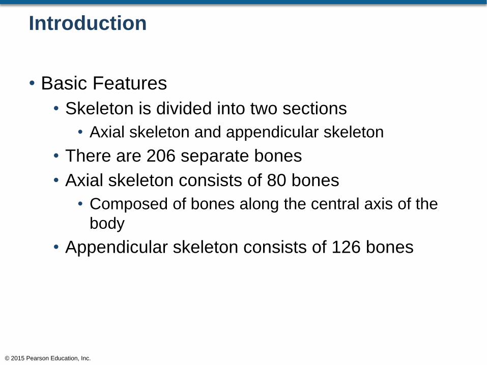
Introduction
• Basic Features
• Skeleton is divided into two sections
• Axial skeleton and appendicular skeleton
• There are 206 separate bones
• Axial skeleton consists of 80 bones
• Composed of bones along the central axis of the
body
• Appendicular skeleton consists of 126 bones
© 2015 Pearson Education, Inc.
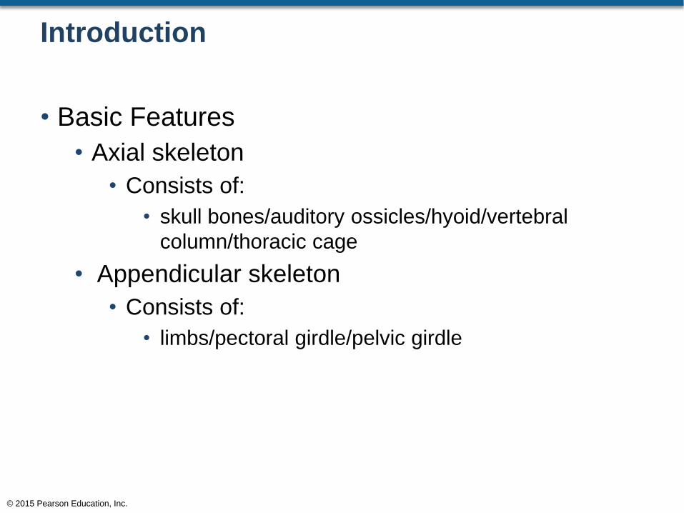
Introduction
• Basic Features
• Axial skeleton
• Consists of:
• skull bones/auditory ossicles/hyoid/vertebral
column/thoracic cage
• Appendicular skeleton
• Consists of:
• limbs/pectoral girdle/pelvic girdle
© 2015 Pearson Education, Inc.
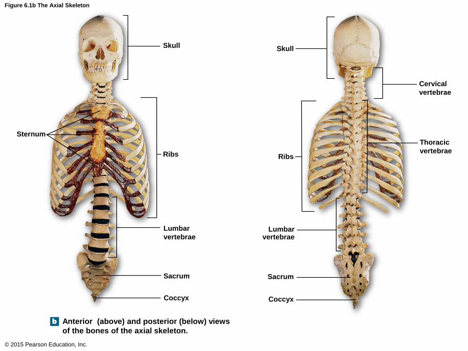
Figure 6.1b The Axial Skeleton
© 2015 Pearson Education, Inc.
Skull
b
Ribs
Sternum
Lumbar
vertebrae
Sacrum
Coccyx
Skull
Cervical
vertebrae
Thoracic
vertebrae Ribs
Lumbar vertebrae
Sacrum
Coccyx
Anterior (above) and posterior (below) views
of the bones of the axial skeleton.
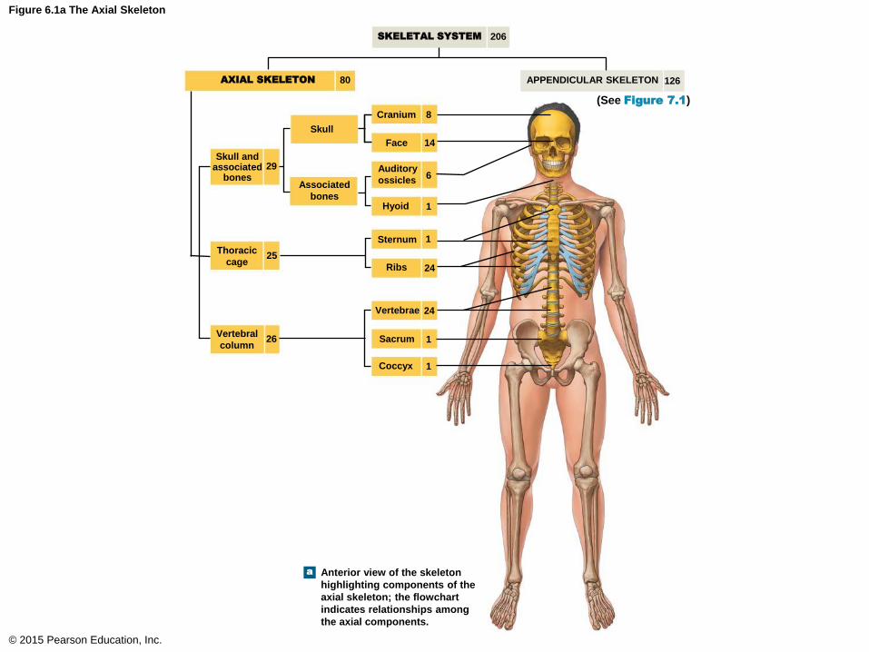
Figure 6.1a The Axial Skeleton
© 2015 Pearson Education, Inc.
SKELETAL SYSTEM
a
AXIAL SKELETON APPENDICULAR SKELETON
206
126 80
(See Figure 7.1)
Skull
Skull and associated
bones 29
Associated
bones
Cranium 8
Face 14
Auditory
ossicles 6
Hyoid 1
Sternum 1
Ribs 24
Thoracic
cage 25
Vertebral
column 26
Vertebrae 24
Sacrum 1
Coccyx 1
Anterior view of the skeleton
highlighting components of the
axial skeleton; the flowchart
indicates relationships among
the axial components.
Introduction
• Function of the Axial Skeleton
• Framework that supports and protects organs in
the dorsal and ventral body cavities
• Protects special sense organs for taste, smell,
hearing, balance, and vision
• Attachment sites for muscles that:
• Adjust the posture of the head, neck, and trunk
• Move the thoracic cage for respiration
• Stabilize the appendicular skeleton
© 2015 Pearson Education, Inc.
The Skull and Associated Bones
• Cranial and Facial Subdivisions of the Skull
• The skull consists of
• Face
• Cranium
• Associated bones
• The face: 14 individual bones
• The cranium: 8 individual bones
• The associated bones: 7 individual bones
© 2015 Pearson Education, Inc.
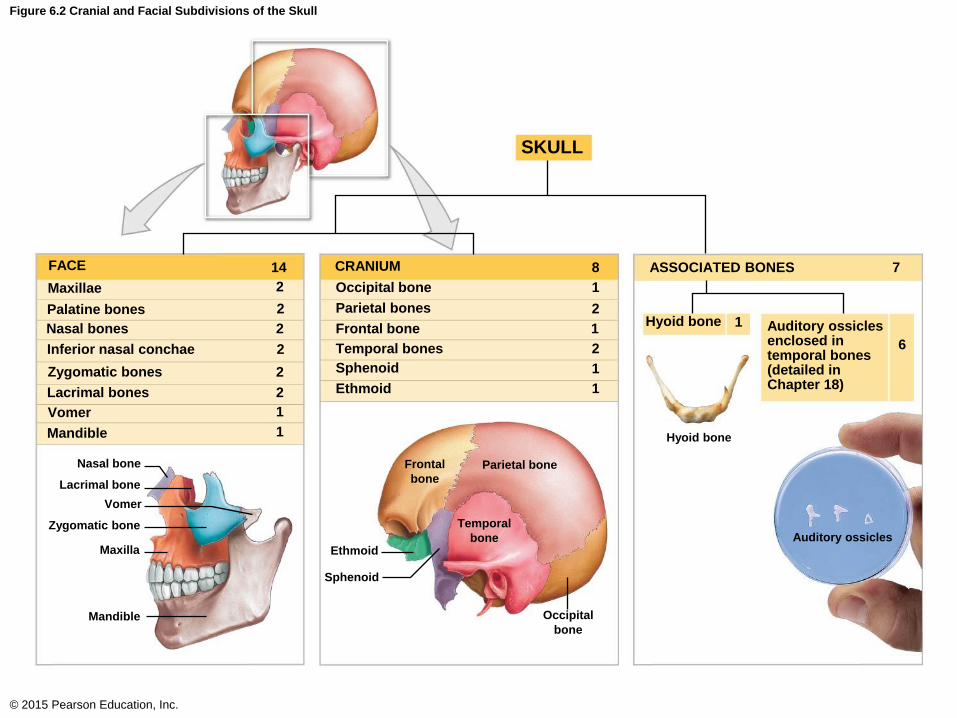
Figure 6.2 Cranial and Facial Subdivisions of the Skull
© 2015 Pearson Education, Inc.
Nasal bones
Nasal bone
Lacrimal bone
Vomer
Zygomatic bone
Maxilla
Mandible
Frontal
bone Parietal bone
Temporal
bone
Occipital
bone
Ethmoid
Sphenoid
Hyoid bone
Auditory ossicles
FACE
Maxillae
Palatine bones
Inferior nasal conchae
Zygomatic bones
Lacrimal bones
Vomer
Mandible
14
2
2
2
2
2
2
1
1
2
2
1
1
1
1
8
1
CRANIUM ASSOCIATED BONES
Occipital bone
Parietal bones
Frontal bone
Temporal bones
Sphenoid
Ethmoid
Hyoid bone
SKULL
Auditory ossicles enclosed in temporal bones (detailed in Chapter 18)
7
6
The Skull and Associated Bones
• Posterior View of the Occipital Bone
• Lambdoid suture
• Suture between the occipital bone and the two
parietal bones (superior skull)
• Sagittal suture
• Suture between the two parietal bones
• External occipital protuberance
• Bulge about midway of the occipital bone
• Occipital bone
• Most posterior bone of the skull
© 2015 Pearson Education, Inc.
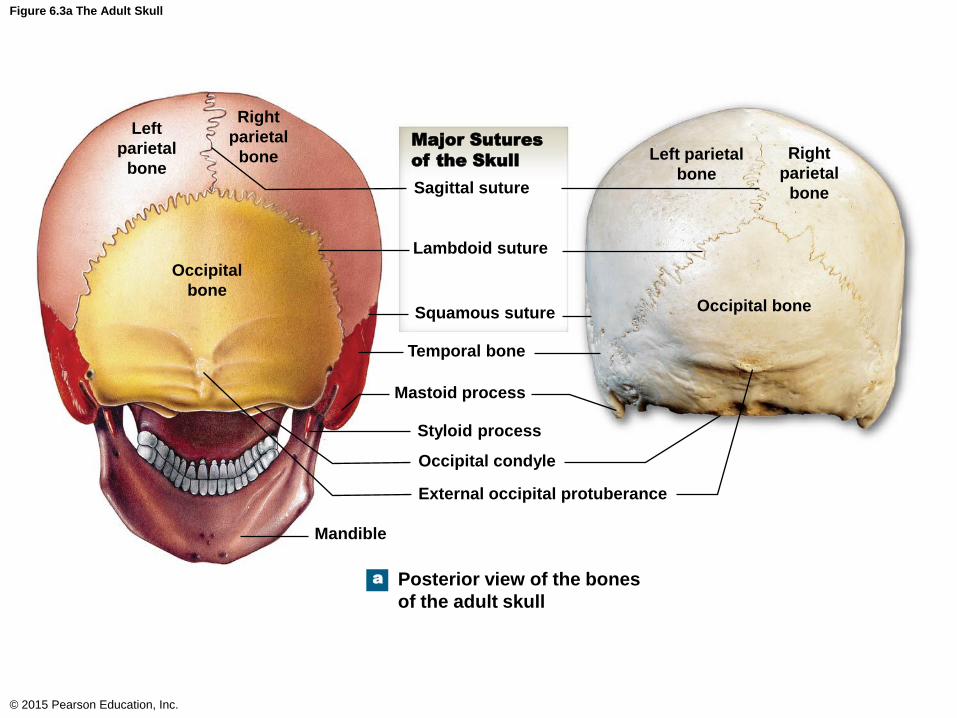
Figure 6.3a The Adult Skull
© 2015 Pearson Education, Inc.
Left
parietal
bone
a
Right
parietal
bone
Occipital
bone
Left parietal
bone
Right
parietal
bone
Occipital bone
Sagittal suture
Lambdoid suture
Squamous suture
Temporal bone
Mastoid process
Styloid process
Occipital condyle
External occipital protuberance
Mandible
Posterior view of the bones
of the adult skull
Major Sutures
of the Skull
The Skull and Associated Bones
• Superior View of the Skull
• Parietal bones
• Left and right parietal bones
• Sagittal suture
• Between the two parietal bones
• Coronal suture
• Between the frontal bone and the two parietal
bones
• Frontal bone
• The most anterior bone of the skullcap
© 2015 Pearson Education, Inc.

Figure 6.3b The Adult Skull
© 2015 Pearson Education, Inc.
b
Sagittal suture
Major Sutures
of the Skull
Lambdoid suture
Coronal suture
Zygomatic bone
Occipital bone Occipital bone
Right
parietal
bone
Right
parietal
bone
Left
parietal
bone
Left
parietal
bone
Frontal
bone
Frontal
bone
Nasal
bones
Superior view of the
bones of the adult skull
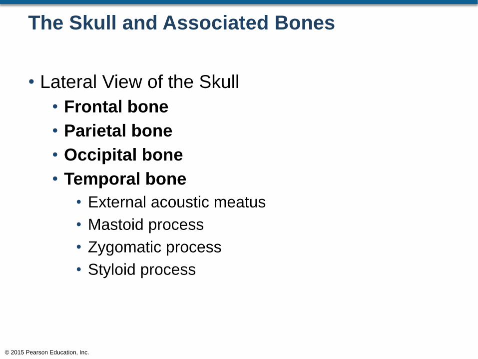
The Skull and Associated Bones
• Lateral View of the Skull
• Frontal bone
• Parietal bone
• Occipital bone
• Temporal bone
• External acoustic meatus
• Mastoid process
• Zygomatic process
• Styloid process
© 2015 Pearson Education, Inc.
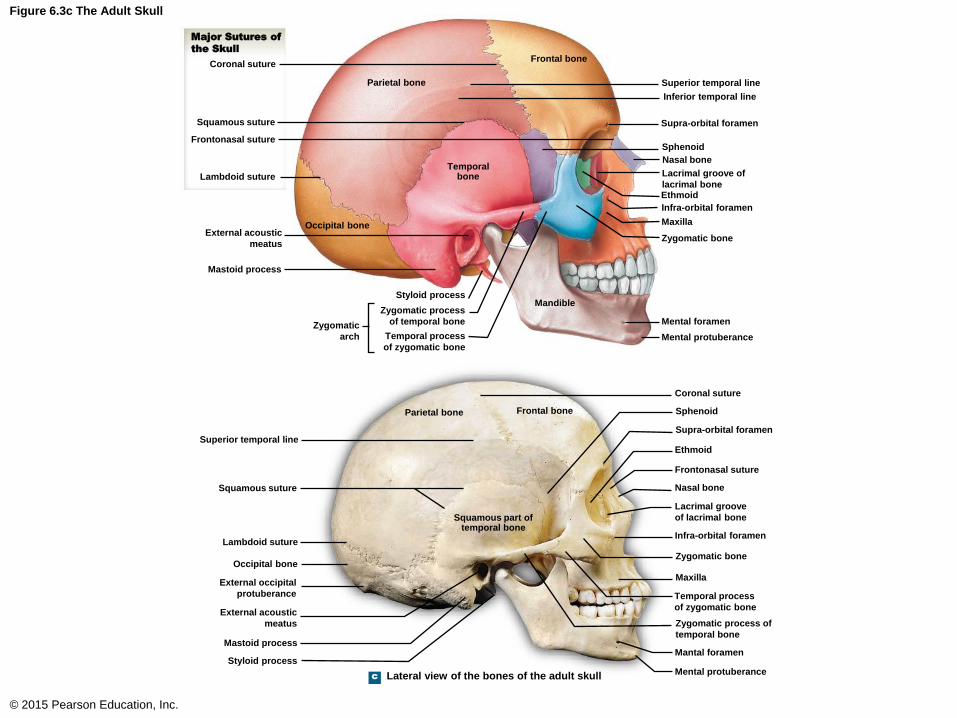
Figure 6.3c The Adult Skull
© 2015 Pearson Education, Inc.
Frontal bone
c
Parietal bone
Temporal bone
Occipital bone
Mandible
Superior temporal line
Inferior temporal line
Supra-orbital foramen
Sphenoid
Nasal bone
Lacrimal groove of
lacrimal bone Ethmoid
Infra-orbital foramen
Maxilla
Zygomatic bone
Mental foramen
Mental protuberance
Styloid process
Zygomatic process
of temporal bone
Temporal process
of zygomatic bone
Zygomatic
arch
Major Sutures of
the Skull
Coronal suture
Squamous suture
Frontonasal suture
Lambdoid suture
External acoustic
meatus
Mastoid process
Frontal bone Parietal bone
Squamous part of temporal bone
Lateral view of the bones of the adult skull
Superior temporal line
Squamous suture
Lambdoid suture
Occipital bone
External occipital
protuberance
External acoustic
meatus
Mastoid process
Styloid process
Coronal suture
Sphenoid
Supra-orbital foramen
Ethmoid
Frontonasal suture
Nasal bone
Lacrimal groove
of lacrimal bone
Infra-orbital foramen
Zygomatic bone
Maxilla
Temporal process
of zygomatic bone
Zygomatic process of
temporal bone
Mantal foramen
Mental protuberance

The Skull and Associated Bones
• Anterior View of the Skull
• Frontal bone
• Supra-orbital foramen
• Nasal bone
• Maxilla bone
• Infra-orbital foramen
• Mandible bone
© 2015 Pearson Education, Inc.

Figure 6.3d The Adult Skull
© 2015 Pearson Education, Inc.
Frontal bone
d
Parietal bone
Supra-orbital foramen
Sphenoid
Temporal bone
Ethmoid
Palatine bone
Lacrimal bone
Zygomaticofacial
foramen
Zygomatic bone
Nasal bone
Maxilla
Inferior nasal concha
Vomer
Mandible
Coronal suture
Frontonasal suture
Optic canal
Superior orbital fissure
Inferior orbital fissure
Temporal process of
zygomatic bone
Infra-orbital foramen
Middle nasal concha
Perpendicular plate
of ethmoid
Mental foramen
Mental protuberance
Frontal bone
Parietal bone
Supra-orbital foramen
Temporal bone
Sphenoid
Zygomatic bone
Infra-orbital foramen
Mastoid process
Maxilla
Mental foramen
Mandible
Coronal suture
Nasal bone
Frontonasal suture
Optic canal
Superior orbital fissure
Lacrimal bone
Middle nasal concha
Temporal process of
zygomatic bone
Inferior nasal concha
Perpendicular
plate of ethmoid
Vomer
Bony nasal septum
Mental protuberance
Anterior view of the bones of the adult skull
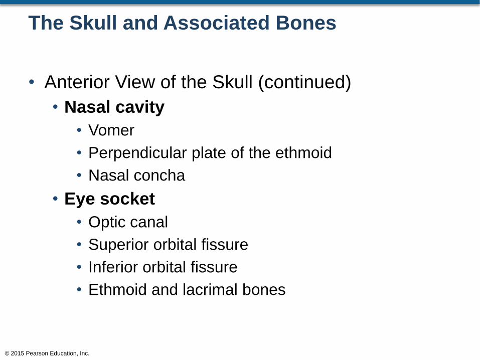
The Skull and Associated Bones
• Anterior View of the Skull (continued)
• Nasal cavity
• Vomer
• Perpendicular plate of the ethmoid
• Nasal concha
• Eye socket
• Optic canal
• Superior orbital fissure
• Inferior orbital fissure
• Ethmoid and lacrimal bones
© 2015 Pearson Education, Inc.
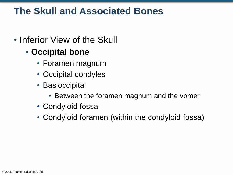
The Skull and Associated Bones
• Inferior View of the Skull
• Occipital bone
• Foramen magnum
• Occipital condyles
• Basioccipital
• Between the foramen magnum and the vomer
• Condyloid fossa
• Condyloid foramen (within the condyloid fossa)
© 2015 Pearson Education, Inc.
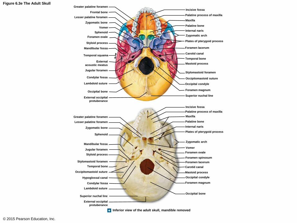
Figure 6.3e The Adult Skull
© 2015 Pearson Education, Inc.
Inferior view of the adult skull, mandible removed e
Vomer
Greater palatine foramen
Frontal bone
Lesser palatine foramen
Zygomatic bone
Sphenoid
Foramen ovale
Styloid process
Mandibular fossa
Temporal squama
External
acoustic meatus
Jugular foramen
Condylar fossa
Lambdoid suture
Occipital bone
External occipital
protuberance
Greater palatine foramen
Incisive fossa
Palatine process of maxilla
Maxilla
Palatine bone
Internal naris
Zygomatic arch
Plates of pterygoid process
Foramen lacerum
Carotid canal
Temporal bone
Mastoid process
Stylomastoid foramen
Occipitomastoid suture
Occipital condyle
Foramen magnum
Superior nuchal line
Incisive fossa
Palatine process of maxilla
Maxilla
Palatine bone
Internal naris
Zygomatic arch
Plates of pterygoid process
Vomer
Foramen ovale
Foramen spinosum
Foramen lacerum
Carotid canal
Mastoid process
Occipital condyle
Foramen magnum
Occipital bone
Lesser palatine foramen
Zygomatic bone
Sphenoid
Mandibular fossa
Jugular foramen
Styloid process
Stylomastoid foramen
Temporal bone
Occipitomastoid suture
Hypoglossal canal
Condylar fossa
Lambdoid suture
Superior nuchal line
External occipital
protuberance
The Skull and Associated Bones
• Inferior View of the Roof of the Mouth
• Palatine process of the maxilla (anterior
palatine)
• Incisive fossa
• Incisive foramen (within the incisive fossa)
• Palatine bone (posterior palatine)
• Greater palatine foramen
• Lesser palatine foramen
© 2015 Pearson Education, Inc.

The Skull and Associated Bones
• Inferior View of the Skull (continued)
• Internal nares
• Also called choana
• Vomer
• Inferior bone of the nasal septum
© 2015 Pearson Education, Inc.
The Skull and Associated Bones
• Inferior View of the Skull (continued)
• Foramen
• Foramen lacerum
• Carotid canal
• Foramen ovale
• Foramen spinosum
• Jugular foramen
• Stylomastoid foramen
© 2015 Pearson Education, Inc.
The Skull and Associated Bones
• Internal View of the Skull
• Frontal bone
• Ethmoid bone
• Crista galli
• Cribriform plate
• Cribriform plate foramina (olfactory foramina)
• Sphenoid bone
• Temporal bone
• Occipital bone
© 2015 Pearson Education, Inc.
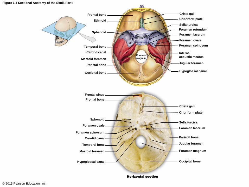
Figure 6.4 Sectional Anatomy of the Skull, Part I
© 2015 Pearson Education, Inc.
Frontal bone
Ethmoid
Sphenoid
Temporal bone
Carotid canal
Mastoid foramen
Parietal bone
Occipital bone
Foramen
magnum
Crista galli
Cribriform plate
Sella turcica
Foramen rotundum
Foramen lacerum
Foramen ovale
Foramen spinosum
Internal
acoustic meatus
Jugular foramen
Hypoglossal canal
Frontal sinus
Sphenoid
Frontal bone
Crista galli
Cribriform plate
Sella turcica
Foramen lacerum Foramen ovale
Foramen spinosum
Temporal bone
Carotid canal
Mastoid foramen
Hypoglossal canal Occipital bone
Parietal bone
Jugular foramen
Foramen magnum
Horizontal section
The Skull and Associated Bones
• Internal View of the Skull (continued)
• Sphenoid bone
• Sella turcica
• Dorsum sellae
• Hypophyseal fossa
• Tuberculum sellae
© 2015 Pearson Education, Inc.

The Skull and Associated Bones
• Internal View of the Skull (continued)
• Foramen
• Optic canals
• Foramen rotundum
• Foramen lacerum
• Foramen ovale
• Foramen spinosum
• Hypoglossal canal
• Foramen magnum
• Carotid canal
• Jugular foramen
© 2015 Pearson Education, Inc.
The Skull and Associated Bones
• Internal View of the Skull (continued)
• Internal acoustic meatus
• Petrous portion of the temporal bone
• Organs for balance and hearing are embedded
in this structure
© 2015 Pearson Education, Inc.
The Skull and Associated Bones
• Sectional Anatomy of the Skull
• Ethmoid bone
• Crista galli (anterior brain attachment)
• Perpendicular plate of the ethmoid
• Sphenoid bone
• Hypophyseal fossa (pituitary gland sits in this fossa
for protection)
© 2015 Pearson Education, Inc.

The Skull and Associated Bones
• Sectional Anatomy of the Skull (continued)
• Occipital bone
• Foramen magnum area
• Clivus (slant from the dorsum sella to the foramen
magnum)
• Nasal cavity
• Perpendicular plate of the ethmoid
• Vomer
© 2015 Pearson Education, Inc.

Figure 6.5 Sectional Anatomy of the Skull, Part II
© 2015 Pearson Education, Inc.
Sphenoid
Coronal suture
Frontal bone
Sphenoidal sinus (right)
Frontal sinus
Crista galli
Nasal bone
Perpendicular plate of ethmoid
Vomer
Palatine bone
Maxilla
Mandible
Parietal bone
Squamous suture
Temporal bone
Lambdoid suture
Hypophyseal fossa
of sella turcica
Internal acoustic meatus
Occipital bone
Hypoglossal canal
Styloid process
Sphenoid
Frontal bone
Frontal sinus
Crista galli
Nasal bone
Perpendicular plate of ethmoid
Vomer
Maxilla
Palatine bone
Mandible
Anterior nasal spine
Coronal suture
Sphenoidal sinuses
(left and right)
Squamous suture
Hypophyseal fossa
of sella turcica
Parietal bone
Lambdoid suture
Occipital bone
Petrous part of temporal bone
Internal acoustic meatus
Jugular foramen
Hypoglossal canal
Margin of
foramen magnum Sagittal section
Occipital condyle
The Skull and Associated Bones
• Sectional Anatomy of the Skull (continued)
• Sinuses
• Frontal sinus
• Sphenoidal sinus
© 2015 Pearson Education, Inc.
Sutures of the Skull
• Lambdoid suture
• Suture between the occipital and parietal bones
• Sagittal suture
• Suture between the parietal bones
• Coronal suture
• Suture between the frontal and parietal bones
© 2015 Pearson Education, Inc.
Sutures of the Skull
• Squamous suture
• Suture between the temporal bone and the
parietal bones
• Frontonasal suture
• Suture between the nasal and frontal bones
© 2015 Pearson Education, Inc.

Figure 6.3a The Adult Skull
© 2015 Pearson Education, Inc.
Left
parietal
bone
a
Right
parietal
bone
Occipital
bone
Left parietal
bone
Right
parietal
bone
Occipital bone
Sagittal suture
Lambdoid suture
Squamous suture
Temporal bone
Mastoid process
Styloid process
Occipital condyle
External occipital protuberance
Mandible
Posterior view of the bones
of the adult skull
Major Sutures
of the Skull
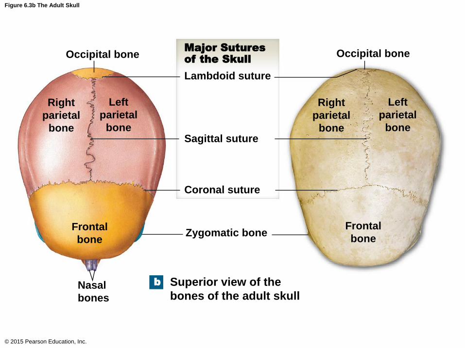
Figure 6.3b The Adult Skull
© 2015 Pearson Education, Inc.
b
Sagittal suture
Major Sutures
of the Skull
Lambdoid suture
Coronal suture
Zygomatic bone
Occipital bone Occipital bone
Right
parietal
bone
Right
parietal
bone
Left
parietal
bone
Left
parietal
bone
Frontal
bone
Frontal
bone
Nasal
bones
Superior view of the
bones of the adult skull

Figure 6.3c The Adult Skull
© 2015 Pearson Education, Inc.
Frontal bone
c
Parietal bone
Temporal bone
Occipital bone
Mandible
Superior temporal line
Inferior temporal line
Supra-orbital foramen
Sphenoid
Nasal bone
Lacrimal groove of
lacrimal bone Ethmoid
Infra-orbital foramen
Maxilla
Zygomatic bone
Mental foramen
Mental protuberance
Styloid process
Zygomatic process
of temporal bone
Temporal process
of zygomatic bone
Zygomatic
arch
Major Sutures of
the Skull
Coronal suture
Squamous suture
Frontonasal suture
Lambdoid suture
External acoustic
meatus
Mastoid process
Frontal bone Parietal bone
Squamous part of temporal bone
Lateral view of the bones of the adult skull
Superior temporal line
Squamous suture
Lambdoid suture
Occipital bone
External occipital
protuberance
External acoustic
meatus
Mastoid process
Styloid process
Coronal suture
Sphenoid
Supra-orbital foramen
Ethmoid
Frontonasal suture
Nasal bone
Lacrimal groove
of lacrimal bone
Infra-orbital foramen
Zygomatic bone
Maxilla
Temporal process
of zygomatic bone
Zygomatic process of
temporal bone
Mantal foramen
Mental protuberance
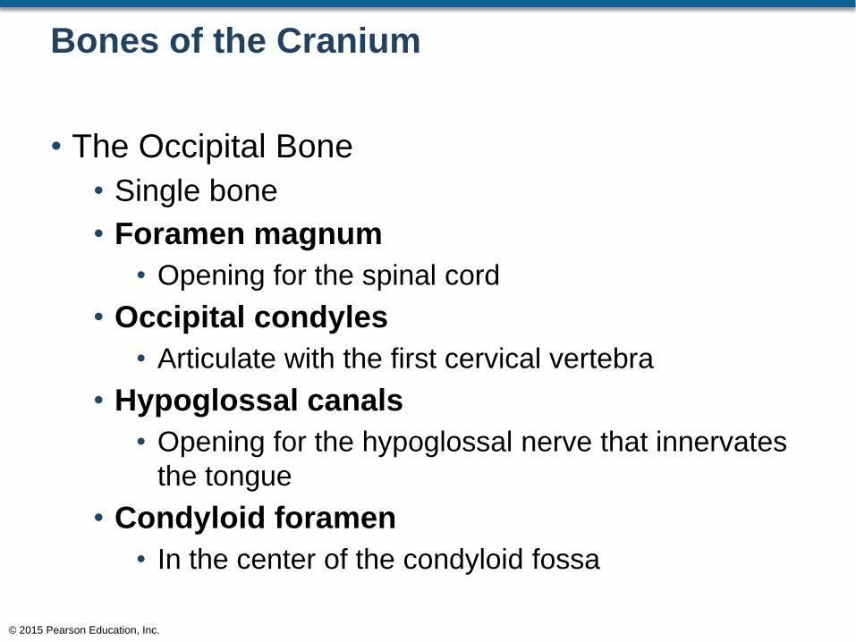
Bones of the Cranium
• The Occipital Bone
• Single bone
• Foramen magnum
• Opening for the spinal cord
• Occipital condyles
• Articulate with the first cervical vertebra
• Hypoglossal canals
• Opening for the hypoglossal nerve that innervates
the tongue
• Condyloid foramen
• In the center of the condyloid fossa
© 2015 Pearson Education, Inc.
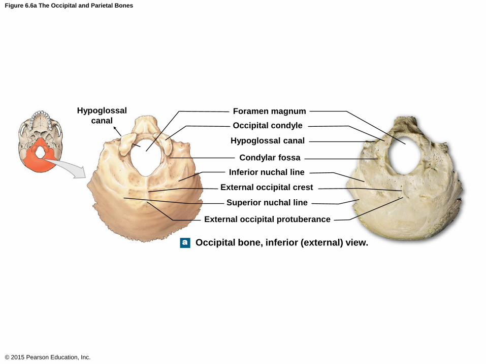
Figure 6.6a The Occipital and Parietal Bones
© 2015 Pearson Education, Inc.
Hypoglossal
canal
a
Hypoglossal canal
Foramen magnum
Occipital condyle
Condylar fossa
Inferior nuchal line
External occipital crest
Superior nuchal line
External occipital protuberance
Occipital bone, inferior (external) view.
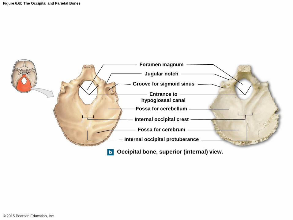
Figure 6.6b The Occipital and Parietal Bones
© 2015 Pearson Education, Inc.
b Occipital bone, superior (internal) view.
Foramen magnum
Jugular notch
Groove for sigmoid sinus
Entrance to
hypoglossal canal
Fossa for cerebellum
Internal occipital crest
Fossa for cerebrum
Internal occipital protuberance
Bones of the Cranium
• Parietal Bones
• Paired bones
• Internal surface retains the impression of cranial
blood vessels
© 2015 Pearson Education, Inc.
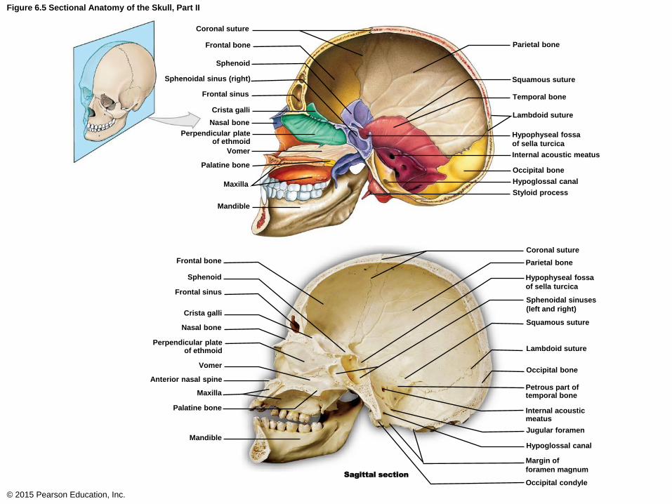
Figure 6.5 Sectional Anatomy of the Skull, Part II
© 2015 Pearson Education, Inc.
Sphenoid
Coronal suture
Frontal bone
Sphenoidal sinus (right)
Frontal sinus
Crista galli
Nasal bone
Perpendicular plate of ethmoid
Vomer
Palatine bone
Maxilla
Mandible
Parietal bone
Squamous suture
Temporal bone
Lambdoid suture
Hypophyseal fossa
of sella turcica
Internal acoustic meatus
Occipital bone
Hypoglossal canal
Styloid process
Sphenoid
Frontal bone
Frontal sinus
Crista galli
Nasal bone
Perpendicular plate of ethmoid
Vomer
Maxilla
Palatine bone
Mandible
Anterior nasal spine
Coronal suture
Sphenoidal sinuses
(left and right)
Squamous suture
Hypophyseal fossa
of sella turcica
Parietal bone
Lambdoid suture
Occipital bone
Petrous part of temporal bone
Internal acoustic meatus
Jugular foramen
Hypoglossal canal
Margin of
foramen magnum Sagittal section
Occipital condyle
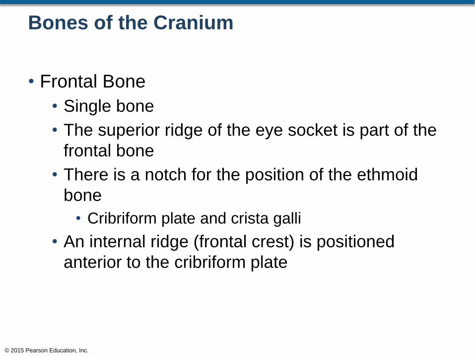
Bones of the Cranium
• Frontal Bone
• Single bone
• The superior ridge of the eye socket is part of the
frontal bone
• There is a notch for the position of the ethmoid
bone
• Cribriform plate and crista galli
• An internal ridge (frontal crest) is positioned
anterior to the cribriform plate
© 2015 Pearson Education, Inc.
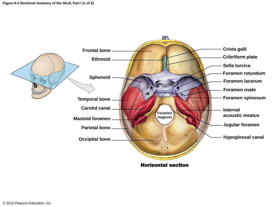
Figure 6.4 Sectional Anatomy of the Skull, Part I (1 of 2)
© 2015 Pearson Education, Inc.
Frontal bone
Ethmoid
Sphenoid
Temporal bone
Carotid canal
Mastoid foramen
Parietal bone
Occipital bone
Horizontal section
Crista galli
Cribriform plate
Sella turcica
Foramen rotundum
Foramen lacerum
Foramen ovale
Foramen spinosum
Internal
acoustic meatus
Jugular foramen
Hypoglossal canal
Foramen
magnum
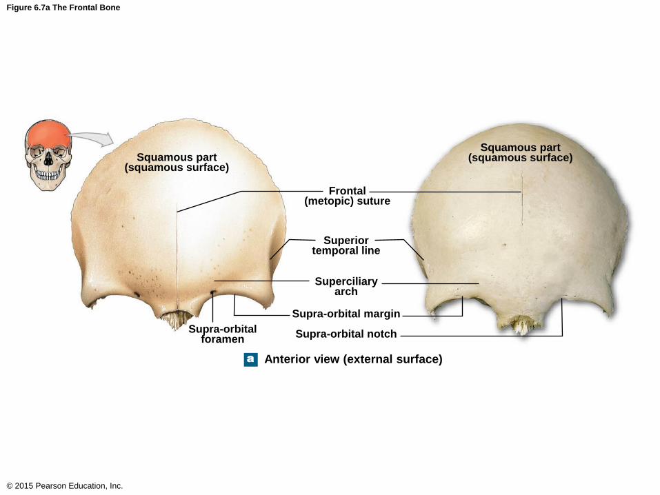
Figure 6.7a The Frontal Bone
© 2015 Pearson Education, Inc.
a
Squamous part (squamous surface)
Anterior view (external surface)
Squamous part (squamous surface)
Frontal (metopic) suture
Superior temporal line
Superciliary arch
Supra-orbital margin
Supra-orbital notch Supra-orbital foramen

Figure 6.7c The Frontal Bone
© 2015 Pearson Education, Inc.
c Posterior view
Margin of coronal suture
Squamous part
Frontal crest
Orbital part
Notch for ethmoid
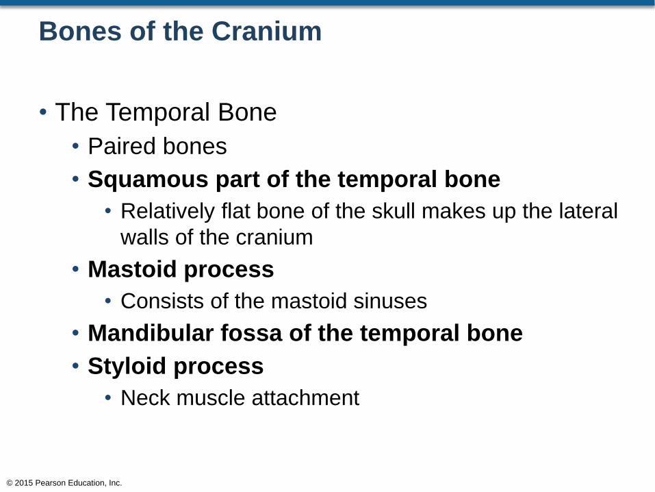
Bones of the Cranium
• The Temporal Bone
• Paired bones
• Squamous part of the temporal bone
• Relatively flat bone of the skull makes up the lateral
walls of the cranium
• Mastoid process
• Consists of the mastoid sinuses
• Mandibular fossa of the temporal bone
• Styloid process
• Neck muscle attachment
© 2015 Pearson Education, Inc.
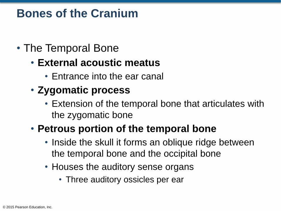
Bones of the Cranium
• The Temporal Bone
• External acoustic meatus
• Entrance into the ear canal
• Zygomatic process
• Extension of the temporal bone that articulates with
the zygomatic bone
• Petrous portion of the temporal bone
• Inside the skull it forms an oblique ridge between
the temporal bone and the occipital bone
• Houses the auditory sense organs
• Three auditory ossicles per ear
© 2015 Pearson Education, Inc.
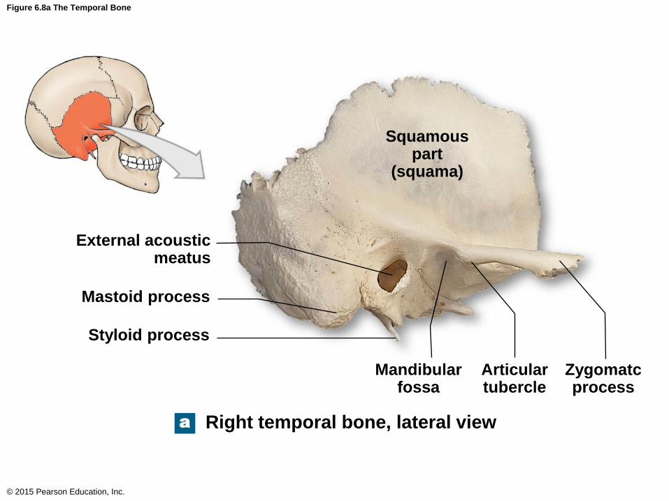
Figure 6.8a The Temporal Bone
© 2015 Pearson Education, Inc.
a
Squamous part
(squama)
External acoustic meatus
Styloid process
Mandibular fossa
Articular tubercle
Zygomatc process
Right temporal bone, lateral view
Mastoid process
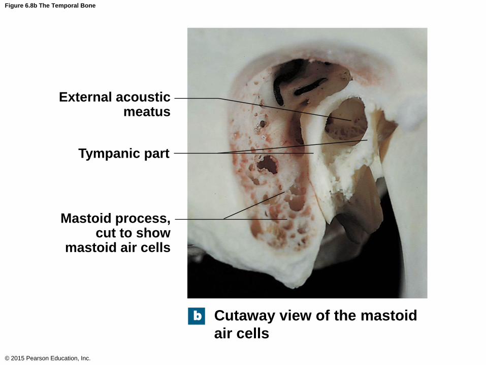
Figure 6.8b The Temporal Bone
© 2015 Pearson Education, Inc.
External acoustic meatus
Tympanic part
Mastoid process, cut to show
mastoid air cells
Cutaway view of the mastoid
air cells
b
Bones of the Cranium
• The Sphenoid Bone
• Single bone
• Sella turcica
• Dorsum sellae
• Hypophyseal fossa (fossa for the pituitary gland)
• Tuberculum sellae
• Anterior clinoid processes
• Posterior clinoid processes
• Optic canals
• Openings for the optic nerves
© 2015 Pearson Education, Inc.
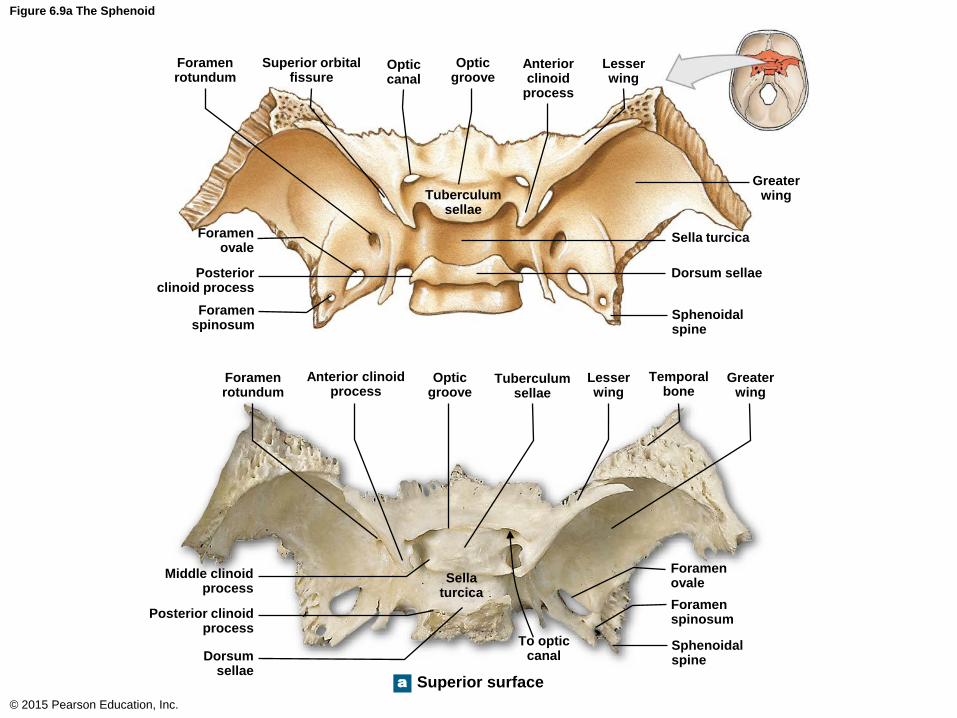
Figure 6.9a The Sphenoid
© 2015 Pearson Education, Inc.
Foramen rotundum
a
Superior orbital fissure
Optic canal
Optic groove
Anterior clinoid
process
Lesser wing
Greater wing
Sella turcica
Dorsum sellae
Sphenoidal spine
Tuberculum sellae
Foramen ovale
Posterior clinoid process
Foramen spinosum
Foramen rotundum
Anterior clinoid process
Optic groove
Tuberculum sellae
Lesser wing
Temporal bone
Greater wing
Foramen ovale
Foramen spinosum
Sphenoidal spine
To optic canal
Sella turcica
Dorsum sellae
Posterior clinoid process
Middle clinoid process
Superior surface
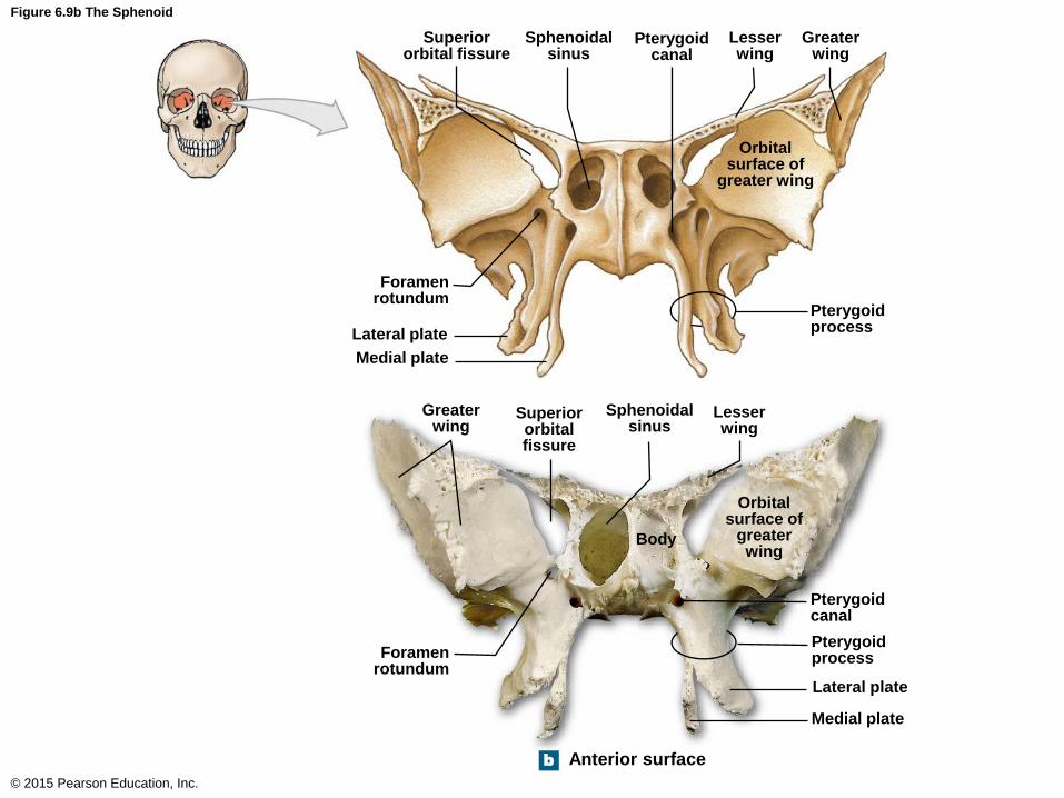
Figure 6.9b The Sphenoid
© 2015 Pearson Education, Inc.
Anterior surface b
Lateral plate
Superior orbital fissure
Sphenoidal sinus
Pterygoid canal
Lesser wing
Greater wing
Orbital surface of
greater wing
Foramen rotundum
Medial plate
Pterygoid process
Greater wing
Superior orbital fissure
Sphenoidal sinus
Lesser wing
Body
Orbital surface of
greater wing
Foramen rotundum
Pterygoid canal
Pterygoid process
Lateral plate
Medial plate
Bones of the Cranium
• The Ethmoid Bone
• Single bone
• Perpendicular plate of the ethmoid
• Superior portion of the nasal septum
• Crista galli
• Superior portion of the perpendicular plate of the
ethmoid
• Cribriform plate
• Borders the crista galli
• Cribriform plate foramina (olfactory foramina)
• Openings for the olfactory nerves
© 2015 Pearson Education, Inc.
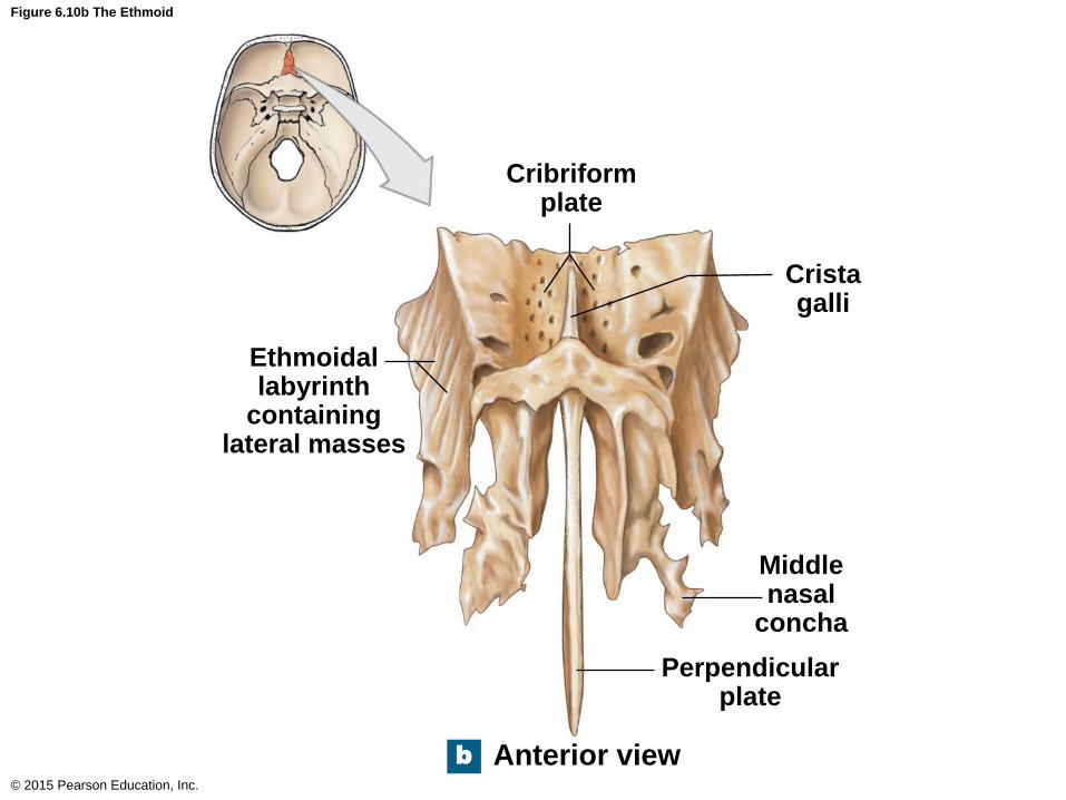
Figure 6.10b The Ethmoid
© 2015 Pearson Education, Inc.
b Anterior view
Ethmoidal labyrinth
containing lateral masses
Cribriform plate
Crista galli
Middle nasal
concha
Perpendicular plate
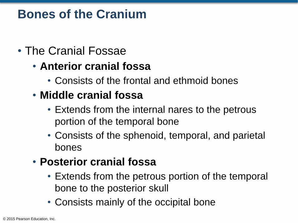
Bones of the Cranium
• The Cranial Fossae
• Anterior cranial fossa
• Consists of the frontal and ethmoid bones
• Middle cranial fossa
• Extends from the internal nares to the petrous
portion of the temporal bone
• Consists of the sphenoid, temporal, and parietal
bones
• Posterior cranial fossa
• Extends from the petrous portion of the temporal
bone to the posterior skull
• Consists mainly of the occipital bone
© 2015 Pearson Education, Inc.
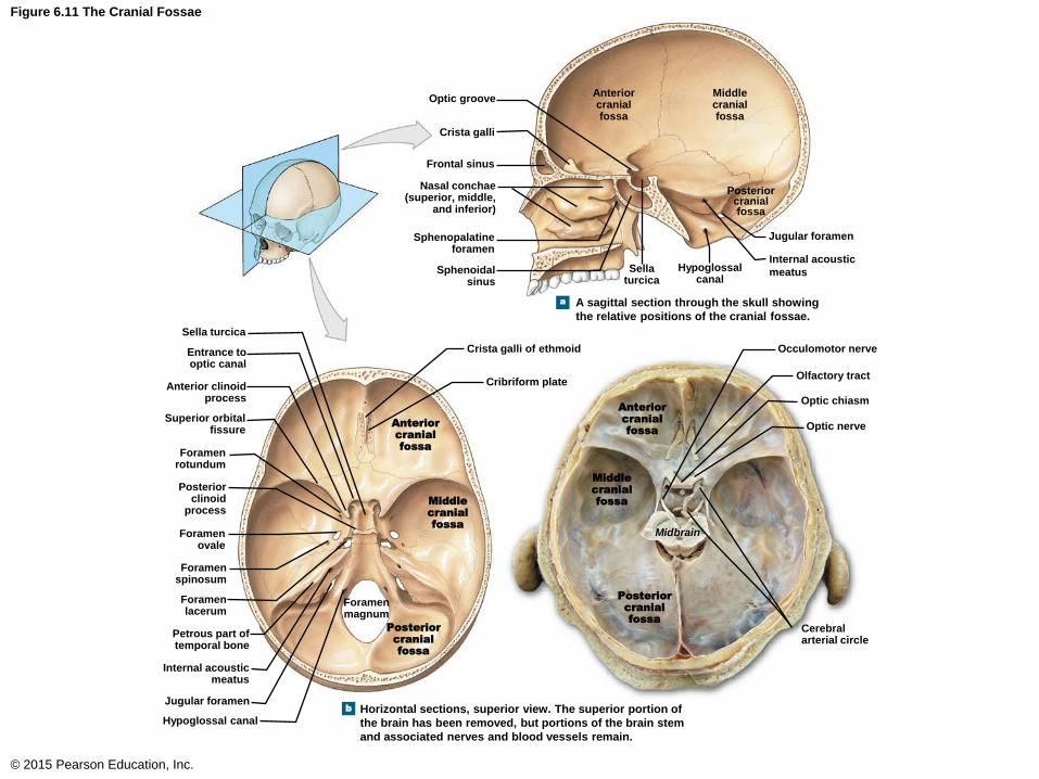
Figure 6.11 The Cranial Fossae
© 2015 Pearson Education, Inc.
Optic groove
a
b
Crista galli
Frontal sinus
Nasal conchae (superior, middle,
and inferior)
Sphenopalatine foramen
Sphenoidal sinus
Anterior cranial fossa
Sella turcica
Hypoglossal canal
Internal acoustic
meatus
Jugular foramen
Posterior cranial fossa
Middle cranial fossa
A sagittal section through the skull showing
the relative positions of the cranial fossae.
Optic nerve
Optic chiasm
Olfactory tract
Occulomotor nerve
Cerebral arterial circle
Crista galli of ethmoid
Cribriform plate
Anterior
cranial
fossa
Midbrain
Foramen magnum
Posterior
cranial
fossa
Middle
cranial
fossa
Anterior
cranial
fossa
Posterior
cranial
fossa
Middle
cranial
fossa
Sella turcica
Entrance to optic canal
Anterior clinoid process
Superior orbital fissure
Foramen rotundum
Posterior clinoid
process
Foramen ovale
Foramen spinosum
Foramen lacerum
Petrous part of temporal bone
Internal acoustic meatus
Jugular foramen
Hypoglossal canal Horizontal sections, superior view. The superior portion of
the brain has been removed, but portions of the brain stem
and associated nerves and blood vessels remain.
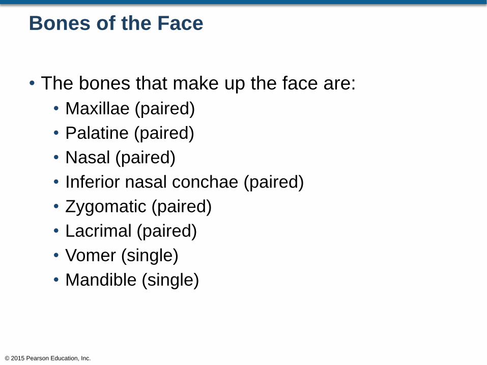
Bones of the Face
• The bones that make up the face are:
• Maxillae (paired)
• Palatine (paired)
• Nasal (paired)
• Inferior nasal conchae (paired)
• Zygomatic (paired)
• Lacrimal (paired)
• Vomer (single)
• Mandible (single)
© 2015 Pearson Education, Inc.

Bones of the Face
• The Maxillae
• Paired bones
• Make up the upper jaw
• Maxillary sinuses
• Anterior nasal spine
• Alveolar processes
• Tooth sockets
© 2015 Pearson Education, Inc.
Bones of the Face
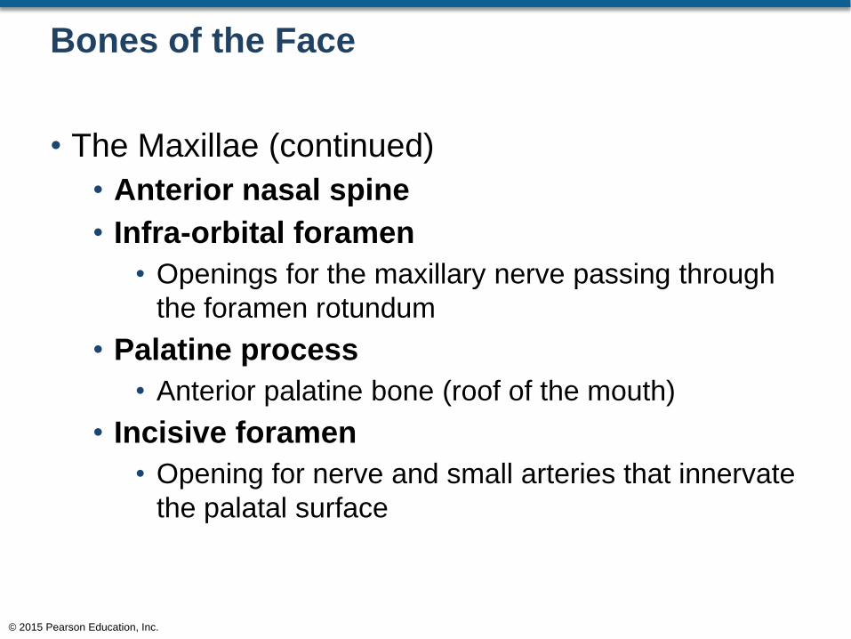
• The Maxillae (continued)
• Anterior nasal spine
• Infra-orbital foramen
• Openings for the maxillary nerve passing through
the foramen rotundum
• Palatine process
• Anterior palatine bone (roof of the mouth)
• Incisive foramen
• Opening for nerve and small arteries that innervate
the palatal surface
© 2015 Pearson Education, Inc.
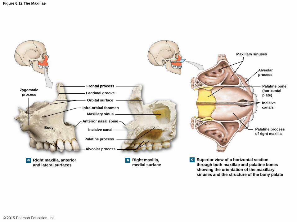
Figure 6.12 The Maxillae
© 2015 Pearson Education, Inc.
Body
a b c
Zygomatic
process
Frontal process
Lacrimal groove
Orbital surface
Infra-orbital foramen
Maxillary sinus
Anterior nasal spine
Incisive canal
Palatine process
Alveolar
process
Maxillary sinuses
Alveolar process
Palatine bone
(horizontal
plate)
Incisive
canals
Palatine process
of right maxilla
Right maxilla, anterior
and lateral surfaces
Right maxilla,
medial surface
Superior view of a horizontal section
through both maxillae and palatine bones
showing the orientation of the maxillary
sinuses and the structure of the bony palate
Bones of the Face
• The Palatine Bones
• Paired bones
• Posterior to the palatine process of the maxillae
• Makes up 1/3 of the roof of the mouth
© 2015 Pearson Education, Inc.

Figure 6.12c The Maxillae
© 2015 Pearson Education, Inc.
Alveolar
process
Maxillary sinuses
Palatine bone
(horizontal
plate)
Incisive
canals
Palatine process
of right maxilla
Superior view of a horizontal section
through both maxillae and palatine bones
showing the orientation of the maxillary
sinuses and the structure of the bony palate
c
Bones of the Face
• The Nasal Bones
• Paired bones
• Articulate with the frontal bone at the frontonasal
suture
• The lateral edges of each nasal bone articulate
with the maxillae
© 2015 Pearson Education, Inc.

Figure 6.3d The Adult Skull (1 of 2)
© 2015 Pearson Education, Inc.
Frontal bone
Parietal bone
Supra-orbital foramen
Sphenoid
Temporal bone
Ethmoid
Palatine bone
Lacrimal bone
Zygomaticofacial
foramen
Zygomatic bone
Nasal bone
Maxilla
Inferior nasal concha
Vomer
Mandible
Coronal suture
Frontonasal suture
Optic canal
Superior orbital fissure
Inferior orbital fissure
Temporal process of
zygomatic bone
Infra-orbital foramen
Middle nasal concha
Perpendicular plate
of ethmoid
Mental foramen
Mental protuberance
d Anterior view of the bones of the adult skull
Bones of the Face
• The Inferior Nasal Conchae
• Paired bones
• Attached to the lateral wall of each nasal cavity
© 2015 Pearson Education, Inc.
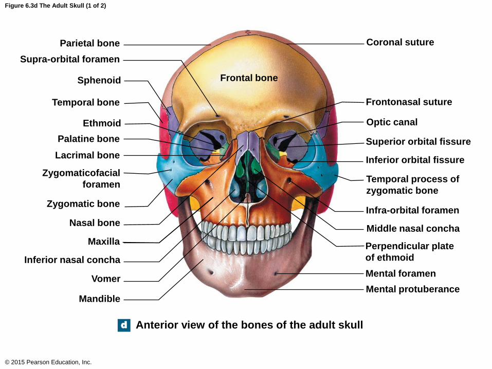
Figure 6.3d The Adult Skull (1 of 2)
© 2015 Pearson Education, Inc.
Frontal bone
Parietal bone
Supra-orbital foramen
Sphenoid
Temporal bone
Ethmoid
Palatine bone
Lacrimal bone
Zygomaticofacial
foramen
Zygomatic bone
Nasal bone
Maxilla
Inferior nasal concha
Vomer
Mandible
Coronal suture
Frontonasal suture
Optic canal
Superior orbital fissure
Inferior orbital fissure
Temporal process of
zygomatic bone
Infra-orbital foramen
Middle nasal concha
Perpendicular plate
of ethmoid
Mental foramen
Mental protuberance
d Anterior view of the bones of the adult skull

Bones of the Face
• The Zygomatic Bones
• Paired bones
• Articulates with the maxillae and the zygomatic
process of the temporal bone and the frontal bone
• Makes up the lateral wall of the eye socket
© 2015 Pearson Education, Inc.

Figure 6.3d The Adult Skull (1 of 2)
© 2015 Pearson Education, Inc.
Frontal bone
Parietal bone
Supra-orbital foramen
Sphenoid
Temporal bone
Ethmoid
Palatine bone
Lacrimal bone
Zygomaticofacial
foramen
Zygomatic bone
Nasal bone
Maxilla
Inferior nasal concha
Vomer
Mandible
Coronal suture
Frontonasal suture
Optic canal
Superior orbital fissure
Inferior orbital fissure
Temporal process of
zygomatic bone
Infra-orbital foramen
Middle nasal concha
Perpendicular plate
of ethmoid
Mental foramen
Mental protuberance
d Anterior view of the bones of the adult skull
Bones of the Face
• The Lacrimal Bones
• Paired bones
• Smallest bones of the skull
• Positioned on the medial aspect of the eye socket
(anterior to the ethmoid bone)
• Consists of a lacrimal foramen (nasolacrimal
canal)
• Drains tears into the nasal cavity
© 2015 Pearson Education, Inc.
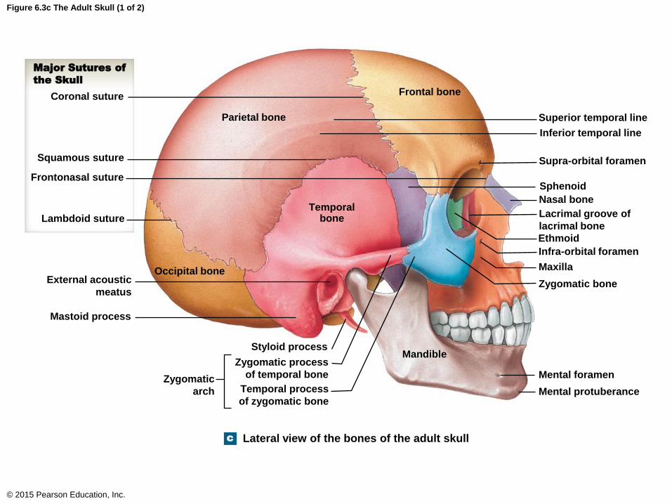
Figure 6.3c The Adult Skull (1 of 2)
© 2015 Pearson Education, Inc.
c
Frontal bone
Parietal bone
Temporal bone
Occipital bone
Styloid process
Zygomatic process
of temporal bone
Temporal process
of zygomatic bone
Zygomatic
arch
Major Sutures of
the Skull
Coronal suture
Squamous suture
Frontonasal suture
Lambdoid suture
External acoustic
meatus
Mastoid process
Mandible
Superior temporal line
Inferior temporal line
Supra-orbital foramen
Sphenoid
Nasal bone
Lacrimal groove of
lacrimal bone
Ethmoid
Infra-orbital foramen
Maxilla
Zygomatic bone
Mental foramen
Mental protuberance
Lateral view of the bones of the adult skull
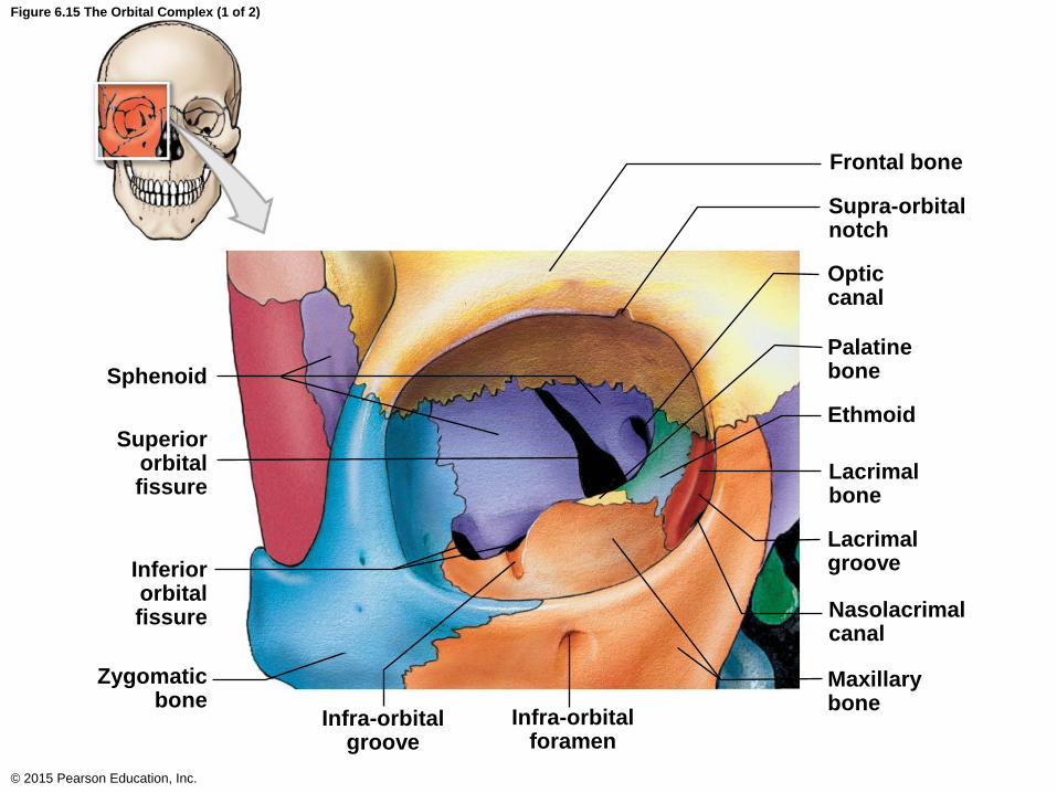
Figure 6.15 The Orbital Complex (1 of 2)
© 2015 Pearson Education, Inc.
Sphenoid
Superior orbital fissure
Inferior orbital fissure
Zygomatic bone
Infra-orbital groove
Infra-orbital foramen
Frontal bone
Supra-orbital notch
Optic canal
Palatine bone
Ethmoid
Lacrimal bone
Lacrimal groove
Nasolacrimal canal
Maxillary bone
Bones of the Face
• The Vomer
• Single bone
• Forms the inferior portion of the nasal septum
© 2015 Pearson Education, Inc.
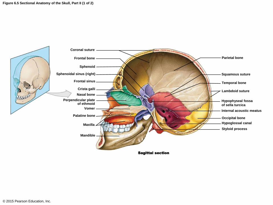
Figure 6.5 Sectional Anatomy of the Skull, Part II (1 of 2)
© 2015 Pearson Education, Inc.
Sphenoid
Coronal suture
Frontal bone
Sphenoidal sinus (right)
Frontal sinus
Crista galli
Nasal bone
Perpendicular plate of ethmoid
Vomer
Palatine bone
Maxilla
Mandible
Sagittal section
Parietal bone
Squamous suture
Temporal bone
Lambdoid suture
Hypophyseal fossa
of sella turcica
Internal acoustic meatus
Occipital bone
Hypoglossal canal
Styloid process

Figure 6.3d The Adult Skull (1 of 2)
© 2015 Pearson Education, Inc.
Frontal bone
Parietal bone
Supra-orbital foramen
Sphenoid
Temporal bone
Ethmoid
Palatine bone
Lacrimal bone
Zygomaticofacial
foramen
Zygomatic bone
Nasal bone
Maxilla
Inferior nasal concha
Vomer
Mandible
Coronal suture
Frontonasal suture
Optic canal
Superior orbital fissure
Inferior orbital fissure
Temporal process of
zygomatic bone
Infra-orbital foramen
Middle nasal concha
Perpendicular plate
of ethmoid
Mental foramen
Mental protuberance
d Anterior view of the bones of the adult skull
Bones of the Face
• The Mandible
• Single bone
• Makes up the lower jaw
• Head (mandibular condyle)
• Articulates with the mandibular fossa of the
temporal bone
• Mandibular notch
• Coronoid process
© 2015 Pearson Education, Inc.
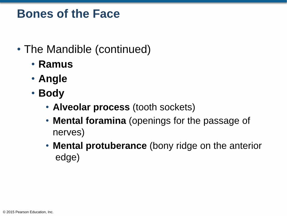
Bones of the Face
• The Mandible (continued)
• Ramus
• Angle
• Body
• Alveolar process (tooth sockets)
• Mental foramina (openings for the passage of
nerves)
• Mental protuberance (bony ridge on the anterior
edge)
© 2015 Pearson Education, Inc.
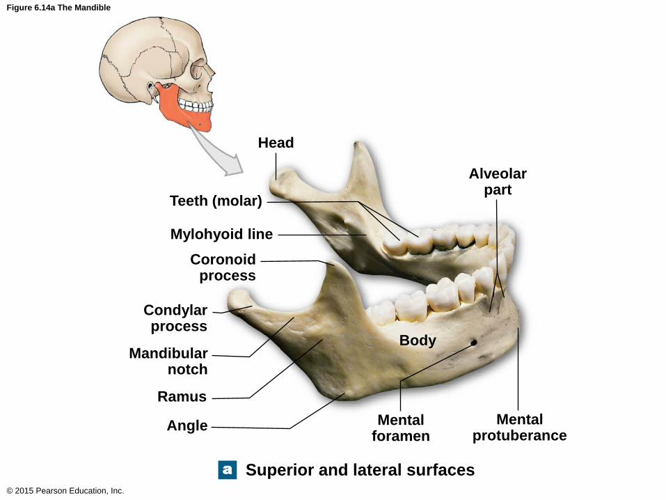
Figure 6.14a The Mandible
© 2015 Pearson Education, Inc.
Head
a
Teeth (molar)
Mylohyoid line
Coronoid process
Condylar process
Mandibular notch
Ramus
Angle Mental foramen
Body
Mental protuberance
Alveolar part
Superior and lateral surfaces
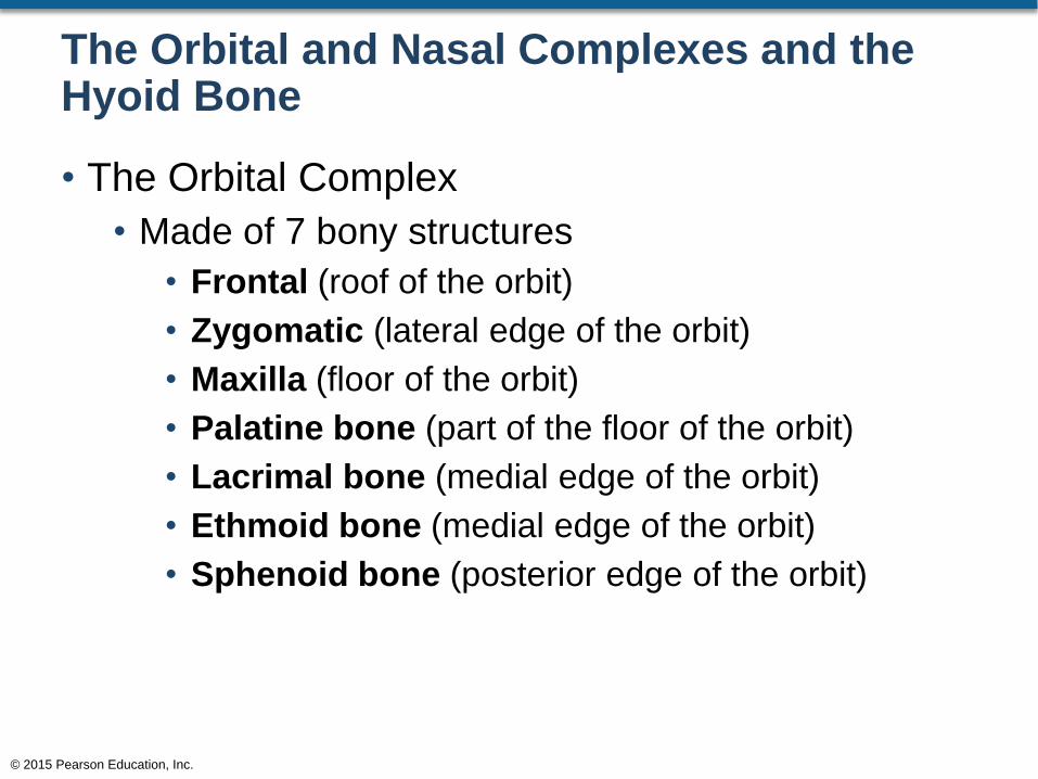
The Orbital and Nasal Complexes and the Hyoid Bone
• The Orbital Complex
• Made of 7 bony structures
• Frontal (roof of the orbit)
• Zygomatic (lateral edge of the orbit)
• Maxilla (floor of the orbit)
• Palatine bone (part of the floor of the orbit)
• Lacrimal bone (medial edge of the orbit)
• Ethmoid bone (medial edge of the orbit)
• Sphenoid bone (posterior edge of the orbit)
© 2015 Pearson Education, Inc.

Figure 6.15 The Orbital Complex
© 2015 Pearson Education, Inc.
Sphenoid
Superior orbital fissure
Inferior orbital fissure
Zygomatic bone
Infra-orbital groove
Infra-orbital foramen
Frontal bone
Supra-orbital notch
Optic canal
Palatine bone
Ethmoid
Lacrimal bone
Lacrimal groove
Nasolacrimal canal
Maxillary bone
Sphenoid
Superior orbital fissure
Inferior orbital fissure
Zygomatic bone
Ethmoid
Infra-orbital groove
Frontal bone
Optic canal
Maxillary bone
Infra-orbital foramen
Nasolacrimal canal
Supra-orbital notch
The Orbital and Nasal Complexes and the Hyoid Bone
• The Orbital Complex (continued)
• Superior orbital fissure (opening for the
following nerves)
• Oculomotor
• Trochlear
• Ophthalmic branch of the trigeminal nerve
© 2015 Pearson Education, Inc.
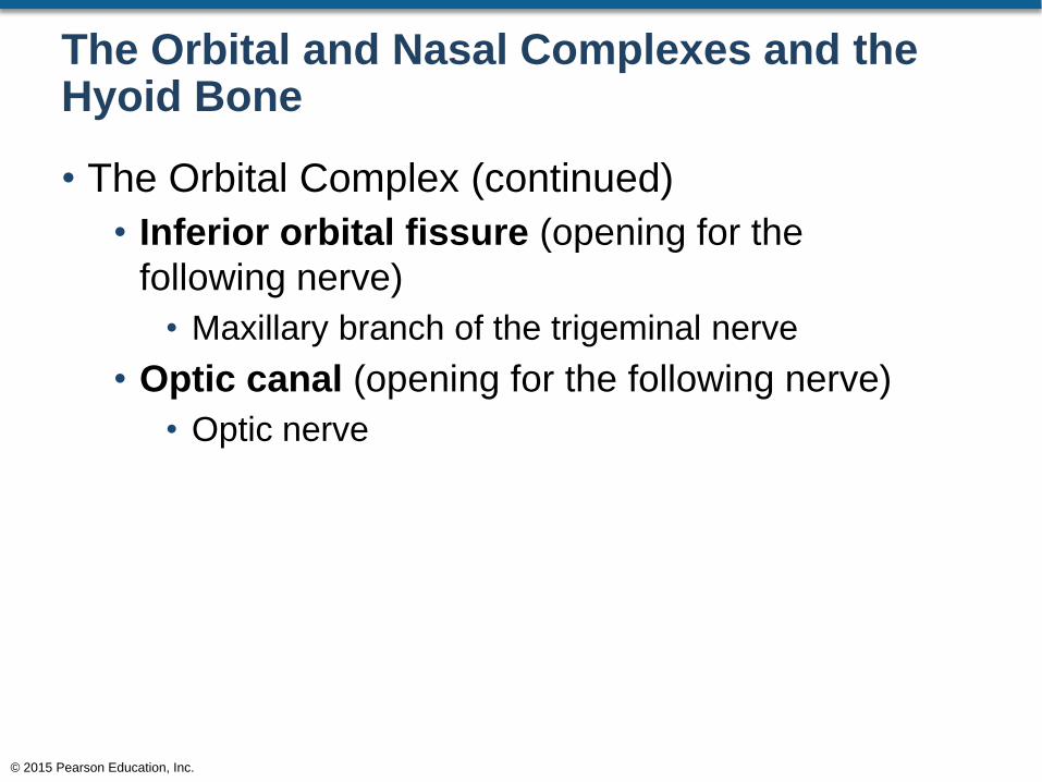
The Orbital and Nasal Complexes and the Hyoid Bone
• The Orbital Complex (continued)
• Inferior orbital fissure (opening for the
following nerve)
• Maxillary branch of the trigeminal nerve
• Optic canal (opening for the following nerve)
• Optic nerve
© 2015 Pearson Education, Inc.
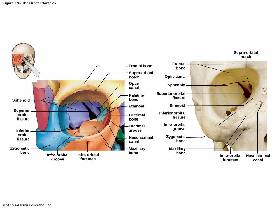
Figure 6.15 The Orbital Complex
© 2015 Pearson Education, Inc.
Sphenoid
Superior orbital fissure
Inferior orbital fissure
Zygomatic bone
Infra-orbital groove
Infra-orbital foramen
Frontal bone
Supra-orbital notch
Optic canal
Palatine bone
Ethmoid
Lacrimal bone
Lacrimal groove
Nasolacrimal canal
Maxillary bone
Sphenoid
Superior orbital fissure
Inferior orbital fissure
Zygomatic bone
Ethmoid
Infra-orbital groove
Frontal bone
Optic canal
Maxillary bone
Infra-orbital foramen
Nasolacrimal canal
Supra-orbital notch
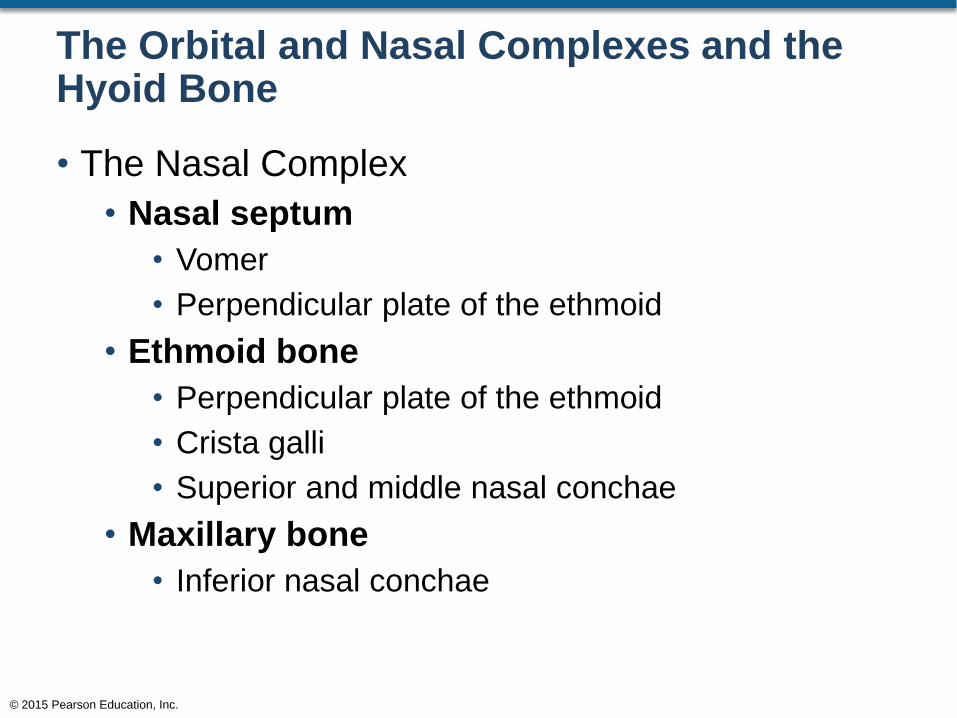
The Orbital and Nasal Complexes and the Hyoid Bone
• The Nasal Complex
• Nasal septum
• Vomer
• Perpendicular plate of the ethmoid
• Ethmoid bone
• Perpendicular plate of the ethmoid
• Crista galli
• Superior and middle nasal conchae
• Maxillary bone
• Inferior nasal conchae
© 2015 Pearson Education, Inc.

Figure 6.5 Sectional Anatomy of the Skull, Part II (1 of 2)
© 2015 Pearson Education, Inc.
Sphenoid
Coronal suture
Frontal bone
Sphenoidal sinus (right)
Frontal sinus
Crista galli
Nasal bone
Perpendicular plate of ethmoid
Vomer
Palatine bone
Maxilla
Mandible
Sagittal section
Parietal bone
Squamous suture
Temporal bone
Lambdoid suture
Hypophyseal fossa
of sella turcica
Internal acoustic meatus
Occipital bone
Hypoglossal canal
Styloid process
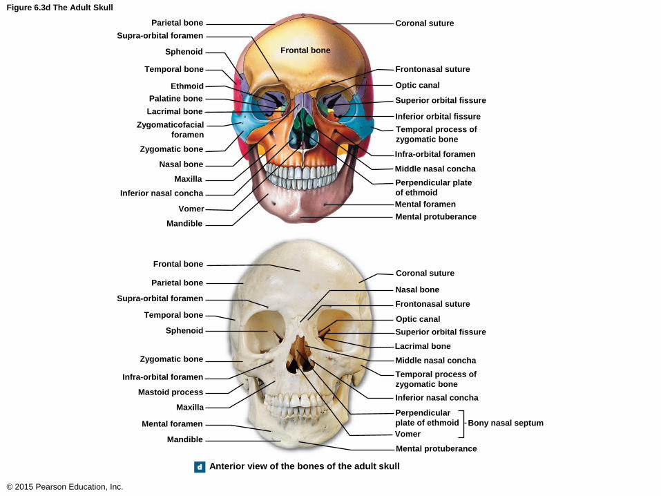
Figure 6.3d The Adult Skull
© 2015 Pearson Education, Inc.
Frontal bone
d
Parietal bone
Supra-orbital foramen
Sphenoid
Temporal bone
Ethmoid
Palatine bone
Lacrimal bone
Zygomaticofacial
foramen
Zygomatic bone
Nasal bone
Maxilla
Inferior nasal concha
Vomer
Mandible
Coronal suture
Frontonasal suture
Optic canal
Superior orbital fissure
Inferior orbital fissure
Temporal process of
zygomatic bone
Infra-orbital foramen
Middle nasal concha
Perpendicular plate
of ethmoid
Mental foramen
Mental protuberance
Frontal bone
Parietal bone
Supra-orbital foramen
Temporal bone
Sphenoid
Zygomatic bone
Infra-orbital foramen
Mastoid process
Maxilla
Mental foramen
Mandible
Coronal suture
Nasal bone
Frontonasal suture
Optic canal
Superior orbital fissure
Lacrimal bone
Middle nasal concha
Temporal process of
zygomatic bone
Inferior nasal concha
Perpendicular
plate of ethmoid
Vomer
Bony nasal septum
Mental protuberance
Anterior view of the bones of the adult skull
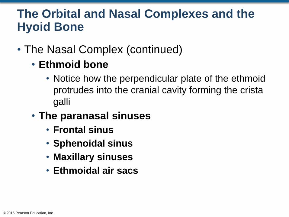
The Orbital and Nasal Complexes and the Hyoid Bone
• The Nasal Complex (continued)
• Ethmoid bone
• Notice how the perpendicular plate of the ethmoid
protrudes into the cranial cavity forming the crista
galli
• The paranasal sinuses
• Frontal sinus
• Sphenoidal sinus
• Maxillary sinuses
• Ethmoidal air sacs
© 2015 Pearson Education, Inc.
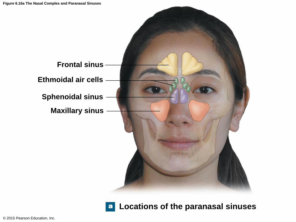
Figure 6.16a The Nasal Complex and Paranasal Sinuses
© 2015 Pearson Education, Inc.
a
Frontal sinus
Ethmoidal air cells
Sphenoidal sinus
Maxillary sinus
Locations of the paranasal sinuses
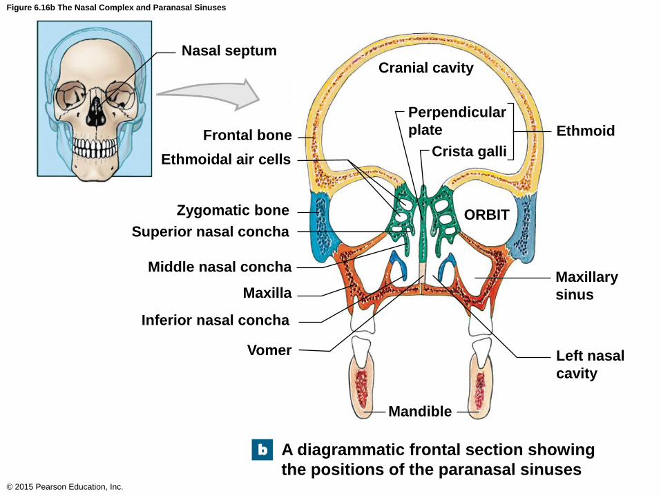
Figure 6.16b The Nasal Complex and Paranasal Sinuses
© 2015 Pearson Education, Inc.
Vomer
Nasal septum
Frontal bone
Ethmoidal air cells
Zygomatic bone
Superior nasal concha
Middle nasal concha
Maxilla
Inferior nasal concha
Cranial cavity
Perpendicular
plate
Crista galli
ORBIT
Mandible
Left nasal
cavity
Maxillary
sinus
Ethmoid
A diagrammatic frontal section showing
the positions of the paranasal sinuses
b
The Orbital and Nasal Complexes and the Hyoid Bone
• The Hyoid Bone
• Does not articulate with any other bone
• It is suspended inferior to the skull
• The inferior portion is connected to the
thyrohyoid ligament
• The superior portion is suspended from the
mandible via muscles
• Stylohyoid muscle
• Digastric muscle
© 2015 Pearson Education, Inc.
The Orbital and Nasal Complexes and the Hyoid Bone
• The Hyoid Bone (continued)
• Bony projections of the hyoid bone
• Greater horn
• Lesser horn
• Body
© 2015 Pearson Education, Inc.
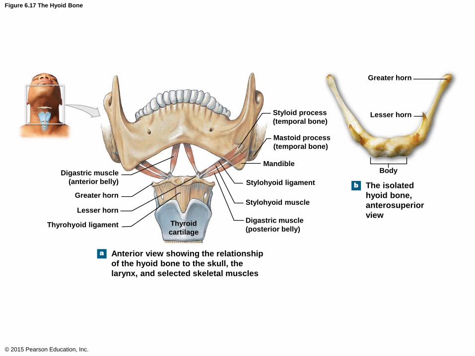
Figure 6.17 The Hyoid Bone
© 2015 Pearson Education, Inc.
Greater horn
a
Lesser horn
Body
The isolated
hyoid bone,
anterosuperior
view
Greater horn
Digastric muscle
(anterior belly)
Lesser horn
Thyrohyoid ligament Thyroid
cartilage
Styloid process
(temporal bone)
Mastoid process
(temporal bone)
Mandible
Stylohyoid ligament
Stylohyoid muscle
Digastric muscle
(posterior belly)
Anterior view showing the relationship
of the hyoid bone to the skull, the
larynx, and selected skeletal muscles
b
The Skulls of Infants, Children, and Adults
• Major features of the infant skull
• 4 major fontanel areas
• Membranous areas where sutures will eventually
form
• Allow for distortion of the skull during childbirth
• Anterior fontanel (baby’s “soft spot”)
• Posterior fontanel
• Sphenoidal fontanels
• Mastoid fontanels
© 2015 Pearson Education, Inc.
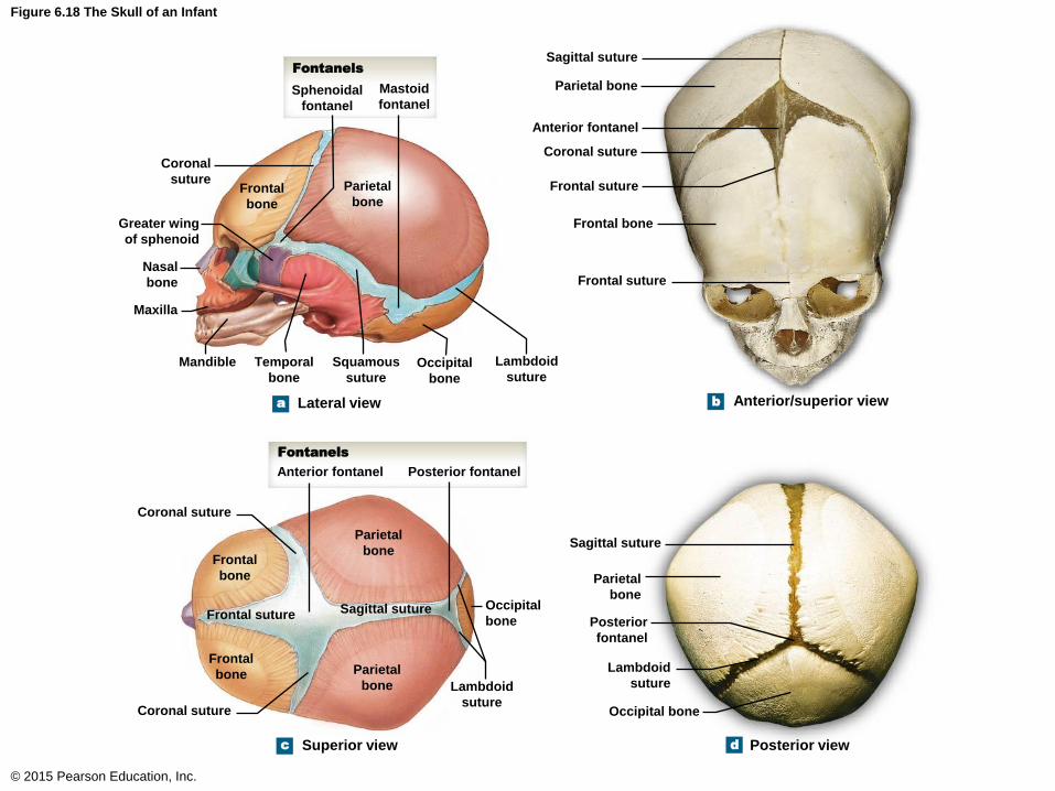
Figure 6.18 The Skull of an Infant
© 2015 Pearson Education, Inc.
Mandible
a b
d c
Sphenoidal
fontanel
Mastoid
fontanel
Fontanels
Coronal
suture Frontal
bone
Parietal
bone
Greater wing
of sphenoid
Nasal
bone
Maxilla
Temporal
bone
Squamous
suture Occipital
bone
Lambdoid
suture
Sagittal suture
Parietal bone
Anterior fontanel
Coronal suture
Frontal suture
Frontal bone
Frontal suture
Lateral view Anterior/superior view
Fontanels
Anterior fontanel
Coronal suture
Frontal
bone
Parietal
bone
Frontal suture Sagittal suture Occipital
bone
Lambdoid
suture
Parietal
bone
Frontal
bone
Coronal suture
Superior view
Sagittal suture
Parietal
bone
Posterior
fontanel
Lambdoid
suture
Occipital bone
Posterior view
Posterior fontanel
Review of the Skull
• There are 22 bones of the skull
• Facial bones
• Maxillae – 2
• Palatine bones – 2
• Nasal bones – 2
• Inferior nasal conchae – 2
• Zygomatic bones – 2
• Lacrimal bones – 2
• Vomer – 1
• Mandible – 1
© 2015 Pearson Education, Inc.
Review of the Skull (continued)
• There are 22 bones of the skull
• Cranial bones
• Occipital bone – 1
• Parietal bones – 2
• Frontal bone – 1
• Temporal bones – 2
• Sphenoid bone – 1
• Ethmoid bone – 1
© 2015 Pearson Education, Inc.
Review of the Skull (continued)
• There are 7 associated bones of the skull
• Associated bones
• Auditory ossicles – 6
• Hyoid bone – 1
© 2015 Pearson Education, Inc.
The Vertebral Column
• The adult vertebral column is made up of 26
bones
• 24 vertebrae
• 7 cervical vertebrae
• 12 thoracic vertebrae
• 5 lumbar vertebrae
• 1 sacrum (5 fused vertebrae)
• 1 coccyx (3 to 5 fused vertebrae)
© 2015 Pearson Education, Inc.
The Vertebral Column
• Functional Anatomy of the Vertebral Column
• Encloses and protects the spinal cord
• Supports the skull
• Supports the weight of the head, neck, and trunk
• Transfers weight to the lower limbs
• Helps maintain the upright position of the body
© 2015 Pearson Education, Inc.

Figure 6.19ab The Vertebral Column
© 2015 Pearson Education, Inc.
a The major divisions of the
vertebral column showing
the four adult spinal curves
b Normal vertebral
column, lateral view
Cervical
Thoracic
Regions are defined
by anatomical
characteristics of
individual vertebrae.
VERTEBRAL REGIONS
Lumbar
Sacral
Coccygeal
a
Primary curves
develop before birth,
and secondary curves
after birth.
SPINAL CURVES
Cervical curve.
A secondary curve;
develops as the infant
learns to balance the
Weight of the head on
the vertebrae of the neck.
Thoracic curve.
A primary curve;
accommodates
the thoracic
organs.
Lumbar curve.
A secondary curve;
balances the weight of
the trunk over the lower
limbs; it develops with
the ability to stand.
Sacral curve.
A primary curve;
accommodates
the abdomino-
pelvic organs.
The Vertebral Column
• Spinal Curves
• There are 4 major curves of the vertebral column
• Cervical curve
• Thoracic curve
• Lumbar curve
• Sacral curve
• These curves, along with muscle attachment to
the various vertebral processes, help to maintain
balance
© 2015 Pearson Education, Inc.
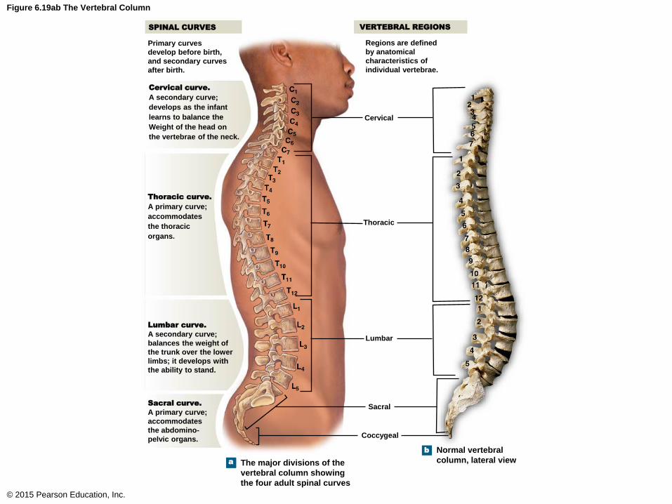
Figure 6.19ab The Vertebral Column
© 2015 Pearson Education, Inc.
a The major divisions of the
vertebral column showing
the four adult spinal curves
b Normal vertebral
column, lateral view
Cervical
Thoracic
Regions are defined
by anatomical
characteristics of
individual vertebrae.
VERTEBRAL REGIONS
Lumbar
Sacral
Coccygeal
a
Primary curves
develop before birth,
and secondary curves
after birth.
SPINAL CURVES
Cervical curve.
A secondary curve;
develops as the infant
learns to balance the
Weight of the head on
the vertebrae of the neck.
Thoracic curve.
A primary curve;
accommodates
the thoracic
organs.
Lumbar curve.
A secondary curve;
balances the weight of
the trunk over the lower
limbs; it develops with
the ability to stand.
Sacral curve.
A primary curve;
accommodates
the abdomino-
pelvic organs.
The Vertebral Column
• Abnormal curvatures of the vertebral column
• Scoliosis
• Abnormal lateral curvature
• Kyphosis
• Exaggerated posterior curvature of the thoracic
region
• Lordosis
• Exaggerated anterior curvature of the lumbar
region
© 2015 Pearson Education, Inc.

Clinical Note 6.1 Kyphosis, Lordosis, and Scoliosis
© 2015 Pearson Education, Inc.
Kyphosis Scoliosis
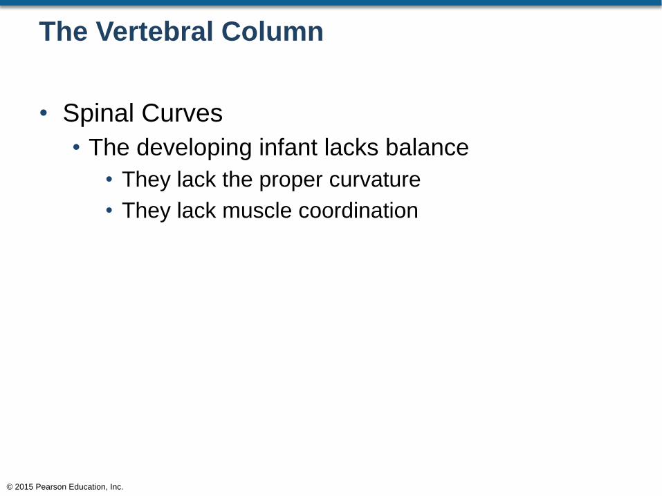
The Vertebral Column
• Spinal Curves
• The developing infant lacks balance
• They lack the proper curvature
• They lack muscle coordination
© 2015 Pearson Education, Inc.
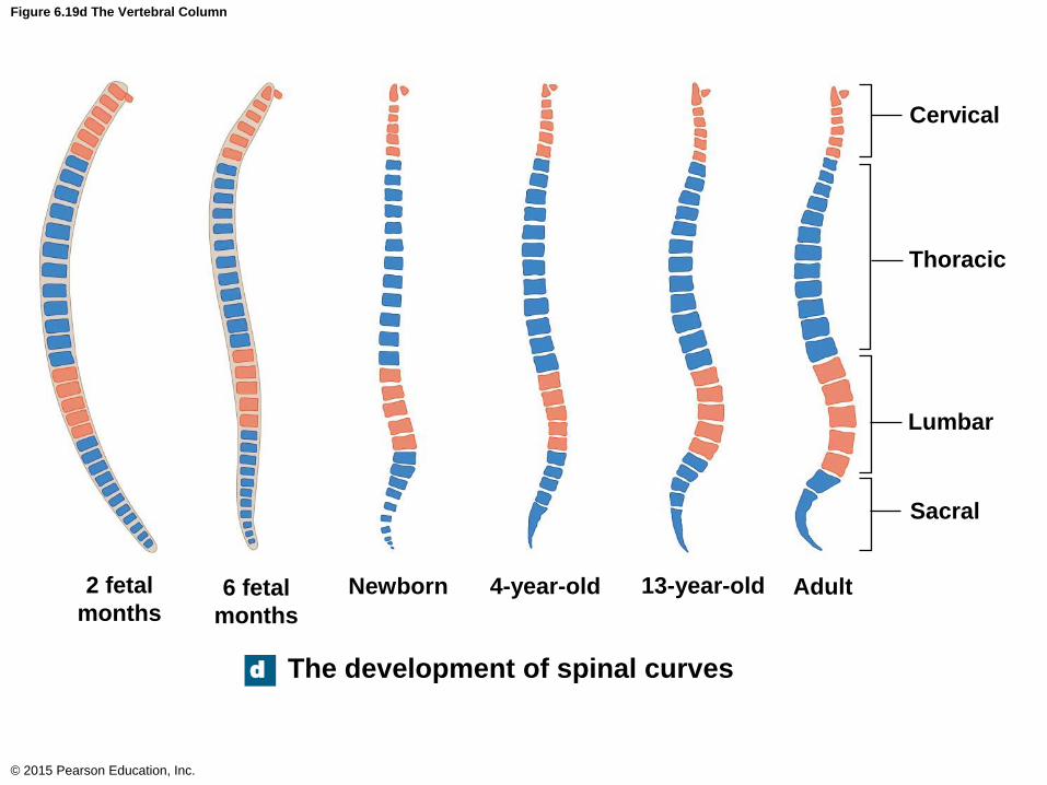
Figure 6.19d The Vertebral Column
© 2015 Pearson Education, Inc.
d The development of spinal curves
Cervical
Thoracic
Lumbar
Sacral
2 fetal
months 6 fetal
months
Newborn 4-year-old 13-year-old Adult
The Vertebral Column
• Vertebral Anatomy
• The vertebral body
• Supports weight along the axis of the body
• An anterior structure
• A vertebral body is separated from another
vertebral body by a pad of cartilage called the
intervertebral disc
© 2015 Pearson Education, Inc.
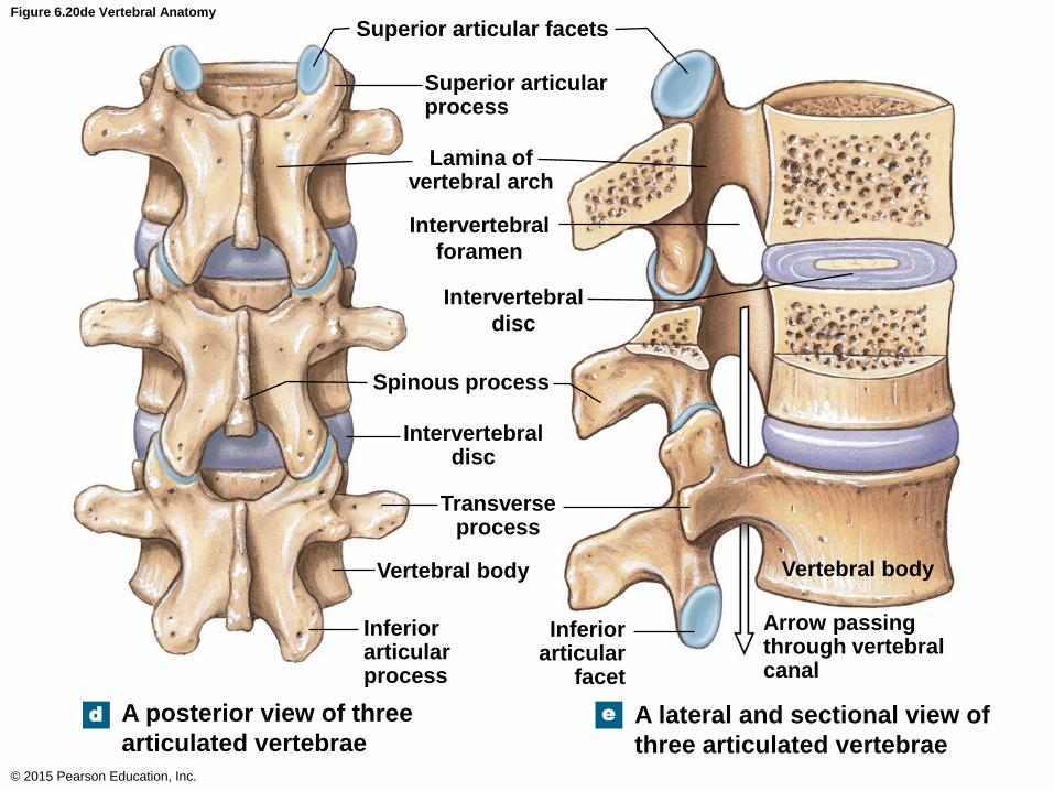
Figure 6.20de Vertebral Anatomy
© 2015 Pearson Education, Inc.
d
Superior articular facets
Superior articular process
Lamina of vertebral arch
Spinous process
Intervertebral disc
Transverse process
Vertebral body
Inferior articular process
A posterior view of three
articulated vertebrae
Intervertebral
foramen
Intervertebral
disc
Inferior articular
facet
Vertebral body
Arrow passing through vertebral canal
A lateral and sectional view of
three articulated vertebrae
e
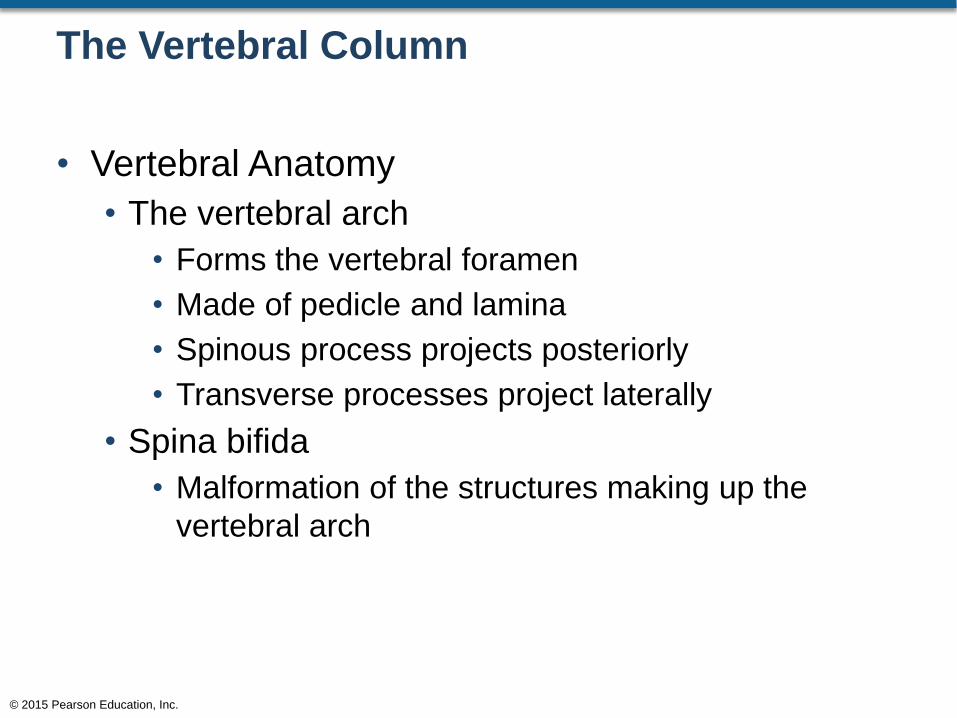
The Vertebral Column
• Vertebral Anatomy
• The vertebral arch
• Forms the vertebral foramen
• Made of pedicle and lamina
• Spinous process projects posteriorly
• Transverse processes project laterally
• Spina bifida
• Malformation of the structures making up the
vertebral arch
© 2015 Pearson Education, Inc.

Figure 6.23a Thoracic Vertebrae
© 2015 Pearson Education, Inc.
T2
T3
T4
T5
T6
T7
T8
T9
T10
T11
T12
a
Spinous process of vertebra prominens
Intervertebral foramen
Lateral view of the
thoracic region of the
vertebral column. The
vertebra prominens
(C7) resembles T1, but
it lacks facets for rib
articulation. Vertebra
T12 resembles the first
lumbar vertebra (L1),
but it has a facet for
rib articulation.
C7
T1
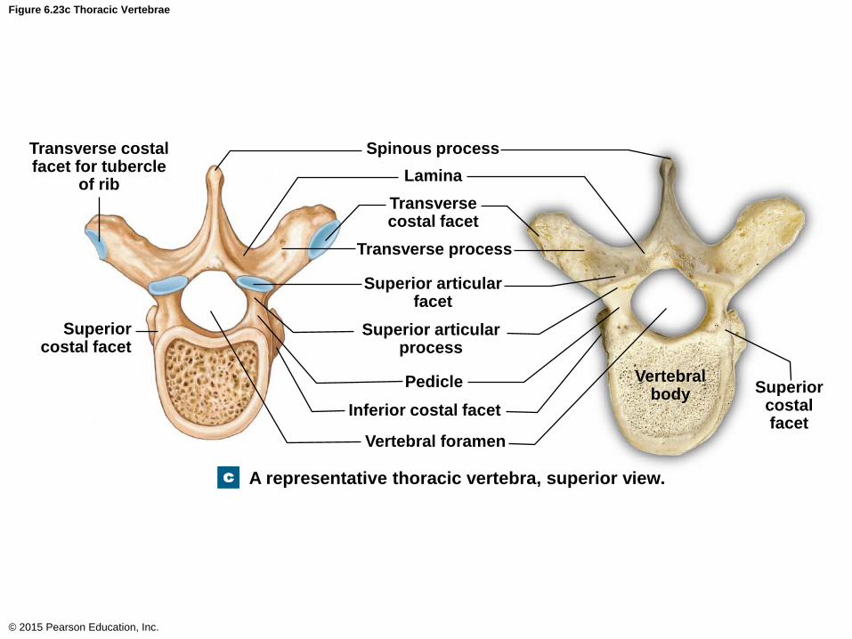
Figure 6.23c Thoracic Vertebrae
© 2015 Pearson Education, Inc.
c
Spinous process
Lamina
Transverse costal facet
Transverse process
Superior articular facet
Superior articular process
Pedicle
Inferior costal facet
Vertebral foramen
Superior costal facet
Transverse costal facet for tubercle
of rib
Vertebral body Superior
costal facet
A representative thoracic vertebra, superior view.
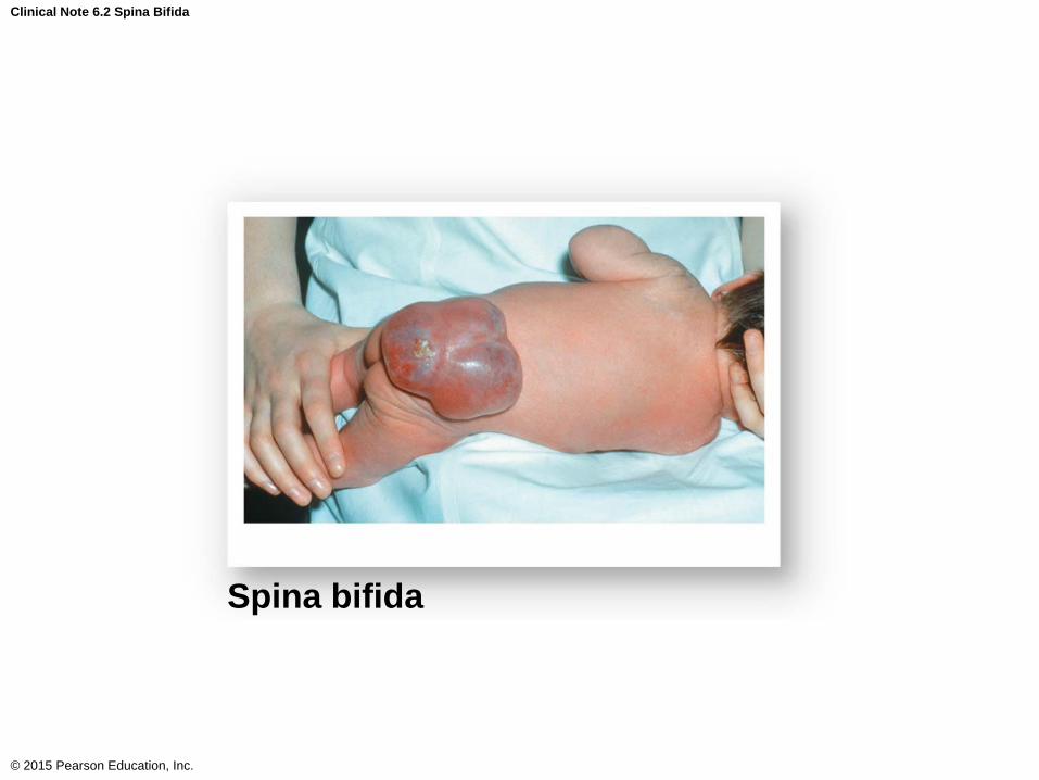
Clinical Note 6.2 Spina Bifida
© 2015 Pearson Education, Inc.
Spina bifida
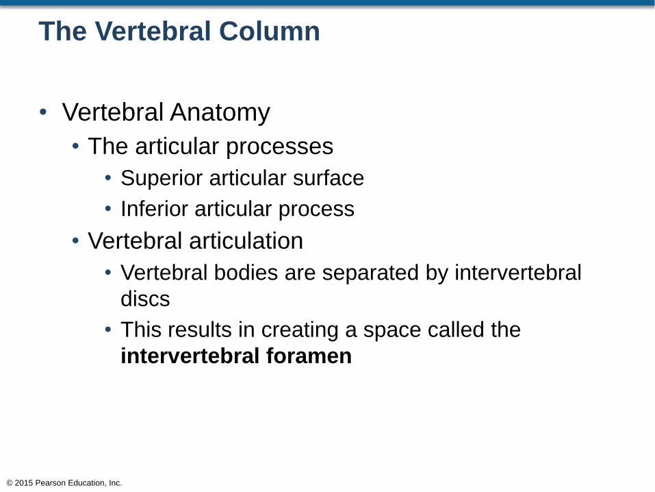
The Vertebral Column
• Vertebral Anatomy
• The articular processes
• Superior articular surface
• Inferior articular process
• Vertebral articulation
• Vertebral bodies are separated by intervertebral
discs
• This results in creating a space called the
intervertebral foramen
© 2015 Pearson Education, Inc.
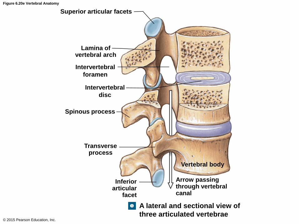
Figure 6.20e Vertebral Anatomy
© 2015 Pearson Education, Inc.
Superior articular facets
Lamina of vertebral arch
Spinous process
Transverse process
Intervertebral
foramen
Intervertebral
disc
Inferior articular
facet
Vertebral body
Arrow passing through vertebral canal
A lateral and sectional view of
three articulated vertebrae
e
The Vertebral Column
• Vertebral Regions
• Numbering system of vertebrae
• Cervical region
• C1, C2, C3, etc.
• Thoracic region
• T1, T2, T3, etc.
• Lumbar region
• L1, L2, L3, etc.
© 2015 Pearson Education, Inc.
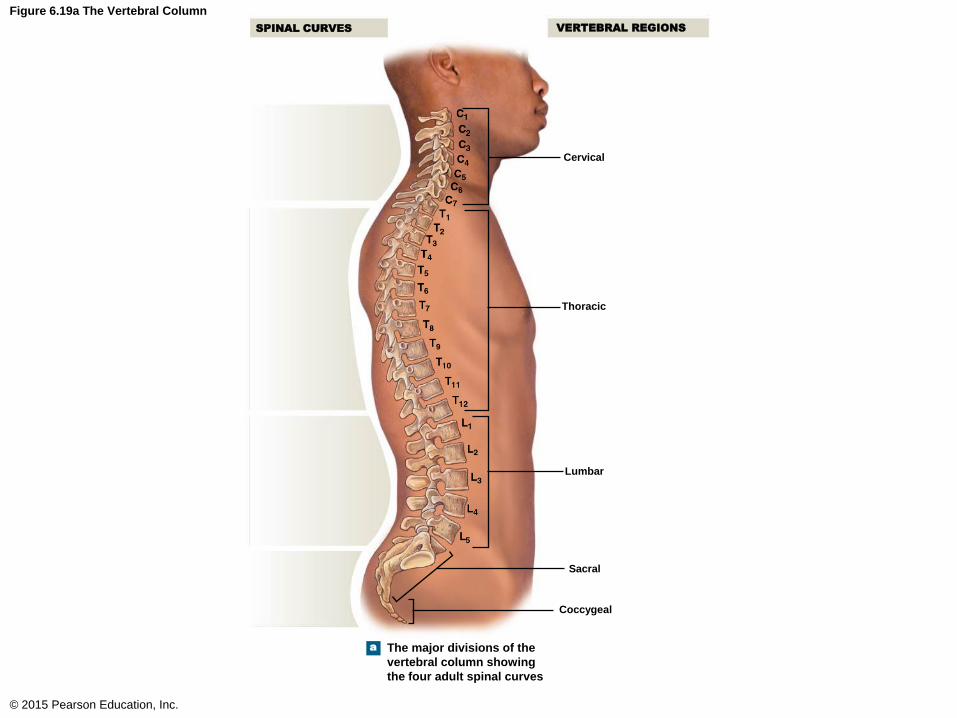
Figure 6.19a The Vertebral Column
© 2015 Pearson Education, Inc.
Cervical
Thoracic
VERTEBRAL REGIONS
Lumbar
Sacral
Coccygeal
a The major divisions of the
vertebral column showing
the four adult spinal curves
SPINAL CURVES
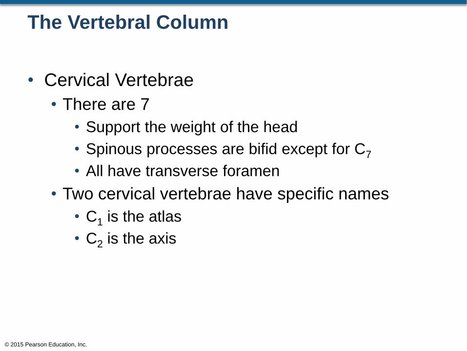
The Vertebral Column
• Cervical Vertebrae
• There are 7
• Support the weight of the head
• Spinous processes are bifid except for C7
• All have transverse foramen
• Two cervical vertebrae have specific names
• C1 is the atlas
• C2 is the axis
© 2015 Pearson Education, Inc.
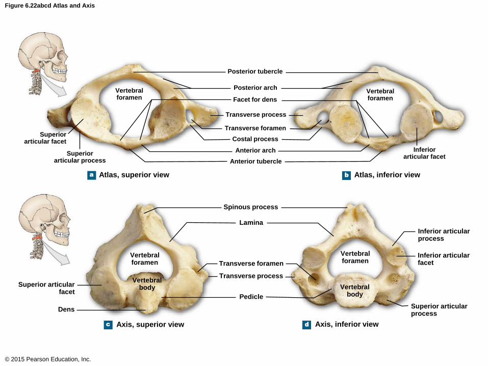
Figure 6.22abcd Atlas and Axis
© 2015 Pearson Education, Inc.
Posterior tubercle
a b
Posterior arch
Facet for dens
Transverse process
Transverse foramen
Costal process
Anterior arch
Anterior tubercle
Vertebral foramen
Superior articular facet
Superior articular process
Vertebral foramen
Inferior articular facet
Atlas, superior view Atlas, inferior view
c d
Vertebral foramen
Vertebral foramen
Vertebral body Vertebral
body
Spinous process
Lamina
Transverse foramen
Transverse process
Pedicle
Superior articular facet
Dens
Axis, superior view Axis, inferior view
Inferior articular process
Inferior articular facet
Superior articular process

The Vertebral Column
• The Atlas (C1)
• Articulates with the occipital condyles of the skull
• Does not have a body
• Has the largest vertebral foramen of all vertebrae
• Allows the head to nod in a “yes” manner
• The Axis (C2)
• Has a dens
• The transverse ligament binds the dens to the
atlas
• Allows the head to move in a “no” manner
© 2015 Pearson Education, Inc.
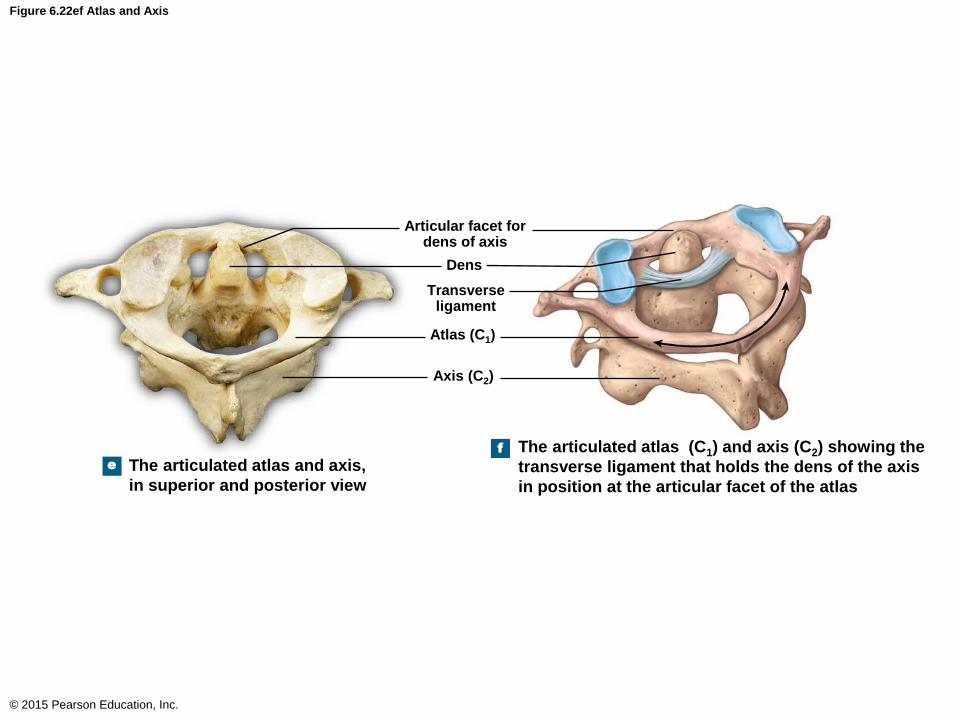
Figure 6.22ef Atlas and Axis
© 2015 Pearson Education, Inc.
e
f
Articular facet for dens of axis
Dens
Transverse ligament
Atlas (C1)
Axis (C2)
The articulated atlas and axis,
in superior and posterior view
The articulated atlas (C1) and axis (C2) showing the
transverse ligament that holds the dens of the axis
in position at the articular facet of the atlas
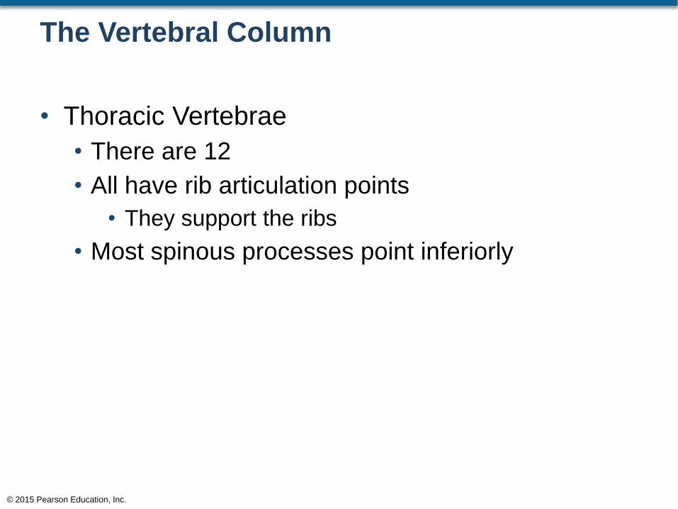
The Vertebral Column
• Thoracic Vertebrae
• There are 12
• All have rib articulation points
• They support the ribs
• Most spinous processes point inferiorly
© 2015 Pearson Education, Inc.
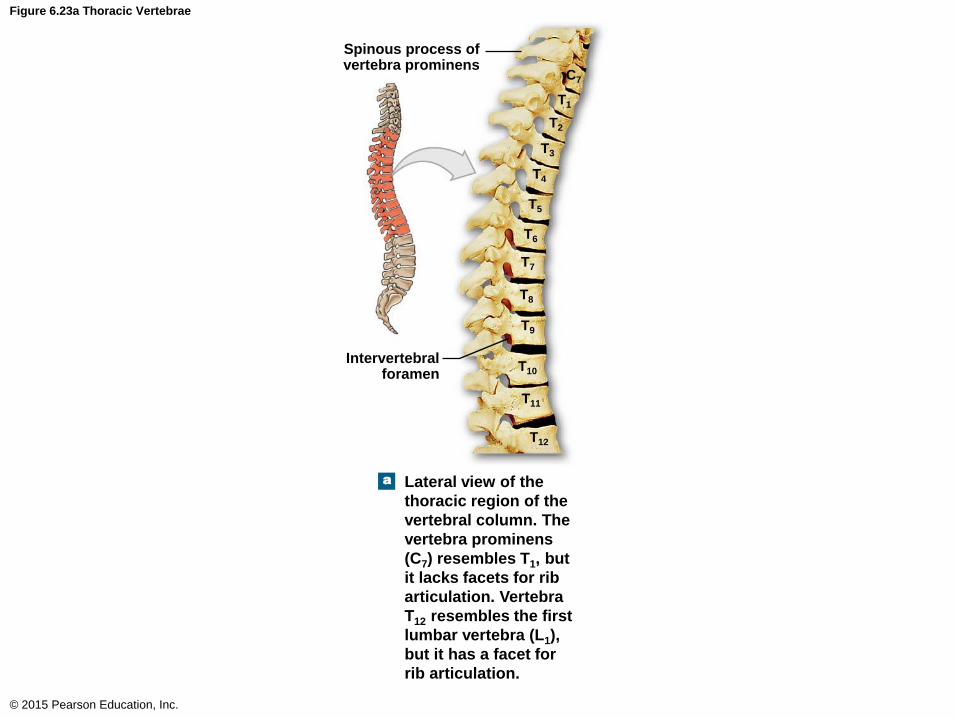
Figure 6.23a Thoracic Vertebrae
© 2015 Pearson Education, Inc.
T2
T3
T4
T5
T6
T7
T8
T9
T10
T11
T12
a
Spinous process of vertebra prominens
Intervertebral foramen
Lateral view of the
thoracic region of the
vertebral column. The
vertebra prominens
(C7) resembles T1, but
it lacks facets for rib
articulation. Vertebra
T12 resembles the first
lumbar vertebra (L1),
but it has a facet for
rib articulation.
C7
T1

Figure 6.23c Thoracic Vertebrae
© 2015 Pearson Education, Inc.
c
Spinous process
Lamina
Transverse costal facet
Transverse process
Superior articular facet
Superior articular process
Pedicle
Inferior costal facet
Vertebral foramen
Superior costal facet
Transverse costal facet for tubercle
of rib
Vertebral body Superior
costal facet
A representative thoracic vertebra, superior view.
The Vertebral Column
• Lumbar Vertebrae
• There are 5
• Support the weight of the torso
• Vertebral bodies are quite large
• Spinous process points posteriorly
© 2015 Pearson Education, Inc.
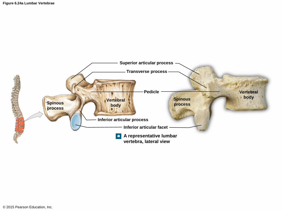
Figure 6.24a Lumbar Vertebrae
© 2015 Pearson Education, Inc.
Pedicle
a
Superior articular process
Transverse process
Spinous
process
Vertebral
body
Inferior articular process
Inferior articular facet
A representative lumbar
vertebra, lateral view
Vertebral
body Spinous
process
The Vertebral Column
• The Sacrum
• There is one sacrum but consists of five fused
vertebrae
• Structures of the sacrum
• Sacral hiatus
• Median sacral crest
• Entrance to sacral canal
• Sacral foramina
© 2015 Pearson Education, Inc.
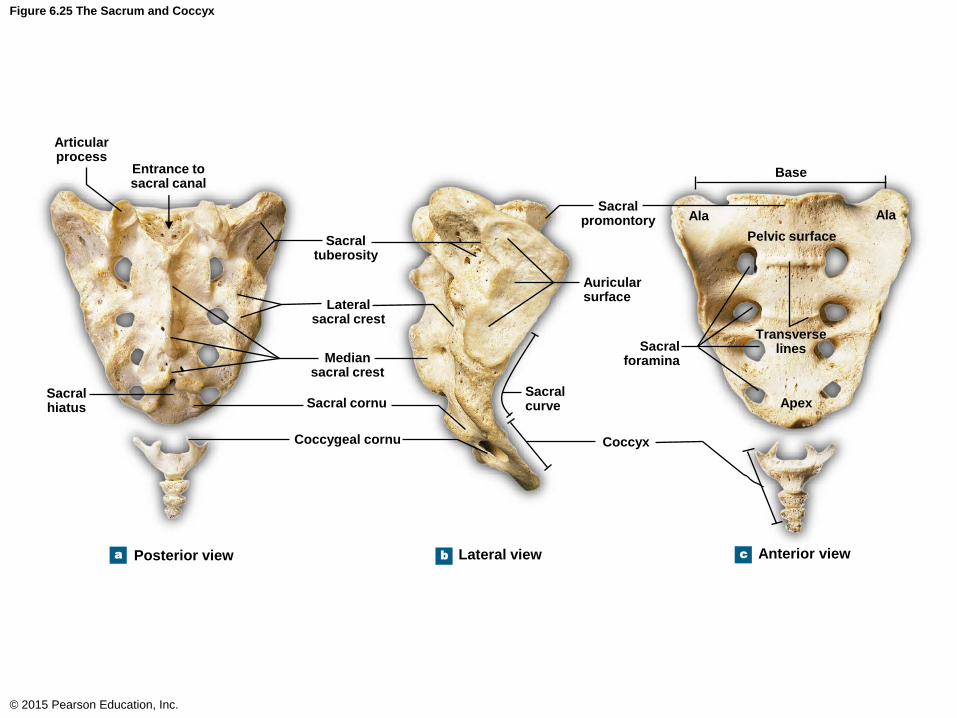
Figure 6.25 The Sacrum and Coccyx
© 2015 Pearson Education, Inc.
Articular process
b a c
Entrance to sacral canal
Sacral hiatus
Sacral tuberosity
Lateral sacral crest
Median sacral crest
Sacral cornu
Coccygeal cornu
Sacral promontory
Auricular surface
Sacral curve
Sacral foramina
Coccyx
Base
Ala Ala
Pelvic surface
Transverse lines
Apex
Posterior view Lateral view Anterior view
The Vertebral Column
• The Coccyx
• Consists of three to five fused vertebrae
• Adult male coccyx points anteriorly
• Adult female coccyx points inferiorly
• Coccyx consists of the coccygeal cornu
© 2015 Pearson Education, Inc.
The Thoracic Cage
• The thoracic cage has two functions
• It protects the heart, lungs, thymus, and other
structures within the cavity
• It serves as the attachment site for muscles
involved in:
• Respiration
• Positioning the vertebral column
• Movements of the pectoral girdle and upper limb
© 2015 Pearson Education, Inc.
The Thoracic Cage
• The Ribs
• Two types of rib classification
• Ribs (one type of classification)
• True ribs: 1–7
• False ribs: 8–12
• Ribs (another type of classification)
• Vertebrosternal ribs: 1–7
• Vertebrochondral ribs: 8–10
• Vertebral ribs (floating ribs): 11–12 (no anterior
cartilage)
© 2015 Pearson Education, Inc.
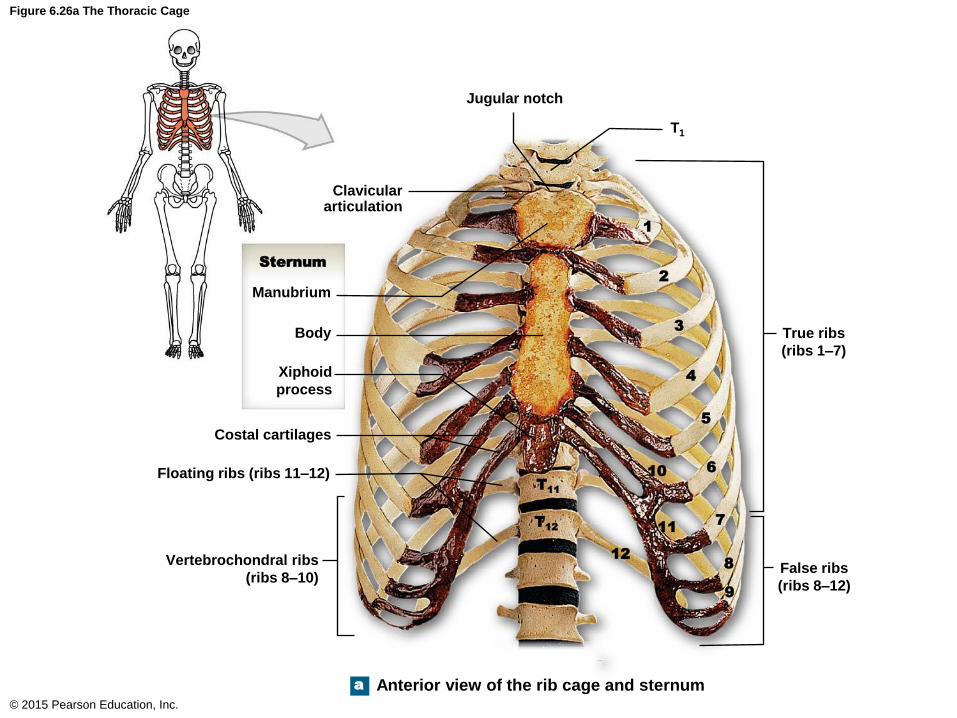
Figure 6.26a The Thoracic Cage
© 2015 Pearson Education, Inc.
a
Jugular notch
1
2
3
4
5
6
7
8
9
10
11
12
T11
T12
T1
Clavicular articulation
Sternum
Manubrium
Body
Xiphoid
process
Costal cartilages
Floating ribs (ribs 11–12)
Vertebrochondral ribs
(ribs 8–10)
True ribs
(ribs 1–7)
False ribs
(ribs 8–12)
Anterior view of the rib cage and sternum
The Thoracic Cage
• The Ribs
• 12 pairs of ribs
• Each rib articulates with a thoracic vertebra
• Structures of a rib
• Head
• Neck
• Tubercle
• Angle
• Costal groove
• Body
© 2015 Pearson Education, Inc.

Figure 6.26d The Thoracic Cage
© 2015 Pearson Education, Inc.
d A posterior and medial view showing
major anatomical landmarks on an
isolated left rib (rib 10)
Attachment to
costal cartilage
(sternal end)
Body
Articular
facets
Head
Neck Tubercle
Angle
Costal
groove

The Thoracic Cage
• The Sternum
• Consists of
• Manubrium
• Body
• Xiphoid
• Jugular notch
© 2015 Pearson Education, Inc.

Figure 6.26a The Thoracic Cage
© 2015 Pearson Education, Inc.
a
Jugular notch
1
2
3
4
5
6
7
8
9
10
11
12
T11
T12
T1
Clavicular articulation
Sternum
Manubrium
Body
Xiphoid
process
Costal cartilages
Floating ribs (ribs 11–12)
Vertebrochondral ribs
(ribs 8–10)
True ribs
(ribs 1–7)
False ribs
(ribs 8–12)
Anterior view of the rib cage and sternum