
CHAPTER 6 MUSCULAR SYSTEM. MUSCLE CELL TYPES Skeletal (striated) Cardiac Smooth (non-striated)
99
CHAPTER 6 MUSCULAR SYSTEM
-
Upload
barbra-sutton -
Category
Documents
-
view
222 -
download
1
Transcript of CHAPTER 6 MUSCULAR SYSTEM. MUSCLE CELL TYPES Skeletal (striated) Cardiac Smooth (non-striated)
C H A P T E R 6
MUSCULAR SYSTEM
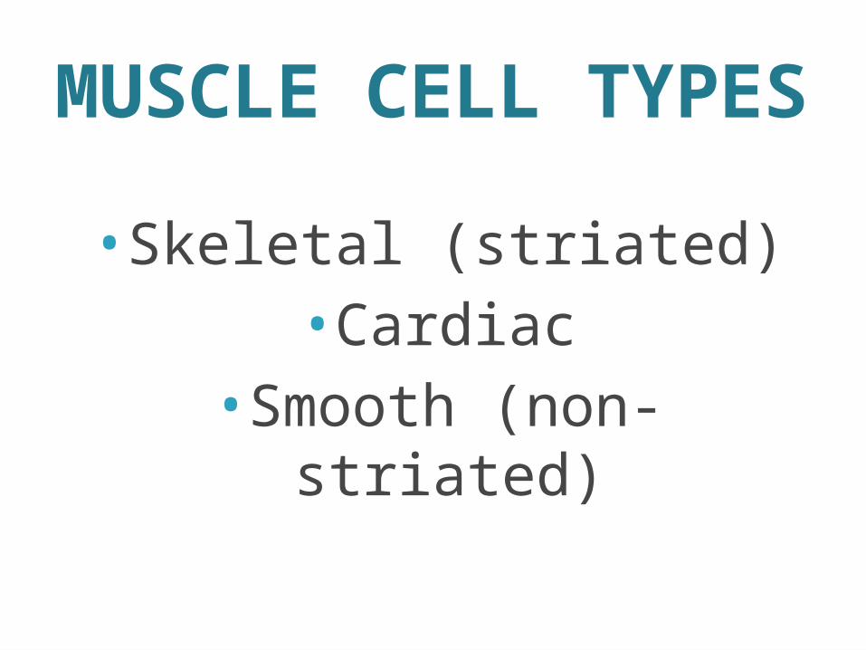
MUSCLE CELL TYPES
•Skeletal (striated)•Cardiac
•Smooth (non-striated)
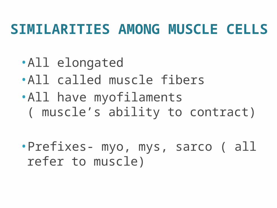
SIMILARITIES AMONG MUSCLE CELLS
• All elongated• All called muscle fibers• All have myofilaments ( muscle’s ability to contract)
• Prefixes- myo, mys, sarco ( all refer to muscle)
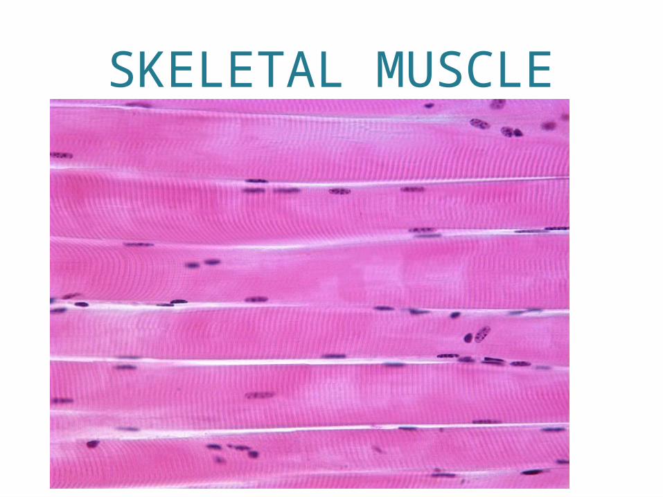
SKELETAL MUSCLE
•Attach to the body’s skeleton•Help form smooth contours of the body•Cigar-shaped•Multinucleated (many nuclei)•Largest of all muscle fibers
SKELETAL MUSCLE•Also known as striated muscle (appear to be striped)
•OR
•Voluntary muscle: only muscle type to conscious control.
SKELETAL MUSCLE
•Soft and fragile
•But, can exert tremendous power
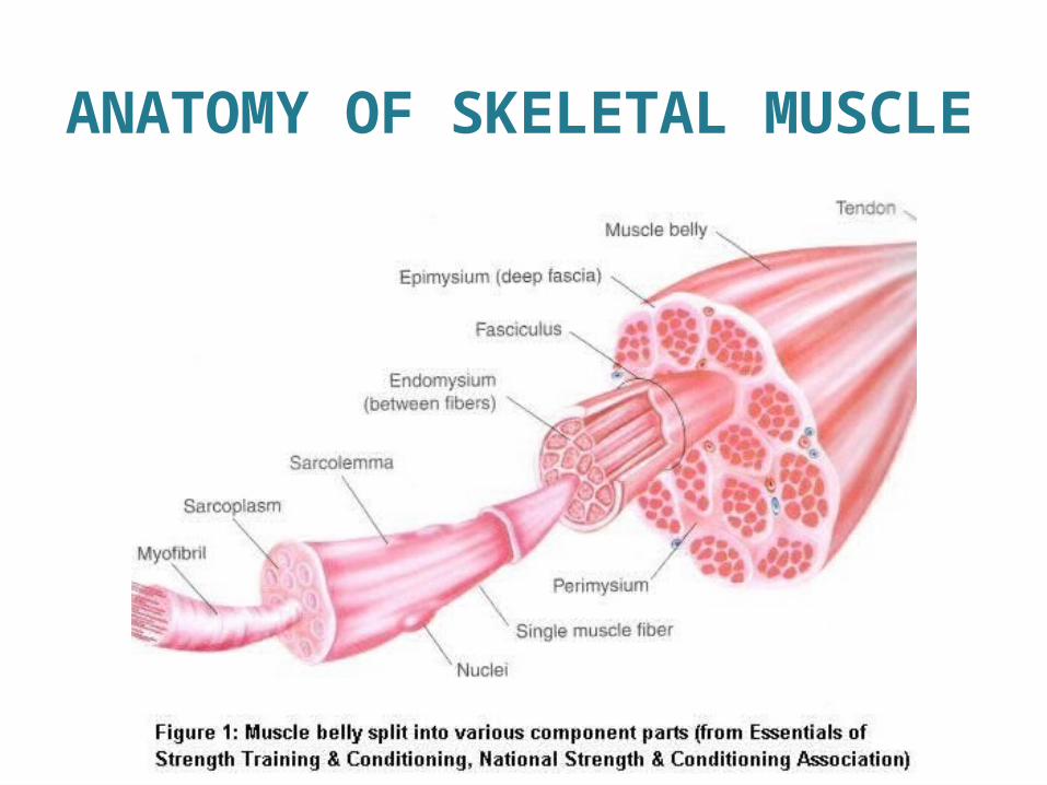
•Fibers are bound together by connective tissue.
SKELETAL MUSCLE• Endomysium- connective tissue sheath that encloses muscle fibers.
•Perimysium- coarse fibrous membrane that encloses several sheathed fibers.
• Fascicle- bundle of fibers.
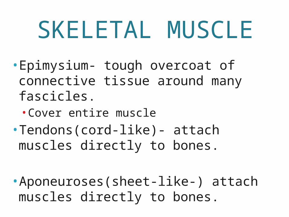
SKELETAL MUSCLE• Epimysium- tough overcoat of connective tissue around many fascicles.• Cover entire muscle
• Tendons(cord-like)- attach muscles directly to bones.
• Aponeuroses(sheet-like-) attach muscles directly to bones.
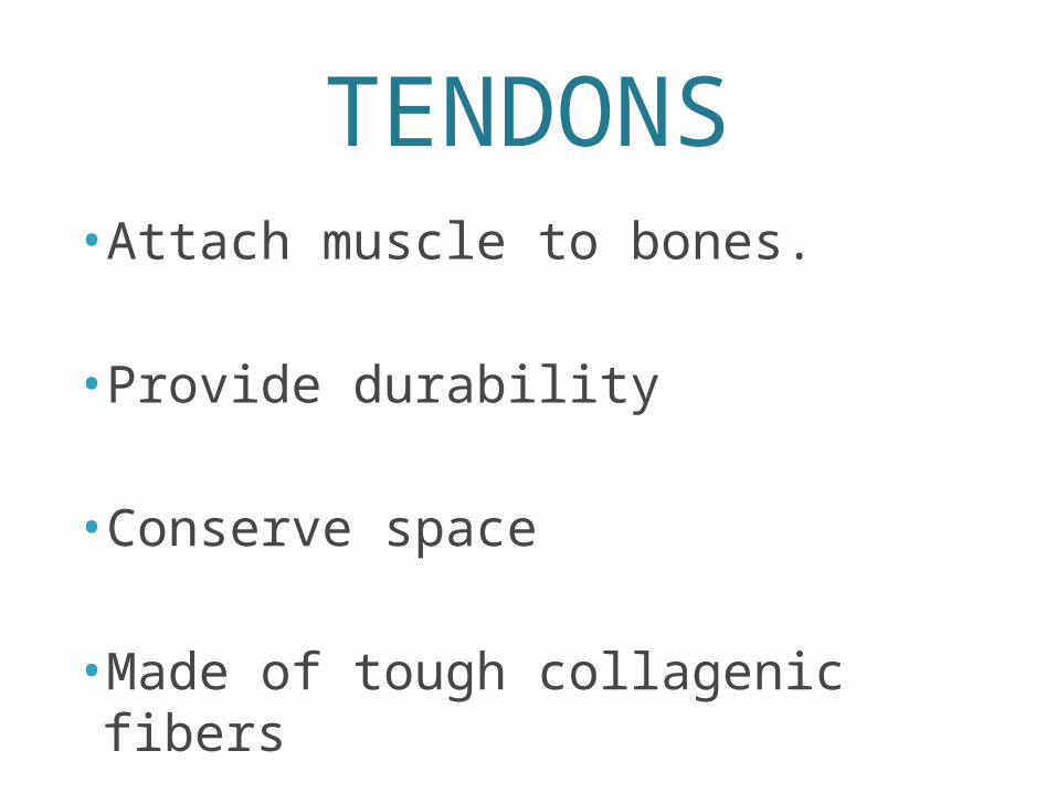
TENDONS•Attach muscle to bones.
•Provide durability
•Conserve space
•Made of tough collagenic fibers

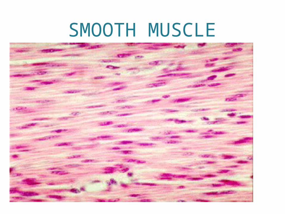
SMOOTH MUSCLE• Contains no striations.
• Involuntary muscle – we cannot consciously control it.
• Found mainly in the walls of hollow visceral organs.• Stomach, bladder, respiratory passages

SMOOTH MUSCLE• Propels substances along a definite track or
pathway.
• Using terms like visceral, nonstraited, involuntary
• Spindle shaped
• Single nuclei
• Arranged in sheets or layers
SMOOTH MUSCLE
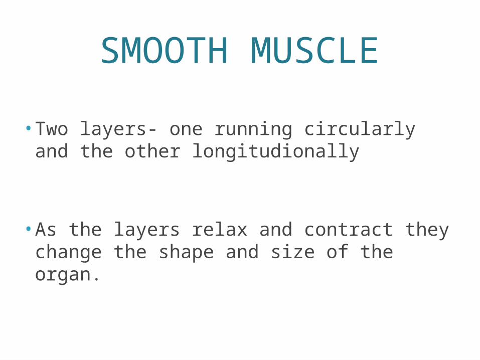
• Two layers- one running circularly and the other longitudionally
• As the layers relax and contract they change the shape and size of the organ.
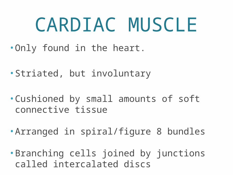
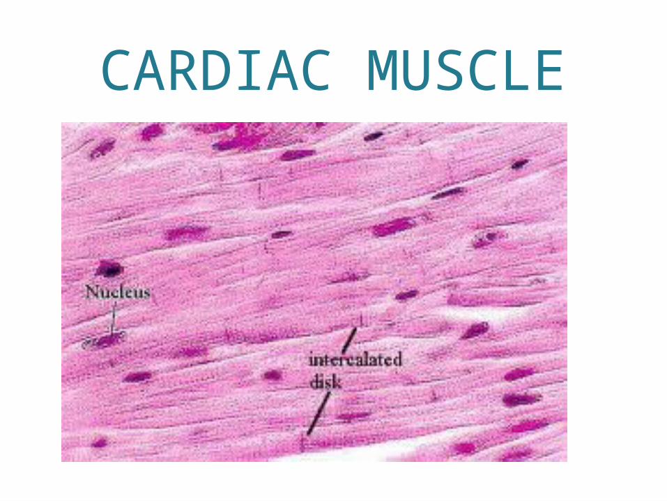
CARDIAC MUSCLE• Only found in the heart.
• Striated, but involuntary
• Cushioned by small amounts of soft connective tissue
• Arranged in spiral/figure 8 bundles
• Branching cells joined by junctions called intercalated discs

MUSCLE FUNCTIONS
•1. produce movement•2. maintains posture•3. stabilizes joints•4. generates heat
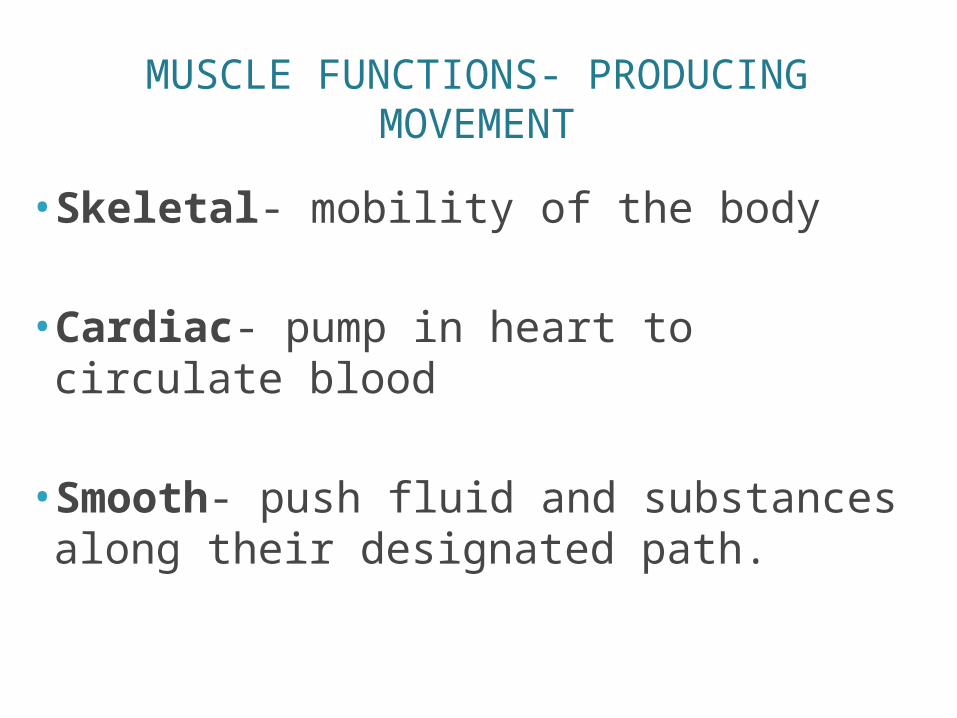
MUSCLE FUNCTIONS- PRODUCING MOVEMENT
•Skeletal- mobility of the body
•Cardiac- pump in heart to circulate blood
•Smooth- push fluid and substances along their designated path.
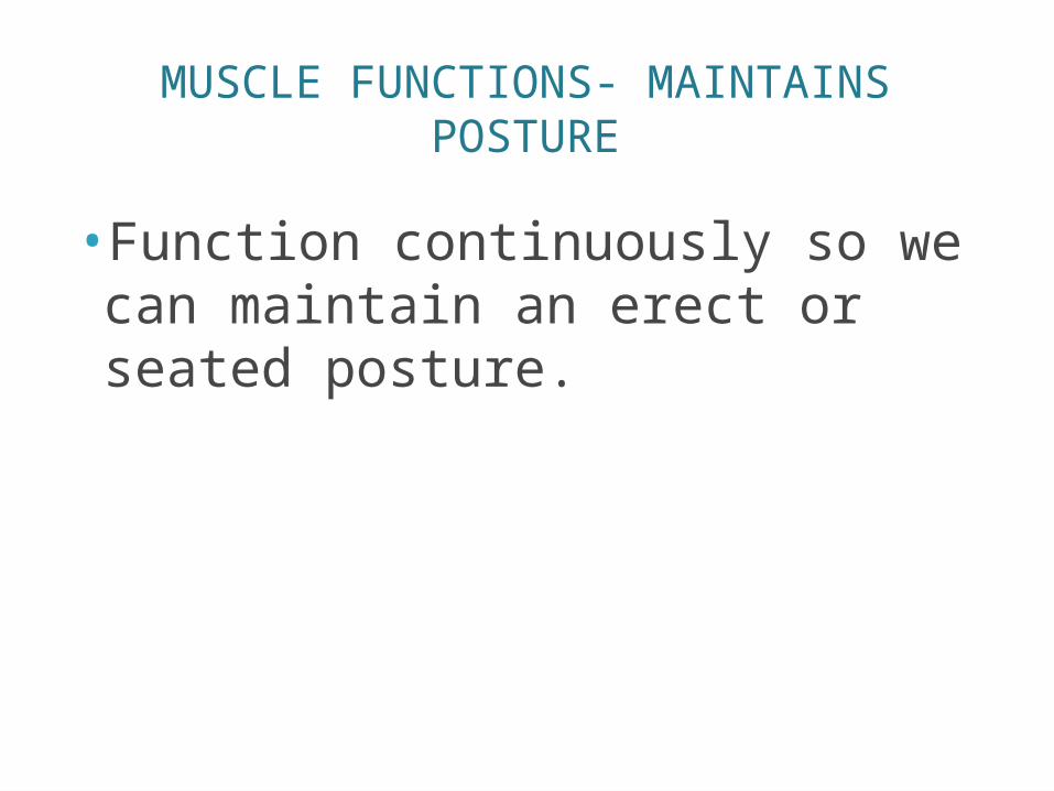
MUSCLE FUNCTIONS- MAINTAINS POSTURE
•Function continuously so we can maintain an erect or seated posture.
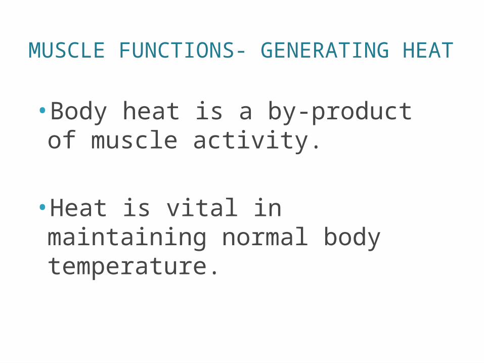
MUSCLE FUNCTIONS- GENERATING HEAT
•Body heat is a by-product of muscle activity.
•Heat is vital in maintaining normal body temperature.
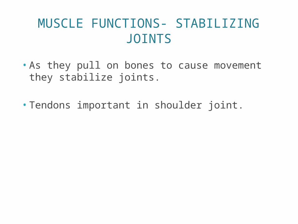
MUSCLE FUNCTIONS- STABILIZING JOINTS
• As they pull on bones to cause movement they stabilize joints.
• Tendons important in shoulder joint.
MICROSCOPIC ANATOMY OF SKELETAL MUSCLE
ANATOMY
• Sarcolemma – plasma membrane• Myofibrils- long ribbon-like organelles that
push nuclei to sides and nearly fill the whole cytoplasm.• Chains of tiny contractile units called sarcomeres
• Sarcomeres-aligned like boxcars in a train• Striped appearance comes from light (I) and
dark (A) bands along the muscle.• Sarcoplasmic reticulum- specialized smooth
ER• Stores calcium and releases it on demand
2 TYPES OF MYOFILAMENTS
• Thick filaments (made of protein myosin)
• Cross bridges- link thick and thin filaments together during contraction.
• Thin filaments (made of protein actin)
SKELETAL MUSCLE ACTIVITY
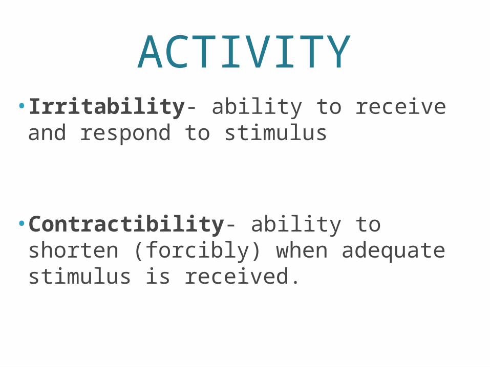
ACTIVITY• Irritability- ability to receive and respond to stimulus
•Contractibility- ability to shorten (forcibly) when adequate stimulus is received.
NERVE STIMULUS AND ACTION POTENTIAL
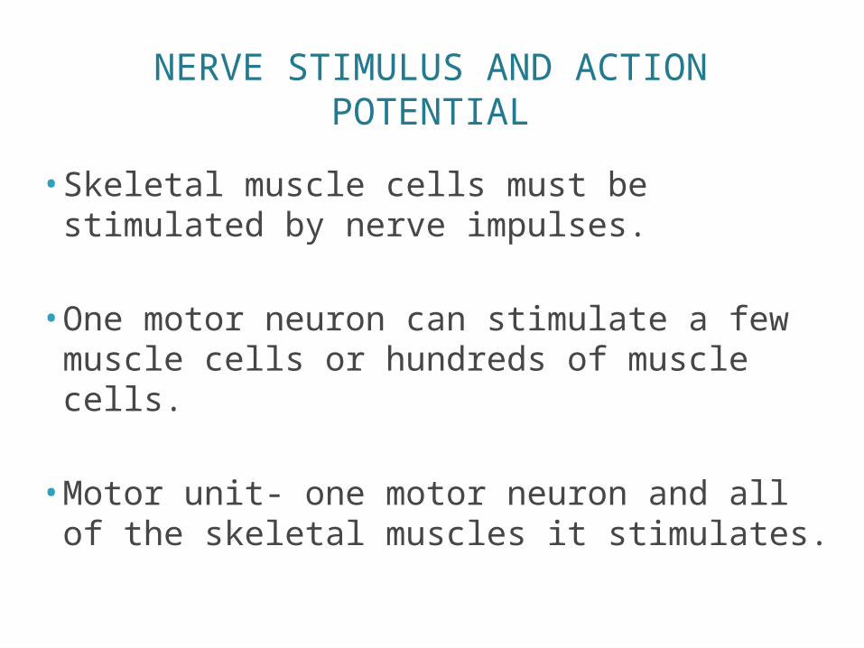
• Skeletal muscle cells must be stimulated by nerve impulses.
• One motor neuron can stimulate a few muscle cells or hundreds of muscle cells.
• Motor unit- one motor neuron and all of the skeletal muscles it stimulates.
NERVE STIMULUS AND ACTION POTENTIAL
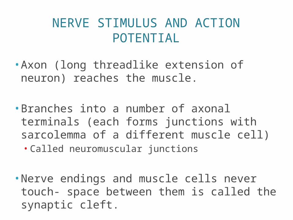
• Axon (long threadlike extension of neuron) reaches the muscle.
• Branches into a number of axonal terminals (each forms junctions with sarcolemma of a different muscle cell)• Called neuromuscular junctions
• Nerve endings and muscle cells never touch- space between them is called the synaptic cleft.
NERVE STIMULUS AND ACTION POTENTIAL
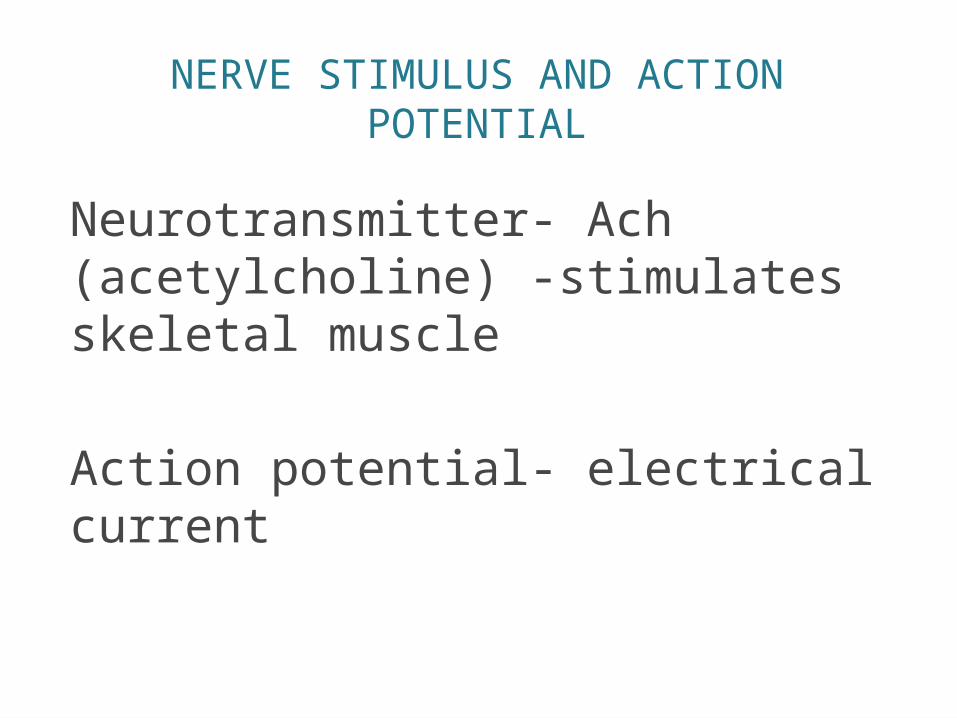
Neurotransmitter- Ach (acetylcholine) -stimulates skeletal muscle
Action potential- electrical current
ACETYLCHOLINE
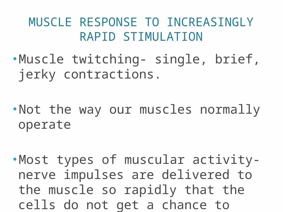
MUSCLE RESPONSE TO INCREASINGLY RAPID STIMULATION
•Muscle twitching- single, brief, jerky contractions.
•Not the way our muscles normally operate
•Most types of muscular activity- nerve impulses are delivered to the muscle so rapidly that the cells do not get a chance to relax completely.
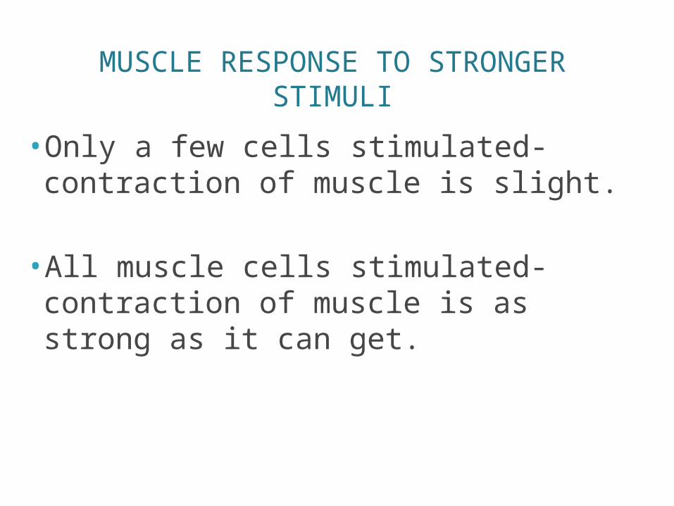
MUSCLE RESPONSE TO STRONGER STIMULI
•Only a few cells stimulated- contraction of muscle is slight.
• All muscle cells stimulated- contraction of muscle is as strong as it can get.
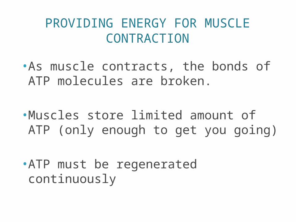
PROVIDING ENERGY FOR MUSCLE CONTRACTION
• As muscle contracts, the bonds of ATP molecules are broken.
•Muscles store limited amount of ATP (only enough to get you going)
• ATP must be regenerated continuously
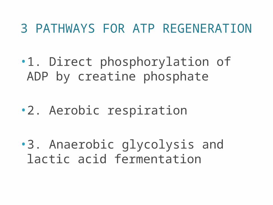
3 PATHWAYS FOR ATP REGENERATION
• 1. Direct phosphorylation of ADP by creatine phosphate
• 2. Aerobic respiration
• 3. Anaerobic glycolysis and lactic acid fermentation
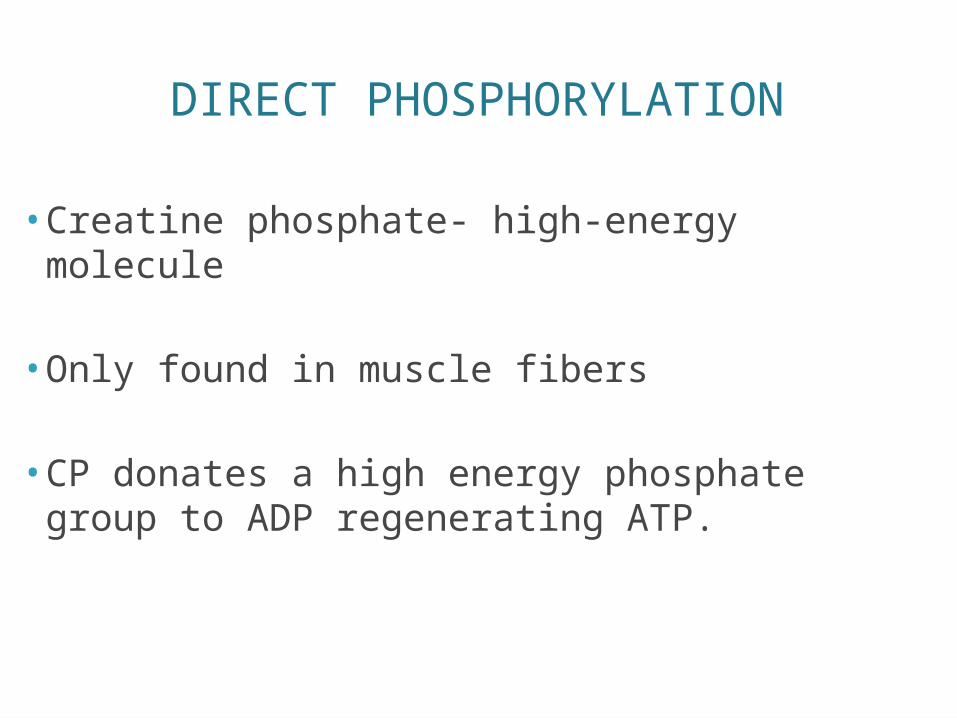
DIRECT PHOSPHORYLATION
• Creatine phosphate- high-energy molecule
• Only found in muscle fibers
• CP donates a high energy phosphate group to ADP regenerating ATP.
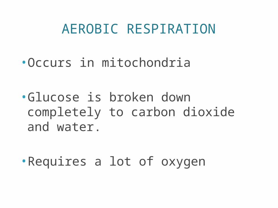
AEROBIC RESPIRATION
•Occurs in mitochondria
•Glucose is broken down completely to carbon dioxide and water.
• Requires a lot of oxygen
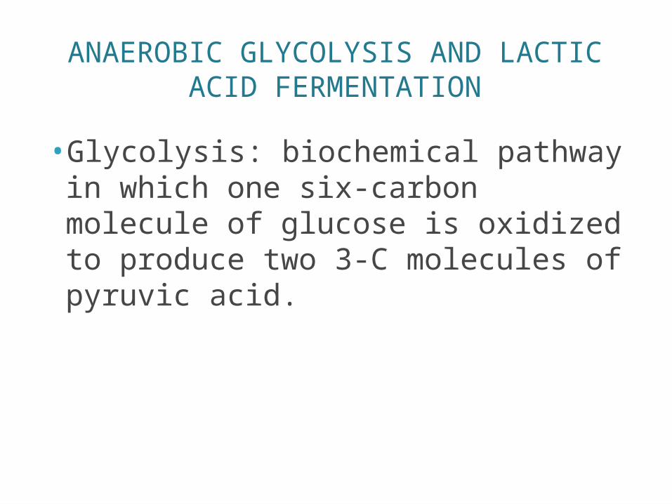
ANAEROBIC GLYCOLYSIS AND LACTIC ACID FERMENTATION
•Glycolysis: biochemical pathway in which one six-carbon molecule of glucose is oxidized to produce two 3-C molecules of pyruvic acid.
MUSCLE FATIGUE AND OXYGEN DEBT
• Muscle fatigue: occurs if we exercise our muscles strenuously for a long time.
• Unable to contract even if it is being stimulated.
• Believed to be a result from oxygen debt
• Oxygen debt occurs during prolonged muscle activity.
• When muscles lack oxygen- lactic acid builds up in muscles

TYPES OF MUSCLE CONTRACTIONS- ISOTONIC
Myofiliments successful in their sliding movementsMuscle shortensMovement occurs
Bending the kneeRotating armsSmiling
TYPES OF MUSCLE CONTRACTIONS- ISOMETRIC
• Myosin filaments are “skidding their wheels”• Tension of the muscle keeps increasing• Trying to slide but cannot
• Like when you try to pick up a 400 pound object
MUSCLE TONE
• Even when muscle is voluntarily relaxed- some fibers are contracting
• Muscle remains firm, healthy , and constantly ready for action.
• Muscle tone- state of continuous partial contraction
EFFECT OF EXERCISE ON MUSCLES
• If you don’t use muscle, you lose it.
• Regular exercise increases muscle size, strength, and endurance.
• Aerobic- (endurance)- jogging, biking, aerobics class• Result in stronger, more flexible muscle • Makes overall body metabolism more efficient• Improves digestion • Enhances neuromuscular coordination• Makes skeleton stronger
TYPES OF BODY MOVEMENTS
• 600 skeletal muscles- attached to a bone or other connective tissue structure.
• Origin- attached to the immovable or less movable bone.
• Insertion- attached to the movable bone
• When the muscle contracts the insertion moves toward the origin.
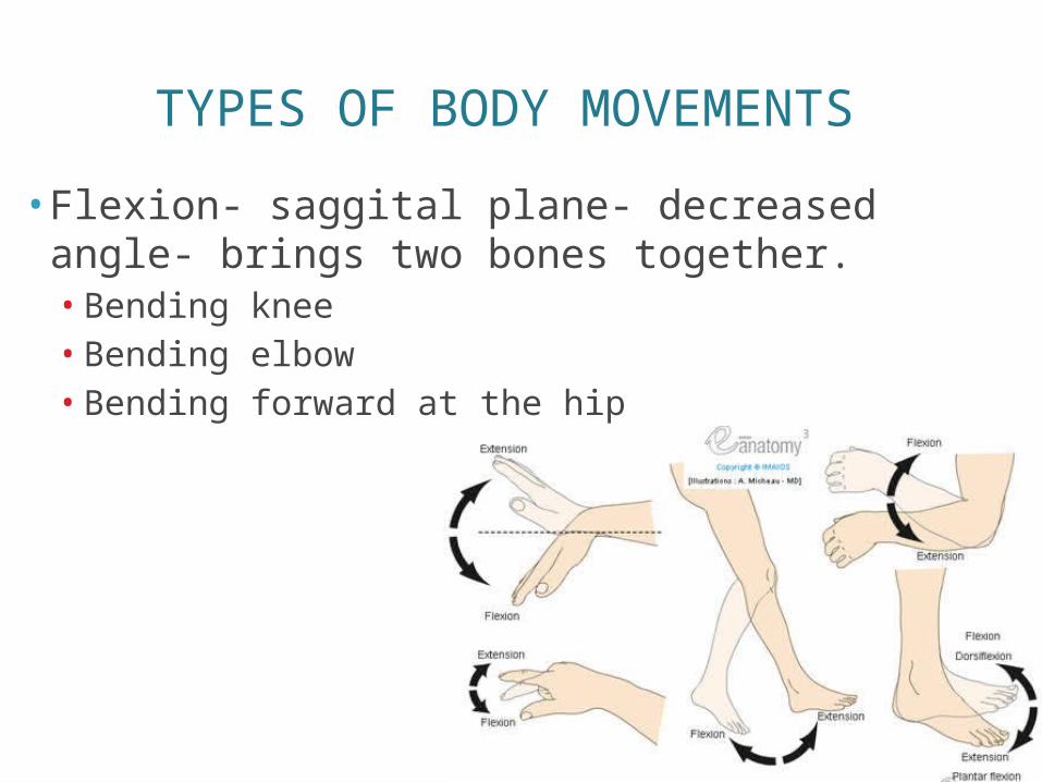
TYPES OF BODY MOVEMENTS
• Flexion- saggital plane- decreased angle- brings two bones together.• Bending knee• Bending elbow• Bending forward at the hip
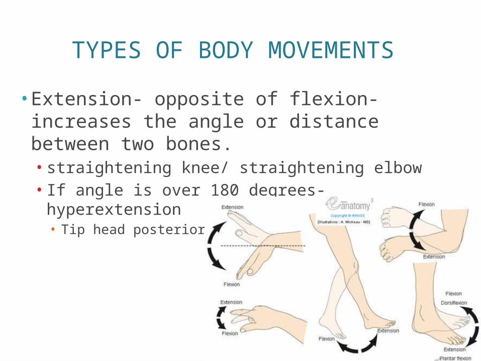
TYPES OF BODY MOVEMENTS
• Extension- opposite of flexion- increases the angle or distance between two bones. • straightening knee/ straightening elbow• If angle is over 180 degrees- hyperextension• Tip head posteriorly so chin points to ceiling.
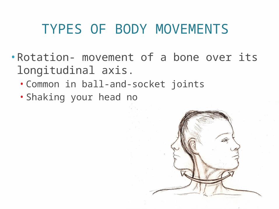
TYPES OF BODY MOVEMENTS
• Rotation- movement of a bone over its longitudinal axis.• Common in ball-and-socket joints• Shaking your head no
TYPES OF BODY MOVEMENTS
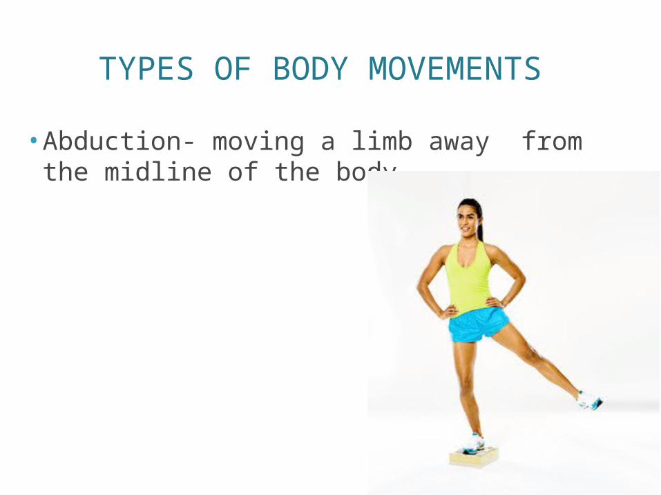
• Abduction- moving a limb away from the midline of the body.
TYPES OF BODY MOVEMENTS
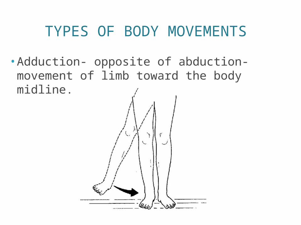
• Adduction- opposite of abduction- movement of limb toward the body midline.
TYPES OF BODY MOVEMENTS
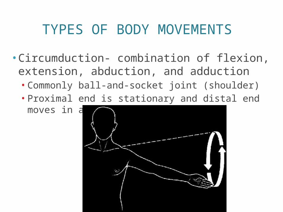
• Circumduction- combination of flexion, extension, abduction, and adduction• Commonly ball-and-socket joint (shoulder)• Proximal end is stationary and distal end moves in a
circle

TYPES OF BODY MOVEMENTS
• Dorsiflexion and plantar flexion- up and down movements of the foot and ankle.
TYPES OF BODY MOVEMENTS
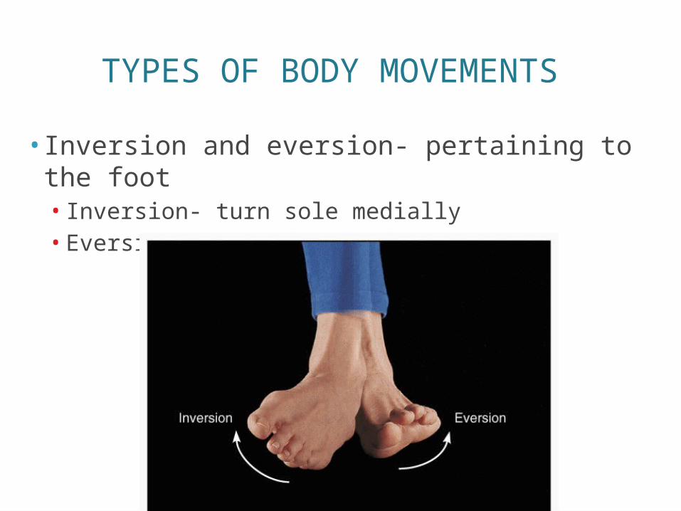
• Inversion and eversion- pertaining to the foot• Inversion- turn sole medially• Eversion- turn sole laterally
TYPES OF BODY MOVEMENTS
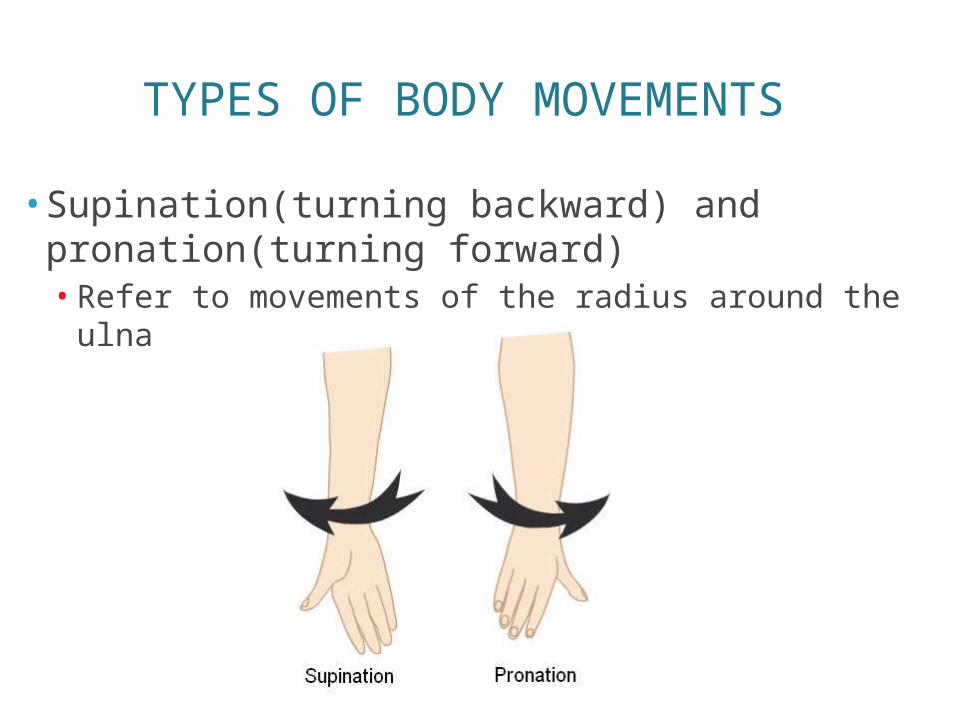
• Supination(turning backward) and pronation(turning forward)• Refer to movements of the radius around the ulna
TYPES OF BODY MOVEMENTS
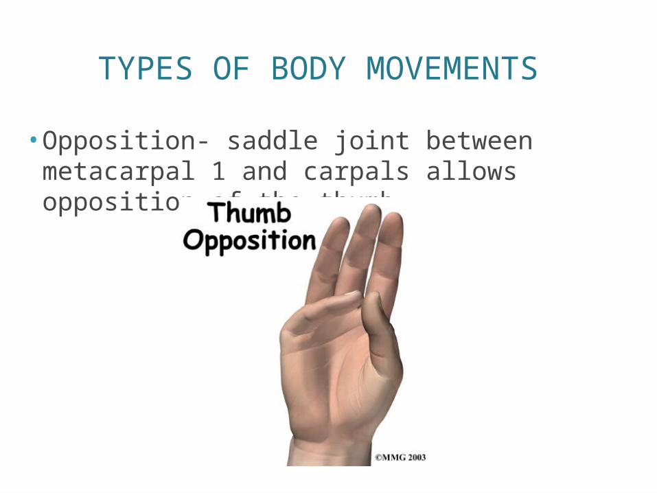
• Opposition- saddle joint between metacarpal 1 and carpals allows opposition of the thumb.
TYPES OF MUSCLES
TYPES OF MUSCLES
• Prime mover- muscle that has major responsibility for causing a particular movement.• Antagonists- muscles that oppose or reverse
a movement
• When prime mover is active- antagonist is stretched and relaxed.• Antagonists can be prime movers• Biceps of the arm (prime mover of elbow flexion) is
antagonized by the triceps (a prime mover of elbow extension)
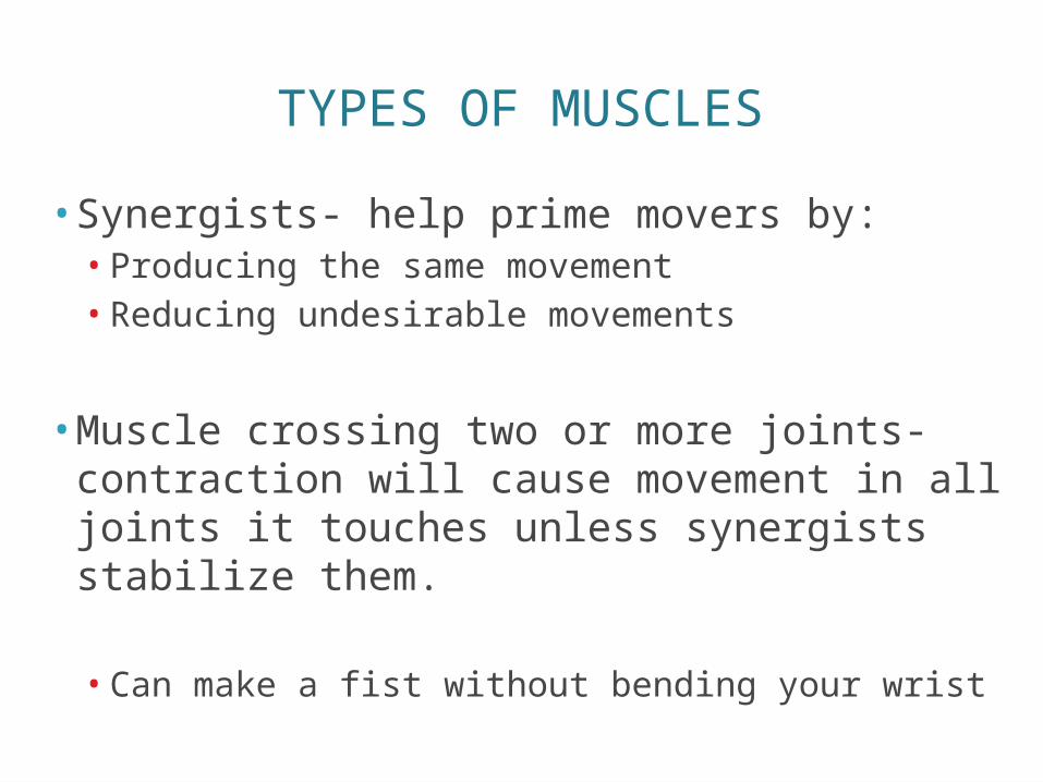
TYPES OF MUSCLES
• Synergists- help prime movers by:• Producing the same movement• Reducing undesirable movements
• Muscle crossing two or more joints- contraction will cause movement in all joints it touches unless synergists stabilize them.
• Can make a fist without bending your wrist
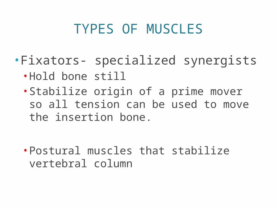
TYPES OF MUSCLES
• Fixators- specialized synergists• Hold bone still• Stabilize origin of a prime mover so all
tension can be used to move the insertion bone.
• Postural muscles that stabilize vertebral column
NAMING SKELETAL MUSCLE
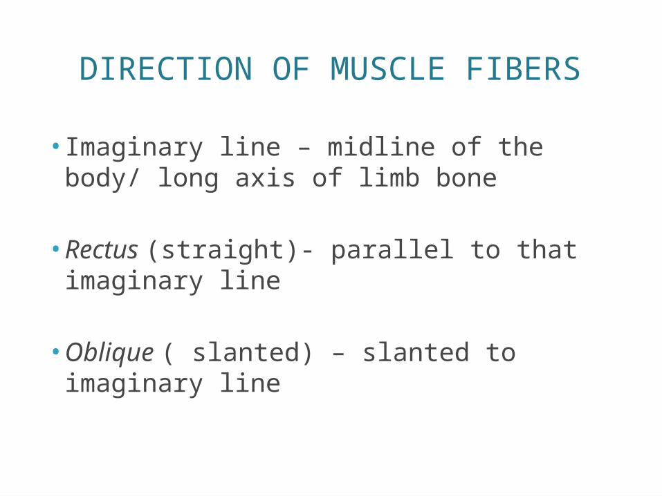
DIRECTION OF MUSCLE FIBERS
• Imaginary line – midline of the body/ long axis of limb bone
• Rectus (straight)- parallel to that imaginary line
• Oblique ( slanted) – slanted to imaginary line
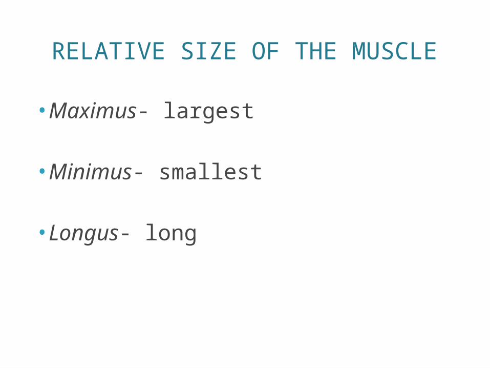
RELATIVE SIZE OF THE MUSCLE
•Maximus- largest
•Minimus- smallest
• Longus- long
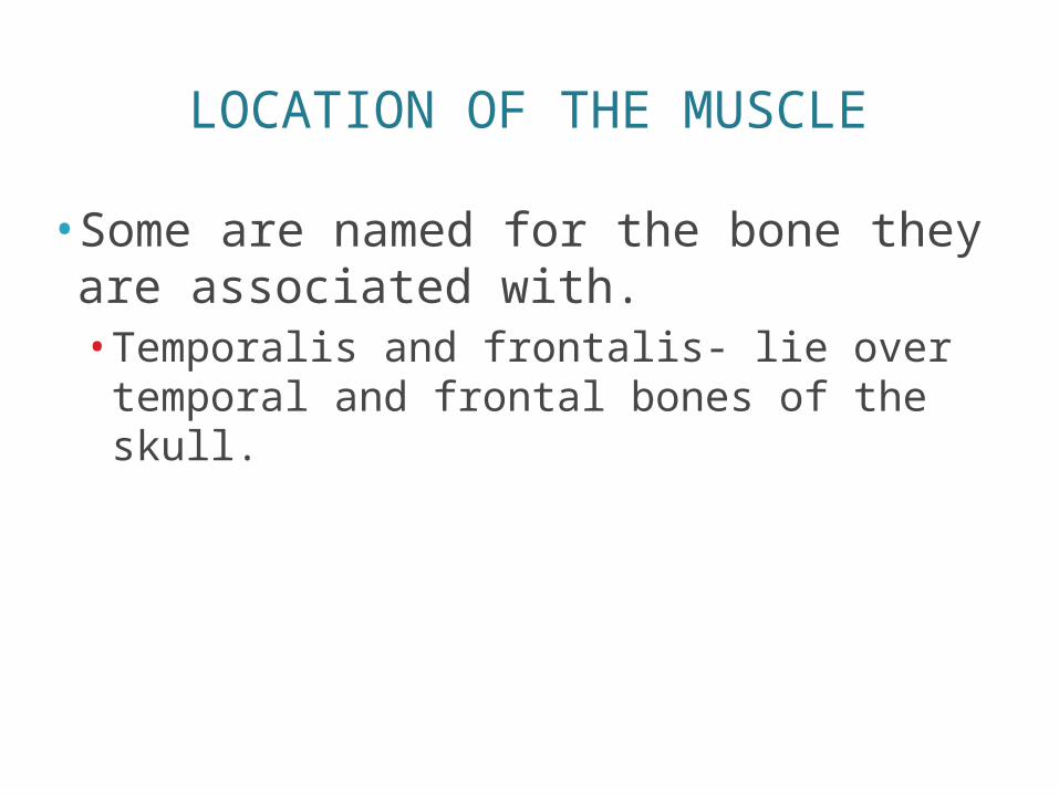
LOCATION OF THE MUSCLE
• Some are named for the bone they are associated with.• Temporalis and frontalis- lie over temporal
and frontal bones of the skull.
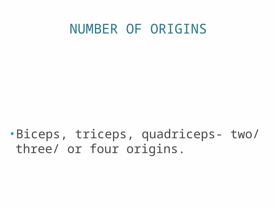
NUMBER OF ORIGINS
• Biceps, triceps, quadriceps- two/ three/ or four origins.
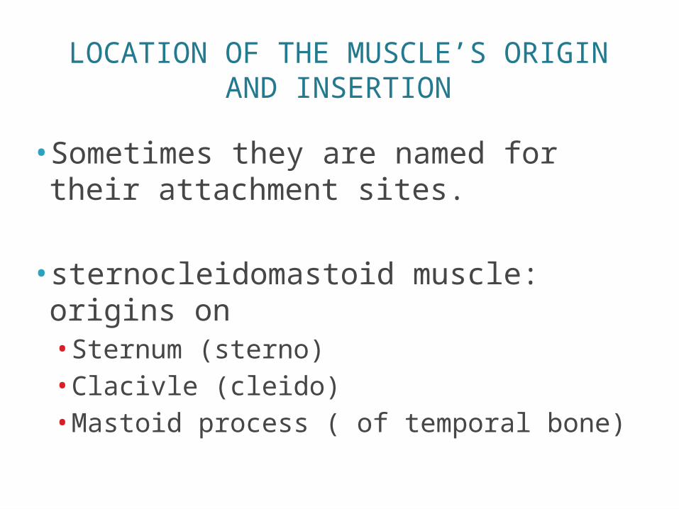
LOCATION OF THE MUSCLE’S ORIGIN AND INSERTION
• Sometimes they are named for their attachment sites.
• sternocleidomastoid muscle: origins on• Sternum (sterno)• Clacivle (cleido)• Mastoid process ( of temporal bone)
SHAPE OF THE MUSCLE
•Named for their shape•Deltoid muscle – triangular
ACTION OF THE MUSCLE
•When muscles are named for their actions- the following words may appear
• Flexor, extensor, adductor• Adductor muscles of the thigh/ extensor muscles of the wrist.
GROSS ANATOMY OF SKELETAL MUSCLES
HEAD AND NECK MUSCLES
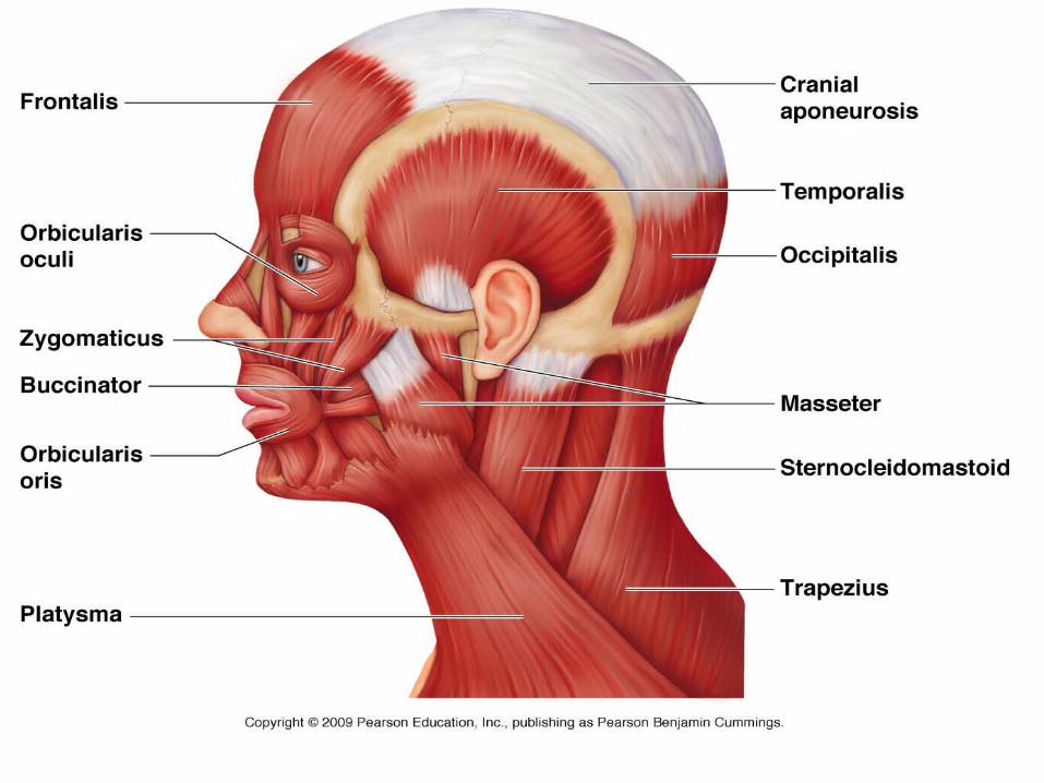
Facial Muscles• Frontalis• Oribicularis Oculi• Oribicularis Oris• Buccinator• Zygomaticus
TrapeziusCranial aperneurosisoccipitalis
Chewing Muscles• Masseter• Temporalis
Neck Muscles• Platysma• Sternocleidomastoid
FACIAL MUSCLES
• Frontalis- covers frontal bone• Allows you to raise your eyebrows• Allows you to wrinkle your forehead
• Orbicularis Oculi- circles around the eyes• Allows you to close your eyes, squint, blink, and
wink
• Orbicularis Oris- circular muscle of lips• Kissing muscle• Closes the mouth and protrudes the lips
FACIAL MUSCLES
• Buccinator: runs across the cheek and inserts into the orbicularis oris.• Flatten cheek in whistling or blowing• Also used as a chewing muscle
• Zygomaticus: extends from corner of mouth to cheekbone.• Smiling muscle- because it raises corners of the
mouth upwards.
CHEWING MUSCLES
• Masseter- covers lower jaw as it runs from the zygomatic process of the temporal bone to the mandible.• Closes the jaw by elevating the mandible
• Temporalis- fan-shaped / covers temporal bone • Inserts into the mandible• Acts as a synergist of the masseter in closing the
jaw

NECK MUSCLES
• Platysma- single sheet-like muscle that covers the anterolateral neck• Acts to pull the corners of the mouth inferiorly• Produces a downward sag of the mouth
• Sternocleidomastoid- paired muscles- one on each side of the neck.
TRUNK MUSCLES
Anterior Muscles• Pectoralis Major• Intercostal Muscles• Mucles of the Abdominal
Girdle• Rectus Abdominus• External Oblique• Internal Oblique• Transverse Abdominus
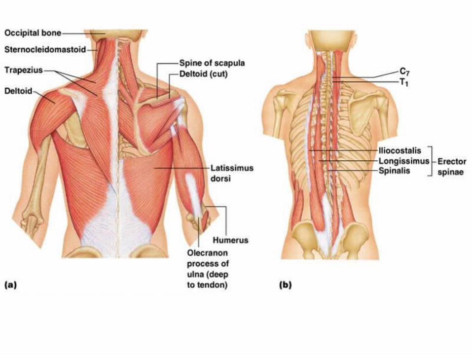
Posterior Muscles• Trapezius• Latissimus Dorsi• Erector Spinae• Deltoid
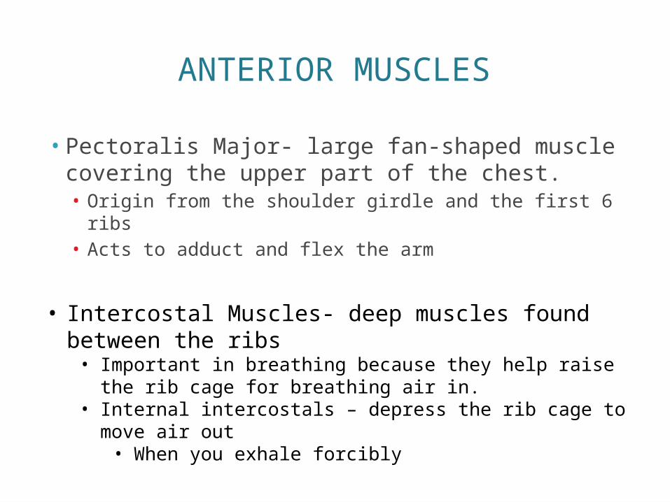
ANTERIOR MUSCLES
• Pectoralis Major- large fan-shaped muscle covering the upper part of the chest.• Origin from the shoulder girdle and the first 6 ribs• Acts to adduct and flex the arm
• Intercostal Muscles- deep muscles found between the ribs• Important in breathing because they help raise the rib
cage for breathing air in. • Internal intercostals – depress the rib cage to move air
out• When you exhale forcibly

ANTERIOR MUSCLES
• Muscles of the Abdominal Girdle- anterior abdominal muscles• Includes: rectus abdominus, external and internal
obliques, and transversus abdominus• Reinforces the body trunk• Form a thick wall
• Rectus abdominus- run from pubis to rib cage/ main function is to flex the vertebral column
• External oblique- make up lateral walls of abdomen/ run from last 4 pairs of ribs to illium/ flex the vertebral column and rotate the trunk.
• Internal oblique- from illiac crest and insert into the last three ribs
• Transversus abdominus- deepest muscle of the abdomen wall/ compresses the abdominal contents.
POSTERIOR MUSCLES
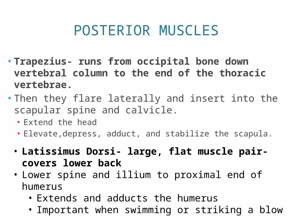
• Trapezius- runs from occipital bone down vertebral column to the end of the thoracic vertebrae.• Then they flare laterally and insert into the scapular
spine and calvicle.• Extend the head• Elevate,depress, adduct, and stabilize the scapula.
• Latissimus Dorsi- large, flat muscle pair- covers lower back
• Lower spine and illium to proximal end of humerus• Extends and adducts the humerus• Important when swimming or striking a blow
POSTERIOR MUSCLES
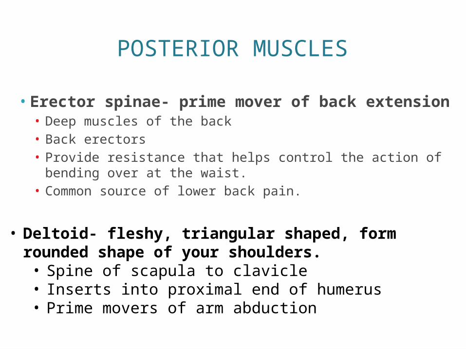
• Erector spinae- prime mover of back extension• Deep muscles of the back• Back erectors• Provide resistance that helps control the action of bending
over at the waist.• Common source of lower back pain.
• Deltoid- fleshy, triangular shaped, form rounded shape of your shoulders.• Spine of scapula to clavicle• Inserts into proximal end of humerus• Prime movers of arm abduction
UPPER LIMB MUSCLES
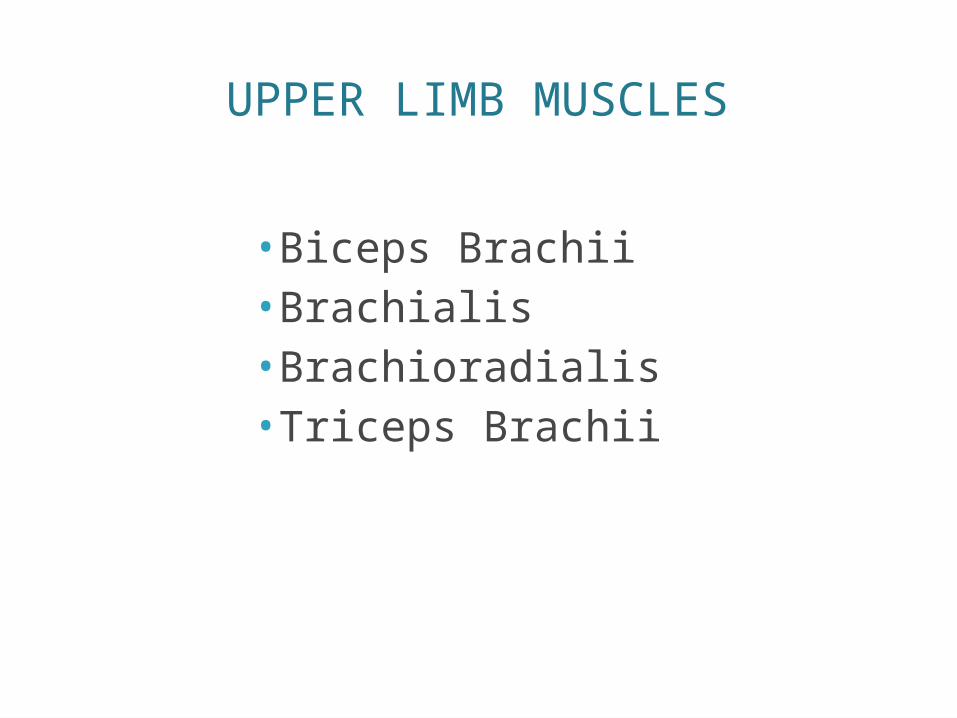
• Biceps Brachii• Brachialis• Brachioradialis• Triceps Brachii
UPPER LIMB MUSCLES
• Biceps Brachii- most familiar muscle of the forearm• Bulges when elbow is flexed• Originates from shoulder girldle and inserts to radial
tuberosity.• Brachialis- lies deep in biceps brachii• Important in elbow flexion
• Brachioradialis- flairy, weak, arises in humerus- inserts into forearm• Resides mainly in forearm
• Triceps Brachii- only muscle fleshing out posterior humerus• Inserts into olecranon process of ulna
LOWER LIMB MUSCLES
• Cause movement at hip, knee, and foot joints
• Largest, strongest muscles
• Specialized for walking and balancing the body
• Pelvic girdle- heavy fused bones- no special group of muscles for it
• Different from shoulder girdle
LOWER LIMB MUSCLES
• Span two joints
• Can cause movement at both joints
• Thigh- massive muscles that hold body upright.
• Thigh muscles cross knee and cause its flexion and extension
HIP JOINT MUSCLE
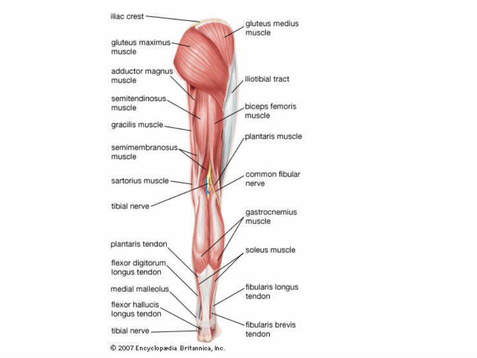
•Gluteus maximus•Gluteus medius
• Iliopsoas• Adductor muscles
HIP JOINT MUSCLE
• Gluteus maximus- forms most of the flesh of the buttock• Powerful hip extensor• Brings thigh in a straight line with pelvis• Most important when climbing stairs and when
jumping• Originates from sacrum and iliac bones and runs to
insert on the gluteal tuberosity of femur
HIP JOINT MUSCLE
• Gluteus medius- runs from ilium to femur• Beneath gluteus maximus• Hip abductor• Important site for giving intramuscular injections
• Iliopsoas- fused muscle composed of two muscles (iliacus and psoas major)• Prime mover of hip flexion• Runs from iliac bone and lower vertebrae deep
inside pelvis to insert on lesser tronchanter• Keeps upper body from falling backward when
we are standing up
HIP JOINT MUSCLE
• Adductor muscles-form muscle mass at medial side of each thigh.• Press thighs together • Tend to become flabby very easily • Origin on pelvis and insert on proximal aspect of
femur
MUSCLES CAUSING MOVEMENT AT THE KNEE JOINT
• Hamstring group• Biceps femoris• Semimembranosu
s• semitendinosus
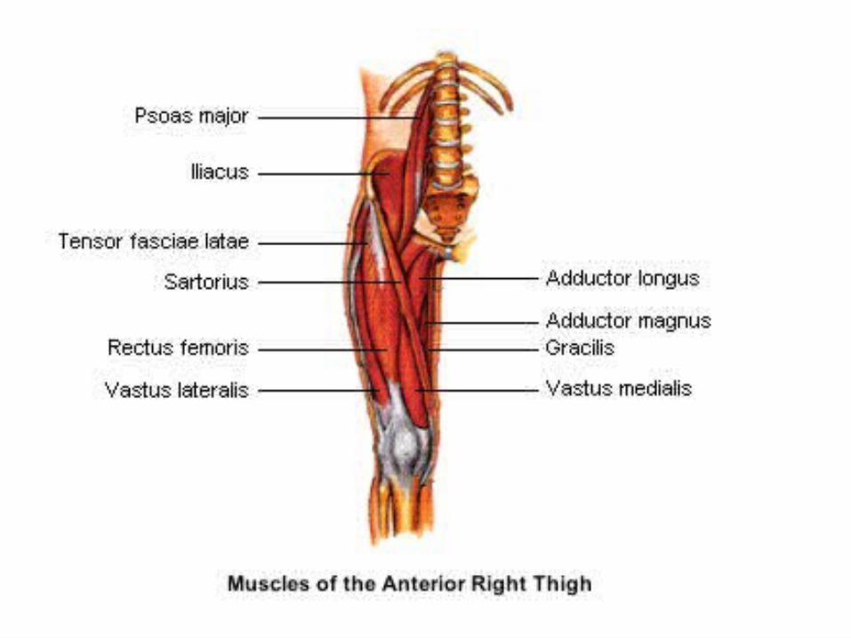
• Sartorius
• Quadriceps Group• Rectus femoris• Vastus muscles
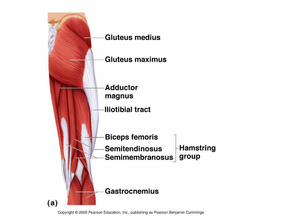
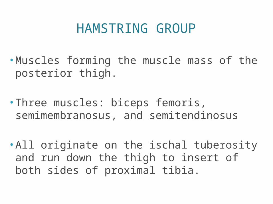
HAMSTRING GROUP
• Muscles forming the muscle mass of the posterior thigh.
• Three muscles: biceps femoris, semimembranosus, and semitendinosus
• All originate on the ischal tuberosity and run down the thigh to insert of both sides of proximal tibia.
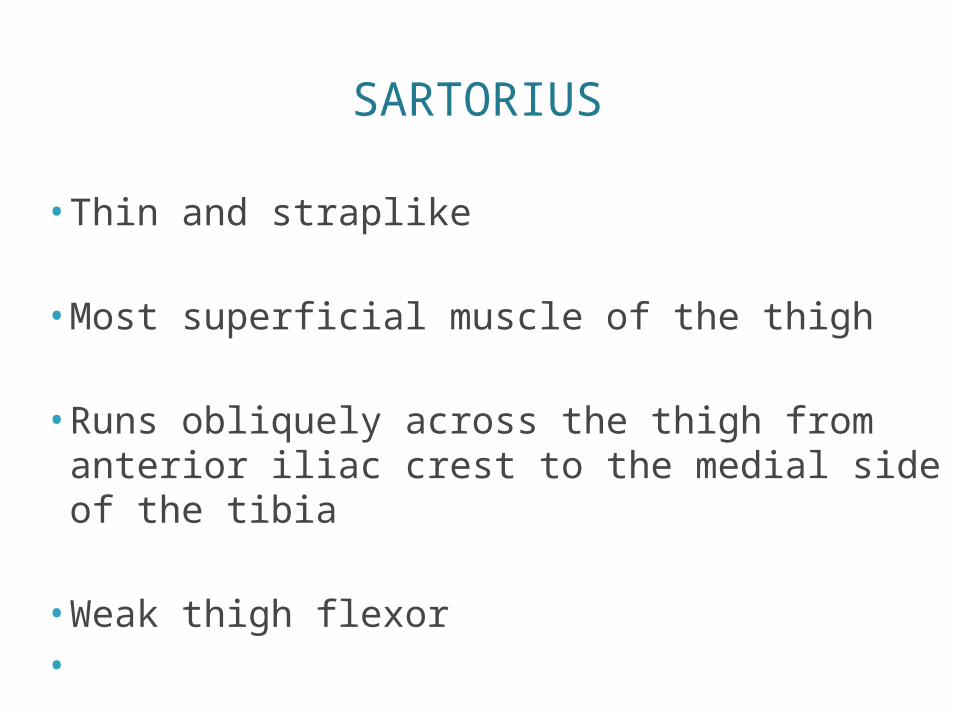
SARTORIUS
• Thin and straplike
• Most superficial muscle of the thigh
• Runs obliquely across the thigh from anterior iliac crest to the medial side of the tibia
• Weak thigh flexor•

QUADRICEPS GROUP
• Rectus femoris and three vastus muscles• Flesh out anterior thigh
• Vastus muscles originate from the femur
• Rectus femoris originates from pelvis
• All four insert into the tibial tuberosity via the patellar ligament
• Acts to extend knee powerfully
• Flex hip
MUSCLES CAUSING MOVEMENT AT THE ANKLE AND FOOT
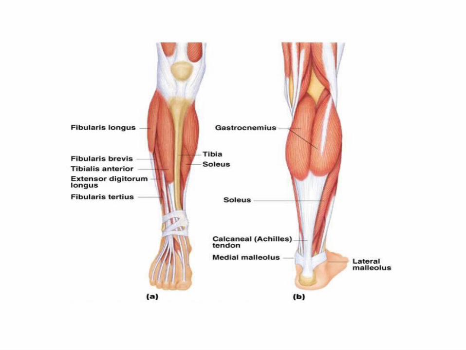
•Tibialis anterior•Extensor digitorum longus
•Fibularis muscles•Gastrocnemius
•Soleus
MUSCLES CAUSING MOVEMENT AT THE ANKLE AND FOOT
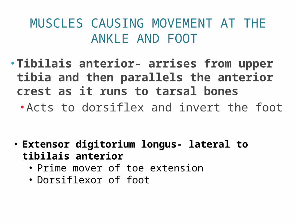
• Tibilais anterior- arrises from upper tibia and then parallels the anterior crest as it runs to tarsal bones• Acts to dorsiflex and invert the foot
• Extensor digitorium longus- lateral to tibilais anterior• Prime mover of toe extension• Dorsiflexor of foot
MUSCLES CAUSING MOVEMENT AT THE ANKLE AND FOOT
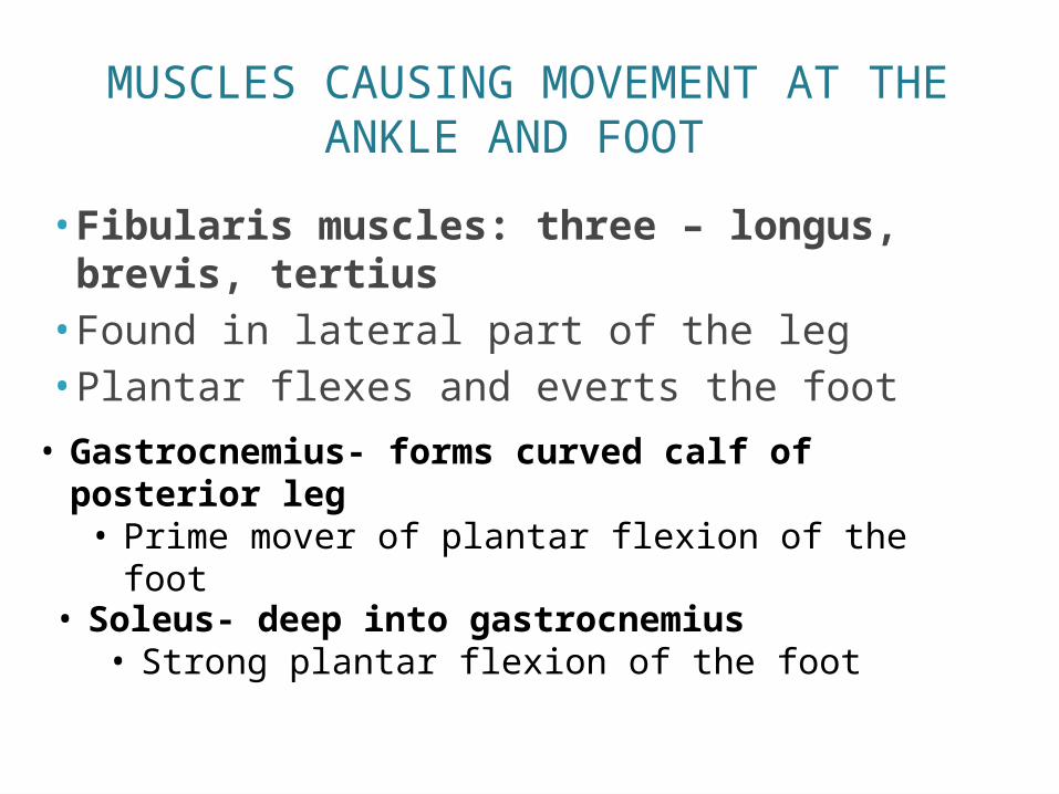
• Fibularis muscles: three – longus, brevis, tertius• Found in lateral part of the leg• Plantar flexes and everts the foot• Gastrocnemius- forms curved calf of
posterior leg• Prime mover of plantar flexion of the foot
• Soleus- deep into gastrocnemius• Strong plantar flexion of the foot