
Chapter 6: Head Trauma Objectives - Famona Sitefamona.sezampro.rs/medifiles/atls/atls06.pdf ·...
28
1 Chapter 6: Head Trauma Objectives: Upon completion of this topic, the physician will be able to demonstrate the techniques of assessment and explain the emergency management of head trauma. Specifically, the physician will be able to: A. Review specific principles of anatomy and physiology related to head injuries. B. Identify and discuss the principles of general management of the unconscious patient and delayed complications. C. Outline the method of evaluating head injuries using a minineurologic examination. D. Identify and discuss management techniques for specific types of head injuries. E. Demonstrate the ability to assess various types of head, maxillofacial, and neck injuries, using a head-trauma manikin. F. Discuss clinical signs and outline priorities for the initial management of injuries identified during patient assessment.
Transcript of Chapter 6: Head Trauma Objectives - Famona Sitefamona.sezampro.rs/medifiles/atls/atls06.pdf ·...

1
Chapter 6: Head Trauma
Objectives:
Upon completion of this topic, the physician will be able to demonstrate the techniquesof assessment and explain the emergency management of head trauma.
Specifically, the physician will be able to:
A. Review specific principles of anatomy and physiology related to head injuries.
B. Identify and discuss the principles of general management of the unconsciouspatient and delayed complications.
C. Outline the method of evaluating head injuries using a minineurologic examination.
D. Identify and discuss management techniques for specific types of head injuries.
E. Demonstrate the ability to assess various types of head, maxillofacial, and neckinjuries, using a head-trauma manikin.
F. Discuss clinical signs and outline priorities for the initial management of injuriesidentified during patient assessment.
2
I. Introduction
Because a head injury occurs every 15 seconds, and a patient dies of a headinjury every 12 minutes, a physician dealing with trauma is confronted with a head-injured patient almost every day. Approximately 50% of all trauma deaths areassociated with head injury, and more than 60% of vehicular trauma deaths are due tohead injury. Because of the immense importance of head injury, the physician who seespatients soon after injury, but is not expert in the comprehensive management of headinjuries, must develop a practical knowledge of the initial care of these patients. Aneurosurgeon may not be available immediately when a patient with a central nervoussystem injury is first seen. Therefore, one of the most important responsibilities ofphysicians initially evaluating such a patient is in the management of ventilation andhypovolemia, thereby obviating potential secondary brain damage. Therefore, a systemthat incorporates consultation with a neurosurgeon, whose opinion may modify themanagement of such a patient, is essential. Early transfer of appropriate patientssubstantially reduces morbidity and mortality.
When conferring with a neurosurgical consultant about a patient with a head injury,the physician should relay the following patient information:
1. Age of patient and mechanism of injury.
2. Respiratory and cardiovascular status.
3. Results of the neurologic examination, especially the level of consciousness,pupillary reactions, and presence of lateralized extremity weakness.
4. Presence and type of noncerebral injuries.
5. The results of diagnostic studies, if obtained.
Neurosurgical consultation or patient transfer should not be delayed to obtaindiagnostic studies, eg, computed tomographic (CT) scan or skull roentgenograms.
II. Anatomy and Physiology
As with all other aspects of medicine, the basis for a rational decision is theintegration of the knowledge of anatomy and physiology with the history and physicalexamination. A short review of these subjects follows.
A. Scalp
The scalp is made up five layers of tissue covering the bone of the top of the skull(calvarium): (1) skin, (2) subcutaneous tissue, (3) galea aponeurotica, (4) a layer of looseareolar tissue, and (5) periosteum, the "pericranium". The loose areolar tissue, separating thegalea from the pericranium, is subject to the commonly encountered subgaleal hematomas,large flaps caused by injury and "scalping" injuries. Because of the scalp's generous bloodsupply, bleeding from a scalp laceration can result in major blood loss, especially in children.

3
B. Skull
The skull is composed of the cranial vault (calvarium) and the base. The calvariumis especially thin in the temporal regions. The base of the skull is irregular and rough,allowing injury to occur as the brain moves within the skull during acceleration anddeceleration.
C. Meninges
The dura is tough, fibrous membrane that adheres firmly to the internal surface of theskull. Because it is not attached to the underlying arachnoid, a potential space (the subduralspace) exists into which hemorrhage can occur - usually from veins that traverse the space(bridging veins). In certain places the dura splits into two surfaces forming venous sinusesthat provide the major venous drainage from the brain. The midline (superior) sagittal sinusis especially vulnerable to injury.
Meningeal arteries lie between the dura and the internal surface of the skull(epidural). The courses of these arteries may be visible on plain skull roentgenograms, if thevessels groove the inner surface of the skull. Laceration of these arteries may result in anepidural hematoma.
Beneath the dura is a second meningeal layer, the thin transparent arachnoid. Thethird layer, the pia, is firmly attached to the brain cortex. Between the arachnoid and the pia(subarachnoid space), the cerebrospinal fluid (CSF) circulates. Hemorrhage into this spaceis, by definition, subarachnoid hemorrhage.
D. Brain
The brain consists of the cerebrum, the cerebellum, and the brain stem. The cerebrumis composed of right and left hemispheres that are separated by the falx, a dural reflection.The left hemisphere, which usually contains the language centers, is often referred to as thedominant hemisphere. The frontal lobe is concerned with emotions and motor function, theoccipital lobe with vision, and the parietal lobe with sensory function. The temporal loberegulates certain memory functions, but can be a relatively silent region on the right side.
The brain can be compared with a funnel. The two cerebral hemispheres comprise thefunnel portion. The brain stem, the major neural pathways to and from the hemispheres,comprises the neck of the funnel. The brain stem is composed of the midbrain, the pons, andthe medulla. The midbrain and upper pons contain the reticular activating system, which isresponsible for an "awake" state. Vital cardiorespiratory centers reside in the lower brainstem, the medulla, which then continues to form the spinal cord. The cerebellum, whichcontrols movement, coordination, and balance, surrounds the pons and medulla in theposterior fossa.
E. Cerebrospinal Fluid
Cerebrospinal fluid is produced by the choroid plexus and is discharged into theventricles of the brain. The fluid leaves the ventricular cavities to circulate through the
4
subarachnoid space.
F. Tentorium
The tentorium divides the head into the supratentorial compartment (comprising theanterior and middle fossae of the skull) and the infratentorial compartment (containing theposterior fossa). The midbrain courses from the cerebrum through the large aperture (incisura)in the tentorium. The third cranial nerve also passes along this opening. Any pathologicprocess that rapidly increases supratentorial pressure, most commonly hemorrhage or edema,can force the medial aspect of the temporal lobe (the uncus) through this opening. The result(uncal or tentorial herniation) causes compression of the third nerve, producing an ipsilateraland dilated, fixed pupil. Contralateral spastic weakness of the arm and leg, caused bycompression of the corticospinal (pyramidal) tract within the cerebral peduncle on the sideof herniation, is also an integral component of uncal herniation.
G. Consciousness
Alteration of consciousness is the hallmark of brain injury.
Two types of brain injury - bilateral injury to the cerebral cortices and injury to thereticular activating system of the brain stem - can produce unconsciousness. Increasedintracranial pressure and decreased cerebral blood flow, regardless of the cause, can alsodepress the level of consciousness.
H. Intracranial Pressure
Intracranial pressure can be described as a relationship among volumes of CSF, blood,and brain, and expressed by the equation of the modified Monroe-Kellie hypothesis:
KICP = VCSF + VBl + VBr
Constancy (K) of the intracranial pressure (ICP) relates to the volume (V) of thecerebrospinal fluid (CSF), plus the volume (V) of blood (Bl), plus the volume (V) of the brain(Br).
The volume of CSF is altered by varying rates of absorption depending on theintracranial pressure, since CSF production is constant. Intracranial venous blood volumedepends on rates of passive drainage influenced by intracranial pressure and intrathoracicpressure. The arterial blood volume is affected by autoregulation, arterial PCO2, and may bedecreased by autoregulation with hyperventilation. Brain volume is relatively static, exceptwhen cerebral edema occurs.
As the volume of mass from an expanding hematoma or cerebral edema increases,CSF or venous blood (or both) are removed from the intracranial compartment at a greaterrate than normal. The result of these adjustments is that the intracranial compartment mayaccommodate an enlarging mass to a volume of 50 to 100 mL, depending on location andrapidity of enlargement. When all normal compensatory measures are exhausted, further smallincreases in intracranial mass volume cause very large increases in intracranial pressure,

5
resulting in diminution of cerebral perfusion pressure. When the mass reaches a criticalsize, further increases in volume result in a rapid increase in intracranial pressure with brainherniation likely.
Cerebral perfusion pressure is, approximately, the mean systemic arterial pressureminus the intracranial pressure. It is a measure of arterial perfusion of brain tissue, andnormally it is greater than 50 mm Hg. Initial elevations of intracranial pressure are oftenassociated with compensatory rises in mean systemic arterial pressure. However, furtherelevations of intracranial pressure are at the expense of cerebral perfusion pressure (ie,cerebral blood flow), and result in brain ischemia, leading to alterations in the level ofconsciousness. Brain death occurs when intracranial pressure approaches systemic arterialpressure, thus preventing any blood flow to the brain.
The definitive management of increased ICP is substantially aided by direct measuresof arterial, central venous, and intracranial pressures under the direction of a neurosurgeon.
III. Assessment of Head Injuries
A. History
To make the correct management decisions, it is helpful to know what kind of headinjury can result from a certain kind of trauma. The cause is usually obvious, but even thougha patient may appear to be in good neurologic condition, mechanism-of-injury risk factorsshould be assessed in deciding how and where to treat the patient (See Chapter 12,Stabilization and Transport.) For example, simply knowing that the patient was injured in afall instead of vehicular crash quadruples the patient's risk of having an intracranialhematoma.
Prehospital personnel should question observers at the scene about the patient'scondition immediately after the incident. Information obtained by ambulance personnel shouldbe documented in writing and provided to the hospital personnel when the patient is deliveredto the emergency department.
B. Initial Assessment
The importance of initial assessment is emphasized because it provides thebaseline for sequential reassessment, which is the critical basis for many subsequentdecisions in patient management.
Because many factors influence the neurologic evaluation, documentation of thecardiorespiratory status must accompany the neurologic examination. Airway, breathing, andcirculatory control and management must take priority. Care must be exercised inattributing changes in the patient's mental status to head injury if the systolic blood pressureis less than 60 mm Hg or systemic hypoxemia exists. Similarly, a drug screen for alcohol andother nervous system depressants must be obtained, so that toxic factors that may influencehead injury assessment are taken into account.

6
C. Assessment of Vital Signs
Although brain injury may alter the vital signs, it is very difficult to be sure thatchanges in the vital signs are due to head injury and not to other factors. Some useful clinicalrules include:
1. Never presume that brain injury is the cause of hypotension. Although bleedingfrom scalp lacerations can cause hemorrhagic shock (especially in small children), intracranialbleeding will not produce shock from blood loss alone. Hypotension arising from the braininjury is a terminal event, resulting from failure of medullary centers.
2. The combination of progressive hypertension associated with bradycardia anddiminished respiratory rate (Cushing response) is a specific response to an acute andpotentially lethal rise in intracranial pressure. In head injury, the course is usually a lesiondemanding immediate operative intervention.
3. Hypertension alone or in combination with hyperthermia may reflect centralautonomic dysfunction caused by certain types of brain injury.
D. AVPU + Minineurologic Examination
The AVPU mnemonic is supplemented by a minineurologic examination that isdirected toward determining the presence and severity of gross neurologic deficits, especiallythose that may require urgent operation. The AVPU method, completed in the primary survey,describes the patient's level of consciousness. (See Chapter 1, Initial Assessment andManagement and Chart 2, Revised Trauma Score in Chapter 12, Stabilization and Transport.)
The minineurologic examination should be conducted repeatedly for any patient witha head injury. The examination assesses: (1) level of consciousness, (2) pupillary function,and (3) lateralized extremity weakness. Generally, a patient with abnormalities of all threecomponents will have a mass lesion that may require surgery.
1. Level of consciousness
a. Glasgow Coma Scale
The Glasgow Coma Scale (GCS) provides a quantitative measure of the patient's levelof consciousness. The GCS is the sum of scores for three areas of assessment: (1) eyeopening, (2) verbal response, and (3) best motor response. Each is graded separately.
1) Eye-opening response (E score)
Scoring of eye opening is not valid if the eyes are swollen shut. This fact must bedocumented.
a) Spontaneous - already open with blinking (normal): E = four (4) points
b) To speech - not necessarily to a request for eye opening: E = three (3) points

7
c) To pain - stimulus should not be applied to the face: E = two (2) points
d) None: E = one (1) point.
2) Verbal response (V score)
Scoring for verbal response is invalid if speech is impossible, for example, in thepresence of endotracheal intubation. This circumstance must be documented.
a) Oriented - knows name, age, etc: V = five (5) points
b) Confused conversation = still answers questions: V = four (4) points
c) Inappropriate words - speech is either exclamatory or random, but recognizablewords are produced: V = three (3) points
d) Incomprehensible sounds - grunts and groans are produced, but no actual words areuttered; do not confuse with partial respiratory obstruction: V = two (2) points
e) None: V = one (1) point.
3) Best motor response (M score)
The best response obtained for any extremity is recorded even though worse responsesmay be present in other extremities. The worst motor activity also is important but is not usedfor the GCS; worst response should be noted separately. For patients not following verbalcommand, a painful stimulus is applied to the fingernail or toenail.
a) Obeys - moves limb to command and pain is not required: M = six (6) points
b) Localizes - changing the location of the pain stimulus causes purposeful motiontoward the stimulus: M = five (5) points
c) Withdraws - pulls away from painful stimulus: M = four (4) points
d) Abnormal flexion - decorticate posture: M = three (3) points
e) Extensor response - decerebrate posture: M = two (2) points
f) No movement: M = one (1) point.
b. Patient categorization
The Glasgow Coma Scale itself can be used to categorize patients:
1) Coma
A patient in coma is defined as having no eye opening (E = 1), no ability to follow

8
commands (M = 1 to 5), and no word verbalizations (V = 1 to 2). This means that allpatients with a GCS Score of less than eight and most of those with a GCS Score that equalseight are in coma. Patients with a GCS Score more than 8 are not in coma.
2) Head injury severity
On the basis of the GCS, patients are classified as having:
a) Severe head injury if the GCS Score is less than or equal to eight.
b) Moderate head injury if the GCS Score is 9 to 12.
c) Minor head injury if the GCS Score is 13 to 15.
2. Assessment of pupillary function
The pupils are evaluated for their equality and response to bright light. A differencein pupil diameters of more than 1 mm is abnormal. Even though eye injury may be present,intracranial injury must be excluded. Light reactivity must be evaluated for the briskness ofresponse; a more sluggish response may indicate an intracranial injury.
3. Lateralized extremity weakness
Spontaneous movements are observed for equality. If spontaneous motion is minimal,the response to a painful stimulus is assessed. A delay in onset of movement, less movement,or need for more stimulus on one side is significant. Often, the motor component of the GCScan be used to score the best and the worst extremity movement. A clearly lateralizedweakness suggests an intracranial mass lesion.
4. Purposes of neurologic examination
The purposes of the minineurologic examination are to determine the severity of thebrain injury, and detect any neurologic deterioration. The GCS can be used to categorizeinjuries as mild, moderate, and severe. Irrespective of the GCS, a patient is considered to havea severe head injury if he exhibits any of the following:
a. Unequal pupils
b. Unequal motor examination
c. An open head injury with leaking CSF or exposed brain tissue
d. Neurologic deterioration
e. Depressed skull fracture.
A decrease in the GCS score of two or more points clearly means the patient'scondition has deteriorated. A decrease of three or more points is a catastrophic deterioration

9
that demands immediate treatment if the cause is remediable. However, these changes arerather gross and are often preceded by more subtle signs of deterioration. Neurologicdeterioration of special concern includes:
f. Increase in severity of a headache or an extraordinarily severe headache
g. Increase in the size of one pupil
h. Development of weakness on one side.
It cannot be overemphasized that the initial neurologic examination is only thebeginning. The initial findings are only the reference with which to compare results ofrepeated neurologic examinations to determine whether a patient's condition is deterioratingor improving. All previous efforts will go to waste if signs of neurologic deterioration arenot appreciated. The brain cannot withstand unrelieved compression for very long beforebrain damage is irreversible. The neurologic assessment is designed to detect this secondarybrain damage so timely treatment can be initiated following neurosurgical consultation.
E. Special Assessment
1. Skull roentgenograms
Skull roentgenograms are of little value in the early management of patients withobvious head injuries, except in cases of penetrating injuries. The unconscious patient shouldhave skull roentgenograms only if precise care of the cardiorespiratory system and continuingreassessment can be assured. Physical examination is usually more valuable than skullroentgenograms. (See VII. Scalp Wounds, in this chapter.) Clinical signs of basal fracturesare more useful than roentgenograms of the skull base in diagnosing a fracture. Increasingly,skull films are not being obtained on patients with minor head injuries because theinformation obtained is rarely helpful one way or the other. When in doubt about the patient'scondition, the physicians should obtain neurosurgical consultation.
2. Computed tomography
The CT scan has revolutionized diagnosis in patients with head injuries and is thediagnostic procedure of choice for patients who have or are suspected of having a serioushead injury. Although not perfect, the CT scan is capable of showing the exact location andsize of most mass lesions. Specific diagnosis allows more precise planning of definitive care,including operation. The CT scan has supplanted less specific and more invasive tests, suchas cerebral angiography.
Except for patients with minor head injuries, all head-injured patients will requireCT scanning at some time. The more serious the injury, the earlier and more emergent is theneed for the scan. Consequently, injured patients seen first at facilities without CT capabilitymay require transfer to more sophisticated hospitals.
Once initial resuscitation has been undertaken and the need for a CT scan determined,care must be taken to (1) maintain adequate resuscitation during the scan, and (2) assure the

10
best possible quality of the scan. The patient must be attended constantly in the CT suite tomonitor vital signs closely and initiate immediate treatment should the patient's statusdeteriorate.
Patient movement results in artifacts and a poor-quality scan. This artifact may masksignificant intracranial lesions requiring urgent surgical intervention. Movement artifact canbe eliminated by sedating restless or uncooperative patients. However, extreme caution mustbe exercised to avoid sedating patients whose restlessness or lack of cooperation is a clinicalmanifestation of hypoxia. Often endotracheal intubation with controlled ventilation becomesnecessary if the scan is considered mandatory, and the patient can be rendered motionlessonly with paralyzing drugs. Ideally, the neurosurgeon should examine the patient beforethe patient is iatrogenically paralyzed or has a CT scan. However, efficient and correctmanagement of the patient, in certain situations, may dictate otherwise.
3. Other tests
Lumbar puncture, electroencephalogram, and isotope scanning have no role in theacute management of head trauma.
Certain reflexes, eg, oculocephalic and vestibulo-ocular, can reflect the integrity of aportion of the brain-stem neural pathways. Although these may permit more specific diagnosisin some instances, their elicitation can be hazardous, their interpretation difficult, and they addlittle to the emergency management of the patient. They are best left to the neurosurgicalconsultant.
IV. Specific Types of Head Injury
After initial assessment and resuscitation, the emergency management of the patientwith head injury is aimed at (1) establishing a specific anatomic diagnosis of the head injury,(2) assuring the metabolic needs of the brain, and (3) preventing secondary brain damagefrom treatable causes of elevated intracranial pressure. The need to prevent systemic causesof secondary brain injury, notably hypoxia, ischemia, and hyperthermia, is axiomatic.
Head injuries include (1) skull fractures, (2) diffuse brain injuries, and (3) focalinjuries. The pathophysiology, severity, need for urgent treatment, and outcome are differentin each group. A review of these head injuries aids in making and simplifying the initialdiagnosis.
A. Skull Fractures
Skull fractures are common, but many are not associated with severe brain injury.Likewise, many severe brain injuries occur without skull fracture. Although identifying a skullfracture is important, diagnosing a brain injury does not depend on it. Searching for a skullfracture should never delay patient management. Attention should be directed to the braininjury. The significance of a skull fracture is that it identifies the patient with a higherprobability of having or developing an incracranial hematoma. For this reason, all patientswith skull fractures should be admitted to the hospital for observation. All patients with skullfractures require neurosurgical consultation.

11
1. Linear, nondepressed fractures
Linear skull fractures are often seen on the roentgenogram as a lucent line. They mayalso be stellate. Linear skull fractures require no specific treatment, and management isdirected toward the underlying brain injury. Fractures across vascular arterial grooves orsuture lines should raise suspicion of the possibility of epidural hemorrhage.
2. Depressed skull fractures
Depressed skull fractures may or may not be neurosurgical emergencies. Managementis directed toward the underlying brain injury. To reduce the risk of possible sequelae, suchas seizure disorder, any fragment depressed more than the thickness of the skull may requireoperative elevation of the bony fragment.
3. Open skull fractures
By definition, open skull fractures have a direct communication between a scalplaceration and the cerebral substance, because the dura is torn. This condition can bediagnosed if brain is visible or if CSF is leaking from the wound. Open fractures require earlyoperative intervention, with elevation or removal of the fragments and closure of the dura, toreduce the possibility of infection.
4. Basal skull fractures
These fractures are often not apparent on skull roentgenograms. Indirectly, intracranialair or an opaque sphenoid sinus gives clues.
The diagnosis is based on physical findings such as CSF leaking from the ear(otorrhea) or the nose (rhinorrhea). When CSF is mixed with blood, it may be difficult todetect. An aid is the "ring sign," detected by allowing a drop of the fluid to fall onto a pieceof filter paper. If CSF is present, blood remains in the center, and one or more concentricrings of clearer fluid develop.
Ecchymosis in the mastoid region (Battle's sign) also indicates a basal skull fracture,as does blood behind the tympanic membrane (hemotympanum). Cribriform plate fracturesare often associated with periorbital ecchymosis (raccoon eyes). These signs may take severalhours to appear and may be absent immediately after injury. Whenever a frontal basal skullfracture is present, danger exists of inadvertent intracranial intubation with the insertion ofa nasogastric tube. This problem may be prevented by the insertion of the gastric tube througha previously, carefully placed soft, trumpet-style nasal airway, or orally.
B. Diffuse Brain Injuries
Diffuse brain injuries are produced when rapid head motions (acceleration ordeceleration) cause widespread interruption of brain function in most areas of the brain. Oftenas with concussion, the disturbance of neural function is temporary. But with more severeinjuries (diffuse axonal injury), microscopic structural damage throughout the brain may causepermanent problems. It is important to try to distinguish these injuries from focal injuries,

12
because diffuse brain injuries do not have mass lesions requiring emergency surgery.
1. Concussion
Concussion is a brain injury accompanied by brief loss of neurologic function. In itsmilder forms, it may cause only temporary loss of consciousness. The period ofunconsciousness is usually short, but more severe forms exist. Many neurologic abnormalitiesmay be described in the first few minutes after concussion, but these disappear quickly,usually before the patient gets to a medical facility. Therefore, any neurologic abnormalityobserved in the patient should not be attributed to a concussion.
Most patients with a concussion will be awake or awakening when seen in theemergency department, even though some mental confusion may persist. After the confusionclears, the patient may be capable of describing portions of his accident, but may notremember the actual impact. The patient may complain of headache, dizziness, or nausea, butthe minineurologic examination will not show localizing signs. These patients should beobserved in the hospital and then at home only if their mental state clears completely. Patientssustaining a severe concussion should be admitted for observation, because of the potentialfor associated serious brain injuries. The decision to admit the patient should be based on thelength of unconsciousness and the reliability of the individuals with whom the patient resides.A rule of thumb is that if a patient has been unconscious for five minutes or more, he shouldbe observed in the hospital for 24 hours. The duration of amnesia and/or retrograde amnesiaalso must be considered. If the patient is under the age of 12 years, it is best to admit forobservation. Persistent vomiting and/or convulsions may occur. Any patient whose confusiondoes not clear completely must be admitted.
2. Diffuse axonal injury
Diffuse axonal injury (DAI) - often called brain-stem injury, closed head injury, ordiffuse injury is characterized by prolonged coma, often lasting days to weeks. DAI is afrequent injury, occurring in 44% of coma-producing head injuries.
Overall mortality is 33%. In its most severe form, DAI has a 50% mortality, usuallyfrom increased intracranial pressure secondary to cerebral edema. DAI results primarily inmicroscopic damage that is scattered widely throughout the brain, and therefore does notrequire surgery. Although the length of coma cannot be determined in the emergencydepartment, recognition of DAI is important because it may mimic other injuries that dorequire emergency surgery. The diagnosis is justified when an emergency CT scan shows nomass lesion in a patient who remains deeply comatose, often with decerebrate or decorticateposturing. Autonomic dysfunction producing high fever, hypertension, and sweating iscommon. The patient should be transferred to a facility equipped to care for long-term comapatients.
C. Focal Injuries
Focal injuries are those in which macroscopic damage occurs in a relatively local area.They consist of contusions, hemorrhages, and hematomas. These injuries may requireemergency surgery because of their mass effects. Diagnostic efforts during the early

13
postinjury period are primarily aimed at diagnosing focal lesions, because they aretreatable and frequently require emergency operative intervention.
1. Contusion
Cerebral contusion can be single or multiple, small or large. Therefore, the patient maypresent in several ways. Most commonly, contusions are associated with serious concussionscharacterized by longer periods of coma and mental confusion or obtundation. Contusions canoccur beneath an area of impact (coup contusions) or in areas remove from impact(contrecoup contusions). The tips of frontal and temporal lobes are especially common sitesof contusion. The contusion itself may produce focal neurologic deficit if it occurs near thesensory or motor areas of the brain, but a deficit may not be apparent if the contusion iselsewhere. If the contusion is large or associated with pericontusional edema, the mass effectmay cause herniation and brain-stem compression, resulting in secondary or delayedneurologic deterioration. A CT scan of the head determines with certainty the presence,location, and size of contusions.
Patients sustaining cerebral contusion should be admitted to the hospital forobservation, because delayed edema or swelling around the contusion may cause neurologicdeterioration. Contusions require surgery only if they cause substantial mass effect, but carefulobservation is necessary to detect patients with delayed bleeding into the contusion. This factis especially important for patients with measurable blood alcohol levels, because acutely orchronically alcoholic patients have a high incidence of delayed bleeding into their contusions.
2. Intracranial hemorrhages
Intracranial hemorrhages can be classified as meningeal or brain hemorrhages. Due togreat variation in location, size, and rapidity of bleeding, there is no typical picture. Computedtomography has made the diagnosis and localization very precise. It must be stressed thatepidural hematoma and temporal lobe intracerebral hematoma can mimic each other, becausethe symptoms and the clinical findings are usually those of tentorial herniation,characteristically seen in epidural hemorrhage.
a. Meningeal hemorrhage
1) Acute epidural hemorrhage
Epidural bleeding almost always occurs from a tear in a dural artery, usually themiddle meningeal artery. A small percentage may occur from a tear in the dural sinus.Although epidural hemorrhage is relatively rare (0.5% in unselected head injuries and 0.9%of coma-producing head injuries), it must be considered because it may be rapidly fatal. Sucharterial tears are usually associated with linear skull fractures over the parietal or temporalareas that cross the grooves of the middle meningeal artery; however, this injury is not anabsolute requirement for the diagnosis.
The signs and symptoms of a typical epidural hemorrhage are (1) loss ofconsciousness (concussion) followed by an intervening lucid interval (the lucid period maynot be a return to full consciousness); (2) a secondary depression of consciousness; and (3)

14
the development of hemiparesis on the opposite side. A dilated and fixed pupil on the sameside as the impact area is a hallmark of this injury. (Less frequently, the dilated, fixed pupilmay be on the side opposite the hematoma or the hemiparesis may be on the same side as thehematoma.) Although the patient is lucid, he may not be entirely symptom free, usuallycomplains of a severe, localized headache, and is sleepy.
This injury requires immediate surgical intervention. If treated early, prognosis isusually excellent, because the underlying brain injury is often not serious. Outcome is directlyrelated to the status of the patient before surgery. For patients not in coma, the mortality fromepidural hematoma approximates zero. For those in light coma it is 9%, and for patients indeep coma it is 20%. Secondary brain injury occurs rapidly if the hematoma is not evacuatedquickly.
2) Acute subdural hematoma
Subdural hematomas also are life threatening and are much more common thanepidural hematomas (30% of severe head injuries). They most commonly occur from ruptureof bridging veins between the cerebral cortex and dura, but also can be seen with lacerationsof the brain or cortical arteries. A skull fracture may or may not be present. In addition to theproblems caused by the mass of subdural blood, underlying primary brain injury is oftensevere. The prognosis is often dismal, and the mortality remains at 60%. However, recentstudies demonstrate improved outcome if the hematoma is evacuated very early.
3) Subarachnoid hemorrhage
This type of hemorrhage results in bloody CSF and meningeal irritation. The patientusually complains of a headache and/or photophobia. No immediate treatment is needed, andthe hemorrhage alone is not life threatening. A lumbar puncture is not needed, because thediagnosis can be made with a CT scan.
b. Brain hemorrhages and lacerations
1) Intracerebral hematomas
Hemorrhages within the brain substance can occur in any location. Computedtomography provides a more precise diagnosis. Many small, deep intracerebral hemorrhagesare associated with other brain injuries (especially DAI). The neurologic deficits depend onthese associated injuries and the region(s) involved, the size of the hemorrhage, and whetherbleeding continues. Hemiplegia may occur. Occipital hemorrhages can cause visual defects.Intraventricular and intracerebellar hemorrhages are associated with a high mortality rate.
2) Impalement injuries
All foreign bodies protruding from the skull should be left in plate until they can beremoved by a neurosurgeon as a formal operation. Roentgenograms of the skull are requiredto determine the object's angle and depth of penetration.

15
3) Bullet wounds
The larger the caliber and the higher the velocity of the bullet, the more likely deathoccurs. Through-and-through and side-to-side injuries, and those lower in the brain, tend tobe ominous.
Patients in coma have a very high mortality, especially with an initial GCS Score ofless than six. Skull films and a CT scan help plant the surgical approach when operation isdeemed necessary. Entrance and exit wounds should be covered with antiseptic-soakeddressings until definitive neurosurgical care is provided. A bullet that does not penetrate theskull may still result in intracranial injury, because of blunt trauma, and such patients requirea CT scan.
c. Time factors
The earlier the hematoma is recognized and evaluated, the better the prognosis.Epidural hematomas, being arterial in origin, usually develop more rapidly than subduralhematomas of venous origin. Epidural hematomas may require four to six hours to becomeclinically apparent, whereas subdural hematomas may take 12 to 24 hours to become evidentin the absence of other lesions such as intracerebral hematomas or diffuse cerebral edema.However, both types of hematomas may become evident within as little as one hour afterinjury, especially in children. Therefore, all patients suspected of harboring a developingintracranial hematoma must be observed closely and transferred to the care of a neurosurgeonas rapidly as possible. Remember, the decision to delay transfer may be as critical a surgicaldecision as the decision to operate.
V. Emergency Management of Head Injuries
A. Establishing a Specific diagnosis
The immediate purpose of a specific diagnosis is to determine which patients need anemergency or urgent neurosurgical operation. This is an issue that demands earlyparticipation by the neurosurgical consultant. Patients requiring emergency surgery arethose with massive depressed or open skull fractures and those with large focal mass lesions.The former category is usually obvious, but the latter must be distinguished from diffuse braininjuries where no mass lesion exists.
1. Assessing the need for surgery
Injuries requiring surgery are likely to occur in different circumstances than injuriesnot requiring surgery. Therefore, a gross estimate of a patient's potential need for surgery canbe based on knowledge concerning three factors: (1) whether the patient is comatose; (2)whether the trauma was vehicular or nonvehicular; and (3) whether there is a lateralizedmotor deficit. (See Table 1, Approximate Percent of Patients Who May Require Surgery.)

16
Table 1. Approximate Percent of Patients Who May Require Surgery
(Excludes patients with minor injuries who are neurologically normal.)
Not in Coma ComatoseMotor Equal Unequal Motor Equal Unequal
Vehicular 20% 40% 20% 30%Nonvehicular 40% 70% 60% 80%
2. Diagnostic triage system
Based on the minineurologic examination, a practical and systematic approach can beestablished for diagnosing head-injured patients. It must be understood that this scheme isonly a guideline. (See Figure 1, Head Injury Triage Scheme.) Because of the complexity andmultiplicity of brain injuries, this system is not and cannot be either foolproof or all-inclusive.However, it offers one useful approach to determining not only the initial diagnosis, but alsothe severity of injury, so that emergency treatment decisions can be made.
a) The first priority is determining the level of consciousness, based on the GCS. Ifa patient has a score equal to or less than eight, the physician must immediately determine(1) whether the pupils are unequal, or (2) whether there is a lateralized motor deficit. If eitherof these exist, a large focal lesion (epidural, subdural, or intracerebral hematoma, orlarge contusion) must be presumed present. Preparation must be made for emergencyoperation to evacuate the lesion. Simultaneously, efforts must be taken to decrease theintracranial pressure. Presumptive evidence of a progressive lesion, capable of producinglethal brain herniation (usually an epidural hematoma), is one of the few neurosurgicalemergencies in the injured patient that may require operative intervention within minutes.
b) A comatose patient with equal pupils and motor responses may still have a surgicalmass lesion but is more likely to have diffuse axonal injury, acute brain swelling, or cerebralischemia. After immediate measures have been taken to reduce intracranial pressure, anemergency CT is needed to exclude a mass lesion.
c) A patient who is not comatose (ie, GCS > 8) still may harbor an injury requiringoperation. If pupillary asymmetry or lateralized weakness is present, the patient must besuspected to have a focal lesion that is not (yet) of sufficient size to compress the brain stemand cause coma. A CT scan is urgently required. If the pupils and movements are equal, thepresence or absence of an open head injury must be determined by a search for CSF leakingfrom the nose, ears, or a scalp wound.
d) If there is no open injury (in the noncomatose patient who has equal pupils andmovements), a complete neurologic examination must be performed. If the examination is notnormal, a focal lesion must be excluded by CT scan. If the examination is normal, and thepatient has been unconscious only briefly and is in the low-risk group for an intracraniallesion (see Table 2, Relative Risk of Intracranial Lesion), discharge from the hospital may beconsidered after appropriate instructions are given to the patient. If any of these conditionsare not met, the patient should be admitted to the hospital for postconcussion observation.

17
Remember, a normal neurologic examination includes a normal mental statusexamination. No patient with signs of head injury should be discharged because he is intactexcept for being intoxicated. Do not presume that altered mental status is due to alcohol.The patient should be observed until his mental status is completely normal, or he should beadmitted to the hospital for observation.
Figure 1. Head Injury Triage Scheme
Level of Consciousness GCS ≤ 8:
1. Yes. Pupils Unequal or Lateralized Deficit:1.1. Yes.Best Diagnosis: Large Mass.Action: Admit. Intubate. Hyperventilate. Mannitol. STAT CT. Operate.Neurosurgery: STAT.1.2. No.Best Diagnosis: Diffuse axonal injury.Action: Admit. Intubate. Hyperventilate. STAT CT. ICU. Monitor.Neurosurgery: STAT.2. No. Pupils Unequal or Lateralized Deficit:2.1. Yes.Best Diagnosis: Possible mass.Action: Admit. Urgent CT. ICU. Observe.Neurosurgery: Urgent.2.2. No.2.2.1. Open Injury?2.2.2.1. Yes.Best Diagnosis: Basilar fracture. Penetrating injury.Action: Admit. Urgent CT. Observe or Operate.Neurosurgery: Urgent.2.2.2.2. No.2.2.2.2.1. Neurologically Normal?2.2.2.2.1.1. No.Best Diagnosis: Contusion, small mass post-concussion.Action: Admit. Elective CT. Observe.Neurosurgery: Urgent.2.2.2.2.1.2. Yes.2.2.2.2.1.2.1. No LOC, LOC < 5 Min or Low Risk?2.2.2.2.1.2.1.1. No.Best Diagnosis: Concussion, fracture.Action: Admit. Elective CT. Observe.Neurosurgery: Selective.2.2.2.2.1.2.1.2. Yes.Best Diagnosis: Minor injury.Action: Discharge with instructions.Neurosurgery: Selective.
18
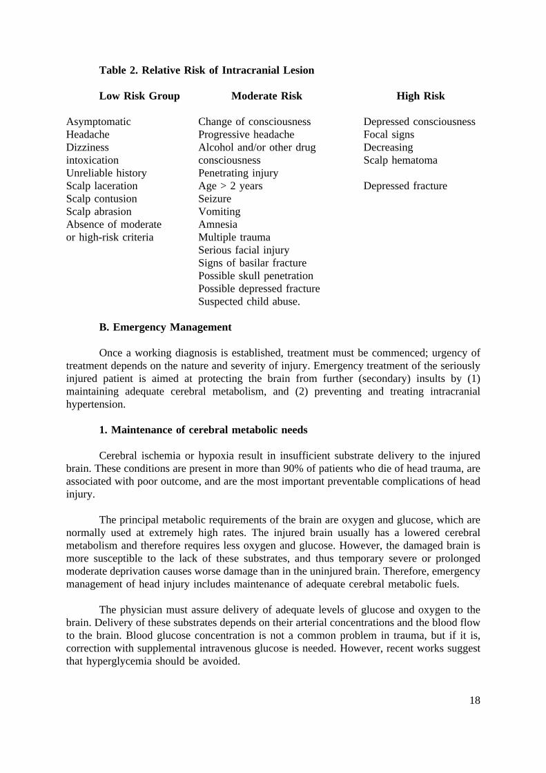
Table 2. Relative Risk of Intracranial Lesion
Low Risk Group Moderate Risk High Risk
Asymptomatic Change of consciousness Depressed consciousnessHeadache Progressive headache Focal signsDizziness Alcohol and/or other drug Decreasingintoxication consciousness Scalp hematomaUnreliable history Penetrating injuryScalp laceration Age > 2 years Depressed fractureScalp contusion SeizureScalp abrasion VomitingAbsence of moderate Amnesiaor high-risk criteria Multiple trauma
Serious facial injurySigns of basilar fracturePossible skull penetrationPossible depressed fractureSuspected child abuse.
B. Emergency Management
Once a working diagnosis is established, treatment must be commenced; urgency oftreatment depends on the nature and severity of injury. Emergency treatment of the seriouslyinjured patient is aimed at protecting the brain from further (secondary) insults by (1)maintaining adequate cerebral metabolism, and (2) preventing and treating intracranialhypertension.
1. Maintenance of cerebral metabolic needs
Cerebral ischemia or hypoxia result in insufficient substrate delivery to the injuredbrain. These conditions are present in more than 90% of patients who die of head trauma, areassociated with poor outcome, and are the most important preventable complications of headinjury.
The principal metabolic requirements of the brain are oxygen and glucose, which arenormally used at extremely high rates. The injured brain usually has a lowered cerebralmetabolism and therefore requires less oxygen and glucose. However, the damaged brain ismore susceptible to the lack of these substrates, and thus temporary severe or prolongedmoderate deprivation causes worse damage than in the uninjured brain. Therefore, emergencymanagement of head injury includes maintenance of adequate cerebral metabolic fuels.
The physician must assure delivery of adequate levels of glucose and oxygen to thebrain. Delivery of these substrates depends on their arterial concentrations and the blood flowto the brain. Blood glucose concentration is not a common problem in trauma, but if it is,correction with supplemental intravenous glucose is needed. However, recent works suggestthat hyperglycemia should be avoided.

19
Oxygen content depends on arterial hemoglobin and oxygen concentrations. Arterialoxygen concentration can be assessed with blood gases and supplemental oxygen deliveredto maintain the arterial PO2 at greater than 80 mm Hg. Blood transfusion may be necessaryto provide sufficient hemoglobin to maintain normal oxygen carrying capacity.
Cerebral blood flow is dependent on systemic arterial pressure and arterial PaCO2,especially shortly after head trauma. Normalization of blood pressure and maintenance ofPaCO2 at 26 to 28 mm Hg is sufficient to maintain adequate cerebral blood flow under mostcircumstances. Keep in mind that the PaCO2 cannot be allowed to rise too high as it mayaggravate intracranial pressure (see 2.a.).
2. Preventing / treating intracranial hypertension
The purpose of these treatments is to prevent, control, or diminish intracranialhypertension, regardless of its cause. In addition to mass lesions, acute brain swelling (avascular engorgement phenomena) and brain edema can complicate any primary brain injuryby causing or aggravating intracranial hypertension. It is for this reason that, optimally,neurosurgical consultation should be obtained when these measures are instituted.
a. Induced hypocapnia
Arterial carbon dioxide concentration profoundly affects cerebral circulation. Whenabnormally elevated, cerebrovasodilatation occurs, increasing intracranial blood volume andintracranial pressure. (See II. Anatomy and Physiology, H. Intracranial Pressure in thischapter.) Conversely, reduction of PaCO2 reduces intracranial blood volume and, secondarily,intracranial pressure. Additionally, hyperventilation tends to reduce intracerebral acidosis andincrease cerebral metabolism, both of which are beneficial. Therefore, hyperventilation isrecommended to reduce arterial PaCO2, maintaining it at 26 to 28 mm Hg. This procedureusually requires endotracheal intubation, controlled ventilation, and intermittent iatrogenicparalysis. Intubation should be performed early for the comatose patient. Care must be takento assure that injudicious intubation does not cause further intracranial problems due toincreased intracranial pressure elevations from coughing or gagging during intubation. (SeeChapter 2, Airway and Ventilatory Management.) Continued intubation may require sedationor iatrogenic paralysis. Hypocapnia can reduce cerebral circulation to the point where cerebralischemia occurs. Blood gases must be monitored closely.
b. Fluid control
Intravenous fluids should be administered judiciously to prevent overhydration, whichaugments cerebral edema. since the nature and volume of fluids administered will depend onthe systemic status of the patient, consultations between the trauma surgeon and theneurosurgeon are necessary. Intravenous fluid used for maintenance must not be hyposmolar,as cerebral edema may likewise follow.
c. Diuretics
Diuretics such as mannitol, which cause diuresis by producing intravascularhyperosmolarity, are widely used for severe brain injury. Mannitol undoubtedly can be very

20
effective in shrinking brain volume and lowering intracranial pressure. However, other effectsmay cause major problems. In the emergency department, this agent should be administeredonly with the consent of a neurosurgeon or to gain time when neurosurgical capabilities willbe delayed and the patient's condition is deteriorating. Mannitol is most commonly andeffectively used for a suspected surgical mass lesion when time is needed to prepare foroperation (ie, hematoma with deteriorating level of consciousness, hemiparesis, and dilatingpupil). For the average patient, 1 g/kg, is administered rapidly and intravenously. Care mustbe taken to prevent subcutaneous infiltration.
The neurosurgeon also may recommend loop diuretics, such as furosemide, (40 to 80mg IV for adults). These medications act by medically decompressing the brain and may buyup to several hours of reduced intracranial pressure. A urinary catheter is required.
Because of the rapid production of urine, vascular volume may be significantlyaffected. Therefore, the patient's blood pressure must be closely monitored, especially if thepatient has sustained multiple trauma or is a child. Urine volume may be replaced withisotonic fluids in these patients.
d. Steroids
Steroids are not recommended for the treatment of acute head injury.
VI. Other Manifestations of Head Injury
A. Seizures
Seizures, either full-blown convulsions or focal seizures, can occur with any injury.Seizures are common at the time of injury or shortly thereafter, and do not necessarily portendthe development of chronic epilepsy. The neurosurgeon may recommend no treatment.Prolonged or repetitive seizures may be associated with intracranial hemorrhage and shouldbe treated aggressively, because they may cause cerebral hypoxia, brain swelling, andincreased intracranial pressure. A common treatment regimen consists of 10 mg of diazepamvia an intravenous bolus. Respiratory exchange should be closely monitored. If the patienttolerates diazepam and has another convulsion, the dose may be repeated once with caution.Phenytoin, 1 g IV at a rate of 50 mg/minute, should be started as soon as possible, whilemonitoring the patient's blood pressure and electrocardiogram. If the convulsions persistdespite diazepam and phenytoin, the patient should be given phenobarbital or an anesthetic.The injured brain tolerates anesthesia well.
B. Restlessness
Restlessness frequently accompanies brain injury and/or cerebral hypoxia. Itsdevelopment in a previously quiet patient may be the first clinical expression of an expandingintracranial mass. Restlessness can often be controlled by correcting the cause. Primaryattention should be directed to the possibility of cerebral hypoxia and its etiology. Pain frominjuries, a distended bladder, painful bandages, tight casts, or systemic hypoxia may alsoinduce restlessness. Only when these etiologic factors have been treated or excluded is theuse of drugs justified to control the restlessness.

21
The relief of severe pain is an important part in the management of the trauma patient.Effective analgesia implies the use of intravenous opiates which may adversely affect thepatient. They also may cause respiratory depression and mask neurologic signs. Therefore, forthese reasons, opiates and other strong analgesics/sedatives should be withheld, if possible,until neurosurgical consultation has taken place. This presents less of a problem for theneurosurgeon if CT scan is readily available. Chlorpromazine, 10 to 25 mg IV, may behelpful for severe agitation. This agent must be administered carefully to avoid hypotension.
C. Hyperthermia
Hyperthermia is disastrous for the patient with an injured brain. Elevations in bodytemperature increase the rate of brain metabolism and levels of carbon dioxide. The higherthe temperature, the greater the risk to the patient. A head-injured patient with hyperthermiashould be placed on a hypothermia blanket, and shivering should be controlled withchlorpromazine.
VII. Scalp Wounds
Despite the dramatic appearance of scalp wounds, they are usually tolerated well andcause few complications. Scalp wounds usually heal well if general principles of good woundmanagement are followed. The location and type of injury to the scalp give the physician abetter understanding of the force and direction of the energy transmitted to the brain.
A. Blood Loss
Blood loss from scalp wounds can be extensive, especially in children. If the adultpatient is in shock, bleeding from the scalp alone is usually not the cause. The generalprinciples of scalp wound care include locating and stopping of the bleeding. In the case ofsevered large vessels (ie, superficial temporal arteries), the vessels should be clamped withhemostats and ligated. Moderately severe bleeding from a deep scalp laceration can usuallybe controlled temporarily by applying direct pressure or placing hemostats on the galea andreflecting them backwards. For the unconscious patient, self-retaining retractors can be used.
Most scalp bleeding, with or without direct ligation of bleeding vessels, may becontrolled by a snugly applied circumferential head dressing. If necessary, the hair may beclipped to facilitate application.
B. Inspection of the Scalp Wound
One should inspect the wound carefully and search under direct vision for signs of askull fracture or foreign material. Blind probing with an instrument or the gloved fingershould be avoided. Wound inspection also includes looking for CSF leaks, which may indicatenot only a skull fracture, but also an arachnoid and dural tear. A neurosurgeon should beconsulted before the wound is closed for all cases involving open fractures or depressedfractures of the skull.

22
C. Repairing the Scalp Wound
The first step in wound care is to irrigate the scalp wound with copious amounts ofsaline. Debris, including hair, must be removed. Do not remove bone fragments, because theymay be tamponading intracranial bleeding. The neurosurgeon should remove bone fragmentsin the operating room. When in doubt, always consult a neurosurgeon. If transfer to adefinitive-care facility is required, definitive surgical closure of a scalp wound can be delayed.However, temporary repair may be done to prevent further contamination of the wound.
When possible, primary definitive repair should be achieved under sterile techniquewith meticulous attention to the wound, as scalp wounds may become infected. The areaimmediately around the laceration should be shaved. The galea is closed first, usinginterrupted sutures. The skin is then sutured and a nonadherent dressing applied. The headwrap, if applied, should be just tight enough to apply mild pressure and prevent subgalealaccumulation of blood.
VIII. Definitive Surgical Management
Within two hours of injury, essential diagnostic studies should be completed. Althoughmeeting this standard is difficult, delay can be extremely costly for the patient. If aneurosurgeon is not available at the facility initially receiving the patient, expeditious transferof the patient to a hospital with an available neurosurgeon is necessary.
In exceptional circumstances, rapidly expanding intracranial hematomas (usuallyepidural hematomas) may be imminently life threatening and may not allow time for transferif neurosurgical care is some distance away. Although this circumstance is rare in urbansettings, it may occur occasionally in rural areas. In such circumstances, emergency burr holescan be considered if a surgeon, properly trained in the procedure, is available.
If definitive neurosurgical care is not immediately available, the purpose of anemergency burr hole is to preserve life by evacuating life-threatening intracranial hematomas.The performance of this procedure may be especially important for patients whose neurologicstatus rapidly deteriorates and for patients who do not respond to nonsurgical measures.However and before proceeding with the procedure, the physician must consider the followingproblems:
1. Not all severely head-injured patients have hematomas.
2. A burr hole, placed as little as 2 cm away from a hematoma, may not locate it.
3. A burr hole itself may cause brain damage or intracranial hemorrhage.
4. Burr-hole evacuation of a hematoma may not be life saving.
5. A burr hole can consume as much time as getting the patient to a neurosurgeon.
Performance of emergency burr holes should be done only in exceptionalcircumstances and only with the advice and consent of a neurosurgeon. The indications

23
for a burr hole performed by a nonneurosurgeon are few, and widespread use as adesperation maneuver is not recommended or supported by the Committee on Trauma.The procedure should be employed only if, in the judgment of a neurosurgeon, the burrhole can be performed in a timely manner, and when definitive neurosurgical carecannot be rendered expeditiously. In almost all instances this means that definitiveneurosurgical care is many miles or several hours away.
The neurosurgeon usually recommends the burr hole be placed on the side of thelargest pupil in comatose patients with decerebrate and decorticate posturing whoseposturing does not respond to endotracheal hyperventilation and dehydrating agents (eg,1 g/kg mannitol). The Committee on Trauma strongly recommends that those whoanticipate the need for this procedure receive proper training in the procedure from aneurosurgeon.
IX. Summary
A. Secure and maintain an open airway.
B. Ventilate the patient to maintain oxygenation and to avoid hypercarbia.
C. Treat shock, if present, and look for cause.
D. Except for shock, restrict fluid intake to maintenance levels.
E. Perform a minineurologic examination.
F. Establish an initial working diagnosis.
G. Prevent secondary brain injury.
H. Search for associated injuries.
I. Obtain roentgenograms or CT scan as needed, but only after the patient is stable.
J. Consult a neurosurgeon and consider early transfer.
K. Should the patient's condition worsen, consider other diagnoses and forms oftreatment; consult with a neurosurgeon, and consider transfer.
L. Continually reassess the patient (neurologic examination and GCS Score) to identifychanges that necessitate neurosurgical intervention.

24
Skill Station IX: Head and Neck Trauma Assessment and Management
Resources and Equipment
This list is the recommended equipment to conduct this skill session in accordancewith the stated objectives for and intent of the procedures outlined. Additional equipment maybe used providing it does not detract from the stated objectives and intent of this skill, orfrom performing the procedures in a safe method as described and recommended by the ACSCommittee on Trauma.
1. Mr. HURT - Head trauma manikin.2. Ophthalmoscope.3. Otoscope.4. Table and chairs; or gurney, pillows, and sheet.5. Semirigid cervical collar.6. Cervical traction tongs (preferably Gardner-Wells tongs).7. 2 coconuts.
Objectives
1. Performance at this station will allow participants to practice and demonstrate theirassessment and diagnostic skills in determining the type and extent of injuries with Mr.HURT (head trauma manikin).
2. Upon completion of this station, the participant will be able to discuss theimportance of clinical signs and symptoms found through assessment.
3. Upon completion of this station, the participant will be able to discuss and establishpriorities for initial primary management of the patient with head trauma.
4. Participation at this station will allow the student to discuss other diagnostic aidsthat can be used to determine the area of injury within the brain and the extent of injury.
5. Upon completion of this station, the participant will be able to demonstrateproficiency in the use and application of the cervical traction tongs.

25
Skill Procedures: Head and Neck Trauma Assessment and Management
Note: Universal precautions are required whenever caring for the trauma patient.
I. Head Trauma Assessment and Management
A. Primary Survey
1. ABCs.
2. Immobilize and stabilize the cervical spine.
3. Perform a brief neurologic examination.
a. Pupillary response.
b. AVPU.
B. Secondary Survey and Management
1. Inspect the head.
a. Lacerations.
b. Nose and ears for presence of CSF leakage.
2. Palpate the head.
a. Fractures.
b. Lacerations for underlying fractures.
3. Inspect all scalp lacerations.
a. Brain tissue.
b. Depressed skull fractures.
c. Debris.
d. CSF leaks.
4. Perform a minineurologic examination and determine the patient's GCS Score.
a. Eye-opening response.
b. Verbal response.

26
c. Best limb motor response.
5. Palpate the c-spine for fractures and apply a semirigid cervical collar, if needed.
6. Determine the extent of injury.
7. Reassess the patient continuously, observing for signs of deterioration.
a. Frequency.
b. Parameters to be assessed.
C. Head and Neck Injuries
1. Open, depressed skull fractures on the right
This type of injury is indicative of the amount of energy force that has been deliveredto the cranium. An open, depressed skull fracture requires early operative intervention, withmanagement directed toward the underlying brain injury.
2. Zygomatic fracture (Le Fort III fracture) on the right
A Le Fort III fracture is a separation of the mid-third of the face at thezygomaticofrontal, zygomaticotemporal, and nasal sutures, and across the orbital floor. It isa compound fracture with major hemorrhage that jeopardizes the integrity of the airway.Airway control and protection of the airway with concomitant immobilization of the cervicalspine is imperative. Inability to intubate the patient may be an indication for surgicalcricothyroidotomy, depending on the clinical judgment and skill level of the examiner.Nasotracheal intubation is contraindicated.
3. Maxillary fracture (Le Fort I fracture) on the right
Traumatic malocclusion is an important sign indicating a major disruption of the facialbones. Associated bleeding also may compromise the airway. Management includes: (1)securing a patent airway, (2) maintaining c-spine immobilization, and (3) hemorrhage control.
4. Mandibular fracture on the left
This is an important injury to recognize, especially if it occurs in combination withother facial fractures. In the unconscious patient, the tongue falls posteriorly and occludes theairway. Basic airway management procedures, eg, insertion of an oropharyngeal airway, arenecessary to prevent airway obstruction.
5. Mandibular/neck laceration on the left
Penetrating injuries of the neck are located in three anatomic areas: (1) Zone 1 -structures at the thoracic outlet, (2) Zone 2 - between the clavicles and mandible, and (3)Zone 3 - between the angle of the mandible and the base of the skull.

27
This is an important wound to recognize. In this particular manikin, the platysma hasbeen violated. Active bleeding, external or internal, may be present. Control and protectionof the airway is necessary in this unconscious patient.
6. Nasal fracture
Bleeding can be significant from nasal fractures and can place the integrity of theairway in jeopardy, especially in the unconscious patient.
7. Multiple fractures, C-6 vertebra
A step-off deformity, although difficult to palpate, is present. Management includescontinued immobilization of the c-spine and obtaining a lateral c-spine roentgenogram.
8. Unequal pupils
A unilateral, fixed and dilated pupil in an unconscious patient is a sign of graveconsequence. Appropriate resuscitation and surgical intervention must be instituted rapidly.The inability of the dilated pupil to react to light denotes one lesion. The third cranial nerveon the side of the dilated pupil may be compressed by increased intracranial pressure due toa mass lesion or epidural bleeding. Rapid neurosurgical intervention is necessary to preventsecondary brain injury. Controlled hyperventilation and fluid resuscitation restriction may berequired for this patient. The eyes also are exophthalmic.
9. Hemotympanum on the right
This sign denotes a basal skull fracture. Patients with this type of fracture may havesignificant traumatic CSF leaks. Cribriform plate fractures are often associated with thisinjury. Insertion of a nasogastric tube and nasotracheal intubation are contraindicated.
II. Application of Cervical Traction Tongs (Optional Skill Procedure)
Note: Differences of opinion exist among neurosurgeons regarding the indications andtechniques for the use of cervical traction in the patient with a known cervical spine injury.Therefore, protocols for its use should be established at each local institution in consultationwith a neurosurgeon.
This skill procedure is based on the use of Gardner-Wells tongs for the patient withmid-cervical fracture-dislocation, eg, C-4 on C-5.
A. Identify the site for application of the tongs. The tongs are applied above the earsand below the "equator" (temporal ridges).
B. Surgically prepare the scalp with an antiseptic preparation. It is not necessary toshave the scalp. An aerosol spray that coats and penetrates the hair to the skin isrecommended. Then rub in the antiseptic.
C. Locally inject an anesthetic and allow time for it to take effect.

28
D. If using a spray antiseptic, respray the scalp.
E. Place the tong apparatus with the metal instruction plate facing forward.
F. Advance the spring-loaded points until the 1-mm protrusion indicator within itsknob shows that the proper squeeze is being exerted. (As the points are advanced, the skinis stretched increasingly snug, effectively sealing the point of entry and preventing bleeding.)
G. When bone is encountered, the stiff spring yields until the outer end of the spring-loaded point barely protrudes beyond the flat surface of the knurled end. (Indicates 30 poundsof "squeeze" being applied.)
H. Tilt the tong back and forth several times to seat the points. Then check theindicator protrusion and retighten it if necessary.
I. Support the tongs with slight, hand-applied traction, parallel to the bed or patientcart, until the assistant attaches the rope and pulley system.
J. Apply five pounds of weight to the rope, and carefully release hold of the tongs.
K. Additional weight can be applied according to protocol or at the direction of theneurosurgeon.
Contraindications
1. Paget's disease of the skull.
2. Neonates or infants.
3. Massive open/comminuted skull fracture.
Complications
1. Localized sepsis at puncture sites.
2. Laceration of a scalp artery.


