CHAPTER 5 PURIFICATION AND CHARACTERIZATION OF BACTERIOCIN...
17
71 CHAPTER 5 PURIFICATION AND CHARACTERIZATION OF BACTERIOCIN PRODUCED BY STREPTOCOCCUS PHOCAE PI80 5.1. Introduction Studies of the bacteriocins of Streptococci extend back to the 1960s (Mindich, 1966). However, the most recent isolation and characterization of streptococcal bacteriocins have been focused on pathogenic streptococci, where most of the characterized bacteriocins have been found to originate from a few species. Lantibiotics are the most prevalent peptide bacteriocins in streptococci, where most are elongated cationic type A lantibiotics. Two-peptide lantibiotics have also been isolated from Streptococci (Ingolf et al., 2007). Streptococcus phocae was first isolated from clinical specimens taken from seals (Skaar et al., 1994), and although there are a few reports on its isolation from seals and Atlantic salmon (Gibello et al., 2005; Henton et al., 1999; Vossen et al., 2004), there have been no reports of any isolates from shrimp. Moreover, all of the reported isolates have been from diseased animals and were beta hemolytic. However, there has been no previous report on bacteriocin production by S. phocae. Accordingly, the main focus of this study was to characterize an antibacterial substance produced by a nonhemolytic strain, Streptococcus phocae PI80, isolated from the gut of Indian white shrimp ( Penaeus indicus) and then to purify it to homogeneity. This study also attempted to understand the mechanism of the cell damage induced by this antibacterial agent.
-
Upload
dangnguyet -
Category
Documents
-
view
226 -
download
3
Transcript of CHAPTER 5 PURIFICATION AND CHARACTERIZATION OF BACTERIOCIN...

71
CHAPTER 5
PURIFICATION AND CHARACTERIZATION OF BACTERIOCIN PRODUCED BY
STREPTOCOCCUS PHOCAE PI80
5.1. Introduction
Studies of the bacteriocins of Streptococci extend back to the 1960s (Mindich, 1966). However,
the most recent isolation and characterization of streptococcal bacteriocins have been focused on
pathogenic streptococci, where most of the characterized bacteriocins have been found to
originate from a few species. Lantibiotics are the most prevalent peptide bacteriocins in
streptococci, where most are elongated cationic type A lantibiotics. Two-peptide lantibiotics
have also been isolated from Streptococci (Ingolf et al., 2007). Streptococcus phocae was first
isolated from clinical specimens taken from seals (Skaar et al., 1994), and although there are a
few reports on its isolation from seals and Atlantic salmon (Gibello et al., 2005; Henton et al.,
1999; Vossen et al., 2004), there have been no reports of any isolates from shrimp. Moreover, all
of the reported isolates have been from diseased animals and were beta hemolytic. However,
there has been no previous report on bacteriocin production by S. phocae. Accordingly, the main
focus of this study was to characterize an antibacterial substance produced by a nonhemolytic
strain, Streptococcus phocae PI80, isolated from the gut of Indian white shrimp (Penaeus
indicus) and then to purify it to homogeneity. This study also attempted to understand the
mechanism of the cell damage induced by this antibacterial agent.

72
5.2. Materials and methods
5.2.1. Strains and culture conditions
A bacteriocin producer strain Streptococcus phocae PI80 was isolated from the gut of Indian
white shrimp (Peneaus indicus). The strain was identified by morphological, physiological,
biochemical tests and a homology search based on the 16S rDNA sequence (Gopalakannan,
2006). A DNA fragment corresponding to the 16S ribosomal region was PCR-amplified by using
chromosomal DNA as the template. Partial sequencing was carried out in Macrogen, Korea.
Bacterial strains and media used for the study are listed in Table 6. E. coli DH5 was used as an
indicator strain for the main test. All strains were maintained as frozen stocks at -80°C. Working
cultures were maintained in agar medium and subcultured in liquid media before use. MRS
medium (Himedia, Mumbai, India) supplemented with 1% NaCl was routinely used for culturing
Streptococcus phocae PI80.
5.2.2. Bacteriocin assay
Liquid cultures inoculated with 0.1% of an inoculum of Streptococcus phocae PI80 were grown
for 16 h at 37°C with constant shaking at 100 rpm. The cells were then separated by centrifuging
the culture medium at 8,500 ×g and 4oC for 15 min, whereas the cell-free culture supernatants
(CFCS) were neutralized with 1 N sodium hydroxide to pH 6.6 ± 0.1 and used immediately.
A well-diffusion assay procedure was used for the antibacterial assay, as described previously
(Schillinger & Lucke, 1989). Eighty microliter of the CFCS was placed in an 8-mm well of a
TSA (Vibrios), BHI (L. monocytogenes), and seawater agar plate (Aeromonas) (20 ml)
previously swabbed (10-2
dilution) with the appropriate indicator strain (Table 6). After 24 h of
73
incubation at a temperature optimal for the indicator strain, clear zones of inhibition appeared
when the strain was sensitive.
5.2.3. Effect of heat, solvent and enzyme treatment on phocaecin PI80
Dialyzed samples of phocaecin PI80 were used in these tests. E.coli DH5 was used as an
indicator organism. About 1 ml aliquots of bacteriocin sample were exposed to temperatures of
50°, 60°, 70°, 80°, 90° and 100°C for 10, 15 and 30 min and tested for antibacterial activity.
Sensitivity to solvents was determined by incubating partially purified bacteriocin sample with
acetone, ethanol, isopropanol, methanol, butanol, acetonitrile and chloroform at for 1 h at 37°C
and tested for antibacterial activity.
Sensitivity to enzymes was determined as described previously (Tiwari & Srivastava, 2008).
Bacteriocin sample incubated in the presence of following enzymes: protease, chymotrypsin,
pepsin, trypsin, lipase, catalase, peroxidase and diastase for 2 h at 37°C at final concentration of
1 mg/ml. All enzymes were purchased from HiMedia, Mumbai, India. After incubation the
enzymes were heat inactivated (70°C at 10 min.) and tested for inhibition against indicator
bacterium E.coli DH5.
5.2.4. Purification of phocaecin PI80
The CFCS was obtained as described in ―Bacteriocin assays.‖ The supernatant was subjected to
ultrafiltration (GE Healthcare, Uppsala, Sweden) using a 10 kDa cutoff membrane cartridge
filter, as the bacteriocin was suspected to be below this molecular mark. Ammonium sulfate was
slowly added to the resulting filtrate to produce 90% saturation (61.3 g/100 ml) and stirred
overnight at 4°C. The precipitated proteins were then collected by centrifugation at 10,000 ×g for

74
20 min at 4oC and resuspended in a minimal quantity of a 10 mM ammonium acetate buffer (pH
6.0). The suspension was then dialyzed overnight at 4°C against the same buffer in dialysis
tubing (Spectrumlabs, U.S.A.) with a 1 kDa cutoff. Thereafter, the dialyzed sample was applied
to a Sephadex G-25 (Sigma, U.S.A.) gel filtration column (C10/20) connected to an Akta Prime
plus protein purification system (GE Healthcare, Uppsala, Sweden). The gel column was
equilibrated with 10 mM ammonium acetate buffer (pH 6.0), and 1 ml of the dialyzed sample
was eluted using 10 mM ammonium acetate buffer (pH 6.0) containing 0.01 M sodium chloride
at a flow rate of 0.5 ml/min and fraction size of 1.0 ml. For each fraction, a bacteriocin activity
assay and protein profiling by Tricine SDS-PAGE were performed.
The active fraction was concentrated using a lyophilizer (Savant, U.S.A.), and the concentrated
sample further purified by reverse phase liquid chromatography (RP-HPLC) (Tahiri et al., 2004)
using a Shimadzu Analytical HPLC system (Shimadzu, Japan). Briefly, 25 μl of the concentrated
bacteriocin (fraction collected from column purification showing inhibition of pathogens) was
injected into an analytical C18 reverse-phase column (Luna 5 μm, 4.6×250 mm; Phenomenex,
CA, U.S.A.). The elution was performed at a flow rate of 1 ml/min using a linear gradient from
90% solvent A [0.1% (w/v) trifluoro-acetic acid (TFA) in 5% (v/v) acetonitrile in water] and 10%
solvent B (0.1% TFA in 100% acetonitrile) to 42% and 58% of solvents A and B, respectively,
within 46 min. The peptide fractions were detected spectrophotometrically by measuring the
absorbance at 220 nm and collected manually. The fractions were then concentrated using a
lyophilizer and dissolved in an ammonium acetate buffer (10 mM, pH 6.0) and used in the
bacteriocin activity assay and molecular mass determination.

75
5.2.5. Molecular mass determination
The molecular size of phocaecin PI80 was determined by separation of the fraction obtained after
an HPLC analysis using Tricine SDS-PAGE (10%) (Schägger & Jagow, 1987). A low molecular
mass protein marker with sizes ranging from 3.0 - 205 kDa (Bangalore Genei, Bangalore, India)
was used. The gel was stained using the silver staining method (Morrissey, 1981).
The molecular mass of phocaecin PI80 was further confirmed by mass-assisted laser desorption
ionization time of flight (MALDI-TOF) mass spectrometry. The mass of the peptide was
determined by the Proteomic facility of the Molecular Biophysics Unit at the Indian Institute of
Sciences, Bangalore, India.
5.2.6. Bacteriocin activity in polyacrylamide gels
The Tricine SDS-PAGE was run under nonreducing conditions (Svetoslav, 2007). The boiling
and addition of 2-mercaptoethanol in the probe buffer was suspended. The gel was washed 4
times for 10 min in distilled water with gentle agitation to remove the SDS. The gel was placed
in a petri dish containing a 2% agar medium and then overlaid with a precooled 0.7% agar
medium inoculated with the indicator organism. The plate was incubated for 24 h at 37°C.
5.2.7. Mode of Action and determination of K+ efflux by phocaecin PI80
The mode of action of phocaecin PI80 was determined by measuring the potassium ion leakage
in the external medium (Ghrairi et al., 2005). Briefly, the Streptococcus phocae PI80 cells were
grown in a MRS broth at 37°C for 16 h. The phocaecin PI80 was then partially purified from the
culture filtrate using Sephadex G-25 gel chromatography (Akta prime plus) and the bacteriocin
activity found to be 1,280 AU/ml. The indicator strains E. coli DH5α, E. coli CSH57, L.

76
monocytogenes and V. parahaemolyticus were grown overnight at 37°C and diluted to 10-2
.
Equal volumes of the purified bacteriocin sample and indicator bacteria were incubated for 1 h at
37°C and centrifuged to remove the particulate material. The bacteriocin + water served as
common control for the experiment, whereas a second control of just the bacterial cells was kept
as an individual indicator organism so as to rule out any K+ interference due to the medium or
cell lysis caused by reasons other than the bacteriocin. The standards (1-100 ppm) were prepared
by dissolving KCl in distilled water. The K+ efflux was determined by measuring the
concentration of K+ using Inductively Coupled Plasma Optical Emission Spectrometry (ICP-
OES) (Jobin Vyon, Japan).
5.3. Result
5.3.1. Identification of Streptococcus phocae from gut of Penaeus indicus
Isolate was identified as Streptococcus phocae by morphological, biochemical and phylogenetic
16S rDNA sequence method. Sequence was identified and deposited in Genbank (Accession No.
EU117220) S. phocae PI80 was found to be Gram positive, cocci, non- motile, catalase negative,
alpha hemolytic in sheep blood agar plate, oxidase negative and capable of reducing nitrate
present in the medium.
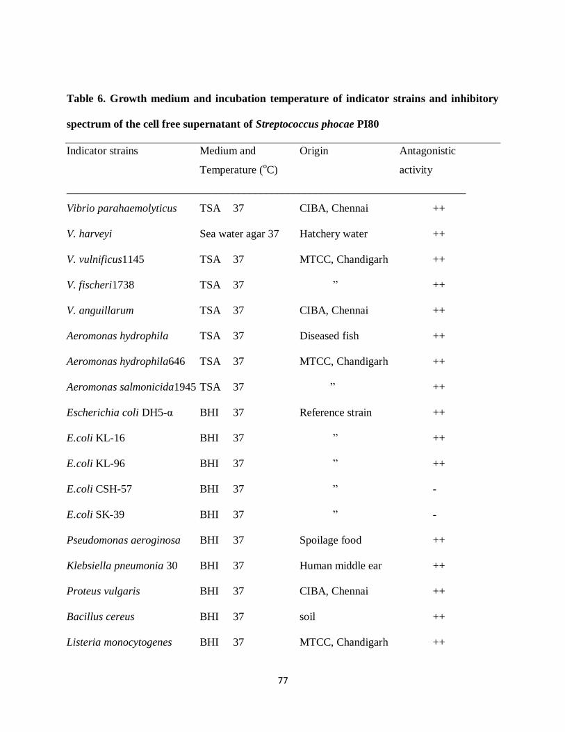
5.3.2. Inhibitory spectrum of phocaecin PI80
Inhibition was found against wide spectrum of bacterial species which includes both Gram
positive as well as negative (Table 6). Streptococcus phocae PI80 was found to be active against
important pathogens of food and livestock (fish and shrimp): Listeria monocytogenes, Vibrio
parahaemolyticus, Aeromonas hydrophila, Pseudomonas aeroginosa.
77
Table 6. Growth medium and incubation temperature of indicator strains and inhibitory
spectrum of the cell free supernatant of Streptococcus phocae PI80
Indicator strains Medium and Origin Antagonistic
Temperature (oC) activity
________________________________________________________________________
Vibrio parahaemolyticus TSA 37 CIBA, Chennai ++
V. harveyi Sea water agar 37 Hatchery water ++
V. vulnificus1145 TSA 37 MTCC, Chandigarh ++
V. fischeri1738 TSA 37 ‖ ++
V. anguillarum TSA 37 CIBA, Chennai ++
Aeromonas hydrophila TSA 37 Diseased fish ++
Aeromonas hydrophila646 TSA 37 MTCC, Chandigarh ++
Aeromonas salmonicida1945 TSA 37 ‖ ++
Escherichia coli DH5-α BHI 37 Reference strain ++
E.coli KL-16 BHI 37 ‖ ++
E.coli KL-96 BHI 37 ‖ ++
E.coli CSH-57 BHI 37 ‖ -
E.coli SK-39 BHI 37 ‖ -
Pseudomonas aeroginosa BHI 37 Spoilage food ++
Klebsiella pneumonia 30 BHI 37 Human middle ear ++
Proteus vulgaris BHI 37 CIBA, Chennai ++
Bacillus cereus BHI 37 soil ++
Listeria monocytogenes BHI 37 MTCC, Chandigarh ++
78
Lactobacillus plantarum BHI 37 ‖ ++
L. acidophilus BHI 37 ‖ ++
L. rhamnosus BHI 37 ‖ ++
________________________________________________________________________
+, inhibition zone less than10 mm in diameter; ++, inhibition zone larger than10 mm in diameter;
-, no inhibition zone recorded.
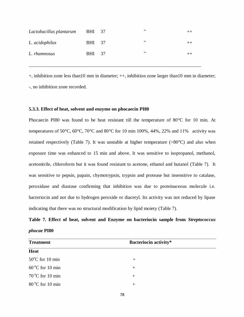
5.3.3. Effect of heat, solvent and enzyme on phocaecin PI80
Phocaecin PI80 was found to be heat resistant till the temperature of 80°C for 10 min. At
temperatures of 50°C, 60°C, 70°C and 80°C for 10 min 100%, 44%, 22% and 11% activity was
retained respectively (Table 7). It was unstable at higher temperature (>80°C) and also when
exposure time was enhanced to 15 min and above. It was sensitive to isopropanol, methanol,
acetonitrile, chloroform but it was found resistant to acetone, ethanol and butanol (Table 7). It
was sensitive to pepsin, papain, chymotrypsin, trypsin and protease but insensitive to catalase,
peroxidase and diastase confirming that inhibition was due to proteinaceous molecule i.e.
bacteriocin and not due to hydrogen peroxide or diacetyl. Its activity was not reduced by lipase
indicating that there was no structural modification by lipid moiety (Table 7).
Table 7. Effect of heat, solvent and Enzyme on bacteriocin sample from Streptococcus
phocae PI80
Treatment Bacteriocin activity*
Heat
50oC for 10 min +
60 oC for 10 min +
70 oC for 10 min +
80 oC for 10 min +

79
90 oC for 10 min
Solvent
Acetone +
Ethanol +
Isopropanol
Methanol
Butanol +
Acetonitrile
Chloroform
Enzymes
Trypsin
Diastase +
Catalase +
Protease
Lipase +
Pepsin
Chymotrypsin
Peroxidase +
+, inhibition zone; –, no inhibition zone
* Bacteriocin activity was determined against E. coli DH5α
5.3.4. Purification of phocaecin PI80
To purify phocaecin PI80, CFCS was ultrafiltered through 10 kDa membrane and then
ammonium sulphate precipitated, dialyzed and subjected to Sephadex G-25 gel chromatography
(Fig.1). Active fraction obtained was found to retain 44% of initial activity found in CFCS. This
active fraction was lyophilized and dissolved in minimum volume of Solvent A for HPLC

80
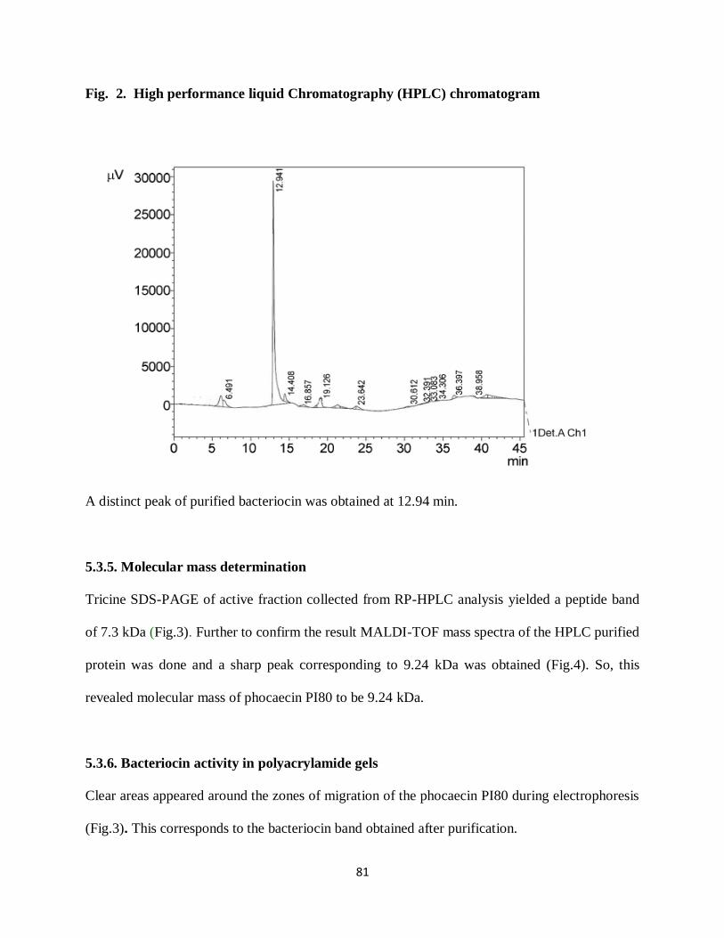
analysis. On injection of SephadexG-25 purified sample to reverse – phase HPLC a distinct peak
was eluted at 27.34% of acetonitrile, corresponding to retention time of 12.94 min (Fig.2). The
peak was shown to be active against E. coli DH5α. This active peak was further used for
molecular mass determination.
Fig. 1. Sephadex G-25 Chromatogram of Bacteriocin Sample
Bacteriocin sample was purified by Sephadex G-25 gel chromatography. Sample was eluted at
0.5 ml/min flow rate. Bacteriocin protein was eluted at about 19.57 min in the fraction numbers 4
to 10.
81
Fig. 2. High performance liquid Chromatography (HPLC) chromatogram
A distinct peak of purified bacteriocin was obtained at 12.94 min.
5.3.5. Molecular mass determination
Tricine SDS-PAGE of active fraction collected from RP-HPLC analysis yielded a peptide band
of 7.3 kDa (Fig.3). Further to confirm the result MALDI-TOF mass spectra of the HPLC purified
protein was done and a sharp peak corresponding to 9.24 kDa was obtained (Fig.4). So, this
revealed molecular mass of phocaecin PI80 to be 9.24 kDa.
5.3.6. Bacteriocin activity in polyacrylamide gels
Clear areas appeared around the zones of migration of the phocaecin PI80 during electrophoresis
(Fig.3). This corresponds to the bacteriocin band obtained after purification.

82
Fig. 3. Tricine SDS- PAGE of Purified Bacteriocin from Streptococcus phocae PI80
A. Molecular weight marker (3.0-205 kDa)
B. Purified active bacteriocin sample showing a single band of 7.3 kDa
C. Non-reducing Tris-tricine SDS-PAGE in 10% migrating gel overlaid
with agar (0.7%) inoculated with the sensitive strain E. coli DH5α. An activity zone
Corresponding approximately to 7.3 kDa band provides evidence for its antibacterial
activity.

83
Fig. 4. Mass Assisted Laser Desorption Ionization Time of Flight (MALDI-TOF) mass
spectrum of Purified bacteriocin
The single active peak fraction of HPLC was subjected to mass spectrometry. MALDI-TOF
analysis showing a distinct peak at 9244.419 m/z.
5.3.7. Mode of action of phocaecin PI80
Potassium ion leakage by the susceptible cells due to the action of phocaecin PI80 alone was
found out by subtracting the value obtained (ppm) from test reaction (bacteriocin + cells) to
control values of bacteriocin (control a) and bacterial cell (control b). After incubation of E. coli
DH5α, L. monocytogenes and V. parahemolyticus with partially purified bacteriocin sample K+
efflux of 434 ppm, 98 ppm, 120 ppm was observed respectively by ICP-OES. E. coli CSH57
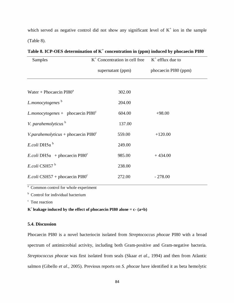
84
which served as negative control did not show any significant level of K+
ion in the sample
(Table 8).
Table 8. ICP-OES determination of K+ concentration in (ppm) induced by phocaecin PI80
Samples K+ Concentration in cell free K
+ efflux due to
supernatant (ppm) phocaecin PI80 (ppm)
Water + Phocaecin PI80a 302.00
L.monocytogenes b 204.00
L.monocytogenes + phocaecin PI80c 604.00 +98.00
V. parahemolyticus b 137.00
V.parahemolyticus + phocaecin PI80c 559.00 +120.00
E.coli DH5α b 249.00
E.coli DH5α + phocaecin PI80c 985.00 + 434.00
E.coli CSH57 b 238.00
E.coli CSH57 + phocaecin PI80c 272.00 - 278.00
a Common control for whole experiment
b Control for individual bacterium
c Test reaction
K+
leakage induced by the effect of phocaecin PI80 alone = c- (a+b)
5.4. Discussion
Phocaecin PI80 is a novel bacteriocin isolated from Streptococcus phocae PI80 with a broad
spectrum of antimicrobial activity, including both Gram-positive and Gram-negative bacteria.
Streptococcus phocae was first isolated from seals (Skaar et al., 1994) and then from Atlantic
salmon (Gibello et al., 2005). Previous reports on S. phocae have identified it as beta hemolytic

85
and mostly isolated from diseased animals (Gibello et al., 2005; Stefanie & George, 2005).
However, the S. phocae was isolated from shrimp in our laboratory, and the strain was found to
be non-hemolytic and non-pathogenic, which was confirmed by testing the strain with the
common carp Cyprinus carpio (Gopalakannan, 2006) and shrimp Penaeus monodon (Swain et
al., 2009). Moreover, no previous report has been found regarding the isolation of bacteria from
Penaeus indicus for the purpose of bacteriocin production and probiotics. Therefore, this is the
first report of a bacteriocin from S. phocae. Phocaecin PI80 exhibited strong anti-listeric activity
against L. monocytogenes. Other Streptococcal strains that also show anti-listerial activity
include S. thermophilus 110 (Stefanie & George, 2005), S. thermophilus 13 (Marciset et al.,
1997), S. thermophilus 81 (Ivanova et al., 1998), and S. thermophilus 347 (Villani et al., 1995).
Bacteriocins that are active against L. monocytogenes have already been used successfully for
food preservation. For example, nisin at a concentration of 2.5 mg/l effectively inhibits the
growth of L. monocytogenes in ricotta-type cheese for a period of 8 weeks or more (Davies et al.,
1997). In addition, bacteriocins have also been found to be effective against L. monocytogenes
when used in combination with an increased NaCl concentration (Thomas & Wimpenny, 1996).
As S. phocae has already shown antagonistic activity towards Aeromonas hydrophila, V.
parahaemolyticus, V. harveyi, V. fischeri 1738 and V. anguillarum, it is suggested that
Streptococcus phocae PI80 may be effective as a probiotic agent for shrimp farming, as well as
in the preservation of seafood to enhance the shelf life.
In this study, Phocaecin PI80 was found to be heat stable up to 70oC for 10 min, making it
neither very heat stable nor very heat labile, whereas bacteriocins previously extracted from
Streptococcus species are either heat stable or heat labile (Ivanova et al., 1998; Mathot et al.,
2003; Stefanie & George, 2005; Tadashi et al., 1982). Phocaecin PI80 also showed sensitivity
86
towards proteolytic enzymes, protease, chymotrypsin, pepsin and trypsin, suggesting a
proteinaceous nature. Similar characteristics have been observed for Thermophilin 110 (Stefanie
& George, 2005), Thermophilin 580 (Mathot et al., 2003) and Thermophilin 13 (Marciset et al.,
1997).
Phocaecin PI80 was purified to homogeneity, and MALDI-TOF mass spectrometry confirmed
the purity of the sample and determined the molecular mass of the bacteriocin to be 9.24 kDa. In
the case of SDS-PAGE, the bacteriocin migrated with an apparent molecular mass of about 7.3
kDa. This discrepancy could be attributed to the nonlinear migration of small peptides on SDS-
PAGE gels (Piard et al., 1992). Similarly, lacticin H-559 and gassericin A migrate to positions of
2.5 kDa and 3.8 kDa respectively, in the case of SDS-PAGE, whereas mass spectrometry shows
their molecular mass to be 3.34 kDa and 5.65 kDa, respectively (Hun et al., 1999; Kawai et al.,
1998). Phocaecin PI80 could be classified as a class II bacteriocin based on its molecular mass
(Klaenhammer, 1993). Some of the bacteriocins that have already been isolated from bacteria
belonging to the genus Streptococcus have either a low molecular mass or high molecular mass
(Ingolf et al., 2007). Dysgalactin isolated from Streptococcus dysgalactiae subsp. equisimilis has
a molecular mass of 21 kDa (Wong, 1981). Similarly, Stellalysin (29 kDa) and SA-M57 (17
kDa) were isolated from Streptococcus constellatus subsp. constellatus and M-type 57
Streptococcus pyogenes, respectively (Simpson & Tagg, 1983; Wong, 1981).
In most of the previous studies, the mode of bacteriocin activity is typically monitored by a
decrease in the viability of an indicator organism (Amer, 2007; Stefanie & George, 2005), yet
this effect alone provides little information on the bacteriocin action. Previous studies have
indicated that LAB bacteriocins are poreforming peptides that catalyze the potassium efflux from
sensitive strains (Abee et al., 1994; Mantovani et al., 2002; McAuliffe et al., 1998). Similarly,

87
phocaecin PI80 induced a massive leakage of K+ from L. monocytogenes, E. coli DH5α, and V.
parahaemolyticus, when increasing the outside concentration. Therefore, this finding suggests
that phocaecin PI80 renders the membrane of sensitive cells permeable, allowing the efflux of
K+. This release also probably occurs through pore formation (Castellano et al., 2003; Nes &
Holo, 2000), where the resultant pore formation in the cytoplasmic membrane induces the
leakage of small intracellular substances from sensitive cells (Beatriz et al., 1996).
Consequently, to establish a safe and effective antimicrobial agent, additional investigation is
currently under way to further characterize the efficacy of phocaecin PI80 as a seafood
biopreservative and S. phocae PI80 as a potential probiont for shrimp farming.