Chapter 44
54
right © 2005 Pearson Education, Inc. publishing as Benjamin Cummings PowerPoint Lectures for Biology, Seventh Edition Neil Campbell and Jane Reece Lectures by Chris Romero Chapter 44 Osmoregulation and Excretion
-
Upload
holly-duffy -
Category
Documents
-
view
25 -
download
0
description
Chapter 44. Osmoregulation and Excretion. Overview: A Balancing Act. Physiological systems of animals operate in a fluid environment Relative concentrations of water and solutes must be maintained within fairly narrow limits. - PowerPoint PPT Presentation
Transcript of Chapter 44
Copyright © 2005 Pearson Education, Inc. publishing as Benjamin Cummings
PowerPoint Lectures for Biology, Seventh Edition
Neil Campbell and Jane Reece
Lectures by Chris Romero
Chapter 44Chapter 44
Osmoregulation and Excretion

Copyright © 2005 Pearson Education, Inc. publishing as Benjamin Cummings
Overview: A Balancing Act
• Physiological systems of animals operate in a fluid environment
• Relative concentrations of water and solutes must be maintained within fairly narrow limits

Copyright © 2005 Pearson Education, Inc. publishing as Benjamin Cummings
•Freshwater animals show adaptations that reduce water uptake and conserve solutes
•Desert and marine animals face desiccating environments that can quickly deplete body water

Copyright © 2005 Pearson Education, Inc. publishing as Benjamin Cummings
• Osmoregulation regulates solute concentrations and balances the gain and loss of water
• Excretion gets rid of metabolic wastes

Copyright © 2005 Pearson Education, Inc. publishing as Benjamin Cummings
Concept 44.1: Osmoregulation balances the uptake and loss of water and solutes
• Osmoregulation is based largely on controlled movement of solutes between internal fluids and the external environment

Copyright © 2005 Pearson Education, Inc. publishing as Benjamin Cummings
Osmosis
• Cells require a balance between osmotic gain and loss of water
• Various mechanisms of osmoregulation in different environments balance water uptake and loss

Copyright © 2005 Pearson Education, Inc. publishing as Benjamin Cummings
Osmotic Challenges
• Osmoconformers, consisting only of some marine animals, are isoosmotic with their surroundings and do not regulate their osmolarity
• Osmoregulators expend energy to control water uptake and loss in a hyperosmotic or hypoosmotic environment

Copyright © 2005 Pearson Education, Inc. publishing as Benjamin Cummings
• Most animals are stenohaline; they cannot tolerate substantial changes in external osmolarity
• Euryhaline animals can survive large fluctuations in external osmolarity

Copyright © 2005 Pearson Education, Inc. publishing as Benjamin Cummings
Marine Animals
• Most marine invertebrates are osmoconformers
• Most marine vertebrates and some invertebrates are osmoregulators
• Marine bony fishes are hypoosmotic to sea water
• They lose water by osmosis and gain salt by diffusion and from food
• They balance water loss by drinking seawater

LE 44-3aLE 44-3a
Gain of water andsalt ions from foodand by drinkingseawater
Osmotic water lossthrough gills and other partsof body surface
Excretion ofsalt ionsfrom gills
Osmoregulation in a saltwater fish
Excretion of salt ions and small amountsof water in scantyurine from kidneys

Copyright © 2005 Pearson Education, Inc. publishing as Benjamin Cummings
Freshwater Animals
• Freshwater animals constantly take in water from their hypoosmotic environment
• They lose salts by diffusion and maintain water balance by excreting large amounts of dilute urine
• Salts lost by diffusion are replaced by foods and uptake across the gills

LE 44-3bLE 44-3b
Excretion oflarge amounts ofwater in diluteurine from kidneys
Osmotic water gainthrough gills and other partsof body surface
Osmoregulation in a freshwater fish
Uptake ofsalt ionsby gills
Uptake ofwater and someions in food
Copyright © 2005 Pearson Education, Inc. publishing as Benjamin Cummings
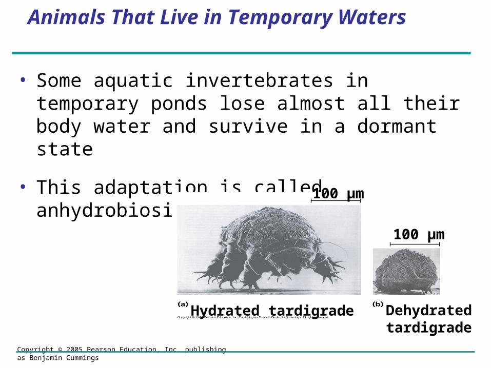
Animals That Live in Temporary Waters
• Some aquatic invertebrates in temporary ponds lose almost all their body water and survive in a dormant state
• This adaptation is called anhydrobiosis
Hydrated tardigrade Dehydrated tardigrade
100 µm
100 µm

Copyright © 2005 Pearson Education, Inc. publishing as Benjamin Cummings
Land Animals
•Land animals manage water budgets by drinking and eating moist foods and using metabolic water
Waterbalance in a kangaroo rat(2 mL/day)
Waterbalance ina human
(2,500 mL/day)
Watergain
Waterloss
Derived frommetabolism (1.8 mL)
Ingestedin food (0.2 mL)
Derived frommetabolism (250 mL)
Ingestedin food (750 mL)
Ingestedin liquid (1,500 mL)
Evaporation (900 mL)
Feces (100 mL)Urine(1,500 mL)
Evaporation (1.46 mL)
Feces (0.09 mL)Urine(0.45 mL)

LE 44-6LE 44-6
Control group(Unclipped fur)
Experimental group(Clipped fur)
Wat
er l
ost
per
day
(L/1
00 k
g b
od
y m
ass) 4
3
2
1
0
Desert animals get major water savings from simple anatomical features

Copyright © 2005 Pearson Education, Inc. publishing as Benjamin Cummings
Transport Epithelia
• Transport epithelia are specialized cells that regulate solute movement
• They are essential components of osmotic regulation and metabolic waste disposal
• They are arranged in complex tubular networks
• An example is in salt glands of marine birds, which remove excess sodium chloride from the blood

LE 44-7aLE 44-7a
Nostrilwith saltsecretions
Nasal salt gland

LE 44-7bLE 44-7b
Vein
Capillary
Secretorytubule
Transportepithelium
Directionof saltmovement
Centralduct
Artery
Bloodflow
Lumen ofsecretory tubule
NaCl
Secretory cellof transportepithelium

Copyright © 2005 Pearson Education, Inc. publishing as Benjamin Cummings
Concept 44.2: An animal’s nitrogenous wastes reflect its phylogeny and habitat
•The type and quantity of an animal’s waste products may greatly affect its water balance
•Among the most important wastes are nitrogenous breakdown products of proteins and nucleic acids
Nitrogenous bases
Nucleic acids
Amino acids
Proteins
—NH2
Amino groups
Most aquatic animals, including most bony fishes
Mammals, most amphibians, sharks, some bony fishes
Many reptiles (including birds), insects, land snails
Ammonia Urea Uric acid

Copyright © 2005 Pearson Education, Inc. publishing as Benjamin Cummings
Forms of Nitrogenous Wastes
Ammonia
• Animals that excrete nitrogenous wastes as ammonia need lots of water
• They release ammonia across the whole body surface or through gills

Copyright © 2005 Pearson Education, Inc. publishing as Benjamin Cummings
Forms of Nitrogenous Wastes
Urea
• The liver of mammals and most adult amphibians converts ammonia to less toxic urea
• The circulatory system carries urea to the kidneys, where it is excreted

Copyright © 2005 Pearson Education, Inc. publishing as Benjamin Cummings
Forms of Nitrogenous Wastes
Uric Acid
• Insects, land snails, and many reptiles, including birds, mainly excrete uric acid
• Uric acid is largely insoluble in water and can be secreted as a paste with little water loss

Copyright © 2005 Pearson Education, Inc. publishing as Benjamin Cummings
The Influence of Evolution and Environment on Nitrogenous Wastes
• The kinds of nitrogenous wastes excreted depend on an animal’s evolutionary history and habitat
• The amount of nitrogenous waste is coupled to the animal’s energy budget
Copyright © 2005 Pearson Education, Inc. publishing as Benjamin Cummings
Concept 44.3: Diverse excretory systems are variations on a tubular theme
• Excretory systems regulate solute movement between internal fluids and the external environment

Copyright © 2005 Pearson Education, Inc. publishing as Benjamin Cummings
Excretory Processes
•Most excretory systems produce urine by refining a filtrate derived from body fluids
•Key functions of most excretory systems:
– Filtration: pressure-filtering of body fluids
– Reabsorption: reclaiming valuable solutes
– Secretion: adding toxins and other solutes from the body fluids to the filtrate
– Excretion: removing the filtrate from the system
Filtration
Reabsorption
Secretion
Excretion
Excretorytubule
Capillary
Filtrate
Urin
e

Copyright © 2005 Pearson Education, Inc. publishing as Benjamin Cummings
•A protonephridium is a network of dead-end tubules lacking internal openings
•The smallest branches of the network are capped by a cellular unit called a flame bulb
•These tubules excrete a dilute fluid and function in osmoregulation
Protonephridia(tubules)
Tubule
Nephridioporein body wall
Flamebulb
Interstitial fluidfilters throughmembrane wherecap cell and tubulecell interdigitate(interlock)
Tubule cell
Cilia
Nucleusof cap cell
Protonephridia: Flame-Bulb Systems

Copyright © 2005 Pearson Education, Inc. publishing as Benjamin Cummings
Metanephridia
• Each segment of an earthworm has a pair of open-ended metanephridia
• Metanephridia consist of tubules that collect coelomic fluid and produce dilute urine for excretion
Collectingtubule
Nephridio-pore
Capillarynetwork
Coelom
Bladder
MetanephridiumNephrostome

Copyright © 2005 Pearson Education, Inc. publishing as Benjamin Cummings
Malpighian Tubules
• In insects and other terrestrial arthropods, Malpighian tubules remove nitrogenous wastes from hemolymph and function in osmoregulation
• Insects produce a relatively dry waste matter, an important adaptation to terrestrial life
Salt, water, andnitrogenous
wastes
Digestive tract
Midgut(stomach)
Malpighiantubules
RectumIntestineHindgut
Reabsorption of H2O,ions, and valuableorganic molecules
Malpighiantubule
HEMOLYMPH
Anus
Rectum
Feces and urine

Copyright © 2005 Pearson Education, Inc. publishing as Benjamin Cummings
Vertebrate Kidneys
• Kidneys, the excretory organs of vertebrates, function in both excretion and osmoregulation

Copyright © 2005 Pearson Education, Inc. publishing as Benjamin Cummings
Concept 44.4: Nephrons and associated blood vessels are the functional unit of the mammalian kidney
• The mammalian excretory system centers on paired kidneys, which are also the principal site of water balance and salt regulation
• Each kidney is supplied with blood by a renal artery and drained by a renal vein
• Urine exits each kidney through a duct called the ureter
• Both ureters drain into a common urinary bladder
Animation: Nephron Introduction

Copyright © 2005 Pearson Education, Inc. publishing as Benjamin Cummings
Structure and Function of the Nephron and Associated Structures
• The mammalian kidney has two distinct regions: an outer renal cortex and an inner renal medulla
• The nephron, the functional unit of the vertebrate kidney, consists of a single long tubule and a ball of capillaries called the glomerulus
Excretory organs and major associated blood vessels
RenalmedullaRenalcortexRenalpelvis
Section of kidney from a ratKidney structure
Ureter
Kidney
GlomerulusBowman’s capsule
Proximal tubulePeritubular capillaries
Afferentarteriolefrom renalartery
Efferentarteriole from glomerulus
DistaltubuleCollectingduct
SEM20 µm
Branch ofrenal vein
Filtrate and blood flow
Vasarecta
DescendinglimbAscendinglimb
LoopofHenle
Renalmedulla
Nephron
Torenalpelvis
Renalcortex
Collectingduct
Juxta-medullarynephron
Corticalnephron
Posterior vena cava
Renal artery and vein
Aorta
Ureter
Urinary bladder
Urethra

Copyright © 2005 Pearson Education, Inc. publishing as Benjamin Cummings
Filtration of the Blood
• Filtration occurs as blood pressure forces fluid from the blood in the glomerulus into the lumen of Bowman’s capsule
• Filtration of small molecules is nonselective
• The filtrate in Bowman’s capsule mirrors the concentration of solutes in blood plasma

Copyright © 2005 Pearson Education, Inc. publishing as Benjamin Cummings
Pathway of the Filtrate
• From Bowman’s capsule, the filtrate passes through three regions of the nephron: the proximal tubule, the loop of Henle, and the distal tubule
• Fluid from several nephrons flows into a collecting duct

Copyright © 2005 Pearson Education, Inc. publishing as Benjamin Cummings
Blood Vessels Associated with the Nephrons
• Each nephron is supplied with blood by an afferent arteriole, a branch of the renal artery that divides into the capillaries
• The capillaries converge as they leave the glomerulus, forming an efferent arteriole
• The vessels divide again, forming the peritubular capillaries, which surround the proximal and distal tubules

Copyright © 2005 Pearson Education, Inc. publishing as Benjamin Cummings
From Blood Filtrate to Urine: A Closer Look
•Filtrate becomes urine as it flows through the mammalian nephron and collecting duct
•Secretion and reabsorption in the proximal tubule greatly alter the filtrate’s volume and composition
•Reabsorption of water continues as filtrate moves into the descending limb of the loop of Henle
http://www.colorado.edu/kines/Class/IPHY3430-200/13urinar.html

Copyright © 2005 Pearson Education, Inc. publishing as Benjamin Cummings
• In the ascending limb of the loop of Henle, salt diffuses from the permeable tubule into the interstitial fluid
• The distal tubule regulates the K+ and NaCl concentrations of body fluids
• The collecting duct carries filtrate through the medulla to the renal pelvis and reabsorbs NaCl

LE 44-14LE 44-14
Filtrate
H2O
Salts (NaCl and others)
HCO3–
H+
Urea
Glucose; amino acids
Some drugs
Key
Active transport
Passive transportINNERMEDULLA
OUTERMEDULLA
NaCl
H2O
CORTEX
Descending limbof loop ofHenle
Proximal tubule
NaCl Nutrients
HCO3–
H+
K+
NH3
H2O
Distal tubule
NaCl HCO3–
H+K+
H2O
Thick segmentof ascendinglimb
NaCl
NaCl
Thin segmentof ascendinglimb
Collectingduct
Urea
H2O

Copyright © 2005 Pearson Education, Inc. publishing as Benjamin Cummings
Concept 44.5: The mammalian kidney’s ability to conserve water is a key terrestrial adaptation
• The mammalian kidney conserves water by producing urine that is much more concentrated than body fluids

Copyright © 2005 Pearson Education, Inc. publishing as Benjamin Cummings
Solute Gradients and Water Conservation
• The cooperative action and precise arrangement of the loops of Henle and collecting ducts are largely responsible for the osmotic gradient that concentrates the urine
• NaCl and urea contribute to the osmolarity of the interstitial fluid, which causes reabsorption of water in the kidney and concentrates the urine
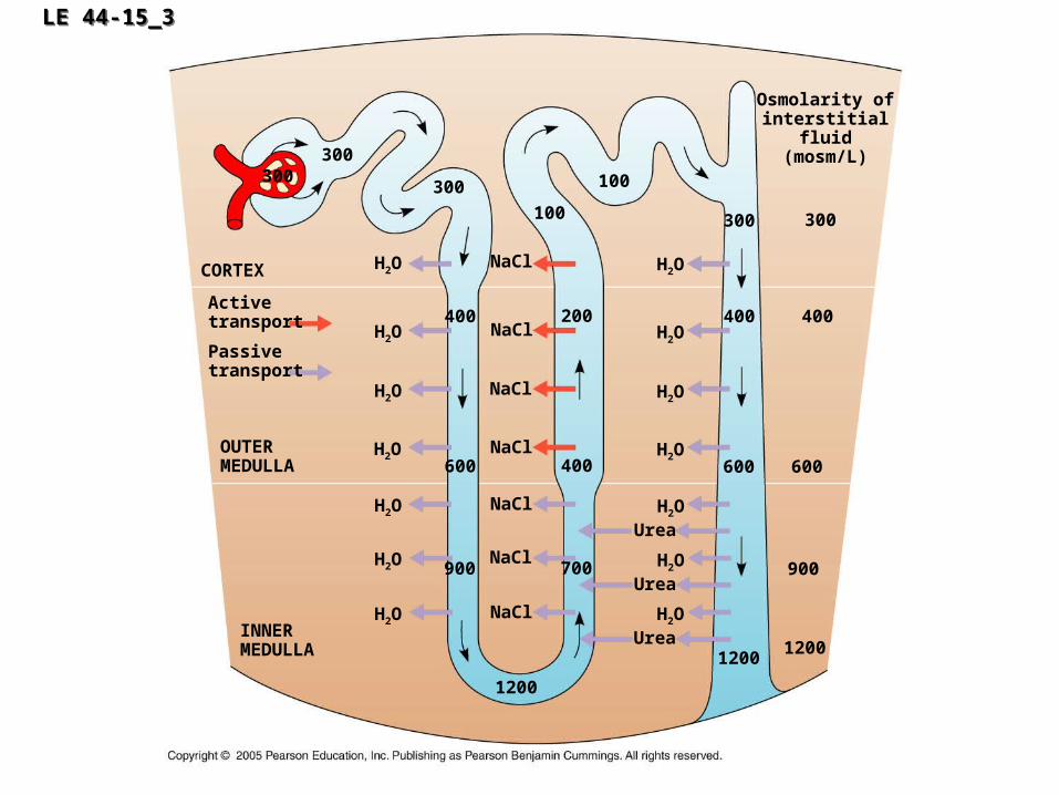
LE 44-15_3LE 44-15_3
INNERMEDULLA
OUTERMEDULLA
CORTEX
Osmolarity ofinterstitial
fluid(mosm/L)
NaCl
Urea
H2O
Activetransport
Passivetransport
300300
300 100
100
400 200H2O
H2O
H2O
H2O
H2O
H2O
600 400
900 700
1200
300
400
H2O
600
12001200
600
900
300
400NaCl
NaCl
NaCl
NaCl
NaCl
NaCl
UreaH2O
UreaH2O
H2O
H2O
H2O
H2O

Copyright © 2005 Pearson Education, Inc. publishing as Benjamin Cummings
• The countercurrent multiplier system involving the loop of Henle maintains a high salt concentration in the kidney
• This enables the kidney to form concentrated urine

Copyright © 2005 Pearson Education, Inc. publishing as Benjamin Cummings
• The collecting duct conducts filtrate through the osmolarity gradient, and more water exits the filtrate by osmosis
• Urea diffuses out of the collecting duct as it traverses the inner medulla
• Urea and NaCl form the osmotic gradient that enables the kidney to produce urine that is hyperosmotic to the blood

Copyright © 2005 Pearson Education, Inc. publishing as Benjamin Cummings
Regulation of Kidney Function
• The osmolarity of the urine is regulated by nervous and hormonal control of water and salt reabsorption in the kidneys
• Antidiuretic hormone (ADH) increases water reabsorption in the distal tubules and collecting ducts of the kidney

LE 44-16aLE 44-16a
Osmoreceptorsin hypothalamus
Hypothalamus
ADH
Pituitarygland
Increasedpermeability
Distaltubule
Thirst
Drinking reducesblood osmolarity
to set point
Collecting duct
H2O reab-sorption helpsprevent further
osmolarityincrease
Homeostasis:Blood osmolarity
STIMULUSThe release of ADH istriggered when osmo-receptor cells in the
hypothalamus detect anincrease in the osmolarity
of the blood

Copyright © 2005 Pearson Education, Inc. publishing as Benjamin Cummings
• The renin-angiotensin-aldosterone system (RAAS) is part of a complex feedback circuit that functions in homeostasis
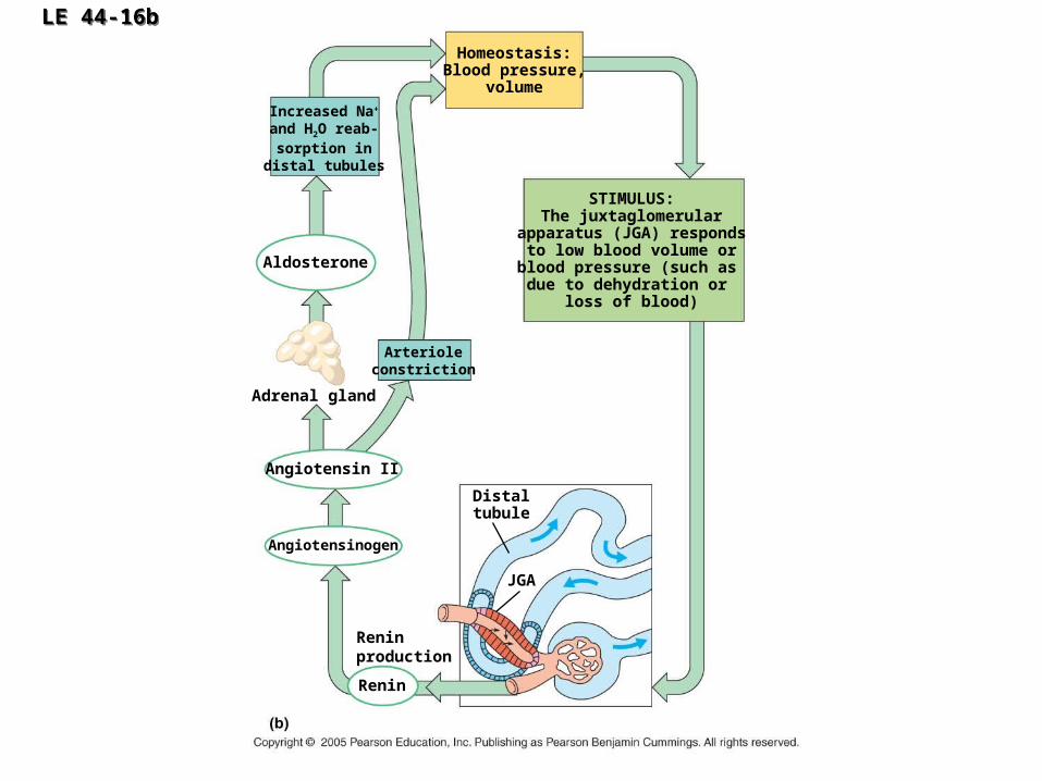
LE 44-16bLE 44-16b
Distaltubule
Aldosterone
Homeostasis:Blood pressure,
volume
STIMULUS:The juxtaglomerular
apparatus (JGA) respondsto low blood volume or
blood pressure (such as due to dehydration or
loss of blood)
Increased Na+
and H2O reab-sorption in
distal tubules
Reninproduction
Arterioleconstriction
Adrenal gland
Angiotensin II
Angiotensinogen
JGA
Renin

Copyright © 2005 Pearson Education, Inc. publishing as Benjamin Cummings
• Another hormone, atrial natriuretic factor (ANF), opposes the RAAS

Copyright © 2005 Pearson Education, Inc. publishing as Benjamin Cummings
• The South American vampire bat, which feeds on blood, has a unique excretory system
• Its kidneys offload much of the water absorbed from a meal by excreting dilute urine

Copyright © 2005 Pearson Education, Inc. publishing as Benjamin Cummings

Copyright © 2005 Pearson Education, Inc. publishing as Benjamin Cummings
Concept 44.6: Diverse adaptations of the vertebrate kidney have evolved in different environments
• The form and function of nephrons in various vertebrate classes are related to requirements for osmoregulation in the animal’s habitat

LE 44-18aLE 44-18a
Bannertail kangaroo rat(Dipodomys spectabilis)
Beaver (Castor canadensis)

LE 44-18bLE 44-18b
Rainbow trout(Oncorrhynchus mykiss)
Frog (Rana temporaria)

LE 44-18cLE 44-18c
Roadrunner(Geococcyx californianus)
Desert iguana(Dipsosaurus dorsalis)

LE 44-18dLE 44-18d
Northern bluefin tuna (Thunnus thynnus)


