
Chapter 4 Regulation of AKT Signaling Pathway in...
30
Chapter 4 Regulation of AKT Signaling Pathway in Response to DNA Minor Groove Binding Ligand, DMA and Radiation in Human Embryonic Kidney (HEK293) Cells
Transcript of Chapter 4 Regulation of AKT Signaling Pathway in...
Chapter 4 Regulation of AKT Signaling
Pathway in Response to DNA Minor Groove Binding Ligand, DMA and
Radiation in Human Embryonic Kidney (HEK293) Cells

Introduction

Chapter 4
160
4.1. INTRODUCTION Exposure of cells to a variety of stresses induces compensatory activations of multiple intracellular signalling pathways [1,2]. These include DNA damage recognition and repair, induction of cell cycle checkpoints, senescence and/or apoptosis [3,4] . Damage to other cellular components, including the cell membrane, mitochondria, endoplasmic reticulum, and non-DNA constituents of chromatin, may also initiate or modify stress signalling in response to IR [5]. These activations can play critical roles in controlling cell survival and repopulation effects in a stress-specific and cell type dependent manner. There is a complex network of signals induced by ionizing radiation and other cellular stresses in animal cells, with particular attention to signalling by growth factor and death receptors. This includes radiation-induced signalling via the EGFR and IGFI-R to the PI3K, MAPK, JNK, and p38 pathways as well as FAS-R and TNF-R signalling. Other pathways activated by stress such as ionizing radiation include those downstream of death receptors, including pro-caspases and the transcription factor NFkB.
The phosphatidylinositol 3-kinase (PI3K)/AKT (also known as protein kinase B) regulates fundamental cellular functions, such as transcription, translation, proliferation, growth, and survival, and is often over activated in a wide range of tumor types [6-8]. AKT is a serine/threonine, kinase that is recruited to the plasma membrane and is activated by phosphorylation in response to growth factor or stress signalling. Once activated, AKT modulates the function of numerous downstream substrates involved in the regulation of survival, growth, and cell cycle progression. This modulation includes inhibition of proapoptotic factors such as BAD [9] and caspase-9 [10] and activation of anti-apoptotic factors such as IKK [11] and CREB [12]. Some of other substrates are, procaspase-9, the forkhead family of transcription factors (FKHR/AFX/FOX), glycogen synthase kinase- 3 (GSK-3), p21Cip1, and Raf. IKK and CREB are activated by AKT phosphorylation, whereas Raf, Bad, procaspase-9, FKHR, and GSK-3 are inactivated. Mechanisms by which AKT modulates activity of these proteins to promote cell survival and cell cycle progression will be discussed in the following sections [13,14] (Figure 4.1).

Chapter 4
161
Figure 4.1: Overview of the PI3K/AKT pathway (source: website www.invitrogen.com)
Mechanisms by which PKB (AKT) can promote proliferation. p21 and the related family members p27/Kip1 (p27) and p57/Kip2 block the cell cycle by reversibly inhibiting several CDK (cyclin-dependent kinase) complexes. The HER2/neu receptor is a member of the growth factor glycoprotein family that includes the epidermal growth factor receptor and is overexpressed in 30% of human breast and ovarian cancers. The HER2/neu receptor can activate PKB, which then phosphorylates p21 on Thr145 [15]. Interestingly, this does not prevent p21 from interacting with and inhibiting CDKs, but instead mislocalises p21 in the cytoplasm, preventing it from entering the nucleus, where it must reside to inhibit cell proliferation.
Chapter 4
162
p27 CDK inhibitor transcription is also regulated by PKB. Activation of PKB or overexpression of constitutively active forms of this enzyme decreases the cellular levels of p27, there by promoting cell proliferation [16-18]. More recent studies indicate PKB phosphorylates the forkhead transcription factor, which is required for transcription of p27. PKB phosphorylation results in its binding to 14-3-3 proteins, which sequester it in the cytoplasm of cells. From cytoplasm of the cells p27-14-3-3 complex cannot induce p27 [19,20]. Other key regulators which are govern by AKT for cell cycle progression are the cyclin D proteins (D1, D2 and D3), which accumulate during G1 phase and are required for the activity of CDK4 and CDK6. The levels of cyclin D are controlled at the levels of transcription, translation and protein stability. Many growth factors promote transcription of this cell cycle regulator, and multiple signal transduction pathways, including the ERK1/ERK2 pathway [21-24].
Mechanism of Apoptosis inhibition by PKB (AKT). PKB can promote cell survival by inhibiting proteins that mediate apoptosis. Many substrates are identified which are recognized by AKT. Bcl2 family member BAD, promotes apoptosis by interacting with Bcl-XL on the mitochondrial membrane. Phosphorylation of BAD by PKB (and other AGC kinases) enables it to interact with 14-3-3 proteins, which prevents it from binding to Bcl-XL and thereby suppress apoptosis [25]. The forkhead transcription factor may be required for the expression of pro-apoptotic molecules such as the Fas ligand. Phosphorylation of forkhead transcription factor traps it in cytoplasm resulting in reduced expression of these proteins and thereby promote cell survival [26]. However, the recent finding suggests that the dominant negative forkhead mutant used in these studies potently inhibits PKB activity in vivo [27] indicates that some of the reported effects of overexpression of forkhead mutants may be indirect and result from the suppression of the phosphorylation of other PKB substrates [24].
It has also been reported recently that low-dose radiation triggers DNA repair, cell survival, and cell proliferation via the induction of cellular protective mechanisms that are distinctively different from high- dose radiation responses [28-31]. Kim et al. in 2007 had shown that, a low dose of ionizing radiation enhances cell proliferation through the activation of ERK1/2 and p38 [32]. The activation of the MAPKs pathway

Chapter 4
163
seems to be associated with cell protection as well as cell survival against ionizing radiation [33].
The serine/threonine kinase AKT (protein kinase B, PKB) is also known to be associated with cell protection against ionizing radiation-induced apoptosis [34,35]. Suppression of AKT activation with LY294002, a phosphatidylinositol-3 kinase (PI3K) inhibitor, sensitized cells to ionizing radiation in a dose-dependent manner (Edwards et al, 2002). Also, growth factors such as bFGF inhibited radiation-induced apoptosis via the PI3K/AKT pathway [36]. AKT also plays a vital role in, increasing glucose uptake [37], and promotion of cell cycle progression [38,39] . Recently, it has been reported that full activation of AKT, in response to ionizing radiation, is mediated by ATM [40][33].
Amifostine, along with other thiol-containing drugs, such as N-acetyl cysteine (NAC), mesna, and oltipraz, which have been approved for clinical use, are capable of participating in intracellular reductive/oxidative processes that have broad clinical implications. These include effects on the activation of redox-sensitive transcription factors, alteration of gene expression, and modification of protein activities. Amifostine protects cells from cytotoxic damage by scavenging oxygen-free radicals caused by radiation and radiomimetic drugs and by binding to reactive nucleophiles, which have the potential to react with DNA. The redox-sensitive transcription factor NFkB can be activated in mammalian cells by the active thiol form of the cytoprotector amifostine [41-45]. Mirjana et al in 2010 showed that amifostine pre-treated rats showed the activation of the AKT signalling pathways that mediate cellular survival [46].
Hoechst 33342, a DNA minor groove binding ligand, is a known radioprotector. The radioprotection activity of Hoechst 33342 is mediated by electron transfer. Many studies have been done in this process and it was found and reported that the radioprotective activity of the ligand can be improved by the addition of electron-donating substituents [47,48]. But there are some limitation with Hoechst 33342 like, inhibition of topoisomerase , chromatin structure dependent DNA repair leads to a higher level of residual DNA damage, resulting in a higher level of cytogenetic damage, cell cycle perturbations, delayed apoptosis and genomic instability, thereby enhancing

Chapter 4
164
the cytotoxicity [49]. Therefore, development of small DNA minor groove binding molecule with affordable radioprotection and reduced earlier mentioned disadvantages can play a significant role in field of radiation biology. On the basis of above mentioned observations, we in our laboratory we have synthesized, few new disubstituted benzimidazoles, Out of these new molecules we investigated the radiomodifying effects of DMA and TBZ, by studying their cell proliferation and cell survival compared with parent compound Hoechst 33342 on mammalian cell lines, exposed to low linear energy transfer (LET) .The results of these studies showed that DMA afford 84% radioprotection in BMG1 cells [50][51]
In the present study, in order to examine the reason for increased radioprotective activity and less cytotoxicity of the DMA, we studied the effects of DMA and radiation on the transcriptional response of HEK 293 cell line. From gene expression analysis study in response to DMA and/or ionizing radiation treated HEK done through microarray hybridization and analysis ,we found that DMA modulates gene expression in such a way that the cell cycle, transcription and the signal transduction machinery is induced whereas the apoptosis and oxidative stress response is suppressed. Protein expression analysis by 2D PAGE in response to DMA and/or ionizing radiation treated HEK cells led to identification of differentially expressed proteins which were categorized into following three main pathways, (a) Chaperones and folding catalysts, (b) structural proteins, and (c) metabolic enzymes. Some proteins identified were found to be interacting partner of AKT protein directly or indirectly. In Real Time PCR we observed modulation of a number of genes YWHAE, YWHAZ, NFkB, RIPK3, HSP70, AKT, GSK3B, PMS2 and ENO1. Real time quantitation of the identified proteins also confirmed differential regulation of proteins. Further study of the effect of DMA on AKT gene mediated signalling cascade implicated the involvement of AKT as the regulator of gene expression modulation in response to DMA and/or ionizing radiation.

Present Work
Chapter 4
165
4.2. PRESENT WORK
The serine/threonine kinase AKT is known to be associated with cell protection against ionizing radiation-induced apoptosis. It covalently modifies its substrate and thus associated protein to modulate different pathways associated with cell survival, cell proliferation and suppression of apoptosis. Inhibition or suppression of active form of AKT should result in sensitization or low cell survival. Thus suppression of AKT activation with LY294002, a phosphatidylinositol-3 kinase (PI3K) inhibitor, sensitized cells to ionizing radiation in a dose-dependent manner. To examine the effect of DMA on AKT signaling pathway in the presence and absence of radiation, HEK cells were treated with DMA and ionizing radiation and the few proteins of this pathway were studied by Western blot. We detected the phosphorylation level of AKT using the antibody against phosphorylated AKT protein on Ser-473.
Activation of AKT will lead to further subsequent activation of its substrates. It was clear from our results, that the treatment of DMA increased the level of phosphorylated AKT in HEK cells, and this was consistent with the alteration of DNA-PKcs. DNA-PKcs was recently reported to phosphorylate and activate AKT on Ser-473. To confirm the activation of AKT signalling pathway, we further detected the phosphorylation level of GSK3β, a substrate of activated AKT. The result indicated that the phospho-GSK3β level was simultaneously increased in the DMA treated HEK cells in presence of radiation. To know the exact role of AKT in radioprotection in presence of DMA , AKT was specifically inhibited through siRNA treatement , which is more specific than LY294002 treatment. To further confirm this observation, we did clonogenecity assay with DMA in AKT depleted HEK cells and control siRNA treated cells. We found that in control siRNA treated cells, the percentage of radioprotection shown by DMA was 15% and in AKT siRNA it was 3% at 5Gy of radiation dose. The results showed that, there was a decrease in radioprotective activity of DMA at 5 Gy of radiation dose by 12 % in AKT depleted HEK cell (treated with AKT siRNA) in comparison to control siRNA treated HEK cells. This delineate study reveals that AKT is one of the possible molecule involved in enhanced radioprotective effect, exhibited by DMA.

Figure 4.2: Proposed hypothesis for the mechanism of action of chaperones, folding catalysts, stress responsive genes and activation of transcription in DMA treated cells lead to cellular proliferation. Thus, in cells treated with DNA minor groove binding ligand DMA, induction of AKT and related
166
Proposed hypothesis for the mechanism of action of DMA. Suppression of chaperones, folding catalysts, stress responsive genes and activation of transcription in DMA
r proliferation. Thus, in cells treated with DNA minor groove binding ligand DMA, induction of AKT and related proteins help in cell survival and proliferation.
Chapter 4
DMA. Suppression of chaperones, folding catalysts, stress responsive genes and activation of transcription in DMA
r proliferation. Thus, in cells treated with DNA minor groove binding in cell survival and proliferation.

Materials and Methods

Chapter 4
167
4.3. MATERIALS AND METHODS 4.3.1. Cell Culture HEK, Human Embryonic Kidney cells (from National Center for Cell Science, Pune) were maintained as monolayers at 37°C, 5% CO2 humidified atmosphere in 25 cm2
tissue culture flasks using Dulbecco’s Modified Eagle’s Medium (Sigma-Aldrich, USA) supplemented with 10% Fetal Bovine Serum (PAN Biotech, Germany), Antibiotic Antimycotic solution 100 X (PAN Biotech, Germany). Cells were passaged routinely using 0.05% Trypsin solution (PAN Biotech, Germany) in phosphate buffered saline (PBS) for trypsinization and cultured in Dulbecco’s Modified Eagle’s Medium (Sigma-Aldrich, USA) supplemented with 10% Fetal Bovine Serum (PAN Biotech, Germany), Antibiotic Antimycotic solution 100 X (PAN Biotech, Germany). Four treatment conditions were studied- control (untreated cells), ligand treated (50µM DMA treated cells), radiation treated (5 Gy radiation treated cells), ligand + radiation treated (50µM DMA+ 5 Gy radiation, cells were treated with DMA for 1 h and irradiated as described below). 4.3.2. Ligand (DMA) and Radiation Exposure Conditions HEK, Human Embryonic Kidney cells (2x10⁵ cells) were plated in 6 well plates in complete DMEM and incubated at 37°C, 5% CO2 humidified atmosphere. HEK, were exposed to 50 µM DMA for 1h. Following DMA treatment, cells were irradiated at room temperature with a Co-60 source (A cell, AECL, CANADA) at a dose of 5 Gy. Following this procedure, HEK cells were further incubated for 4 h and subsequently harvested for further experiment. 4.3.3. Knockdown of AKT Gene Expression Using siRNA HEK 293, Human Embryonic Kidney cells were transfected with AKT siRNA and control-siRNA (Santa Cruz Biotechnology Inc.) according to the manufacturer’s instructions. HEK cells were plated (2X10⁵ cells/well) in antibiotic free DMEM supplemented with FBS. After 24 h, a 30 min pre-incubated mix of solution A [ 0-10 µl siRNA (0-100 picomole concentration) + 95 µl transfection medium] and solution B [6 µl

Chapter 4
168
transfection reagent +94 µl transfection medium] was added to cells and the cells were layered over with 800 µl transfection medium. After 5h, cells were fed with 1 ml DMEM supplemented with 2X antibiotic and 2X FBS. After 24 h, media was changed to complete DMEM. 24 h later, cells were harvested and total RNA was isolated using TriReagent (Ambion), digested by DNAse (RNAse free, MBI Fermentas) and purified using Qiagen™ RNeasy minikit purification column. The RNA was subsequently quantified using NanoDrop spectrophotometer (Thermo Fischer). cDNA was synthesized using High Capacity cDNA Reverse Transcription kit (Applied Biosystems). Semi-quantitative RT-PCR was performed with primers specific for AKT and ACTB (β Actin) in control siRNA and AKT siRNA treated HEK cells for analysing knockdown of AKT and ACTB gene expression. 4.3.4. Immunoblot Analysis HEK293, Human Embryonic Kidney cells were treated with increasing concentration from 0 to 100µM of DMA (0µ, 10µM, 25µM, 50µM, 100µM) for one hour and irradiated at 5 Gy. Two samples 0µM (no DMA treatment) and 50µM (DMA treated) were not irradiated to be used as untreated only and DMA only control. The cells were collected and centrifuged at 2000 rpm for 2 min. The cell pellet was resuspended in RIPA buffer (50mM Tris-HCl pH 7.6, 150mM NaCl, 1% sodium deoxycholate, 10% sodium orthovanadate and cocktail of protease inhibitors) and allowed to swell on ice for 30 min. Following centrifugation at 25,000 g for 30 min, the supernatant was collected as total cell lysate and protein concentration was determined using Bradford’s Assay (Bangalore Genei). Samples were prepared in SDS gel loading buffer (625 mM Tris pH 6.8, 10% SDS, 25% glycerol, 100 mM DTT and 0.015% bromophenol blue). Western blotting was performed using standard protocols. Equal amounts of protein (50 µg) from each sample was separated on a denaturing 12% SDS-PAGE gel and transferred to nitrocellulose membrane (MDI). The membranes were incubated with primary antibodies against human AKT, pAKT,GSK3β, pGSK3β and DNAPKCs (Santa Cruz Biotechnology Inc.) for 1 h, washed thrice with PBS-T for 10 min each and then incubated with horseradish peroxidase-conjugated mouse anti-human immunoglobulin (secondary antibody, Abcam) for 1 h before detection by an enhanced chemiluminescence (ECL) system (Pierce).

Chapter 4
169
4.3.5. Clonogenecity Assay with AKT Depleted Cells Cell radiosensitivity to killing was determined by the clonogenic survival assay. Exponential growing HEK cells post siRNA (siRNA-AKT and control siRNA) oligonucleotide transfections were treated with 50µM DMA and after 1h of incubation cells were irradiated with 0 Gy, 1 Gy , 2 Gy, 5 Gy and 8 Gy of radiation dose at r.t (room temperature) and trypsinized immediately at 37ºC. Cells were seeded into 60 mm dishes in triplicate, at various densities (from 100 cells per dish to 15000 cells per dish) aiming approximately 100 colonies /dish. After an incubation period of 10 days, cells were stained with crystal violet and colonies of >50 cells were counted. Same experiment was done with control siRNA-transfected HEK cells.
Percentage (%) radioprotection was calculated by following formula:
Survival fraction in siRNA DMA treated cells% of p r otection in radiation treated cells 100Survival fraction in only siRNA treated cells+
= ×

Results and Discussion

4.4. RESULTS AND DISCUSSION4.4.1. Knock Down of To knock down the expression of line, siRNA technologytotally inhibit AKT gene expression, HEK cconcentration of AKTFollowing RNA isolation and cDNA synthesis, semiperformed with primers specific for
Figure 4.3: Knock down of treated cells with AKT primer , Lane 1 to 10 shows the increasing concentration of control siRNA from 0 to 100pM (Lane 1 Lane 6 - 50pM, Lane 7 ladder). (B) RT-PCR with AKT siRNA treated cells with AKT primer , Lane 1 to 10 shows the increasing concentration of AKT siRNA from 0 to 100pM (Lane 1 20 pM, Lane 4 -30 pM, Lane 5 80pM, Lane 10 -100pM,)ACTB primer , Lane 1 to 10 shows the increasing concentration of AKT siRNA from 0 to 100pM (Lane 1 -0pM, Lane 2 -10 pM, Lane 3 Lane 7 – 60 pM, Lane 8 -70pM, Lane 9
170
DISCUSSION of AKT Gene Expression by siRNA
To knock down the expression of AKT gene in HEK, Human Embryonic Kidney cells siRNA technology was used. To standardize at which concentration AKT siRNA
totally inhibit AKT gene expression, HEK cells were transfected withof AKT siRNA and control-siRNA separately and kept for 48h
Following RNA isolation and cDNA synthesis, semi-quantitative RTperformed with primers specific for AKT and ACTB (Figure 4.3).
Knock down of AKT gene expression using siRNA. (A) RT-PCR withAKT primer , Lane 1 to 10 shows the increasing concentration of control siRNA
from 0 to 100pM (Lane 1 - 0pM, Lane 2 -10 pM, Lane 3 -20 pM, Lane 4 -30 pM, Lane 5 50pM, Lane 7 – 60 pM, Lane 8 -70pM, Lane 9 -80pM, Lane 10
PCR with AKT siRNA treated cells with AKT primer , Lane 1 to 10 shows the ng concentration of AKT siRNA from 0 to 100pM (Lane 1 -0pM, Lane 2
30 pM, Lane 5 – 40 pM, Lane 6 - 50pM, Lane 7 – 60 pM, Lane 8 100pM,) M (DNA ladder). (C) RT-PCR with AKT siRNA treated cells wit
ACTB primer , Lane 1 to 10 shows the increasing concentration of AKT siRNA from 0 to 100pM 10 pM, Lane 3 -20 pM, Lane 4 -30 pM, Lane 5 – 40 pM, Lane 6 70pM, Lane 9 -80pM, Lane 10 -100pM,) M (DNA lad
Chapter 4
uman Embryonic Kidney cells To standardize at which concentration AKT siRNA
ells were transfected with increasing separately and kept for 48h.
itative RT-PCR was
PCR with control siRNA
AKT primer , Lane 1 to 10 shows the increasing concentration of control siRNA 30 pM, Lane 5 – 40 pM,
80pM, Lane 10 -100pM,) M (DNA PCR with AKT siRNA treated cells with AKT primer , Lane 1 to 10 shows the
0pM, Lane 2 -10 pM, Lane 3 -60 pM, Lane 8 -70pM, Lane 9 -
PCR with AKT siRNA treated cells with ACTB primer , Lane 1 to 10 shows the increasing concentration of AKT siRNA from 0 to 100pM
40 pM, Lane 6 - 50pM, 100pM,) M (DNA ladder).
Chapter 4
171
PCR with AKT gene was checked in control siRNA transfected cells and it was found there was no inhibition of AKT gene with increasing concentration of control siRNA from 0 to 100pM , suggesting that control siRNA does not affect AKT gene expression. Then PCR with AKT gene was checked in AKT siRNA transfected cell and it was found that at 50pM concentration there was complete inhibition of AKT gene and the inhibition was maintained upto 100 pM. So effective concentration AKT siRNA for complete inhibition of AKT gene was 50pM. Similarly to check the nonspecific effect of AKT siRNA, we did PCR with ACTB gene on AKT siRNA transfected cells. We found there was no effect on ACTB gene expression with increasing concentration of AKT siRNA upto 100pM. 4.4.2. Immunoblot Analysis From the previous results we observed the differential regulation of AKT gene in response to DMA treatment in presence and absence of ionizing radiation, hence the present study was planned to test the effect of DMA, to promote AKT activation in together with few other protein of AKT pathway. Immunoblot technique was elucidate the underlying mechanism involved in the modulation of cellular DNA damage and repair, by DMA, the present study was planned on the observation of differential regulation of AKT gene in response to DMA treatment in presence and absence of ionizing radiation. Ser/Thr kinase AKT (also known as protein kinase B) is the major mediator of survival signals that protect cells from apoptosis induced by radiation or other genotoxic agents [52]. HEK cell were treated with increasing concentration of DMA (0 to 100µM) at 5Gy of radiation Dose. Samples were subsequently prepared and processed for western blot. The phosphorylation level of AKT was detected using the antibody against phosphorylated AKT protein on Ser-473. There was no change in AKT protein expression on increasing the concentration from 0 to 100µM, but in DMA treated samples the level of phosphorylated AKT was increased, upto 50µM in presence of ionising radiation at 5Gy, beyond that on increasing the DMA concentration the level of phosphorylated AKT was not increasing. We also found a minor increase in phosphorylation of AKT in only DMA treated HEK cells. In only radiation treated samples, the phosphorylation of AKT was very less in comparison to DMA treated samples. To further confirm the activation AKT pathway, the phosphorylation level of
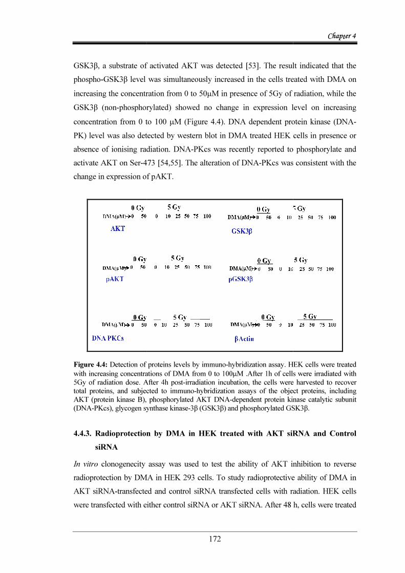
GSK3β, a substrate of activated AKT was detectedphospho-GSK3β level increasing the concentration fromGSK3β (non-phosphorylated) showed no change in expression level on increasing concentration from 0 to 100 PK) level was also detected by western blot in DMA treated HEK cells in presence or absence of ionising radiation.activate AKT on Ser-473change in expression of pAKT.
Figure 4.4: Detection of proteins levels by immunowith increasing concentrations of DMA from 0 to 1005Gy of radiation dose. After 4h posttotal proteins, and subjectAKT (protein kinase B), phosphorylated AKT(DNA-PKcs), glycogen synthase kinase 4.4.3. Radioprotection by DMA in HEK treated with AKT siRNA
siRNA In vitro clonogenecity assayradioprotection by DMA in HEK 293 cells.AKT siRNA-transfected and control siRNA transfected cells were transfected with either control siRNA or AKT siRNA. After 48
172
β, a substrate of activated AKT was detected [53]. The result indicated that the was simultaneously increased in the cells treated with
concentration from 0 to 50µM in presence of 5Gy of radiation, while the phosphorylated) showed no change in expression level on increasing
concentration from 0 to 100 µM (Figure 4.4). DNA dependent protein kinase (DNAwas also detected by western blot in DMA treated HEK cells in presence or
absence of ionising radiation. DNA-PKcs was recently reported to phosphorylate and 473 [54,55]. The alteration of DNA-PKcs was consistent with the
change in expression of pAKT.
Detection of proteins levels by immuno-hybridization assay. HEK cells were increasing concentrations of DMA from 0 to 100µM .After 1h of cells were irradiated with
After 4h post-irradiation incubation, the cells were harvested to recover total proteins, and subjected to immuno-hybridization assays of the object proteins, including
ein kinase B), phosphorylated AKT DNA-dependent protein kinase catalytic subunit synthase kinase-3β (GSK3β) and phosphorylated GSK3β
oprotection by DMA in HEK treated with AKT siRNA
lonogenecity assay was used to test the ability of AKT inhibition to reverse radioprotection by DMA in HEK 293 cells. To study radioprotective ability of DMA in
ected and control siRNA transfected cells with radiationwere transfected with either control siRNA or AKT siRNA. After 48
Chapter 4
. The result indicated that the was simultaneously increased in the cells treated with DMA on
M in presence of 5Gy of radiation, while the phosphorylated) showed no change in expression level on increasing
DNA dependent protein kinase (DNA-was also detected by western blot in DMA treated HEK cells in presence or
PKcs was recently reported to phosphorylate and was consistent with the
hybridization assay. HEK cells were treated
After 1h of cells were irradiated with irradiation incubation, the cells were harvested to recover
hybridization assays of the object proteins, including dependent protein kinase catalytic subunit
3β (GSK3β) and phosphorylated GSK3β.
oprotection by DMA in HEK treated with AKT siRNA and Control
bility of AKT inhibition to reverse study radioprotective ability of DMA in
with radiation. HEK cells were transfected with either control siRNA or AKT siRNA. After 48 h, cells were treated

Chapter 4
173
with DMA for 1h and radiation at following radiation dose 0Gy, 1 Gy, 2Gy, 5Gy and 8 Gy. After irradiation cell were seeded in 60 mm pteri dish .After 10 days colonies were counted and survival fraction and percentage protection was calculated. The results showed that there was significant radiation protection in DMA treated control siRNA transfected cells in comparison to only control siRNA cells transfected. Whereas there was a very little radiation protection in DMA treated AKT siRNA transfected cells in comparison to only AKT siRNA transfected cells with increasing dose of radiation (Figure 4.5). It was observed that in control siRNA treated cells DMA provide 15 % radioprotection to HEK cells whereas it was observed 3% in AKT siRNA treated cell followed by DMA treatment. The results showed that, there was a decrease in radioprotective activity of DMA at 5 Gy of radiation dose by 12 % in AKT depleted HEK cell (treated with AKT siRNA) in comparison to control siRNA treated HEK cells.
Figure 4.5: Clonogenecity assay showing cell survival in control siRNA and AKT siRNA treated HEK 293 cells after DMA treatemnt. Four type of treatment condition were taken (a) Only control siRNA transfected cells, (b) DMA treated control siRNA transfected cells (c) Only AKT siRNA transfected cells, (b) DMA treated AKT siRNA transfected cells. Different no. of cells were seeded in different petri plate for each radiation dose. Cells were incubated with DMA (50µM) for 1 h followed by irradiation at indicated dose. Pteri dish were kept for 10 days for colony formation. Colonies were counted and plating efficiency and survival fraction was calculated for each dose of radiation.
Radiation Dose (Gy)0 2 4 6 8 10
Surv
ival
Fra
ctio
n
0.01
0.1
1
10Control siRNAControl siRNA + DMA AKT siRNA AKT siRNA+ DMA

Chapter 4
174
In this study we have tried to find out the plausible mechanism of DMA radioprotection activity. Our study suggests that DMA protects HEK 293 cells from radiation induced killing via activation of AKT pathway. AKT has been implicated in the survival of cells. AKT signaling inactivates several proapoptotic factors ,along with, it also activates transcription factors that up-regulate antiapoptotic genes [56,57] The antiapoptotic role of the AKT pathway has been well documented by many investigators in response to numerous noxious stimuli, and in some cell types, the antiapoptotic effects of ERBB receptor signaling have been attributed to activation of the PI3K/AKT pathway [58,59] .In Addition to direct effect of AKT pathway , this pathway also interact with other major signalling pathway. This cross talk extends its regulatory importance in terms of the stress response especially their interaction with the NFkB , WNT and MAPK pathway. AKT promote cell survival either by phosphorylating and activating transcription factor CREB (cAMP-response element binding protein) or IkB kinases (IKKs) that regulate the activity of NF-kB (nuclear factor kappaB [57]. It has been reported that under PDGF stimulation, TNF can induce the activity of PI3K which in turn activates AKT, eventually leading to an increase in NF-κB DNA binding activity [60,61]. Earlier in chapter 3 we found NFkB and AKT gene is upregulated in DMA and DMA treated HEK cells in Real Time PCR experiment. Thus this result suggest the role of NFkB as a factor favouring AKT mediated cell survival by DMA.
The AKT downstream target, GSK3β, plays a key role in WNT signalling. WNT pathway is known to regulate many different biological events including cell survival [62]. mTOR the well-defined AKT downstream target, , can be effectively activated by WNT signalling via GSK3β [63]. Inoki et al. in 2006 showed that PI3K-AKT signalling and its downstream targets have the potential to activate the Wnt pathway and vice versa. In our microarray data WNT signalling pathway was found to differentially expressed in the DMA and DMA + Radiation treated cells. In western blot experiment we found that there is increase in level of phosphorylated GSK3β with increasing concentration of DMA in ionizing radiation treated cells. These finding suggest that AKT together with GSK3β and WNT signalling pathway might play a play a role in reducing DNA Damage and enhancing DNA repair in DMA treated HEK cells.
Chapter 4
175
In addition to NFkB and WNT signalling, AKT also interact with MAPK signalling pathway, including JNK/SAPK, p38 family kinases and ERKs. MAPKs are serine/threonine protein kinases that respond to a wide range of extracellular stimuli (for example, growth factors, cytokines, stress) and have been identified as regulators of many cellular activities (e.g. oxidative and osmotic stress, apoptosis, mitosis, cell differentiation and proliferation, immune response, memory, etc.). The stress-responsive JNK/p38 pathway can be inhibited by AKT via phosphorylation of the JNK/p38 upstream factor ASK1 (Apoptotic signal regulated kinase 1, or MAPKKK5) at S83, which prevents ASK1 from being activated by apoptotic stimuli. In addition, p38 regulates the activation of AKT through a complex with other factors including HSP27 (heat shock protein 27) and MAPK2 (MAPK-activated protein kinase-2) in human neutrophils [64,65] . In microarray and real time data we found MAPkinase pathway as major pathway which was differentially expressed in DMA treated cells in presence and absence of radiation.
4.5. CONCLUSIONS DMA, a Bis-benzimidazoles, is a minor grove binder and thus affect the expression level of different gene/protein. The minor groove binding of the DNA leads to the compression of the major groove that antagonizes transcription factor binding to the DNA major groove in an allosteric fashion. It also affects the expression of AKT, which is one of the crucial parts of the cell survival pathway. Although the basal level of AKT did not show any change in the expression in presence of DMA alone or in combination with IR. The active form of AKT i.e. the phosphorylated AKT increased in dose dependent manner on increasing the concentration of DMA from 0 to 50 µM in presence of radiation. The result indicated that the phosphorylated AKT protein was increased in DMA-treated cells, which is consistent with the enhancement of DNA-PKcs level. As the consequence, the phosphorylation level of GSK3β, which is a substrate of the activated AKT, was concurrently enhanced. Therefore, it is likely that DMA lead to activation of the AKT pathway via the induction of DNA-PKcs, and this activated survival signal pathway may also contribute to the radioprotective effect of DMA. As radiation induces DNA damage, the radioprotective effect of DMA increases

Chapter 4
176
the phosphorylation of AKT which enhances the survival pathway and thus activated or covalently modulates the other substrates and promotes the DNA repair and advances the cell toward cell survival. Thus DMA modulates the transcription of AKT through DNA binding in concentration dependent manner. In conclusion, we demonstrate that DMA a novel Bis-benzimidazoles analogue, effectively minimize ionizing radiation-induced DNA damage in cultured cells. This radioprotection of DMA is in part attributed to promotion of DNA damage repair and activation of AKT signal pathway.

References

Chapter 4
177
4.6. REFERENCES
1. Dent P, Yacoub A, Contessa J, Caron R, Amorino G, Valerie K et al.: Stress and radiation-induced activation of multiple intracellular signaling pathways. Radiat Res 2003, 159: 283-300.
2. Amundson SA, Bittner M, Fornace AJ, Jr.: Functional genomics as a window on radiation stress signaling. Oncogene 2003, 22: 5828-5833.
3. Peltola V, Parvinen M, Huhtaniemi I, Kulmala J, Ahotupa M: Comparison of effects of 0.5 and 3.0 Gy X-irradiation on lipid peroxidation and antioxidant enzyme function in rat testis and liver. J Androl 1993, 14: 267-274.
4. Yamaoka K, Sato EF, Utsumi K: Induction of two species of superoxide dismutase in some organs of rats by low dose X-irradiation. Physiol Chem Phys Med NMR 1994, 26: 205-214.
5. Martindale JL, Holbrook NJ: Cellular response to oxidative stress: signaling for suicide and survival. J Cell Physiol 2002, 192: 1-15.
6. Sroka IC, Nagle RB, Bowden GT: Membrane-type 1 matrix metalloproteinase is regulated by sp1 through the differential activation of AKT, JNK, and ERK pathways in human prostate tumor cells. Neoplasia 2007, 9: 406-417.
7. Vivanco I, Sawyers CL: The phosphatidylinositol 3-Kinase AKT pathway in human cancer. Nat Rev Cancer 2002, 2: 489-501.
8. Osaki M, Oshimura M, Ito H: PI3K-Akt pathway: its functions and alterations in human cancer. Apoptosis 2004, 9: 667-676.
9. del PL, Gonzalez-Garcia M, Page C, Herrera R, Nunez G: Interleukin-3-induced phosphorylation of BAD through the protein kinase Akt. Science 1997, 278: 687-689.
10. Cardone MH, Roy N, Stennicke HR, Salvesen GS, Franke TF, Stanbridge E et al.: Regulation of cell death protease caspase-9 by phosphorylation. Science 1998, 282: 1318-1321.

Chapter 4
178
11. Kane LP, Shapiro VS, Stokoe D, Weiss A: Induction of NF-kappaB by the Akt/PKB kinase. Curr Biol 1999, 9: 601-604.
12. Wang JM, Chao JR, Chen W, Kuo ML, Yen JJ, Yang-Yen HF: The antiapoptotic gene mcl-1 is up-regulated by the phosphatidylinositol 3-kinase/Akt signaling pathway through a transcription factor complex containing CREB. Mol Cell Biol 1999, 19: 6195-6206.
13. Elia U, Flescher E: PI3K/Akt pathway activation attenuates the cytotoxic effect of methyl jasmonate toward sarcoma cells. Neoplasia 2008, 10: 1303-1313.
14. Chang F, Lee JT, Navolanic PM, Steelman LS, Shelton JG, Blalock WL et al.: Involvement of PI3K/Akt pathway in cell cycle progression, apoptosis, and neoplastic transformation: a target for cancer chemotherapy. Leukemia 2003, 17: 590-603.
15. Zhou BP, Liao Y, Xia W, Spohn B, Lee MH, Hung MC: Cytoplasmic localization of p21Cip1/WAF1 by Akt-induced phosphorylation in HER-2/neu-overexpressing cells. Nat Cell Biol 2001, 3: 245-252.
16. Gesbert F, Sellers WR, Signoretti S, Loda M, Griffin JD: BCR/ABL regulates expression of the cyclin-dependent kinase inhibitor p27Kip1 through the phosphatidylinositol 3-Kinase/AKT pathway. J Biol Chem 2000, 275: 39223-39230.
17. Graff JR, Konicek BW, McNulty AM, Wang Z, Houck K, Allen S et al.: Increased AKT activity contributes to prostate cancer progression by dramatically accelerating prostate tumor growth and diminishing p27Kip1 expression. J Biol Chem 2000, 275: 24500-24505.
18. Sun H, Lesche R, Li DM, Liliental J, Zhang H, Gao J et al.: PTEN modulates cell cycle progression and cell survival by regulating phosphatidylinositol 3,4,5,-trisphosphate and Akt/protein kinase B signaling pathway. Proc Natl Acad Sci U S A 1999, 96: 6199-6204.
19. Medema RH, Kops GJ, Bos JL, Burgering BM: AFX-like Forkhead transcription factors mediate cell-cycle regulation by Ras and PKB through p27kip1. Nature 2000, 404: 782-787.

Chapter 4
179
20. Nakamura N, Ramaswamy S, Vazquez F, Signoretti S, Loda M, Sellers WR: Forkhead transcription factors are critical effectors of cell death and cell cycle arrest downstream of PTEN. Mol Cell Biol 2000, 20: 8969-8982.
21. Cheng M, Sexl V, Sherr CJ, Roussel MF: Assembly of cyclin D-dependent kinase and titration of p27Kip1 regulated by mitogen-activated protein kinase kinase (MEK1). Proc Natl Acad Sci U S A 1998, 95: 1091-1096.
22. Lavoie JN, L'Allemain G, Brunet A, Muller R, Pouyssegur J: Cyclin D1 expression is regulated positively by the p42/p44MAPK and negatively by the p38/HOGMAPK pathway. J Biol Chem 1996, 271: 20608-20616.
23. Schwartz MA, Assoian RK: Integrins and cell proliferation: regulation of cyclin-dependent kinases via cytoplasmic signaling pathways. J Cell Sci 2001, 114: 2553-2560.
24. Lawlor MA, Alessi DR: PKB/Akt: a key mediator of cell proliferation, survival and insulin responses? J Cell Sci 2001, 114: 2903-2910.
25. Downward J: How BAD phosphorylation is good for survival. Nat Cell Biol 1999, 1: E33-E35.
26. Brunet A, Bonni A, Zigmond MJ, Lin MZ, Juo P, Hu LS et al.: Akt promotes cell survival by phosphorylating and inhibiting a Forkhead transcription factor. Cell 1999, 96: 857-868.
27. Rena G, Prescott AR, Guo S, Cohen P, Unterman TG: Roles of the forkhead in rhabdomyosarcoma (FKHR) phosphorylation sites in regulating 14-3-3 binding, transactivation and nuclear targetting. Biochem J 2001, 354: 605-612.
28. Korystov YN, Eliseeva NA, Kublik LN, Narimanov AA: The effect of low-dose irradiation on proliferation of mammalian cells in vitro. Radiat Res 1996, 146: 329-332.
29. Wang GJ, Cai L: Induction of cell-proliferation hormesis and cell-survival adaptive response in mouse hematopoietic cells by whole-body low-dose radiation. Toxicol Sci 2000, 53: 369-376.

Chapter 4
180
30. Suzuki K, Kodama S, Watanabe M: Extremely low-dose ionizing radiation causes activation of mitogen-activated protein kinase pathway and enhances proliferation of normal human diploid cells. Cancer Res 2001, 61: 5396-5401.
31. Li W, Wang G, Cui J, Xue L, Cai L: Low-dose radiation (LDR) induces hematopoietic hormesis: LDR-induced mobilization of hematopoietic progenitor cells into peripheral blood circulation. Exp Hematol 2004, 32: 1088-1096.
32. Kim CS, Kim JM, Nam SY, Yang KH, Jeong M, Kim HS et al.: Low-dose of ionizing radiation enhances cell proliferation via transient ERK1/2 and p38 activation in normal human lung fibroblasts. J Radiat Res 2007, 48: 407-415.
33. Park HS, Yun Y, Kim CS, Yang KH, Jeong M, Ahn SK et al.: A critical role for AKT activation in protecting cells from ionizing radiation-induced apoptosis and the regulation of acinus gene expression. Eur J Cell Biol 2009, 88: 563-575.
34. Soderlund K, Perez-Tenorio G, Stal O: Activation of the phosphatidylinositol 3-kinase/Akt pathway prevents radiation-induced apoptosis in breast cancer cells. Int J Oncol 2005, 26: 25-32.
35. Kim CS, Kim JK, Nam SY, Yang KH, Jeong M, Kim HS et al.: Low-dose radiation stimulates the proliferation of normal human lung fibroblasts via a transient activation of Raf and Akt. Mol Cells 2007, 24: 424-430.
36. Gu Q, Wang D, Wang X, Peng R, Liu J, Jiang T et al.: Basic fibroblast growth factor inhibits radiation-induced apoptosis of HUVECs. I. The PI3K/AKT pathway and induction of phosphorylation of BAD. Radiat Res 2004, 161: 692-702.
37. Gottlob K, Majewski N, Kennedy S, Kandel E, Robey RB, Hay N: Inhibition of early apoptotic events by Akt/PKB is dependent on the first committed step of glycolysis and mitochondrial hexokinase. Genes Dev 2001, 15: 1406-1418.
38. Downward J: Mechanisms and consequences of activation of protein kinase B/Akt. Curr Opin Cell Biol 1998, 10: 262-267.

Chapter 4
181
39. Nicholson KM, Anderson NG: The protein kinase B/Akt signalling pathway in human malignancy. Cell Signal 2002, 14: 381-395.
40. Viniegra JG, Martinez N, Modirassari P, Hernandez LJ, Parada CC, Sanchez-Arevalo Lobo VJ et al.: Full activation of PKB/Akt in response to insulin or ionizing radiation is mediated through ATM. J Biol Chem 2005, 280: 4029-4036.
41. Sen CK, Packer L: Antioxidant and redox regulation of gene transcription. FASEB J 1996, 10: 709-720.
42. Das KC, Lewis-Molock Y, White CW: Activation of NF-kappa B and elevation of MnSOD gene expression by thiol reducing agents in lung adenocarcinoma (A549) cells. Am J Physiol 1995, 269: L588-L602.
43. Antras-Ferry J, Maheo K, Chevanne M, Dubos MP, Morel F, Guillouzo A et al.: Oltipraz stimulates the transcription of the manganese superoxide dismutase gene in rat hepatocytes. Carcinogenesis 1997, 18: 2113-2117.
44. Romano MF, Lamberti A, Bisogni R, Garbi C, Pagnano AM, Auletta P et al.: Amifostine inhibits hematopoietic progenitor cell apoptosis by activating NF-kappaB/Rel transcription factors. Blood 1999, 94: 4060-4066.
45. Murley JS, Kataoka Y, Hallahan DE, Roberts JC, Grdina DJ: Activation of NFkappaB and MnSOD gene expression by free radical scavengers in human microvascular endothelial cells. Free Radic Biol Med 2001, 30: 1426-1439.
46. Mirjana M, Goran P, Nevena G, Melita V, Svetlana D, Ilijana G et al.: The rat acute-phase protein alpha2-macroglobulin plays a central role in amifostine-mediated radioprotection. J Radiol Prot 2010, 30: 567-583.
47. Martin RF, Broadhurst S, D'Abrew S, Budd R, Sephton R, Reum M et al.: Radioprotection by DNA ligands. Br J Cancer Suppl 1996, 27: S99-101.
48. Martin RF, Anderson RF: Pulse radiolysis studies indicate that electron transfer is involved in radioprotection by Hoechst 33342 and methylproamine. Int J Radiat Oncol Biol Phys 1998, 42: 827-831.

Chapter 4
182
49. Adhikari JS, Khaitan D, Arya MB, Dwarakanath BS: Heterogeneity in the radiosensitizing effects of the DNA ligand hoechst-33342 in human tumor cell lines. J Cancer Res Ther 2005, 1: 151-161.
50. Tawar U, Jain AK, Dwarakanath BS, Chandra R, Singh Y, Chaudhury NK et al.: Influence of phenyl ring disubstitution on bisbenzimidazole and terbenzimidazole cytotoxicity: synthesis and biological evaluation as radioprotectors. J Med Chem 2003, 46: 3785-3792.
51. Tawar U, Jain AK, Chandra R, Singh Y, Dwarakanath BS, Chaudhury NK et al.: Minor groove binding DNA ligands with expanded A/T sequence length recognition, selective binding to bent DNA regions and enhanced fluorescent properties. Biochemistry 2003, 42: 13339-13346.
52. Toker A: Protein kinases as mediators of phosphoinositide 3-kinase signaling. Mol Pharmacol 2000, 57: 652-658.
53. Horn S, Endl E, Fehse B, Weck MM, Mayr GW, Jucker M: Restoration of SHIP activity in a human leukemia cell line downregulates constitutively activated phosphatidylinositol 3-kinase/Akt/GSK-3beta signaling and leads to an increased transit time through the G1 phase of the cell cycle. Leukemia 2004, 18: 1839-1849.
54. Dragoi AM, Fu X, Ivanov S, Zhang P, Sheng L, Wu D et al.: DNA-PKcs, but not TLR9, is required for activation of Akt by CpG-DNA. EMBO J 2005, 24: 779-789.
55. Feng J, Park J, Cron P, Hess D, Hemmings BA: Identification of a PKB/Akt hydrophobic motif Ser-473 kinase as DNA-dependent protein kinase. J Biol Chem 2004, 279: 41189-41196.
56. Bonnaud S, Niaudet C, Legoux F, Corre I, Delpon G, Saulquin X et al.: Sphingosine-1-phosphate activates the AKT pathway to protect small intestines from radiation-induced endothelial apoptosis. Cancer Res 2010, 70: 9905-9915.
Chapter 4
183
57. Miraglia AG, Travaglione S, Meschini S, Falzano L, Matarrese P, Quaranta MG et al.: Cytotoxic necrotizing factor 1 prevents apoptosis via the Akt/IkappaB kinase pathway: role of nuclear factor-kappaB and Bcl-2. Mol Biol Cell 2007, 18: 2735-2744.
58. Daly JM, Olayioye MA, Wong AM, Neve R, Lane HA, Maurer FG et al.: NDF/heregulin-induced cell cycle changes and apoptosis in breast tumour cells: role of PI3 kinase and p38 MAP kinase pathways. Oncogene 1999, 18: 3440-3451.
59. Dent P, Yacoub A, Fisher PB, Hagan MP, Grant S: MAPK pathways in radiation responses. Oncogene 2003, 22: 5885-5896.
60. Romashkova JA, Makarov SS: NF-kappaB is a target of AKT in anti-apoptotic PDGF signalling. Nature 1999, 401: 86-90.
61. Madrid LV, Wang CY, Guttridge DC, Schottelius AJ, Baldwin AS, Jr., Mayo MW: Akt suppresses apoptosis by stimulating the transactivation potential of the RelA/p65 subunit of NF-kappaB. Mol Cell Biol 2000, 20: 1626-1638.
62. Gordon MD, Nusse R: Wnt signaling: multiple pathways, multiple receptors, and multiple transcription factors. J Biol Chem 2006, 281: 22429-22433.
63. Inoki K, Ouyang H, Zhu T, Lindvall C, Wang Y, Zhang X et al.: TSC2 integrates Wnt and energy signals via a coordinated phosphorylation by AMPK and GSK3 to regulate cell growth. Cell 2006, 126: 955-968.
64. Rane MJ, Coxon PY, Powell DW, Webster R, Klein JB, Pierce W et al.: p38 Kinase-dependent MAPKAPK-2 activation functions as 3-phosphoinositide-dependent kinase-2 for Akt in human neutrophils. J Biol Chem 2001, 276: 3517-3523.
65. Wu R, Kausar H, Johnson P, Montoya-Durango DE, Merchant M, Rane MJ: Hsp27 regulates Akt activation and polymorphonuclear leukocyte apoptosis by scaffolding MK2 to Akt signal complex. J Biol Chem 2007, 282: 21598-21608.


