Chapter 3 Tissues. 2 Introduction: A.Cells are arranged in tissues that provide specific functions...
44
Chapter 3 Tissues
-
Upload
alannah-fitzgerald -
Category
Documents
-
view
214 -
download
0
Transcript of Chapter 3 Tissues. 2 Introduction: A.Cells are arranged in tissues that provide specific functions...
Chapter 3
Tissues
2
Introduction: A. Cells are arranged in tissues that
provide specific functions for the body.
B. Cells of different tissues are structured differently, which leads to their differences in function.
C. The tissues of the human body include four major types: epithelial, connective, muscle, and nervous.
CopyrightThe McGraw-Hill Companies, Inc. Permission required for reproduction or display.

3
Epithelial Tissues:A. General Characteristics
1. Epithelial tissue is widespread throughout the body, covers
organs, and lines body surfaces.2. Epithelial tissues are anchored to a
basement membrane, are made up of tightly packed cells containing
little intercellular material, generally lack blood vessels, and are replaced frequently.
CopyrightThe McGraw-Hill Companies, Inc. Permission required for reproduction or display.

4
3. Epithelial tissues function in protection, secretion, absorption, excretion, and sensory reception.
CopyrightThe McGraw-Hill Companies, Inc. Permission required for reproduction or display.Table 05.01
5
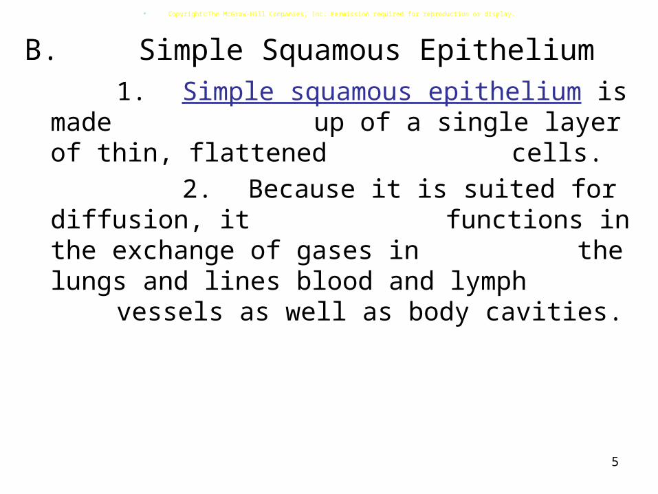
B. Simple Squamous Epithelium1. Simple squamous epithelium is
made up of a single layer of thin, flattened cells.
2. Because it is suited for diffusion, it functions in the exchange of gases
in the lungs and lines blood and lymph vessels as well as body cavities.
CopyrightThe McGraw-Hill Companies, Inc. Permission required for reproduction or display.

6
Fig05.01
(b)(a)
(d)(c)
Free surfaceof tissue
Simplesquamousepithelium
Basementmembrane
Nucleus
Free surfaceof simplesquamousepithelium
Nucleus
Connectivetissue
Copyright ©The McGraw-Hill Companies, Inc. Permission required for reproduction or display.
©The McGraw-Hill Companies, Inc./Al Telser, photographer
©The McGraw-Hill Companies, Inc./Al Telser, photographer
7
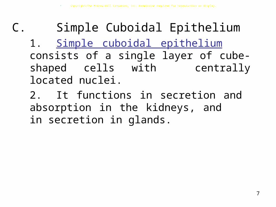
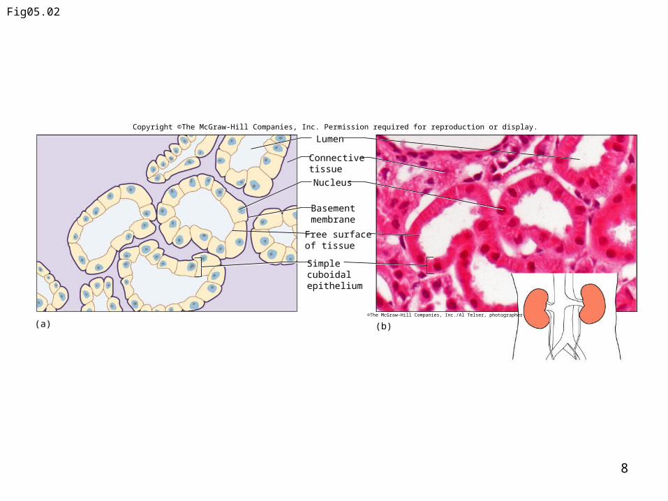
C. Simple Cuboidal Epithelium
1. Simple cuboidal epithelium consists of a single
layer of cube-shaped cells with centrally located nuclei.
2. It functions in secretion and absorption in the
kidneys, and in secretion in glands.
CopyrightThe McGraw-Hill Companies, Inc. Permission required for reproduction or display.
8
Fig05.02
Nucleus
Basement membrane
Free surface of tissue
Simplecuboidalepithelium
Connectivetissue
Lumen
(a) (b)©The McGraw-Hill Companies, Inc./Al Telser, photographer
Copyright ©The McGraw-Hill Companies, Inc. Permission required for reproduction or display.

9
D. Simple Columnar Epithelium1. Simple columnar epithelium is
made up of a row of elongated cells
whose nuclei are all located near the basement membrane. It may be ciliated.
2. It lines the uterus, stomach, and intestines where it protects underlying
tissues, secretes digestive fluids, and absorbs nutrients.
CopyrightThe McGraw-Hill Companies, Inc. Permission required for reproduction or display.

10
3. In the intestine, these cells possess microvilli that increase the
surface area available for absorption.4. Mucus-secreting goblet cells
can be found among columnar cells.
CopyrightThe McGraw-Hill Companies, Inc. Permission required for reproduction or display.Fig05.03
Nucleus
Basementmembrane
Microvilli(free surfaceof tissue)
Connectivetissue
Mucus
Cytoplasm
Goblet cell
(a) (b)©The McGraw-Hill Companies, Inc./Al Telser, photographer
Copyright ©The McGraw-Hill Companies, Inc. Permission required for reproduction or display.
11
Connective Tissues:A. General Characteristics
1. Connective tissues bind, support, protect, serve as frameworks,
fill spaces, store fat, produce blood cells, protect against infection, and repair tissue damage.
2. Unlike epithelial tissues, connective tissues have an abundance of extracellular matrix, or
intercellular material, throughout, and have good blood supplies (except cartilage).
CopyrightThe McGraw-Hill Companies, Inc. Permission required for reproduction or display.

12
3. Major Cell Types
a. The fibroblast is the most common cell type, and is a
fixed, star-shaped cell that secretes fibers and is large in size.
b. Wandering macrophages function as scavenger cells and defend against infection.
c. Mast cells are large and are located near blood vessels
where they release heparin (anticoagulant) and
histamine (promotes inflammation).
CopyrightThe McGraw-Hill Companies, Inc. Permission required for reproduction or display.
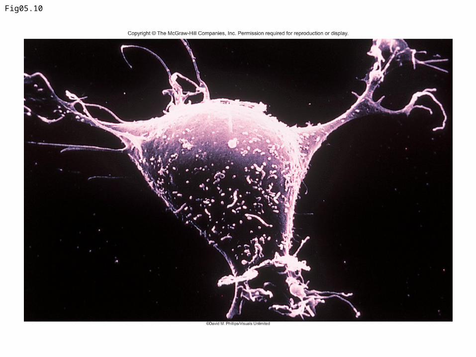
Fig05.10
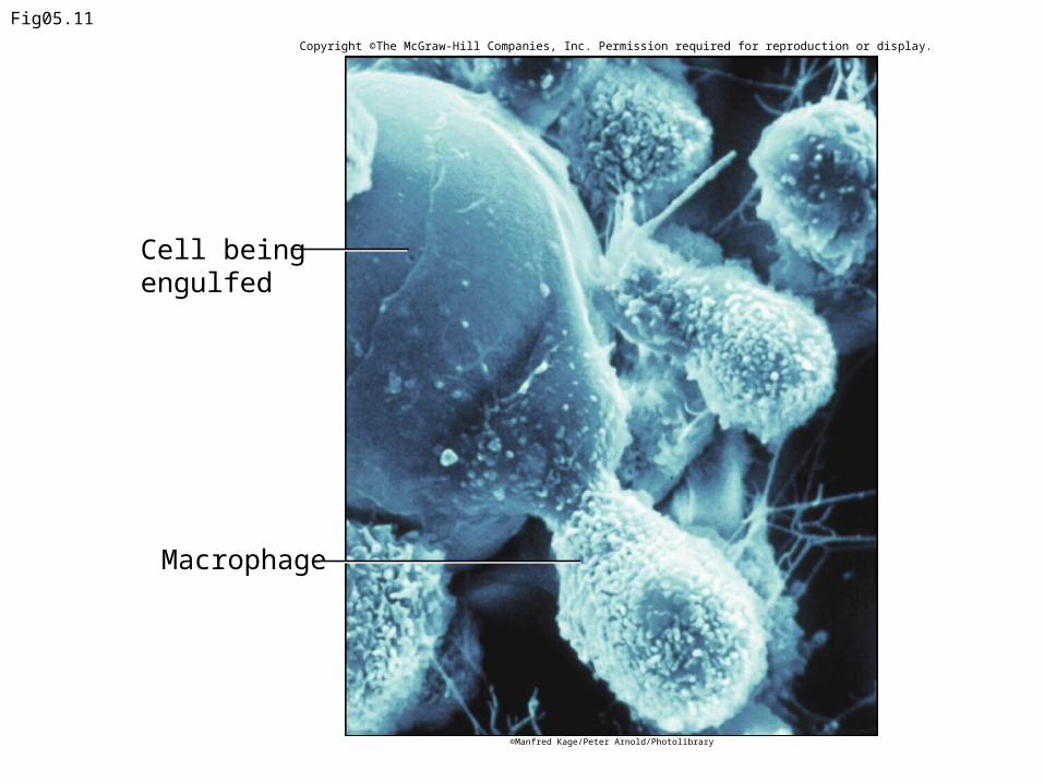
Macrophage
Fig05.11
Cell being engulfed
©Manfred Kage/Peter Arnold/Photolibrary
Copyright ©The McGraw-Hill Companies, Inc. Permission required for reproduction or display.

Fig05.12

16
4. Connective Tissue Fibers a. Strong collagenous fibers
(white fibers), made of the protein collagen, add strength for holding body parts together.
b. Elastic fibers (yellow fibers), made of the protein elastin, are stretchy and add flexibility to certain types of connective tissues.
c. Reticular fibers are thin collagenous fibers that form supportive networks in a variety of tissues.
CopyrightThe McGraw-Hill Companies, Inc. Permission required for reproduction or display.
17
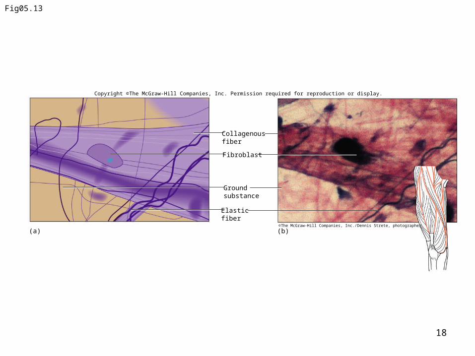
B. Categories of Connective Tissue1. Loose connective Tissue includes
areolar, adipose and reticular connective tissues.
a. Areolar Tissue forms delicate, thin membranes throughout
the body that bind body parts together such as skin and underlying organs.
i. The majority of the cells are fibroblasts that are separated by a gel-like ground substance that contains collagenous and elastic fibers.
CopyrightThe McGraw-Hill Companies, Inc. Permission required for reproduction or display.
18
Fig05.13
Elastic fiber
(a) (b)
Collagenous fiber
Fibroblast
Ground substance
©The McGraw-Hill Companies, Inc./Dennis Strete, photographer
Copyright ©The McGraw-Hill Companies, Inc. Permission required for reproduction or display.
19
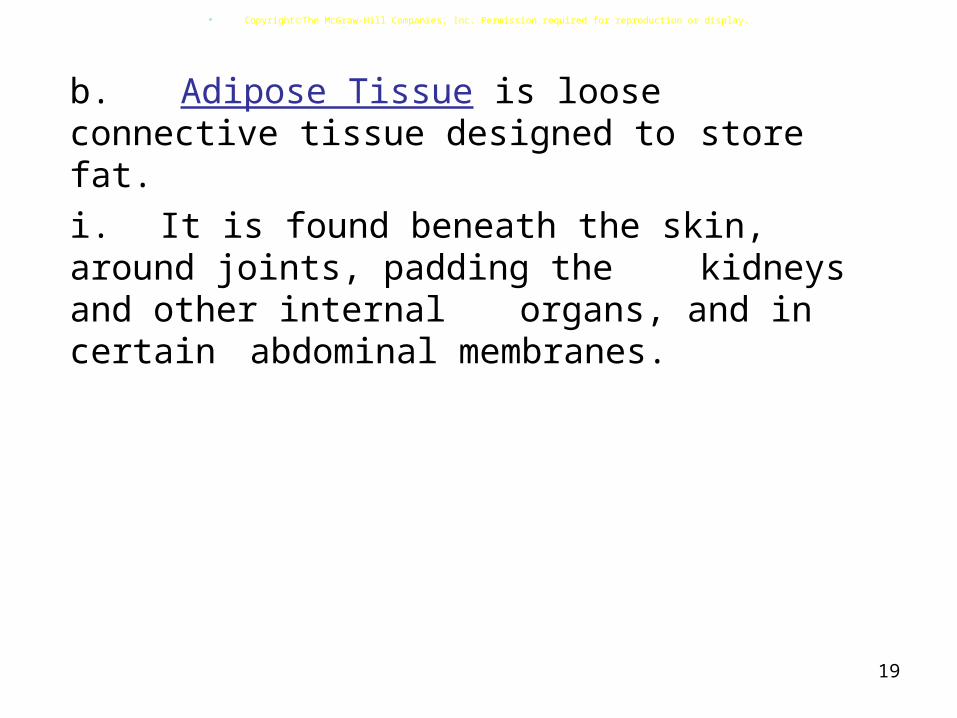
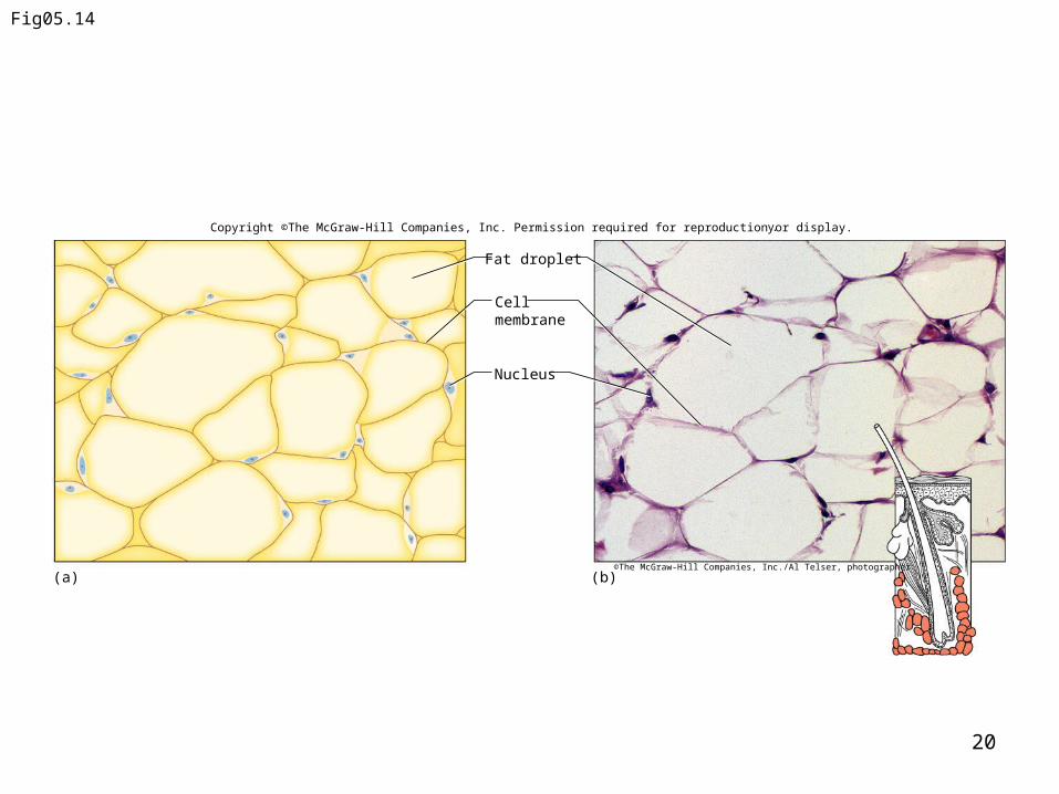
b. Adipose Tissue is loose connective tissue
designed to store fat.i. It is found beneath the
skin, around joints, padding the kidneys and other internal
organs, and in certain abdominal
membranes.
CopyrightThe McGraw-Hill Companies, Inc. Permission required for reproduction or display.
20
Fig05.14
(a) (b)
Fat droplet
Nucleus
Cell membrane
y.
©The McGraw-Hill Companies, Inc./Al Telser, photographer
Copyright ©The McGraw-Hill Companies, Inc. Permission required for reproduction or display.
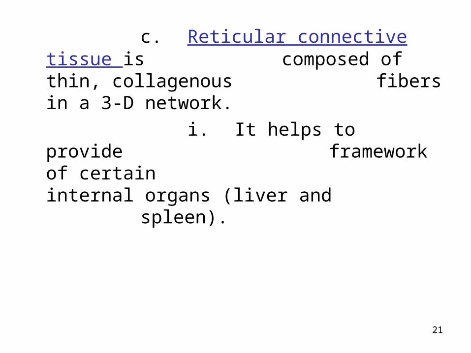
c. Reticular connective tissue is composed of thin, collagenous fibers in a 3-D network.
i. It helps to provide framework of certain internal organs (liver and spleen).
21
22
2. Dense Connective Tissue
a. This tissue consists of densely packed collagenous fibers and is very strong but lacks a good
blood supply.b. It is found as part of
tendons and ligaments.
CopyrightThe McGraw-Hill Companies, Inc. Permission required for reproduction or display.

23
Fig05.15
Fibroblasts
Collagenous fibers
(a) (b)©The McGraw-Hill Companies, Inc./Dennis Strete, photographer
Copyright ©The McGraw-Hill Companies, Inc. Permission required for reproduction or display.

24
3. Cartilage
a. Cartilage is a rigid connective tissue that provides a supportive framework for
various structures. It lacks a vascular system and so
heals slowly.b. Cartilage cells
(chondrocytes) lie within lacunae in the gel-
like fluid matrix.
CopyrightThe McGraw-Hill Companies, Inc. Permission required for reproduction or display.
25
c. Cartilaginous structures are enclosed within a connective tissue called the perichondrium.
d. The most common, hyaline cartilage, is white with abundant fine collagen fibers,
is found at the ends of bones, and supports respiratory
passages.
CopyrightThe McGraw-Hill Companies, Inc. Permission required for reproduction or display.

26
e. Elastic cartilage, with elastic fibers, provides a framework for the external ears and parts
of the larynx.f. Fibrocartilage, with
many collagenous fibers, is a tough tissue that provides a shock-
absorbing function in intervertebral discs and
in the knees and pelvic girdle.
CopyrightThe McGraw-Hill Companies, Inc. Permission required for reproduction or display.
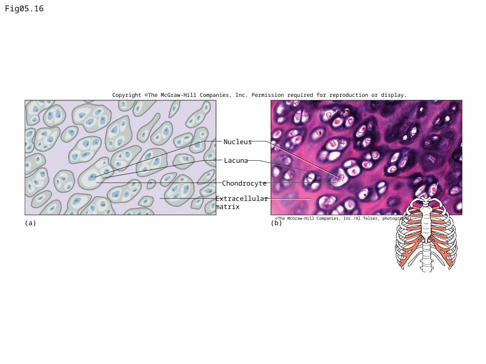
Fig05.16
Chondrocyte
Nucleus
Extracellular matrix
(a) (b)
Lacuna
©The McGraw-Hill Companies, Inc./Al Telser, photographer
Copyright ©The McGraw-Hill Companies, Inc. Permission required for reproduction or display.

Fig05.17
Chondrocyte
Elastic fibers
Nucleus
Extracellular matrix
(a) (b)
Lacuna
©The McGraw-Hill Companies, Inc./Al Telser, photographer
Copyright ©The McGraw-Hill Companies, Inc. Permission required for reproduction or display.

Fig05.18
Chondrocyte
Nucleus
Collagenous fiber
Extracellular matrix
(a) (b)©The McGraw-Hill Companies, Inc./Al Telser, photographer
Copyright ©The McGraw-Hill Companies, Inc. Permission required for reproduction or display.
30
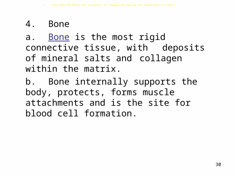
4. Bone
a. Bone is the most rigid connective
tissue, with deposits of mineral salts and collagen within the matrix.
b. Bone internally supports the body, protects, forms muscle attachments and is the site for
blood cell formation.
CopyrightThe McGraw-Hill Companies, Inc. Permission required for reproduction or display.
31
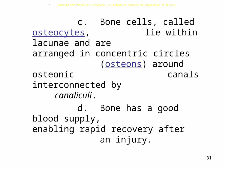
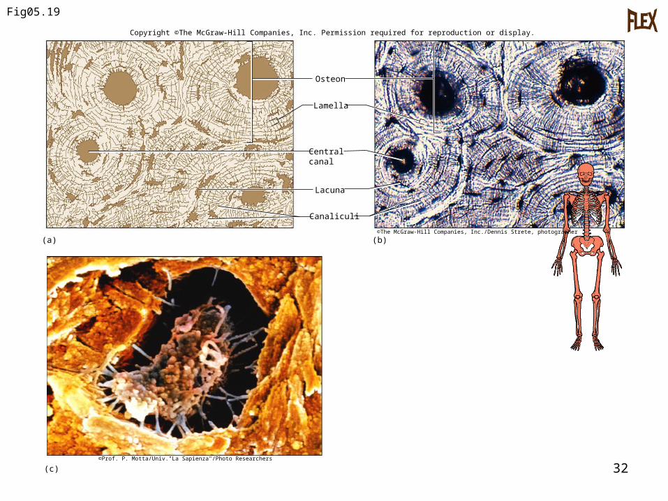
c. Bone cells, called osteocytes, lie within lacunae and are arranged in concentric circles
(osteons) around osteonic canals interconnected
by canaliculi.d. Bone has a good blood
supply, enabling rapid recovery after an injury.
CopyrightThe McGraw-Hill Companies, Inc. Permission required for reproduction or display.
32
Fig05.19
Canaliculi
Lacuna
Central canal
Lamella
Osteon
©The McGraw-Hill Companies, Inc./Dennis Strete, photographer
(c)©Prof. P. Motta/Univ."La Sapienza"/Photo Researchers
Copyright ©The McGraw-Hill Companies, Inc. Permission required for reproduction or display.
(a) (b)
33
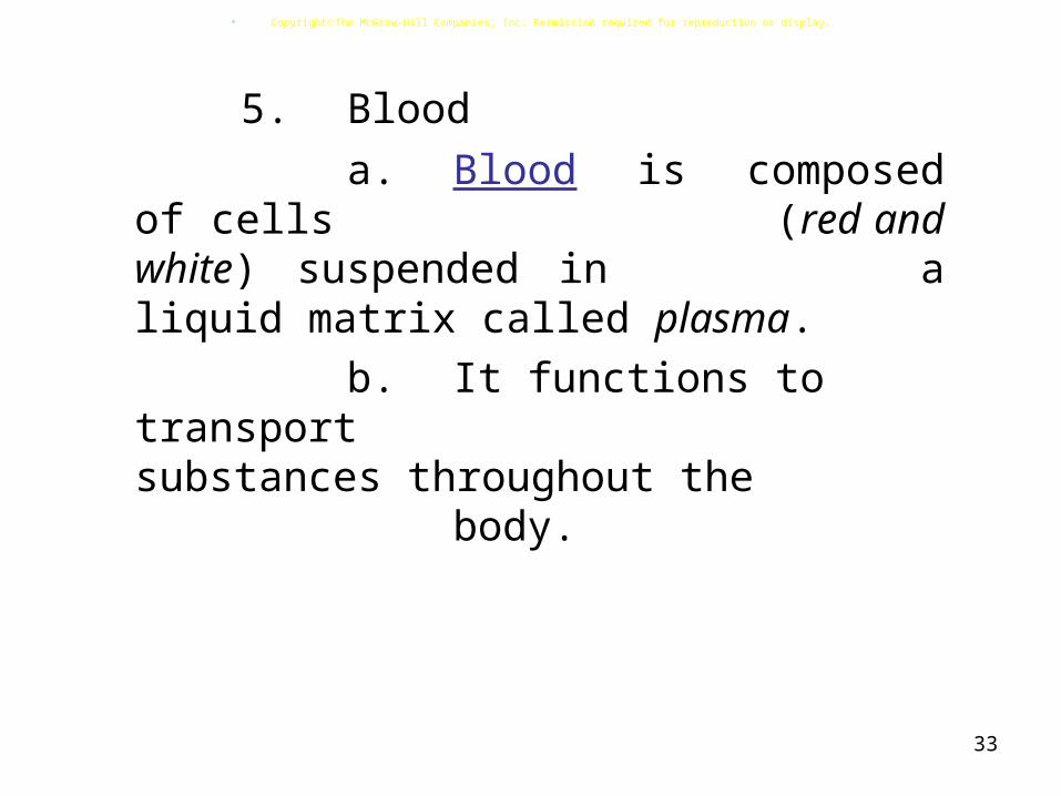
5. Blood a. Blood is composed of
cells (red and white) suspended in a liquid matrix called plasma.
b. It functions to transport substances
throughout the body.
CopyrightThe McGraw-Hill Companies, Inc. Permission required for reproduction or display.
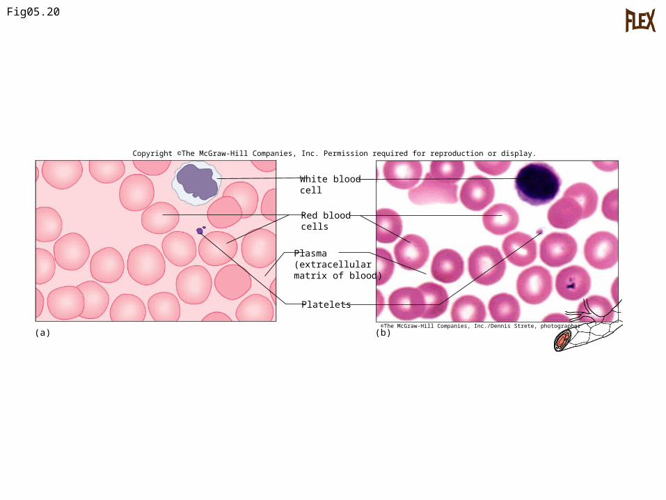
Fig05.20
Red blood cells
Plasma(extracellularmatrix of blood)
Platelets
White blood cell
(a) (b)©The McGraw-Hill Companies, Inc./Dennis Strete, photographer
Copyright ©The McGraw-Hill Companies, Inc. Permission required for reproduction or display.
Types of Membranes:A. Epithelial membranes are thin,
sheetlike structures composed of epithelium and connective tissues, covering body surfaces and lining body cavities.
B. The four main types are: serous, mucous, cutaneous, and synovial.
35

36
Muscle Tissues:A. General Characteristics 1. Muscle cells, or fibers, can contract
and consist of three major types: skeletal, smooth and cardiac.
CopyrightThe McGraw-Hill Companies, Inc. Permission required for reproduction or display.

37
B. Skeletal Muscle Tissue
1. Skeletal muscle is attached to bone and can be controlled by conscious effort (voluntary).
2. The cells (muscle fibers) are long and
cylindrical, striated, have many nuclei and contract
from nervous impulse.
CopyrightThe McGraw-Hill Companies, Inc. Permission required for reproduction or display.

38
Fig05.21
Striations
Portion of amuscle fiber
Nuclei
(a) (b)©The McGraw-Hill Companies, Inc./Al Telser, photographer
Copyright ©The McGraw-Hill Companies, Inc. Permission required for reproduction or display.
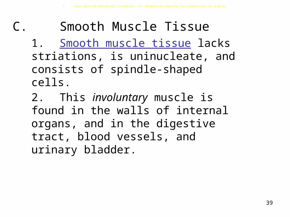
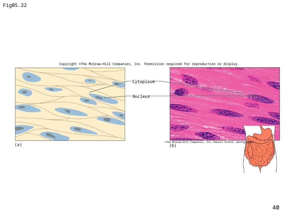
39
C. Smooth Muscle Tissue1. Smooth muscle tissue lacks
striations, is uninucleate, and consists of spindle-shaped cells.
2. This involuntary muscle is found in the walls
of internal organs, and in the digestive tract, blood vessels, and
urinary bladder.
CopyrightThe McGraw-Hill Companies, Inc. Permission required for reproduction or display.
40
Fig05.22
Nucleus
Cytoplasm
(a) (b)©The McGraw-Hill Companies, Inc./Dennis Strete, photographer
Copyright ©The McGraw-Hill Companies, Inc. Permission required for reproduction or display.

41
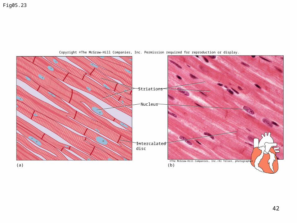
D. Cardiac Muscle Tissue
1. Cardiac muscle tissue is found only in the heart and consists of branching fibers that are connected
to each other with intercalated discs.
2. This involuntary muscle has a single nucleus in each cell but appears striated.
CopyrightThe McGraw-Hill Companies, Inc. Permission required for reproduction or display.
42
Fig05.23
Copyright ©The McGraw-Hill Companies, Inc. Permission required for reproduction or display.
Intercalated disc
Nucleus
Striations
(a) (b)©The McGraw-Hill Companies, Inc./Al Telser, photographer

43
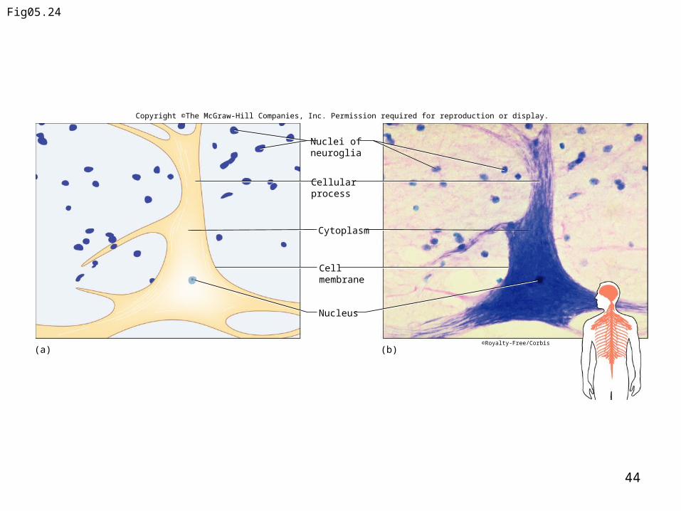
Nervous Tissues: A. Nervous tissues are found in the
brain, spinal cord, and nerves.
B. Neurons, or nerve cells, conduct nervous impulses while
helper cells, or neuroglia, support and nourish the neurons.
CopyrightThe McGraw-Hill Companies, Inc. Permission required for reproduction or display.
44
Fig05.24
Copyright ©The McGraw-Hill Companies, Inc. Permission required for reproduction or display.
(a) (b)
Nucleus
Cell membrane
Cytoplasm
Cellular process
Nuclei of neuroglia
©Royalty-Free/Corbis