CHAPTER 3: IN VITRO PROPAGATION -...
47
41 CHAPTER 3: IN VITRO PROPAGATION
Transcript of CHAPTER 3: IN VITRO PROPAGATION -...

41
CCHHAAPPTTEERR 33::
IINN VVIITTRROO PPRROOPPAAGGAATTIIOONN

3.1. In vitro propagation in Crimson Seedless
3.1.1. Introduction
Owing to highly heterozygous nature, seed dormancy and stenospermocarpy and
for maintenance of purity of clones and varieties, grape is mostly propagated through
vegetative means. The term stenospermocarpy (coined by Stout, 1936) refers to the
condition in which, the embryos abort during berry development and seeds remain in
rudimentary form resulting in seedlessness.
Grapevine was among the first woody plants, where the use of shoot apices and
axillary buds for in vitro propagation of various species and cultivars was reported (Gray
and Fisher, 1985). Tissue culture technique in grapevine was initially used for virus
elimination (Galzy, 1964). Later it was used for producing grapevines free from viroid
(Duran-Vila et al., 1988) and Pierce’s disease (Robacker and Chang, 1992). In vitro
propagation was first obtained by axillary shoot initiation in nodal cuttings (Galzy, 1969).
Fragmented shoot apices and shoot tips were also used as explants (Barlass and Skene,
1978; Harris and Stevenson, 1979; Goussard, 1981). Shoot apex as an explant was
commonly used for micropropagation of herbaceous species (Murashige, 1977; Abbot,
1978) but to a lesser degree in woody species. Use of other explants like meristem in Vitis
rotundifolia (Thies and Graves, 1992), microcutting and axillary buds in Vitis X
Muscadania hybrids (Torregrosa and Bouquet, 1995) have also been documented. Studies
on micropropagation of vines have recently been reviewed by Torregrosa et al. (2001).
Despite a moderate multiplication rate, nodal segment remains a widely used
explant for micropropagation of vines due to its operational feasibility and genotype
stability (Torregrosa et al., 2001). It was reported that survival and shoot production
potential was greater in explants from axillary shoots than from terminal ones (Yu and
Meredith, 1986). The influence of the axillary bud position and growth regulators on in
vitro establishment of V. rotundifolia was earlier studied (Sudarsono and Goldy, 1991). The
use of nodal segments to propagate Arka Neelamani (Vitis vinifera) by in vitro layering
technique (Thomas, 1997; 1998; 2000) and rootstocks (Sahijram et al., 1996) and three
vinifera (Sonaka, Tas-e-Ganesh and Thompson Seedless) cultivars (Mhatre et al., 2000)
has been achieved. The relative ease with which nodal explants of grapevine can be
induced axillary bud proliferation has led to the potential application of micropropagation
as a vehicle for mutation breeding also (Barlass, 1986; Kim et al., 1986).
Various factors influencing successful in vitro propagation of vines have been
studied to little extent. The organic and inorganic constituents of media for shoot
multiplication of Vitis hybrid Remaily Seedless have been defined (Chee and Pool, 1983;
42

1985; 1987). The effects of other growth substances like auxins / cytokinins and
photoperiod on the in vitro development of shoots from cultured shoot apices of Rougeon
grapevines has been reported (Chee and Pool, 1982 and 1988; Reisch, 1986). Zlenko et al.
(1995) reported an optimized medium for clonal propagation of rootstock Kober 5BB and
three vinifera cultivars.
A rhizogenesis medium with reduced salt concentrations for rooting of grapevine
(Vitis spp.) genotypes has been reported without the use of exogenous auxins (Roubelakis-
Angelakis and Zivanovitc, 1991). However in case of V. labrusca Delaware, rooting and
acclimatization of micropropagated plants was achieved with the use of auxins in the
medium (Lewandowski, 1991). Sachet technique, a cheaper and comparatively easier
method for the acclimatization of micropropagated plantlets of grapevine cultivars has been
reported (Ravindra and Thomas, 1995; Bharathy et al., 2003). Ex vitro rooting by auxin
pulse treatment was an advantage in not only reducing the time of in vitro propagation
nearly by one month but also in lowering the cost of plant production. Influence of
different potting mixtures on growth and establishment of in vitro produced plantlets
during hardening has been studied earlier (Mhatre et al., 2000). Application of soilrite
(Mhatre et al., 2000), 1:1:1 mixture of sand: loamy soil: soilrite for grapevine rootstocks
(Sahijram et al., 1996), Peat (Rubelakis and Zivanovitc, 1991), Metromix–500 with starter
nutrient charge, Red:Earth:Peat:Lite mix with starter nutrient charge, Sphagnum Peat:
Perlite (1:1 v/v) and vermiculite (Lewandowski, 1991) has been reported.
Micropropagation complements the conventional methods, when a large amount of
planting material of a particular variety is required in a short period of time. Earlier studies
on in vitro propagation of Vitis have indicated that the degree of success at each stage of
culture is genotype dependent (Barlass and Skene 1980; Monette, 1988; Botti et al., 1993;
Peros et al., 1998). Due to the variability in morphogenic response of in vitro cultured
explants, it is imperative to optimize the culture conditions for different stages of in vitro
propagation for a given genotype.
3.1.2. Materials and Methods
3.1.2.1. Plant material
Twigs of field grown vines of Crimson Seedless were collected from the vineyard
of National Research Centre for Grapes, Pune, India. Single node segments (1.5 - 2 cm
long) were used as explant for culture initiation (Fig. 3.1.1). Nodal segments were surface
sterilized by soaking them in liquid soap solution for 10 min followed by thorough rinses
with running tap water. The explants were then submerged in 0.1% fungicide solution
43

Fig. 3.1.1. Single nodal segments of Crimson Seedless used as explant.

(BavistinTM, BASF, India) for 1 h followed by 2-3 washes with sterile distilled water. Then
the explants were treated with 0.1% (w/v) Mercuric chloride for 10 min followed by
several rinses with sterile distilled water in a laminar flow hood. Excess water was removed
by blotting dry the explants on a sterile filter paper.
3.1.2.2. Bud break and shoot initiation
Different workers reported various basal media for in vitro propagation of different
species and cultivars of Vitis and reported that the influence of basal media on in vitro
propagation was cultivar dependent. Based on the earlier reports, six basal media were
investigated for bud break and other morphogenic responses for the cultivar Crimson
Seedless. For bud break and shoot initiation, nodal segments were inoculated in glass
culture tubes containing the following six different basal media: C2d (Chee and Pool,
1987); ER (Eriksson, 1965); B5 (Gamborg et al., 1968); WPM (Llyod and McCown, 1981);
MS (Murashige and Skoog, 1962) and NN (Nitsch and Nitsch, 1969). Also to achieve high
efficiencies of bud break and shoot initiation, MS basal medium supplemented with a range
of BA concentrations (0.44-44.44 μM) was also tested.
3.1.2.3. Induction of multiple shoots
3.1.2.3.1. Influence of growth regulators
For the induction of multiple shoots, both primary nodal segments from field grown
vines as well as secondary nodal segments excised from in vitro grown shoots from
primary nodal segments were used.
To investigate the influence of various growth regulators on induction of multiple
shoots, nodal segments were inoculated on MS medium supplemented with either BA
(0.44-44.44 μM), KIN (0.46-46.0 μM), 2ip (0.49-49.0 μM), TDZ (0.045-4.5 μM), Zeatin
(0.046-4.6 μM) or BA (8.89 μM) alone and in combination with IAA (0.57–1.71 μM) or
IBA (0.49–1.48 μM) or NAA (0.54–1.61 μM). Explants that induced multiple shoots were
shifted to fresh medium after every 4 weeks for further proliferation.
3.1.24. Elongation of multiple shoots
3.1.2.4.1. Influence of growth regulators
For elongation, uniform size multiple shoot clumps were transferred to culture
bottles containing full strength MS medium supplemented with BA (0.89-6.67 μM). In
another experiment, MS medium supplemented with BA (2.22 μM) alone and in
44

combination with IAA (0.57–1.71 μM) or IBA (0.49–1.48 μM) or NAA (0.54–1.61 μM)
was tested. Each culture bottle contained single clump of multiple shoots.
3.1.2.4.2. Carryover effect of BA concentrations on shoot elongation
This experiment was carried out to test the influence of carry over effect of BA
concentration included in the shoot proliferation medium on the subsequent shoot
elongation. Multiple shoots proliferated on MS basal medium supplemented with BA (2.22
- 17.78 μM) were transferred to MS basal medium supplemented with BA (2.22 μM) and
their elongation was recorded.
3.1.2.5. In vitro rooting
3.1.2.5.1. Influence of strength of the basal medium
To test the influence of strength of basal medium on in vitro rooting, ¼, ½ 1X
strength of MS medium was used.
3.1.2.5.2. Influence of growth regulators and gelling agents
In vitro shoots elongated on MS medium supplemented with BA (2.22 μM) were
transferred to culture tubes containing half strength semi-solid or liquid MS medium
supplemented with IAA (0.57–1.71 μM) or IBA (0.49–1.48 μM) or NAA (0.54–1.61 μM).
Agar (0.65%) or gelrite (0.2%) were used as gelling agents in case of semi-solid media.
Sucrose (2%) was added to all media and pH was adjusted to 5.8 before
autoclaving. All the media were gelled with 0.65% agar unless otherwise mentioned. All
the growth regulators were added to the media before autoclaving except zeatin. Zeatin was
filter sterilized with 0.22 μm pore size sterile syringe filters (Laxbro, India) and added to
the media after autoclaving. All the media were autoclaved at 121oC and 105 KPa for 20
min.
3.1.2.5.3. Influence of ventilation closure types
To investigate the influence of aeration on in vitro rooting of shoots, culture tubes
plugged with cotton and capped and sealed with saran wrap were tested in the present
study.
Sucrose (2%) was added to all media and pH was adjusted to 5.8 before
autoclaving. All the media were gelled with 0.65% agar unless otherwise mentioned. All
the growth regulators were added to the media before autoclaving except zeatin. Zeatin was
filter sterilized with 0.22 μm pore size sterile syringe filters (Laxbro, India) and added to
45
the media after autoclaving. All the media were autoclaved at 121oC and 105 KPa for 20
min.
3.1.2.6. Ex vitro rooting by auxin pulse treatment
Three auxins viz., IAA, IBA and NAA at concentrations of 57.08 μM, 49.0 μM and
53.71 μM, respectively, were used individually for the pulse treatment. Individual
elongated shoots (>5.0 cm) were given pulse treatment of above auxins for 5 or 10 min.
After pulse treatment, shoots were transferred to plastic cups containing a mixture of sterile
peat: soil: vermiculite (1:1:1). Plants were irrigated with ¼ strength of MS salts and
covered with thin and transparent polythene sachets. Untreated shoots, which served as
control, were also transferred to the same potting mixture.
3.1.2.7. Hardening of plantlets
Rooted shoots and nodal segments with direct rooting on basal media were
transferred to plastic cups containing a mixture of soil and sand (1:1). For hardening of in
vitro and ex vitro-rooted shoots, Sachet technique followed by Ravindra and Thomas
(1995) and Bharathy et al. (2003) was used in the present study. Plantlets were covered
with thin, transparent polythene sachets and kept in growth room having 24 h light with an
intensity of 24.4 μmol m-2 s-1 at 25+2oC. After 2 weeks, plantlets were shifted to another
room having ambient temperature. Here, the sachets were cut at top corners and were
removed completely after another 2 weeks. After that plantlets were transferred to
polyhouse for acclimatization.
3.1.2.8. Influence of potting mixtures on hardening
In another study to evaluate influence of different potting mixtures on hardening
and acclimatization, in vitro rooted shoots of Crimson Seedless were collected and their
roots were washed with sterile distilled water to remove media sticking to them. The
different potting mixtures used in the present study were: 1. Soil : Vermiculite (1:1), 2.
Soilrite, 3. Peat : Sand : Soil (1:1:1), 4. Peat : Vermiculite (1:1), 5. Peat : Perlite (2:3), 6.
Vermiculite, 7. Perlite : Coarse sand (3:1) and 8. Soil : Sand (1:1). All the potting mixtures
were sterilized at 121oC and 105 KPa for 30 min. The rooted plantlets were transferred to
plastic cups containing above potting mixtures and hardened as mentioned in section 3.2.7.
Observations on plant height, plant survival, other morphological characters like root
growth and rooting pattern were recorded after 30 d of planting.
46

All the experiments were repeated for a minimum of three times with each
treatment containing a minimum of twelve explants. Observations of all the experiments
were taken at 30 d interval unless otherwise specified.
3.1.3. Results and Discussion
3.1.3.1. Bud break and shoot initiation
Bud break in nodal segments commenced after 7 d of inoculation and continued up
to 30 d (Fig. 3.1.2). Six basal media affected different morphogenic responses. The
maximum bud break response in explants was observed in MS basal medium (90.0%)
followed by NN (89.4%) and WPM (87.2%) after 30 d of inoculation (Table 3.1.1). Nodal
segment that induced shoot of atleast 1 cm in length was counted for shoot initiation.
Percentage of explants that induced shoots was also higher (85.7) in MS followed by NN
and WPM (78.8 each). Average length of the shoots ranged from 1.30 cm in NN medium to
1.75 cm in B5 medium. The results of average shoot length were found to be statistically
non-significant. Among all the basal media tested, MS yielded higher percentage of bud
break and number of explants showing shoot production. Hence, for all further experiments
MS basal medium was selected as nutrient medium. From the earlier studies, it is evident
that full strength MS medium was the most commonly used nutrient medium for in vitro
propagation of different cultivars of grapevine (Mhatre et al. 2000).
Table 3.1.1: Influence of different nutrient media on morphogenic responses in Crimson Seedless. Explant: Primary nodal segment.
Nutrient Medium
Number of explants
Inoculated
% of explants showing
bud break
% of explants showing shoots
Average length of
shoots (cm)
% of explants showing rooting
Average number of roots per explant
% of plants established on transfer
to pots
WPM 47 87.2 78.7 1.57 69.1 6.52 100.0 C2d 46 84.8 78.3 1.36 22.0 2.11 25.0 ER 45 75.6 66.7 1.54 00.0 0.00 00.0 NN 47 89.4 78.8 1.30 55.8 6.75 00.0 B5 69 60.9 53.7 1.75 60.0 6.03 66.7 MS 70 90.0 85.7 1.36 25.0 3.67 33.3 SEM± 1.4 1.6 0.28 3.23 0.75 4.7 CD (p=0.01) 3.2 3.1 0.49 5.60 1.30 8.1
* ** NS ** * ** * Significant at 5% level; ** Significant at 1% level; NS – Non signoficant
Six different basal media had varying influence on in vitro induction of direct
rooting in nodal explants of Crimson seedless. With the exception of ER, all the basal
47

Fig. 3.1.2. Direct rooting in nodal segments of Crimson Seedless. 1 – in B5 medium, 2- in MS medium, 3 – ER medium, 4 – C2d medium, 5 – NN medium and 6 – WPM.
1 2 3
4 5 6

media induced direct rooting (Fig. 3.1.3). WPM induced rooting in maximum number of
nodal segments (69.1%) followed by B5 (60%), where the establishment of rooted plantlets
on potting was 100% and 66.7%, respectively. There was 100% mortality of plantlets
rooted in NN media during hardening process.
0
20
40
60
80
100
120
WPM C2d ER NN B5 MS
Basal medium
% re
spon
seRooting
Plant
Fig. 3.1.3. Influence of basal media on percentage of rooting and plantlet establishment in nodal segments of Crimson Seedless.
WPM not only induced rooting in maximum number of explants but also the
quality of the roots was found better. The roots were long and brownish with a greater
number of secondary roots and root hairs. Attachment of root to nodal segments was
stronger. In case of B5 medium, the roots were white and thick with fewer root hairs (Fig.
3.1.4). In addition, the roots were fleshy and weak, which could be one of the reasons for
poor survival of rooted nodal segments in B5 medium. Direct rooting in nodal segments
has an advantage in propagation, since explants rooted in this manner can directly be potted
and hardened plantlets can be obtained within 2 months. Also use of single node cuttings in
culture instead of 3-4 node cuttings used in vineyard can give rise to larger number of
plants if availability of mother plant material is a serious limitation.
Varying responses of six basal media on bud break and direct rooting observed in
the present study could be due to variations in nutrient compositions of basal media. For
example, amount of CaCl2 is higher in MS and ER media as compared to WPM and NN.
While in C2d, it is substituted by Ca(NO3)2. Similarly, potassium iodide (KI) is absent in
WPM, NN, C2d and ER media, while it is present in B5 and MS though in different
quantities. In addition, the amount of MnSO4 varied in the six basal media studied. It has
been demonstrated earlier that nutrient requirement varies with the morphogenetic process
of the plant. High K and N levels proved favorable to shoot development but restricted root
growth (Galzy, 1969). In another study on grape, it was reported that lower concentrations
48
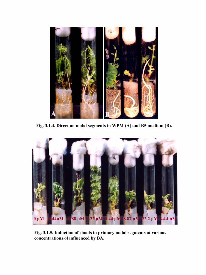
Fig. 3.1.5. Induction of shoots in primary nodal segments at various concentrations of influenced by BA.
0 μM 0.44μM 0.88 μM 2.22 μM 4.44 μM 8.87 μM 22.2 μM 44.4 μM
Fig. 3.1.4. Direct on nodal segments in WPM (A) and B5 medium (B).
A B

of KI and MnSO4 in the medium enhance the shoot production, while substitution of CaCl2
with Ca(NO3)2 produced good quality shoots (Chee and Pool, 1987). In a previous study
with different grapevine cultivars and rootstocks, Roubelakis-Angelakis and Zivanovitc
(1991) reported increased rhizogenesis in single node segments on medium containing
lower amounts of N, K, Ca and Mg. In our study, WPM contains lower amount of N as
compared to other five basal media and this could be a reason for increased rooting
response. Similarly, higher amount of N present in ER medium as compared to other media
could be a reason for the failure of rooting on ER medium. In addition, among all basal
media myo-inositol content was lower in C2d medium (0.056 mM compared to 0.56 mM in
others) and completely absent in ER medium, which might also be reason for lower rooting
response in C2d and no rooting in ER medium. It was earlier reported that higher
concentrations of thiamine in the media improved the number of shoots per explant and
decrease in inositol enhanced the shoot number and shoot length and number of nodes in
grape hybrid Ramsey Seedless (Chee and Pool, 1985).
Incorporation of BA at 4.44 μM in MS basal medium induced bud break in 100%
of explants (data not shown). A gradual increase in the percentage of explants showing 2 or
more shoots with the increase in the concentration of BA in the medium was observed.
Direct rooting observed in nodal segments cultured on BA free medium was absent in MS
basal medium supplemented with BA. Mean length of shoots decreased with increase in
BA concentration (Fig. 3.1.5). Cytokinins play an important role in stimulating cell
division and cell enlargement. Cytokinins have been reported to be essential for in vitro
shoot development in grapevine (Pool and Powell, 1975; Jona and Webb, 1978; Mullins et
al., 1979). Positive influence of BA on differentiation and proliferation of dormant
accessory buds in cotton was reported earlier (Hazra et al., 2001).
As MS basal medium was found to be superior to all other media in terms of
percentage of explants showing bud break and shoot induction in nodal segments, MS
medium was selected as nutrient medium in further experiments for multiple shoot
induction.
3.1.3.2. Multiple shoots induction
3.1.3.2.1. Influence of growth regulators
3.1.3.2.1.1. Primary nodal segments
In case of primary nodal segments, percentage of explants with multiple shoots
induction increased with increased concentrations of BA. Percent response varied from
5.7% in hormone free medium to 62.5 in MS medium supplemented with BA (44.44 μM).
49

The number of shoots produced per explant increased with the higher concentrations of BA
in the medium. The maximum number of shoots (2.6 shoots per explant) was recorded on
MS medium supplemented with BA (8.89 μM) and IBA (1.48 μM) (Table 3.1.2).
Table 3.1.2: Influence of BA and auxins on induction of multiple shoots in Primary nodal segments of Crimson Seedless
Hormone conc. (μM) Number of explants
Inoculated
% of explants showing
multiple shoots
% of explants showing single
shoots
Av. No. of shoots
per explant
Average Shoot length
(cm)
BA (0.0) 70 5.71 82.9 1.03 1.36 BA (0.44) 32 12.5 81.3 1.20 1.51 BA (0.89) 30 23.3 76.7 1.23 1.56 BA (2.22) 36 25.0 69.4 1.32 1.23 BA (4.44) 32 28.13 71.9 1.28 1.22 BA (8.89) 34 29.41 70.6 1.59 1.13 BA (22.22) 34 52.94 41.2 1.69 0.65 BA (44.44) 32 62.5 31.3 2.47 0.55 BA (8.89) 34 29.4 70.6 1.59 1.13 BA (8.89)+IAA (0.57) 36 30.6 66.7 1.37 1.65 BA (8.89)+IAA (1.14) 35 31.4 65.7 1.15 2.01 BA (8.89)+IAA (1.71) 33 33.3 63.6 1.91 1.63 BA (8.89)+IBA (0.49) 32 34.4 62.5 1.97 1.23 BA (8.89)+IBA (0.98) 35 37.1 62.9 2.06 1.22 BA (8.89)+IBA (1.48) 35 28.6 71.4 2.60 1.00 BA (8.89)+NAA (0.54) 34 26.5 64.7 1.81 1.65 BA (8.89)+NAA (1.07) 32 21.9 68.8 1.66 1.78 BA (8.89)+NAA (1.61) 30 26.7 73.3 2.08 1.22 SEM± 3.16 5.65 0.21 0.25 CD (p=0.01) 5.48 9.79 0.37 0.43 ** ** ** **
**Significant at 1% level
There was a significant increase in the number of multiple shoots per explant on the
medium supplemented with IAA or IBA along with BA (8.89 μM). IBA gave rise higher
response compared to IAA. Addition of NAA in the medium adversely affected the
multiple shoots and induced excessive callus at the base of explants.
3.1.3.2.1.2. Secondary nodal segments
To maximize multiplication rates, secondary nodal segments obtained from shoots
grown from primary nodal segments were used as explants for the induction of multiple
shoots, similar to primary nodal segments. Different cytokinins at different concentrations
50

tested differed in percentage of explants showing number of shoots per explant and average
length of the shoots. In general, TDZ (at more than 0.90 μM) and Zeatin (at more than 0.92
μM) concentrations induced higher number of shoots / explant. MS medium supplemented
with TDZ (4.5 μM) or zeatin (4.6 μM) induced the maximum (100%) response with an
average of 3.89 and 3.81 shoots per explant, respectively. However, profuse callusing at the
base of explants was observed in the media with TDZ concentrations higher than 0.45 μM
(Table 3.1.3).
KIN and 2ip were found to be less effective in multiple shoot induction. Shoots in
media with zeatin had higher lengths as compared to media with other cytokinins. Between
the primary and secondary nodal explants at constant BA (44.44 μM) in the medium, the
latter resulted in the higher percentage of explants showing multiple shoots (90%)
compared to primary nodal segments (62.5%).
Cytokinins have been reported to induce axillary and adventitious shoots in
different plants (Madhulatha et al., 2004), however, their efficiencies vary among the
species. The superiority of TDZ over BA and KIN on axillary bud establishment and shoot
proliferation, when used alone or in combination with BA or KIN in three muscardine
grape cultivars has been reported (Sudarsono and Goldy, 1991). It was observed that higher
concentrations of TDZ induced shoot thickening and inhibited further shoot growth.
Similarly, TDZ has been reported to promote bud out growth and number of shoots from a
single bud. Zeatin was found to be more effective cytokinin compared to BA for induction
of adventitious shoots in tubers of two wild Potato cultivars (Anjum and Ali, 2004). While
in another report, the maximum adventitious shoot formation from internode explants of
Adenophora triphylla was affected in the medium supplemented with BA and NAA (Chen
et al., 2001).
In the present study, it was observed that a higher number of shoots per explant
corresponded with shorter shoot lengths. MS basal medium devoid of growth regulators
could not induce multiple shoots in secondary nodal segments, whereas a very small
percentage (5.7) showed multiple shoot induction in primary nodal explants. Multiple
shoots induced in the axils of nodal explants were in form of clumps and the size of these
clumps increased with the sub culture (Fig. 3.1.6A,B). At the initial stages, it was difficult
to count the number of shoots present in a clump with the naked eye, hence it was
necessary to dissect the clump and the numbers of shoots were counted under
stereomicroscope.
51
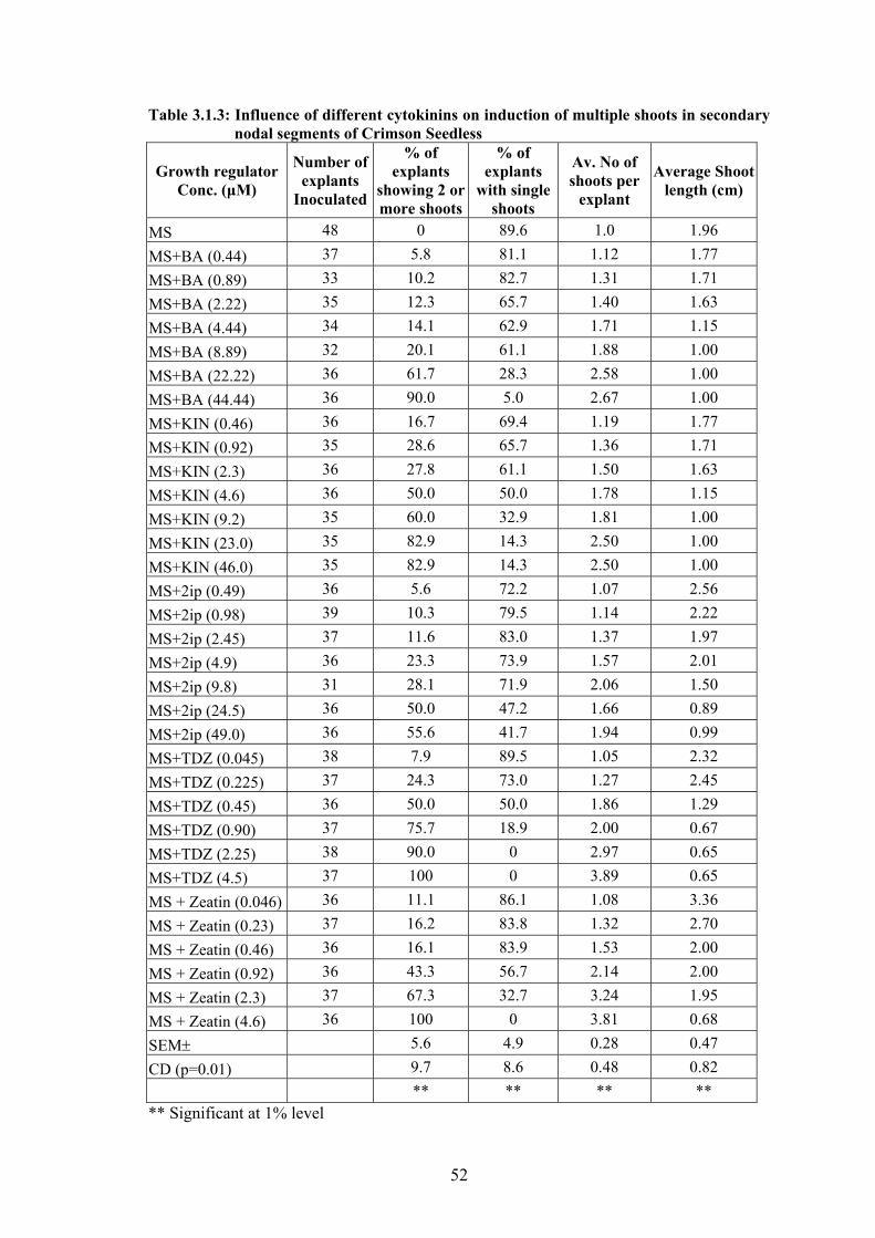
Table 3.1.3: Influence of different cytokinins on induction of multiple shoots in secondary nodal segments of Crimson Seedless
Growth regulator Conc. (μM)
Number of explants
Inoculated
% of explants
showing 2 or more shoots
% of explants
with single shoots
Av. No of shoots per
explant
Average Shoot length (cm)
MS 48 0 89.6 1.0 1.96 MS+BA (0.44) 37 5.8 81.1 1.12 1.77 MS+BA (0.89) 33 10.2 82.7 1.31 1.71 MS+BA (2.22) 35 12.3 65.7 1.40 1.63 MS+BA (4.44) 34 14.1 62.9 1.71 1.15 MS+BA (8.89) 32 20.1 61.1 1.88 1.00 MS+BA (22.22) 36 61.7 28.3 2.58 1.00 MS+BA (44.44) 36 90.0 5.0 2.67 1.00 MS+KIN (0.46) 36 16.7 69.4 1.19 1.77 MS+KIN (0.92) 35 28.6 65.7 1.36 1.71 MS+KIN (2.3) 36 27.8 61.1 1.50 1.63 MS+KIN (4.6) 36 50.0 50.0 1.78 1.15 MS+KIN (9.2) 35 60.0 32.9 1.81 1.00 MS+KIN (23.0) 35 82.9 14.3 2.50 1.00 MS+KIN (46.0) 35 82.9 14.3 2.50 1.00 MS+2ip (0.49) 36 5.6 72.2 1.07 2.56 MS+2ip (0.98) 39 10.3 79.5 1.14 2.22 MS+2ip (2.45) 37 11.6 83.0 1.37 1.97 MS+2ip (4.9) 36 23.3 73.9 1.57 2.01 MS+2ip (9.8) 31 28.1 71.9 2.06 1.50 MS+2ip (24.5) 36 50.0 47.2 1.66 0.89 MS+2ip (49.0) 36 55.6 41.7 1.94 0.99 MS+TDZ (0.045) 38 7.9 89.5 1.05 2.32 MS+TDZ (0.225) 37 24.3 73.0 1.27 2.45 MS+TDZ (0.45) 36 50.0 50.0 1.86 1.29 MS+TDZ (0.90) 37 75.7 18.9 2.00 0.67 MS+TDZ (2.25) 38 90.0 0 2.97 0.65 MS+TDZ (4.5) 37 100 0 3.89 0.65 MS + Zeatin (0.046) 36 11.1 86.1 1.08 3.36 MS + Zeatin (0.23) 37 16.2 83.8 1.32 2.70 MS + Zeatin (0.46) 36 16.1 83.9 1.53 2.00 MS + Zeatin (0.92) 36 43.3 56.7 2.14 2.00 MS + Zeatin (2.3) 37 67.3 32.7 3.24 1.95 MS + Zeatin (4.6) 36 100 0 3.81 0.68 SEM± 5.6 4.9 0.28 0.47 CD (p=0.01) 9.7 8.6 0.48 0.82 ** ** ** ** ** Significant at 1% level
52

Fig. 3.1.6. Multiple shoot induction in nodal segments. A: Multiple shoot clump in bottle, B: Multiple shoots from secondary nodal segment, C: Multiples from axillary bud and D: Multiples from apical meristem. Bar = 5 mm.
A B
C D

Yet in another experiment with secondary nodal explants, various auxins were
supplemented along with BA (8.89 μM) in MS basal medium to investigate their
effectiveness in multiple shoot induction. Among the 3 auxins tested, IBA at 0.98 μM +
BA (8.89 μM) induced multiple shoots in 25.0% of explants (Table 3.1.4) compared to
18.8% with BA (8.89 μM) alone. Results with BA (8.89 μM) and IAA at all three levels
varied marginally. Inclusion of NAA in the medium not only showed lower response of
multiple shoots but also resulted in profuse callusing at basal ends of explants and also
shoots became hyperhydric.
On further subculture of these explants onto their respective media in test tubes did
not improve multiple shoot number in majority of the media. It was observed that node
region swelled and enlarged in size. When these explants were sub cultured in glass bottles
instead of test tubes, a dramatic increase in number of multiple shoots was observed at the
end of 30 d. On an average 19.5 shoots per explant were recorded on MS basal medium
supplemented with BA (8.89 μM) and IBA (1.48 μM) (Table 3.1.4). Also, a large number
of shoot buds were observed in axils (Fig. 3.1.6C) and apices of multiple shoots (Fig.
3.1.6D). The poorest response was recorded on MS supplemented with BA (8.89 μM).
MS alone without any growth regulators (control) did not show any multiple shoot
induction in secondary nodal segments. Similar to present results, Goussard (1982)
reported poor shoot proliferation up to two sub cultures and a sudden linear increase in
shoot proliferation after 2nd sub culture in presence of cytokinins in grapevine cv. Chenin
blac. Histology studies indicated the origin of multiple shoots from the secondary nodal
segments (Fig. 3.1.7).
Due to its favorable response, BA has been the most commonly used cytokinin in
grape tissue cultures. BA in the range of 1.125 – 2.25 mg/l was found to be an effective
growth regulator for induction of shoots in grapevine cultures (Harris and Stevenson, 1982;
Mhatre et al., 2000). Lee and Wetzstein (1990) reported that proportion of small (<1 cm)
shoots increased with increased BA levels and cultures at higher BA levels had dense,
unexpanded shoots with high mortality in vinifera cv. Summit. Thomas (1997) in his
experiments with vinifera cultivar ‘Arka Neelamani’ obtained multiple shoots with poor
elongation at BA (5 μM). Chee and Pool (1985) reported that shoot proliferation of Vitis
hybrid Remaily Seedless increased with increased concentration of BA (0 – 80 μM)
reaching maximum at 5 μM.
53
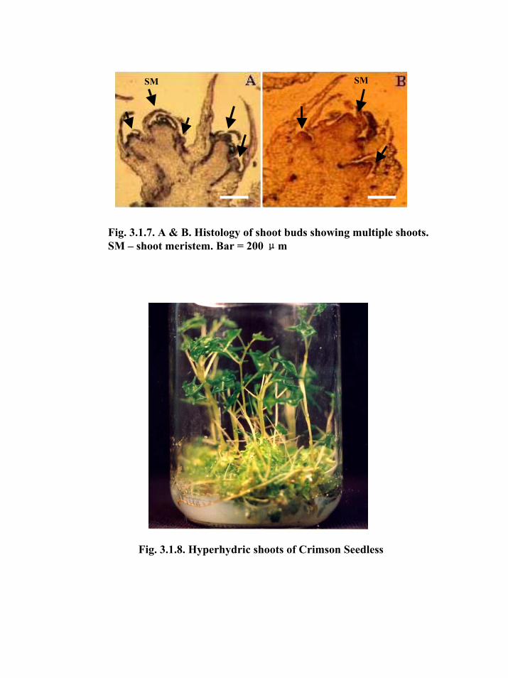
Fig. 3.1.8. Hyperhydric shoots of Crimson Seedless
Fig. 3.1.7. A & B. Histology of shoot buds showing multiple shoots. SM – shoot meristem. Bar = 200 μm
SMSM

Table 3.1.4: Influence of growth regulators on proliferation of multiple shoots in secondary nodal segments of Crimson seedless No. of replicates in each treatment: 32; 1 Explants - shoot clumps in test tubes; 2 Shoot clumps in bottles.
Av. number of shoots per explant Medium Composition
Conc. (μM)
% of explants showing
multiple shoots (30 d)1 (90 d)2
MS+BA (8.89) 18.8 2.33 2.33 MS+BA (8.89)+IAA (0.57) 18.8 2.50 13.00 MS+BA (8.89)+IAA (1.14) 18.8 2.50 15.20 MS+BA (8.89)+IAA (1.71) 15.6 2.00 11.80 MS+BA (8.89)+IBA (0.49) 9.3 2.00 8.67 MS+BA (8.89)+IBA (0.98) 25.0 2.13 16.17 MS+BA (8.89)+IBA (1.48) 15.6 2.60 19.50 MS+BA (8.89)+NAA (0.54) 9.9 2.00 9.50 MS+BA (8.89)+NAA (1.07) 9.4 2.33 6.33 MS+BA (8.89)+NAA (1.61) 12.5 2.00 8.60 MS 0.0 0.00 0.00 CD (p=0.05) 2.16 0.27 8.17
* * ** * Significant at 5%; ** Significant at 1%.
Hyperhydricity in shoots was observed with repeated subculture of the multiple
shoot clumps in treatments (Fig. 3.1.8). This condition could be partially due to presence of
higher concentrations of BA in the medium as reported earlier by Morini et al. (1985).
Though the mechanism of occurrence of this phenomenon has not been elucidated fully,
the possible reason may be higher concentration of cytokinin, NH4+ ions or agar in the
medium (Ziv, 1991). In the present study, problem of hyperhydricity could be overcome by
reducing the concentration of BA in the medium to 4.44 μM. Also to prevent
hyperhydricity in shoots, continuous sub cultures on medium with auxins was avoided by
initiating fresh crop of multiple shoots.
3.1.3.3. Elongation of multiple shoots
3.1.3.3.1. Influence of growth regulators
Since multiple shoots induced were stunted and were in form of clumps, it was
necessary to define a medium for elongation. Results obtained show that BA at 2.22 μM
resulted in the highest number of shoots elongation per clump (Table 3.1.5). Though the
average length of elongated shoots was slightly higher in BA (0.89 μM), the average shoot
length above 3.0 cm was regarded adequate for in vitro rooting. Hence BA at 2.22 μM was
54

selected for further studies. Lee and Wetzstein (1990) reported that higher BA levels (20,
30 and 40 μM) strongly inhibited shoot elongation, in Vitis vinifera cv. Summit. Similarly,
BA at 10-20 μM resulted into condensed form of shoots in grapevine cultivar Arka
Neelamani (Thomas 1997).
Table 3.1.5: Influence of BA on elongation of multiple shoots of Crimson Seedless
BA conc. (μM)
Initial size of the clump
Final size of the clump
(cm)
Increase in clump size
(cm)
No. of shoots elongated per
clump
Av. shoot length (cm)
0.0 4.0 4.00 0.00 3.6 1.70 0.89 4.0 4.05 0.05 12.8 3.68 2.22 4.0 5.50 1.50 15.0 3.37 4.44 4.0 6.28 2.28 12.4 2.97 6.67 4.0 7.00 3.00 4.8 2.43
SEM± 0.80 1.10 0.19 CD (p=0.01) 1.18 1.64 0.28 ** ** ** ** Significant at 1% level
In another experiment, three auxins (IAA, IBA and NAA) at 3 concentrations were
tested for shoot elongation. Results showed that the maximum number of shoots (8.1 per
clump) elongated on MS with BA at 2.22 μM followed by 7.85 shoots per clump on MS
with BA (2.22 μM) + NAA (1.61 μM) (Table 3.1.6) (Fig. 3.1.9). On these media, shoots
grown could be excised easily from the clumps. Though comparatively higher elongation
was achieved on media with NAA or IAA at all the three levels, however there was
excessive callusing at base with adventitious roots. MS basal medium without any growth
regulator (control) affected the least elongation.
Similar to our findings, Mhatre et al. (2000) reported enhanced shoot elongation
with an addition of 0.2 mg/l IAA to the MS medium containing BA at 0.5 mg/l compared
to the medium with BA (0.5 mg/l) alone.
55

Table 3.1.6: Influence of BA and auxins on elongation of multiple shoots of Crimson Seedless
Medium Composition (μM)
No. of clumps
inoculated
Total no. of shoots
elongated
No. of shoots elongated per
clump
Av. shoot length (cm)
MS 12 43 3.6 1.70 MS+BA (2.22) 30 243 8.1 3.28 MS+BA (2.22)+IAA (0.57) 22 148 6.7 3.70 MS+BA (2.22)+IAA (1.14) 10 43 4.3 4.20 MS+BA (2.22)+IAA (1.71) 14 84 6.0 3.50 MS+BA (2.22)+IBA (0.49) 13 85 6.5 2.99 MS+BA (2.22)+IBA (0.98) 12 70 5.83 3.27 MS+BA (2.22)+IBA (1.48) 12 61 5.08 3.22 MS+BA (2.22)+NAA (0.54) 12 94 7.85 4.53 MS+BA (2.22)+NAA (1.07) 12 41 3.43 4.71 MS+BA (2.22)+NAA (1.61) 12 36 3.00 4.97 SEM± 0.94 0.76 CD (p=0.01) 1.62 1.32 ** ** ** Significant at 1% level
From the published reports, it can be inferred that no particular BA concentration is
optimum for different stages of in vitro multiplication of different grapevine cultivars,
indicating that BA concentrations need to be optimized for each cultivar.
3.3.1.3.1. Influence of carry over effect of BA concentrations on shoot elongation
Medium supplemented with BA in previous subculture significantly influenced the
elongation of multiple shoots. Though shoot proliferation and simultaneous elongation was
observed in all the treatments, there was a substantial increase in size of the clump
collected from proliferation medium containing higher levels of BA. Average number of
shoots elongated was maximum (13.7 per clump) in shoot clumps collected from medium
supplemented with BA (8.89 μM) (Table 3.1.7).
The number of elongated shoot per clump gradually increased with the increase in
BA concentration from 2.22 - 8.89 μM but showed reverse trend in higher BA
concentrations. The increase in the number of elongated shoots may be attributed to the
proliferation medium containing higher levels of BA. Average length of elongated shoots
was better in shoot clumps proliferated on media containing lower levels of BA. Influence
of previous-culture medium on subsequent morphogenic responses of grapevine has earlier
been reported (Lee and Wetzstein, 1990).
56

Fig. 3.1.9. Elongated shoots from multiple shoot clumps
Fig. 3.1.10. In vitro rooting of elongated shoots in agar (A), gelrite (B) and liquid (C) rooting media
A B C

Table 3.1.7: Influence of carry over effect of BA concentrations on subsequent shoot elongation BA conc. used
for shoot proliferation
(μM)
No. of clumps
inoculated
Initial av. Clump
size (cm)
Final av. clump size
(cm)
Increase in clump size
(cm)
No. of shoots
elongated per clump
Av. shoot length (cm)
2.22 15 2.4 2.8 0.4 3.4 8.11 4.44 12 2.5 3.0 0.5 5.5 6.77 8.89 11 3.0 3.7 0.7 13.7 4.16
17.78 14 3.0 4.0 1.0 11.8 3.34 SEM± 0.5 1.6 5.2 CD (p=0.01) 0.8 2.8 0.9 NS ** ** * Medium used for elongation – MS + BA (2.22 μM) ** Significant at 1% level
3.1.3.4. In vitro rooting
3.1.3.4.1. Influence of strength of culture medium
Strength of the medium significantly influenced percentage of rooting and rooting
parameters studied. Percent rooting was highest (83.0) in the quarter strength MS medium,
and at par with the half strength medium (Table 3.1.8). Among other parameters studied,
half strength MS basal medium was found to be superior in terms of earliness in rooting,
number of roots per rooted shoot and average length of the roots. Highest number of roots
per rooted shoot (11.6) was recorded in half strength MS basal medium. The difference
between average shoot length in half strength and full strength MS basal was found to be
non-significant.
MS basal medium at quarter, half and full strength resulted in the different rooting
responses. MS at quarter and half strength resulted in 83 and 81.3 % of rooting of shoots,
respectively (Table 3.1.8). Among other parameters studied, half strength MS basal
medium was found to be superior in terms of number of days taken for rooting, number of
roots per rooted shoot and average length of the roots. The highest number of roots (11.6)
per shoot was observed on half strength MS basal medium. The difference between average
shoot length between half strength and full strength was found to be non-significant.
57

Table 3.1.8: Influence of strength of basal medium on in vitro rooting in Crimson Seedless
Basal medium
No. of shoots kept for rooting
Rooting (%)
Av. shoot length (cm)
Days to the first rooting
Av. No. of roots per
shoot
Av. root length (cm)
¼MS 47 83.0 6.5 7 8.2 1.65 ½MS 48 81.3 7.6 7 11.6 2.41 MS 52 68.0 8.4 11 4.9 1.39 SEM± 2.2 0.5 - 1.85 0.43 CD (p=0.01) 8.0 0.9 - 3.20 0.75 ** ** - ** ** ** Significant at 1% level
3.1.3.4.2. Influence of auxins and gelling agents
Though rooting of in vitro shoots could be observed in varying percentages in
medium with agar (Fig. 3.1.10A) or gelrite (Fig. 3.1.10B) semi-solid or liquid medium
(Fig. 3.1.10C) without any auxins (control), however number of roots induced were less as
compared to media supplemented with auxins. First root initials were observed as early as
three days and continued up to 25 d of inoculation. Time taken for the first root initials to
appear was less in control and IAA treatments as compared to others. An early rooting of
shoots was observed on medium gelled with agar compared to liquid medium (Table 3.1.9).
Average shoot length was increased significantly compared to control in all auxin
treatments. The increase in shoot length was more in NAA treatments followed by IBA and
IAA. Overall, average shoot length was more in liquid media but least in agar media.
Enhanced shoot length in liquid media was attributed to easy availability of the nutrients to
the growing plantlet, whereas agar and gelrite solidified media could impose water stress
there by limiting the nutrient uptake by the growing plantlets. Rooting delayed in IAA
treatments, which could be due to the formation of IAA conjugates in the plants and the
slow release of free IAA in plant tissues (Hangarter and Good, 1981).
Rooting response was better in terms of number of roots per rooted shoot in
medium with NAA. Average number of roots per rooted shoot was the minimum (5.3) in
control treatment. Media gelled with agar induced higher number of roots compared to
gelrite or without any gelling agent (liquid). Inclusion of auxin in the medium significantly
increased the rooting percentage of in vitro shoots of Crimson seedless. Both average
number of roots and root length were found to be negatively correlated to each other.
Average root length was more in hormone free medium but decreased with the inclusion of
auxins. Among the 3 auxins, average root length was more in IAA as compared to IBA and
NAA treatments. Media gelled with agar resulted in the least root length.
58

Among different concentrations of auxins tested, NAA at 1.14 and 1.71 μM
produced 100% rooting response irrespective of the gelling agents (Table 3.1.9). NAA at
0.57 μM induced rooting in 99% shoots in all the three forms of media. Establishment of
plantlets was better in shoots rooted in medium with NAA as compared to other auxins.
Though half strength MS medium devoid of growth regulators induced rooting in
84.1% shoots with an average number of 5.2 roots per shoot, however, addition of auxin in
the medium significantly improved the per cent rooting, number of roots per shoot and
survival of plantlets on potting. However, the higher dose of auxin induced heavy callusing
and led to poor establishment of the plants during hardening. Among the 3 auxins tested,
NAA at 1.07 μM induced 100% rooting of in vitro shoots in all three forms of media and
was found to be a better auxin for rooting of in vitro shoots of Crimson seedless (Table
3.1.9).
In earlier reports on grapevine, it was documented that auxin stimulated root
initiation but inhibited subsequent root growth (Galzy, 1969), and that its appropriate
concentration was of critical importance. Similar to present studies, Gray and Benton
(1991) demonstrated that incorporation of NAA (1 μM) in the media significantly increased
the percent rooting, number of roots per shoot and root length in three muscadine cultivars
of Vitis rotundifolia. While IBA was a better auxin for in vitro rooting of various clones of
grapevine (Novak and Juvova, 1982/83) and in V. vinifera cv. Pinot noir (Helior et
al.,1997). In contrary, Harris and Stevenson (1982) obtained maximum rooting with IAA
(0.1 mg/l) in clones of grapevines. Earlier, Roubelakis-Angelakis and Zivanovitc (1991)
reported superiority of semi-solid medium to liquid medium in terms of root initiation and
root elongation.
59

Table 3.1.9: Influence of auxins and gelling agents on in vitro rooting of shoots of Crimson Seedless.
% of shoots rooted Days to the first rooting Av. shoot length (cm) Av. No of roots per shoot Av. root length (cm) Treatment
Agar Gelrite Liquid Agar Gelrite Liquid Agar Gelrite Liquid Agar Gelrite Liquid Agar Gelrite Liquid Control 84.10 95.2 100.0 3 10 3 5.62 7.38 7.11 5.2 7.15 3.50 3.18 3.69 4.84
IAA (0.57) 93.20 100.0 100.0 3 6 6 6.33 6.10 8.92 5.2 11.80 7.73 2.46 3.64 2.75
IAA (1.14) 97.20 90.0 100.0 3 10 7 6.45 8.33 9.0 12.0 18.77 17.25 2.33 3.73 2.56
IAA (1.71) 96.90 70.0 83.3 5 9 8 7.15 12.71 10.0 8.2 12.55 18.2 2.92 4.23 3.74
IBA (0.49) 93.10 100.0 100.0 7 10 7 6.17 10.33 10.13 9.36 10.90 7.04 1.02 2.09 3.82
IBA (0.98) 100.0 100.0 88.9 8 10 7 6.25 8.90 9.94 14.86 9.90 14.75 1.13 1.40 1.85
IBA (1.47) 100.0 90.9 75.0 6 10 9 9.85 7.10 9.17 21.67 9.10 18.83 1.07 1.19 1.98
NAA (0.54) 96.55 100.0 100.0 8 9 6 8.44 10.40 8.60 20.25 17.90 13.19 1.66 3.52 2.67
NAA (1.08) 100.0 100.0 100.0 10 5 6 7.55 7.70 9.71 19.11 16.00 15.71 1.71 2.19 2.96
NAA (1.62) 100.0 100.0 100.0 9 8 8 9.40 8.00 12.0 26.47 13.00 13.25 2.68 2.38 1.29
SEM± 0.82 0.50 0.85 - - - 0.16 0.21 0.20 0.42 0.30 0.21 0.17 0.15 0.19
CD(p=0.01) 2.43 1.49 2.51 - - - 0.49 0.62 0.60 1.23 0.89 0.63 0.49 0.45 0.56
Between solidifying agents
% shoots rooted Ave. Shoot length Ave. No. of roots per shoot Ave. root length
SEM± 3.11 0.61 2.52 -
CD (p=0.01) 4.48 2.21 2.82 -
** ** ** NS
** Significant at 1% level, NS – Non significant
60

3.1.3.4.3. Influence of ventilation closure types
In our previous rooting experiments, it was observed that shoots inoculated in
culture tubes plugged with cotton dried within few days especially when young shoots
were used for rooting. Drying of shoots was also observed if the rooting was delayed
beyond 2 weeks. In view of this problem, an experiment was conducted with culture tubes
plugged with cotton or capped and sealed with saran wrap. We observed a substantial
improvement in rooting of shoots cultured in sealed tubes. There was 25% and 55%
increase in percentage of rooting in half strength and full strength MS basal media,
respectively, if the culture tubes were capped and sealed (Table 3.1.10). Average shoot
length also significantly increased in shoots rooted in culture tubes capped and sealed. In
case of tubes capped and sealed with saran wrap, humidity maintained inside might have
reduced the transpiration rate which in turn prevented the shoots from drying before onset
of rooting.
Table 3.1.10: Influence of ventilation closure types on in vitro rooting in Crimson Seedless
Culture vessel
No. of shoots
inoculated
% of shoots rooted
Av. shoot length (cm)
Days to the first rooting
Av. No. of roots per
shoot
Av. root length (cm)
½MS + Cotton plugs
20 80.0 6.31 7 11.4 2.21
½MS +Plastic caps
22 100.0 8.95 7 11.7 2.60
MS+ Cotton plugs
26 53.9 5.50 11 6.7 1.69
MS+ Plastic caps
24 83.3 6.29 15 3.1 1.09
SEM± 3.1 0.78 - 1.4 0.52 CD (p=0.01) 5.4 1.33 - 2.4 0.90 ** ** - ** ** ** Significant at 1% level
3.1.3.5. Ex vitro rooting
Rooting was visible through the plastic cups after 15 d of potting (Fig. 3.1.11A).
Though rooting of shoots was observed in all the treatments, its efficiency varied. The
maximum response (100%) in terms of percentage of explants induced rooting and
plantlet establishment was recorded with IAA (57.08 μM) pulse for 10 min (Table
3.1.11). It was observed that average number of roots was directly proportional to the
duration of the pulse treatment and increased with its increase from 5 to 10 min.
61

Fig. 3.1.11. Ex vitro rooting of shoots of Crimson Seedless after 15 d of pulse treatment with IAA (57.08 μM) for 10 min (A) .1 – control, 2 –IAA (57.08 μM), 3 – IBA (49.0 μM) 4 – NAA (53.71 μM) for 5 min, 5 – IAA (57.08 μM), 6 – IBA (49.0 μM) and 7 – NAA (53.71 μM) for 10 min.
1 2 3 4 5 6 7A

All the shoots treated with IAA (57.08 μM) pulse for 10 min rooted and were
healthy, whereas untreated shoots showed only 62.5% rooting response with a plantlet
establishment of 56.25% (Table 3.1.11). NAA pulse treated shoots showed 94.7% rooting
response and plantlet survival (90%). Shoots infected with fungus at early stages of ex
vitro rooting did not establish and dried. Pulse of IAA at 57.08 μM for 10 min was found
to be optimum in terms of number of shoots rooted and the number of plantlets survived
during hardening.
Table 3.1.11: Influence of auxin pulse treatment on ex vitro rooting and plant establishment in Crimson Seedless
Treatment % of
shoots rooted
Av. No. of roots per
shoot
Av. root length
(cm)
Av. shoot length (cm)
% of plants established
Control 62.5 10.17 3.65 11.28 56.3 IAA 57.08 μM for 5 min 73.7 9.60 4.65 9.20 52.6 IAA 57.08 μM for 10 min 100.0 11.42 4.34 11.16 100.0 IBA 49.00 μM for 5 min 84.2 11.29 3.24 9.75 73.9 IBA 49.00 μM for 10 min 84.2 11.63 4.70 10.60 79.0 NAA 53.71 μM for 5 min 94.7 8.66 2.43 8.13 84.2 NAA 53.71 μM for 10 min 94.7 16.59 4.60 9.59 94.8 SEM± 4.57 1.07 0.98 1.50 7.16 CD (p=0.01) 7.91 1.86 1.70 2.60 12.4 ** ** ** ** **
** Significant at 1% level
Average shoot length of rooted shoots was maximum (11.28 cm) in control (11.16
cm) and in case of plants treated with IAA pulse of 57.08 μM for 10 min. NAA pulse of
10 min induced a higher number of roots per shoot (16.59) followed by IBA pulse for 5
min (11.63) and 10 min (11.42). Average root length was higher in all IAA treatments.
Roots induced in NAA pulse of 10 min were short, thick and white with fewer root hairs
(Fig. 3.1.11B). Over all, percentage of rooting in in vitro conditions was higher compared
to the ex vitro conditions, which is in agreement with the observations of Gray and
Benton (1991). They opined that though more shoots rooted in vitro than ex vitro (77%
and 46%), the latter was preferable, since acclimatized plantlets were produced in lesser
time and a major in vitro step could be eliminated.
3.1.3.6. Influence of potting mixtures on hardening of plantlets
Different potting mixtures resulted in varying percentages of plantlet survival
(Table 3.1.12; Fig. 3.1.12A). The highest survival percentage (97.4) was achieved on peat
62
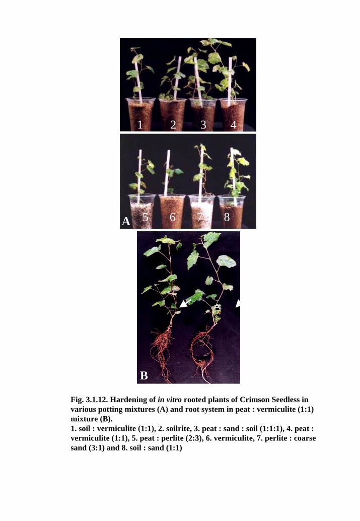
Fig. 3.1.12. Hardening of in vitro rooted plants of Crimson Seedless in various potting mixtures (A) and root system in peat : vermiculite (1:1) mixture (B).1. soil : vermiculite (1:1), 2. soilrite, 3. peat : sand : soil (1:1:1), 4. peat : vermiculite (1:1), 5. peat : perlite (2:3), 6. vermiculite, 7. perlite : coarse sand (3:1) and 8. soil : sand (1:1)
1 2 3 4
B
A
5 6 7 8A

: perlite followed by peat : vermiculite (94.8), and soil : vermiculite (92.3). Potting
mixtures had influence on growth parameters of plantlets i.e. shoot length, number of
roots per plantlet and length of roots. Length of shoots in different mixtures varied in the
range of 11.6 cm to 20.5 cm. The maximum average shoot length of 20.5 cm and the
highest average number of roots per plantlet (30.3) were observed in peat : vermiculite
(Table 3.1.12). Vermiculite alone gave only 40% survival rate, and required frequent
watering due to lack of water retention. In contrast to our results, Kamnoon and
Kantamaht (2000) obtained 100% plant survival in vermiculite while hardening shoot tip
derived plantlets of Maesa ramentace. While in another report, plantlets were
acclimatized initially in soilrite for 4 weeks (Mhatre et al. 2000). Survival of plantlets
depends on several factors including the number of primary and secondary roots and root
hairs in addition to root length. Average root length was lower in case of plantlets
transferred to either vermiculite or peat : perlite. A large number of primary and
secondary roots were observed in the plantlets transferred to peat : vermiculite mixture
(Fig. 3.1.12B). In vermiculite alone, plantlets required frequent watering; growth
remained stunted with light green foliage and pink coloration on stems. There are
innumerable reports in literature demonstrating a requirement of particular combination
of potting mixture for a plant species / cultivar in question. Thus, it can be inferred that
optimization of potting mixtures for different species/genotypes is an essential step in
micropropagation.
Table 3.1.12: Influence of different potting mixtures on growth and survival of in vitro propagated plantlets of Crimson Seedless.
Potting mixture Plantlet survival
(%)
Av. Shoot length (cm)
Av. no of roots per
plant
Av. root length (cm)
Soil : Vermiculite (1:1) 92.3 13.0 11.1 7.24 Soilrite 66.7 14.3 9.5 8.58 Peat : Sand : Soil (1:1:1) 84.6 10.7 16.3 7.17 Peat : Vermiculite (1:1) 94.8 20.5 30.3 7.20 Peat : Perlite (2:3) 97.4 14.4 26.7 4.71 Vermiculite 79.5 11.9 09.1 5.21 Perlite : Coarse sand (3:1) 87.2 11.6 14.1 4.44 Soil : Sand (1:1) 71.8 12.4 10.4 3.84 SEM± 1.8 0.4 0.9 0.22 C.D (p=0.01) 3.2 1.0 2.8 0.66 ** ** ** ** ** Significant at 1% level
63

Fig. 3.1.13. Hardening and acclimatization of in vitro raised plants. A: Hardening process, B: hardened plants and C: Hardened plants in green house of NRCG.
A
B
C

3.1.3.7. Hardening of plantlets
In vitro as well ex vitro rooted shoots and nodal segments with direct rooting
could be hardened successfully by the Sachet technique (Fig. 3.13). It was observed that
covering of plantlets with polythene sachets for minimum of 4 weeks was very crucial.
Though top corners of bags could be cut after 2 weeks, however, complete removal of
bags before 4 weeks caused scorching and drying of leaves. Hardening of plantlets in the
present study was carried out in continuous light at higher intensity. Such a light
treatment has been reported to increase the rates of photosynthesis and boost growth of in
vitro raised plantlets (Kozai et al., 1990).
A total of 509 hardened plants of Crimson Seedless were supplied to National
Research Centre for Grapes (NRCG), Pune for further field evaluation.
3.1.4. Conclusion
The present study demonstrates that Crimson seedless can successfully be
propagated in vitro by two routes. In one route, bud break and rooting was induced in
single node segments from field grown vines and whole plants were established on
potting. By this technique, hardened plantlets could be produced in 2 months. In second
route, axillary buds in primary or secondary nodal segments were induced multiples and
proliferated further in glass bottles instead of culture tubes. The multiple shoots could be
elongated, rooted in vitro or ex vitro and plants could be transferred to soil for hardening.
By second route, plant production could be increased by many fold. Shoots could also be
rooted ex vitro by pulse treatment of auxins. This circumvents one major in vitro stage
and cuts down the cost and time of production. Hardening of plantlets could be achieved
in a simple set up by the Sachet technique. The optimum conditions for in vitro
propagation described in the present paper, will have application in commercial
production of planting stock of this exotic grapevine cultivar Crimson Seedless.
Part of the work has been reported in the following publications: 1. Nookaraju, A., Barreto, S.M. and Agrawal, D.C (2007) Rapid in vitro
propagation of grapevine cv. Crimson Seedless - Influence of basal media and plant growth regulators. Journal of Applied Horticulture (In press).
2. Barreto, M.S., A. Nookaraju, A.M. Joglekar, G.S. Karibasappa and D.C. Agrawal (2007) Variability among Vitis vinifera cultivars to in vitro propagation. Acta Horticulturae (In press).
64
3.2. Molecular characterization of in vitro raised plants of Crimson Seedless
3.2.1. Introduction
Tissue culture environment may cause alterations in cellular controls, leading to
genomic changes in in vitro derived plants (Phillips et al., 1994). However, gross
morphological variations may occur at a much lower frequency than cryptic variations
(Evans et al. 1984). The occurrence of somaclonal and random genetic variations are
potential drawbacks, when the propagation of an elite tree is intended for the clonal
uniformity. On the other hand, stable somaclonal variation of a specific type may be of
advantage for the improvement of certain traits. In commercial industry, dealing with
micropropagation technology, the foremost concern is the maintenance of true-to-type
nature of the in vitro propagated plants. Beyond the influence of the culture conditions
(culture media, type of explant, culture duration, temperature, pH etc.), the occurrence
and extent of in vitro genetic instability may also be highly genotype specific (Meins
1983; Karp and Bright 1985; Vazquez 2001). The frequency of somaclonal variation was
found to be dependent on regeneration method and explant source. In general,
micropropagated plants obtained from pre-formed meristems have been reported to be
uniform (Ostry et al. 1994), however, possibility of somaclonal variations can not be
ruled out completely (Rani and Raina, 2000).
In earlier reports, variations among in vitro derived plants have been observed
with different molecular markers. Allozyme markers have been used for examining
uniformity and cryptic somaclonal variations, but these markers have limitation in
number and extent of polymorphism. Recently, DNA markers have attracted much
attention, since these are more informative and are developmentally regulated. Among the
DNA markers, Randomly Amplified Polymorphic DNA (RAPD) has serious limitation
due to its reliability and reproducibility. Moreover, RAPDs are dominant diallelic
markers; thus, individual parental alleles cannot usually be differentiated in diploid
organisms. The sensitivity, reproducibility, co-dominance and strong discriminatory
power of microsatellite DNA / SSR (Simple Sequence Repeats) markers or SSR-targeted
primers like ISSRs make them particularly suitable for assessment of uniformity and
detection of somaclonal variations (Rajora and Rehman, 2001). More over, ISSRs offer
greater probability than any other PCR marker system in the repeat regions of the
genome, which are reported to be the potent regions of genetic variations and this renders
ISSRs useful as a supplementary system to any of the random, dominant marker systems
65

(Pandit et al., 2007). Molecular markers can be used for characterization of somaclonal
variations with greater precision and less effort than phenotypic and karyologic analyses
(Cloutier and Landry, 1994).
Earlier, molecular markers have been successfully used for studying variation
among micropropagated plants of several crops. Studies on microsatellite markers in
trembling aspen (Rahman and Rajora 2001), AFLPs in pecan somatic embryos
(Vendrame et al. 1999), RAPD and ISSR in peach (Hashmi et al. 1997) and almonds
(Martins et al. 2004) and RAPD and cpDNA microsatellites in Foeniculum vulgare
(Bennici et al. 2004) have been reported.
Grape is a perennial crop and the morphological variation generated in tissue
culture could be visible only at the maturity and fruiting. Consequently, early detection of
variations among the tissue culture raised plants using molecular techniques may be
desirable. The aim of the present study was to check the genetic uniformity of in vitro
derived plants of Crimson Seedless using molecular markers. Two molecular marker
techniques used were ISSR and microsatellites as these two types of markers amplify
different regions on the non-coding sequences of the genome.
3.2.2. Materials and Methods
3.2.2.1. Collection and storage of leaf samples for DNA extraction
Young expanding light green leaves were collected from 24 randomly selected in
vitro propagated plants of Crimson Seedless maintained in green house of NRC for
Grapes and also from 12 mother vines of Crimson Seedless growing in the vineyard. Leaf
samples collected in microfuge tubes (1.5 ml) which were immediately placed in liquid
nitrogen. The leaf samples were stored at –70oC untill used for DNA extraction. The
selected plants were given serial numbers as CS-1 to CS-24 and mother plants as CSP-1
to CSP-12.
3.2.2.2. DNA extraction
Genomic DNA extraction was carried out according to the method reported by
Lodhi et al. (1994) with minor modifications.
1. About 100 mg of leaf tissue was ground into a fine powder in liquid nitrogen.
2. The powder was taken in a 1.5 ml microfuge tube and 1 ml of CTAB buffer and 10
mg of polyvinylpyrrolidone (Sigma P2307) were added to it.
3. The above mixture was incubated for 25 min at 65oC with occasional mixing.
4. The tubes were then taken out of water bath and cooled to room temperature.
66

5. Equal volume of chloroform: isoamylalcohol (24:1) was added to the mixture and
the tubes were inverted for 12 times and centrifuged at 10,000 rpm for 10 min.
6. The upper aqueous phase was retained in a fresh microfuge tube, to which 3 μl of
RNase A (10 mg/ml) was added and incubated at 37oC for 20 min.
7. Then equal volume of chloroform: isoamylalcohol was added to the above mixture
and the tubes were inverted for 12 times and centrifuged at 10,000 rpm for 5 min.
8. Again the upper aqueous phase was collected in a fresh tube and half the volume of
5 M NaCl and twice the volume of 95% (-20oC) ethanol were added. The contents
were mixed thoroughly by inversion several times and incubated on ice for 1 h.
9. Centrifugation was performed at 10,000 rpm for 8 min and the supernatant was
discarded by retaining the pellet.
10. The pellet was washed with 70% ethanol (-20oC) and air-dried.
11. The pellet was resuspended in 75 μl of TE buffer, pH 8.0.
Quantity of DNA was estimated by ultraviolet absorbence spectrophotometry by
absorption at 260 nm and the purity of the DNA was assessed with the ratios of
absorptions at 260 nm and 280 nm. Each DNA sample was diluted to 20 ng/μl in sterile
double distilled water and stored at 40C.
3.2.2.3. PCR Amplification
3.2.2.3.1. ISSR
As a result of a preliminary screening with 50 ISSR (UBC, Vancouver, USA)
primers, 22 primers (UBC 807–826, 873, 878 and 881) producing scorable bands were
selected for the study. The PCR reaction was performed in 25µl reaction mix containing
0.3U of Taq DNA polymerase (Genetix Ltd.) in 1X buffer, 2 mM MgCl2, 30 ng of
template DNA, 200 µM of each dNTPs (Genetix Ltd.) and 4.5–6 pmol primer. PCR was
performed using the following program: Initial denaturation at 94oC for 5 min, followed
by 35 cycles of denaturation at 94oC for 30 s, annealing at 52oC for 45 s, extension at
72oC for 2 min with a final extension at 72oC for 7 min. Amplifications were performed
in a mastercycler (Eppendorf, Germany) for both ISSR and microsatellites.
PCR products were visualized on 2% agarose gel stained with ethidium bromide
(0.5 μg/ml) and compared using a standard molecular weight marker λ / PstI (MBI
Fermentas Ltd.). Ethidium bromide stained gels were visualized under UV light and
photographed using a gel documentation system (Gel Doc 2000TM, Bio–Rad Lab. Inc.).
67
3.2.2.3.2. Microstellites / SSR
Five grape microsatellite primer pairs selected namely were, VS1, VVS2
(Thomas and Scott, 1993), VVMD5, VVMD31 (Bowers et al.,1996), VMCNG4c8
(Zyprian et al.,2003). The reaction was performed in 25µl reaction mix containing 0.3U
of Taq DNA polymerase in 1X buffer, 2 mM MgCl2, 30 ng of template DNA, 200 µM of
each dNTPs and 7.5 pmol. of each primer. PCR was performed using the following
program: Initial denaturation at 94oC for 5 min, followed by 35 cycles of denaturation at
94oC for 30 s, annealing at 45oC for VS1 and VVMD5, 48oC for VVS2 and VMCNG4c8,
50oC for VVMD31 for 45 s, extension at 72oC for 2 min with a final extension at 72oC
for 7 min. PCR products were visualized on 2% agarose gel stained with ethidium
bromide (0.5 μg/ml) and compared using a standard molecular weight marker 100 bp
ladder (MBI Fermentas Ltd.).
Each PCR experiment was repeated three times for repeatability of the banding
pattern and reliability of results. Each PCR experiment contained a negative control
consisting of a complete reaction mix, minus template DNA, to test for the presence of
possible non-specific amplification.
3.2.2.4 Data analysis
All the gels of ISSR and SSR-PCR were analyzed using Quantity One 1-D
Analysis software (Bio -Rad Laboratories Inc.) and the banding patterns were scored as
either present (1) or absent (0) of band per each primer. Genetic similarities between in
vitro raised plants and mother plants of Crimson Seedless were measured by the Dice
similarity co-efficient based on the proportion of shared alleles (Nei and Li, 1979).
Similarity estimate, D was calculated as 2Nab/ (Na + Nb) for each primer and also
collectively for all 22 primers.
Where Nab is the number of shared bands, Na is number of bands in sample a and
NB is the number of bands in sample b. B
The genetic similarity matrix was generated using the Windist software option of
the Winboot package (Yap et al., 1996) with Dice co-efficiencies. For calculation and
generation similarity matrix, only one mother plant was considered as all the mother
plants showed similar banding pattern with all the primers.
68

3.2.3. Results and Discussion
Visual assessment of 6-12 month old in vitro raised plants of Crimson Seedless
growing in the polyhouse did not show any morphological differences. As reported in
literature, clonal uniformity based on morphological and phenological traits is not precise,
and the same can be assessed by using recently developed molecular markers. Number of
reports in the field is increasing at an amazing rate. In the present study, we decided to
check if there is any change in micropropagated plants of Crimson Seedless at molecular
level using a few DNA markers. Our initial attempts with RAPD primers did not show
any polymorphism and there was a problem of reproducibility of results, therefore we
restricted this study to ISSR and SSR markers only.
DNA extraction followed in the present study permitted to obtain a good quality
of DNA for the PCR experiments. The quantity of DNA obtained was adequate for ISSR
and SSR analyses. Also the conditions of PCR amplification such as concentrations of
magnesium chloride, Tag DNA polymerase, Template DNA, primer and annealing
temperature were optimized to have sharp bands with high intensity without shearing
effect. Moreover, the amplifications were reproducible when PCR was repeated for each
primer.
3.2.3.1. ISSR
From 22 ISSR primers used in the present study, a total of 3152 scorable bands
(number of plants analyzed x number of band classes with all the primers) were obtained.
The number of bands per each primer ranged from 2 to 16 with an average of 6.01 bands
per primer. The band sizes ranged from 350 bp to 4005 bp. Among the 134 distinct band
classes recorded, 130 (97%) were monomorphic for all the clones and mother plants and
4 (3%) bands were polymorphic for 5 clones (Table 3.2.1). All these four polymorphic
bands were generated by one ISSR primer UBC 825.
69

Fig. 3.2.1. ISSR-PCR profiles of mother vines with UBC 812 (A) and in vitroraised plants of Crimson Seedless with 817 (B) and 823 (C).C – No template control, M –λ/pstI digest
A
B
C
M C 1 2 3 4 5 6 7 8 9 10 11 12
5077
805
514
C
C

Table 3.2.1: List of ISSR primers used in the study and number of band classes generated ISSR primer Primer sequence No. of distinct
bands generated %
similarity Band sizes
(range in bp) UBC 807 (AG)8T 4 100 520-1500
UBC 808 (AG)8C 8 100 700-1250
UBC 809 (AG)8G 7 100 350-1440
UBC 810 (GA)8T 6 100 400-2000
UBC 811 (GA)8C 4 100 750-950
UBC 812 (GA)8A 8 100 600-3000
UBC 813 (CT)8T 4 100 620-3500
UBC 815 (CT)8G 4 100 850-3500
UBC 816 (CA)8T 3 100 1100-1700
UBC 817 (CA)8A 7 100 560-2000
UBC 818 (CA)8G 8 100 800-1400
UBC 819 (GT)8A 5 100 900-5000
UBC 820 (GT)8C 3 100 1400-1500
UBC 821 (GT)8T 3 100 1600-2600
UBC 822 (TC)8A 6 100 700-2200
UBC 823 (TC)8C 4 100 1300-1800
UBC 824 (TC)8G 2 100 910-1600
UBC 825 (AC)8T 9 55.5 440-4005
UBC 826 (AC)8C 7 100 810-2440
UBC 873 (GACA)4 16 100 470-2500
UBC 878 (GGAT)4 8 100 650-2550
UBC 881 GGGT(GGGGT)2G 8 100 800-2500
PCR profiles of the mother plants for all the ISSR primers showed no
polymorphism (Fig. 3.2.1A). Among the primers, the primer UBC 825 showed minor
variations, while all other primers showed uniformity among the in vitro propagated
plants (Fig. 3.2.1B,C). Primer UBC 825 showed different banding patterns among the
plants numbered CS-7, CS-9, CS-19, CS-23 and CS-24 (Fig. 3.2.2A). The results show
that 4005 bp, 2947 bp, 1207 bp, 920 bp and 650 bp size bands were common to all the
plants. Where as, 1731bp band was present in all the plants except in CS-9, CS-23 and
CS-24; 1115 bp band was present only in CS-7, CS-9, CS-23 and CS-24; 1077 bp band is
70

present only in CS-9, CS-23 and CS-24 and 440 bp band is present only in CS-9, CS-19,
CS-23 and CS-24 (Table 3.2.2). From the results, it seems that there are four different
groups of plants. Plants numbered CS-9, CS-23 and CS-24 are similar, while CS-7 and
CS-19 are different from each other and rest. Remaining 19 plants have shown identical
banding among them and as that of mother.
Table 3.2.2: Presence of polymorphic ISSR bands in 24 in vitro raised plants of Crimson Seedless with primer UBC 825
ISSR bands Plant number 4005bp 2947bp 1731bp 1207bp 1115bp 1077bp 920bp 650bp 440bp CS-1 1 1 1 1 0 0 1 1 0 CS-2 1 1 1 1 0 0 1 1 0 CS-3 1 1 1 1 0 0 1 1 0 CS-4 1 1 1 1 0 0 1 1 0 CS-5 1 1 1 1 0 0 1 1 0 CS-6 1 1 1 1 0 0 1 1 0 CS-7 1 1 1 1 1 0 1 1 0 CS-8 1 1 1 1 0 0 1 1 0 CS-9 1 1 0 1 1 1 1 1 1 CS-10 1 1 1 1 0 0 1 1 0 CS-11 1 1 1 1 0 0 1 1 0 CS-12 1 1 1 1 0 0 1 1 0 CS-13 1 1 1 1 0 0 1 1 0 CS-14 1 1 1 1 0 0 1 1 0 CS-15 1 1 1 1 0 0 1 1 0 CS-16 1 1 1 1 0 0 1 1 0 CS-17 1 1 1 1 0 0 1 1 0 CS-18 1 1 1 1 0 0 1 1 0 CS-19 1 1 1 1 0 0 1 1 1 CS-20 1 1 1 1 0 0 1 1 0 CS-21 1 1 1 1 0 0 1 1 0 CS-22 1 1 1 1 0 0 1 1 0 CS-23 1 1 0 1 1 1 1 1 1 CS-24 1 1 0 1 1 1 1 1 1 Mother plants
1 1 1 1 0 0 1 1 0
1- presence; 0- absence of band
Indices of similarities among in vitro raised plants of Crimson Seedless are
depicted by the pair wise matrix of genetic similarities shown in Fig. 3.2.3. Estimation of
genetic similarity co-efficient based on ISSR band data indicated that out of 24 in vitro
raised plants selected, 19 plants were 100% identical to each other and to mother plants
and remaining 5 regenerated plants have shown more than 98% similarity to the mother
plants. Genetic similarity indexes among all the individuals ranged from 0.984 to 1.000,
71

M C 1 2 3 4 5 6 7 8 9 10 11 12 13 14 15 16 17 18 19 20 21 22 23 24
Fig. 3.2.2. ISSR-PCR profiles of in vitro raised plants (A) and the mother plants (B) of Crimson Seedless with primer UBC 825. C – No template control, M – λ/pstI digest
M C 1 2 3 4 5 6 7 8 9 10 11 12
4507
1159805
4507
1159
805
A
B

with a mean of 0.997. Plants numbered CS-7 and CS-19 had 0.996 and CS-9, CS-23 and
CS-24 had 0.984 similarity to mother plants (Fig. 3.2.3), which suggests the reliability of
the protocol used for in vitro propagation of the grapevine cultivar Crimson Seedless.
Interestingly, for the same primer (UBC 825), the DNAs of mother plants
growing at vineyard of NRCG did not show any polymorphism (Fig. 3.2.2B). So it can be
inferred that the minor molecular variations were not from the mother plants and could be
a result of in vitro culture. Larkin and Scowcroft (1981) reported that the use of synthetic
growth regulators for callus production and long-term culture tend to produce genetic as
well as epigenic variation in many plant species. While Martins et al. (2004) on testing of
10 ISSR primers found absolute genetic uniformity without any somaclonal variation
among 22 selected almond tissue culture plants derived from the axillary branching.
In the present study with ISSR markers, two types of polymorphism appeared to
be present i.e., fragment size differences and the absence of a fragment (null phenotype).
Difference in the band intensity was also observed. The presence of variations (as
revealed by ISSR-PCR) among the morphologically indistinct plants indicates that visible
evaluation may not be sensitive enough to detect minor variations at DNA level (Rahman
and Rajora, 2001). This also suggests that the results obtained by molecular markers may
be quite different from those obtained by using morphological characterization
(Ramanatha Rao and Oliveira, 2002).
72

1 2 3 4 5 6 7 8 9 10 11 12 13 14 15 16 17 18 19 20 21 22 23 24 Mother plant
_________________________________________________________________________________________________________________________________________________________________________________________
1 1.000000
2 1.000000 1.000000
3 1.000000 1.000000 1.000000
4 1.000000 1.000000 1.000000 1.000000
5 1.000000 1.000000 1.000000 1.000000 1.000000
6 1.000000 1.000000 1.000000 1.000000 1.000000 1.000000
7 0.996198 0.996198 0.996198 0.996198 0.996198 0.996198 1.000000
8 1.000000 1.000000 1.000000 1.000000 1.000000 1.000000 0.996198 1.000000
9 0.984848 0.984848 0.984848 0.984848 0.984848 0.984848 0.988679 0.984848 1.000000
10 1.000000 1.000000 1.000000 1.000000 1.000000 1.000000 0.996198 1.000000 0.984848 1.000000
11 1.000000 1.000000 1.000000 1.000000 1.000000 1.000000 0.996198 1.000000 0.984848 1.000000 1.000000
12 1.000000 1.000000 1.000000 1.000000 1.000000 1.000000 0.996198 1.000000 0.984848 1.000000 1.000000 1.000000
13 1.000000 1.000000 1.000000 1.000000 1.000000 1.000000 0.996198 1.000000 0.984848 1.000000 1.000000 1.000000 1.000000
14 1.000000 1.000000 1.000000 1.000000 1.000000 1.000000 0.996198 1.000000 0.984848 1.000000 1.000000 1.000000 1.000000 1.000000
15 1.000000 1.000000 1.000000 1.000000 1.000000 1.000000 0.996198 1.000000 0.984848 1.000000 1.000000 1.000000 1.000000 1.000000 1.000000
16 1.000000 1.000000 1.000000 1.000000 1.000000 1.000000 0.996198 1.000000 0.984848 1.000000 1.000000 1.000000 1.000000 1.000000 1.000000 1.000000
17 1.000000 1.000000 1.000000 1.000000 1.000000 1.000000 0.996198 1.000000 0.984848 1.000000 1.000000 1.000000 1.000000 1.000000 1.000000 1.000000 1.000000
18 1.000000 1.000000 1.000000 1.000000 1.000000 1.000000 0.996198 1.000000 0.984848 1.000000 1.000000 1.000000 1.000000 1.000000 1.000000 1.000000 1.000000 1.000000
19 0.996198 0.996198 0.996198 0.996198 0.996198 0.996198 0.992424 0.996198 0.988679 0.996198 0.996198 0.996198 0.996198 0.996198 0.996198 0.996198 0.996198 0.996198 1.000000
20 1.000000 1.000000 1.000000 1.000000 1.000000 1.000000 0.996198 1.000000 0.984848 1.000000 1.000000 1.000000 1.000000 1.000000 1.000000 1.000000 1.000000 1.000000 0.996198 1.000000
21 1.000000 1.000000 1.000000 1.000000 1.000000 1.000000 0.996198 1.000000 0.984848 1.000000 1.000000 1.000000 1.000000 1.000000 1.000000 1.000000 1.000000 1.000000 0.996198 1.000000 1.000000
22 1.000000 1.000000 1.000000 1.000000 1.000000 1.000000 0.996198 1.000000 0.984848 1.000000 1.000000 1.000000 1.000000 1.000000 1.000000 1.000000 1.000000 1.000000 0.996198 1.000000 1.000000 1.000000
23 0.984848 0.984848 0.984848 0.984848 0.984848 0.984848 0.988679 0.984848 1.000000 0.984848 0.984848 0.984848 0.984848 0.984848 0.984848 0.984848 0.984848 0.984848 0.988679 0.984848 0.984848 0.984848 1.000000
24 0.984848 0.984848 0.984848 0.984848 0.984848 0.984848 0.988679 0.984848 1.000000 0.984848 0.984848 0.984848 0.984848 0.984848 0.984848 0.984848 0.984848 0.984848 0.988679 0.984848 0.984848 0.984848 1.000000 1.000000
Mother plant 1.000000 1.000000 1.000000 1.000000 1.000000 1.000000 0.996198 1.000000 0.984848 1.000000 1.000000 1.000000 1.000000 1.000000 1.000000 1.000000 1.000000 1.000000 0.996198 1.000000 1.000000 1.000000 0.984848 0.984848 1.000000 _________________________________________________________________________________________________________________________
Fig. 3.2.3. Matrix of similarities for ISSR markers for in vitro raised plants and mother plants of Crimson Seedless
73
3.2.3.2. SSR
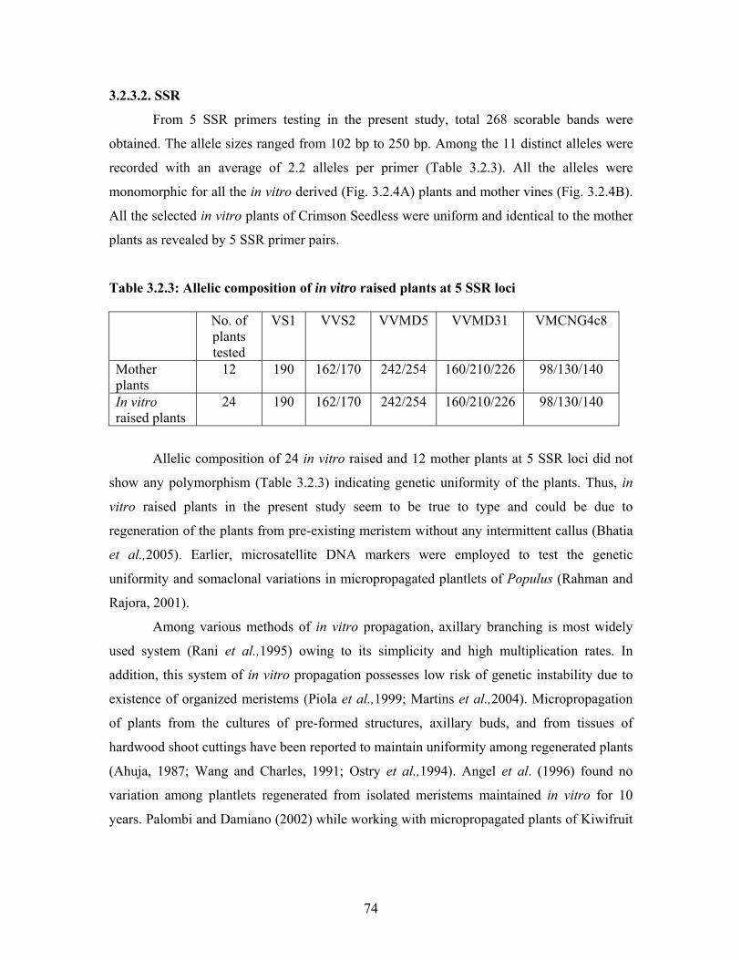
From 5 SSR primers testing in the present study, total 268 scorable bands were
obtained. The allele sizes ranged from 102 bp to 250 bp. Among the 11 distinct alleles were
recorded with an average of 2.2 alleles per primer (Table 3.2.3). All the alleles were
monomorphic for all the in vitro derived (Fig. 3.2.4A) plants and mother vines (Fig. 3.2.4B).
All the selected in vitro plants of Crimson Seedless were uniform and identical to the mother
plants as revealed by 5 SSR primer pairs.
Table 3.2.3: Allelic composition of in vitro raised plants at 5 SSR loci No. of
plants tested
VS1 VVS2 VVMD5 VVMD31 VMCNG4c8
Mother plants
12 190 162/170 242/254 160/210/226 98/130/140
In vitro raised plants
24 190 162/170 242/254 160/210/226 98/130/140
Allelic composition of 24 in vitro raised and 12 mother plants at 5 SSR loci did not
show any polymorphism (Table 3.2.3) indicating genetic uniformity of the plants. Thus, in
vitro raised plants in the present study seem to be true to type and could be due to
regeneration of the plants from pre-existing meristem without any intermittent callus (Bhatia
et al.,2005). Earlier, microsatellite DNA markers were employed to test the genetic
uniformity and somaclonal variations in micropropagated plantlets of Populus (Rahman and
Rajora, 2001).
Among various methods of in vitro propagation, axillary branching is most widely
used system (Rani et al.,1995) owing to its simplicity and high multiplication rates. In
addition, this system of in vitro propagation possesses low risk of genetic instability due to
existence of organized meristems (Piola et al.,1999; Martins et al.,2004). Micropropagation
of plants from the cultures of pre-formed structures, axillary buds, and from tissues of
hardwood shoot cuttings have been reported to maintain uniformity among regenerated plants
(Ahuja, 1987; Wang and Charles, 1991; Ostry et al.,1994). Angel et al. (1996) found no
variation among plantlets regenerated from isolated meristems maintained in vitro for 10
years. Palombi and Damiano (2002) while working with micropropagated plants of Kiwifruit
74
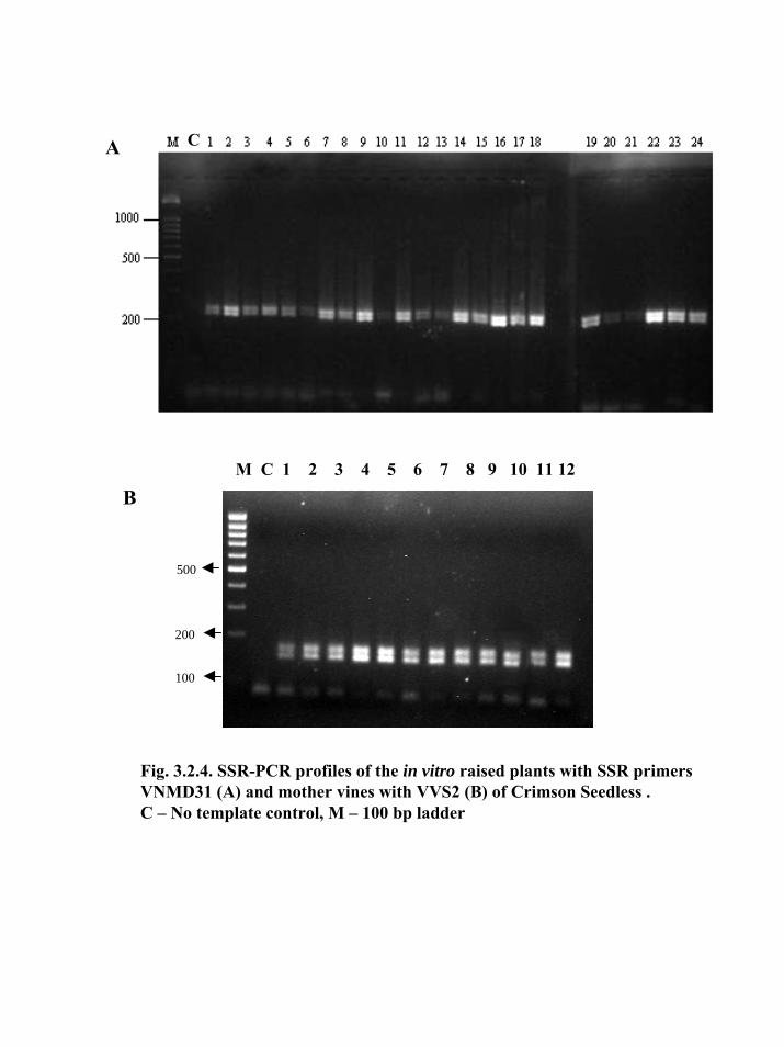
Fig. 3.2.4. SSR-PCR profiles of the in vitro raised plants with SSR primers VNMD31 (A) and mother vines with VVS2 (B) of Crimson Seedless .C – No template control, M – 100 bp ladder
A
BM C 1 2 3 4 5 6 7 8 9 10 11 12
500
200
100
C

revealed that the use of two or more marker techniques is advantageous for evaluation of
genetic uniformity and somaclonal variations.
The number of ISSR and SSR primers used in the study and the total number of
markers generated (134+11), together with absence of any notable morphological differences
in the in vitro raised plants demonstrate the reliability of our procedure.
3.2.4. Conclusion
The present study on the molecular characterization of in vitro raised plants revealed
the uniformity among the plants as assessed with 21 ISSR and 5 SSR primers. Only one
primer UBC 825 showed minor variations in the banding patterns among five in vitro raised
plants of Crimson Seedless, which could be due to minor genetic variations which occur
during cell division and differentiation under in vitro conditions. As assessed by ISSR, all the
in vitro raised plants showed more than 99% similarity with the mother plants. Thus, present
study indicates that ISSR and SSR markers were useful and sensitive markers for
determining the genetic uniformity and detecting variations among tissue culture raised
plants of Crimson Seedless and demonstrate the reliability of our in vitro propagation
procedure.
75