Chapter 3 Construction of scFv Fragments from Hybridoma or ... · Chapter 3 Construction of scFv...
24
Chapter 3 Construction of scFv Fragments from Hybridoma or Spleen Cells by PCR Assembly Jonas V. Schaefer, Annemarie Honegger, and Andreas Plu ¨ ckthun Abbreviations BSA Bovine serum albumin DMSO Dimethylsulfoxide HRP Horse radish peroxidase IPTG Isopropylthiogalactoside PEG Polyethylene glycol PBS Phosphate buffered saline scFv Single-chain Fv fragment cfu Colony forming units tet Tetracycline 3.1 Introduction Today, antibodies can be obtained from naive repertoires (Winter et al. 1994; Vaughan et al. 1996) or libraries of fully synthetic genes (Knappik et al. 2000), and in the last decade, numerous libraries have been described (reviewed in Mondon et al. 2008). Nonetheless, hybridomas have remained the predominant source of antibodies, and a wealth of well characterized and even unique clones exist and are continuing to be generated. There is, thus, great interest in immorta- lizing these clones, in the extreme case, as a computer file of the sequences, as well as in accessing the antibody in a variety of new formats. To obtain enough material J.V. Schaefer, A. Honegger, and A. Plu ¨ckthun (*) Biochemisches Institut, Universita ¨t Zu ¨rich, Winterthurerstr. 190, 8057 Zu ¨rich, Switzerland e-mail: [email protected] R. Kontermann and S. Du ¨bel (eds.), Antibody Engineering Vol. 1, DOI 10.1007/978-3-642-01144-3_3, # Springer-Verlag Berlin Heidelberg 2010 21
Transcript of Chapter 3 Construction of scFv Fragments from Hybridoma or ... · Chapter 3 Construction of scFv...
Chapter 3
Construction of scFv Fragments
from Hybridoma or Spleen Cells
by PCR Assembly
Jonas V. Schaefer, Annemarie Honegger, and Andreas Pluckthun
Abbreviations
BSA Bovine serum albumin
DMSO Dimethylsulfoxide
HRP Horse radish peroxidase
IPTG Isopropylthiogalactoside
PEG Polyethylene glycol
PBS Phosphate buffered saline
scFv Single-chain Fv fragment
cfu Colony forming units
tet Tetracycline
3.1 Introduction
Today, antibodies can be obtained from naive repertoires (Winter et al. 1994;
Vaughan et al. 1996) or libraries of fully synthetic genes (Knappik et al. 2000),
and in the last decade, numerous libraries have been described (reviewed in
Mondon et al. 2008). Nonetheless, hybridomas have remained the predominant
source of antibodies, and a wealth of well characterized and even unique clones
exist and are continuing to be generated. There is, thus, great interest in immorta-
lizing these clones, in the extreme case, as a computer file of the sequences, as well
as in accessing the antibody in a variety of new formats. To obtain enough material
J.V. Schaefer, A. Honegger, and A. Pluckthun (*)
Biochemisches Institut, Universitat Zurich, Winterthurerstr. 190, 8057 Zurich, Switzerland
e-mail: [email protected]
R. Kontermann and S. Dubel (eds.), Antibody Engineering Vol. 1,DOI 10.1007/978-3-642-01144-3_3, # Springer-Verlag Berlin Heidelberg 2010
21
pvogt
Text Box
Reprinted from: J.V. Schaefer, A. Honegger, and A. Plückthun: Construction of scFV Fragments from Hybridoma of Spleen Cells by PCR Assembly. In: Antibody Engineering, R. Kontermann and S. Dübel, Springer Verlag, Heidelberg, Germany (2010) pp. 21-44
for detailed biochemical and biophysical analyses of the deduced antibodies after
immunization, their cloning into formats compatible with recombinant expression
is beneficial, if not essential. For this purpose, the antibody genes must be cloned,
and the binding properties of the recombinant protein have to be verified. In
addition to existing hybridomas, the immune response of an animal upon exposure
to various antigens may often be of particular scientific interest in itself and also
lead to the discovery of new and potent binders. Therefore, there is merit in
immortalizing the results from new immunizations as well. In this case, it is not
necessary to take the detour of first making hybridomas, but instead, mRNA
isolated from spleen can be directly used for the creation of an immune library,
from which binders can be subsequently isolated by phage display and their
sequences determined.
Once the antibody genes have been successfully cloned and after the presumed
binding properties of the recombinant antibodies have been experimentally verified,
their sequences can be used for modeling (www.bioc.uzh.ch/antibody/), and their
structure subsequently be determined by crystallography (Honegger et al. 2005) or
NMR (Freund et al. 1994; Tugarinov et al. 2000). The recombinant single-chain Fv
format (Huston et al. 1988; Glockshuber et al. 1990) is an ideal starting point for all
engineering efforts, from sensors (Backmann et al. 2005; Morfill et al. 2007) to
therapeutic fusion proteins (Di Paolo et al. 2003), or imaging reagents (Adams et al.
1993) to multivalent and multispecific reagents (Pluckthun and Pack 1997), just to
name a few illustrative examples. Recombinant expression of these proteins also
allows one to evolve the affinity further than the immune system normally does,
e.g., to low picomolar KD for scFv fragments (Zahnd et al. 2004; Luginbuhl et al.
2006). Finally, some natural antibodies may not be of sufficient stability, which can
also be corrected by engineering (Worn and Pluckthun 2001; Ewert et al. 2004). In
addition, the murine antibody can be humanized for its use in therapy – a procedure
rapidly achievable at the scFv stage.
The key prerequisite for the use of recombinant antibody technologies, starting
from immune repertoires or defined hybridomas, is the reliable cloning of func-
tional immunoglobulin genes. Even though hybridomas are considered to express
“monoclonal” antibodies, hybridoma clones may encode more than one functional
or even nonfunctional heavy or light chains (Kutemeier et al. 1992). As has
been reported previously, several kappa chain-secreting hybridomas, possessing
X63Ag8.653 myeloma cells as fusion partner, also occasionally transcribe a func-
tional lambda chain, competing with the Vk gene for in-frame scFv antibody
assembly (Krebber et al. 1997). As these false or heterogeneous genes might also
be amplified and subsequently assembled into the scFv fragments, it is highly
recommended to include an enrichment procedure in the cloning protocol. This
step can be circumvented and replaced by screening of clones at the scFv level, but
the phage enrichment is generally much faster if incorrect sequences abound.
Obviously, selection by phage display or by another selection technology such as
ribosome display (Hanes and Pluckthun 1997; Hanes et al. 1998; for detailed
protocols see Schaffitzel et al. 2005; Amstutz et al. 2006; Zahnd et al. 2007) is
mandatory when starting from spleens of immunized mice.
22 J.V. Schaefer et al.

This chapter largely follows our earlier protocols (Pluckthun et al. 1996; Krebber
et al. 1997; Burmester and Pluckthun 2001). A number of variable antibody
domains of hybridomas were accessible with those procedures and reagents
whose genes could not be cloned in other experimental setups. The present protocol
is based on a standard phage display system, which was optimized for robustness,
vector stability, and directional cloning using a single rare cutting restriction
enzyme as well as tight control of the expression of the scFv-gene III fusion
(Krebber et al. 1997). As the procedures for the construction of scFv fragment
libraries from immunized mice and that of cloning one specific antibody from
hybridomas are essentially the same, we combined them in just one protocol.
However, there are slight differences in the initial preparation of the cells, and
high ligation and transformation yields for library cloning are, of course, essential,
as explained under “notes.”
The current version of this protocol contains improvements in the methods but,
most importantly, newly designed primer sequences for the amplification of VH and
VL genes. They are based on our analysis of a reference set of murine germline
sequences found in the most recent version of the IMGT database (http://imgt.cines.
fr/textes/vquest/refseqh.html), which thus incorporates most of the knowledge of
the mouse genome (for a description of the original database, see Lefranc and
Lefranc 2001). Our key criterion was a faithful amplification of the variable region
genes preserving as much sequence identity as possible, avoiding the generation of
nonnatural residue combinations, which could result in sequences problematic for
folding and stability (Honegger and Pluckthun 2001; Jung et al. 2001). We also
tried to ensure similar annealing temperatures with the different genes, as well as
keeping the degeneracy on the DNA level as small as possible. Furthermore, we
avoided pronounced secondary structures within the oligonucleotides such as
hairpin loops or primer-dimers (which were checked against themselves using the
appropriate analysis tools in the Vector NTI software (Invitrogen)). The primers
shown below are the result of this iterative process and have also been tested with a
slightly different overhang.
The cloning strategy outlined in this protocol (Fig. 3.1) allows the simple
conversion of the expression format from the initial scFv fragments to other formats
and fusion proteins. Insertion of the assembled scFv gene into the described
standard vectors pAK100 and pJB12 leads to the expression of a scFv-gene III
fusion applicable for phage display, due to read-through of the amber codons
whenever expressed in strains with amber suppressor tRNA such as Escherichiacoli XL1-Blue. In bacterial strains lacking such suppressor tRNA, the amber stop
codons result in translation termination and production of unfused scFv fragments.
For purposes of IMAC purification or whenever other fusions will be constructed, it
is, however, advantageous to reclone the fragments directly into appropriate vectors
(Figs. 3.4 and 3.5) (Pluckthun et al. 1996), carrying stronger translation initiation
sites. Conversely, it is not advantageous to make expression too strong for phage
display, as discussed below. Although not explicitly mentioned, a very similar
strategy of cloning (Fig. 3.1) only requiring altered reverse primers can be used
for the design of Fab versions of the desired antibodies.
3 Construction of scFv Fragments from Hybridoma or Spleen Cells by PCR Assembly 23

light chain mRNAAAAAAn
pd(N)6
heavy chain mRNAAAAAAn
pd(N)6
2. Synthesis of cDNA
1. Isolation of mRNA (from hybridoma or spleen cells)
7. Transformation of E. coli XL1-Blue cells with final phage display vector, cell propagation and infection with VCSM13 helper phage
antigen
antigen
antigen
scFvphage particle withphagemid ss-DNA
8. Enrichment and detection of binding scFv sequences by phage display
6. Ligation of Sfi l digested insert and phage display vector (pAK100 or pJB12)
VLFLAGs VHpelB l inker *
DYKD (G4S)4
5. Sfi l digestion of the assembled and amplified scFv fragment
TCGGCCGG
CGGCC GGCCTCGG
CCGGAVLFLAGs VHl inker
light chain cDNA
VLrev mix
heavy chain cDNAVHfor mix
VHrev mix
VLfor mix
3. PCR amplification of VL and VH domains
outer-rev
outer-for
4. Assembly of VL and VH by SOE-PCR (splicing by overlap extension)
VH
VLFLAGs
g I I I 250-406
Fig. 3.1 Schematic overview of the amplification and cloning procedure. After its isolation from
hybridoma or spleen cells, the mRNA provides the basis for cDNA synthesis, utilizing random
hexamer primers. The cDNA is used afterward as template for PCR amplification of VL and VH
domains (symbolized by the gray boxes, not drawn to scale), which are subsequently assembled by
SOE-PCR into the scFv format by the outer primer pair outer-for and outer-rev. For antibody
cloning into the phagemid, only the rare cutting enzyme SfiI is used, guaranteeing directional
cloning due to the resulting different overhangs at the cleavage site as indicated. In addition, self-
ligation of insert or vector molecules is excluded by the asymmetry generated in the cut vector.
FLAGs indicates the shortened N-terminal 4-amino acid FLAG tag (Knappik and Pluckthun 1994)
24 J.V. Schaefer et al.

3.2 Materials
l 5 � 106 cells from a growing or frozen hybridoma culture or spleen cells,
respectivelyl PCR primers (Figs. 3.2 and 3.3) and corresponding plasmids (Figs. 3.4
and 3.5)l Helper phage (e.g., Stratagene VCSM13 # 200251)l F+, supE, recA strain (e.g., E. coli XL1-Blue) (available in electrocompetent/
chemocompetent form from Stratagene)l Anti-M13 antibody HRP-conjugate (GE Healthcare; # 27-9421-01)l PEG 6000 (Fluka)l Sterile, RNase-free equipment: pipet tips, tubes, RNase-free ultra high purity
(UHP) water, baked nondisposable glassware, and sterile, disposable plasticwarel Standard molecular biology equipment and reagents for:
– Determining the isotype of mAbs (Roche IsoStrip Mouse Monoclonal Anti-
body Isotyping Kit)
– Purifying RNA (Invitrogen TRIzol reagent and Qiagen RNeasy Mini Kit)
– Performing a cDNA synthesis reaction (Qiagen QuantiTect Reverse Tran-
scription Kit)
– Performing PCR reactions
– Purifying PCR products (Macherey Nagel PCR clean-up Gel Extraction Kit)
– Cutting and gel-purifying DNA (Sigma-Aldrich GenElute Gel Extraction
Kit)
– Concentrating DNA (Amicon Microcon 30 for volumes less than 500 ml)– Ligating and transforming DNA
– Growing bacteria and phages
– Conducting an Enzyme Linked Immunosorbent Assay (ELISA)
– Performing sodium dodecyl sulfate-polyacrylamide gel electrophoresis
(SDS-PAGE) and subsequent immunoblotting.
Fig. 3.1 (continued) and the asterisk symbolizes either the myc tag or the trypsin cleavage site,
present in pAK100 and pJB12, respectively. After infection with VCSM13 helper phages, the
transformed XL1-Blue cells produce phages, displaying the scFv antibody on their surface. The
subsequent enrichment of these phages by panning against the antigen allows the selection of
functional antibody sequences from the library generated from the spleen cells. In addition, this
approach also supports the isolation of specific scFv fragments if the hybridoma cell line initially
contained only a small fraction of mRNA coding for this particular antibody. Phage ELISA then
identifies the antigen-binding clones. Subsequently, the binding properties of the unfused scFv in
the absence of phage need to be verified after recloning into a more powerful expression vector
(Figs. 3.4 and 3.5) and purification from E. coli (not shown in the diagram)
<
3 Construction of scFv Fragments from Hybridoma or Spleen Cells by PCR Assembly 25
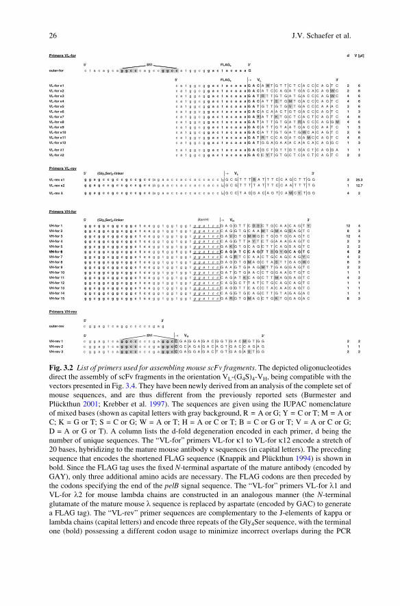
Primers VL-for d V [µl]
5' Sfi GALFI s 3'
outer-for c t a c a g c a g g c c c a g c c g g c c a t g g c g g a c t a c a a a G
GALF'5 s ® VL 3'
VL-for 1 c a t g g c g g a c t a c a a a G A C A W T G T T C T C A C C C A G T C 2 6
VL-for 2 c a t g g c g g a c t a c a a a G A C A T C C A G A T G A C A C A G W C 2 6
VL-for 3 c a t g g c g g a c t a c a a a G A T R T T G T G A T G A C C C A G W C 4 6
VL-for 4 c a t g g c g g a c t a c a a a G A C A T T S T G M T G A C C C A G T C 4 6
VL-for 5 c a t g g c g g a c t a c a a a G A T G T T G T G V T G A C C C A A A C 3 6
VL-for 6 c a t g g c g g a c t a c a a a G A C A C A A C T G T G A C C C A G T C 1 3
VL-for 7 c a t g g c g g a c t a c a a a G A Y A T T K T G C T C A C T C A G T C 4 6
VL-for 8 c a t g g c g g a c t a c a a a G A T A T T G T G A T R A C C C A G G M 4 6
VL-for 9 c a t g g c g g a c t a c a a a G A C A T T G T A A T G A C C C A A T C 1 3
VL-for 10 c a t g g c g g a c t a c a a a G A C A T T G T G A T G W C A C A G T C 2 6
VL-for 11 c a t g g c g g a c t a c a a a G A T R T C C A G A T G A M C C A G T C 4 6
VL-for 12 c a t g g c g g a c t a c a a a G A T G G A G A A A C A A C A C A G G C 1 3
VL-for 1 c a t g g c g g a c t a c a a a G A C G C T G T T G T G A C T C A G G A 1 1
VL-for 2 c a t g g c g g a c t a c a a a G A C C Y T G T G C T C A C T C A G T C 2 2
Primers VL-rev5' (Gly4Ser)3-linker ® VL 3'
VL-rev 1 g g a g c c g c c g c c g c c (a g a a c c a c c a c c a c c )2 G C G T T T B A T T T C C A G C T T G G 3 25.3
VL-rev 2 g g a g c c g c c g c c g c c (a g a a c c a c c a c c a c c )2 G C G T T T T A T T T C C A A T T T T G 1 12.7
VL-rev g g a g c c g c c g c c g c c (a g a a c c a c c a c c a c c )2 G C C T A G G A C A G T C A M C Y T G G 4 2
Primers VH-for
5' (Gly4Ser)2-linker BamHI ® VH 3'
VH-for 1 g g c g g c g g c g g c t c c g g t g g t g g t g g a t c c G A G G T T C D S C T G C A A C A G T Y 12 4
VH-for 2 g g c g g c g g c g g c t c c g g t g g t g g t g g a t c c C A G G T G C A A M T G M A G S A G T C 8 3
VH-for 3 g g c g g c g g c g g c t c c g g t g g t g g t g g a t c c G A V G T G M W G C T G G T G G A G T C 12 4
VH-for 4 g g c g g c g g c g g c t c c g g t g g t g g t g g a t c c C A G G T T A Y T C T G A A A G A G T C 2 2
VH-for 5 g g c g g c g g c g g c t c c g g t g g t g g t g g a t c c G A K G T G C A G C T T C A G S A G T C 2 2
VH f 6 t t t t t C A G A T C C A G T T S G Y G C A G T C 4 2VH-for 6 g g c g g c g g c g g c t c c g g t g g t g g t g g a t c c C A G A T C C A G T T S G Y G C A G T C 4 2
VH-for 7 g g c g g c g g c g g c t c c g g t g g t g g t g g a t c c C A G R T C C A A C T G C A G C A G Y C 4 2
VH-for 8 g g c g g c g g c g g c t c c g g t g g t g g t g g a t c c G A G G T G M A G C T A S T T G A G W C 8 3
VH-for 9 g g c g g c g g c g g c t c c g g t g g t g g t g g a t c c G A A G T G A A G M T T G A G G A G T C 2 2
VH-for 10 g g c g g c g g c g g c t c c g g t g g t g g t g g a t c c G A T G T G A A C C T G G A A G T G T C 1 1
VH-for 11 g g c g g c g g c g g c t c c g g t g g t g g t g g a t c c C A G A T K C A G C T T M A G G A G T C 4 2
VH-for 12 g g c g g c g g c g g c t c c g g t g g t g g t g g a t c c C A G G C T T A T C T G C A G C A G T C 1 1
VH-for 13 g g c g g c g g c g g c t c c g g t g g t g g t g g a t c c C A G G T T C A C C T A C A A C A G T C 1 1
VH-for 14 g g c g g c g g c g g c t c c g g t g g t g g t g g a t c c C A G G T G C A G C T T G T A G A G A C 1 1
VH-for 15 g g c g g c g g c g g c t c c g g t g g t g g t g g a t c c G A R G T G M A G C T G K T G G A G A C 8 3
Primers VH-rev
'3'5
outer-rev c g g a g t c a g g c c c c c g a g
5' Sfi I ® VH 3'
VH-rev 1 c g g a g t c a g g c c c c c g a g g c C G A G G A G A C G G T G A C M G T G G 2 2
VH-rev 2 c g g a g t c a g g c c c c c g a g g c C G C A G A G A C A G T G A C C A G A G 1 1
VH-rev 3 c g g a g t c a g g c c c c c g a g g c C G A G G A G A C T G T G A G A S T G G 2 2
Fig. 3.2 List of primers used for assembling mouse scFv fragments. The depicted oligonucleotidesdirect the assembly of scFv fragments in the orientation VL-(G4S)4-VH, being compatible with the
vectors presented in Fig. 3.4. They have been newly derived from an analysis of the complete set of
mouse sequences, and are thus different from the previously reported sets (Burmester and
Pluckthun 2001; Krebber et al. 1997). The sequences are given using the IUPAC nomenclature
of mixed bases (shown as capital letters with gray background, R = A or G; Y = C or T; M = A or
C; K = G or T; S = C or G; W = A or T; H = A or C or T; B = C or G or T; V = A or C or G;
D = A or G or T). A column lists the d-fold degeneration encoded in each primer, d being the
number of unique sequences. The “VL-for” primers VL-for k1 to VL-for k12 encode a stretch of
20 bases, hybridizing to the mature mouse antibody k sequences (in capital letters). The preceding
sequence that encodes the shortened FLAG sequence (Knappik and Pluckthun 1994) is shown in
bold. Since the FLAG tag uses the fixed N-terminal aspartate of the mature antibody (encoded by
GAY), only three additional amino acids are necessary. The FLAG codons are then preceded by
the codons specifying the end of the pelB signal sequence. The “VL-for” primers VL-for l1 and
VL-for l2 for mouse lambda chains are constructed in an analogous manner (the N-terminal
glutamate of the mature mouse l sequence is replaced by aspartate (encoded by GAC) to generate
a FLAG tag). The “VL-rev” primer sequences are complementary to the J-elements of kappa or
lambda chains (capital letters) and encode three repeats of the Gly4Ser sequence, with the terminal
one (bold) possessing a different codon usage to minimize incorrect overlaps during the PCR
26 J.V. Schaefer et al.

3.3 Method
3.3.1 Isolation of mRNA and cDNA Synthesis
1. Take 5 � 106 cells from a frozen or growing hybridoma culture (for isotype
determination, use the Roche IsoStrip Mouse Monoclonal Antibody Isotyping
Kit) or spleen cells, respectively (see note). Perform a total RNA preparation,
combining homogenization of cells in the presence of TRIzol Reagent (Invitro-
gen) with RNA purification using the Qiagen RNeasy Mini Kit as described by
the manufacturers. According to the supplier, the latter kit can be used for up to
1 � 107 cells, but in order to get highly pure mRNA, take only 5 � 106 cells.
Note: For RNA preparation from mouse spleen (typically yielding 5 � 107
B-cells each), first separate it from connective tissue with sharp forceps or
scissors (if frozen, also cut the frozen tissue into smaller pieces and pulverize
using a mortar) and homogenize it using the Tissue Lyser (Qiagen) or similar
homogenizers in the presence of 1 ml TRIzol Reagent per 50 mg of tissue. Make
sure not to use too many cells as spleens are typically rich in nucleases, and,
therefore, enough RNAse-deactivating components from the TRIzol Reagent
should be present in the solution. TRIzol Reagent is a commercial monophasic
preparation of guanidinium isothiocyanate and phenol and only the addition of
chloroform separates the solution in two phases. If desired, polyA+ mRNA can
subsequently be isolated from the total RNA using the Oligotex Direct mRNA
Mini Kit (Qiagen) – however, in most cases, this should not be necessary for the
subsequent production of cDNA. Therefore, we do not recommend including
this additional purification step, as it might lead to loss of mRNAs present only
in smaller quantities. Since specific amplification primers are used, we consider
it rather advantageous to work with a higher total amount of RNA.
2. Separate the RNA from DNA and proteins by phenol-chloroform extraction with
subsequent silica membrane purification as described by the manufacturer
(Invitrogen). Transfer the upper aqueous phase to a new, RNase-free tube.
Add an equal volume of 100% ethanol dropwise, as its presence is required
for the RNeasy columns to bind the RNA during the initial application. Transfer
up to 700 ml of the mixture, including any precipitate that may have formed,
Fig. 3.2 (continued) assembly reaction. Please note that for these primers, the two identical linker
repeats are presented by a parenthesis with the subscript 2. The “VH-for” primers encode the other
part of the linker (overlap with VL-rev shown in bold) as well as a BamHI recognition site
(underlined). The 20 bases given in capital letters hybridize with the mature mouse VH sequences.
The last 20 nucleotides (nt) at the 30 end of the “VH-rev” primers hybridize with the JH region. The
first nt shown in capital letters will cause a silent mutation at the end of VH in order to code for the
first nt of the second SfiI recognition site (bold and highlighted). The final assembly of the scFv
gene by SOE-PCR is carried out with the outer-for and outer-rev primer set. The outer primer
outer-for encodes the first SfiI site (bold and highlighted). The last column lists the volume that
should be used when mixing the primers (see text)
<
3 Construction of scFv Fragments from Hybridoma or Spleen Cells by PCR Assembly 27
VL
12
34
56
7 a
min
o a
cid
po
siti
on
102
103
104
105
106
107
108
VL
-fo
r1
RK
M,IE
LK
TS
QT
LV
N,ID
VL
-rev
1
VL
-fo
r2
RK
IE
LK
TT,
SQ
TM
QI
DV
L-r
ev2
VL
-fo
r3
GL
VT
V,LR,
KT,
AT,
SQ
TM
VV,I
DV
L-r
ev
VL
-fo
r4
DI
L,V
L,M
TQ
S
VL
-fo
r5
DV
VL,
M,V
TQ
T
VL
-fo
r6
DT
TV
TQ
S
VL
-fo
r7
DI
L,V
LT
QS
VL
-fo
r8
DI
VI,M
TQ
A,D
VL
-fo
r9
DI
VM
TQ
S
VL
-fo
r10
DI
VM
S,T
QS
VL
-fo
r11
DI,V
QM
N,T
QS
VL
-fo
r12
DG
ET
TQ
A
VL
-fo
r1
DA
VV
TQ
E
VL
-fo
r2
DP
,VV
LT
QS
VH
12
34
56
7 a
min
o a
cid
po
siti
on
107
108
109
110
111
112
113
VL
VH
VH
-fo
r 1
SS
VT
VT
TS,L,
FQ
QL
R,Q,L,
HV
EV
H-r
ev 1
VH
-fo
r 2
AS
VT
VL
TS
Q,E
Q,K
M,LQ
VQ
VH
-rev
2
VH
-fo
r 3
SS
VT
LT,
ST
SE
VL
Q,M,
KV
E,D
VH
-rev
3
VH
-fo
r 4
QV
I,TL
KE
S
VH
-fo
r 5
D,E
VQ
LQ
E,Q
S
VH
-fo
r 6
QI
QF
,LA
,VQ
S
VH
-fo
r 7
QI,V
QL
P,S
VH
-fo
r 8
EV
K,Q
LL,
VE
S,T
VH
-fo
r 9
EV
KI,L
EE
S
VH
-fo
r 10
DV
NL
EV
S
VH
-fo
r 11
QI,M
QL
K,Q
ES
VH
-fo
r 12
QA
YL
S
VH
-fo
r 13
QV
HL
S
VH
-fo
r 14
QV
QL
VE
T
VH
-fo
r 15
EV
K,Q
LV
,LE
T
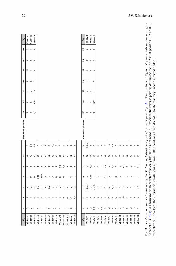
Fig.3.3
Deduced
aminoacidsequence
oftheVdo
main,
hybridizingpa
rtof
primersfrom
Fig.3.2.Theresidues
ofVLandVHarenumbered
accordingto
Kabatetal.(1991).Allforw
ardprimersdetermineonly
thefirst2ntofresidue7,whereasthereverse
primersdeterminethelast2ntofposition102or107,
respectively.Therefore,thealternativetranslationsat
those
latter
positionsgiven
donotindicatethat
they
encodeamixed
codon
28 J.V. Schaefer et al.

dim
eriz
atio
n
dire
ct d
etec
tion
enha
nced
exp
ress
ion
phag
e di
spla
y
Skp
coe
xpre
ssio
n
tryp
sin
clea
vage
site
C-t
erm
inal
det
ectio
n
IMA
Cpu
rific
atio
n
pAK100
pAK300
pAK400
pAK500
pAK600
pJB12
pJB23
pJB33
pAK / pJB vector series
•
• •
• ••
• •
• •
•
•
•
• •
•
• •
• •
KDIR
pelB tet
Sfi I EcoRI Hind III
myc *
alkaline phosphatase
pelB tet (His)6
lac p/o
SD T7g10
pelB tet dHLX •
skp
colEI-orilacI
tet
gIII 250-406 trypsin cleavage siteamber codon
f1-ori
camr
t lpp
pJB12(7416 bp)
Sfi I (3539)
pelB
lac p/ot HP
XbaI (1363)
Sfi I (1438)
(4059)
XbaI Sfi I
pelB tet
pelB tet
gIII 250-406*
pelB tet
pelB tet
pelB tetlac p/o
SD T7g10
gIII 250-406
HindIII
HindIII
HindIII
Hind III
Hind III
HindIII
HindIII
Xba I
Xba I
Xba I
Xba I
Xba I
Sfi I EcoRISfi I
Sfi I EcoRISfi I
Sfi I EcoRISfi I
Sfi I EcoRISfi I
Sfi I EcoRISfi I
Sfi I Sfi I
Sfi I Sfi I
XbaI
Xba I
colEI-orilacI
pelB
tet
myc tag amber codon
camr
f1-ori
lac p/ot HP
gIII 250-406
t lpp
pAK100(6426 bp) Sfi I (1438)
Sfi I (3539)
HindIII
HindIII
(4059)
XbaI (1363)
VL
VH
linker
FLAGs
(His)6
(His)6
(His)6
(His)5
Fig. 3.4 Overview of pAK/pJB vector series. The pAK/pJB vector series (see also Fig. 3.5) can be
used either for phage display (pAK100 and pJB12) by the strategy outlined in Fig. 3.1, or for the
expression of the antibody in a variety of formats. All vectors contain a chloramphenicol resistance
cassette (camr) and additionally a tetracycline resistance “stuffer” cassette (tetA and tetR;2,101 bp), which will be replaced by the antibody gene (the tet cassette allows the monitoring of
complete SfiI digested vector by plating of transformed DH5a cells on tetracycline plates).
Furthermore, these vectors contain the lacI repressor gene, a strong upstream terminator (tHP) to
avoid read-through and premature expression, the lac promoter/operator and the pelB (pectate
lyase gene of Erwinia carotovora) leader sequence (modified to contain a SfiI site) as well as a
3 Construction of scFv Fragments from Hybridoma or Spleen Cells by PCR Assembly 29

to one RNeasy spin column and continue according to the manufacturer’s
instructions.
Note: RNA in harvested tissue is not protected from degradation until the
sample is mixed with TRIzol Reagent, flash-frozen or disrupted and homoge-
nized in the presence of RNase-inhibiting or protein-denaturing reagents. There-
fore, proceed with this step as fast as possible. Generally, DNase digestion is not
required with RNeasy Kits since its silica membrane efficiently removes most of
the DNA. However, if desired, residual DNA can be removed by optional on-
column DNase digestion using the RNase-Free DNase Set (Qiagen). It is
important not to overload the RNeasy spin column, as this will significantly
reduce RNA yield and quality.
3. Elute the purified RNA by the addition of 30 ml RNase-free water. The mRNA
solution is now ready for cDNA synthesis or can alternatively be stored at
�80�C for up to one month.
Note: Diethylpyrocarbonate (DEPC)-treated UHP water can also be used.
However, as DEPC is a suspected carcinogen the use of filtrated RNase-free
water is recommended.
4. For reverse transcription, take approximately 0.1–0.5 mg RNA and 1 ml randomhexamer primers provided in the kit in 20 ml total reaction volume. The precise
procedure is described in the QuantiTect Reverse Transcription Kit (Qiagen).
Note: Ribonuclease H activity of Quantiscript Reverse Transcriptase specifi-
cally degrades only the RNA in RNA:DNA hybrids, but it has no effect on pure
RNA. Hence, an additional RNA degradation step using another RNase H
enzyme is not necessary prior to subsequent PCR reaction.
Note: Specific primers hybridizing to the constant regions can be used as
well, e.g., if only a particular antibody class should be amplified from spleen
cells. In general, however, the random hexamer primers work robustly.
Fig. 3.4 (continued) downstream terminator (tlpp). The rationale for these elements has been
described in detail previously (Krebber et al. 1997). The origins for phage replication and plasmid
replication are as described in Ge et al. (1995). The antibody gene is fused in frame either to
gIII250–406 for phage display, to a his tag for IMAC purification (Lindner et al. 1992) and
C-terminal detection with a recombinant anti-his tag scFv-phosphatase fusion protein (Lindner
et al. 1997), to dimerization helices (Pack et al. 1993, Pluckthun and Pack 1997, see also chapter 7)
or to alkaline phosphatase for both dimerization and direct detection (Lindner et al. 1997). In
pAK100, the in-frame fusion contains a myc tag (Munro and Pelham 1986), offering an additional
detection possibility next to the short N-terminal 4-amino acid FLAG tag (DYKD; Knappik and
Pluckthun 1994) present in all the vectors being encoded by the primers shown in Fig. 3.2. The
plasmid pJB12 contains a trypsin cleavage site (KDIR) and can therefore be conveniently used for
selection of high-affinity binders as described by Dziegiel et al. (1995) and Johansen et al. (1995).
The asterisk in these two vectors pAK100 and pJB12 represents an amber codon. The scFv
expression level in pAK400 and pJB33 is enhanced due to the strong Shine Dalgarno sequence
SDT7g10 (from T7 phage gene 10). Because of the compatibility of the vectors, this feature can
easily be introduced in all of them. In the pJB vector series the co-expressed periplasmic protein
Skp (Bothmann and Pluckthun 1998), encoded on this vector, can increase the functional yield of
antibody fragments expressed in the periplasm without the need of cotransformation with another
plasmid coding for further chaperones. This feature can also be introduced into any of the other
vectors. The complete sequences of all vectors are available from the authors upon request
<
30 J.V. Schaefer et al.

3.3.2 PCR Amplification and scFv Assembly
3.3.2.1 PCR Amplification of VL and VH Domains
1. Use the primers described in Fig. 3.2, which have been dissolved in 100 mMstock solutions in either sterile water or sterile TE buffer to prepare appropriate
mixtures (VL-for mix, VL-rev mix, VH-for mix, and VH-rev mix). Mix them
according to the degree of degeneration, indicated as “d” in Fig. 3.2 (equaling
the number of different unique sequences encoded by mixed bases in the primer)
by adding the stated volumes (in ml) towards the final primer mix. The fraction of
lambda-specific primers in both the forward and reverse VL mixture amounts for
~5% of the total volume, accounting for the low percentage of this light chain
type in mouse antibodies. The nominal total primer concentration of these
mixtures is still 100 mM, ranging from 3 to 40 mM for each of the individual
oligonucleotides.
Note: As described in the introduction, problems in the cloning of monoclo-
nal antibodies can occur if the hybridoma transcribes more than one functional
or even nonfunctional heavy or light chain variable region gene. Therefore, it is
highly recommended to omit any lambda chain-specific primer in the PCR if the
isotyping already indicates that the hybridoma of interest secretes IgGs posses-
sing kappa light chains.
2. For PCR amplification of VL and VH, use the product of the completed first-
strand cDNA reaction and prepare the following mixtures:
PCR mix for amplification of VL PCR mix for amplification of VH
2 ml cDNA 2 ml cDNA1 ml dNTPs (10 mM each) 1 ml dNTPs (10 mM each)
5 ml 10� ThermoPol buffer (NEB) 5 ml 10� ThermoPol buffer (NEB)
0.5 ml VL-for primer mix (100 mM) 0.5 ml VH-for primer mix (100 mM)
0.5 ml VL-rev primer mix (100 mM) 0.5 ml VH-rev primer mix (100 mM)
2.5 ml DMSO 2.5 ml DMSO
0.5 ml VentR Polymerase 2 U/ml (NEB) 0.5 ml VentR Polymerase 2 U/ml (NEB)38 ml H2O 38 ml H2O
Note: This standard protocol is optimized for VentR polymerase, a DNA
polymerase with a 5–15 fold lower error rate than Taq DNA Polymerase (due to
an intrinsic 30!50 proofreading exonuclease activity). If using other proofreadingpolymerases (e.g., Phusion High-Fidelity DNA Polymerase from Finnzymes),
reaction and PCR program conditions might have to be adapted. If the proposed
PCR mix does not lead to any product, varying the cDNA template amount
might be beneficial. If the thermocycler does not have a heated cover, add one
drop of mineral oil to the reaction tube to prevent evaporation.
3. Perform the following PCRcycles after an initial denaturation of theDNA template
for 3 min at 95�C: 5 cycles of 30 s at 95�C, 30 s initial annealing at 55�C, and45 s elongation at 72�C, followed by 20 cycles of 30 s at 95�C, 30 s at 63�C, and
3 Construction of scFv Fragments from Hybridoma or Spleen Cells by PCR Assembly 31

45 s at 72�C. After the last cycle is completed, an additional 5 min elongation
step at 72�C should be performed before cooling the thermocycler to 4�C.Note: We recommend using a hot start, keeping the PCR tubes on ice and not
placing them into the thermocycler until the block has reached 95�C, to mini-
mize unspecific amplification. For successful amplification of VL and VH,
complete annealing of the 30-ends of the primers with the template DNA is
essential. The recommended annealing temperature of 55�C should be suitable
for approx. 97% of the sequences found in a reference set of murine germline
sequences in the IMGT database. However, as it is not clear a priori which
somatic mutations a given monoclonal antibody may carry in the primer regions,
we recommend using a gradient PCR program (covering a range between 70�
and 50�C in steps of 2�) to determine the optimum annealing temperature and to
amplify the antibody genes without unspecific secondary bands. Alternatively,
the PCR might also be run in a “touchdown” manner (Don et al. 1991), starting
at an annealing temperature of 70�C and ending at 50�C. As after 5 cycles the
amplified PCR product will serve itself as template DNA, the annealing temper-
ature of the last 20 cycles can be increased to 63�C.4. Analyze 1/10 volume of each PCR mixtures by agarose gel electrophoresis,
purify the VL and VH genes using the PCR clean-up Gel Extraction Kit
(Macherey Nagel) according to the manufacturer’s instructions and determine
the DNA concentration of both genes.
Note: Using the listed primer mixtures, the expected lengths of the PCR
products of VL and VH are between 375–402 bp and 386–440 bp, respectively.
Purification of the PCR products is important to remove any residual primers
which might interfere with the subsequent assembly PCR. For the case of
multiple bands on the agarose gel, gel-purify the band of correct size using the
GenElute Gel Extraction Kit (Sigma-Aldrich). If the final DNA concentration is
too low afterward, perform a second PCR using these purified fragments as
template for gaining sufficient yields of high-quality DNA.
3.3.2.2 Assembly of VL and VH by SOE-PCR (Splicing by Overlap Extension)
1. For the assembly PCR, use approximately 10 ng of the PCR product of both
domains in a total volume of 50 ml, containing 200 mM dNTPs, 3–5% DMSO,
1 mM outer-for, and outer-rev primer (each) and 1 unit VentR DNA Polymerase
(NEB). Following a 3 min 95�C step, perform 5 cycles of 1 min at 95�C, 1 min at
63�C, and 1 min at 72�C, followed by another 5 cycles of 1 min at 95�C, 30 s at
56�C, and 1 min at 72�C and finally 25 cycles of 1 min at 95�C, 90 s at 72�C.Note: Hot start PCR and initial assembly of VL and VH in the absence of the
primers is usually not necessary but can be performed. It is important to include
DMSO in the PCR mix as well as to keep the primer concentration as low as
indicated to prevent any risk of primer-dimer formation.
Note: The assembly, as used here, places VL in front of VH. This has the
advantage of placing a shortened FLAG tag, consisting of only four amino acids,
32 J.V. Schaefer et al.

at the N-terminus of the construct. Since its last amino acid, Asp, is the same as
the first residue of the VL domain, only three additional amino acids are needed
(Knappik and Pluckthun 1994) for allowing specific detection using this tag. A
slight asymmetry in the VH/VL heterodimer with respect to the pseudo two-fold
axis (Pluckthun et al. 1996) is taken care of with a 20-amino acid linker, leading
to monomeric scFv fragments.
3.3.3 Digestion and Cloning of scFv Genes
1. Purify the product of the assembly PCR using the PCR clean-up Gel Extraction
Kit (Macherey Nagel) according to the manufacturer’s instructions, eluting the
product in 30 ml of the recommended buffer. In case there are several bands on
the analytical agarose gel, carry out a gel purification of the correct band, as
described in 3.3.3.4.
2. Perform a SfiI digest of the amplified scFv gene for 3–4 h at 50�C (At 37�C, theactivity of SfiI would be 10 fold-lower). To the 30 ml purified PCR product, add
5 ml 10� NEbuffer 4 (NEB), 5 ml 10� BSA (final concentration, 100 mg/ml),9 ml H2O, and 1 ml (=20 units) SfiI (NEB).
3. Digest appropriate amounts of vector (pAK100 or pJB12, see Fig. 3.4) with SfiIin the presence of the above-mentioned buffer, including BSA. Use 10 units SfiIfor 1 mg vector in 50 ml and incubate 4 h at 50�C. Dephosphorylate the cut vectorby adding Calf Intestinal Alkaline Phosphatase (CIP, NEB; 0.5 unit/mg vector) tothe digestion mix after 2 h and continue incubation for another 2 h at 50�C.
Note: Dephosphorylation should not be necessary because of the asymmetric
overhangs. However, we always include this step to eliminate any risk of
religation of single-cut vector.
Note: pAK100 or pJB12 are phage display vectors (Fig. 3.4). When starting
from hybridomas, one can also directly clone the VL and VH genes into an scFv
expression vector with a stronger promoter, such as pAK400, which does not
encode a fusion with gIII. However, depending on the number of additional V
genes expressed in the hybridoma, a large number of clones may have to be
screened from individual colonies.
4. Purify the digested scFv antibody genes and vector by preparative agarose gel
electrophoresis in combination with the GenElute Gel Extraction Kit (Sigma-
Aldrich).
Note: For obtaining pure preparations of a fully digested vector, it is very
important not to overload the agarose gel. Furthermore, the gel electrophoresis
has to be run long enough to separate small amounts of undigested vector from
the digested vector band. For large-scale vector or insert preparation, electro-
elution might be an efficient and convenient alternative. If the concentration of
eluted DNA is too low for further applications, Microcon 30 columns (Amicon)
can be used for concentration.
3 Construction of scFv Fragments from Hybridoma or Spleen Cells by PCR Assembly 33

5. Ligate 50 ng scFv gene fragment with the vector (molar ratio of vector to
insert 1:5) with 5 units T4 DNA ligase (NEB) in the presence of 1� T4 DNA
ligase buffer in 10 ml volume. Incubate for 2 h at room temperature or overnight
at 16�C.Note: The ATP-concentration is very crucial for the successful ligation by T4
DNA ligase. Therefore, we recommend using T4 DNA ligase buffer aliquots,
which have been properly stored at �20�C and not thawed repeatedly. To allow
an easy subcloning of the scFv fragment into vectors for optimized soluble
expression and other purposes, compatible vector sets are available (Figs. 3.4
and 3.5).
6. Transform 50 ml chemocompetent XL1-Blue cells (Stratagene) with 5 ml of theligation mix by heat-shock for 45 s at 42�C, add 500 ml of 2� YT medium after
2 min incubation on ice, and incubate for 45 min, shaking at 37�C.Note: Make sure not to exceed a ratio of ligation mix/cells of 1:10 (v/v).
Chemocompetent E. coli are used, if only a very small diversity of clones is
expected, e.g., when cloning from a hybridoma. If a larger diversity and thus
many clones are required (e.g., when cloning from spleen cells), follow the
instructions for electroporation described in steps 3.3.5.1–3.3.5.3.
7. Plate the transformed cells on 2� YT, 1% glucose, chloramphenicol (30 mg/ml)
agar plates, and incubate overnight at 37�C.Note: You may check the ratio of desired ligation product to background by
including transformation with “religated” plasmid in the absence of any insert.
Alternatively, the background signal can be analyzed by testing for tetracycline
resistance after transformation of other E. coli strains not possessing an intrinsic tetresistance (like Invitrogen’s DH5a) with the ligation mix. The portion of vector
with unremoved or religated tet cassette is typically in the range of 0.01–0.1%.
3.3.4 Preparation of Electrocompetent E. coli
1. For preparation of electrocompetent E. coli XL1-Blue cells (Stratagene), use
2 ml of a dense overnight pre-culture to inoculate 500 ml medium (2� YT,
15 mg/ml tetracycline). Shake it at 25�C until an OD600 of 0.6 is reached, then
chill the culture on ice as quickly as possible for 30 min (cool the whole shake
flask in a large ice bath).
Note: Sufficient agitation during growth seems to be very important for
preparation of electrocompetent cells, reaching reproducible efficiencies of
3–6 � 109 cfu/mg pUC19 DNA. Therefore, use 5 l baffled shake flasks with
only 500 ml medium and make sure that the amplitude of the shaker is high
enough to vigorously circulate the medium.
Note: The use of electrocompetent bacteria is an alternative to 3.3.3.6
and needed when a large diversity is expected, typically when cloning from
spleen cells.
34 J.V. Schaefer et al.

a
pAK100scFv, pAK300scFv, pAK500scFv,pAK600scFv, pJB12scFv, pJB23scFv
lacIdne ¬
. . . C A G C T G G C A C G A C A G G T T T C C C G A C T G G A A A G C G G G C A G T G A G C G
. . . Q L A R Q V S R L E S G Q *
tHP terminator
G T A C C C G A T A A A A G C G G C T T C C T G A C A G G A G G C C G T T T T G T T T T G C A G C
CAP binding site
C C A C C T C A A C G C A A T T A A T G T G A G T T A G C T C A C T C A T T A G G C A C C C C A G G
lac-operator01-53-
C T T T A C A C T T T A T G C T T C C G G C T C G T A T G T T G T G T G G A A T T G T G A G C G G A
® mRNA
SD1 ® lacZ
T A A C A A T T T C A C A C A G G A A A C A G C T A T G A C C A T G A T T A C G A A T T T C T A G AM T M I T N F *
SD2 ® pelB signal sequence
T A A C G A G G G C A A A T C A T G A A A T A C C T A T T G C C T A C G G C A G C C G C T G G A T TM K Y L L P T A A A G L
Sfi I FLAGs ® VL
G T T A T T A C T C G C G G C C C A G C C G G C C A T G G C G G A C T A C A A A G A Y . . .
L L L A A Q P A M A D Y K D . . .
pAK400scFv, pJB33scFv
SD2 ® pelB signal sequence
. . . G A A G G A G A T A T A C A T A T G A A A T A C C T A T T G C C T A C G G C A G C C . . .
T7g10 M K Y L L P T A A . . .
b
pAK100scFv
¬ VH Sfi I EcoRI myc tag
. . . C G G C C T C G G G G G C C G A A T T C G A G C A G A A G C T G A T C T C T G A G G A A G A C
. . . A S G A E F E Q K L I S E E D
® gene III 250-406
C T G T A G G G T G G T G G C T C T G G T T C C G G T G A T T T T G A T T A T G A A A A G . . .
L * G G G S G S G D F D Y E K . . .
pJB12scFv
¬ VH Sfi I EcoRI trypsin cleavage site
. . . C G G C C T C G G G G G C C G A A T T C G A G C A G A A G G A T A T C C G T G A G G A A G A C
. . . A S G A E F E Q K D I R E E D
® gene III 250-406
C T G T A G G G T G G T G G C T C T G G T T C C G G T G A T T T T G A T T A T G A A A A GC T G T A G G G T G G T G G C T C T G G T T C C G G T G A T T T T G A T T A T G A A A A G . . .
L * G G G S G S G D F D Y E K . . .
Fig. 3.5 (Continued)
3 Construction of scFv Fragments from Hybridoma or Spleen Cells by PCR Assembly 35
cpAK300scFv, pAK400scFv
¬ VH Sfi I (His)6 tag HindIII
. . . C G G C C T C G G G G G C C G A T C A C C A T C A T C A C C A T C A T T A G T A A G C T T . .
. . . A S G A D H H H H H H * *
pJB23scFv, pJB33scFv
¬ VH Sfi I EcoRI (His)6 tag
. . . C G G C C T C G G G G G C C G A A T T C C A C C A C C A T C A C C A C C A T T A A T G A
. . . A S G A E F H H H H H H * *
HindIII
A A G C T T . . .
dpAK500scFv
¬ VH Sfi I EcoRI
. . . C G G C C T C G G G G G C C G A A T T C C C C A A A C C T A G C A C C C C C C C T G G C A G
. . . A S G A E F P K P S T P P G S
® dHLX
C A G T G G T G A A C T G G A A G A G C T G C T T A A G C A T C T T A A A G A A C T T C T G A A G
S G E L E E L L K H L K E L L K
G G C C C C C G C A A A G G C G A A C T C G A G G A A C T G C T G A A A C A T C T G A A G G A G C T
G P R K G E L E E L L K H L K E L
(His)5 tag
G C T T A A A G G T G G G A G C G G A G G C G C G C C G C A C C A T C A T C A C C A T T G A C G T C
L K G G S G G A P H H H H H *
HindIII
T A A G C T T . . .
pAK600scFv
¬ VH Sfi I EcoRI ® alkaline phosphatase (AP)
. . . C G G C C T C G G G G G C C G A A T T C C G G A C A C C A G A A A T G C C T G T T C T G . . .
A S G A E F R T P E M P V L . . .
PAdne ¬ HindIII
. . . C T C T T C T A C A C C A T G A A A G C C G C T C T G G G G C T G A A A T A A G C T T . . .
. . . L F Y T M K A A L G L K *
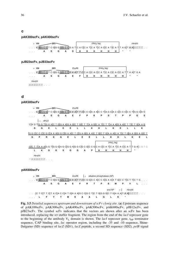
Fig. 3.5 Detailed sequences upstream and downstream of scFv clonig site. (a) Upstream sequence
of pAK100scFv, pAK300scFv, pAK400scFv, pAK500scFv, pAK600scFv, pJB12scFv, and
pJB23scFv. The symbol scFv indicates that the vectors are shown after an scFv has been
introduced, replacing the tet stuffer fragment. The region from the end of the lacI repressor geneto the beginning of the antibody VL domain is shown. The lacI repressor gene, tHP terminator
sequence, CAP binding site, lac operator region, including the -35 and -10 sequence, Shine-
Dalgarno (SD) sequence of lacZ (SD1), lacZ peptide, a second SD sequence (SD2), pelB signal
36 J.V. Schaefer et al.

2. Centrifuge the bacterial culture in 50 ml aliquots in disposable tubes for 5 min at
5,000 g. Remove as much supernatant as possible (leave the tube upside down
for 15–30 s on a clean tissue). Then, fill each tube with 1 volume of ice-cold
distilled water (i.e., the same volume as the original culture aliquot) and remove
the water immediately (the cell pellet is very solid after this first centrifugation
step and will not be resuspended by the brief rinsing with distilled water).
Note: All these steps should be carried out using ice-cold solutions and be
performed in the cold room. Use only ultra pure water to wash cells and to
prepare 10% glycerol, as the presence of impurities such as salts in the water
might cause the subsequent transformations to fail.
3. Fill each tube with 1 vol distilled water (i.e., the same volume as the original
culture aliquot), resuspend the pellet carefully and incubate for 10 min on ice.
Note: Make sure that the cells are sufficiently solubilized to yield a homoge-
neous suspension. Cells are best resuspended by swirling rather than pipetting.
Never vortex the cell suspension!
4. Transfer the cells into new 50 ml tubes and centrifuge at 5,000 g for 10 min.
Carefully remove the supernatant and resuspend the pellets each in 50 ml pre-
chilled 10% (v/v) glycerol (Fluka). Incubate on ice for 10 min.
5. Centrifuge resuspended cells at 5,000 g for 15 min and remove the supernatant
(you might lose a small portion of cells – do not put the tubes upside down on
tissue in this step!). Carefully resuspend the cells in 1/500 of the original culture
volume (= 1 ml) 10% (v/v) glycerol, freeze the cells in 100 ml aliquots by
dipping the tubes immediately into liquid nitrogen and store them at �80�C.Note: Electrocompetent cells can be kept at �80�C for up to 12 months.
6. To determine the transformation efficiency, add 1 ml of 10 pg/ml pUC19
DNA (in water) to 40 ml of barely thawed cells (see step 3.3.5.2). Fifty colonies
per 1/1,000 of the transformation volume plated correspond to an efficiency of
5 � 109 cfu/mg pUC19 DNA.
Fig. 3.5 (continued) sequence, N-terminal SfiI site (underlined and highlighted), four amino acid
FLAGs tag (underlined), and the start of the VL domain (sequence GAY; bold) are indicated abovethe sequence. In addition, also the corresponding amino acid sequence is shown. In pAK400 and
pJB33, the 15 bp upstream from the pelB start codon are replaced by another sequence, including
the SD sequence of the phage T7 gene10, while everything else is identical. Because of the
modularity of the vectors, this feature can be easily introduced into any of the other vectors (see
Fig. 3.4) (b) Downstream sequence of pAK100scFv and pJB12scFv. The last two bases of VH
(bold), the SfiI and EcoRI restriction sites, myc tag (boxed) or trypsin cleavage site and the start ofgeneIII250–406 are indicated above the sequence. Asterisks indicate amber stop codon, leading to
scFv-gene III fusions upon expression in E. coli strains with amber suppressor tRNA, such as XL1-
Blue. (c) Downstream sequence of pAK300scFv, pAK400scFv, pJB23scFv, and pJB33scFv. The
last two bases of VH (bold), the SfiI and EcoRI restriction sites and (His)6 tag (boxed) are indicatedabove the sequence. (d) Sequences of the downstream EcoRI/HindIII fusion cassettes as used in
pAK500 and pAK600. The dHLX dimerization motif (double underlined) was taken from Pack
et al. (1993). The complete sequence of the mature E. coli alkaline phosphatase (AP) gene can be
found in Shuttleworth et al. (1986). For the EcoRI/HindIII cloning cassette the two internal EcoRIsites of the AP gene have been removed by silent mutations. The complete sequences of all vectors
are available from the authors upon request
<
3 Construction of scFv Fragments from Hybridoma or Spleen Cells by PCR Assembly 37

3.3.5 Library Preparation/Construction
1. For desalting the DNA prior to electroporation, apply the ligation mix to
StrataClean Resin (Stratagene; hydroxylated silica, binding proteins with a
high affinity, while having a low affinity for DNA at near neutral pH), followed
by precipitation in 70% ethanol. Since most salts and small organic molecules
are soluble in 70% ethanol, they can be separated from DNA by centrifugation.
Resuspend the precipitated DNA in ultra pure water.
2. For each transformation, use desalted ligation mixtures corresponding to
20–100 ng insert. Add the DNA to 40 ml of barely thawed cells on ice and
mix by flipping the tube shortly and gently. Immediately transfer the cell-DNA
mix to chilled electroporation cuvette (bubble free), pulse according to the
guidelines of the electroporator’s manufacturer, and add 1 ml of SOC medium
(20 g/l bacto-tryptone, 5 g/l yeast extract, 10 mM NaCl, 2.5 mM KCl, 20 mM
glucose, 10 mM MgCl2, 10 mM MgSO4) to cells immediately after the pulse.
Note: For efficient transformation (�108 clones per mg insert DNA), the time
constant using 2 mm cuvettes should be�5 ms, reflecting properly washed cells.
Also, make sure that no air-bubbles are trapped in the cell-DNA mix as they will
interfere with the electroporation.
3. Resuspend cells completely in SOC medium and shake for 1 h at 37�C.Afterward, plate dilutions on 2� YT, 1% glucose, chloramphenicol (30 mg/ml)
agar plates. Use a sterile spreader or sterile glass beads to evenly distribute
the culture over the surface of the 12 � 12 cm plate (do not exceed about 5,000
clones per square plate) and incubate overnight at 37�C. The next day, scrape thecolonies off the plates in 3–4 ml 2� YT, containing 30% glycerol, and subse-
quently store them at �80�C.Note: Take care that your library is homogeneously mixed.
4. For phage panning as described in 3.3.6, inoculate cultures with at least tenfold
more viable cells than colonies obtained after transformation, in order to have
sufficient oversampling. When starting from spleen cells, perform three rounds
of phage panning as described in 3.3.6 before testing single clones. When
starting from a hybridoma, one round should be sufficient, and in ideal cases,
single clones can be tested right away by phage ELISA.
Note: The first panning round is the most crucial, as you might lose any
desired, but less abundant antibody sequence by too extensive washing. There-
fore, do not exceed ten washing steps in this first panning round. The panning
procedure is analogous to the phage ELISA (3.3.6), except that a pool of phages
is grown and that phages are eluted from antigen (at the end of 3.3.6.4), which
are afterward added again to exponentially growing bacteria. This is described in
detail elsewhere (Barbas et al. 2001; Lee et al. 2007) and also in this volume.
Note: The screening of single clones can be performed in three ways. First, at
the level of phages (phage ELISA), as described in Sect. 3.3.3.6, second, after
retransforming of an suppressor tRNA-deficient strain such as, e.g., E. coli strainJM83 (Yanisch-Perron et al. 1985), still with the amber codon, containing
38 J.V. Schaefer et al.

pAK100 derived plasmids, or third, after recloning into a more efficient expres-
sion vector such as pAK400 (Fig. 3.5), which carries no gene III. In second and
third option, the soluble scFv is screened by ELISA.
Note: This protocol does not describe the periplasmic expression of scFv
fragments in E. coli (Glockshuber et al. 1990) and their subsequent purification
(reviewed in Pluckthun et al. 1996). More details can be found in chapter 27
“Improving expression of scFv fragments by coexpression of periplasmic cha-
perones” in this volume.
3.3.6 Screening for Binders by Phage ELISA
1. When starting from hybridoma, pick 10 colonies (from spleen, as many as you
can handle) and grow them separately at 37�C in 2 ml 2� YT, 1% glucose,
chloramphenicol (30 mg/ml) until they reach an OD600 of 0.5. This level of
glucose fully represses expression, and, thus, the growth temperature can be
37�C. Dilute 1:10 in 2� YT, 1% glucose, chloramphenicol (30 mg/ml), contain-
ing 1 mM IPTG, and 1 � 1010 pfu VCSM13 helper phage (Stratagene) per ml,
and grow overnight at 26�C or 37�C (for some murine scFvs with aggregation
tendencies, growth at 26�C after infection may be necessary). The phage titer
after overnight incubation is in the range of 1011–1012 cfu per ml supernatant.
Note: XL1-Blue should be grown on agar plates and in media containing
tetracycline (tet) as the F0-plasmid encoding for the F-pili required for infection
of bacteria also carries the tet resistance gene. The phage titer (in cfu) should bedetermined in order to rule out any problems during phage production. To do so,
take a log-phase culture of XL1-Blue cells (OD600 = 0.4–0.6) and incubate
aliquots of this culture with serial dilutions of your phage preparation. After
15 min incubation at 37�C, plate appropriate amounts (30–150 cfu/plate) on
2� YT, 1% glucose, chloramphenicol (30 mg/ml) agar plates.
Note: We are aware of the fact that the presence of such a high level of
glucose during IPTG-induction is rather unusual. However, for phage display,
the induction level does not have to be very high. Based on our experiences with
this vector series described here, a combination of IPTG addition and the
presence of glucose seems to be crucial for the successful expression of some
scFv fragments, notably those with nonideal biophysical properties, and appro-
priate for most, but may have to be checked for each scFv individually in case of
unusual properties.
2. Centrifuge the culture 10 min at 16,000 g and 4�C. Take 1.6 ml supernatant and
mix it with 0.4 ml 20% PEG 6000 (Fluka), 2.5 M NaCl in a 2 ml Eppendorf tube
in order to precipitate the phages (Sambrook and Russell 2001).
Note: We recommend that the PEG solution be freshly prepared.
3. Incubate on ice for 30–60 min and centrifuge for 15 min at 5,600 g and 4�C.Note: It is important not to centrifuge phages at too high a g force, as
otherwise, it will be difficult to resuspend them homogeneously, resulting in a
3 Construction of scFv Fragments from Hybridoma or Spleen Cells by PCR Assembly 39

decreased phage titer. The size of the white pellet does not necessarily reflect a
high or low phage titer.
4. Resuspend the phage pellet in 400 ml PBS (with 10% (v/v) glycerol). For
complete resuspension, incubate the phage solution on an orbital shaker at
800 rpm for 15 min at 4�C. Pellet insoluble matter (cell debris) by centrifugation
for 10 min at 11,000 g and 4�C and transfer the phage solution to a fresh tube.
Use 100 ml phage solution per well in an ELISA assay to distinguish phages
displaying functional scFv antibody from those which display nonfunctional or
nonproductive antibody fragments.
5. If soluble antigen is available, include a competition ELISA control showing
that free antigen is able to compete with bound antigen for phage binding to
distinguish nonspecific “sticky” from specifically binding phages. In principle,
the same ELISA protocol that was used for the hybridoma screening procedure
can be used.
Note: For weak binders, it might be important to use more phages for ELISA
analysis. In this case, the culture volume should be increased ten times. If no
functional clone shows up in ELISA of single clones, perform one round of
phage panning in order to enrich the functional binders. The enrichment should
be checked by comparison of eluted phages from a specific surface versus a
surface without antigen. In addition, it is recommended to analyze the phage
solution by immunoblot, using an anti-M13 HRP-conjugated antibody (GE
Healthcare), to ensure the correct fusion of the scFv to the gIII-protein as well
as its correct display on the phage surface.
3.4 Troubleshooting
This part of the protocol contains general comment about potential pitfalls of the
recommended standard method. The most critical steps were already highlighted
directly following the instructions in the different subsections.
(a) In case of low transformation yields, check whether the problem is the trans-
formation itself or rather the ligation. To investigate the quality of ligation,
analyzing an aliquot by agarose gel electrophoresis might indicate any pro-
blems caused by nucleases. Furthermore, it might be informative to compare
the ligation efficiency of SfiI digested PCR product with inserts derived from
plasmid digestion. In order to check both the ligation and the transformation
efficiency, a defined amount of pUC19 DNA can be added to the ligation
mixture. Because of the chloramphenicol resistance of the cloning vector and
the ampicillin resistance of pUC19 DNA, it is possible to calculate the ligation
efficiency by plating double transformed cells on ampicillin or chlorampheni-
col plates, respectively, and comparing the number of clones. The transforma-
tion efficiency (in presence of the ligation mixture) can be judged by
comparison of the colony number after transformation with pUC19 DNA alone.
40 J.V. Schaefer et al.

(b) The quality of the oligonucleotides used in this procedure is crucial for the
successful and reliable amplification of various antibody genes as well as their
subsequent assembly into scFvs. The number of proposed primers is important
for a broad representation of the immune response, as any sequence absent from
the complex mixture will obviously decrease the functional library size. We
also strongly recommend using primers that have been accurately purified after
their synthesis (either by HPLC or, for longer primers, by PAGE) to ensure that
no single-base deletions are present in any of the oligonucleotides. These
deletions as well as any insertions would cause frameshifts in the final gene
assembly, resulting in a number of nonfunctional library members. Therefore,
we also suggest – especially for library cloning – to sequence the genes of
several random clones as well as to check for full-length scFv by western blot
analysis detecting its fusion partner gene III (see note at 3.3.6.5).
(c) In case of severe problems in the PCR amplification of the VH and VL genes
(steps 3.3.2), it might be worth considering to divide this reaction into two
separate ones. Using the proposed primers without any overhang at their 50-end(which either codes for the FLAGs-tag and the SfiI cleavage site, or the
(Gly4Ser)4-linker), the pure antibody DNA should be amplified in a first PCR
reaction, and, subsequently, a second PCR should be performed for ream-
plification and introduction of the appropriate overhangs with the original
full-length primers. This procedure increases the degree of matching in both
reactions, and might therefore help in the annealing step of the primers.
(d) Whenever expression of the scFv gene is not required, the bacteria should be
grown in the presence of 1% glucose. Glucose will cause a tight suppression of
the lac promoter, thereby ensuring the genetic stability of the inserted scFv
genes. Likewise, we suggest growing XL1-Blue always on agar plates and in
media containing tetracycline (tet) to keep the bacteria infective, as the tetresistance is located on the F0-plasmid that also contains the genes encoding
F-pilus formation. Always use fresh XL1-Blue colonies, as subcloning might
occasionally lead to the formation of tet resistant cells, which are no longer
infectable. As the F-pilus expression is reduced when the bacteria are past log
phase as well as when grown at temperatures below 34�C, we also recommend
growing them at 37�C to OD600 = 0.4–0.6.
(e) When working with libraries, double transformants can and will occur (Gold-
smith et al. 2007). It is thus highly recommended that the scFv fragments of
interest be recloned into a new vector (e.g., from pAK100 to pAK400), when
they are analyzed at the level of pure unfused protein, thereby also introducing
a stronger translation initiation region. It should be noted that diluted retrans-
formation cannot resolve plasmid mixtures, as in E. coli, plasmids can form
reversible concatamers.
Acknowledgements This protocol has evolved over the years, and heavily relies on the original
versions developed by Anke Krebber and Jorg Burmester, with important discussions and con-
tributions to the reagents and procedures gradually added from Peter Lindner, Lutz Jermutus, Jorg
Willuda, Daniel Steiner, Barbara Klinger, and Cornelia Rinderknecht.
3 Construction of scFv Fragments from Hybridoma or Spleen Cells by PCR Assembly 41

References
Adams GP, McCartney JE, Tai MS, Oppermann H, Huston JS, Stafford WF 3rd, Bookman MA,
Fand I, Houston LL, Weiner LM (1993) Highly specific in vivo tumor targeting by monovalent
and divalent forms of 741F8 anti-c-erbB-2 single-chain Fv. Cancer Res 53:4026–4034
Amstutz P, Binz HK, Zahnd C, Pluckthun A (2006) Ribosome display: in vitro selection of
protein-protein interactions. In: Celis J (ed) Cell biology: a laboratory handbook, 3rd edn.
Elsevier, Amsterdam, pp 497–509
Backmann N, Zahnd C, Huber F, Bietsch A, Pluckthun A, Lang HP, Guntherodt HJ, Hegner M,
Gerber C (2005) A label-free immunosensor array using single-chain antibody fragments. Proc
Natl Acad Sci USA 102:14587–14592
Barbas C, Burton D, Scott J, Silverman G (2001) Phage display: a laboratory manual. Cold Spring
Harbor Laboratory Press, Cold Spring Harbor, NY
Bothmann H, Pluckthun A (1998) Selection for a periplasmic factor improving phage display and
functional periplasmic expression. Nat Biotechnol 16:376–380
Burmester J, Pluckthun A (2001) Construction of scFv fragments from hybridoma or spleen cells
by PCR assembly. In: Kontermann R, Dubel S (eds) Antibody engineering. Springer, Berlin,
pp 19–40
Di Paolo C, Willuda J, Kubetzko S, Lauffer I, Tschudi D, Waibel R, Pluckthun A, Stahel RA,
Zangemeister-Wittke U (2003) A recombinant immunotoxin derived from a humanized epi-
thelial cell adhesion molecule-specific single-chain antibody fragment has potent and selective
antitumor activity. Clin Cancer Res 9:2837–2848
Don RH, Cox PT, Wainwright BJ, Baker K, Mattick JS (1991) ‘Touchdown’ PCR to circumvent
spurious priming during gene amplification. Nucleic Acids Res 19:4008
Dziegiel M, Nielsen LK, Andersen PS, Blancher A, Dickmeiss E, Engberg J (1995) Phage display
used for gene cloning of human recombinant antibody against the erythrocyte surface antigen,
rhesus D. J Immunol Methods 182:7–19
Ewert S, Honegger A, Pluckthun A (2004) Stability improvement of antibodies for extracellular
and intracellular applications: CDR grafting to stable frameworks and structure-based frame-
work engineering. Methods 34:184–199
Freund C, Ross A, Pluckthun A, Holak TA (1994) Structural and dynamic properties of the Fv
fragment and the single-chain Fv fragment of an antibody in solution investigated by hetero-
nuclear three-dimensional NMR spectroscopy. Biochemistry 33:3296–3303
Ge L, Knappik A, Pack P, Freund C, Pluckthun A (1995) Expressing antibodies in Escherichia
coli. In: Borrebaeck C (ed) Antibody engineering, 2nd edn. Oxford University Press, Oxford,
pp 229–236
Glockshuber R, Malia M, Pfitzinger I, Pluckthun A (1990) A comparison of strategies to stabilize
immunoglobulin Fv-fragments. Biochemistry 29:1362–1367
Goldsmith M, Kiss C, Bradbury AR, Tawfik DS (2007) Avoiding and controlling double transfor-
mation artifacts. Protein Eng Des Sel 20:315–318
Hanes J, Pluckthun A (1997) In vitro selection and evolution of functional proteins by using
ribosome display. Proc Natl Acad Sci USA 94:4937–4942
Hanes J, Jermutus L, Weber-Bornhauser S, Bosshard HR, Pluckthun A (1998) Ribosome display
efficiently selects and evolves high-affinity antibodies in vitro from immune libraries. Proc
Natl Acad Sci USA 95:14130–14135
Honegger A, Pluckthun A (2001) The influence of the buried glutamine or glutamate residue in
position 6 on the structure of immunoglobulin variable domains. J Mol Biol 309:687–699
Honegger A, Spinelli S, Cambillau C, Pluckthun A (2005) A mutation designed to alter crystal
packing permits structural analysis of a tight-binding fluorescein-scFv complex. Protein Sci
14:2537–2549
Huston JS, Levinson D, Mudgett-Hunter M, Tai MS, Novotny J, Margolies MN, Ridge RJ,
Bruccoleri RE, Haber E, Crea R, Oppermann H (1988) Protein engineering of antibody binding
42 J.V. Schaefer et al.

sites: recovery of specific activity in an anti-digoxin single-chain Fv analogue produced in
Escherichia coli. Proc Natl Acad Sci USA 85:5879–5883
Johansen LK, Albrechtsen B, Andersen HW, Engberg J (1995) pFab60: a new, efficient vector for
expression of antibody Fab fragments displayed on phage. Protein Eng 8:1063–1067
Jung S, Spinelli S, Schimmele B, Honegger A, Pugliese L, Cambillau C, Pluckthun A (2001)
The importance of framework residue H6, H7, and H10 in antibody heavy chains: Experimen-
tal evidence for a new structural subclassification of antibody VH domains. J Mol Biol
309:701–716
Kabat E, Wu T, Reid-Miller M, Perry H, Gottesman K, Foeller C (1991) Sequences of proteins of
immunological interest, 5th edn. US Department of Health and Human Services, Public
Service, NIH
Knappik A, Pluckthun A (1994) An improved affinity tag, based on the FLAG peptide for the
detection and purification of recombinant antibody fragments. Biotechniques 17:754–761
Knappik A, Ge L, Honegger A, Pack P, Fischer M, Wellnhofer G, Hoess A, Wolle J, Pluckthun A,
Virnekas B (2000) Fully synthetic human combinatorial antibody libraries (HuCAL), based
on modular consensus frameworks and CDRs randomized with trinucleotides. J Mol Biol
296:57–86
Krebber A, Bornhauser S, Burmester J, Honegger A, Willuda J, Bosshard HR, Pluckthun A
(1997) Reliable cloning of functional antibody variable domains from hybridomas and spleen
cell repertoires employing a reengineered phage display system. J Immunol Methods
201:35–55
Kutemeier G, Harloff C, Mocikat R (1992) Rapid isolation of immunoglobulin variable genes
from cell lysates of rat hybridomas by polymerase chain reaction. Hybridoma 11:23–32
Lee CM, Iorno N, Sierro F, Christ D (2007) Selection of human antibody fragments by phage
display. Nat Protoc 2:3001–3008
Lefranc M, Lefranc G (2001) The Immunoglobulin FactsBook. Academic Press, London
Lindner P, Guth B, Wulfing C, Krebber C, Steipe B, Muller F, Pluckthun A (1992) Purification of
native proteins from the cytoplasm and periplasm of Escherichia coli using IMAC and histidine
tails: a comparison of proteins and protocols. Methods 4:41–56
Lindner P, Bauer K, Krebber A, Nieba L, Kremmer E, Krebber C, Honegger A, Klinger B,
Mocikat R, Pluckthun A (1997) Specific detection of his-tagged proteins with recombinant
anti-His tag scFv-phosphatase or scFv-phage fusions. Biotechniques 22:140–149
Luginbuhl B, Kanyo Z, Jones RM, Fletterick RJ, Prusiner SB, Cohen FE, Williamson RA, Burton
DR, Pluckthun A (2006) Directed evolution of an anti-prion protein scFv fragment to an
affinity of 1 pM and its structural interpretation. J Mol Biol 363:75–97
Mondon P, Dubreuil O, Bouayadi K, Kharrat H (2008) Human antibody libraries: a race to
engineer and explore a larger diversity. Front Biosci 13:1117–1129
Morfill J, Blank K, Zahnd C, Luginbuhl B, Kuhner F, Gottschalk KE, Pluckthun A, Gaub HE
(2007) Affinity-matured recombinant antibody fragments analyzed by single-molecule force
spectroscopy. Biophys J 93:3583–3590
Munro S, Pelham HR (1986) An Hsp70-like protein in the ER: identity with the 78 kD glucose-
regulated protein and immunoglobulin heavy chain binding protein. Cell 46:291–300
Pack P, Kujau M, Schroeckh V, Knupfer U, Wenderoth R, Riesenberg D, Pluckthun A (1993)
Improved bivalent miniantibodies, with identical avidity as whole antibodies, produced by
high cell density fermentation of Escherichia coli. Biotechnology (N Y) 11:1271–1277
Pluckthun A, Pack P (1997) New protein engineering approaches to multivalent and bispecific
antibody fragments. Immunotechnology 3:83–105
Pluckthun A, Krebber A, Krebber C, Horn U, Knupfer U, Wenderoth R, Nieba L, Proba K,
Riesenberg D (1996) Producing antibodies in Eschericia coli: Fom PCR to fermentation. In:
McCafferty J, Hoogenboom H (eds) Antibody engineering: a practical approach. IRL press,
Oxford, pp 203–252
Sambrook J, Russell D (2001) Molecular cloning. A laboratory manual, 3rd edn. Cold Spring
Harbor Laboratory Press, Cold Spring Harbor, NY
3 Construction of scFv Fragments from Hybridoma or Spleen Cells by PCR Assembly 43

Schaffitzel C, Zahnd C, Amstutz P, Luginbuhl B, Pluckthun A (2005) In vitro selection and
evolution of protein-ligand interactions by ribosome display. In: Golemis E, Adams P (eds)
Protein-protein interactions: a molecular cloning manual, 2nd edn. Cold Spring Harbor Labo-
ratory Press, Cold Spring Harbor, NY, pp 517–548
Shuttleworth H, Taylor J, Minton N (1986) Sequence of the gene for alkaline phosphatase from
Escherichia coli JM83. Nucleic Acids Res 14:8689
Tugarinov V, Zvi A, Levy R, Hayek Y, Matsushita S, Anglister J (2000) NMR structure of an anti-
gp120 antibody complex with a V3 peptide reveals a surface important for co-receptor binding.
Structure 8:385–395
Vaughan TJ, Williams AJ, Pritchard K, Osbourn JK, Pope AR, Earnshaw JC, McCafferty J, Hodits
RA, Wilton J, Johnson KS (1996) Human antibodies with sub-nanomolar affinities isolated
from a large nonimmunized phage display library. Nat Biotechnol 14:309–314
Winter G, Griffiths AD, Hawkins RE, Hoogenboom HR (1994) Making antibodies by phage
display technology. Annu Rev Immunol 12:433–455
Worn A, Pluckthun A (2001) Stability engineering of antibody single-chain Fv fragments. J Mol
Biol 305:989–1010
Yanisch-Perron C, Vieira J, Messing J (1985) Improved M13 phage cloning vectors and host
strains: nucleotide sequences of the M13mp18 and pUC19 vectors. Gene 33:103–119
Zahnd C, Spinelli S, Luginbuhl B, Amstutz P, Cambillau C, Pluckthun A (2004) Directed in vitro
evolution and crystallographic analysis of a peptide binding scFv antibody with low picomolar
affinity. J Biol Chem 279:18870–18877
Zahnd C, Amstutz P, Pluckthun A (2007) Ribosome display: selecting and evolving proteins
in vitro that specifically bind to a target. Nat Methods 4:269–279
44 J.V. Schaefer et al.