The Importance of Endospore-Forming Bacteria Originating from ...

Chapter 3 Cell Structure and Function
MDufilho 8/20/2017 1

Processes of Life
What is the difference between a living thing and a
non-living thing?
What are the processes of life?
8/20/2017 2

Figure 3.1 Examples of types of cells.
8/20/2017 MDufilho 3

How are these cells similar?
8/20/2017 MDufilho 4
Prokaryote Eukaryote
Prokaryotic and Eukaryotic Cells: An Overview
• Tell Me Why
• In 1985, an Israeli scientist discovered a
single-celled microbe, Epulopiscium
fishelsoni. This organism is visible with
the naked eye. Why did the scientist think
Epulopiscium was eukaryotic?
• What discovery revealed that the microbe
is really a giant bacterium?
8/21/2014 MDufilho 5

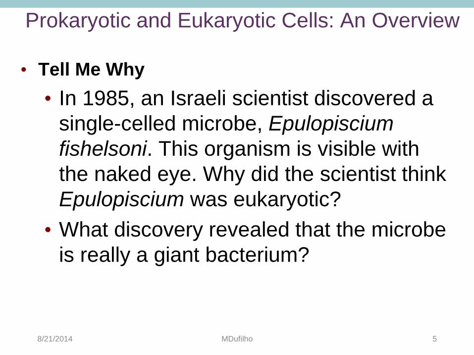
External Structures of Bacterial Cells
• Two Types of Glycocalyces
• Capsule
• Composed of organized repeating units of organic
chemicals
• Firmly attached to cell surface
• May prevent bacteria from being recognized by host
• Slime layer
• Loosely attached to cell surface
• Water soluble
• Sticky layer allows prokaryotes to attach to surfaces
8/20/2017 MDufilho 6
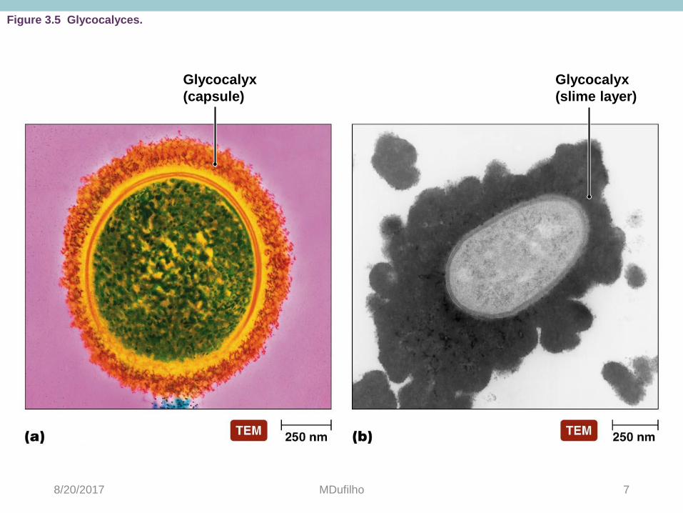
Figure 3.5 Glycocalyces.
Glycocalyx
(capsule)
Glycocalyx
(slime layer)
8/20/2017 MDufilho 7
External Structures of Bacterial Cells
• Flagella
• Are responsible for movement
• Have long structures that extend
beyond cell surface
• Are not present on all bacteria
• Structure
• Composed of filament, hook,
and basal body
• Basal body anchors the
filament and hook to cell wall
8/20/2017 MDufilho 8

Figure 3.6 Proximal structure of bacterial flagella.
Filament
Direction
of rotation
during run
Peptidoglycan
layer (cell wall)
Cytoplasmic
membrane
Cytoplasm
Protein rings
Rod
Filament
Outer
membrane
Peptidoglycan
layer
Cytoplasmic
membrane Cytoplasm
Cell
wall
Outer
protein
rings
Rod
Integral
protein
Inner
protein
rings
Integral
protein
Basal
body
Gram – Gram +
8/20/2017 MDufilho 9

Figure 3.8 Axial filament.
Axial filament
rotates around
cell
Outer
membrane
Cytoplasmic
membrane
Axial filament Spirochete
corkscrews
and moves
forward
Endoflagella
rotate
Axial filament
8/20/2017 MDufilho 10
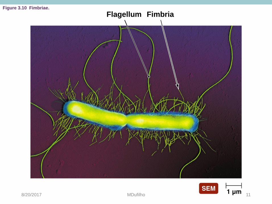
Figure 3.10 Fimbriae.
Flagellum Fimbria
8/20/2017 MDufilho 11

Figure 3.11 Pili.
Pilus
8/20/2017 MDufilho 12
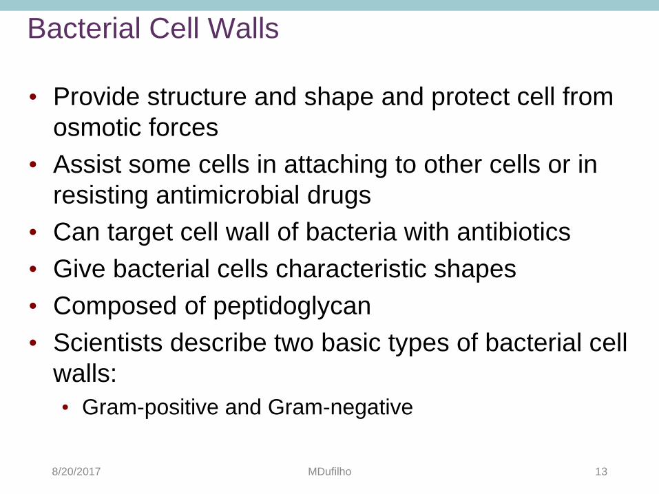
Bacterial Cell Walls
• Provide structure and shape and protect cell from
osmotic forces
• Assist some cells in attaching to other cells or in
resisting antimicrobial drugs
• Can target cell wall of bacteria with antibiotics
• Give bacterial cells characteristic shapes
• Composed of peptidoglycan
• Scientists describe two basic types of bacterial cell
walls:
• Gram-positive and Gram-negative
8/20/2017 MDufilho 13

Sugar
chain
Tetrapeptide
(amino acid)
crossbridge
Connecting chain
of amino acids
Figure 3.14 Possible structure of peptidoglycan.
8/20/2017 MDufilho 14

Bacterial Cell Walls
• Gram-Positive Bacterial Cell Walls
• Relatively thick layer of peptidoglycan
• Contain unique chemicals called teichoic acids
• Appear purple following Gram staining procedure
• Up to 60% mycolic acid in acid-fast bacteria helps cells
survive desiccation
8/20/2017 MDufilho 15

Prokaryotic Cell Walls
• Gram-Negative Bacterial Cell Walls
• Have only a thin layer of peptidoglycan
• Bilayer membrane outside the peptidoglycan contains
phospholipids, proteins, and lipopolysaccharide (LPS)
• Lipid A portion of LPS can cause fever, vasodilation,
inflammation, shock, and blood clotting
• May impede the treatment of disease
• Appear pink following Gram staining procedure
8/20/2017 MDufilho 16

Prokaryotic Cell Walls
• Bacteria Without Cell Walls
• A few bacteria lack cell walls
• Often mistaken for viruses due to small
size and lack of cell wall
• Have other features of prokaryotic cells
such as ribosomes
8/20/2017 MDufilho 17

Bacterial Cytoplasmic Membranes
• Structure
• Referred to as phospholipid bilayer
• Composed of lipids and associated proteins:
• Integral proteins
• Peripheral proteins
• Fluid mosaic model describes current understanding of
membrane structure
8/20/2017 MDufilho 18
Bacterial Cytoplasmic Membranes
• Functions?
8/20/2017 MDufilho 19
Cytoplasm of Bacteria
• Cytosol
• Liquid portion of cytoplasm
• Mostly water
• Contains cell's DNA in region called the nucleoid
• Inclusions
• May include reserve deposits of chemicals
8/20/2017 MDufilho 20

Figure 3.23 Granules of PHB in the bacterium Azotobacter chroococcum.
Polyhydroxybutyrate
8/20/2017 MDufilho 21

Cytoplasm of Bacteria
• Endospores
• Unique structures produced by some bacteria
• Defensive strategy against unfavorable conditions
• Vegetative cells transform into endospores when
multiple nutrients are limited
• Resistant to extreme conditions such as heat, radiation,
chemicals
8/20/2017 MDufilho 22

Figure 3.24 Formation of an endospore.
Steps in Endospore Formation
DNA is replicated.
Cytoplasmic membrane
invaginates to form
forespore.
Cytoplasmic membrane
grows and engulfs
forespore within a
second membrane.
Vegetative cell's DNA
disintegrates.
A cortex of pepti-
doglycan is deposited
between the membranes;
meanwhile, dipicolinic
acid and calcium ions
accumulate within the
center of the endospore.
Spore coat forms
around endospore.
Endospore matures:
Completion of spore coat.
Increase in resistance
to heat and chemicals by
unknown processes.
Endospore is released from
original cell.
Cell wall Cytoplasmic
membrane
DNA
Vegetative cell
Forespore
First
membrane
Second
membrane
Cortex
Spore coat
Outer
spore coat
Endospore
8/20/2017 MDufilho 23

Cytoplasm of Prokaryotes
• Nonmembranous Organelles
• Ribosomes
• Sites of protein synthesis
• Composed of polypeptides and ribosomal RNA
• Cytoskeleton
• Composed of three or four types of protein fibers
• Can play different roles in the cell:
• Cell division
• Cell shape
• Segregate DNA molecules
• Move through the environment
8/20/2017 MDufilho 24
External Structure of Eukaryotic Cells
• Glycocalyces
• Not as organized as prokaryotic capsules
• Help anchor animal cells to each other
• Strengthen cell surface
• Provide protection against dehydration
• Function in cell-to-cell recognition and communication
8/20/2017 MDufilho 25

Eukaryotic Cell Walls and Cytoplasmic
Membranes • Fungi, algae, plants, and some protozoa have cell
walls
• Composed of various polysaccharides:
• Cellulose found in plant cell walls
• Fungal cell walls composed of cellulose, chitin, and/or
glucomannan
• Algal cell walls composed of a variety of
polysaccharides
8/20/2017 MDufilho 26

Eukaryotic Cell Walls and Cytoplasmic
Membranes • All eukaryotic cells have cytoplasmic membrane
• Are a fluid mosaic of phospholipids and proteins
• Contain steroid lipids to help maintain fluidity
• Contain regions of lipids and proteins called
membrane rafts
• Control movement into and out of cell
8/20/2017 MDufilho 27

Cytoplasm of Eukaryotes
• Flagella
• Structure and Arrangement
• Differ structurally and functionally from prokaryotic flagella
• Within the cytoplasmic membrane
• Shaft composed of tubulin arranged to
form microtubules
• Filaments anchored to cell by basal body;
no hook
• May be single or multiple; generally found
at one pole of cell
• Function
• Do not rotate but undulate rhythmically
8/20/2017 MDufilho 28

Figure 3.31c Eukaryotic flagella and cilia.
Cytoplasmic membrane
Cytosol
Central pair
microtubules
"9 + 2" arrangement Microtubules
(doublet)
Cytoplasmic membrane
Portion cut away to show transition area from doublets to triplets and the end of central microtubules
"9 + 0" arrangement
Microtubules
(triplet)
Basal body
8/20/2017 MDufilho 29

Cytoplasm of Eukaryotes
• Cilia
• Shorter and more numerous than flagella
• Coordinated beating propels cells through their
environment
• Also used to move substances past the surface of the
cell
8/20/2017 MDufilho 30

Cytoplasm of Eukaryotes
• Other Nonmembranous Organelles
• Ribosomes
• Larger than prokaryotic ribosomes (80S versus 70S)
• Composed of 60S and 40S subunits
• Cytoskeleton
• Extensive network of fibers and tubules
• Anchors organelles
• Produces basic shape of the cell
• Made up of tubulin microtubules, actin microfilaments,
and intermediate filaments
8/20/2017 MDufilho 31

Table 3.5 Nonmembranous and Membranous Organelles of Cells
8/20/2017 MDufilho 32

Cytoplasm of Eukaryotes
• Endosymbiotic Theory
• Eukaryotes formed from union of small aerobic
prokaryotes with larger anaerobic prokaryotes
• Smaller prokaryotes became internal parasites:
• Parasites lost ability to exist independently
• Larger cell became dependent on parasites for aerobic
ATP production
• Aerobic prokaryotes evolved into mitochondria
• Similar scenario for origin of chloroplasts
• Theory is not universally accepted
8/20/2017 MDufilho 33