3 4 Immobilized Enzyme Systems3.4 Immobilized Enzyme Systems
C H A P T E R T W E N T Y - S E V E N
M
IS
*{
ethods
SN 0
QIAGInstitu
Immobilized-Metal Affinity
Chromatography (IMAC): A Review
Helena Block,* Barbara Maertens,* Anne Spriestersbach,*
Nicole Brinker,* Jan Kubicek,* Roland Fabis,* Jorg Labahn,†
and Frank Schafer*
Contents
1. O
in
076
ENte
verview on IMAC Ligands and Immobilized Ions
Enzymology, Volume 463 # 2009
-6879, DOI: 10.1016/S0076-6879(09)63027-5 All rig
GmbH, Qiagen Strasse 1, Hilden, Germanyof Structural Biology (IBI-2), Research Center Julich, Julich, Germany
Else
hts
440
2. IM
AC Applications 4442.1.
D etection and immobilization 4452.2.
P urification of protein fractions 4462.3.
P urification of His-tagged proteins 4482.4.
G eneral considerations of protein purification by IMAC 4512.5.
C opurifying proteins on IMAC and what to do about it 4542.6.
IM AC for industrial-scale protein production 4582.7.
H igh-throughput automation of IMAC 4602.8.
S pecial applications: Purification of membrane proteins 4612.9.
S pecial applications: Purification of zinc-finger proteins 4632
.10. P rotein purification protocols 4642
.11. C leaning and sanitization 4662
.12. S implified metal-ion stripping and recharging protocol 4673. C
onclusions 467Ackn
owledgments 468Refe
rences 468Abstract
This article reviews the development of immobilized-metal affinity chromatog-
raphy (IMAC) and describes its most important applications. We provide an
overview on the use of IMAC in protein fractionation and proteomics, in protein
immobilization and detection, and on some special applications such as purifi-
cation of immunoglobulins and the Chelex method. The most relevant applica-
tion—purification of histidine-tagged recombinant proteins—will be reviewed
vier Inc.
reserved.
439
440 Helena Block et al.
in greater detail with focus of state-of-the-art materials, methods, and proto-
cols, and the limitations of IMAC and recent advances to improve the technology
and the methods will be described.
1. Overview on IMAC Ligands
and Immobilized Ions
The concept of immobilized-metal affinity chromatography (IMAC)has first been formulated and its feasibility shown by Porath et al. (1975).It was based on the known affinity of transition metal ions such as Zn2þ,Cu2þ, Ni2þ, and Co2þ to histidine and cysteine in aqueous solutions(Hearon, 1948) and extended to the idea to use metal ions ‘‘strongly fixed’’to a support to fractionate protein solutions. As the chelating ligand used to fixthemetal to agarose Porath used iminodiacetic acid (IDA)which is still in usetoday in many commercial IMAC resins. Already in 1975, Porath speculatedthat the affinity of immobilizedmetals to histidine-containing proteinsmightnot be the only application for IMAC—and he was right after all.
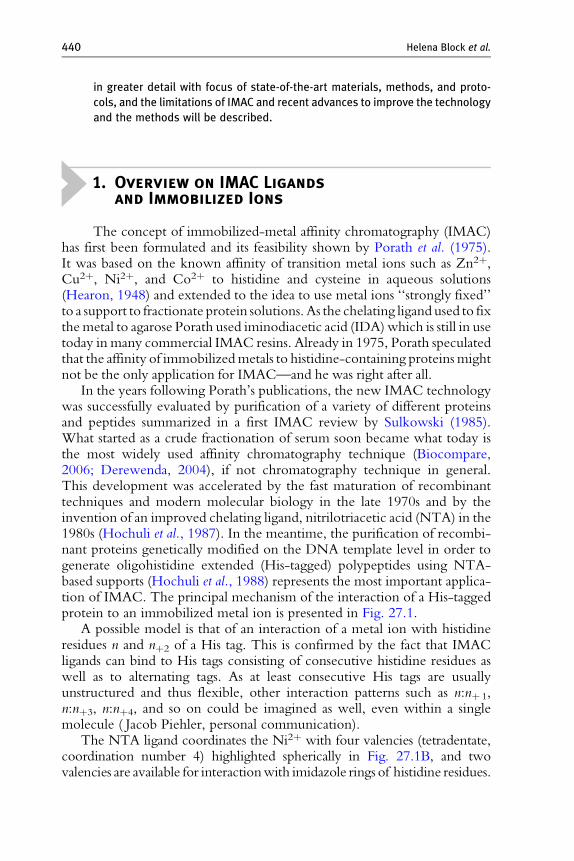
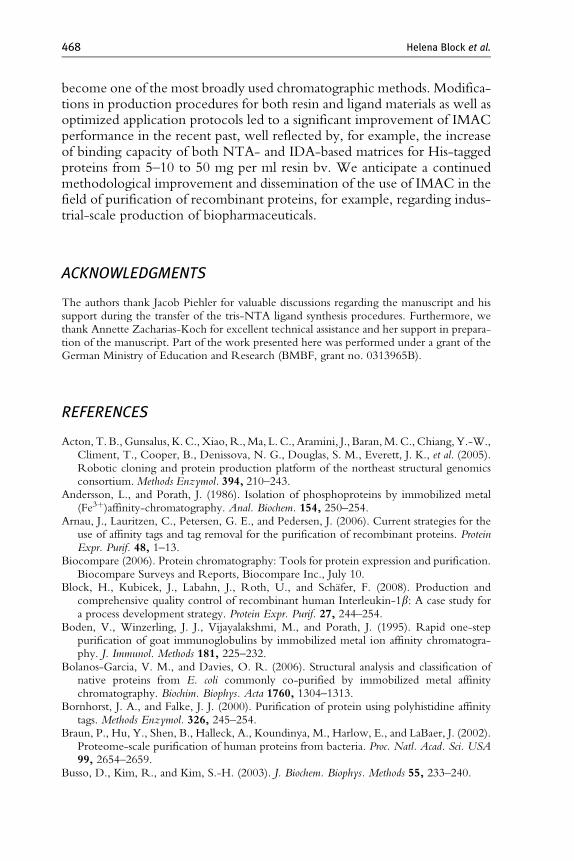
In the years following Porath’s publications, the new IMAC technologywas successfully evaluated by purification of a variety of different proteinsand peptides summarized in a first IMAC review by Sulkowski (1985).What started as a crude fractionation of serum soon became what today isthe most widely used affinity chromatography technique (Biocompare,2006; Derewenda, 2004), if not chromatography technique in general.This development was accelerated by the fast maturation of recombinanttechniques and modern molecular biology in the late 1970s and by theinvention of an improved chelating ligand, nitrilotriacetic acid (NTA) in the1980s (Hochuli et al., 1987). In the meantime, the purification of recombi-nant proteins genetically modified on the DNA template level in order togenerate oligohistidine extended (His-tagged) polypeptides using NTA-based supports (Hochuli et al., 1988) represents the most important applica-tion of IMAC. The principal mechanism of the interaction of a His-taggedprotein to an immobilized metal ion is presented in Fig. 27.1.
A possible model is that of an interaction of a metal ion with histidineresidues n and nþ2 of a His tag. This is confirmed by the fact that IMACligands can bind to His tags consisting of consecutive histidine residues aswell as to alternating tags. As at least consecutive His tags are usuallyunstructured and thus flexible, other interaction patterns such as n:nþ 1,n:nþ3, n:nþ4, and so on could be imagined as well, even within a singlemolecule ( Jacob Piehler, personal communication).
The NTA ligand coordinates the Ni2þ with four valencies (tetradentate,coordination number 4) highlighted spherically in Fig. 27.1B, and twovalencies are available for interactionwith imidazole rings of histidine residues.
Protein
A
Ni-IDA
N
NN
N
NN
N
N
NH2O
CH2CH2
Ni2+
O
NN
N
N
O
Protein
B
Ni-NTA
N
NN
N
NN
N
N
N
CH2
CH
CH2
Ni2+
ON
N
NN
O
O
Protein
C
Ni-TED
N
NN
N
NN
NN
N
NNCH2
CH2
CH2
CH2 CH2Ni2+
O
O
N
NN
O
Figure 27.1 Model of the interaction between residues in the His tag and the metal ionin tri- (IDA), tetra- (NTA), and pentadentate IMAC ligands (TED).
IMAC 441

442 Helena Block et al.
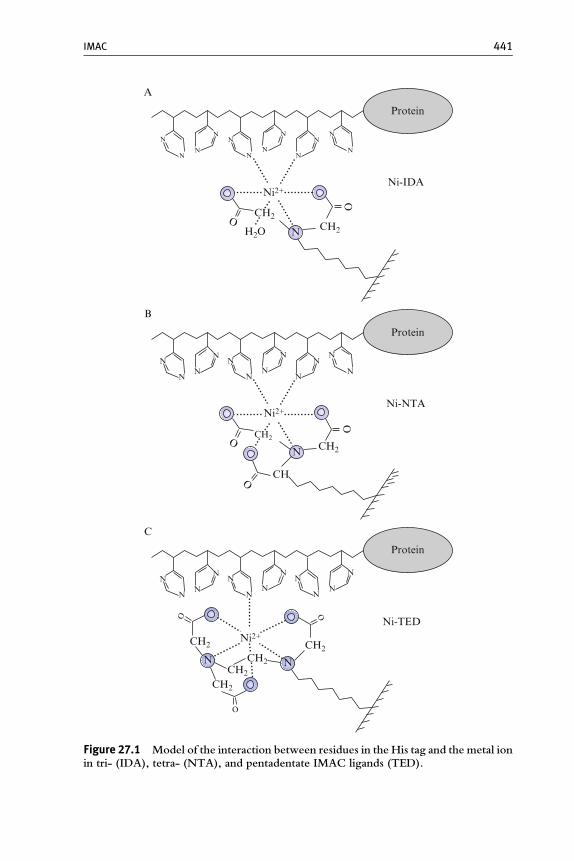
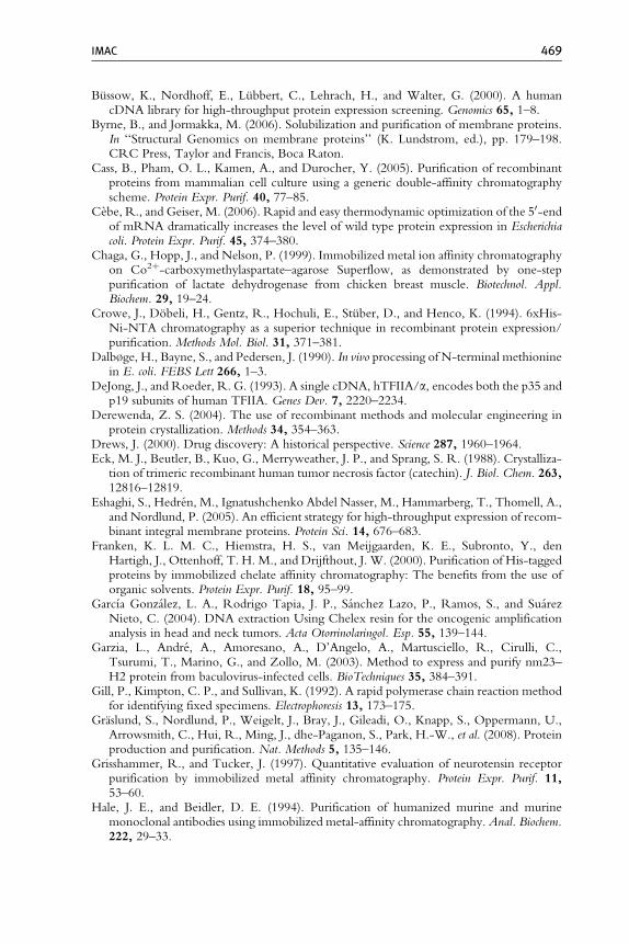
This ratio has turned out to be most effective for purification of His-taggedproteins. Another tetradentate ligand is carboxymethyl aspartate (CM-Asp;Chaga et al., 1999), commercially available as cobalt-charged Talon resin.In contrast to tetradentate ligands, IDA coordinates a divalent ion with threevalencies (tridentate, coordination number 3, Fig. 27.1A) leaving threevalencies free for imidazole ring interaction while it is unclear whetherthe third is sterically able to participate in the interaction. The coordinationnumber seems to play an important role regarding the quality of the purifiedprotein fraction. While protein recovery is usually similar between IDA-and NTA-based chromatography (Fig. 27.2D), a higher leaching of metalions from IDA ligands compared to NTA is observed in general (Hochuli,1989) and even increased under reducing conditions (Fig. 27.2C). Althoughthe metal content in the elution fractions (E in Fig. 27.2C) is higher but stillwithin the same order of magnitude, significantly more Ni2þ ions leachfrom the IDA resin in the equilibration and wash steps (W in Fig. 27.2C).Besides considerable metal leaching, purification of His-tagged proteinsusing an IDA matrix frequently results in lower purity compared toNTA-based purification (Fig. 27.2A and D).
A B
C
His6-HIV-RT
Ni-IDA Ni-NTA Ni-TED
M E1 E2 E1 E2 E1 E2
150
log
[Ni]
(ppb
)
10,000
1000
100
10
1
Ni-IDANi-NTANi-TED100
[FU
]
50
0
1300470
4
W E
Ni-IDA Ni-NTA Ni-TED
W E W E
100
2
54
2624 28 30[s]
Figure 27.2 (Continued)
EMG1D
NTA
LFWE
IDA NTA IDA NTA IDA NTA IDA NTA IDA NTA IDA
FYN1 JNK1 MAPKAP5 p38a PIM1
FWE LFWEFWE LFWEFWE LFWEFWE LFWEFWE LFWEFWE
E a b g d
1 2 3 4 1 2 3 4 1 2 3 41 2 3 4 M200976655
3631
21
14
6
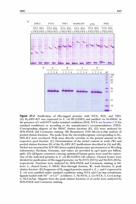
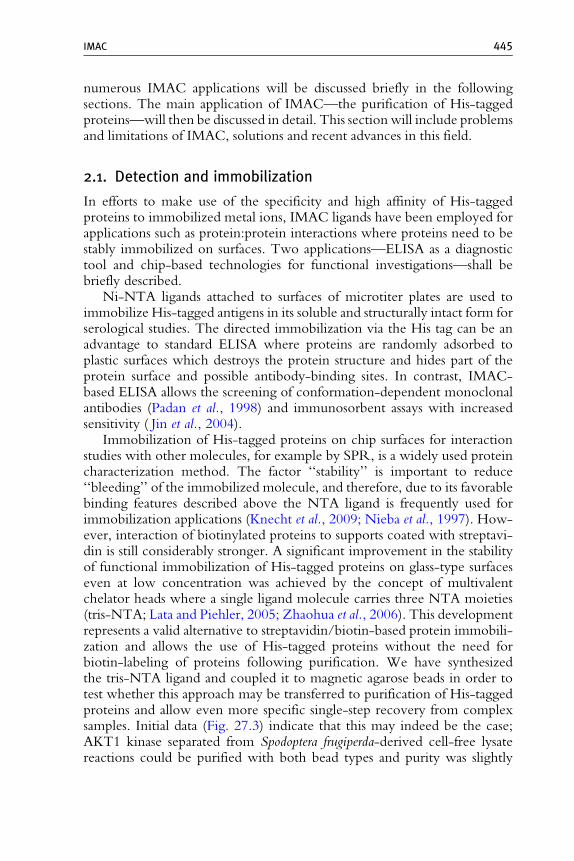
Figure 27.2 Purification of His-tagged proteins with NTA, IDA, and TED.(A) H6-HIV-RT was expressed in E. coli BL21(DE3) and purified via Ni-IMAC inthe presence of 1 mMDTT under standard conditions (IDA, NTA; see Section 2.10 forstandard conditions) or according to the manufacturer’s recommendations (TED).Corresponding aliquots of the IMAC elution fractions (E1, E2) were analyzed bySDS–PAGE and Coomassie staining. (B) Bioanalyzer 2100 lab-on-a-chip analysis ofpooled elution fractions. The peaks from the electropherograms corresponding to H6-HIV-RT were overlayed. Peak areas directly correlate to the protein amount in therespective pool fraction. (C) Determination of the nickel content in wash (W) andpooled elution fractions (E) of the H6-HIV-RT purifications described in (A) and (B).Nickel was measured by ICP-MS (intercoupled plasma mass spectrometry) at WesslingLaboratories, Bochum, Germany, and values are provided in mg/l (parts per billion,ppb). (D) QIAgene constructs carrying optimized human genes were used for expres-sion of the indicated proteins in E. coli BL21(DE3) LB cultures. Cleared lysates weredivided for purification of His-tagged proteins via Ni-NTA (NTA) and Ni-IDA (IDA),respectively. Fractions were analyzed by SDS–PAGE and Coomassie staining as fol-lows: L, cleared lysate; F, IMAC flow-through fraction; W, wash fraction; E, peakelution fraction. (E) Fragments of H6-tagged proteins named a, b, g, and d expressed inE. coli were purified under standard conditions using NTA and Cm-Asp tetradentateligands loaded with Ni2þ or Co2þ as follows: 1, Ni-NTA; 2, Co-NTA; 3, Co-CmAsp;4, Ni-CmAsp. Aliquots from the peak elution fractions (2 ml each) were analyzed bySDS–PAGE and Coomassie staining.
IMAC 443
444 Helena Block et al.
The reason for the lower purity may be that leaching of metal from thetridentate ligand generates charged groups which could act as a cationexchanger and bind positively charged groups on the surface of proteins.The lowest metal leaching is obtained if a pentadentate ligand is used(Fig. 27.2C) which coordinates the ions extremely tightly, and such resinsmay represent a valid alternative if low metal ion leaching into the proteinpreparation is very important. However, in this case only one coordinationsite remains for His tag binding and recovery of His-tagged protein is usuallyconsiderably lower than with IDA or NTA (Fig. 27.2B).
The choice of the metal ion immobilized on the IMAC ligand dependson the application. While trivalent cations such as Al3þ, Ga3þ, and Fe3þ(Andersson and Porath, 1986; Muszynska et al., 1986; Posewitz and Tempst,1999) or tetravalent Zr4þ (Zhou et al., 2006) are preferred for capture ofphosphoproteins and phosphopeptides, divalent Cu2þ, Ni2þ, Zn2þ, andCo2þ ions are used for purification of His-tagged proteins. Combinations ofa tetradentate ligand that ensures strong immobilization, and a metal ion thatleaves two coordination sites free for interaction with biopolymers (Ni2þ,Co2þ) has gained most acceptance and leads to similar recovery and purityof eluted protein. As a typical result using such combinations, Fig. 27.2Eshows the purification of several protein fragments derived from differentgenes that have been expressed and purified as His-tagged proteins by Ni2þand Co2þ immobilized on NTA and Cm-Asp tetradentate ligands.
2. IMAC Applications
Initially developed for purification of native proteins with an intrinsicaffinity to metal ions (Porath et al., 1975), IMAC has turned out to be atechnology with a very broad portfolio of applications. On the chro-matographic purification side, the range of proteins was expanded from theprimary metalloproteins to antibodies, phosphorylated proteins, and recom-binant His-tagged proteins. IMAC is being used in proteomics approacheswhere fractions of the cellular protein pool are enriched and analyzeddifferentially (phosphoproteome, metalloproteome) by mass spectrometricaltechniques; here, IMAC formats can be traditionally bead based or the ligandcan be used on functionalized surfaces such as SELDI (surface-enhanced laserdesorption/ionization) chips. Other chip-based applications include surfaceplasmon resonance (SPR) and allow the immobilization of His-taggedproteins for quantitative functional and kinetic investigations. In addition,the IMAC principle has been used as an inhibitor depletion step prior toPCR amplification of nucleic acids from complex samples such as blood in atechnology called Chelex (Walsh et al., 1991). The most relevant of the
IMAC 445
numerous IMAC applications will be discussed briefly in the followingsections. The main application of IMAC—the purification of His-taggedproteins—will then be discussed in detail. This section will include problemsand limitations of IMAC, solutions and recent advances in this field.
2.1. Detection and immobilization
In efforts to make use of the specificity and high affinity of His-taggedproteins to immobilized metal ions, IMAC ligands have been employed forapplications such as protein:protein interactions where proteins need to bestably immobilized on surfaces. Two applications—ELISA as a diagnostictool and chip-based technologies for functional investigations—shall bebriefly described.
Ni-NTA ligands attached to surfaces of microtiter plates are used toimmobilize His-tagged antigens in its soluble and structurally intact form forserological studies. The directed immobilization via the His tag can be anadvantage to standard ELISA where proteins are randomly adsorbed toplastic surfaces which destroys the protein structure and hides part of theprotein surface and possible antibody-binding sites. In contrast, IMAC-based ELISA allows the screening of conformation-dependent monoclonalantibodies (Padan et al., 1998) and immunosorbent assays with increasedsensitivity ( Jin et al., 2004).
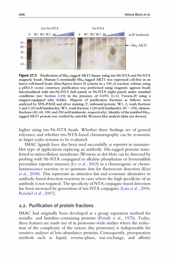
Immobilization of His-tagged proteins on chip surfaces for interactionstudies with other molecules, for example by SPR, is a widely used proteincharacterization method. The factor ‘‘stability’’ is important to reduce‘‘bleeding’’ of the immobilized molecule, and therefore, due to its favorablebinding features described above the NTA ligand is frequently used forimmobilization applications (Knecht et al., 2009; Nieba et al., 1997). How-ever, interaction of biotinylated proteins to supports coated with streptavi-din is still considerably stronger. A significant improvement in the stabilityof functional immobilization of His-tagged proteins on glass-type surfaceseven at low concentration was achieved by the concept of multivalentchelator heads where a single ligand molecule carries three NTA moieties(tris-NTA; Lata and Piehler, 2005; Zhaohua et al., 2006). This developmentrepresents a valid alternative to streptavidin/biotin-based protein immobili-zation and allows the use of His-tagged proteins without the need forbiotin-labeling of proteins following purification. We have synthesizedthe tris-NTA ligand and coupled it to magnetic agarose beads in order totest whether this approach may be transferred to purification of His-taggedproteins and allow even more specific single-step recovery from complexsamples. Initial data (Fig. 27.3) indicate that this may indeed be the case;AKT1 kinase separated from Spodoptera frugiperda-derived cell-free lysatereactions could be purified with both bead types and purity was slightly
tris-Ni-NTA Ni-NTA
F
9766
36
21
14
W1 W2 W3 40 F W1 W2 W3 40250 250 mM imidazole
His6-AKT1
Figure 27.3 Purification of His6-tagged AKT1 kinase using tris-Ni-NTA and Ni-NTAmagnetic beads. Human C-terminally His6-tagged AKT1 was expressed cell-free in aninsect cell-based lysate (EasyXpress Insect II system) in a 100 ml reaction volume usinga pIX4.0 vector construct; purification was performed using magnetic agarose beadsfunctionalized with tris-Ni-NTA (left panel) or Ni-NTA (right panel) under standardconditions (see Section 2.10) in the presence of 0.05% (v/v) Tween-20 using amagnet-equipped tube holder. Aliquots of purification fractions as follows wereanalyzed by SDS–PAGE and silver staining: F, unbound protein; W1, 2, wash fractions1 and 2 (10 mM imidazole); W3, wash fraction 3 (20 mM imidazole); 40! 250, elution-fractions (40, 60, 100, and 250 mM imidazole, respectively). Identity of the purified His6-tagged AKT1 protein was verified by anti-His Western blot analysis (data not shown).
446 Helena Block et al.
higher using tris-Ni-NTA beads. Whether these findings are of generalrelevance and whether tris-NTA-based chromatography can be economicin larger scales remains to be evaluated.
IMAC ligands have also been used successfully as reporter in immuno-blot type of applications replacing an antibody. His-tagged proteins trans-ferred to nitrocellulose membrane (Western or dot blot) can be detected byprobing with Ni-NTA conjugated to alkaline phosphatase or horseraddishperoxidase reporter enzymes (Lv et al., 2003) in a chromogenic or chemi-luminescence reaction or to quantum dots for fluorescent detection (Kimet al., 2008). This represents an attractive fast and economic alternative toantibody-based detection reactions in cases where the high specificity of anantibody is not required. The specificity of NTA conjugate-based detectionhas been increased by generation of tris-NTA conjugates (Lata et al., 2006;Reichel et al., 2007).
2.2. Purification of protein fractions
IMAC had originally been developed as a group separation method formetallo- and histidine-containing proteins (Porath et al., 1975). Today,these features are made use of in proteome-wide studies where the reduc-tion of the complexity of the system (the proteome) is indispensable forsensitive analyses of low-abundance proteins. Consequently, preseparationmethods such as liquid, reverse-phase, ion-exchange, and affinity
IMAC 447
chromatography—such as IMAC—have gradually been used in proteomicsto enrich proteins that may otherwise be lost in detection (Loo, 2003; Stasykand Huber, 2004). The application of IMAC in proteomics has recentlybeen reviewed (Sun et al., 2005) and is focused on the enrichment ofphosphoproteins and phosphopeptides and on metal-binding proteins.In the enrichment step, a complex sample such as a cell lysate or blood ispassed over the IMAC matrix, washed and the fraction of interest eluted byvariation of pH or with high concentrations of imidazole. This fraction isthen analyzed by mass spectrometry (MS) or fractionated further by two-dimensional gel electrophoresis followed by MS or by additional liquidchromatography coupled to MS (LC–MS).
Whereas Fe3þ, Al3þ, and Ga3þ are the preferred ions for phospho-protein research and are usually immobilized to IDA, the ions useful forIMAC-based analysis of the metalloproteome are the elements copper,nickel, zinc, and iron which are essential for life. The metalloproteome isdefined as a set of proteins that have metal-binding ability and several aspectsof this proteomics discipline have been reviewed recently (Shi and Chance,2008; Sun et al., 2005). Proteins with metal-binding affinity can be enrichedby either making use of their ability to bind to certain immobilized Me2þions (e.g., to Me2þ-NTA) or by making use of their bound Me2þ ion bycatching as a Me2þ-protein on an uncharged IMAC ligand (e.g., NTA).
Also, chip-based proteome profiling IMACmethods have been reported(Slentz et al., 2003) and are in use as a tool in clinical screening applicationsfor phospho group- and histidine-containing proteins and peptides(SELDI–IMAC).
IMAC can also be used to bind and separate at least mono- and dinu-cleotides based on a complex phenomenon accounted for by differentialinterplay of affinities of the potential binding sites (oxygen in the phosphategroup, nitrogen and oxygen on the bases, hydroxyl groups on the ribose) tothe immobilized metal (Hubert and Porath, 1980, 1981).
A quite different group-specific separation application of IMAC isrepresented by the affinity of antibodies to immobilized metal ions. As themolecular basis for this interaction an endogenous metal-binding site on theheavy chain (Hale and Beidler, 1994) and an arrangement of histidineresidues on the antibody (Porath and Olin, 1983) have been discussed.Adsorption of immunoglobulins from different sources on IMAC matriceshas been reported by many authors (human IgG, Porath and Olin, 1983;humanized murine IgG, Hale and Beidler, 1994; goat IgG, Boden et al.,1995). Antibody purification has been successfully performed using variousIMAC formats including gels (Hale and Beidler, 1994; Vancan et al., 2002),methacrylate polymer (Meszarosova et al., 2003), and membraneous hollowfibers (Serpa et al., 2005). The mild elution of the protein with salts, costs,and the robustness of IMAC matrices have been identified as advantageousover traditional protein A or G chromatography (Serpa et al., 2005).
448 Helena Block et al.
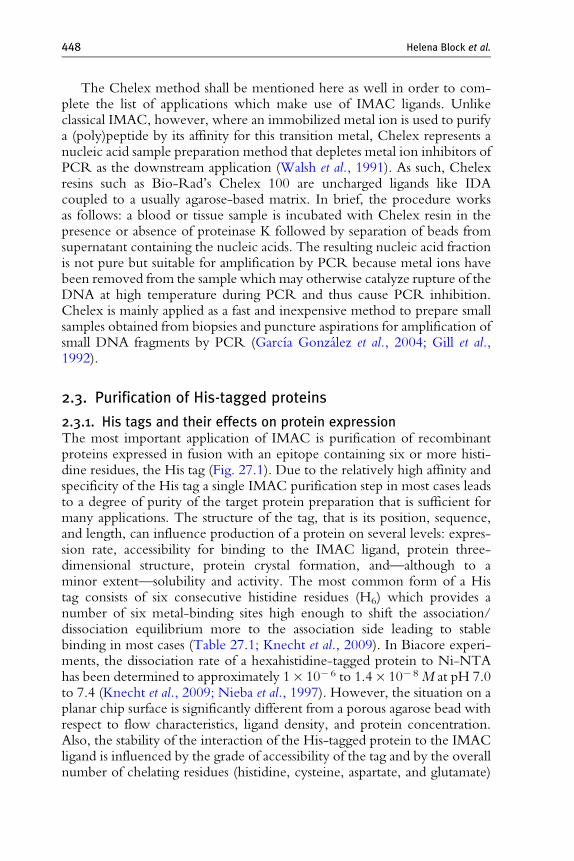
The Chelex method shall be mentioned here as well in order to com-plete the list of applications which make use of IMAC ligands. Unlikeclassical IMAC, however, where an immobilized metal ion is used to purifya (poly)peptide by its affinity for this transition metal, Chelex represents anucleic acid sample preparation method that depletes metal ion inhibitors ofPCR as the downstream application (Walsh et al., 1991). As such, Chelexresins such as Bio-Rad’s Chelex 100 are uncharged ligands like IDAcoupled to a usually agarose-based matrix. In brief, the procedure worksas follows: a blood or tissue sample is incubated with Chelex resin in thepresence or absence of proteinase K followed by separation of beads fromsupernatant containing the nucleic acids. The resulting nucleic acid fractionis not pure but suitable for amplification by PCR because metal ions havebeen removed from the sample which may otherwise catalyze rupture of theDNA at high temperature during PCR and thus cause PCR inhibition.Chelex is mainly applied as a fast and inexpensive method to prepare smallsamples obtained from biopsies and puncture aspirations for amplification ofsmall DNA fragments by PCR (Garcıa Gonzalez et al., 2004; Gill et al.,1992).
2.3. Purification of His-tagged proteins
2.3.1. His tags and their effects on protein expressionThe most important application of IMAC is purification of recombinantproteins expressed in fusion with an epitope containing six or more histi-dine residues, the His tag (Fig. 27.1). Due to the relatively high affinity andspecificity of the His tag a single IMAC purification step in most cases leadsto a degree of purity of the target protein preparation that is sufficient formany applications. The structure of the tag, that is its position, sequence,and length, can influence production of a protein on several levels: expres-sion rate, accessibility for binding to the IMAC ligand, protein three-dimensional structure, protein crystal formation, and—although to aminor extent—solubility and activity. The most common form of a Histag consists of six consecutive histidine residues (H6) which provides anumber of six metal-binding sites high enough to shift the association/dissociation equilibrium more to the association side leading to stablebinding in most cases (Table 27.1; Knecht et al., 2009). In Biacore experi-ments, the dissociation rate of a hexahistidine-tagged protein to Ni-NTAhas been determined to approximately 1� 10� 6 to 1.4� 10� 8 M at pH 7.0to 7.4 (Knecht et al., 2009; Nieba et al., 1997). However, the situation on aplanar chip surface is significantly different from a porous agarose bead withrespect to flow characteristics, ligand density, and protein concentration.Also, the stability of the interaction of the His-tagged protein to the IMACligand is influenced by the grade of accessibility of the tag and by the overallnumber of chelating residues (histidine, cysteine, aspartate, and glutamate)
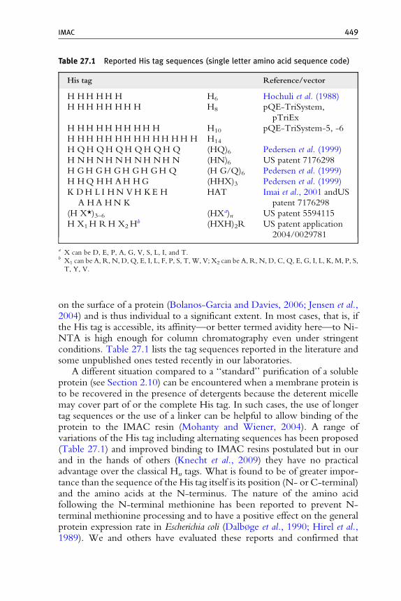
Table 27.1 Reported His tag sequences (single letter amino acid sequence code)
His tag Reference/vector
H HH HH H H6 Hochuli et al. (1988)
H HH HH HH H H8 pQE-TriSystem,
pTriEx
H HH HH HH HH H H10 pQE-TriSystem-5, -6
H HH HH HH HH HH HH H H14
H QH QH QH QH QH Q (HQ)6 Pedersen et al. (1999)
H NH NH NH NH NH N (HN)6 US patent 7176298
H GH GH GH GH GH Q (H G/Q)6 Pedersen et al. (1999)
H HQ HH AH HG (HHX)3 Pedersen et al. (1999)
K DH L I HN VH KE H
A HA HN K
HAT Imai et al., 2001 andUS
patent 7176298
(H X*)3–6 (HXa)n US patent 5594115
H X1H RH X2Hb (HXH)2R US patent application
2004/0029781
a X can be D, E, P, A, G, V, S, L, I, and T.b X1 can be A, R, N, D, Q, E, I, L, F, P, S, T, W, V; X2 can be A, R, N, D, C, Q, E, G, I, L, K, M, P, S,T, Y, V.
IMAC 449
on the surface of a protein (Bolanos-Garcia and Davies, 2006; Jensen et al.,2004) and is thus individual to a significant extent. In most cases, that is, ifthe His tag is accessible, its affinity—or better termed avidity here—to Ni-NTA is high enough for column chromatography even under stringentconditions. Table 27.1 lists the tag sequences reported in the literature andsome unpublished ones tested recently in our laboratories.
A different situation compared to a ‘‘standard’’ purification of a solubleprotein (see Section 2.10) can be encountered when a membrane protein isto be recovered in the presence of detergents because the deterent micellemay cover part of or the complete His tag. In such cases, the use of longertag sequences or the use of a linker can be helpful to allow binding of theprotein to the IMAC resin (Mohanty and Wiener, 2004). A range ofvariations of the His tag including alternating sequences has been proposed(Table 27.1) and improved binding to IMAC resins postulated but in ourand in the hands of others (Knecht et al., 2009) they have no practicaladvantage over the classical Hn tags. What is found to be of greater impor-tance than the sequence of the His tag itself is its position (N- or C-terminal)and the amino acids at the N-terminus. The nature of the amino acidfollowing the N-terminal methionine has been reported to prevent N-terminal methionine processing and to have a positive effect on the generalprotein expression rate in Escherichia coli (Dalb�ge et al., 1990; Hirel et al.,1989). We and others have evaluated these reports and confirmed that

450 Helena Block et al.
namely, lysine and arginine in position 2 of a protein N-terminus show thiseffect (Pedersen et al., 1999; Schafer et al., 2002a; Svensson et al., 2006). Thehigh success rate of using N-terminal His (and Strep II) tags is, however, notonly based on the stimulatory effect of the second amino acid on expressionbut also seems to have stabilizing impact on the mRNA structure in thetranslation initiation region. We compared bacterial and eukaryotic expres-sion of N- versus C-terminal positioning of the His (and Strep II) tag onseveral proteins and analyzed expression level and solubility (Fig. 27.4). N-terminal tags improved protein expression in most cases.
Systematic investigation of the 50 region of mRNAs showed that hairpinloops forming in the translation initiation area frequently are the reason forlow expression as they prevent the ribosome:mRNA binding (Cebe andGeiser, 2006). Sequence optimization can be performed to destabilizehairpin formation and improve expression, and a similar result was obtainedwhen the proteins were expressed with an N-terminal H6 tag. The effectsdescribed in Fig. 27.4 can be explained with this initiator effect of prevent-ing secondary structures on the mRNA in the translation initiation region.Similar observations were also made by others (Busso et al., 2003; Svenssonet al., 2006) and this may be the reason for the attractiveness of N-terminal
M30
50
30
50
15
50307550
T S T S T S T S T S T S T S T S T S T S T S
Insect cell-freeE.coli cell-freeA B
N-H 6C-H 6
N-S llC-S ll
N-H 6/C
-S ll
N-S 11/C
-H 6
N-H 6C-H 6
N-S llC-S ll
N-H 6/C
-S ll
N-S II/C
-H 6
T S
TNFa
TFIIAab
MKK3
TFIIAg
TBP
IRAK4
Figure 27.4 Effect of His tag position on protein expression. Proteins were expressedfrom PCR product templates generated by two-step PCR using the EasyXpress LinearTemplate Kit in E. coli- (A) and insect cell-derived (B) lysates (EasyXpress ProteinSynthesis and EasyXpress Insect II Kits, respectively). Initiator (adapter) primers in theLinear Template Kit were designed in order to prevent formation of secondary struc-ture in the translation initiation region on the mRNA and introduce the following tagsequence(s): N-H6, N-terminal His6 tag; C-H6, C-terminal His6 tag; N-SII, N-terminalStrep II tag; C-SII, C-terminal Strep II tag; N-H6/C-SII, N-terminal His6 andC-terminal Strep II tags; N-SII/C-H6, N-terminal Strep II and C-terminal His6 tags.Corresponding aliquots of total (T) and soluble protein (S, supernatant after centrifu-gation at 15,000� g for 10 min) were loaded on a SDS gel. Protein bands werevisualized by Western blot analysis using a mixture of Penta anti-His and anti-Streptag antibodies. Protein sizes are (kDa) TNFa, 21; TBP, 38; TFIIAab, 55; TFIIAg, 12.5;MKK3, 39; IRAK4, 55. M, His-tagged protein size markers (kDa).
IMAC 451
His tags. However, in some cases such as the one of IRAK4 the C-terminalHis tag has a more pronounced effect on both expression rate and solubility(Fig. 27.4). Recently, expression of an insect toxin in E. coli was reportedwhere the similar observation of higher solubility and thermostability of theC-terminally His-tagged form was made (Xu et al., 2008). The authorsdiscussed that the C-terminal tag stabilized the overall protein structure.Other groups found the His tag to contribute slightly negative to solubilitywhen compared to the untagged protein but to improve yield when fused totheC-terminus (Woestenenk et al., 2004). All in all, these data suggest that anevaluation of at least N- and C-terminally tagged variants of a protein willincrease the chance to obtain reasonable expression and quality of a recom-binant protein. For expression of proteins to be secreted, tags should beplaced at the C-terminus to prevent interference with membrane trafficking.
2.4. General considerations of protein purification by IMAC
There are several advantages of IMAC for purification of His-taggedproteins compared to other affinity chromatography principles as the reasonfor being the most widely used chromatographic technique (Biocompare,2006; Derewenda, 2004). Besides low costs and the simplicity of use therobustness of IMAC is certainly its most striking feature: (i) the His-tag:ligand interaction works under both native and denaturing conditions suchas 8 M urea or 6 M guanidinium hydrochloride (Hochuli et al., 1988)enabling subsequent on-column refolding ( Jungbauer et al., 2004) as wellas (ii) both oxidizing and reducing conditions, (iii) protein binding with-stands a broad spectrum of various chemicals of different types (Table 27.1summarizes chemical compatibilities for Ni-NTA IMAC and some limita-tions), (iv) its relatively high affinity and specificity allows high capturingefficiency even in the presence of high protein titers, and (v) the scalabilityof the purification procedure.
Despite the broad compatibility, IMAC has its limitations. Obviously,the use of chelating agents has to be avoided which can be a disadvantage asEDTA, a potent inhibitor of metalloproteases, can only be applied in lowconcentrations. Care should also be taken with the use of other potentiallychelating groups such as Tris, ammonium salts, and certain amino acids(Table 27.2).
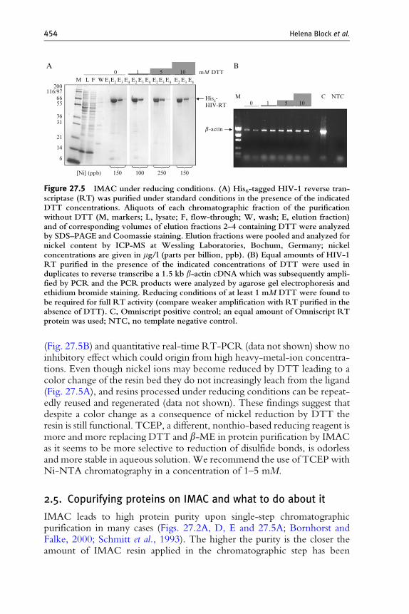
Until recently, the use of strong reducing agents such as DTT in IMACprocessing has been regarded as problematic because of reduction of nickeland, as a consequence, suspected increase of nickel concentrations inprotein preparations. However, we found that moderate concentrations ofDTT are fully compatible with NTA-based purification. This is shown, forexample, in Fig. 27.5A for HIV-1 reverse transcriptase (RT) purified withunaffected efficiency in the presence of up to 10 mM DTT. Also, RTactivity is not influenced by these conditions and both end-point
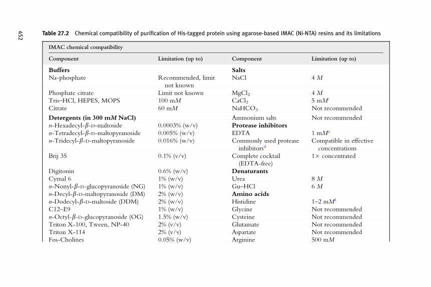
Table 27.2 Chemical compatibility of purification of His-tagged protein using agarose-based IMAC (Ni-NTA) resins and its limitations
IMAC chemical compatibility
Component Limitation (up to) Component Limitation (up to)
Buffers Salts
Na-phosphate Recommended, limit
not known
NaCl 4 M
Phosphate citrate Limit not known MgCl2 4 M
Tris–HCl, HEPES, MOPS 100 mM CaCl2 5 mMc
Citrate 60 mM NaHCO3 Not recommended
Detergents (in 300 mM NaCl) Ammonium salts Not recommended
n-Hexadecyl-b-D-maltoside 0.0003% (w/v) Protease inhibitors
n-Tetradecyl-b-D-maltopyranoside 0.005% (w/v) EDTA 1 mMa
n-Tridecyl-b-D-maltopyranoside 0.016% (w/v) Commonly used protease
inhibitorsdCompatible in effective
concentrations
Brij 35 0.1% (v/v) Complete cocktail
(EDTA-free)
1� concentrated
Digitonin 0.6% (w/v) Denaturants
Cymal 6 1% (w/v) Urea 8 M
n-Nonyl-b-D-glucopyranoside (NG) 1% (w/v) Gu–HCl 6 M
n-Decyl-b-D-maltopyranoside (DM) 2% (w/v) Amino acids
n-Dodecyl-b-D-maltoside (DDM) 2% (w/v) Histidine 1–2 mMb
C12–E9 1% (w/v) Glycine Not recommended
n-Octyl-b-D-glucopyranoside (OG) 1.5% (w/v) Cysteine Not recommended
Triton X-100, Tween, NP-40 2% (v/v) Glutamate Not recommended
Triton X-114 2% (v/v) Aspartate Not recommended
Fos-Cholines 0.05% (w/v) Arginine 500 mM
452
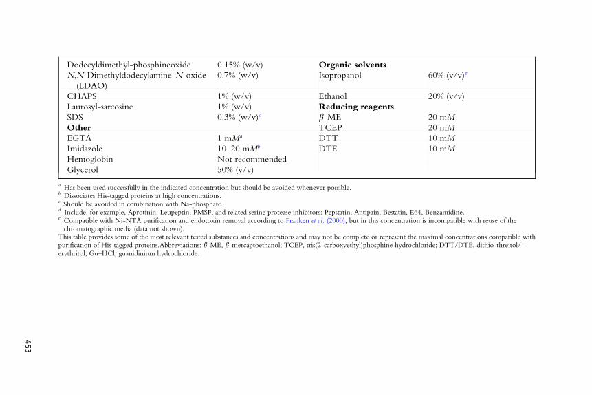
Dodecyldimethyl-phosphineoxide 0.15% (w/v) Organic solvents
N,N-Dimethyldodecylamine-N-oxide
(LDAO)
0.7% (w/v) Isopropanol 60% (v/v)e
CHAPS 1% (w/v) Ethanol 20% (v/v)
Laurosyl-sarcosine 1% (w/v) Reducing reagents
SDS 0.3% (w/v)a b-ME 20 mM
Other TCEP 20 mM
EGTA 1 mMa DTT 10 mM
Imidazole 10–20 mMb DTE 10 mM
Hemoglobin Not recommended
Glycerol 50% (v/v)
a Has been used successfully in the indicated concentration but should be avoided whenever possible.b Dissociates His-tagged proteins at high concentrations.c Should be avoided in combination with Na-phosphate.d Include, for example, Aprotinin, Leupeptin, PMSF, and related serine protease inhibitors: Pepstatin, Antipain, Bestatin, E64, Benzamidine.e Compatible with Ni-NTA purification and endotoxin removal according to Franken et al. (2000), but in this concentration is incompatible with reuse of thechromatographic media (data not shown).
This table provides some of the most relevant tested substances and concentrations and may not be complete or represent the maximal concentrations compatible withpurification of His-tagged proteins.Abbreviations: b-ME, b-mercaptoethanol; TCEP, tris(2-carboxyethyl)phosphine hydrochloride; DTT/DTE, dithio-threitol/-erythritol; Gu–HCl, guanidinium hydrochloride.
453
200
0 1 5 10 mM DTT
M0 1 5 10
C
b -actin
His6-HIV-RT
M L F W E1E2 E3 E4 E2 E3 E4 E2E3 E4 E2 E3 E4
A B
116/976655
3631
21
14
6
[Ni] (ppb) 150 100 250 150
NTC
Figure 27.5 IMAC under reducing conditions. (A) His6-tagged HIV-1 reverse tran-scriptase (RT) was purified under standard conditions in the presence of the indicatedDTT concentrations. Aliquots of each chromatographic fraction of the purificationwithout DTT (M, markers; L, lysate; F, flow-through; W, wash; E, elution fraction)and of corresponding volumes of elution fractions 2–4 containing DTT were analyzedby SDS–PAGE and Coomassie staining. Elution fractions were pooled and analyzed fornickel content by ICP-MS at Wessling Laboratories, Bochum, Germany; nickelconcentrations are given in mg/l (parts per billion, ppb). (B) Equal amounts of HIV-1RT purified in the presence of the indicated concentrations of DTT were used induplicates to reverse transcribe a 1.5 kb b-actin cDNA which was subsequently ampli-fied by PCR and the PCR products were analyzed by agarose gel electrophoresis andethidium bromide staining. Reducing conditions of at least 1 mM DTT were found tobe required for full RT activity (compare weaker amplification with RT purified in theabsence of DTT). C, Omniscript positive control; an equal amount of Omniscript RTprotein was used; NTC, no template negative control.
454 Helena Block et al.
(Fig. 27.5B) and quantitative real-time RT-PCR (data not shown) show noinhibitory effect which could origin from high heavy-metal-ion concentra-tions. Even though nickel ions may become reduced by DTT leading to acolor change of the resin bed they do not increasingly leach from the ligand(Fig. 27.5A), and resins processed under reducing conditions can be repeat-edly reused and regenerated (data not shown). These findings suggest thatdespite a color change as a consequence of nickel reduction by DTT theresin is still functional. TCEP, a different, nonthio-based reducing reagent ismore and more replacing DTT and b-ME in protein purification by IMACas it seems to be more selective to reduction of disulfide bonds, is odorlessand more stable in aqueous solution. We recommend the use of TCEP withNi-NTA chromatography in a concentration of 1–5 mM.
2.5. Copurifying proteins on IMAC and what to do about it
IMAC leads to high protein purity upon single-step chromatographicpurification in many cases (Figs. 27.2A, D, E and 27.5A; Bornhorst andFalke, 2000; Schmitt et al., 1993). The higher the purity is the closer theamount of IMAC resin applied in the chromatographic step has been
IMAC 455
correlated to the amount of recombinant His-tagged protein present in thesample to be processed. The reason is that proteins naturally displayingsurface motifs suitable to interact with an immobilized metal ion may bindto the resin although usually with slightly lower affinity than a flexible Histag. Most His-tagged proteins will therefore displace proteins with natural oraccidental surface motifs. However, there are some proteins where the localdensity of chelating amino acids such as histidine is so high that they will bindto immobilized metal ions almost inevitably. In general, mammalian systemshave a higher natural abundance than bacterial systems of proteins containingconsecutive histidines (Crowe et al., 1994), and a prominent example inhuman cells is the alpha subunit of transcription factor TFIIA which hasseven consecutive and surface-exposed histidine residues and can be purifiedvia IMAC from natural sources under native conditions (DeJong andRoeder, 1993; Ma et al., 1993) and which is observed frequently as a signalband of 55 (the ab precursor) or 35 kDa (the a subunit) of TFIIA inWesternblots using an anti-His tag antibody. Another example is the human tran-scription factor YY1 with 11 consecutive histidines (Shi et al., 1991). In E.coli, the proteins observed to copurify with His-tagged target proteins can bedivided into four groups: (i) proteins with natural metal-binding motifs, (ii)proteins with histidine clusters on their surfaces, (iii) proteins that bind toheterologously expressed His-tagged proteins, for example by a chaperonemechanism, and (iv) proteins with affinity to agarose-based supports(Bolanos-Garcia and Davies, 2006). Whether or not one of the E. coliproteins is copurified is not easily predictable. For example, a protein fromgroup (ii) sometimes reported to copurify with Ni-NTA is the 21 kDa SlyD,however, we have never observed this one in our lab upon purification fromE. coli BL21(DE3), DH5a, M15(pREP4), and other strains. This may beexplained by the fact that many of these impurities are stress-responsiveproteins, suggesting that the cultivation conditions and the bacterial strainhave an influence on their abundance and, a consequence, their appearanceas a contaminating species in the target protein preparation; it is thereforerecommended to induce as little stress as possible during cultivation of E. colicells (e.g., by using shake flasks without baffles). Furthermore, some copuri-fying proteins seem to have a binding preference for Co over Ni (or otherions) and others vice versa.
Several options to get rid of these copurified proteins or prevent theiradsorption early on have been evaluated and some of them shall be discussed inthe following section. These options include (i) performing additional purifi-cation steps, (ii) adjusting the His-tagged protein to resin ratio, (iii) to use anengineered host strain that does not express certain proteins, (iv) using analternative support, and (v) tag cleavage followed by reverse chromatography.
Suitable additional purification steps include classical chromatographictechniques such as ion-exchange (IEX) and size exclusion chromatography(SEC) whereas IEX has the higher separation power. However, as SEC not
456 Helena Block et al.
only serves to separate molecules by size and helps to remove aggregates ofhigh molecular weight but also can be used to desalt the preparation andprovide conditions suitable for certain downstream applications, it isfrequently used as a standardized procedure by, for example, high-throughputlabs such as structural biology consortia (Acton et al., 2005; Graslund et al.,2008) who perform protein crystallization or NMR spectroscopy. AlthoughIMAC–SEC (as opposed to IMAC–IEX) can be performed as a standar-dized procedure without having to take into account the protein biochem-istry such as pI the separation range of a given SEC column will not besuitable for all separation tasks and a range of SEC columns may have to beheld in place. Another issue with the application of IEX and SEC is that inorder to make use of the full power provided by these technologies, costlyequipment such as an automated chromatography system is required whichfrequently precludes multiparallel processing leading to low throughput.Affinity purification such as IMAC can usually be done as a bind–wash–elute procedure in a bench top/gravity flow mode. By introduction of asecond affinity tag (e.g., StrepII, GST, Flag) into the expression construct atwo-step purification leading to high-purity protein preparations is enabledas a bench-top two-step affinity chromatography procedure (Cass et al.,2005; Prinz et al., 2004).
As mentioned earlier, adjusting the amount of His-tagged protein to berecovered to the binding capacity of the used IMAC resin can help toimprove protein purity by preventing copurification of proteins with certainaffinity to IMAC resins. However, the amount of His-tagged protein isusually unknown unless a pilot experiment is performed to estimate thecontent of target protein. While this is an option, the better way would beto exclude the presence of such copurifying proteins by expressing the targetprotein in an engineered strain where the respective genes have beenknocked out. However, to our knowledge results from work with suchstrains have not yet been reported and the experience with knockout strainsin protein production is still low. Also, it does not seem realistic that an E. colistrain can be generated which lacks 17 proteins reported to bind to IMACresins including as important functions as superoxide dismutase and iron-uptake regulation (Bolanos-Garcia and Davies, 2006) and which is still wellviable under stress situations such as protein overproduction.
A different approach to improve the purity of proteins recovered fromIMAC has been reported that made use of dextran-coating of an agarosematrix, the constituting material of the most widely used chromatographicsupports (Sepharoses, Superflow, Agaroses), and which prevented copur-ification of proteins with affinity to these matrices (Mateo et al., 2001).Dextran-coated beads, however, are not readily commercially available asIMAC resins and this measure only helps to preclude proteins with affinityto agarose and not to the immobilized metal or to the target protein. Silica-based IMAC supports also prevent adsorption of proteins with affinity for

IMAC 457
agarose and, in addition, have good pressure stability which makes themsuitable for high-resolution HPLC applications but the silica resinsfrequently suffer from low binding capacity and limited resistance to highpH sanitization procedures. A very recent method that avoids the need touse solid chromatographic supports completely is called affinity precipita-tion (Hilbrig and Freitag, 2003) and has been applied to IMAC (Matiassonet al., 2007). Here, the IMAC ligand is chemically coupled to a responsivepolymer which, after binding to the His-tagged protein, can be aggregatedupon change of environmental conditions such as pH or temperature andcan thus be precipitated by centrifugation. Protocols for its use are stillrelatively complicated but as soon as robust and easy to use commercialmaterials are available this method may have the potential to play animportant role in IMAC applications. Using a ligand in solution couldovercome steric hindrance of the binding of some His-tagged proteins toan immobilized ligand as well as mass transport limitations of porouschromatographic media. Moreover, it is in line with a trend in industrial-scale chromatography toward single-use disposable materials.
Yet another approach has recently been reported that can be applied forprotein separation from lysates after cell-free expression (Kim et al., 2006).An E. coli-derived lysate was preincubated with Ni-NTA magnetic agarosebeads to remove proteins with affinity to Ni-NTA prior to templateaddition and protein expression; the expression capacity of the S30 extractwas found to remain unaltered and the Ni-NTA purified His-taggedprotein fractions to be of higher purity than without pretreatment.
While the aforementioned strategies to improve the purity of MACprotein preps have proven useful in many cases they are not generallyapplicable and successful. There is a method, however, that almost meetsthe criterium of universal applicability regarding improvement of purity,and it has the additional benefit of resulting in a protein native or near-native structure: proteolytic His tag cleavage using a His-tagged proteasefollowed by reverse IMAC (Block et al., 2008). This strategy overcomes thecopurification issue by passing the proteolytically processed protein undersimilar or identical conditions over the same column, and the proteins thatbound to the IMAC resin as impurities in the initial purification step willbind to the same resin again while the cleaved, that is untagged, targetprotein is collected in the flow-through fraction (reverse or subtractiveIMAC mode). It can be performed with both exo- and endoproteases thatthemselves carry an (uncleavable) His-tag (Nilsson et al., 1997; Polayes et al.,2008) but the exoproteolytical approach has the advantage that it is fasterand results in a protein with native structure with no vector-derived aminoacids (Arnau et al., 2006; Block et al., 2008; Pedersen et al., 1999). Thisapproach is especially suitable for demanding downstream applications suchas protein crystallization or biopharmaceutical production. Notably, themethod requires only a single chromatography column to achieve an
458 Helena Block et al.
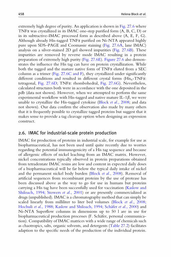
extremely high degree of purity. An application is shown in Fig. 27.6 whereTNFa was crystallized in its IMAC one-step purified form (A, B, C, D) orin its subtractive-IMAC processed form as described above (A, E, F, G).Although already His-tagged TNFa purified on Ni-NTA appeared highlypure upon SDS–PAGE and Coomassie staining (Fig. 27.6A, lane IMAC)analysis on a silver-stained 2D gel showed impurities (Fig. 27.6B). Theseimpurities are removed by reverse mode IMAC resulting in a proteinpreparation of extremely high purity (Fig. 27.6E). Figure 27.6 also demon-strates the influence the His tag can have on protein crystallization. Whileboth the tagged and the mature native form of TNFa eluted from a SECcolumn as a trimer (Fig. 27.6C and F), they crystallized under significantlydifferent conditions and resulted in different crystal forms (His6-TNFa:tetragonal, Fig. 27.6D; TNFa: rhombohedral, Fig. 27.6G). Nevertheless,calculated structures both were in accordance with the one deposited in thepdb (data not shown). However, when we attempted to perform the sameexperimental workflow with His-tagged and native mature IL-1b, we wereunable to crystallize the His-tagged cytokine (Block et al., 2008; and datanot shown). Our data confirm the observation also made by many othersthat it is frequently possible to crystallize tagged proteins but suggest that itmakes sense to provide a tag cleavage option when designing an expressionconstruct.
2.6. IMAC for industrial-scale protein production
IMAC for production of proteins in industrial scale, for example for use asbiopharmaceutical, has not been used until quite recently due to worriesregarding the potential immunogenicity of a His tag sequence and becauseof allergenic effects of nickel leaching from an IMAC matrix. However,nickel concentrations typically observed in protein preparations obtainedfrom tetradentate IMAC resins are low and content in expected daily dosesof a biopharmaceutical will be far below the typical daily intake of nickeland the permanent nickel body burden (Block et al., 2008). Removal ofartificial sequences from recombinant proteins by the use of protease hasbeen discussed above as the way to go for use in humans but proteinscarrying a His tag have been successfully used for vaccination (Kaslow andShiloach, 1994; Stowers et al., 2001) or are presently commercialized asdrugs (unpublished). IMAC is a chromatography method that can simply bescaled linearly from milliliter to liter bed volumes (Block et al., 2008;Hochuli et al., 1988; Kaslow and Shiloach, 1994; Schafer et al., 2000) andNi-NTA Superflow columns in dimensions up to 50 l are in use forbiopharmaceutical production processes (F. Schafer, personal communica-tion). Compatibility of IMACmatrices with a wide range of chemicals suchas chaotropics, salts, organic solvents, and detergents (Table 27.2) facilitatesadaption to the specific needs of the production of the individual protein.
M C F WA
D
C B E F
G
His6-TNFa
His6-
TNFa
TNFa
TNFa
TNFα
10′ 30′IMAC
TAGZy
me
sIMAC
130170
0.4mm
100725543
34
26
17
His6-TNFa
0.3mm
mAU
800
600
400
200
0
0 20 40 60 80 100ml
82.36
pH 3 pH 10 pH 10pH 3 mAU
800
600
400
200
0
0 20 40 60 80 100 ml
85.12
Figure 27.6 Removal of copurifying proteins by His tag cleavage and reverse IMAC. (A) His6-TNFa expressed in E. coli was purified via Ni-NTASuperflow and processed using the TAGZyme exoproteolytic system as described (Sch€afer et al., 2002a). (B, E) 2D gel electrophoresis and silverstaining of His6-TNFa and TNFa, respectively, was performed as described (Block et al., 2008). The subband pattern in the first dimension betweenpI 6.7 and 5.8 for both His6-TNFa and TNFa is in accordance with the report for TNFa produced in yeast (Eck et al., 1988). (C, F) Analytical sizeexclusion chromatography (SEC) on HR 10/30 Superdex 200 was run with 1� TAGZyme buffer (Sch€afer et al., 2002a). (D, G) His6-TNFatetragonal crystal (D) formed in 2.7 M MgSO4, MES, pH 5.5 and diffracted to 2.5 A (homelab X-ray source, FR591 Nonius Bruker);TNFa rhombohedral crystal (G) formed in 1.8 M NH4SO3, 200 mM Tris–HCl, pH 7.8 and diffracted to 2.0 A (ESRF synchrotron). The size oftypical crystals (mm) is indicated by the bar in (D) and (G).

460 Helena Block et al.
For example, bacterial endotoxins (lipopolysaccharides) can be eliminatedfrom the protein product during chromatography by including a wash stepusing a detergent (Triton X-114, Block et al., 2008) or an organic solvent(60% isopropanol; Franken et al., 2000). The suitability of IMAC forindustrial production purposes has been demonstrated and it can beexpected that IMAC-based processes become increasingly used in the futuredue to its robustness and relatively low requirements for individualoptimization.
2.7. High-throughput automation of IMAC
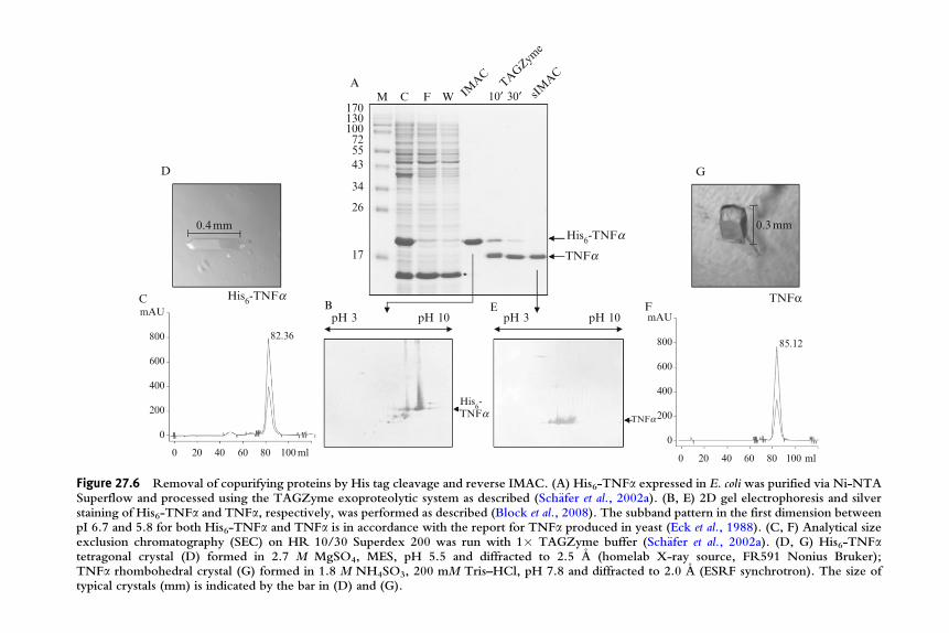
Due to its robustness, universal applicability, and widespread use, IMAC isalso an ideal tool for multiparallel screening for protein expression andsolubility. This is mostly done in the convenient 96-well format usingagarose- or magnetic bead-based IMAC resins in SBS footprint filter ormicroplates on 96-well magnets or plate centrifuges (Braun et al., 2002;Bussow et al., 2000). As the complete expression and the simple bind–wash–elute IMAC purification workflow is easily miniaturizable in microplateformats it was also suitable for hands-on free automation using liquidhandling laboratory robots (see Lesley, 2001, for an overview). The auto-mated steps covered the workflow to various extents, ranging from proteinpurification from manually generated E. coli lysates (Lanio et al., 2000),E. coli or insect cell lysis, lysate clarification, and protein purification (Garziaet al., 2003; Schafer et al., 2002b; Scheich et al., 2003), to automation ofcomplete workflows from construct cloning to protein analysis (Acton et al.,2005; Hunt, 2005; Koehn and Hunt, 2009). Recently, we added anotherseries of protocols and consumables for purification of His-tagged proteinsfrom E. coli or eukaryotic cells or cell-free lysates to the list of options: aconceptually new lab automation instrument allows isolation of microgramto milligram amounts of proteins from a variable number of samples (with arandom-access sample feeding option) using ready-to-go prefilled cartridgesthat provide enzymes, buffers, and Ni-NTA magnetic beads for lysis andpurification. Figure 27.7 describes an expression and purification screeningusing a set of 24 optimized-gene constructs for production of humanproteins. Between 1.4 and 35 mg of highly pure protein was obtainedunder native conditions. Proteins that could not be purified under nativeconditions were obtained upon purification under denaturing conditions,andWestern blot analyses using an anti-His antibody showed the absence ofcross-contaminations between wells (data not shown). Protein resultingfrom such high-throughput purification experiments can be used forfunctional assays (e.g., interaction studies), characterization of proteinrandom mutagenesis, solubility analyses, and clone screening.
Thenext step following an expression screening is frequently a scale-upwitha limited number of proteins or clones for production of milligram amounts
M
200
116/976655
36
31
21
14
PIM
1EM
G1
IL-4IL
-7M
APKAPK
5
ETS1SM
ARCD1
CREB1
p38a
IL-6
PIM
2
BIRC1
JunLe
f1FY
NCD
C2
YY1SM
AD2
JNK1
CM-C
SF
IFNgTN
Fa
IFNa
NFκB
1A
Figure 27.7 High-throughput protein purification screening on a new automationplatform. E. coli BL21 (DE3) cells were transformed with QIAgene constructs carryingoptimized human genes for expression of the indicated proteins in 1 ml LB cultures in a96-deep well block. Cells were harvested by centrifugation and the pellet block placedonto the sample input drawer of the QIAsymphony SP instrument. Cells wereresuspended and lysed and His6-tagged proteins purified from the crude lysates usingNi-NTA magnetic agarose beads and buffer solutions provided in the QIAsymphonycartridge setup. A 5 ml of each elution fraction was analyzed by SDS–PAGE andCoomassie staining. Expected protein sizes (kDa) for the individual proteins wereas follows: PIM1, 40; EMG1,30; IL-4, 15.5; IL-7,18; MAPKAPK5,55; ETS1,55;SMARCD1,60; CREB1,50; p38a, 40; NFkB1A, 40; IL-6,21; IFNa, 20; PIM2, 40;BIRC5, 30; Jun, 50; Lef1, 55; JNK1, 45; CM-CSF, 15; IFNg, 17; TNFa, 17; FYN, 40;CDC2, 35; YY1, 66; SMAD2, 55. M, markers (kDa).
IMAC 461
of protein for animal immunization, structural, or pharmacokinetic studies.Single proteins can be purified using standard AkTA or FPLC systems,and an AkTA system for slightly increased throughput has been developed(AkTAxpress). However, systems with a significantly higher throughput,lower complexity, and more dedicated to one-step (mainly IMAC) affinitypurification have been reported (Steen et al., 2006; Stromberg et al., 2005)and are in use in high-throughput projects such as the human protein atlasproject (Hober and Uhlen, 2008).
2.8. Special applications: Purification of membrane proteins
Membrane proteins have received the highest attention of all protein classesin the past few years due to their enormous importance as drug targets.In fact, more than 50% of all currently commercialized drugs as well as thoseunder development target membrane proteins (Drews, 2000). Furthermore,membrane proteins account for approximately 30% of the human
462 Helena Block et al.
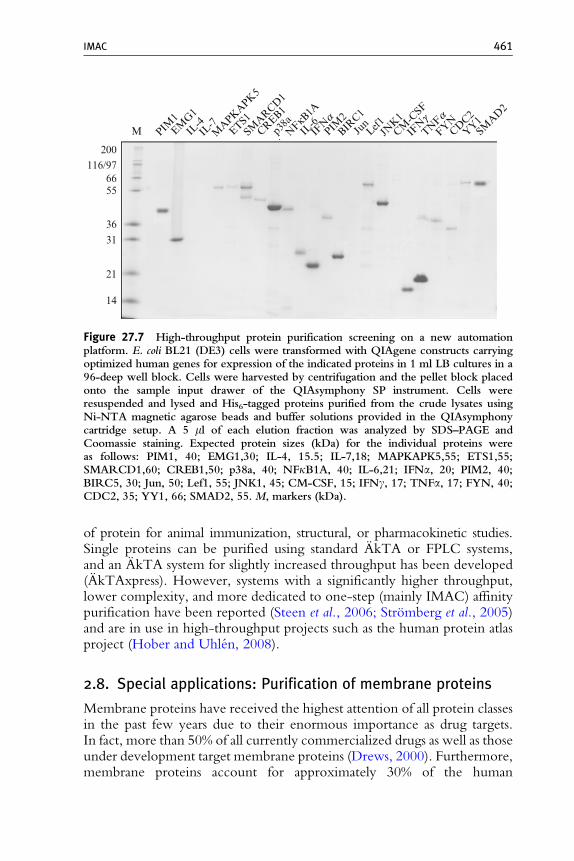
proteome. However, in contrast to soluble proteins, very little is knownabout the biology and structure of membrane proteins which is reflected bythe underrepresentation of membrane protein structures in public databasessuch as the pdb (<1%). While structure determination remains a challenge,purification of membrane proteins by a more or less standardized IMACprocedure has recently become significantly simpler by the development ofnew detergents and consequent screening for the most suitable detergent forboth resolubilization and purification (Eshaghi et al., 2005; Klammt et al.,2005; Lewinson et al., 2008). We have screened more than 50 of the mostfrequently used detergents for their IMAC compatibility and their resolubili-zation and Ni-NTA purification efficiency testing more than 10 bacterial andhuman membrane proteins and have reduced the number to seven powerfuldetergents as follows: Cymal 6 (Cy6), n-nonyl-b-D-glucopyranoside (NG),n-octyl-b-D-glucopyranoside (OG), n-decyl-b-D-maltopyranoside (DM),n-dodecyl-b-D-maltoside (DDM), FOS-choline-16 (FC16), and N,N-dimethyldodecylamine-N-oxide (LDAO). In screenings, we achieved apositive result for efficient resolubilization from E. coli or insect cellmembrane fractions and IMAC purification for at least one detergent ineach case. In Fig. 27.8, the detergent screening and Ni-NTA purification ofHis-tagged human Caveolin 1 is shown as an example.
IMAC compatible detergents are listed in Table 27.2 whereas this listmay not be complete. Nevertheless, binding of a His-tagged membraneprotein to IMAC resins seems not to work in combination with certain
A
80
M M L R F W E1 E2OG DM
LDAO
DDM
Cy6 NG
FC16
7060504030
20
15
B
72553628
17
11
His6-CAV1
His6-CAV1
Figure 27.8 Detergent screening and IMAC purification of His6-tagged humanCaveolin 1 expressed in E. coliC41(DE3) in the presence of LDAO. The gene optimizedfor expression in E. coliwas synthesized and cloned into pQE-T7with anN-terminal His6tag. (A) Detergent screening; the membrane fraction from 200 ml E. coli culture dividedinto seven portions was mixed with detergent as indicated, centrifuged at 20,000� g andaliquots of detergent-soluble protein analyzed byWestern blot (Penta anti-His antibody).Detergents are given in the text. (B) Caveolin 1 purification upscale; His6-taggedCaveolin 1 (CAV1) was purified from 200 ml E. coli culture volume via Ni-NTASuperflow in the presence of 30 mM LDAO. Fractions were analyzed by SDS–PAGEand Coomassie staining: M, markers (kDa); L, E. coli total lysate; R, supernatant fromresolubilization (loaded onto IMAC column); F, IMAC flow-through fraction; W, washfraction; E, elution fractions.

IMAC 463
detergents, including some of the listed ones even though resolubilization isfine. These phenomena depend on the protein–detergent combination andseem to arise from the detergent micelle around the protein partially orcompletely hiding the His tag. The use of longer tag sequences such as a H10
tag has become popular and seems to overcome such limitations of mem-brane protein recovery and improve affinity to IMAC resins (Byrne andJormakka, 2006; Grisshammer and Tucker, 1997; Mohanty and Wiener,2004; Rumbley et al., 1997).
2.9. Special applications: Purification of zinc-finger proteins
Another big protein superfamily which—due to technical issues—shall bementioned in the context of IMAC is the group of proteins containing zinc-finger motifs. The C2H2 zinc-finger transcription factors alone with over 600members represent more than 2% of the human proteome (Knight andShimeld, 2001). In these proteins, zinc ions are coordinated by a defined spatialorganization of two cysteine and two histidine residues each per finger, and asingle polypeptide usually contains four or five of these motifs. Purification ofsuch a metalloprotein via a metal ion chelated in a similar, that is tetradentate,manner deserves some reflection regarding the bestway of IMACpurification.
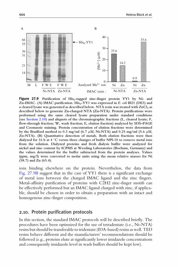
Despite the strong interaction of metal ions with tetradentate IMACligands, it may not be excluded that a metal from the resin could exchangewith the zinc in the zinc finger. Nickel, the most frequently used metal inIMAC protein purification has quite similar physicochemical properties tozinc and might therefore well replace it in such a metal-binding motif, andthe nickel concentration in an IMAC resin (approximately 15 mM ) isusually considerably higher than the concentration of a His-tagged targetprotein (mM range). In order to analyze a potential metal ion exchangebetween matrix and metalloprotein, we expressed the His6-tagged C2H2zinc-finger containing transcription factor YY1 in E. coli and purified theprotein in parallel using Ni-NTA and Zn-NTA. YY1 could be purified tohigh purity by both nickel- and zinc-based IMAC (Fig. 27.9A, lane E).
Both YY1 preparations were subjected to determination of nickel andzinc content by ICP-MS (intercoupled plasma mass spectrometry), a quan-titative method frequently applied to detect trace metals in biologicalsystems (Shi and Chance, 2008). The zinc-IMAC purified protein prepara-tion contained 35.6 mM zinc corresponding to approximately six Zn2þ ionsper YY1 polypeptide and some Ni2þ which can be attributed to residualtraces from the buffers (Fig. 27.9B, right). The protein recovered by nickel-IMAC, however, contained more than 25 mM Ni2þ and 14.2 mM Zn2þ(Fig. 27.9B, left), corresponding to again approximately six Me2þ ions perYY1 polypeptide. The Me2þ:polypeptide molar ratio of 6 is higher than thefour zinc-finger motifs in YY1 reported in databases (http://www.uniprot.org/uniprot/P25490) which may in part be explained with additional metal
A
6655
3631
21
14
M L F W E
Ni-NTA Zn-NTA
Analyzed Me2+ ion:
IMAC resin :
F W E
B
His6-YY1
40
30
20
10
0Ni Zn Ni Zn
Ni-NTA Zn-NTA
[Me2
+] (mM
) 25.6
14.2
0.9
35.6
Figure 27.9 Purification of His6-tagged zinc-finger protein YY1 by Ni- andZn-IMAC. (A) IMAC purification. His6-YY1 was expressed in E. coli Bl21 (DE3) anda cleared lysate was generated as described below. NTA resin was treated with ZnCl2 asdescribed below to generate Zn-charged NTA (Zn-NTA). Protein purifications wereperformed using the same cleared lysate preparation under standard conditions(see Section 2.10) and aliquots of the chromatographic fractions (L, cleared lysate; F,flow-through fraction; W, wash fraction; E, elution fraction) analyzed by SDS–PAGEand Coomassie staining. Protein concentration of elution fractions were determinedby the Bradford method to 0.3 mg/ml (6.7 mM, Ni-NTA) and 0.25 mg/ml (5.6 mM,Zn-NTA). (B) Quantitative detection of metals. Both elution fractions were thendialyzed for 16 h at 4 �C versus three changes of buffer NPI-10 to remove metal ionsfrom the solution. Dialyzed proteins and fresh dialysis buffer were analyzed fornickel and zinc content by ICPMS at Wessling Laboratories (Bochum, Germany) andthe values determined for the buffer subtracted from the protein analyses. Values(ppm, mg/l) were converted to molar units using the mean relative masses for Ni(58.7) and Zn (65.4).
464 Helena Block et al.
ions binding elsewhere on the protein. Nevertheless, the data fromFig. 27.9B suggest that in the case of YY1 there is a significant exchangeof metal ions between the charged IMAC ligand and the zinc fingers.Metal-affinity purification of proteins with C2H2 zinc-finger motifs canbe effectively performed but an IMAC ligand charged with zinc, if applica-ble, should be chosen in order to obtain a preparation with an intact andhomogenous zinc-finger composition.
2.10. Protein purification protocols
In this section, the standard IMAC protocols will be described briefly. Theprocedures have been optimized for the use of tetradentate (i.e., Ni-NTA)resins but should be transferable to tridentate (IDA-based) resins aswell. TEDresins behave different and the manufacturers’ recommendations should befollowed (e.g., proteins elute at significantly lower imidazole concentrationsand consequently imidazole level in wash buffers should be kept low).
IMAC 465
Here, we will provide recommendations for agarose-based Ni-NTAresins (Superflow, Agarose) regarding purification, cleaning, and rechar-ging. These can be regarded as an update of the more detailed descriptionprovided in the Ni-NTA handbook (QIAGEN, 2003). Buffers provided inthe following may be supplemented according to the needs of the individualprotein (e.g., in order to generate reducing conditions, to stabilize theprotein with glycerol, or to provide the presence of cofactors).
2.10.1. Purification of His-tagged proteins under native conditions
1. Lyse cells using the basis bufferNPI-10 (50mMNaH2PO4, 300mMNaCl,10 mM imidazole, pH 8.0) supplemented with a suitable lysis reagent.
ForE. coli lysis, we recommend to use standard hen egg white lysozymeat 1 mg/ml final concentration because lysis is extremely efficient (if cellshad been frozen) and lysozyme is inexpensive. Lysozyme is reliably washedfrom IMAC resins and does not occur in elution fractions (Figs. 27.5A,27.6A, and 27.8; Block et al., 2008). Other suitable methods are based ondetergents (e.g., 1 % (v/v) CHAPS or proprietary solutions) or physicaltreatment (sonication, high pressure/depressurization homogenization).For cultures derived from insect or mammalian cells, 1% Igepal CA-630(former name NP-40) is recommended. For reduction of lysate viscosity,the addition of a nuclease is helpful, and Benzonase (3 units/ml bacterialculture) has been shown to be robust and its removal in wash steps fromIMAC resins works reliably and can be checked by a commercial ELISA(Block et al., 2008). Incubate the lysate on ice for 30 min.
2. Generate a cleared lysate by centrifugation for 30 min at �10,000� gand 2–8 �C. Collect the supernatant.
3. Load the cleared lysate onto the resin equilibrated with 5 bed volumes(bv) of NPI-10 and allow to flow through at approximately 1 ml/min(column of 1 ml bv) or let flow through (gravity flow application).
A suitable linear flow rate during binding is 155 cm/h correspondingto 1 ml/min of a column with a bed diameter of ~ 7 mm (1 ml HisTrapand Ni-NTA Superflow Cartridges). If applicable to the workflow, werecommend to perform binding in batch mode as this is most efficientwith agarose-based resins in general; for this, add the required volume oflysate to the equilibrated resin and incubate for 1 h rotating end-over-end at 2–8 �C.
4. Wash the Ni-NTA column with 10 bv wash buffer NPI-20 (50 mMNaH2PO4, 300 mM NaCl, 20 mM imidazole, pH 8.0).
If binding had been performed in batch, pour the binding suspensioninto a suitable flow-through column.
5. Elute His-tagged protein with 5 bv of elution buffer NPI-500 (50 mMNaH2PO4, 300 mM NaCl, 500 mM imidazole, pH 8.0).
466 Helena Block et al.
2.10.2. Purification of His-tagged proteins underdenaturing conditions
1. Lyse cells using the basis buffer B (100 mM NaH2PO4, 10 mMTris–HCl, 8 M urea, pH 8.0).E. coli as well as most eukaryotic cells are efficiently lysed by 7–9 M
urea but occasionally, His-tagged proteins forming inclusion bodies donot completely dissolve; in such cases, we recommend to replace thechaotrope urea by guanidinium hydrochloride (buffer A: 100 mMNaH2PO4, 10 mM Tris–HCl, 6 M Gu–HCl, pH 8.0). Reduction oflysate viscosity by the addition of Benzonase (3 units/ml final concen-tration) is also possible and effective under denaturing conditions buthere the maximum urea concentration is 7 M (use of Gu–HCl is notpossible in combination with Benzonase). Incubate the lysate for 30 minat ambient temperature.
2. Generate a cleared lysate by centrifugation for 30 min at�10,000�g androom temperature. Collect the supernatant.
3. Load the cleared lysate onto the resin equilibrated with 5 bv of buffer B(or buffer A where applicable) and allow to flow through at approxi-mately 1 ml/min (column of 1 ml bv) or by gravity flow.A suitable linear flow rate during binding is 155 cm/h corresponding
to 1 ml/min of a column with a bed diameter of ~ 7 mm (1 ml HisTrapor Ni-NTA Superflow Cartridges). If applicable to the workflow andthe resin format used, we recommend to perform binding in batch modeas this is most efficient with agarose-based resins in general; for this, addthe required volume of resin slurry to the lysate and incubate for 1 hrotating end-over-end.
4. Wash the Ni-NTA column with 10 bv wash buffer C (100 mMNaH2PO4, 10 mM Tris–HCl, 8 M urea, pH 6.3).If binding had been performed in batch, pour the binding suspension
into a suitable flow-through column prior to the wash step. If lysate wasgenerated using buffer A, wash and elution steps may be performed byswitching to urea-based buffers or by continuing with Gu–HCl-basedbuffers with pH values adjusted accordingly.
5. Elute His-tagged protein with 5 bv of elution buffer E (100 mMNaH2PO4, 10 mM Tris–HCl, 8 M urea, pH 4.5).
2.11. Cleaning and sanitization
A simple and effective cleaning-in-place (CIP) method for Ni-NTA IMACresins used to purify proteins from ‘‘standard’’ samples such asE. coli or humancell lysates or supernatants is contacting the resinwith 0.5MNaOH for 30min(Schafer et al., 2000). The resins have been stored in up to 1 M NaOHfor several months and shown to withstand these conditions without
IMAC 467
compromising its performance even upon >100 cycles of purification/CIPcycles (data not shown). This CIP procedure reliably denatures and desorbsproteins originating from the loaded sample that might have bound unspeci-fically to agarose during purification and is generally suitable for sanitization(depyrogenation, viral, andmicrobial clearance; Levison et al., 1995). Cleaningprotocolsmayhave to be adapted ifmore ‘‘unusual’’ samples such as lysates richin lipids are loaded onto a column. Bases, acids, and other reagents that may beused for cleaning include ethanol (100%), isopropanol (30%, v/v), SDS(2%, w/v), acetic acid (0.2M ), NaOH (1M ), or detergents (see Table 27.2).
For repeated reuse of a Ni-NTA column, we recommend to perform theCIP description provided above followed by reequilibration. For long-termstorage (several years), resin may be kept in either 30% (v/v) ethanol or, if aninflammable reagent is preferred, in 0.01–0.1 M NaOH. Storage in 10 mMNaN3 is possible as well. It is usually not required to strip off the metal ionsand recharge Ni-NTA, even after repeated reuse or long-term storage.
2.12. Simplified metal-ion stripping and recharging protocol
However, in cases where the resin has been seriously damaged or if bindingcapacity decreased over time, for example, by repeated loading of lipid-richor samples containing chelating components, Ni-NTA may be easilystripped and recharged with nickel or a different metal ion. Starting withstep 3, this simplified protocol is also suitable to initially charge NTA resinpurchased uncharged.
1. Wash cleaned (see above) resin with 10 bv of deionized H2O (dH2O).2. Strip off metal ions by passing 5 bv of 100 mM EDTA, pH 8.0 over the
resin bed.3. Wash resin with 10 bv of dH2O.4. Pass 2 bv of a 100 mM metal ion aqueous solution (e.g., NiSO4 or
NiCl2) over the resin bed.Other metal ions that have been successfully and stably immobilized
to NTA include copper (CuCl2, CuSO4), zinc (ZnCl2, ZnSO4), cobalt(CoCl2, CoSO4), and iron (FeCl3, Fe2(SO4)3).
5. Wash resin with 10 bv of dH2O to remove any unbound metal ions.6. Add storage buffer or equilibrate the column with at least 5 bv of starting
buffer for immediate use.
3. Conclusions
Wehave presented thewide variety of applications the IMACprincipleoffers for research in general and for production of His-tagged proteinsin particular. Its robustness and versatility are the reasons why IMAC has
468 Helena Block et al.
become one of the most broadly used chromatographic methods. Modifica-tions in production procedures for both resin and ligand materials as well asoptimized application protocols led to a significant improvement of IMACperformance in the recent past, well reflected by, for example, the increaseof binding capacity of both NTA- and IDA-based matrices for His-taggedproteins from 5–10 to 50 mg per ml resin bv. We anticipate a continuedmethodological improvement and dissemination of the use of IMAC in thefield of purification of recombinant proteins, for example, regarding indus-trial-scale production of biopharmaceuticals.
ACKNOWLEDGMENTS
The authors thank Jacob Piehler for valuable discussions regarding the manuscript and hissupport during the transfer of the tris-NTA ligand synthesis procedures. Furthermore, wethank Annette Zacharias-Koch for excellent technical assistance and her support in prepara-tion of the manuscript. Part of the work presented here was performed under a grant of theGerman Ministry of Education and Research (BMBF, grant no. 0313965B).
REFERENCES
Acton, T. B., Gunsalus, K.C.,Xiao,R.,Ma, L.C., Aramini, J., Baran,M.C., Chiang, Y.-W.,Climent, T., Cooper, B., Denissova, N. G., Douglas, S. M., Everett, J. K., et al. (2005).Robotic cloning and protein production platform of the northeast structural genomicsconsortium.Methods Enzymol. 394, 210–243.
Andersson, L., and Porath, J. (1986). Isolation of phosphoproteins by immobilized metal(Fe3þ)affinity-chromatography. Anal. Biochem. 154, 250–254.
Arnau, J., Lauritzen, C., Petersen, G. E., and Pedersen, J. (2006). Current strategies for theuse of affinity tags and tag removal for the purification of recombinant proteins. ProteinExpr. Purif. 48, 1–13.
Biocompare (2006). Protein chromatography: Tools for protein expression and purification.Biocompare Surveys and Reports, Biocompare Inc., July 10.
Block, H., Kubicek, J., Labahn, J., Roth, U., and Schafer, F. (2008). Production andcomprehensive quality control of recombinant human Interleukin-1b: A case study fora process development strategy. Protein Expr. Purif. 27, 244–254.
Boden, V., Winzerling, J. J., Vijayalakshmi, M., and Porath, J. (1995). Rapid one-steppurification of goat immunoglobulins by immobilized metal ion affinity chromatogra-phy. J. Immunol. Methods 181, 225–232.
Bolanos-Garcia, V. M., and Davies, O. R. (2006). Structural analysis and classification ofnative proteins from E. coli commonly co-purified by immobilized metal affinitychromatography. Biochim. Biophys. Acta 1760, 1304–1313.
Bornhorst, J. A., and Falke, J. J. (2000). Purification of protein using polyhistidine affinitytags. Methods Enzymol. 326, 245–254.
Braun, P., Hu, Y., Shen, B., Halleck, A., Koundinya, M., Harlow, E., and LaBaer, J. (2002).Proteome-scale purification of human proteins from bacteria. Proc. Natl. Acad. Sci. USA99, 2654–2659.
Busso, D., Kim, R., and Kim, S.-H. (2003). J. Biochem. Biophys. Methods 55, 233–240.
IMAC 469
Bussow, K., Nordhoff, E., Lubbert, C., Lehrach, H., and Walter, G. (2000). A humancDNA library for high-throughput protein expression screening. Genomics 65, 1–8.
Byrne, B., and Jormakka, M. (2006). Solubilization and purification of membrane proteins.In ‘‘Structural Genomics on membrane proteins’’ (K. Lundstrom, ed.), pp. 179–198.CRC Press, Taylor and Francis, Boca Raton.
Cass, B., Pham, O. L., Kamen, A., and Durocher, Y. (2005). Purification of recombinantproteins from mammalian cell culture using a generic double-affinity chromatographyscheme. Protein Expr. Purif. 40, 77–85.
Cebe, R., and Geiser, M. (2006). Rapid and easy thermodynamic optimization of the 50-endof mRNA dramatically increases the level of wild type protein expression in Escherichiacoli. Protein Expr. Purif. 45, 374–380.
Chaga, G., Hopp, J., and Nelson, P. (1999). Immobilized metal ion affinity chromatographyon Co2þ-carboxymethylaspartate–agarose Superflow, as demonstrated by one-steppurification of lactate dehydrogenase from chicken breast muscle. Biotechnol. Appl.Biochem. 29, 19–24.
Crowe, J., Dobeli, H., Gentz, R., Hochuli, E., Stuber, D., and Henco, K. (1994). 6xHis-Ni-NTA chromatography as a superior technique in recombinant protein expression/purification. Methods Mol. Biol. 31, 371–381.
Dalb�ge, H., Bayne, S., and Pedersen, J. (1990). In vivo processing of N-terminal methioninein E. coli. FEBS Lett 266, 1–3.
DeJong, J., and Roeder, R. G. (1993). A single cDNA, hTFIIA/a, encodes both the p35 andp19 subunits of human TFIIA. Genes Dev. 7, 2220–2234.
Derewenda, Z. S. (2004). The use of recombinant methods and molecular engineering inprotein crystallization. Methods 34, 354–363.
Drews, J. (2000). Drug discovery: A historical perspective. Science 287, 1960–1964.Eck, M. J., Beutler, B., Kuo, G., Merryweather, J. P., and Sprang, S. R. (1988). Crystalliza-
tion of trimeric recombinant human tumor necrosis factor (catechin). J. Biol. Chem. 263,12816–12819.
Eshaghi, S., Hedren, M., Ignatushchenko Abdel Nasser, M., Hammarberg, T., Thomell, A.,and Nordlund, P. (2005). An efficient strategy for high-throughput expression of recom-binant integral membrane proteins. Protein Sci. 14, 676–683.
Franken, K. L. M. C., Hiemstra, H. S., van Meijgaarden, K. E., Subronto, Y., denHartigh, J., Ottenhoff, T. H. M., and Drijfthout, J. W. (2000). Purification of His-taggedproteins by immobilized chelate affinity chromatography: The benefits from the use oforganic solvents. Protein Expr. Purif. 18, 95–99.
Garcıa Gonzalez, L. A., Rodrigo Tapia, J. P., Sanchez Lazo, P., Ramos, S., and SuarezNieto, C. (2004). DNA extraction Using Chelex resin for the oncogenic amplificationanalysis in head and neck tumors. Acta Otorrinolaringol. Esp. 55, 139–144.
Garzia, L., Andre, A., Amoresano, A., D’Angelo, A., Martusciello, R., Cirulli, C.,Tsurumi, T., Marino, G., and Zollo, M. (2003). Method to express and purify nm23–H2 protein from baculovirus-infected cells. BioTechniques 35, 384–391.
Gill, P., Kimpton, C. P., and Sullivan, K. (1992). A rapid polymerase chain reaction methodfor identifying fixed specimens. Electrophoresis 13, 173–175.
Graslund, S., Nordlund, P., Weigelt, J., Bray, J., Gileadi, O., Knapp, S., Oppermann, U.,Arrowsmith, C., Hui, R., Ming, J., dhe-Paganon, S., Park, H.-W., et al. (2008). Proteinproduction and purification. Nat. Methods 5, 135–146.
Grisshammer, R., and Tucker, J. (1997). Quantitative evaluation of neurotensin receptorpurification by immobilized metal affinity chromatography. Protein Expr. Purif. 11,53–60.
Hale, J. E., and Beidler, D. E. (1994). Purification of humanized murine and murinemonoclonal antibodies using immobilized metal-affinity chromatography. Anal. Biochem.222, 29–33.
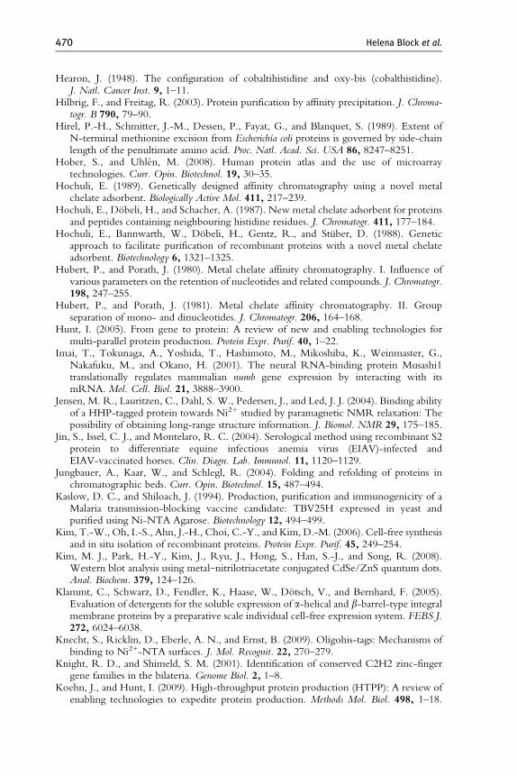
470 Helena Block et al.
Hearon, J. (1948). The configuration of cobaltihistidine and oxy-bis (cobalthistidine).J. Natl. Cancer Inst. 9, 1–11.
Hilbrig, F., and Freitag, R. (2003). Protein purification by affinity precipitation. J. Chroma-togr. B 790, 79–90.
Hirel, P.-H., Schmitter, J.-M., Dessen, P., Fayat, G., and Blanquet, S. (1989). Extent ofN-terminal methionine excision from Escherichia coli proteins is governed by side-chainlength of the penultimate amino acid. Proc. Natl. Acad. Sci. USA 86, 8247–8251.
Hober, S., and Uhlen, M. (2008). Human protein atlas and the use of microarraytechnologies. Curr. Opin. Biotechnol. 19, 30–35.
Hochuli, E. (1989). Genetically designed affinity chromatography using a novel metalchelate adsorbent. Biologically Active Mol. 411, 217–239.
Hochuli, E., Dobeli, H., and Schacher, A. (1987). New metal chelate adsorbent for proteinsand peptides containing neighbouring histidine residues. J. Chromatogr. 411, 177–184.
Hochuli, E., Bannwarth, W., Dobeli, H., Gentz, R., and Stuber, D. (1988). Geneticapproach to facilitate purification of recombinant proteins with a novel metal chelateadsorbent. Biotechnology 6, 1321–1325.
Hubert, P., and Porath, J. (1980). Metal chelate affinity chromatography. I. Influence ofvarious parameters on the retention of nucleotides and related compounds. J. Chromatogr.198, 247–255.
Hubert, P., and Porath, J. (1981). Metal chelate affinity chromatography. II. Groupseparation of mono- and dinucleotides. J. Chromatogr. 206, 164–168.
Hunt, I. (2005). From gene to protein: A review of new and enabling technologies formulti-parallel protein production. Protein Expr. Purif. 40, 1–22.
Imai, T., Tokunaga, A., Yoshida, T., Hashimoto, M., Mikoshiba, K., Weinmaster, G.,Nakafuku, M., and Okano, H. (2001). The neural RNA-binding protein Musashi1translationally regulates mammalian numb gene expression by interacting with itsmRNA. Mol. Cell. Biol. 21, 3888–3900.
Jensen, M. R., Lauritzen, C., Dahl, S. W., Pedersen, J., and Led, J. J. (2004). Binding abilityof a HHP-tagged protein towards Ni2þ studied by paramagnetic NMR relaxation: Thepossibility of obtaining long-range structure information. J. Biomol. NMR 29, 175–185.
Jin, S., Issel, C. J., and Montelaro, R. C. (2004). Serological method using recombinant S2protein to differentiate equine infectious anemia virus (EIAV)-infected andEIAV-vaccinated horses. Clin. Diagn. Lab. Immunol. 11, 1120–1129.
Jungbauer, A., Kaar, W., and Schlegl, R. (2004). Folding and refolding of proteins inchromatographic beds. Curr. Opin. Biotechnol. 15, 487–494.
Kaslow, D. C., and Shiloach, J. (1994). Production, purification and immunogenicity of aMalaria transmission-blocking vaccine candidate: TBV25H expressed in yeast andpurified using Ni-NTA Agarose. Biotechnology 12, 494–499.
Kim, T.-W., Oh, I.-S., Ahn, J.-H., Choi, C.-Y., and Kim, D.-M. (2006). Cell-free synthesisand in situ isolation of recombinant proteins. Protein Expr. Purif. 45, 249–254.
Kim, M. J., Park, H.-Y., Kim, J., Ryu, J., Hong, S., Han, S.-J., and Song, R. (2008).Western blot analysis using metal–nitrilotriacetate conjugated CdSe/ZnS quantum dots.Anal. Biochem. 379, 124–126.
Klammt, C., Schwarz, D., Fendler, K., Haase, W., Dotsch, V., and Bernhard, F. (2005).Evaluation of detergents for the soluble expression of a-helical and b-barrel-type integralmembrane proteins by a preparative scale individual cell-free expression system. FEBS J.272, 6024–6038.
Knecht, S., Ricklin, D., Eberle, A. N., and Ernst, B. (2009). Oligohis-tags: Mechanisms ofbinding to Ni2+-NTA surfaces. J. Mol. Recognit. 22, 270–279.
Knight, R. D., and Shimeld, S. M. (2001). Identification of conserved C2H2 zinc-fingergene families in the bilateria. Genome Biol. 2, 1–8.
Koehn, J., and Hunt, I. (2009). High-throughput protein production (HTPP): A review ofenabling technologies to expedite protein production. Methods Mol. Biol. 498, 1–18.
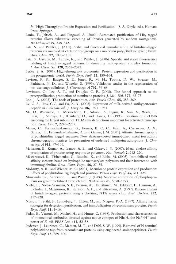
IMAC 471
In ‘‘High Throughput Protein Expression and Purification’’ (S. A. Doyle, ed.). HumanaPress, Springer.
Lanio, T., Jeltsch, A., and Pingoud, A. (2000). Automated purification of His6-taggedproteins allows exhaustive screening of libraries generated by random mutagenesis.BioTechniques 29, 338–342.
Lata, S., and Piehler, J. (2005). Stable and functional immobilization of histidine-taggedproteins via multivalent chelator headgroups on a molecular poly(ethylene glycol) brush.Anal. Chem. 77, 1096–1105.
Lata, S., Gavutis, M., Tampe, R., and Piehler, J. (2006). Specific and stable fluorescencelabeling of histidine-tagged proteins for dissecting multi-protein complex formation.J. Am. Chem. Soc. 128, 2365–2372.
Lesley, S. A. (2001). High-throughput proteomics: Protein expression and purification inthe postgenomic world. Protein Expr. Purif. 22, 159–164.
Levison, P. R., Badger, S. E., Jones, R. M. H., Toome, D. W., Streater, M.,Pathirana, N. D., and Wheeler, S. (1995). Validation studies in the regeneration ofion-exchange celluloses. J. Chromatogr. A 702, 59–68.
Lewinson, O., Lee, A. T., and Douglas, C. R. (2008). The funnel approach to theprecrystallization production of membrane proteins. J. Mol. Biol. 377, 62–73.
Loo, J. A. (2003). The tools of proteomics. Adv. Protein Chem. 65, 353–369.Lv, G. S., Hua, G.C, and Fu, X. Y. (2003). Expression of milk-derived antihypertensive
peptide in Escherichia coli. J. Dairy Sci. 86, 1927–1931.Ma, D., Watanabe, H., Mermelstein, F., Admon, A., Oguri, K., Sun, X., Wada, T.,
Imai, T., Shiroya, T., Reinberg, D., and Handa, H. (1993). Isolation of a cDNAencoding the largest subunit of TFIIA reveals functions important for activated transcrip-tion. Genes Dev. 7, 2246–2257.
Mateo, C., Fernandez-Lorente, G., Pessela, B. C. C., Vian, A., Carrascosa, A. V.,Garcia, J. L., Fernandez-Lafuente, R., and Guisan, J. M. (2001). Affinity chromatographyof polyhistidine tagged enzymes: New dextran-coated immobilized metal ion affinitychromatography matrices for prevention of undesired multipoint adsorptions. J. Chro-matogr. A 915, 97–106.
Matiasson, B., Kumar, A., Ivanov, A. E., and Galaev, I. Y. (2007). Metal-chelate affinityprecipitation of proteins using responsive polymers. Nat. Protocols 2, 213–220.
Meszarosova, K., Tishchenko, G., Bouchal, K., and Bleha, M. (2003). Immobilized-metalaffinity sorbents based on hydrophilic methacrylate polymers and their interaction withimmunoglobulins. React. Funct. Polym. 56, 27–35.
Mohanty, A. K., and Wiener, M. C. (2004). Membrane protein expression and production:Effects of polyhistidine tag length and position. Protein Expr. Purif. 33, 311–325.
Muszynska, G., Andersson, L., and Porath, J. (1986). Selective adsorption of phosphopro-teins on gel-immobilized ferric chelate. Biochemistry 25, 6850–6853.
Nieba, L., Nieba-Axamann, S. E., Persson, A., Hamalainen, M., Edebratt, F., Hansson, A.,Lidholm, J., Magnusson, K., Karlsson, A. F., and Pluckthun, A. (1997). Biacore analysisof histidine-tagged proteins using a chelating NTA sensor chip. Anal. Biochem. 252,217–228.
Nilsson, J., Stahl, S., Lundeberg, J., Uhlen, M., and Nygren, P.-A. (1997). Affinity fusionstrategies for detection, purification, and immobilization of recombinant proteins. ProteinExpr. Purif. 11, 1–16.
Padan, E., Venturi, M., Michel, H., and Hunte, C. (1998). Production and characterizationof monoclonal antibodies directed against native epitopes of NhaH, the Naþ/Hþ anti-porter of E. coli. FEBS Lett. 441, 53–58.
Pedersen, J., Lauritzen, C., Madsen, M. T., and Dahl, S. W. (1999). Removal of N-terminalpolyhistidine tags from recombinant proteins using engineered aminopeptidases. ProteinExpr. Purif. 15, 389–400.
472 Helena Block et al.
Polayes, D.A., Parks, T.D., Johnston, S.A., and Dougherty, W.G. (2008). Application ofTEV protease in protein production. Methods Mol. Med. 13, 169–183. In ‘‘MolecularDiagnosis of Infectious Diseases’’ (U. Reischl. ed.). Humana Press, Springer.
Porath, J., and Olin, B. (1983). Immobilized metal ion affinity adsorption and immobilizedmetal ion affinity chromatography of biomaterials. Serum protein affinities forgel-immobilized iron and nickel ions. Biochemistry 29, 1621–1630.
Porath, J., Carlsson, J., Olsson, I., and Belfrage, G. (1975). Metal chelate affinity chroma-tography, a new approach to protein fractionation. Nature 258, 598–599.
Posewitz, M. C., and Tempst, P. (1999). Immobilized gallium (III) affinity chromatographyof phosphopeptides. Anal. Chem. 71, 2883–2892.
Prinz, B., Schultchen, J., Rydzewski, R., Holz, C., Boettner, M., Stahl, U., and Lang, C.(2004). Establishing a versatile fermentation procedure for human proteins expressed inthe yeasts Saccharomyces cerevisiae and Pichia pastoris for structural genomics. J. Struct. Funct.Genomics 5, 29–44.
QIAGEN (2003). The QIAexpressionist. Handbook for High-Level Expression and Purifi-cation of 6xHis-Tagged Proteins. 5th edn. Hilden, Germany: QIAGEN, Hilden,Germany.
Reichel, A., Schaible, D., Al Furoukh, N., Cohen, M., Schreiber, G., and Piehler, J. (2007).Noncovalent, site-specific biotinylation of histidine-tagged proteins. Anal. Chem. 79,8590–8600.
Rumbley, J. N., Furlong Nickels, E., and Gennis, R. B. (1997). One-step purification ofcytochrome bo3 from Escherichia coli and demonstration that associated quinone is notrequired for the structural integrity of the oxidase. Biochim. Biophys. Acta 1340, 131–142.
Schafer, F., Blumer, J., Romer, U., and Steinert, K. (2000). Ni-NTA for large-scaleprocesses—systematic investigation of separation characteristics, storage and CIP condi-tions, and leaching. QIAGEN. QIAGEN News 4, 11–15. Available from http://www1.qiagen.com/literature/qiagennews/0400/Ni-NTA%20for%20large-scale.pdf
Schafer, F., Schafer, A., and Steinert, K. (2002a). A highly specific system for efficientenzymatic removal of tags from recombinant proteins. J. Biomol. Tech. 13, 158–171.
Schafer, F., Romer, U., Emmerlich, M., Blumer, J., Lubenow, H., and Steinert, K. (2002b).Automated high-throughput purification of 6xHis-tagged proteins. J. Biomol. Tech. 13,131–142.
Scheich, C., Sievert, V., and Bussow, K. (2003). An automated method for high-throughputprotein purification applied to a comparison of His-tag and GST-tag affinity chromatog-raphy. BMC Biotechnol. 3, 12. Available from www.biomedcentral.com/1472-6750/3/12
Schmitt, J., Hess, H., and Stunnenberg, H. G. (1993). Affinity purification of histidine-tagged proteins. Mol. Biol. Rep. 18, 223–230.
Serpa, G., Augusto, E. F. P., Tamashiro, W. M. S. C., Ribeiro, M. B., Miranda, E. A., andBueno, S. M. A. (2005). Evaluation of immobilized metal membrane affinity chroma-tography for purification of an immunoglobulinG1monoclonal antibody. J. Chromatogr. B816, 259–268.
Shi, W., and Chance, M. R. (2008). Metallomics and metalloproteomics. Cell. Mol. Life Sci.65, 3040–3048.
Shi, Y., Seto, E., Chang, L.-S., and Shenk, T. (1991). Transcriptional repression by YY1,a human GLI-Kruppel-related protein, and relief of repression by adenovirus E1Aprotein. Cell 67, 377–388.
Slentz, B. E., Penner, N. A., and Regnier, F. E. (2003). Protein proteolysis and themulti-dimensional electrochromatographic separation of histidine-containing peptidefragments. J. Chromatogr. A 984, 97–103.
Stasyk, T., and Huber, L. A. (2004). Zooming in: Fractionation strategies in proteomics.Proteomics 4, 3704–3716.
IMAC 473
Steen, J., Uhlen, M., Hober, S., and Ottosson, J. (2006). High-throughput protein purifica-tion using an automated set-up for high-yield affinity chromatography. Protein Expr.Purif. 46, 173–178.
Stowers, A. W., Zhang, Y., Shimp, R. L., and Kaslow, D. C. (2001). Structural conformersproduced during malaria vaccine production in yeast. Yeast 18, 137–150.
Stromberg, P., Rotticci-Mulder, J., Bjornestedt, R., and Schmidt, S. R. (2005). Preparativeparallel protein purification (P4). J. Chromatogr. B 818, 11–18.
Sulkowski, E. (1985). Purification of proteins by IMAC. Trends Biotechnol. 3, 1–7.Sun, X., Chiu, J.-F., and He, Q.-Y. (2005). Application of immobilized metal affinity
chromatography in proteomics. Expert Rev. Proteomics 2, 649–657.Svensson, J., Andersson, C., Reseland, J. E., Lyngstadaas, P., and Bulow, L. (2006). Histidine
tag fusions increases expression levels of active recombinant amelogenin in Escherichia coli.Protein Expr. Purif. 48, 134–141.
Vancan, S., Miranda, E. A., and Bueno, S. M. A. (2002). IMAC of human IgG: Studies withIDA-immobilized copper, nickel, zinc, and cobalt ions and different buffer systems.Process Biochem. 37, 573–579.
Walsh, P. S., Metzger, D. A., and Higuchi, R. (1991). Chelex 100 as a medium for simpleextraction of DNA for PCR-based typing from forensic material. BioTechniques 10,506–513.
Woestenenk, E. A., Hammarstrom, M., van den Berg, S., Hard, T., and Berglund, H.(2004). His tag effect on solubility of human proteins produced in Escherichia coli:Comparison between four expression vectors. J. Struct. Funct. Genomics 5, 217–229.
Xu, C.-G., Fan, X.-J., Fu, Y.-J., and Liang, A.-H. (2008). Effect of the His-tag on theproduction of soluble and functional Buthus martensii Karsch insect toxin. Protein Expr.Purif. 59, 103–109.
Zhaohua, H., Park, J. I., Watson, D. S., Hwang, P., and Szoka, F. C. (2006). Facile synthesisof multivalent nitrilotriacetic acid (NTA) and NTA conjugates for analytical and drugdelivery applications. Bioconjug. Chem. 17, 1592–1600.
Zhou, H., Xu, S., Ye, M., Feng, S., Pan, C., Jiang, X., Li, X., Han, G., Fu, Y., and Zou, H.(2006). Zirconium phosphonate-modified porous silicon for highly specific capture ofphosphopeptide and MALDI-TOF MS analysis. J. Proteome Res. 5, 2431–2437.