CHAPTER 26 The Cardiovascular System 26-2 Learning Outcomes (cont.) 26.1 Describe the structures of...
45
© 2014 by M cG raw -H illEducation. This is proprietary m aterialsolely for authorized instructor use.N ot authorized for sale or distribution in any m anner. This docum ent m ay not be copied,scanned,duplicated,forw arded,distributed,or posted on a w ebsite,in w hole or part. CHAPTER 26 The Cardiovascular System
-
Upload
adele-ford -
Category
Documents
-
view
213 -
download
0
Transcript of CHAPTER 26 The Cardiovascular System 26-2 Learning Outcomes (cont.) 26.1 Describe the structures of...

© 2014 by McGraw-Hill Education. This is proprietary material solely for authorized instructor use. Not authorized for sale or distribution in any manner. This document may not be copied, scanned, duplicated, forwarded, distributed, or posted on a website, in whole or part.
CHAPTER
26The Cardiovascular
System

26-2
© 2014 by McGraw-Hill Education. This is proprietary material solely for authorized instructor use. Not authorized for sale or distribution in any manner. This document may not be copied, scanned, duplicated, forwarded, distributed, or posted on a website, in whole or part.
Learning Outcomes (cont.)
26.1 Describe the structures of the heart and the function of each.
26.2 Explain the cardiac cycle, including the cardiac conduction system.
26.3 Compare pulmonary and systemic circulation.

26-3
© 2014 by McGraw-Hill Education. This is proprietary material solely for authorized instructor use. Not authorized for sale or distribution in any manner. This document may not be copied, scanned, duplicated, forwarded, distributed, or posted on a website, in whole or part.
Learning Outcomes (cont.)
26.4 Differentiate among the different types of blood vessels and their functions.
26.5 Explain blood pressure and tell how it is controlled.
26.6 Describe the causes, signs and symptoms, and treatments of various diseases and disorders of the cardiovascular system.

26-4
© 2014 by McGraw-Hill Education. This is proprietary material solely for authorized instructor use. Not authorized for sale or distribution in any manner. This document may not be copied, scanned, duplicated, forwarded, distributed, or posted on a website, in whole or part.
Introduction
• The cardiovascular system – Heart – Blood vessels
– Sends blood to• Lungs for oxygen• Digestive system for nutrients
– Also circulates waste products for removal

26-5
© 2014 by McGraw-Hill Education. This is proprietary material solely for authorized instructor use. Not authorized for sale or distribution in any manner. This document may not be copied, scanned, duplicated, forwarded, distributed, or posted on a website, in whole or part.
The Heart• Cone-shaped organ
• Size of a loose fist
• Slightly left of the midline
• Bordered by– Lungs
– Sternum and vertebral column
– Diaphragm

26-6
© 2014 by McGraw-Hill Education. This is proprietary material solely for authorized instructor use. Not authorized for sale or distribution in any manner. This document may not be copied, scanned, duplicated, forwarded, distributed, or posted on a website, in whole or part.
Cardiac Membranes
• Pericardium
– Covers the heart and large blood vessels
– Fibrous outer layer
– Visceral pericardium ~ epicardium
– Parietal pericardium
• Pericardial sac
• Pericardial cavity – pericardial fluid Cardiac
Membranes

26-8
© 2014 by McGraw-Hill Education. This is proprietary material solely for authorized instructor use. Not authorized for sale or distribution in any manner. This document may not be copied, scanned, duplicated, forwarded, distributed, or posted on a website, in whole or part.
The Heart Wall
• Epicardium
• Myocardium
• Endocardium– Thin, smooth layer
– Contains part of electrical conduction systemHeart Wall

26-9
© 2014 by McGraw-Hill Education. This is proprietary material solely for authorized instructor use. Not authorized for sale or distribution in any manner. This document may not be copied, scanned, duplicated, forwarded, distributed, or posted on a website, in whole or part.
Heart Chambers and Valves
• Four chambers– Two atria
• Upper chambers• Interatrial septum
– Atrioventricular septum separates the atria from the ventricles
– Two ventricles• Lower chambers• Interventricular
septum
Click for View of Heart

26-10
© 2014 by McGraw-Hill Education. This is proprietary material solely for authorized instructor use. Not authorized for sale or distribution in any manner. This document may not be copied, scanned, duplicated, forwarded, distributed, or posted on a website, in whole or part.
Heart Chambers and Valves (cont.)
• Valves between atria and ventricles
– Tricuspid ~ three cusps
– Bicuspid ~ two cusps
• Valves between ventricles and arteries– Pulmonary semilunar
– Aortic semilunar Click for View of Heart

26-12
© 2014 by McGraw-Hill Education. This is proprietary material solely for authorized instructor use. Not authorized for sale or distribution in any manner. This document may not be copied, scanned, duplicated, forwarded, distributed, or posted on a website, in whole or part.
Apply Your Knowledge
Match the following:
__ Tricuspid valve A. Outermost layer of the heart wall
__ Bicuspid valve B. Covering of the heart and aorta
__ Intraventricular C. Between the right atrium and the right septum ventricle
Ventricles D. Pump blood into the arteries
Pericardium E. Primarily cardiac muscle
Epicardium F. Between the left atrium and the left ventricle
Myocardium G. Inner most layer of the heart wall
Endocardium H. Separates the ventricles
B
F
C
ANSWER:
A
D
E
G
H

26-13
© 2014 by McGraw-Hill Education. This is proprietary material solely for authorized instructor use. Not authorized for sale or distribution in any manner. This document may not be copied, scanned, duplicated, forwarded, distributed, or posted on a website, in whole or part.
Cardiac Cycle
One heart beat = one cardiac cycle
1.Right atrium contracts tricuspid valve opens
blood flows into right ventricle
2.Left atrium contracts bicuspid valve closes
blood flows into left ventricle

26-14
© 2014 by McGraw-Hill Education. This is proprietary material solely for authorized instructor use. Not authorized for sale or distribution in any manner. This document may not be copied, scanned, duplicated, forwarded, distributed, or posted on a website, in whole or part.
Cardiac Cycle (cont.)
3. Right ventricle contracts tricuspid valve
closes pulmonary semilunar valve opens
blood is pushed into pulmonary artery
4. Left ventricle contacts bicuspid valve closes
aortic semilunar valve opens blood is
pushed into aorta

26-15
© 2014 by McGraw-Hill Education. This is proprietary material solely for authorized instructor use. Not authorized for sale or distribution in any manner. This document may not be copied, scanned, duplicated, forwarded, distributed, or posted on a website, in whole or part.
Cardiac Cycle (cont.)
• Influenced by– Exercise– Parasympathetic nerves– Sympathetic nerves– Cardiac control center– Body temperature– Potassium ions– Calcium ions
26-16
© 2014 by McGraw-Hill Education. This is proprietary material solely for authorized instructor use. Not authorized for sale or distribution in any manner. This document may not be copied, scanned, duplicated, forwarded, distributed, or posted on a website, in whole or part.
Heart Sounds
• Lubb – First sound– Occurs when ventricles contract and the tricuspid and
bicuspid valves snap shut
• Dubb
– Second sound – Occurs when atria contract and the pulmonary and
aortic valves snap shut
• Murmur – abnormal heart sound
Click for Heart Sounds

26-17
© 2014 by McGraw-Hill Education. This is proprietary material solely for authorized instructor use. Not authorized for sale or distribution in any manner. This document may not be copied, scanned, duplicated, forwarded, distributed, or posted on a website, in whole or part.
Cardiac Conduction System
• Sinoatrial node – Right atrium– Natural pacemaker– Sends impulse to AV node
• Atrioventricular node – Between atria and just above ventricles– Sends impulse to the bundle of His
Click for Conduction
System

26-18
© 2014 by McGraw-Hill Education. This is proprietary material solely for authorized instructor use. Not authorized for sale or distribution in any manner. This document may not be copied, scanned, duplicated, forwarded, distributed, or posted on a website, in whole or part.
Cardiac Conduction System (cont.)
• Bundle of His– Between ventricles– Right and left bundle branches– Sends impulse to Purkinje fibers
• Purkinje fibers– Lateral walls of ventricles– Ventricles contract
Click for Conduction
System

26-20
© 2014 by McGraw-Hill Education. This is proprietary material solely for authorized instructor use. Not authorized for sale or distribution in any manner. This document may not be copied, scanned, duplicated, forwarded, distributed, or posted on a website, in whole or part.
Apply Your Knowledge
Match the following:
__ Right ventricle A. Two branches; sends impulse to Purkinje fibers
__ Left ventricle B. Located in the medulla oblongata
__ Cardiac control C. Contraction pushes blood into the lungs center
__ SA node D. In the lateral walls of ventricles
__ Bundle of His E. Natural pacemaker
__ Purkinje fibers F. Contraction sent blood to the aortaD
A
E
B
F
C
ANSWER:

26-21
© 2014 by McGraw-Hill Education. This is proprietary material solely for authorized instructor use. Not authorized for sale or distribution in any manner. This document may not be copied, scanned, duplicated, forwarded, distributed, or posted on a website, in whole or part.
Circulation
• Pulmonary circulation– Oxygenates blood– Removes carbon dioxide
• Systemic Circulation– Delivers oxygen and nutrients – Removes carbon dioxide and waste products

26-22
© 2014 by McGraw-Hill Education. This is proprietary material solely for authorized instructor use. Not authorized for sale or distribution in any manner. This document may not be copied, scanned, duplicated, forwarded, distributed, or posted on a website, in whole or part.
Lungs
Circulation (cont.)
Right Atrium
Right Ventricle
Left Atrium
BicuspidValve
Left Ventricle
Pulmonary Semilunar
Valve
TricuspidValve
Body
AorticSemilunar
Valve
Pulmonary Circulation
Systemic Circulation

26-23
© 2014 by McGraw-Hill Education. This is proprietary material solely for authorized instructor use. Not authorized for sale or distribution in any manner. This document may not be copied, scanned, duplicated, forwarded, distributed, or posted on a website, in whole or part.
Apply Your Knowledge
What are the functions of the pulmonary and systemic circulations?
ANSWER: The pulmonary circulation functions to oxygenate the blood and remove carbon dioxide. The systemic circulation delivers oxygen and nutrients to and removes carbon dioxide and waste products from the body’s cells.

26-24
© 2014 by McGraw-Hill Education. This is proprietary material solely for authorized instructor use. Not authorized for sale or distribution in any manner. This document may not be copied, scanned, duplicated, forwarded, distributed, or posted on a website, in whole or part.
Blood Vessels
• Form a closed pathway that carries blood from the heart to cells and back to the heart

26-25
© 2014 by McGraw-Hill Education. This is proprietary material solely for authorized instructor use. Not authorized for sale or distribution in any manner. This document may not be copied, scanned, duplicated, forwarded, distributed, or posted on a website, in whole or part.
Arteries and Arterioles
• Thick muscular wall• Carry blood under
high pressure• Arterioles
• Coronary arteries – Supply heart muscle– Coronary sinus
• Pulmonary arteries– Heart to lungs– Deoxygenated blood
• Aorta

26-26
© 2014 by McGraw-Hill Education. This is proprietary material solely for authorized instructor use. Not authorized for sale or distribution in any manner. This document may not be copied, scanned, duplicated, forwarded, distributed, or posted on a website, in whole or part.
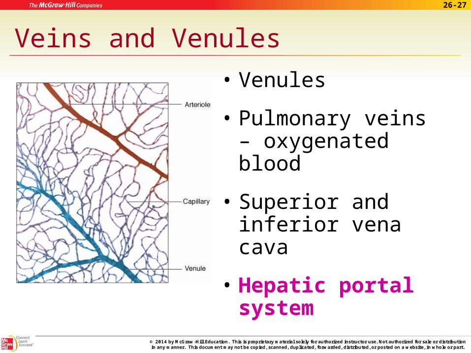
Veins and Venules
• Veins– Deoxygenated blood
– Skeletal muscle contractions move blood
– Valves prevent back flow
– Sympathetic nervous system effect
26-27
© 2014 by McGraw-Hill Education. This is proprietary material solely for authorized instructor use. Not authorized for sale or distribution in any manner. This document may not be copied, scanned, duplicated, forwarded, distributed, or posted on a website, in whole or part.
Veins and Venules • Venules
• Pulmonary veins – oxygenated blood
• Superior and inferior vena cava
• Hepatic portal system

26-28
© 2014 by McGraw-Hill Education. This is proprietary material solely for authorized instructor use. Not authorized for sale or distribution in any manner. This document may not be copied, scanned, duplicated, forwarded, distributed, or posted on a website, in whole or part.
Capillaries
• Branches of arterioles
• Connect arterioles to venules
• Exchange vessels
– Oxygen and nutrients
– Carbon dioxide and waste products
– Water

26-29
© 2014 by McGraw-Hill Education. This is proprietary material solely for authorized instructor use. Not authorized for sale or distribution in any manner. This document may not be copied, scanned, duplicated, forwarded, distributed, or posted on a website, in whole or part.
Apply Your KnowledgeARTERIES:Match the following:
coronary arteries A. exchange vessels
arterioles B. supply blood to the heart tissues
veins C. largest veins in the body
arteries D. supplies the forearm and hand
capillaries E. carry blood toward heart
vena cavae F. drain the knees
radial artery G. strongest blood vessels
popliteal veins H. small branches of arteries
C
A
B
D
E
F
G
H

26-30
© 2014 by McGraw-Hill Education. This is proprietary material solely for authorized instructor use. Not authorized for sale or distribution in any manner. This document may not be copied, scanned, duplicated, forwarded, distributed, or posted on a website, in whole or part.
Blood Pressure
• The force blood exerts on the inner walls of blood vessels
• Highest in arteries
• Lowest in veins
• Rises and falls as ventricles contract and relax

26-31
© 2014 by McGraw-Hill Education. This is proprietary material solely for authorized instructor use. Not authorized for sale or distribution in any manner. This document may not be copied, scanned, duplicated, forwarded, distributed, or posted on a website, in whole or part.
Blood Pressure
• Systolic pressure – Systole
– Ventricles contract
– Blood pressure in arteries greatest
• Diastolic pressure– Diastole
– Ventricles relax
– Blood pressure in arteries lowest
• Pulse

26-32
© 2014 by McGraw-Hill Education. This is proprietary material solely for authorized instructor use. Not authorized for sale or distribution in any manner. This document may not be copied, scanned, duplicated, forwarded, distributed, or posted on a website, in whole or part.
Blood Pressure (cont.)
• Factors affecting blood pressure – Cardiac output
– Blood volume– Vasoconstriction and vasodilation
– Blood viscosity

26-33
© 2014 by McGraw-Hill Education. This is proprietary material solely for authorized instructor use. Not authorized for sale or distribution in any manner. This document may not be copied, scanned, duplicated, forwarded, distributed, or posted on a website, in whole or part.
Blood Pressure (cont.)
• Controlled by the amount of blood pumped out of the heart
• Starling's law of the heart – Blood entering the left ventricle stretches its
wall
– The more the wall is stretched the harder it will contract

26-34
© 2014 by McGraw-Hill Education. This is proprietary material solely for authorized instructor use. Not authorized for sale or distribution in any manner. This document may not be copied, scanned, duplicated, forwarded, distributed, or posted on a website, in whole or part.
Blood Pressure (cont.)
• Baroreceptors – Help regulate blood pressure
– Located in the aorta and carotid arteries
– Send information to cardiac center in the medulla oblongata

26-35
© 2014 by McGraw-Hill Education. This is proprietary material solely for authorized instructor use. Not authorized for sale or distribution in any manner. This document may not be copied, scanned, duplicated, forwarded, distributed, or posted on a website, in whole or part.
Apply Your Knowledge
What is the difference between the systolic pressure and diastolic pressure?
ANSWER: Systolic pressure is the result of the contraction of the ventricles increasing the pressure in the arteries. Diastolic pressure is the result of the relaxation of the ventricles lowering the pressure in the arteries.
26-36
© 2014 by McGraw-Hill Education. This is proprietary material solely for authorized instructor use. Not authorized for sale or distribution in any manner. This document may not be copied, scanned, duplicated, forwarded, distributed, or posted on a website, in whole or part.
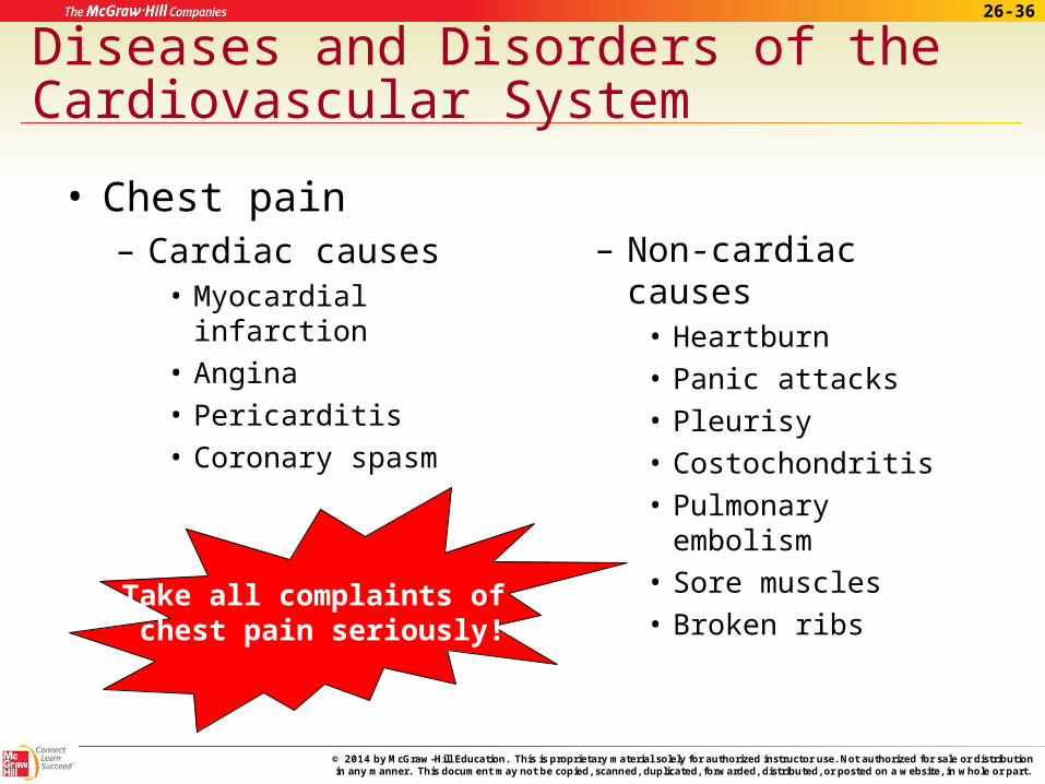
Diseases and Disorders of the Cardiovascular System
• Chest pain – Cardiac causes
• Myocardial infarction• Angina• Pericarditis• Coronary spasm
– Non-cardiac causes• Heartburn• Panic attacks• Pleurisy• Costochondritis • Pulmonary embolism• Sore muscles• Broken ribs
Take all complaints of chest pain seriously!
26-37
© 2014 by McGraw-Hill Education. This is proprietary material solely for authorized instructor use. Not authorized for sale or distribution in any manner. This document may not be copied, scanned, duplicated, forwarded, distributed, or posted on a website, in whole or part.
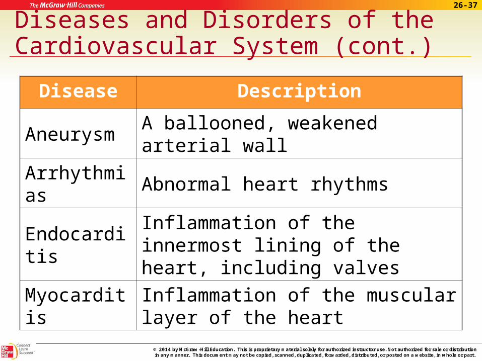
Diseases and Disorders of the Cardiovascular System (cont.)
Disease Description
Aneurysm A ballooned, weakened arterial wall
Arrhythmias Abnormal heart rhythms
EndocarditisInflammation of the innermost lining of the heart, including valves
MyocarditisInflammation of the muscular layer of the heart

26-38
© 2014 by McGraw-Hill Education. This is proprietary material solely for authorized instructor use. Not authorized for sale or distribution in any manner. This document may not be copied, scanned, duplicated, forwarded, distributed, or posted on a website, in whole or part.
Diseases and Disorders of the Cardiovascular System (cont.)
Disease Description
Pericarditis Inflammation of the membranes that surround the heart (pericardium)
Congestive heart failure
Weakening of the heart over time; heart is unable to pump enough blood to meet body’s needs
Coronary artery disease (CAD)
Atherosclerosis; narrowing of coronary arteries caused by hardening of the fatty plaque deposits within them
26-39
© 2014 by McGraw-Hill Education. This is proprietary material solely for authorized instructor use. Not authorized for sale or distribution in any manner. This document may not be copied, scanned, duplicated, forwarded, distributed, or posted on a website, in whole or part.
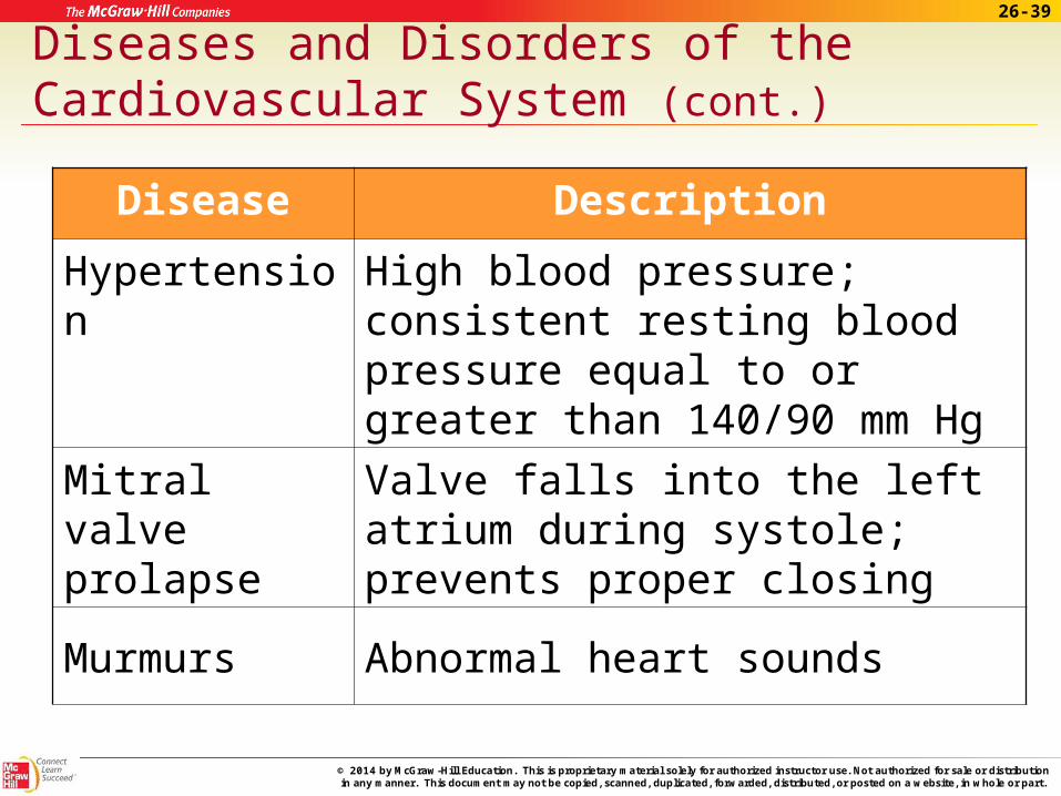
Diseases and Disorders of the Cardiovascular System (cont.)
Disease Description
Hypertension High blood pressure; consistent resting blood pressure equal to or greater than 140/90 mm Hg
Mitral valve prolapse
Valve falls into the left atrium during systole; prevents proper closing
Murmurs Abnormal heart sounds

26-40
© 2014 by McGraw-Hill Education. This is proprietary material solely for authorized instructor use. Not authorized for sale or distribution in any manner. This document may not be copied, scanned, duplicated, forwarded, distributed, or posted on a website, in whole or part.
Diseases and Disorders of the Cardiovascular System (cont.)
Disease Description
Myocardial infarction
Heart attack; damage to cardiac muscle due to a lack of blood supply
Stenosis of the heart valves
The valve does not fully open; aortic stenosis, mitral stenosis
Thrombophlebitis Blood clots and inflammation develop in a vein
Varicose veins Twisted, dilated veins

26-41
© 2014 by McGraw-Hill Education. This is proprietary material solely for authorized instructor use. Not authorized for sale or distribution in any manner. This document may not be copied, scanned, duplicated, forwarded, distributed, or posted on a website, in whole or part.
Apply Your KnowledgeANSWER:True or false:
A myocardial infarction results from a lack of oxygen to the cardiac muscle
Myocarditis is inflammation of the innermost lining of the heart.
A potentially fatal arrhythmia is ventricular fibrillation.
Murmurs are abnormal heart rhythms.
Bravo!
T
T
F
F
Endocarditis
sounds

26-42
© 2014 by McGraw-Hill Education. This is proprietary material solely for authorized instructor use. Not authorized for sale or distribution in any manner. This document may not be copied, scanned, duplicated, forwarded, distributed, or posted on a website, in whole or part.
In Summary
26.1 The structures of the heart include the pericardium, epicardium, myocardium, and endocardium.
The chambers of the heart consist of the upper atria and the lower ventricles.
The septa are the interatrial, interventricular, and atrioventricular.
The four valves within the heart are the tricuspid, the bicuspid, the pulmonary semilunar, and the aortic semilunar valves.

26-43
© 2014 by McGraw-Hill Education. This is proprietary material solely for authorized instructor use. Not authorized for sale or distribution in any manner. This document may not be copied, scanned, duplicated, forwarded, distributed, or posted on a website, in whole or part.
In Summary (cont.)
26.2 One cardiac cycle consists of one complete heartbeat.
The atria contract and relax together, and the ventricles contract and relax together.
As each chamber contracts, associated valves open and close to control the flow of blood through the heart.
Contractions are initiated by the cardiac conduction system.

26-44
© 2014 by McGraw-Hill Education. This is proprietary material solely for authorized instructor use. Not authorized for sale or distribution in any manner. This document may not be copied, scanned, duplicated, forwarded, distributed, or posted on a website, in whole or part.
In Summary (cont.)
26.3 Pulmonary circulation: Right atrium tricuspid valve right ventricle pulmonary semilunar valve pulmonary trunk pulmonary arteries lungs pulmonary veins left atrium.
Systemic circulation: Left atrium bicuspid valve left ventricle aortic semilunar
valve aorta arteries arterioles capillaries venules veins venae cavae right atrium.

26-45
© 2014 by McGraw-Hill Education. This is proprietary material solely for authorized instructor use. Not authorized for sale or distribution in any manner. This document may not be copied, scanned, duplicated, forwarded, distributed, or posted on a website, in whole or part.
In Summary (cont.)
26.4 Types of blood vessels include arteries and arterioles, which bring blood from the heart to the body; capillaries, which act as the connectors between the arterioles and venules.
The largest artery in the body is the aorta. Others arteries are listed in the chapter.
The largest veins in the body are the superior and inferior venae cavae. Others veins are listed in the chapter.

26-46
© 2014 by McGraw-Hill Education. This is proprietary material solely for authorized instructor use. Not authorized for sale or distribution in any manner. This document may not be copied, scanned, duplicated, forwarded, distributed, or posted on a website, in whole or part.
In Summary (cont.)
26.5 Blood pressure is the force exerted on the inner wall of blood vessels by blood as it flows through vessels.
It is highest in arteries and lowest in veins. Clinically, blood pressure refers to the force of blood within the arteries.
Blood pressure is largely controlled by the amount of blood pumped out of the heart, but various other events also may raise and
lower blood pressure.

26-47
© 2014 by McGraw-Hill Education. This is proprietary material solely for authorized instructor use. Not authorized for sale or distribution in any manner. This document may not be copied, scanned, duplicated, forwarded, distributed, or posted on a website, in whole or part.
In Summary (cont.)
26.6 Many different types of cardiac and blood diseases are described within this chapter.
The signs, symptoms, and treatments are as varied as the diseases themselves.
The last section of this chapter outlines the most common of these diseases, their signs and symptoms, as well as their treatments.

26-48
© 2014 by McGraw-Hill Education. This is proprietary material solely for authorized instructor use. Not authorized for sale or distribution in any manner. This document may not be copied, scanned, duplicated, forwarded, distributed, or posted on a website, in whole or part.
Your work is to discover your world and then with all your heart give yourself to it.
~ Buddha
End of Chapter 26