Chapter 23 The Gastrointestinal Tract. Learning Objectives (1 of 2) Identify major types of cleft...
94
Chapter 23 The Gastrointestinal Tract
-
Upload
princess-page -
Category
Documents
-
view
220 -
download
3
Transcript of Chapter 23 The Gastrointestinal Tract. Learning Objectives (1 of 2) Identify major types of cleft...

Chapter 23
The Gastrointestinal Tract

Learning Objectives (1 of 2)
• Identify major types of cleft lip and cleft palate deformity
• Explain pathogenesis and prevention of dental caries and periodontal disease
• Describe common congenital anomalies of the GIT, clinical manifestations, diagnosis, treatment
• Describe three most common lesions of the esophagus that lead to esophageal obstruction
• Explain pathogenesis, complications, and treatment of peptic ulcer
• Describe types and clinical manifestations of acute and chronic enteritis

Learning Objectives (2 of 2)
• Differentiate acute appendicitis and Meckel’s diverticulitis in terms of pathogenesis, clinical manifestations, and treatment
• Describe pathogenesis of diverticulitis and the role of diet in its development
• Discuss causes, clinical manifestations, complications– Intestinal obstruction– Colon cancer– Diverticulosis
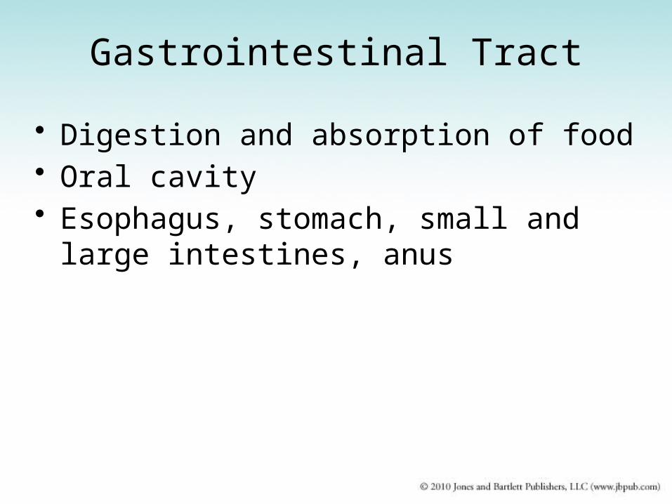
Gastrointestinal Tract
• Digestion and absorption of food• Oral cavity• Esophagus, stomach, small and large
intestines, anus

Cleft Lip and Cleft Palate• Embryologically, face and palate formed by
coalescence of cell masses that merge to form facial structures
• Palate formed by two masses of tissues that grow medially and fuse at midline to separate as nose and mouth
• Maldevelopment leads to defects– 1 per 1000 births– Multifactorial inheritance pattern
• Surgical correction (cheiloplasty)– Cleft lip: soon after birth– Cleft palate: 1 to 2 years of age followed by speech
therapy to correct nasal speech


Types of cleft lip and palate abnormalities viewed from below


A complete bilateral cleft lip and cleft palate with anterior protrusion of tissues between clefts
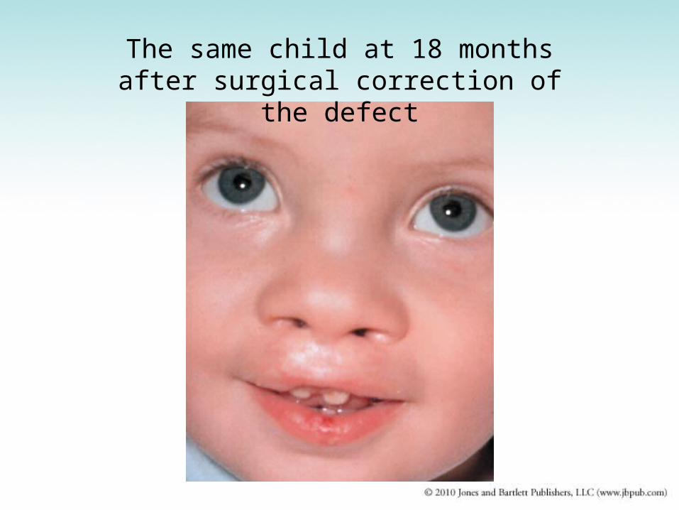
The same child at 18 months after surgical correction of the defect

Abnormalities of Tooth Development
• Teeth: specialized structures that develop in tissues of the jaws
• Two sets– Temporary or deciduous teeth (20 teeth)– Permanent teeth (32 teeth)
• Missing teeth or extra teeth: common abnormality
• Enamel forms at specific times during embryologic period
• Tetracycline: administered during enamel formation causes permanent yellow-gray to brown discoloration of the crown

Figure 23.10a
IncisorsCentral (6–8 mo)
IncisorsCentral (7 yr)
Canine (eyetooth)(16–20 mo)
Canine (eyetooth)(11 yr)Premolars(bicuspids)First premolar(11 yr)
MolarsFirst molar(10–15 mo)
MolarsFirst molar (6–7 yr)
Lateral (8–10 mo) Lateral (8 yr)
Second molar(about 2 yr)
Second molar(12–13 yr)Third molar(wisdom tooth)(17–25 yr)(a)
Permanentteeth
Deciduous(milk) teeth Second premolar
(12–13 yr)

Figure 23.11
Crown
Neck
Root
EnamelDentinDentinal tubulesPulp cavity (containsblood vessels and nerves)Gingiva (gum)
Cementum
Root canalPeriodontalligament
Apical foramen
Bone

Abnormalities of Tooth Development

Dental Caries and Periodontal Disease
• Oral cavity: diverse collection of aerobic and anaerobic bacteria that mix with saliva, forming sticky film on teeth (dental plaque)
• Plaque + action of bacteria result in tooth decay (caries)• Dental cavity: loss of tooth structure from bacterial action• Gingivitis: inflammation of the gums due to masses of
bacteria and debris accumulating around base of teeth• Periodontal disease: inflammation extends to tissues that
support teeth; forms small pockets of infection between teeth and gums– Two types: gingivitis and periodontitis

Tooth and Gum Disease
• Dental caries (cavities): gradual demineralization of enamel and dentin – Dental plaque (sugar, bacteria, and debris)
adheres to teeth– Acid from bacteria dissolves calcium salts– Proteolytic enzymes digest organic matter – Prevention: daily flossing and brushing

Tooth and Gum Disease
• Gingivitis– Plaque calcifies to form calculus (tartar)– Calculus disrupts the seal between the
gingivae and the teeth – Anaerobic bacteria infect gums– Infection reversible if calculus removed

Tooth and Gum Disease
• Periodontitis– Immune cells attack intruders and body
tissues• Destroy periodontal ligament• Activate osteoclasts
– Consequences• Possible tooth loss, promotion of
atherosclerosis and clot formation in coronary and cerebral arteries

Stomatitis
• Inflammation of the oral cavity• Causes
– Irritants: alcohol, tobacco, hot or spicy foods– Infectious agents: Herpes virus, Candida
albicans fungus, bacteria that cause trench mouth

Carcinoma of the Oral Cavity
• Arises from squamous epithelium– Lips– Cheek– Tongue– Palate– Back of throat

Esophagus (1 of 3)
• Muscular tube that extends from pharynx to stomach with sphincters at both upper and lower ends– Upper sphincter relaxes to allow passage of swallowed
food– Lower (gastroesophageal or cardiac) sphincter relaxes
to allow passage of food to the stomach• Diseases
– Failure of cardiac sphincter to function properly– Tears in lining of esophagus from retching and vomiting– At gastroesophageal junction from repetitive,
intermittent, vigorous contractions that increase intraabdominal pressure
– Esophageal obstruction from carcinoma, food impaction, or stricture

Figure 23.13
Tongue
Trachea
Pharynx
Epiglottis
Glottis
Bolus of food
Epiglottis
Esophagus
Uvula
Bolus
Bolus
Relaxed muscles
Circular musclescontract
Bolus of food
Longitudinal musclescontract
Stomach
Relaxedmuscles
Gastroesophagealsphincter opens
Gastroesophagealsphincter closed
Upper esophageal sphincter iscontracted. During the buccal phase, thetongue presses against the hard palate,forcing the food bolus into the oropharynxwhere the involuntary phase begins.
Food is movedthrough the esophagusto the stomach byperistalsis.
The gastroesophagealsphincter opens, and foodenters the stomach.
The uvula and larynx rise to prevent foodfrom entering respiratory passageways. Thetongue blocks off the mouth. The upperesophageal sphincter relaxes, allowing foodto enter the esophagus.
The constrictor muscles of thepharynx contract, forcing foodinto the esophagus inferiorly. Theupper esophageal sphinctercontracts (closes) after entry.
1 2
4
3
5

Figure 23.13, step 4
Relaxed muscles
Bolus of food
Stomach
Circular musclescontract
Longitudinal musclescontract
Gastroesophagealsphincter closed
Food is moved throughthe esophagus to thestomach by peristalsis.
4


Esophagus (2 of 3)• Symptoms
– Difficulty swallowing (dysphagia)– Substernal discomfort or pain– Inability to swallow (complete obstruction)– Regurgitation of food into trachea– Choking and coughing
• Two major disturbances of cardiac sphincter– 1. Cardiospasm: sphincter fails to open properly due to
malfunction of nerve plexus; esophagus becomes dilated proximal to constricted sphincter from food retention• Treatment: periodic stretching of sphincter; surgery
– 2. Incompetent cardiac sphincter: sphincter remains open; gastric juices leak back into esophagus

Esophagus (3 of 3)• Complications of incompetent cardiac sphincter
– Reflux esophagitis: inflammation– Ulceration and scarring of squamous mucosal lining– Barrett’s esophagus: glandular metaplasia; change from
squamous to columnar epithelium; ↑risk for cancer -a disorder in which the lining of the esophagus is damaged by stomach acid.
• Esophageal obstruction– Carcinoma: can arise anywhere in esophagus– Tumor narrows lumen of esophagus, infiltrates
surrounding tissue, invades trachea (tracheoesophageal fistula)
– Food impaction: distal part– Stricture: from scar tissue due to necrosis and
inflammation from corrosive chemicals such as lye

Gastric mucosal tear caused by retching and vomiting
Mallory–Weiss syndrome or gastro-esophageal laceration
syndrome refers to bleeding from tears (a Mallory-Weiss tear) in the
mucosa at the junction of the stomach and esophagus, usually
caused by severe retching, coughing, or vomiting.

Figure 23.30b
Liver
Lesser omentumGallbladder
StomachDuodenum
Transverse colon
Small intestine
Cecum
Urinary bladder(b)

Figure 23.15a
Mucosa
Surfaceepithelium
Lamina propria
Muscularismucosae
Oblique layer
Circular layer
Longitudinallayer
Serosa
(a) Layers of the stomach wall (l.s.)Stomach wall
Muscularis externa(contains myentericplexus)
Submucosa(contains submucosalplexus)

Figure 23.15b
(b) Enlarged view of gastric pits and gastric glands
Mucous neck cells
Parietal cell
Surface epithelium(mucous cells)
Gastric pits
Chief cell
Enteroendocrine cell
Gastric pit
Gastric gland

Figure 23.15c
(c) Location of the HCl-producing parietal cells and pepsin-secreting chief cells in a gastric gland
Pepsinogen
Mitochondria
PepsinHCl
Chief cell
Enteroendocrinecell
Parietal cell

Figure 23.18
Stomach lumenChief cell
Parietal cell
Inter-stitialfluid
Carbonicanhydrase
Alkalinetide
HCO3–
Bloodcapillary
CO2
Cl–
CO2 + H2O
H2CO3
HCO3–- Cl–
antiporter
HCO3–
H+
Cl– Cl–l
K+ K+
H+
H+-K+
ATPase
HCI

Acute Gastritis• Inflammation of the gastric lining• Self-limited inflammation of short duration• May be associated with mucosal ulceration or
bleeding• From nonsteroidal anti-inflammatory drugs (NSAID)
that inhibit cyclooxygenase (COX) enzyme: aspirin, ibuprofen, naproxen– COX-1: promotes synthesis of prostaglandin that protects
gastric mucosa– COX-2: promotes synthesis of prostaglandin that mediate
inflammation• Drugs that selectively inhibit COX-2 increase risk for heart attack
and stroke• Alcohol: a gastric irritant; stimulates gastric acid
secretion

H. Pylori Gastritis (1 of 2)• Small, curved, gram-negative organisms that
colonize surface of gastric mucosa• Grow within layer of mucus covering epithelial
cells• Produce urease that decomposes urea, a
product of protein metabolism, into ammonia• Ammonia neutralizes gastric acid allowing
organisms to flourish; organisms also produce enzymes that break down mucus layer

H. Pylori Gastritis (2 of 2)
• Common infection that increases with age (50% by age 50)
• Spreads via person-to-person through close contact and fecal-oral route
• Increased risk of gastric carcinoma: intestinal metaplasia
• Increased risk of malignant lymphoma (mucosa-associated lymphoid tissue, MALT)

Figure 23.16
Bacteria
Mucosalayer ofstomach
(a) A gastric ulcer lesion (b) H. pylori bacteria

Peptic Ulcer
• Pathogenesis– Digestion of mucosa due to increased acid secretions
and digestive enzymes (gastric acid and pepsin)– Helicobacter pylori injures mucosa directly or through
increased acid secretion by gastric mucosa– Common sites: distal stomach or proximal
duodenum• Complications: hemorrhage, perforation,
peritonitis, obstruction from scarring• Treatment
– Antacids: block acid secretion by gastric epithelial cells– Antibiotic therapy: against H. pylori– Surgery if medical therapy fails

Figure 23.16
Bacteria
Mucosalayer ofstomach
(a) A gastric ulcer lesion (b) H. pylori bacteria

Gastric ulcer, eroded a blood vessel at
base of ulcer causing profuse bleeding
Large, chronic duodenal ulcer

Carcinoma of the Stomach
• Manifestations– Vague upper abdominal discomfort– Iron-deficiency anemia (chronic blood loss
from ulcerated surface of tumor)• Diagnosis: biopsy by means of
gastroscopy• Treatment: surgical resection of affected
part, surrounding tissue and lymph nodes• Long-term survival: relatively poor; often
far-advanced at time of diagnosis

Carcinoma of the Stomach

Inflammatory Diseases of the Intestines
• Acute enteritis– Intestinal infections; common; of short duration– Nausea, vomiting, abdominal discomfort, loose stools
• Chronic enteritis: less common, more difficult to treat
• Regional enteritis or Crohn’s disease: distal ileum– Chronic inflammation and ulceration of mucosa with
thickening and scarring of bowel wall– Inflammation may be scattered with normal intervening
areas or “skip areas”– Treatment: drugs and possible surgical resection of
affected part of bowel

Ulcerative Colitis (1 of 2)• Ulcerative colitis: large intestines and
rectum– Inflammation is limited to mucosa, bowel not
thickened unlike in Crohn’s– Frequently begins in rectal mucosa and
spreads until entire colon is involved• Complications
– Bleeding; bloody diarrhea– Perforation: from extensive inflammation with
leakage of intestinal contents into peritoneal cavity
– Long-standing disease may develop cancer of colon and/or rectum

Ulcerative Colitis (2 of 2)
• Treatment– Symptomatic and supportive measures– Antibiotics, corticosteroids to control flare-ups– Immunosuppressive drugs– Surgical resection

Inflammatory Diseases of the Intestines (1 of 3)
• Antibiotic-associated colitis: broad-spectrum antibiotics destroy normal intestinal flora– Allows growth of anaerobic spore-forming bacteria,
Clostridium difficile not inhibited by antibiotic taken– Organisms produce toxins causing inflammation and
necrosis of colonic mucosa– Diarrhea, abdominal pain, fever
• Diagnosis: stool culture, toxin in stool• Treatment: stop antibiotic treatment; give
vancomycin or metronidazole– Drugs that decrease intestinal motility will prolong illness

Inflammatory Diseases of the Intestines (2 of 3)
• Appendicitis: most common inflammatory lesion of the bowel– Narrow caliber of appendix may be plugged with fecal
material– Secretions of appendix drain poorly, create pressure
in appendiceal lumen, compressing blood supply– Bacteria invade appendiceal wall causing
inflammation• Manifestations
– Generalized abdominal pain localizing in right lower quadrant; rebound tenderness; rigidity
• Treatment: surgery

Inflammatory Diseases of the Intestines (3 of 3)
• Meckel’s diverticulum– Outpouching at distal ileum, 12-18 inches proximal to
cecum– From persistence of a remnant of the vitelline duct,
narrow tubular channel connecting small intestine with yolk sac embryologically
– Found in 2% of population; usually asymptomatic
• May become infected causing features and complications similar to acute appendicitis
• Lining may consist of ectopic acid-secreting gastric mucosa and may cause peptic ulcer

Regional enteritis, mucosa ulcerated and covered with
inflammatory exudate

Inflammatory Disease Intestines

Disturbances in Bowel Function (1 of 2)
• Food intolerance: Crampy abdominal pain, distention, flatulence, loose stools
• Lactose intolerance– Unable to digest lactose into glucose and galactose for
absorption due to lactase deficiency– Enzyme abundant in infants and young children– Unabsorbed lactose remains in intestinal lumen and
raises osmotic pressure of bowel contents– Fermented by bacteria in colon, yielding lactic acid that
further increases intraluminal pressure– Common in Asians; 90% in Native Americans; 70%
Blacks

Disturbances Bowel Function (2 of 2)
• Gluten intolerance (Celiac disease; Gluten enteropathy or Nontropical sprue)– Gluten: protein in wheat, rye, barley; imparts
elasticity to bread dough– Chronic diarrhea impairing absorption of fats and
nutrients; weight loss, vitamin deficiencies– Leads to atrophy of intestinal villi– Diagnosis: clinical features and biopsy of
intestinal mucosa– Treatment: gluten-free diet

Irritable Bowel Syndrome• Also known as spastic colitis or mucous colitis• Episodes of crampy abdominal discomfort, loud
gurgling bowel sounds, and disturbed bowel function without structural or biochemical abnormalities
• Alternating diarrhea and constipation• Excessive mucus secreted by colonic mucosal
glands• Diagnosis: by exclusion
– Rule out pathogenic infections, food intolerance, and inflammatory conditions
• Treatment– Reduce emotional tension– Improve intestinal motility

Intestinal Infections in Homosexual Men
• Shigella• Salmonella• Entamoeba Histolytica• Giardia• Transmission: anal-oral sexual practices• Treatment: treat underlying cause

Colon Diverticulosis and Diverticulitis
• Diverticulosis: outpouchings or diverticula of colonic mucosa through weak areas in the muscular wall of large intestine– Low-residue diet predisposes to condition as increased
intraluminal pressure must be generated to propel stools through colon
– Acquired, usually asymptomatic, seen in older people– Common site: sigmoid colon
• Diverticulitis: inflammation incited by bits of fecal material trapped within outpouchings
• Complications: inflammation, perforation, bleeding, scarring, abscess

Diverticulosis of colon. Exterior of colon, illustrating several diverticula projecting
through the wall of the colon.
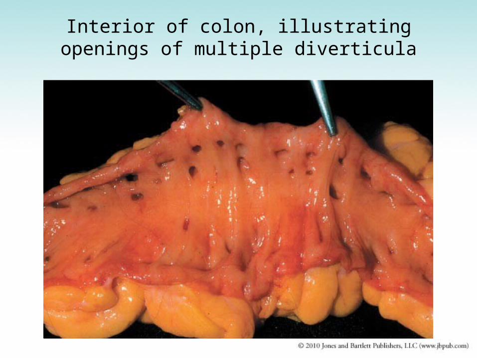
Interior of colon, illustrating openings of multiple diverticula

Diverticula of colon demonstrated by injection of barlum contrast material into
colon (barium enema)

Intestinal Obstructions (1 of 5)• Conditions blocking normal passage of
intestinal contents• Always considered as a serious condition• Severity depends on location of
obstruction, completeness, interference with blood supply
• High intestinal obstruction– Severe, crampy abdominal pain from
vigorous peristalsis– Vomiting with loss of H2O and electrolytes,
may result in dehydration

Intestinal Obstructions (2 of 5)
• Low intestinal obstruction– Symptoms less acute– Mild, crampy abdominal pain– Moderate distention of abdomen
• Common causes of intestinal obstruction– Adhesions– Hernia– Tumor– Volvulus– Intussusception

Intestinal Obstructions (3 of 5)
• Adhesions– Adhesive bands of connective tissue– May cause loop of bowel to become kinked,
compressed, twisted– Causes obstruction proximal to site of
adhesion• Hernia
– Protrusion of loop of bowel through a small opening, usually in abdominal wall
– Herniated loop pushes through peritoneum to form hernial sac

Intestinal Obstructions (4 of 5)
• Hernia– Inguinal hernia: common in men; loop of small
bowel protrudes through a weak area in inguinal ring and descends downward into scrotum
– Umbilical and femoral hernia: common in both sexes• Umbilical hernia: loop of bowel protrudes into
umbilicus through defect in the abdominal wall• Femoral hernia: loop of intestine extends under
inguinal ligament along course of femoral vessels into the groin

Intestinal Obstructions (5 of 5)• Reducible hernia: herniated loop of bowel can be
pushed back into abdominal cavity• Incarcerated hernia: cannot be pushed back• Strangulated hernia: loop of bowel is tightly
constricted obstructing the blood supply to the herniated bowel; requires prompt surgical intervention
• Volvulus: rotary twisting of bowel impairing blood supply; common site: sigmoid colon
• Intussusception: telescoping of a segment of bowel into adjacent segment; from vigorous peristalsis or tumor– Common site: terminal ileum

Fibrous adhesions between a loop of small intestine and
omentum
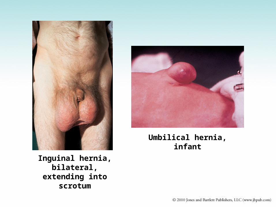
Inguinal hernia, bilateral,
extending into scrotum
Umbilical hernia, infant

Intussusception resulting from a colon tumor

Volvulus A. Rotary twisting of sigmoid colon on its mesenteryB. Obstruction of colon and interruption of blood
supply

Mesenteric Thrombosis
• Thrombosis of superior mesenteric artery– Artery supplies blood to small bowel and
proximal half of colon– May develop arteriosclerosis– Become occluded by thrombus, embolus, or
atheroma– Obstruction causes extensive bowel
infarction

Figure 19.23d

Tumors of the Colon• Benign pedunculated polyps
– Frequent– Tip may erode causing bleeding– Removed by colonoscopy
• Carcinoma– Cecum and right half of colon
• Does not cause obstruction as caliber is large and bowel contents are relatively soft
• Tumor can ulcerate, bleed; leads to chronic iron-deficiency anemia
• Symptoms of anemia: weakness and fatigue– Left half of colon
• Causes obstruction and symptoms of lower intestinal obstruction

Figure 23.29a
Left colic(splenic) flexure
Transversemesocolon
Epiploicappendages
Descendingcolon
Teniae coli
Sigmoidcolon
Cut edge ofmesentery
External anal sphincter
Rectum
Anal canal(a)
Right colic(hepatic) flexureTransversecolon SuperiormesentericarteryHaustrum
Ascendingcolon IIeum
IIeocecal valve
Vermiform appendix
Cecum

Colon Carcinoma

Imperforate Anus• Congenital anomaly, colon fails to acquire a
normal anal opening• Two types• 1. Rectum and anus normally formed and
extends to level of skin but no anal orifice– Easily treated by incising tissue covering anal opening
• 2. Entire distal rectum fails to develop, with associated abnormalities of urogenital and skeletal system– Corrected surgically but technically more difficult– Less satisfactory results


Imperforate Anus

Hemorrhoids• Hemorrhoids are vascular structures in the anal canal
which help with stool control. They become pathological when swollen or inflamed. In their physiological state they act as a cushion composed of arterio-venous channels and connective tissue that aid the passage of stool. The symptoms of pathological hemorrhoids depend on the type present. Internal hemorrhoids usually present with painless rectal bleeding while external hemorrhoids present with pain in the area of the anus.
• Recommended treatment consists of increasing fiber intake, oral fluids to maintain hydration, NSAID analgesics, sitz baths, and rest. Surgery is reserved for those who fail to improve following these measures.

Figure 23.29b
(b)
Rectal valveRectum
Anal canal
Levator animuscle
Anus
Anal sinuses
Anal columns
Internal analsphincter
External analsphincter
Hemorrhoidalveins
Pectinate line

Figure 19.28a

Figure 19.28c

Figure 23.29b
(b)
Rectal valveRectum
Anal canal
Levator animuscle
Anus
Anal sinuses
Anal columns
Internal analsphincter
External analsphincter
Hemorrhoidalveins
Pectinate line

Hemorrhoids• Varicose veins of hemorrhoidal venous plexus that
drains rectum and anus• Constipation and straining predispose to
development• Relieved by high-fiber diet rich in fruits and
vegetables, stool softeners, rectal ointment, or surgery– Internal hemorrhoids
• Veins of the lower rectum• May erode and bleed, become thrombosed, or prolapse
– External hemorrhoids• Veins of anal canal and perianal skin• May become thrombosed, causing discomfort

Diagnosis of GI Disease• Endoscopic procedures
– To directly visualize and biopsy abnormal areas such as esophagus, stomach, intestines
• Radiologic examination– To examine areas that cannot be readily
visualized– To evaluate motility problems– To visualize contours of GIT mucosa– To identify location and extent of disease
• Examples: Upper gastrointestinal tract – UGI• Colon – BE (barium enema)


Duodenum – 10 inchesJejunum – 8 feetIleum – 12 feetLarge Intestine – 4.9 feet


Barium

Barium Swallow

Barium Enema

Colon carcinoma demonstrated by barium enema
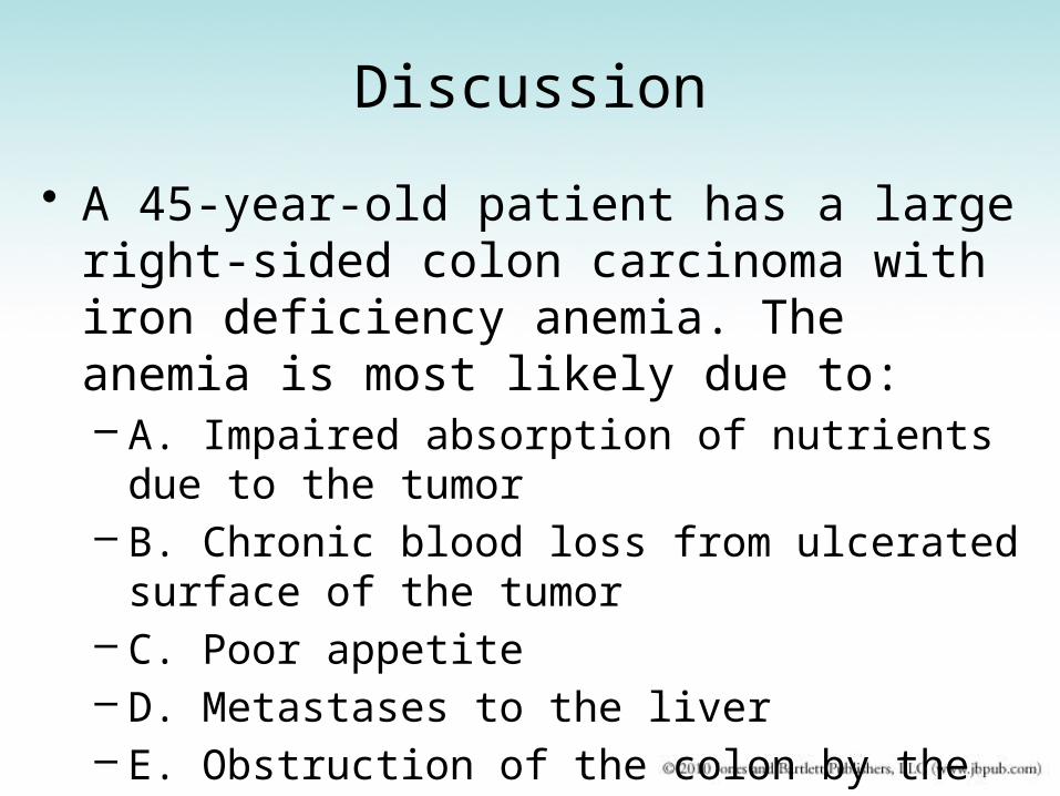
Discussion
• A 45-year-old patient has a large right-sided colon carcinoma with iron deficiency anemia. The anemia is most likely due to:– A. Impaired absorption of nutrients due to the
tumor– B. Chronic blood loss from ulcerated surface of
the tumor– C. Poor appetite– D. Metastases to the liver– E. Obstruction of the colon by the tumor