Chapter 2: Functional degradable polymers by...
39
Supporting information for “Poly(oligo (ethylene glycol) vinyl acetate)s: A versatile new class of thermoresponsive and biocompatible polymers” Guillaume G. Hedir, Maria C. Arno, Marvin Langlais, Jonathan T. Husband, Rachel K. O’Reilly and Andrew P. Dove 1
Transcript of Chapter 2: Functional degradable polymers by...
Supporting information
for
“Poly(oligo (ethylene glycol) vinyl acetate)s: A versatile new class of
thermoresponsive and biocompatible polymers”
Guillaume G. Hedir, Maria C. Arno, Marvin Langlais, Jonathan T. Husband, Rachel K. O’Reilly and Andrew P. Dove
1

1. Materials
The following reagents were used as received: vinyl acetate (VAc: Sigma-Aldrich, > 99%), 2-[2-(2-
methoxyethoxy)ethoxy] acetic acid (C6H12O5: VWR International, AR grade), 2-(2-methoxyethoxy)
acetic acid (C4H8O4: Sigma-Aldrich, technical grade), tetra(ethylene glycol) monomethyl ether
(TEGME: Sigma-Aldrich, technical grade), potassium hydroxide (KOH: Fisher Scientific, 90%),
palladium acetate (Pd(OAc)2, Sigma-Aldrich, 98%), potassium permanganate (KMnO4, Sigma-Aldrich,
> 99%), magnesium sulfate (MgSO4: anhydrous, Fisher Scientific, LR grade), hexylamine (C6H15N,
Fisher Scientific, > 99%), silica gel (SiO2: Apollo Scientific, 40 – 63 μm). 1,1’-azobis-
(cyclohexanocarbonitrile) (ABCN: Sigma-Aldrich, 98%) was recrystallized from methanol prior to use.
The following solvents were used as received: dichloromethane (DCM: VWR International, AR
grade), ethyl acetate (EtOAc, Fisher Scientific, LT grade), tetrahydrofuran (THF, VWR
International, AR grade), hexane (C6H14, Sigma-Aldrich, HPLC grade) and methanol (CH3OH,
Fisher Scientific, LT grade), diethyl ether (VWR International, AR grade). The enzyme used for
the degradation, Lipase, immobilized beads from Candida antarctica (Sigma-Aldrich, beads >
2 U/mg) were used as received and kept at a low temperature (5 °C) prior use. 2-Methylene-1,3-
dioxepane (MDO) was synthesized using the previously described method of Bailey et al.,[1] the chain
transfer agent p-methoxyphenyl xanthate (CTA) was synthesized using the procedure described in
our previous report.[2] The water used for the dissolution of the polymers for cloud point
measurements was purified using an ion exchange cartridge (18.2 MΩ/cm). N-succinimidyl
iodoacetate was obtained from Thermo Fisher Scientific. Human lysozyme (recombinant, expressed
in rice, lyophilized powder) was obtained from Sigma-Aldrich. SDS-PAGE gradient gels were
purchased from Biorad.
2. Characterization
Nuclear magnetic resonance (1H and 13C NMR) spectra were recorded at 400 MHz, 100 (or 125) MHz,
respectively, in CDCl3 on a Bruker DPX-400 spectrometer at 298 K. Chemical shifts are reported as δ
in parts per million (ppm) and referenced to the chemical shift of the residual solvent resonances
(CDCl3 1H: δ = 7.26 ppm; 13C: δ = 77.16 ppm). Size exclusion chromatography (SEC) analysis was
performed on a system composed of a Varian 390-LC-Multi detector suite using a Varian Polymer
Laboratories guard column (PLGel 5 μM, 50 × 7.5 mm), two mixed-D Varian Polymer Laboratories
columns (PLGel 5 μM, 300 × 7.5 mm) and a PLAST RT autosampler. Detection was conducted using a
differential refractive index (RI) and an ultraviolet (UV) detector set to λ = 280 nm. The analyses were
2

performed at 313 K in CHCl3 (HPLC grade) containing 0.5% w/w triethyl amine (NEt3) at a flow rate of
1.0 mL/min. Polystyrene (PS) (162 – 2.4 × 10⁵ g/mol) standards were used for calibration. Molecular
weights and dispersities were determined using Cirrus v3.3 SEC software. DLS analysis was
performed on a Malvern Zetasizer Nano ZS instrument operating at 25 °C with a 4 Mw He-Ne 633 nm
laser module. Measurements were made at an angle of 173° (back scattering), and results were
analyzed using Malvern DTS 6.3 software. All determinations were made in triplicate unless
otherwize stated (with 10 measurements recorded for each run). SLS experiments were performed
at angles of observation ranging from 20° up to 150° with an ALV CG3 spectrometer operating at λ0 =
633 nm and at 20 ± 1 °C. Solutions (1 mg/mL) were filtered through 0.45 μm nylon filters prior to
analysis. Data were collected in duplicate with 100 s run times. Calibration was achieved with filtered
toluene and the background was measured with filtered solvent (NaCl 0.1 M). The aggregation
number, Nagg of the self-assemblies was calculated using the REPES algorithm. [3] All turbidimetric
analysis was performed at a concentration of 5.0 mg/mL, unless otherwize stated, by UV-visible
spectrophotometry using an Agilent Cary 60/visible spectrophotometer. The absorbance was set at
550 nm and a heating rate of 1 °C/min was applied. Data was normalized between 0 and 100, plotted
in terms of transmittance (%), and the cloud point was defined as the temperature at which the
normalized transmittance equals 50%. SDS-PAGE was performed using 4-16 % BioRad SDS-PAGE gels.
Samples were prepared by heating protein solutions combined with loading buffer in a 1:1 ratio for 5
minutes at 80 °C and approximately 15 μL of protein solution was loaded into each well. Gels were
run at a constant voltage, 120 V, for around 60 minutes and subsequently stained with Coomassie
Brilliant Blue. MALDI-ToF mass spectra were recorded on a Bruker Daltonics Ultraflex and an Autoflex
MALD-ToF mass spectrometer in positive ion TOF detection mode performed using an acceleration
voltage of 25 kV. Solutions in THF of dithranol as matrix (30 mg/mL), sodium or potassium
trifluoroacetate as ionization agents (2 mg/mL) and analyte (1 mg/mL) were mixed prior to being
spotted on the MALDI plate and air-dried. The samples were measured in reflector ion mode and
calibrated by comparison to SpheriCal (Polymer Factory) single molecular weight standards (500 –
1800 g/mol).
3. Experimental
3.1. Procedure for the synthesis of MeO2VAc monomer
A solution of 2-[2-(2-methoxyethoxy)ethoxy] acetic acid (32.02 g, 179.7 mmol), KOH (1.0 g, 17.8
mmol) and Pd(OAc)2 (2.0 g, 8.90 mmol) in vinyl acetate (VAc) (153.30 g, 1780.9 mmol) was stirred at
3

60 °C for 16 h. The solution was then filtered over celite and thoroughly washed with diethyl ether
(100 mL) in order to remove the excess of catalyst. The excess of vinyl acetate and solvent (diethyl
ether) were evaporated under reduced pressure using rotary evaporation. The crude product was
purified and isolated by column chromatography (Silica, 7:3, EtOAc/hexane) before being dried over
anhydrous MgSO4, and then reduced to dryness using rotary evaporation, to yield to a pale brown
liquid. Rf (EtOAc/hexane 7:3) 0.46. The monomer was further purified by vacuum distillation (0.3
mmHg / 130 °C) to yield a colorless liquid (22.3 g, 109.30 mmol, 61%). 1H NMR (400 MHz, CDCl3,
ppm) δ: 7.26 (dd, COOCHCH2, 1H, 3JH-H = 13.82 Hz, 3JH-H = 7.68 Hz), 4.90 (dd, COOCHCHH, 1H, 3JH-H =
13.82 Hz, 2JH-H = 1.75 Hz), 4.60 (dd, COOCHCHH, 1H, 3JH-H = 6.28 Hz, 2JH-H = 1.84 Hz), 4.20 (s, OCOCH2O,
2H), 3.74 – 3.48 (m, (CH2CH2O)2, 8H), 3.33 (s, CH2CH2OCH3, 3H). 13C NMR (100 MHz, CDCl3) δ: 167.8
(CH2CHOCO), 140.5 (CH2CHCOO), 98.3 (CH2CHCOO), 70.9 – 70.4 (CH2CH2O), 68.1 (OCOCH2O), 58.9
(CH2CH2OCH3). Elemental analysis: Calculated for C9H16O5: C, 52.93%; H, 7.90%. Found: C, 52.46%; H,
7.98%.
3.2. General procedure for the homopolymerization of MeO2VAc
In an inert environment, MeO2VAc (0.30 g, 1.47 mmol), p-methoxyphenyl xanthate (4.30 mg, 0.015
mmol), ABCN (0.50 mg, 2.04 × 10⁻³ mmol) were placed into an ampoule and sealed. The solution was
subjected to a further three freeze-pump-thaw cycles and backfilled with nitrogen. The resulting
solution was stirred and heated to 90 °C for 6 h before the polymerization was quenched by plunging
the ampoule into an ice bath. An aliquot was taken prior to precipitation for monomer conversion
analysis by 1H NMR spectroscopy. The polymer was dissolved in a small amount of DCM (0.50 mL)
and precipitated several times into hexane until no further monomer residue was observed. The
light yellow solid was dried under vacuum at room temperature for 24 h. 1H NMR (400 MHz, CDCl3,
ppm) δ: 7.10 – 6.90 (dd, Ar, 4H, 3JH-H = 9.02 Hz), 5.10 – 4.72 (m, CH2CH backbone, 1H), 4.20 – 3.80 (m,
OCOCH2(OCH2CH2)2 side chain, 2H), 3.75 – 3.45 (m, (CH2CH2O)2 side chain, 2H, (CH2CH2O)2 side chain,
2H, CH3OCOCH2 end-group, 3H, Ar-OCH3, 3H), 2.50 (m, CH3OCOCH2 end-group, 2H), 3.45 – 3.30 (m,
(CH2CH2O)2CH3 side chain, 3H), 1.95 – 1.60 (m, CH2CH backbone, 2H). Conversion: MeO2VAc = 38%.
Mn (1H NMR, CDCl3) = 9.2 kg/mol, Mn (SEC, CHCl3) = 5.3 kg/mol, ÐM = 1.39.
3.3. Procedure for the synthesis of MeOVAc monomer
A solution of 2-(2-methoxyethoxy) acetic acid (32.02 g, 187.9 mmol), KOH (1.0 g, 17.8 mmol) and
Pd(OAc)2 (2.0 g, 8.90 mmol) in vinyl acetate (VAc) (161.3 g, 1873.4 mmol) was stirred at 60 °C for 16
h. The solution was filtered over celite and thoroughly washed with diethyl ether (100 mL) in order 4

to remove the excess of catalyst. The excess of vinyl acetate and solvent (diethyl ether) were
evaporated under reduced pressure using rotary evaporation. The crude product was purified and
isolated by column chromatography (Silica, 7:3, EtOAc/hexane) before being dried over anhydrous
MgSO4, and then reduced to dryness using rotary evaporation to yield to a pale brown liquid. Rf
(EtOAc/hexane 7:3) 0.38. The monomer was further purified by vacuum distillation (0.3 mmHg / 90
°C) to yield a colorless liquid (17.5 g, 109.25 mmol, 58%). 1H NMR (400 MHz, CDCl3, ppm) δ: 7.33 (dd,
COOCHCH2, 1H, 3JH-H = 13.02 Hz, 3JH-H = 6.02 Hz), 4.95 (dd, COOCHCHH, 1H, 3JH-H = 13.89 Hz, 2JH-H = 1.69
Hz), 4.62 (dd, COOCHCHH, 1H, 3JH-H = 6.22 Hz, 2JH-H = 1.70 Hz), 4.24 (s, OCOCH2O, 2H), 3.74 (t, CH2CH2O,
2H), 3.60 (t, CH2CH2O, 2H), 3.38 (s, CH2CH2OCH3, 3H). 13C NMR (100 MHz, CDCl3) δ: 167.6
(CH2CHCOO), 140.5 (CH2CHCOO), 98.5 (CH2CHCOO), 71.9 (CH2CH2O), 70.9 (CH2CH2O), 68.1
(COOCH2O), 58.9 (CH2CH2OCH3). Elemental analysis: Calculated for C7H12O4: C, 52.49%; H, 7.55%.
Found: C, 52.30%; H, 7.57%.
3.4. Procedure for the synthesis of MeO3VAc monomer
The monomer MeO3VAc was synthesized in a two steps procedure. In the first step, 2-2-[2-(2-
methoxyethoxy)ethoxy]ethoxy acetic acid was formed as follows: A solution of tetra(ethylene glycol)
monomethyl ether (TEGME) (10.4 g, 49.9 mmol), KOH (5.4 g, 96.4 mmol) and potassium
permanganate (KMnO4) (15.2 g, 96.4 mmol) in distilled water (1000 mL) was stirred at 0 °C for 2 h.
The brown solution was left to stir for a further 16 h at room temperature. The solution was filtered
on a Büchner filter, in order to remove the KMnO4 salt, and reduced to ¾ of its original volume using
rotary evaporation. The solution was acidified to pH = 2 using HCl (1 M) and the desired product was
extracted from the water phase against DCM (200 mL). The solution was dried over anhydrous
MgSO4, and then reduced to dryness using rotary evaporation to yield to a colorless liquid (6.1 g,
27.4 mmol, 55%). 1H NMR (400 MHz, CDCl3, ppm) δ: 11.30 (s, CH2COOH, 1H), 4.15 (s, CH2COOH, 2H),
3.74 – 3.48 (m, CH2CH2O, 12H), 3.38 (s, CH2CH2OCH3, 3H). 13C NMR (100 MHz, CDCl3) δ: 171.3
(CH2COOH), 71.9 – 70.9 (CH2CH2O), 68.4 (COOCH2O), 58.5 (CH2CH2OCH3). In the second step,
VMeO3Ac was synthesized as follows: A solution of 2-2-[2-(2-methoxyethoxy)ethoxy]ethoxy acetic
acid (4.1 g, 18.4 mmol), KOH (0.10 g, 0.18 mmol) and Pd(OAc)2 (0.20 g, 0.89 mmol) in vinyl acetate
(VAc) (16.0 g, 185.8 mmol) was stirred at 60 °C for 16 h. The solution was filtered over celite and
thoroughly washed with diethyl ether (10 mL) in order to remove the excess of catalyst. The excess
of vinyl acetate and solvent (diethyl ether) were evaporated under reduced pressure using rotary
evaporation. The crude product was purified and isolated by column chromatography (Silica, 7:3,
5

EtOAc/hexane), before being dried over anhydrous MgSO4, and reduced to dryness using rotary
evaporation to yield to a colorless liquid, (1.2 g, 4.82 mmol, 26%). Rf (EtOAc/hexane 7:3) 0.51. 1H
NMR (400 MHz, CDCl3, ppm) δ: 7.28 (dd, COOCHCH2, 1H, 3JH-H = 13.88 Hz, 3JH-H = 7.23 Hz), 4.95 (dd,
COOCHCHH, 1H, 3JH-H = 13.82 Hz, 2JH-H = 1.68 Hz), 4.65 (dd, COOCHCHH, 1H, 3JH-H = 6.48 Hz, 2JH-H = 1.72
Hz), 4.20 (s, OCOCH2O, 2H), 3.78 – 3.50 (m, CH2CH2O, 12H), 3.38 (s, CH2CH2OCH3, 3H). 13C NMR (100
MHz, CDCl3) δ: 167.4 (CH2CHCOO), 140.7 (CH2CHCOO), 98.1 (CH2CHCOO), 70.9 – 70.6 (CH2CH2O), 68.5
(COOCH2O), 58.5 (CH2CH2OCH3). Elemental analysis: Calculated for C11H20O6: C, 53.22%; H, 8.18%.
Found: C, 53.29%; H, 8.31%.
3.5. General procedure for the homopolymerization of MeOVAc
In an inert environment, MeOVAc (0.50 g, 3.10 mmol), p-methoxyphenyl xanthate (8.50 mg, 0.031
mmol), ABCN (0.76 mg, 3.10 × 10⁻³ mmol) were placed into an ampoule and sealed. The solution was
subjected to a further three freeze-pump-thaw cycles and backfilled with nitrogen. The resulting
solution was stirred and heated to 90 °C for 4.5 h before the polymerization was quenched by
plunging the ampoule into an ice bath. An aliquot was taken prior to precipitation for monomer
conversion analysis by 1H NMR spectroscopy. The polymer was dissolved in a small amount of DCM
(0.50 mL) and precipitated several times into hexane until no further monomer residue was
observed. The light yellow solid was dried under vacuum at room temperature for 24 h. 1H NMR (400
MHz, CDCl3, ppm) δ: 7.10 – 6.90 (dd, Ar, 4H, 3JH-H = 9.00 Hz), 5.10 – 4.80 (m, CH2CH backbone, 1H),
4.20 – 3.80 (m, OCOCH2OCH2CH2 side chain, 2H), 3.75 – 3.45 (m, CH2CH2O side chain, 2H, CH2CH2O
side chain, 2H, CH3OCOCH2 end-group, 3H, Ar-OCH3, 3H), 3.45 – 3.30 (m, CH2CH2OCH3 side chain, 3H),
2.50 (m, CH3OCOCH2 end-group, 2H), 1.95 – 1.60 (m, CH2CH backbone, 2H). Conversion: MeOVAc =
42%. Mn (1H NMR, CDCl3) = 7.7 kg/mol, Mn (SEC, CHCl3) = 4.7 kg/mol, ÐM = 1.46.
3.6. Procedure for the homopolymerization of MeO3VAc
In an inert environment, MeO3VAc (0.20 g, 0.81 mmol), p-methoxyphenyl xanthate (2.20 mg, 0.0081
mmol), ABCN (0.19 mg, 7.80 × 10⁻⁴ mmol) were placed into an ampoule and sealed. The solution was
subjected to a further three freeze-pump-thaw cycles and backfilled with nitrogen. The resulting
solution was stirred and heated to 90 °C for 7 h before the polymerization was quenched by plunging
the ampoule into an ice bath. An aliquot was taken prior to precipitation for monomer conversion
analysis by 1H NMR spectroscopy. The polymer was dissolved in a small amount of DCM (0.50 mL)
and precipitated several times into hexane until no further monomer residue was observed. The
light yellow solid was dried under vacuum at room temperature for 24 h. 1H NMR (400 MHz, CDCl3, 6

ppm) δ: 7.10 – 6.90 (dd, Ar, 4H, 3JH-H = 9.04 Hz), 5.10 – 4.80 (m, CH2CH backbone, 1H), 4.20 – 3.90 (m,
OCOCH2(OCH2CH2)3 side chain, 2H), 3.80 – 3.45 (m, (CH2CH2O)3 side chain, 2H, (CH2CH2O)3 side chain,
2H, CH3OCOCH2 end-group, 3H, Ar-OCH3, 3H), 2.50 (m, CH3OCOCH2 end-group, 2H), 3.45 – 3.30 (m,
(CH2CH2O)3CH3 side chain, 3H), 1.95 – 1.60 (m, CH2CH backbone, 2H). Conversion: MeO3VAc = 57%.
Mn (1H NMR, CDCl3) = 27.3 kg/mol, Mn (SEC, CHCl3) = 5.8 kg/mol, ÐM = 1.22.
3.7. Procedure for the aminolysis of poly(MeO2VAc)78
Poly(MeO2VAc) obtained from the RAFT/MADIX polymerization of MeO2VAc (0.24 g, 1.6 x 10⁻² mmol,
Mnobs = 15.1 kg/mol, Mn SEC = 7.4 kg/mol, ÐM = 1.51) and D,L dithiothreitol (0.50 mg, 3.2 x 10 -3 mmol)
were dissolved in 1.5 mL of THF and placed into an ampoule and sealed. The solution was subjected
to three freeze-pump-thaw cycles. Hexylamine (25 μL, 3.4 × 10 -4 mol) was added under N2 flow and
the solution was further degassed by freeze-pump thaw and backfilled with N2 before being left to
stir at room temperature for 6 h. The modified polymer was then recovered by three precipitations
from CHCl3 into hexane and dried under vacuum at room temperature for 24 h, leading to a pale
yellow residue. 1H NMR (400 MHz, CDCl3, ppm) δ: 7.10 – 6.90 (dd, Ar, 4H, 3JH-H = 9.02 Hz), 5.10 – 4.72
(m, CH2CH backbone, 1H), 4.20 – 3.80 (m, OCOCH2(OCH2CH2)2 side chain, 2H), 3.75 – 3.45 (m,
(CH2CH2O)2 side chain, 2H, (CH2CH2O)2 side chain, 2H, CH3OCOCH2 end-group, 3H, Ar-OCH3, 3H), 2.50
(m, CH3OCOCH2 end-group, 2H), 3.45 – 3.30 (m, (CH2CH2O)2CH3 side chain, 3H), 1.95 – 1.60 (m, CH2CH
backbone, 2H). Conversion: MeO2VAc = 38%. Mn (1H NMR, CDCl3) = 9.2 kg/mol, Mn (SEC, CHCl3) = 7.1
kg/mol, ÐM = 1.53.
3.8. Procedure for the thiol terminated polymer conjugation to human lysozyme
1 mg of human lysozyme was solubilized in 1mL of 10 mM, pH = 8, phosphate buffer. To this, a
solution of 0.6 mg of N-succinimidyl iodoacetate in 25 μL DMSO was added and left to react in the
dark for 45 minutes. This solution was then purified by spin filter (5K MW cut-off). To the purified
protein in 10 mM, pH = 8, phosphate buffer (30 mg) of thiol terminated 16 kDa poly(MeO 2VAc) was
added and left to react in the dark for 90 mins. To cap unreacted linkers, cysteine (15 mM) was
added and reacted for 20 minutes. After re-purification by spin filter, samples were analyzed by SDS-
PAGE.
3.9. General procedure for the copolymerization of VAc and MeO2VAc
In an inert environment, MeO2VAc (0.35 g, 1.71 mmol), VAc (0.15 g, 1.72 mmol), p-methoxyphenyl
xanthate (9.45 mg, 3.4 × 10⁻² mmol) and ABCN (0.81 mg, 3.21 × 10⁻³ mmol) were placed into an 7

ampoule and sealed. The resulting solution was subjected to a further three freeze-pump-thaw
cycles and then backfilled with nitrogen. The resulting solution was stirred and heated to 90 °C for 15
h before the polymerization was quenched by plunging the ampoule into an ice bath. An aliquot was
taken prior precipitation for conversion analysis by 1H NMR spectroscopy. The polymer was dissolved
in a small amount of DCM (0.20 mL) and precipitated several times into hexane until no further
monomer residue was observed. The final light yellow residue was dried under vacuum at room
temperature for 24 h. 1H NMR (400 MHz, CDCl3, ppm) δ: 7.10 – 6.90 (dd, Ar, 4H, 3JH-H = 8.95 Hz), 5.25
– 4.75 (m, CH2CH VAc backbone, 1H, m, CH2CH MeO2VAc backbone, 1H), 4.30 – 4.00 (m,
CH2(CH2CH2O)2, 2H), 3.90 – 3.45 (m, CH3OCOCH2 end-group, 2H, m, CH2(CH2CH2O)2, 2H, m,
CH2(CH2CH2O)2, 2H, m, Ar-OCH3 end-group, 3H), 3.45 – 3.25 (m, (CH2CH2O)2CH3, 3H), 2.40 - 2.25 (m,
CH3OCOCH2 end-group, 2H), 2.15 – 1.95 (m, OCOCH3 VAc, 3H), 1.95 - 1.45 (m, CH2CH VAc backbone,
2H, m CH2CH MeO2VAc backbone, 2H). Conversions: MeO2VAc = 64%, VAc = 61%. Mn (1H NMR, CDCl3)
= 12.7 kg/mol, Mn (SEC, CHCl3) = 8.2 kg/mol, ÐM = 1.62.
3.10. General procedure for the copolymerization of MDO and MeO2VAc
In an inert environment, MeO2VAc (0.50 g, 2.45 mmol), MDO (0.11 g, 0.96 mmol), p-methoxyphenyl
xanthate (9.90 mg, 3.6 × 10⁻² mmol) and ABCN (0.80 mg, 3.27 × 10⁻³ mmol) were placed into an
ampoule and sealed. The resulting solution was subjected to a further three freeze-pump-thaw
cycles and then backfilled with nitrogen. The resulting solution was stirred and heated to 90 °C for 9
h before the polymerization was quenched by plunging the ampoule into an ice bath. An aliquot was
taken prior precipitation for conversion analysis by 1H NMR spectroscopy. The polymer was dissolved
in a small amount of DCM (0.20 mL) and precipitated several times into hexane until no further
monomer residue was observed. The final light yellow solid was dried under vacuum at room
temperature for 24 h. 1H NMR (400 MHz, CDCl3, ppm) δ: 7.10 – 6.90 (dd, Ar, 4H, 3JH-H = 8.95 Hz), 5.25
– 4.75 (m, CH2CH backbone, 1H), 4.20 – 3.80 (m, OCOCH2(OCH2CH2)2, 2H, COOCH2CH2CH2 backbone,
4H), 3.75 – 3.45 (m, (CH2CH2O)2 side chain, 2H, (CH2CH2O)2 side chain, 2H, CH3OCOCH2 end-group, 3H,
Ar-OCH3, 3H), 3.45 – 3.25 (m, (CH2CH2O)2CH3 side chain, 3H, CH2CH2SCSO end-group, 2H), 2.60 – 2.15
(m, CH3OCOCH2 end-group, 2H, CH2COOCH2CH2 backbone, 2H), 1.95 – 1.45 (m, CHCH2 backbone, 2H,
COOCH2CH2CH2CH2 backbone, 2H, COOCH2CH2CH2CH2 backbone, 2H, CH2CH2CH2SCS end-group, 2H),
1.45 – 1.10 (m, COOCH2CH2CH2CH2, 2H, CH2CH2SCS end-group, 2H). Conversions: MeO2VAc = 45%,
MDO = 28%. Mn (1H NMR, CDCl3) = 12.2 kg/mol, Mn (SEC, CHCl3) = 7.7 kg/mol, ÐM = 1.40.
8

3.11. General procedure for the copolymerization of MDO and MeOVAc
In an inert environment, MeOVAc (0.40 g, 2.50 mmol), MDO (0.046 g, 0.38 mmol), CTA 4 (7.80 mg,
2.86 × 10⁻² mmol) and ABCN (0.35 mg, 2.86 × 10 -3 mmol) were placed into an ampoule and sealed.
The resulting solution was subjected to a further three freeze-pump-thaw cycles and then backfilled
with nitrogen. The resulting solution was stirred and heated to 90 °C for 4.5 h before the
polymerization was quenched by plunging the ampoule into an ice bath. An aliquot was taken prior
to precipitation for conversion analysis by 1H NMR spectroscopy. The polymer was dissolved in a
small amount of DCM (0.50 mL) and precipitated several times into hexane until no further
monomer residue was observed. The final light yellow solid was dried under vacuum at room
temperature for 24 h. 1H NMR (400 MHz, CDCl3, ppm) δ: 7.10 – 6.90 (dd, Aromatic, 4H, 3JH-H = 8.98
Hz), 5.25 – 4.75 (m, CH2CH backbone, 1H), 4.20 – 3.80 (m, OCOCH2OCH2CH2 side chain, 2H,
COOCH2CH2CH2 backbone, 2H), 3.75 – 3.45 (m, CH2CH2O side chain, 2H, CH2CH2O side chain, 2H,
CH3OCOCH2 end-group, 3H, Aromatic-OCH3, 3H), 3.45 – 3.25 (m, CH2CH2OCH3 side chain, 3H,
CH2CH2SCSO end-group, 2H), 2.60 – 2.15 (m, CH3OCOCH2 end-group, 2H, CH2COOCH2CH2 backbone,
2H), 1.95 – 1.45 (m, CHCH2 backbone, 2H, COOCH2CH2CH2CH2 backbone, 2H, COOCH2CH2CH2CH2
backbone, 2H, CH2CH2CH2SCS end-group, 2H), 1.45 – 1.10 (m, COOCH2CH2CH2CH2, 2H, CH2CH2SCS
end-group, 2H). Conversions: MeOVAc = 43%, MDO = 31%. Mn (1H NMR, CDCl3) = 6.4 kg/mol, Mn
(SEC, CHCl3) = 4.5 kg/mol, ÐM = 1.53.
3.12. General procedure for the degradation experiments
The degradation under enzymatic conditions was performed as follows: 500 mg copolymer was
placed in a 10 mL vial and 10 mL of a phosphate buffer solution (PBS, pH = 7.4) was added to dissolve
the sample. Enzymes beads (Lipase immobilized from Candida antarctica, 0.10 g) were added to the
polymer solution and stirred at either 25 or 37 °C. In order to keep the enzymes activities constant
throughout the experiment, the beads were replaced every 24 h. Samples were taken a different
time points and the solvent (PBS solution) was removed by freeze-drying for 16 h. The polymer
residues were re-dissolved in CHCl3 and filtered in order to remove the residual salt, and analyzed by
SEC in CHCl3.
3.13. General procedure for the cell viability studies
For the cell viability study, the degradation products of poly(MeO2VAc-co-MDO) were obtained using
the degradation procedure described in section 3.12 with the Lipase enzyme beads at 37 °C. After 5
9

days, the enzyme beads were removed and the degradation product mixture was freeze dried three
times before being re-dissolved in water and used for cells study. For the preparation of the
degradation products of poly(MeO2VAc), the polymer was degraded in a solution of KOH in methanol
(0.1M) for 24 h at 37 °C. After degradation, the mixture was neutralized to pH 7 using a solution of
HCl (0.1 M) and the solvent was removed in vacuo. The degraded products were re-dissolved in
water and freeze dried three times before being use for cells study. MC3T3 cells were obtained from
Public Health England. Cells were cultured in 175 cm2 tissue culture flasks using MEM alpha medium
(Gibco), as advized by supplier, with addition of 10% FBS and 1% pen/strep, at 37 °C, 5% CO 2. Cells
were used three weeks after culture and seeded on 12 well plates (2000 cells/cm 2) for viability
assays. Cells were left to adhere and proliferate on the wells for 72 h, then incubated with a
concentration range (from 0 to 1 mg/mL) of poly(MDO-co-MeO2VAc), poly(MeO2VAc), and
degradation products of both. Viability was measured after 72 h using PrestoBlue metabolic assay
(Invitrogen), and results are reported as percentages compared to controls (0 mg/mL of
polymers/degradation products). Experiments were performed in triplicate.
0.00.51.01.52.02.53.03.54.04.55.05.56.06.57.07.58.0ppm
2.82
8.05
2.00
0.97
0.91
0.74
O O
O
OO
a
ab
c d
b c
d
e
f g
hi
e, f, g, h
i
Figure S1. 1H NMR spectrum of di(ethylene glycol) methyl ether vinyl acetate, MeO2VAc, synthesized by palladium catalyzed vinyl exchange reaction between 2-[2-(2-methoxyethoxy)ethoxy] acetic acid and vinyl
acetate (300 MHz, CDCl3).
10

0102030405060708090110130150170190210ppm
O O
O
OOa
bc d
e, g
e
g
f, h
hi
a
b
cf
d
CDCl3
i
Figure S2. 13C NMR spectrum of di(ethylene glycol) methyl ether vinyl acetate, MeO2VAc, synthesized by palladium catalyzed vinyl exchange reaction between 2-[2-(2-methoxyethoxy)ethoxy] acetic acid and vinyl
acetate (100 MHz, CDCl3).
0
0.2
0.4
0.6
0.8
1
1.2
100 1000 10000 100000 1000000
Nor
mal
ized
dw
/log
M
Molecular weight (g/mol)
1.5 h3 h6 h9 h12 h
Figure S3. Size exclusion chromatograms of poly(MeO2VAc) obtained by the RAFT/MADIX polymerization with p-methoxyphenyl xanthate the chain transfer agent for different reaction times, (SEC, CHCl3).
11

0
0.2
0.4
0.6
0.8
1
1.2
100 1000 10000 100000 1000000
Nor
mal
ized
dw
/dlo
gM
Molecular weight (g/mol)
RIUV (280 nm)
Figure S4. Size exclusion chromatograms of poly(MeO2VAc)40 synthesized using RAFT/MADIX polymerization after 6 h; blue trace using RI detection and red dashed trace using UV detection at λ = 280 nm, (SEC, CHCl3).
Table S1. Characterization data for the homopolymerization of MeO2VAc for different polymerization time points.
Time(h)
MeO2VAc conv.a
(%)DP b Mn
SEC c
(kg/mol)Mn
theo. d
(kg/mol)Mn
obs. e
(kg/mol) ÐM c
1.5 15 16 3.5 3.1 3.7 1.133 30 35 4.8 6.1 7.8 1.286 38 40 5.3 7.8 9.2 1.399 53 60 6.7 10.8 12.4 1.53
12 68 86 9.3 13.3 17.8 1.73a monomer conversions determined by 1H NMR spectroscopy (CDCl3), b obtained by 1H NMR spectroscopy
analysis (CDcl3), c obtained by SEC analysis in CHCl3, c theoretical molecular weight based on monomer conversion (1H NMR spectroscopy), d observed molecular weight obtained by 1H NMR spectroscopy end-group
analysis.
12

OO
n
O
O
O
O
S
S
O
O
2
0.00.51.01.52.02.53.03.54.04.55.05.56.06.57.07.58.0ppm
174.3
0
272.4
1
734.0
1
182.5
5
85.9
8
4.00
1 2
12
3
3
45
6
78
99
10
9
6
4, 5, 7, 10
***
8
CHCl3
Figure S5. 1H NMR spectrum of poly(MeO2VAc)86 synthesized using RAFT/MADIX polymerization after 12 h and p-methoxyphenyl xanthate as the chain transfer agent, (300 MHz, CDCl3), * indicates residual dichloromethane
traces and ** residual hexane traces.
0
5
10
15
20
25
30
35
1 10 100 1000 10000
Popu
latio
n (%
)
Dh (nm)
NumberVolumeIntensity
Figure S6. Dynamic light scattering traces of poly(MeO2VAc)40 in water at a concentration of 5 mg/mL.
13

R² = 0.9517
0
10
20
30
40
50
60
70
80
0.00E+00 5.00E+14
τ-1/
s-1
q2 (1/m2)
A
B
0.0001
0.00011
0.00012
0.00013
0.00014
0.00015
0.00016
0.00017
0 5E+14
Kc/R
θ
q2 (1/m2)
Figure S7. Multiple angle dynamic (A) and static (B) light scattering data of poly(MeO2VAc)40 in water at a concentration of 5 mg/mL confirming the unimeric population (Aggregation number, Nagg = 1, and
Hydrodynamic radius, Rh = 1.4 nm, with a refractive index increment (dn/dc) of 0.13).
14

0
20
40
60
80
100
50 70 90 110
Nor
mal
ized
tran
smitt
ance
(%)
Temperature (°C)
heatingcooling
Figure S8. Plot of normalized transmittance vs. temperature obtained for the heating/cooling cycle (0.5 °C/min) of poly(MeO2VAc)40 at a polymer concentration of 5 mg/mL.
Table S2. Characterization data for the polymer, poly(MeO2VAc), with different DPs of 11, 31, 66, 93, 280, 370 and their associated cloud point values.
DP a Mn SEC b
(kg/mol)Mn
obs. c
(kg/mol)Mn
theo. d
(kg/mol) ÐM b
Cloud point(°C) e
11 1.6 2.5 4.2 1.20 8431 3.5 6.6 7.1 1.33 8266 4.4 13.7 9.1 1.50 8293 6.0 19.2 12.7 1.61 86
280 14.0 57.1 28.4 2.00 81370 12.8 75.5 29.3 2.27 84
a determined by 1H NMR spectroscopy (CDCl3), b obtained by SEC analysis in CHCl3, c observed molecular weight obtained by 1H NMR spectroscopy end-group analysis, d theoretical molecular weight based on monomer
conversion (1H NMR spectroscopy), e turbidimetry experiment at a polymer concentration of 5 mg/mL in water.
15

0
20
40
60
80
100
0 20 40 60 80 100
Nor
mal
ized
tran
smitt
ance
(%)
Temperature (°C)
DP 11DP 31DP 66DP 93DP 280DP 370
0102030405060708090
100
0 20000 40000 60000 80000
Clou
d po
int (
°C)
Molecular weight (g/mol)
A
B
Figure S9. (A) Plots of normalized transmittance vs. temperature obtained for poly(MeO2VAc) polymers having different observed molecular weight, Mn obs and DP: DP = 11; Mn = 2.5 kg/mol, DP = 31; Mn = 6.6 kg/mol, DP =
66; Mn = 13.7 kg/mol, DP = 93; Mn = 19.2 kg/mol , DP = 280; Mn = 57.1 kg/mol, DP = 370; Mn = 75.5 kg/mol and (B) cloud points as a function of increasing Mn.
16

0
20
40
60
80
100
0 20 40 60 80 100
Nor
mal
ized
tran
smitt
ance
(%)
Temperature (°C)
0.5 mg/mL1 mg/mL1.5 mg/mL2.5 mg/mL5 mg/mL10 mg/mL
0
20
40
60
80
100
0 2 4 6 8 10 12
Clou
d po
int (
°C)
Concentration (mg/mL)
A
B
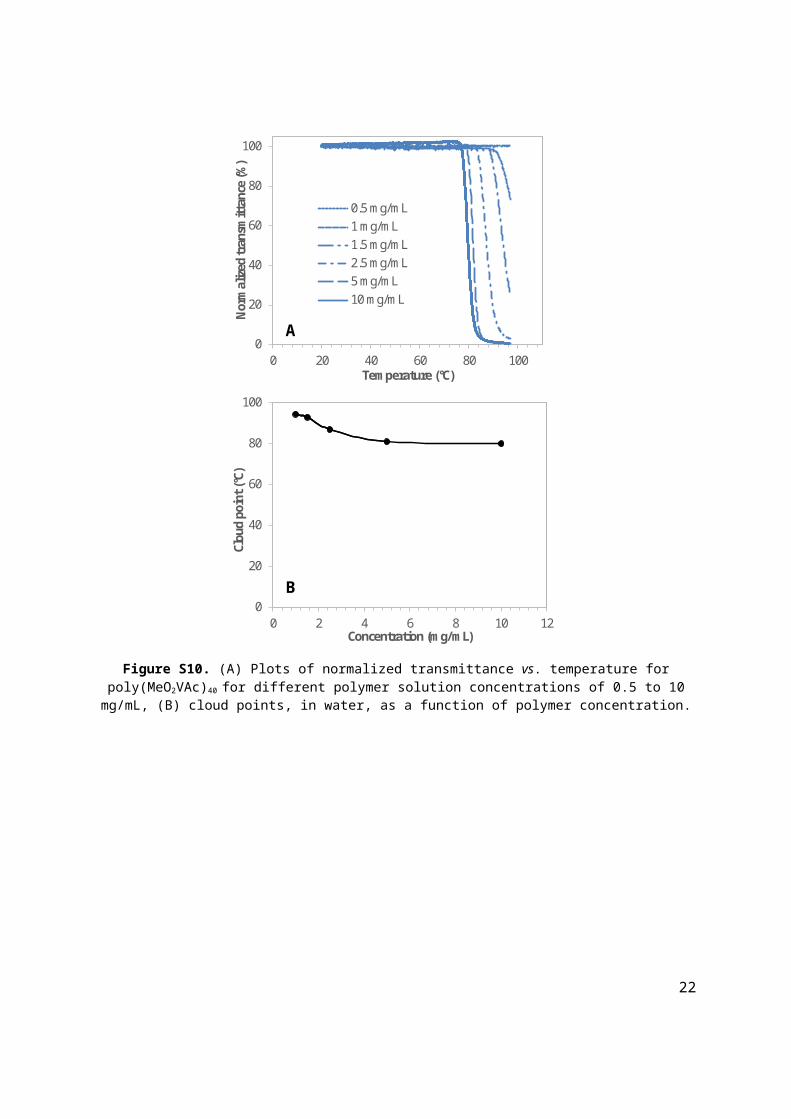
Figure S10. (A) Plots of normalized transmittance vs. temperature for poly(MeO2VAc)40 for different polymer solution concentrations of 0.5 to 10 mg/mL, (B) cloud points, in water, as a function of polymer concentration.
17

O O
O
O
-0.50.00.51.01.52.02.53.03.54.04.55.05.56.06.57.07.58.08.5ppm
3.0
0
4.0
6
2.0
0
1.0
0
0.9
9
1.1
0
ab
c d
b c
de
fg
e, f
g
H2Oa + CHCl3
Figure S11. 1H NMR spectrum of ethylene glycol methyl ether vinyl acetate, MeOVAc, synthesized by the palladium catalyzed vinyl exchange reaction between 2,2-(methoxy)ethoxy acetic acid and vinyl acetate (400
MHz, CDCl3).
0102030405060708090110130150170190210ppm
O O
O
Oab
c
d
efg
a
b
c
de f
g
CHCl3
Figure S12. 13C NMR spectrum of ethylene glycol methyl ether vinyl acetate, MeOVAc, synthesized by palladium catalysed vinyl exchange reaction between 2,2-(methoxy)ethoxy acetic acid and vinyl acetate (100
MHz, CDCl3).
18

-0.50.00.51.01.52.02.53.03.54.04.55.05.56.06.57.07.58.08.5ppm
3.00
12.1
9
2.01
1.00
1.00
1.26
O O
O
OO
O
a + CHCl3
ab
c e
g
h i
f jk
b c
d
d
k
e, f, g, h, i, j
H2O
Figure S13. 1H NMR spectrum of tri(ethylene glycol) methyl ether vinyl acetate, VMeO3Ac, synthesized by palladium catalyzed vinyl exchange reaction between 2-2-[2-(2-methoxyethoxy)ethoxy]ethoxy acetic acid and
vinyl acetate (400 MHz, CDCl3).
0102030405060708090110130150170190210ppm
O O
O
OO
Oab
c d e
g
h i
f j k
a
b
c
k
d
CHCl3
e, h, i
f, g, j
Figure S14. 13C NMR spectrum of tri(ethylene glycol) methyl ether vinyl acetate, VMeO3Ac, synthesized by palladium catalyzed vinyl exchange reaction between 2-2-[2-(2-methoxyethoxy)ethoxy]ethoxy acetic acid and
vinyl acetate (100 MHz, CDCl3).
19

0.00.51.01.52.02.53.03.54.04.55.05.56.06.57.07.58.08.5ppm
192.8
2
218.6
0
304.9
5
149.7
3
71.1
6
4.00
27
3
4, 5, 8, 9
6
1CHCl3
10
n
12 S
10O
O9
O
3O
O4
5O
6
S
O7
8
7
O
Figure S15. 1H NMR spectrum of poly(MeOVAc) synthesized using RAFT/MADIX polymerization and p-methoxyphenyl xanthate as the chain transfer agent, (300 MHz, CDCl3).
Figure S16. Size exclusion chromatogram of poly(MeOVAc) obtained after 4.5 h of RAFT/MADIX polymerization using p-methoxyphenyl xanthate as the chain transfer agent, (SEC, CHCl3).
20

0.00.51.01.52.02.53.03.54.04.55.05.56.06.57.07.58.08.5ppm
Jan24-2017Chemist Hedir GuillaumeGH-iv-63 pptPROTON.w CDCl3 /opt/topspin GH 54
326.
42
393.
14
1553
.93
245.
91
109.
99
4.00
n
12 S
10O
O9
O
3O
O4
5O
6
S
O7
8
7
O
3
27
3
4, 5, 8, 9
6
1
CHCl3
10
Figure S17. 1H NMR spectrum of poly(MeO3VAc) synthesized using RAFT/MADIX polymerization and p-methoxyphenyl xanthate as the chain transfer agent, (400 MHz, CDCl3).
Figure S18. Size exclusion chromatogram of poly(MeO3VAc) synthesized using RAFT/MADIX polymerization after 7 h, (SEC, CHCl3).
21
0.00.51.01.52.02.53.03.54.04.55.05.56.06.57.07.58.08.5ppm
Mar03-2017Chemist Guillaume HedirGH-iv-64PROTON.w CDCl3 /opt/topspin3.2 GH 28
149.5
7
208.4
3
571.2
1
133.7
4
56.71
4.00
S
OO
O
SO
O
O
O
O
2
n
12
3
6
4, 5, 7, 9
10
1 2
3
45
6
710
88
9
8
CHCl3
Figure S19. 1H NMR spectrum of poly(MeO2VAc) confirming the presence of the xanthate aromatic end-group signals at δ = 7.10 – 6.90 ppm, (400 MHz, CDCl3).
0102030405060708090110130150170190210230250ppm
Mar03-2017Chemist Guillaume HedirGH-iv-64C13APT.w CDCl3 /opt/topspin3.2 GH 28
S
OO
O
SO
O
O
O
O
2
n 1
1
2
3 45
54
2 3
Figure S20. 13C NMR spectrum of poly(MeO2VAc) confirming the presence of the xanthate aromatic end-group signals at δ = 158.1, 147.1, 122.7, 114.6 and 55.6 ppm, (100 MHz, CDCl3).
22

1000 2000 3000 4000 5000 60000
25
50
75
100
125
150
Nor
mal
ised
Inte
nsity
(%)
m/z
4
68
1012
14
16
1820
DP Exp. m/z Calc. m/z
4 1118.15 1118.24
6 1526.57 1526.50
8 1929.39 1929.45
10 2336.52 2336.61
12 2744.09 2744.20
14 3152.35 3152.40
16 3562.34 3562.61
18 3973.07 3973.21
20 4379.87 4380.07
S
OO
O
SO
O
O
O
O
2
nNa+
Figure S21. MALDI-ToF MS spectrum of poly(MeO2VAc)16.
SH
OO
O
O
O
O
2
n
0.00.51.01.52.02.53.03.54.04.55.05.56.06.57.07.58.08.5ppm
Feb15-2017GH-iv-71 ppt after amino
1 2
12
3
3
45
6
78
6
4, 5, 7
8
CHCl3
Figure S22. 1H NMR spectrum of poly(MeO2VAc)78 after aminolysis revealing the disappearance of the aromatic end-group at δ = 7.10 – 6.90 ppm, (400 MHz, CDCl3).
23

0102030405060708090110130150170190210230250ppm
Mar03-2017Chemist Guillaume HedirGH-iv-67C13APT.w CDCl3 /opt/topspin3.2 GH 29
SH
OO
O
O
O
O
2
n
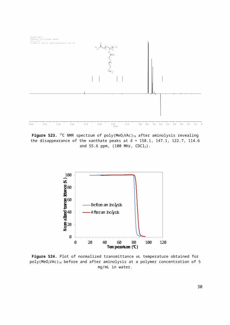
Figure S23. 13C NMR spectrum of poly(MeO2VAc)78 after aminolysis revealing the disappearance of the xanthate peaks at δ = 158.1, 147.1, 122.7, 114.6 and 55.6 ppm, (100 MHz, CDCl3).
0
20
40
60
80
100
0 20 40 60 80 100 120
Nor
mal
ized
tran
smitt
ance
(%)
Temperature (°C)
Before aminolysis
After aminolysis
Figure S24. Plot of normalized transmittance vs. temperature obtained for poly(MeO2VAc)78 before and after aminolysis at a polymer concentration of 5 mg/mL in water.
24

0
0.2
0.4
0.6
0.8
1
1.2
100 1000 10000 100000 1000000
Nor
mal
ized
dw
/dlo
gM
Molecular weight (g/mol)
Before aminolysisAfter aminolysis
Mn = 7.4 kg/molÐM = 1.51
Mn = 7.1 kg/molÐM = 1.53
Figure S25. Size exclusion chromatograms of poly(MeO2VAc)78 before and after aminolysis, (SEC, CHCl3).
Scheme S1. Schematic representation of (A) the aminolysis post-polymerization modifications of poly(MeO2VAc)78 and (B) subsequent conjugation to iodoacyl functionalized human lysozyme. The protein
structure of human lysozyme is from PDB entry 1LZ1.
25
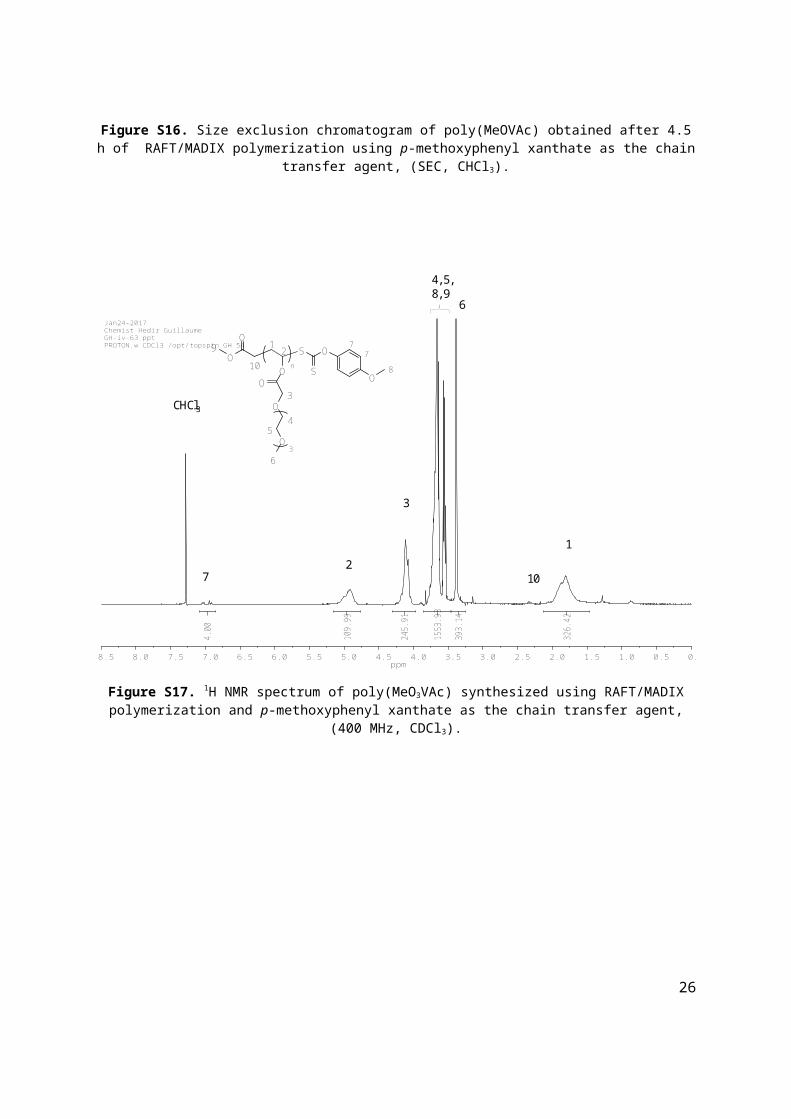
Figure S26. SDS-PAGE of poly(MeO2VAc)78 human lysozyme conjugate. Lane 1: ladder; lane 2: native human lysozyme; lane 3: human lysozyme reacted with succinimidyl iodoacetate; lane 4: human lysozyme
poly(MeO2VAc) conjugate.
Table S3. Characterization data for the copolymers of poly(MeO2VAc-co-VAc) obtained by RAFT/MADIX polymerization.
Initial monomer
feedMe
O2VAc:VAc a
Time(h)
VMeO2Acconv.a
(%)
VAc conv.a
(%)
Polymercomp.a
MeO2VAc:VAc
Mn SEC b
(kg/mol)
Mntheo. c
(kg/mol)
Mnobs. d
(kg/mol) ÐM b
90:10 16 68 51 94:6 10.0 13.2 10.3 1.7180:20 16 67 54 89:11 8.7 12.1 12.1 1.6160:40 8 37 31 75:25 4.2 5.6 6.5 1.24
50:50 9 62 57 56:44 8.2 9.0 12.7 1.62
40:60 9 69 54 50:50 9.7 8.7 10.5 1.59
35:65 8 65 57 42:58 9.8 8.1 11.3 1.61
30:70 8 33 25 35:65 5.2 6.6 7.0 1.50a determined by 1H NMR spectroscopy (CDCl3), b obtained by SEC analysis in CHCl3, c theoretical molecular
weight based on monomer conversion (1H NMR spectroscopy), d observed molecular weight obtained by 1H NMR spectroscopy end-group analysis.
26

0
0.5
1
1.5
2
2.5
100 1000 10000 100000 1000000
Nor
mal
ized
dw
/dlo
gM
Molecular weight (g/mol)
RIUV (280 nm)
Figure S27. Size exclusion chromatograms of poly(MeO2VAc-co-VAc) obtained by RAFT/MADIX polymerization for an initial monomer feed of 60:40 (MeO2VAc:VAc) and after 8 h of polymerization, blue trace using RI
detection and red dashed trace using UV detection at λ = 280 nm, (SEC, CHCl3).
0.00.51.01.52.02.53.03.54.04.55.05.56.06.57.07.58.08.5ppm
Jan13-2017Chemist Hedir GuillaumeGH-iv-61 50/50 pptPROTON.w CDCl3 /opt/topspin GH 2
300.
96
138.
54
379.
05
92.7
1
79.4
1
4.00
OS
OO
O
SO
O
O2
OO
O
x x-1
CHCl3
12
3 4
5
6 7
8
910
11
12
13
124, 7
8
1, 9, 10, 13
11
12
3, 6
H2O
2
5
Figure S28. 1H NMR spectrum of poly(VAc-co-MeO2VAc) synthesized using RAFT/MADIX polymerization and p-methoxyphenyl xanthate as the chain transfer agent, (400 MHz, CDCl3).
27

0
20
40
60
80
100
0 20 40 60 80 100
Nor
mal
ized
Tra
nsm
ittan
ce (%
)
Temperature ( C)
(a) (b) (c) (d) (e) (f) (g) (h)
VAc content
MeO2VAc content
Figure S29. Intensity of the normalized transmitted laser light vs. temperature for poly(MeO2VAc-co-VAc) copolymers of various compositions (a) 35 mol%, (b) 42 mol%, (c) 50 mol%, (d) 56 mol%, (e) 75 mol%, (f) 89
mol%, (g) 94 mol% and (h) 100% in MeO2VAc, at a concentration of 5 mg/mL in water.
y = 0.9397x - 9.5769R² = 0.985
0
10
20
30
40
50
60
70
80
90
20 40 60 80 100
Clou
d po
int (
°C)
Amount of MeO2VAc (%)Figure S30. Influence of MeO2VAc content on cloud point observed for the copolymer poly(MeO2VAc-co-VAc).
28

0
20
40
60
80
100
0 20 40 60 80 100
Nor
mal
ized
tran
smitt
ance
(%)
Temperature (°C)
(a) (b)
Figure S31. Plot of normalized transmittance vs. temperature for the heating (solid line) and cooling (dash line) cycle of poly(MeO2VAc-co-VAc) samples with (a) 42 mol% and (b) 89 mol% of MeO2VAc, at a concentration of 5
mg/mL in water.
Table S4. Characterization data for the copolymers of poly(MeO2VAc-co-MDO) obtained by RAFT/MADIX polymerization.
Initial monomer
feedMe
O2VAc:MDO a
Time(h)
VMeO2Acconv.a
(%)
MDO conv.a
(%)
Polymercomp.a
MeOnVAc:MDO
Mn SEC b
(kg/mol)
Mntheo. c
(kg/mol)
Mnobs. d
(kg/mol) ÐM b
90:10 5 43 22 95:5 6.8 8.1 15.5 1.6480:20 5 32 16 89:11 4.5 6.4 8.1 1.49
70:30 7 47 20 80:20 5.4 7.6 9.7 1.54
60:40 9 41 24 71:29 4.3 6.4 7.2 1.49
50:50 9 47 22 67:33 5.4 5.9 9.4 1.51
30:70 16 52 24 53:47 5.5 5.2 7.1 1.62a determined by 1H NMR spectroscopy (CDCl3), b obtained by SEC analysis in CHCl3, c theoretical molecular
weight based on monomer conversion (1H NMR spectroscopy), d observed molecular weight obtained by 1H NMR spectroscopy end-group analysis.
29
0
0.2
0.4
0.6
0.8
1
1.2
100 1000 10000 100000 1000000
Nor
mal
ized
dw
/dlo
gM
Molecular weight (g/mol)
RIUV (280 nm)
Figure S32. Size exclusion chromatograms of poly(MeO2VAc-co-MDO) obtained by the RAFT/MADIX polymerization after 9 h, blue trace using RI detection and red dashed trace using UV detection at λ = 280 nm,
(SEC, CHCl3).
0.51.01.52.02.53.03.54.04.55.05.56.06.57.07.58.08.5ppm
27.3
9
191.6
8
501.0
4
136.5
0
55.9
5
4.00
217
CHCl3
8, 3, 13
4, 5, 18, 19
6
20, 7, 12 **
1, 9, 11, 14
10, 1516
#
x 1-x
12
7O
8
9
10
1112
O13
14
15
16O
O
20O
O19
O
S O
S
1717
O18
3O
O4
5O
6n = 2
n
Figure S33. 1H NMR spectrum of poly(MDO-co-MeO2VAc) synthesized using RAFT/MADIX polymerization and p-methoxyphenyl xanthate as the chain transfer agent, (300 MHz, CDCl3),** branches resulting from the 1,4-
and 1,7-hydrogen abstraction, # residual acetone.
30

(a) (b)
0
20
40
60
80
100
0 20 40 60 80 100
Nor
mal
ized
tran
smitt
ance
(%)
Temperature (°C)
Figure S34. Plot of normalized transmittance vs. temperature for the heating (solid line) and cooling (dash line) cycle of poly(MeO2VAc-co-MDO) samples with (a) 80 mol% and (b) 95 mol% of MeO2VAc, at a concentration of
5 mg/mL in water.
Table S5. Characterization data for the copolymers of Poly(MDO(0.10)-co-MeOVAc(0.90))33 and Poly(MDO(0.33)-co-MeO2VAc(0.67))26 used during the enzymatic degradation.
Polymer
Monomer feed a
MeO2VAc:M
DO
Polymer comp.a
MeOnVAc:MDO
Mn SEC b
(kg/mol)Mn
theo. c
(kg/mol)Mn
obs. d
(kg/mol) ÐM b
CP (°C)
Poly(MDO(0.10)-co-MeOVAc(0.90))33
82:18 90:10 4.5 5.3 6.4 1.53 32.9
Poly(MDO(0.33)-co-MeO2VAc(0.67))26
58:42 67:33 3.6 4.1 5.2 1.44 29.9
a determined by 1H NMR spectroscopy (CDCl3), b obtained by SEC analysis in CHCl3, c theoretical molecular weight based on monomer conversion (1H NMR spectroscopy), d observed molecular weight obtained by 1H
NMR spectroscopy end-group analysis.
31

Table S6. Characterization data for the copolymers of MDO with MeOVAc, MeO2VAc and MeO3VAc comparing the initial monomer feed and final copolymer compositions.
MeOnVAc Monomer feed a
MeOnVAc:MDO
Polymer comp.a
MeOnVAc:MDO
Mn SEC b
(kg/mol)Mn
theo. c
(kg/mol) ÐM b
MeO1VAc 70:30 80:20 3.6 4.5 1.52MeO2VAc 70:30 78:22 5.4 7.6 1.54MeO3VAc 60:40 73:27 6.3 7.7 1.47
a determined by 1H NMR spectroscopy (CDCl3), b obtained by SEC analysis in CHCl3, c theoretical molecular weight based on monomer conversion (1H NMR spectroscopy).
4. References
[1] W. J. Bailey, Z. Ni, S.-R. Wu, J. Polym. Sci., Part A: Polym. Chem. 1982, 20, 3021-3030. [2] C. A. Bell, G. G. Hedir, R. K. O'Reilly, A. P. Dove, Polym. Chem. 2015, 6, 7447-7454. [3] J. Jakeš, Collect. Czech. Chem. Commun. 1995, 60, 1781-1797.
32