The influence of welding parameters on macrostructure and ...
Upload
nguyenngocCategory
view
218download
3
chapter
1Structure and Function of the Muscular, Neuromuscular, Cardiovascular, and Respiratory Systems

Chapter Objectives
• Describe the macrostructure and micro-structure of muscle and bone.
• Describe neuromuscular function.• Describe the characteristics of different
muscle fiber types.• Describe the characteristics of the cardio-
vascular and respiratory systems.
Skeletal System
• Composed of 206 bones in the adult body
• Provides leverage, support, and protection
• Pulled on by muscles to allow the body to push or pull against external objects
(continued)
Skeletal System (continued)
• Figure 1.1 (next slide)– (a) Front view of an adult male human skeleton– (b) Rear view of an adult male human skeleton

Figure 1.1

Axial Versus Appendicular Skeleton
• Axial: Consists of the skull, vertebral column (C1-coccyx), ribs, and sternum
• Appendicular: Consists of shoulder girdle; bones of the arms, wrists, hands, and pelvic girdle; and bones of the legs, ankles, and feet

Types of Joints
• Joint: Junctions of bones
• Fibrous: Allow virtually no movement– Example: Sutures of the skull
• Cartilaginous: Allow limited movement– Example: Intervertebral
• Synovial: Allow considerable movement– Example: Elbows and knees
(continued)

Types of Joints (continued)
• Uniaxial: Operate as a hinge, rotate about one axis– Example: Elbow
• Biaxial: Operate in two perpendicular axes– Example: Ankle and wrist
• Multiaxial: Allow movement in all three axes– Example: Shoulder and hip

Vertebral Column
• Vertebral bones separated by flexible disks that allow for movement– Cervical vertebrae (neck region): 7– Thoracic vertebrae (upper back): 12– Lumbar vertebrae (lower back): 5– Sacral vertebrae (make up rear of pelvis): 5– Coccygeal vertebrae (form vestigial tail extending
down from the pelvis): 3-5

Muscular System
• Macrostructure and microstructure– Each skeletal muscle is an organ that contains
muscle tissue, connective tissue, nerves, and blood vessels.
– Fibrous connective tissue, or epimysium, covers the body’s more than 430 skeletal muscles.
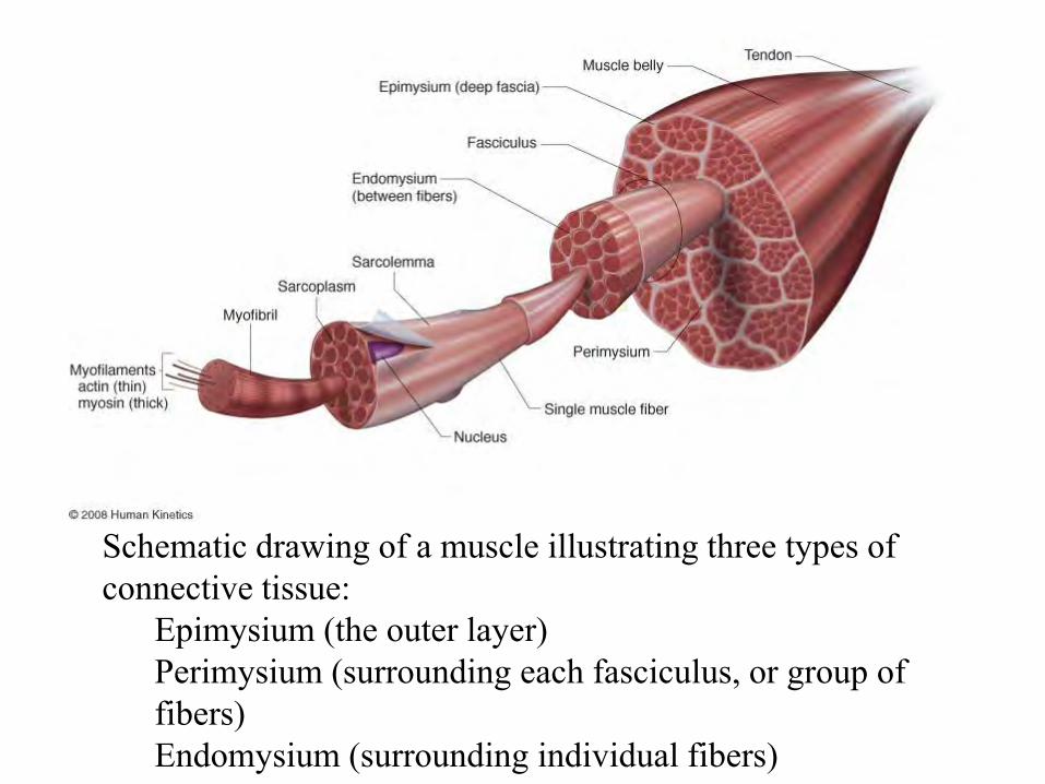
Figure 1.1
Schematic drawing of a muscle illustrating three types of connective tissue:
Epimysium (the outer layer)Perimysium (surrounding each fasciculus, or group of fibers)Endomysium (surrounding individual fibers)

Motor Unit
• Figure 1.4 (next slide)– A motor unit consists of a motor neuron and the
muscle fibers it innervates.– There are typically several hundred muscle fibers in
a single motor unit.
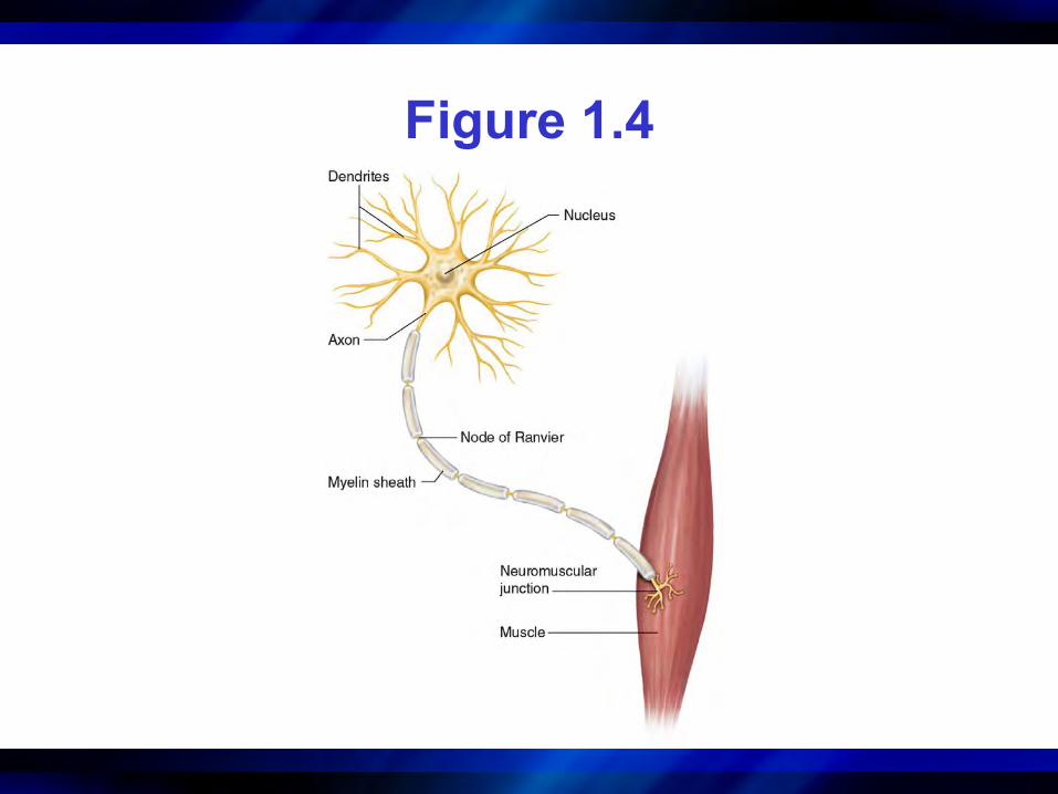
Figure 1.4

Figure 1.4
• (Myosin & Actin)– The arrangement
of myosin (thick) and actin (thin) filaments gives skeletal muscle its striated appearance.

Muscular System
• Sliding-Filament Theory of Muscular Contraction– The sliding-filament theory states that the actin
filaments at each end of the sarcomere slide inward on myosin filaments, pulling the Z-lines toward the center of the sarcomere and thus shortening the muscle fiber.

Figure 1.5• (a) In stretched muscle the I-
bands and H-zone are elongated, and there is low force potential due to reduced cross-bridge–actin alignment.
• (b) Muscle force is optimal when a muscle is at its resting length or slightly above due to optimal cross-bridge–actin alignment.
• (c) With completely contracted muscle, there is low force potential due to reduced cross-bridge–actin alignment, overlapping of actin, and abutting of myosin against Z-discs.

Figure 1.2• A motor
unit consists of a motor neuron and the muscle fibers it innervates.
Neuromuscular System (continued)
• Activation of muscles– The extent of control of a muscle depends on the
number of muscle fibers within each motor unit.• Muscles that function with great precision may have as
few as one muscle fiber per motor neuron. • Muscles that require less precision may have several
hundred fibers served by one motor neuron.

Key Point
Contractile Process of
Skeletal Muscle
Key Term
• all-or-none principle: All of the muscle fibers in the motor unit contract and develop force at the same time. There is no such thing as a motor neuron stimulus that causes only some of the fibers to contract. Similarly, a stronger action potential cannot produce a stronger contraction.

Figure 1.6• Twitch, twitch summation, & tetanus of a
motor unit: – a = single twitch– b = force resulting from
summation of 2 twitches– c = unfused tetanus– d = fused tetanus

Neuromuscular System
• Motor Unit Recruitment Patterns During Exercise– The force output of a muscle can be varied
through change in the frequency of activation of individual motor units or change in the number of activated motor units recruited.

Neuromuscular System
• Muscle Fiber Types– Type I (slow-twitch) – Type II (fast-twitch)
• Type IIab (fast-twitch); now named as Type IIax• Type IIb (fast-twitch); now named as Type IIx

Table 1.1

Table 1.2

Neuromuscular System
• How Can Athletes Improve Force Production?– Recruit large muscles or muscle groups during an
activity.– Increase the cross-sectional area of muscles involved in
the desired activity. – Preload a muscle just before a concentric action to
enhance force production during the subsequent muscle action (ie, Stretch-Shortening Cycle → Plyometrics).
– Use preloading during training to develop strength early in the range of motion.

Neuromuscular System
• Proprioception– Proprioceptors are specialized sensory receptors
that provide the central nervous system with information needed to maintain muscle tone and perform complex coordinated movements.
– Provides information concerning kinesthetic sense, or conscious appreciation of the position of body parts in space with respect to gravity
– Processed at subconscious levels– Includes muscle spindles and golgi tendon organs
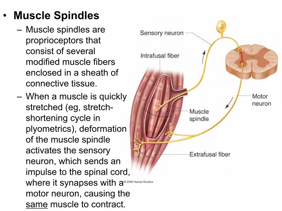
Figure 1.7• Muscle Spindles
– Muscle spindles are proprioceptors that consist of several modified muscle fibers enclosed in a sheath of connective tissue.
– When a muscle is quickly stretched (eg, stretch-shortening cycle in plyometrics), deformation of the muscle spindle activates the sensory neuron, which sends an impulse to the spinal cord, where it synapses with a motor neuron, causing the same muscle to contract.
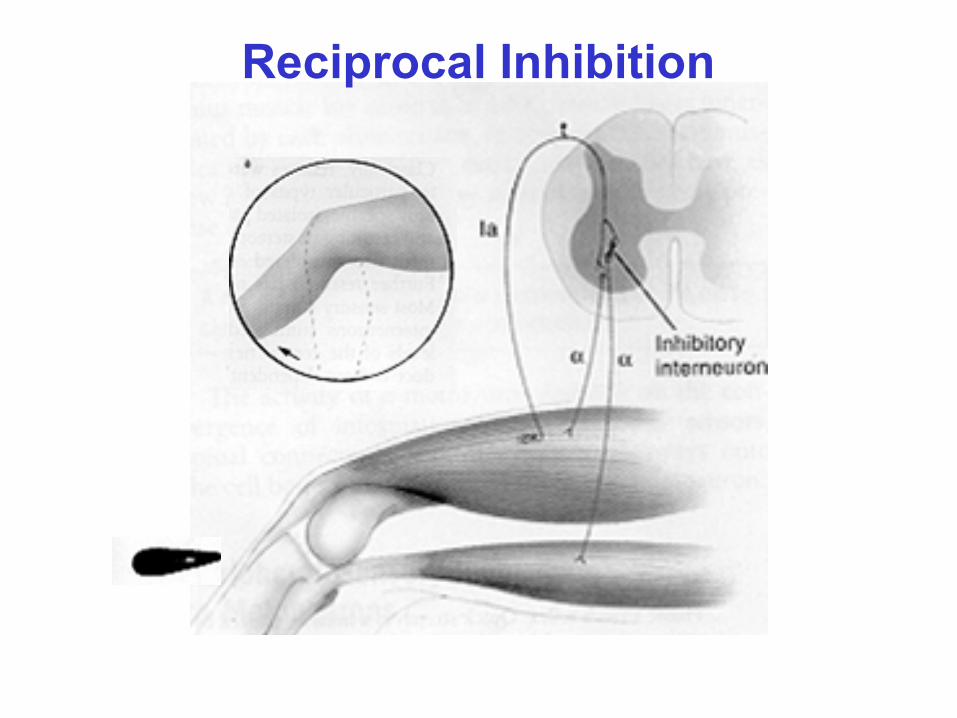
Figure 1.7Reciprocal Inhibition

Neuromuscular System
• Golgi Tendon Organs (GTO)– Golgi tendon organs are proprioceptors located in
tendons near the myotendinous junction.– They occur in series (i.e., attached end to end) with
extrafusal muscle fibers.– When an extremely heavy load is placed on the
muscle, discharge of the GTO occurs.– The sensory neuron of the GTO activates an
inhibitory interneuron in the spinal cord, which in turn synapses with and inhibits a motor neuron serving the same muscle.

Figure 1.8

Neuromuscular System
• Older Muscle – Muscle function is reduced in older adults. – Reductions in muscle size and strength are
amplified in weight-bearing extensor muscles.– Muscle atrophy with aging results from losses in
both number and size of muscle fibers, especially Type II muscle fibers.
– Inactivity plays a major role but cannot account for all of the age-related loss of muscle and function.

Cardiovascular System
• Heart– The heart is a muscular organ made up of
two interconnected but separate pumps.• The right ventricle pumps blood to the lungs.• The left ventricle pumps blood to the rest of the
body.
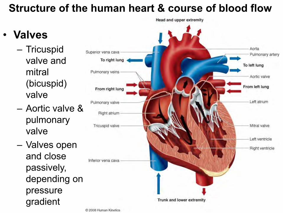
Figure 1.9• Valves– Tricuspid
valve and mitral (bicuspid) valve
– Aortic valve & pulmonary valve
– Valves open and close passively, depending on pressure gradient
Structure of the human heart & course of blood flow
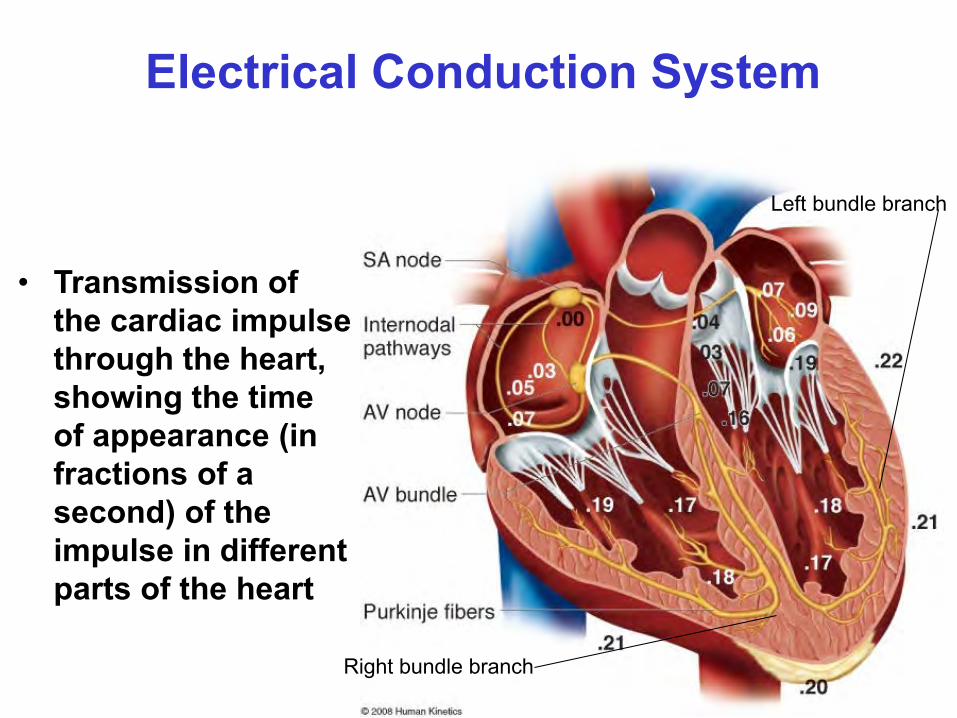
Figure 1.11
• Transmission of the cardiac impulse through the heart, showing the time of appearance (in fractions of a second) of the impulse in different parts of the heart
Electrical Conduction System
Right bundle branch
Left bundle branch
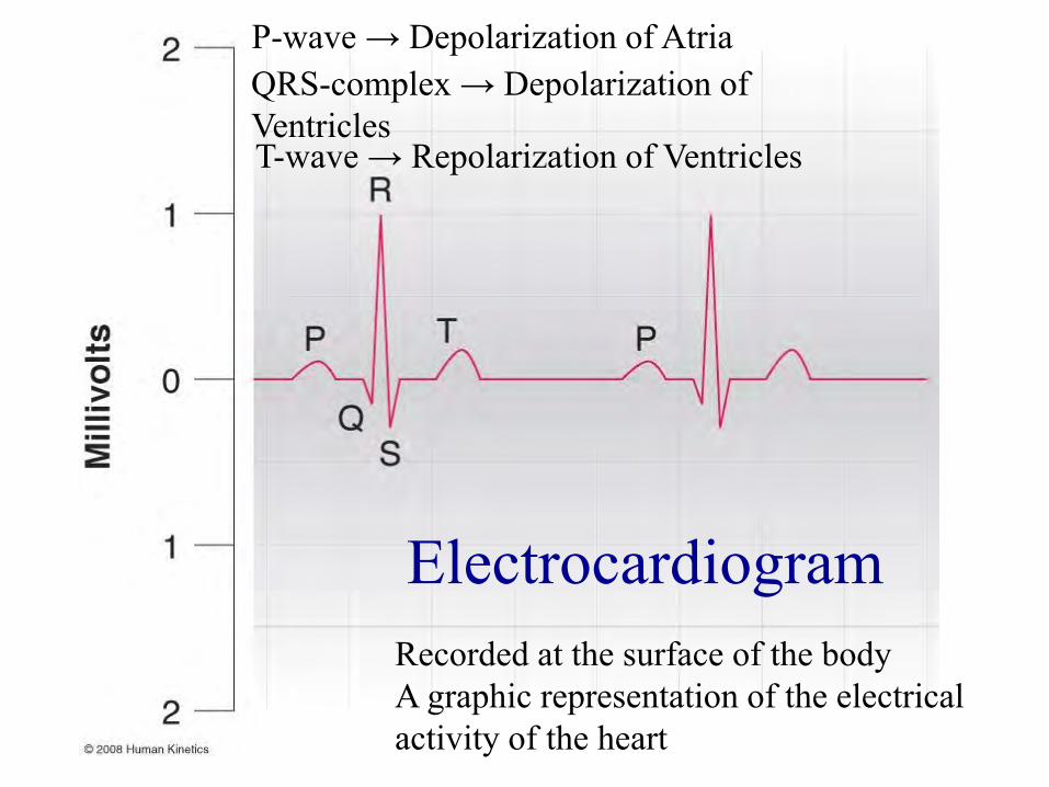
Figure 1.12
Recorded at the surface of the bodyA graphic representation of the electrical activity of the heart
Electrocardiogram
P-wave → Depolarization of AtriaQRS-complex → Depolarization of VentriclesT-wave → Repolarization of Ventricles

Cardiovascular System • Blood Vessels
– Blood vessels operate in a closed-circuit system. – Arteries (Carries oxygenated blood away from heart)– Capillaries (microscopic vessels that exchange oxygen,
nutrients, and other substances between blood and interstitial fluid)
– Veins (Carries deoxygenated blood towards heart)• Blood
– Hemoglobin transports oxygen and serves as an acid–base buffer.
– Red blood cells facilitate carbon dioxide removal.

Figure 1.13
• Arterial (right) and venous (left) components of the circulatory system.
• The percent values indicate the distribution of blood volume throughout the circulatory system at rest.
Distribution of Blood
Key Point
• The cardiovascular system transports nutrients and removes waste products while helping to maintain the environment for all the body’s functions. The blood transports oxygen from the lungs to the tissues for use in cellular metabolism, and it transports carbon dioxide from the tissues to the lungs, where it is removed from the body.

Figure 1.14Gross anatomy of the human respiratory system
Respiratory System
• Exchange of Air– The amount and movement of air and expired gases
in and out of the lungs are controlled by expansion and recoil of the lungs.
• Exchange of respiratory gases– The primary function of the respiratory system is the
basic exchange of oxygen and carbon dioxide.

Respiratory System (continued)
• Pleural pressure– Pressure in the narrow space between the lung
pleura and the chest wall pleura • Membranes enveloping the lungs and lining the chest walls
(continued)
Respiratory System (continued)
• Alveolar pressure– Pressure inside the alveoli when the glottis is open
and no air is flowing into or out of the lungs– To cause inward flow of air during inspiration, the
pressure in the alveoli must fall to a value slightly below atmospheric pressure
– During expiration, alveolar pressure must rise above atmospheric pressure
(continued)

Figure 1.15
• Contraction & expansion of thoracic cage during expiration and inspiration, illustrating diaphragmatic contraction, elevation of rib cage, & function of intercostals.– Vertical and anteroposterior diameters increase during
inspiration.