Chapter 18 Circulatory System. Heart (general) Size of a fist Size of a fist Almost one pound in...
16
Chapter 18 Chapter 18 Circulatory System Circulatory System
-
Upload
drusilla-willis -
Category
Documents
-
view
221 -
download
3
Transcript of Chapter 18 Circulatory System. Heart (general) Size of a fist Size of a fist Almost one pound in...

Chapter 18Chapter 18
Circulatory SystemCirculatory System

Heart (general)Heart (general)
• Size of a fistSize of a fist
• Almost one pound Almost one pound in weightin weight
• Middle of chest Middle of chest points to leftpoints to left
• Just superior to Just superior to zyphoid processzyphoid process

Layers of HeartLayers of Heart
• Epicardium Epicardium (pericardial sack)(pericardial sack)– Protects heartProtects heart– Anchors it to Anchors it to
surrounding tissuesurrounding tissue– Prevents overfilling of Prevents overfilling of
heart with bloodheart with blood– Pericarditis – hinders Pericarditis – hinders
production of serous production of serous fluid sometimes under fluid sometimes under severe friction severe friction inflammatory fluid inflammatory fluid rushes in and can lead rushes in and can lead to cardiac tamponade to cardiac tamponade (heart plug)(heart plug)

Myocardium (heart muscle)Myocardium (heart muscle)
• Fibers embedded in Fibers embedded in layer form skeleton layer form skeleton to support and to support and prevent over prevent over dilation or dilation or expansion.expansion.
• More prevalent More prevalent around valves and around valves and major vessels off of major vessels off of heart.heart.

Endocardium Endocardium
• Thin tissue layer Thin tissue layer which lines the which lines the inside of the heart.inside of the heart.

Chambers of the Heart Chambers of the Heart (Atria)(Atria)• Atria – located on the Atria – located on the
top of hearttop of heart
• Very thin relative to Very thin relative to ventricles.ventricles.
• Only have to pump to Only have to pump to ventricles.ventricles.
• Fossa ovalis – opening Fossa ovalis – opening in the fetal heart (seen in the fetal heart (seen as shallow depression as shallow depression in atrial septum.in atrial septum.

Right AtriumRight Atrium
• Right atrium – Right atrium – receives oxygen poor receives oxygen poor blood.blood.– superior vena cava – superior vena cava –
diaphragm and abovediaphragm and above– inferior vena cava – inferior vena cava –
belowbelow– Coronary sinus – vein Coronary sinus – vein
from heartfrom heart– Veins are any large Veins are any large
vessel moving blood vessel moving blood toward heart.toward heart.
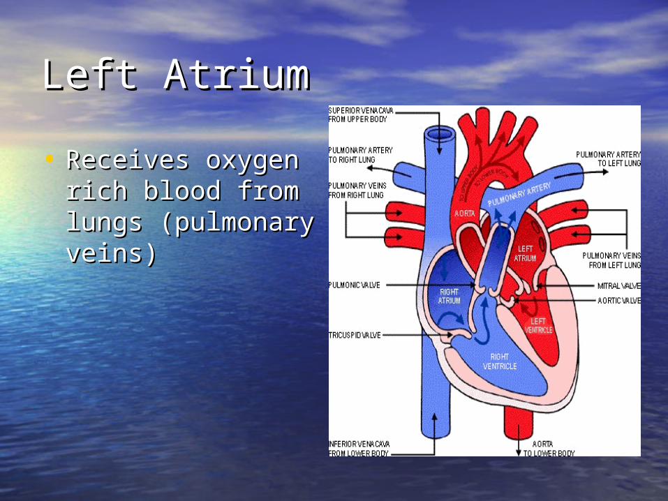
Left AtriumLeft Atrium
• Receives oxygen Receives oxygen rich blood from rich blood from lungs (pulmonary lungs (pulmonary veins)veins)
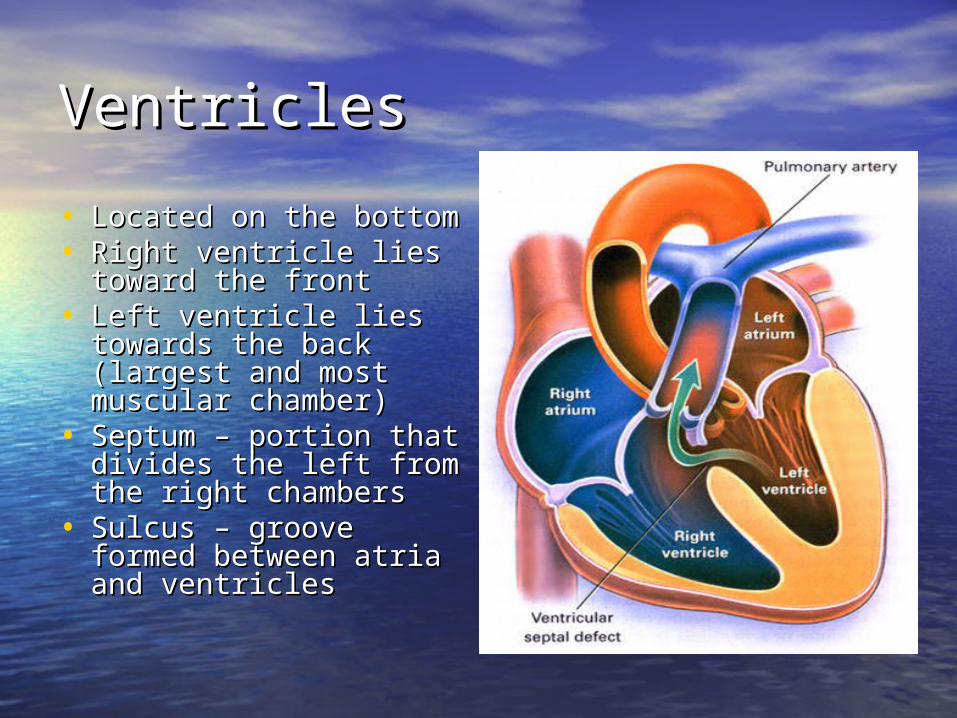
VentriclesVentricles
• Located on the bottomLocated on the bottom• Right ventricle lies Right ventricle lies
toward the fronttoward the front• Left ventricle lies Left ventricle lies
towards the back towards the back (largest and most (largest and most muscular chamber)muscular chamber)
• Septum – portion that Septum – portion that divides the left from divides the left from the right chambersthe right chambers
• Sulcus – groove Sulcus – groove formed between atria formed between atria and ventriclesand ventricles

Circulatory CircuitCirculatory Circuit
• Pulmonary circuit – right Pulmonary circuit – right sideside
• Systemic circuit – left Systemic circuit – left sideside
• Coronary circuit – feeds Coronary circuit – feeds the heart to thick for the heart to thick for diffusion – blockages or diffusion – blockages or narrowing of these narrowing of these vessels can be fatal if vessels can be fatal if not corrected (bi – pass)not corrected (bi – pass)

ValvesValves
• Atioventricular valvesAtioventricular valves– Tricuspid valve – 3 flaps of Tricuspid valve – 3 flaps of
tissue – right sidetissue – right side– Bicuspid valve (mitral valve) Bicuspid valve (mitral valve)
2 flaps of tissue – left side2 flaps of tissue – left side– Chordae tendinae – Chordae tendinae –
connective tissue that connective tissue that attaches to valves and attaches to valves and inferior muscle surface inferior muscle surface located in ventricles.located in ventricles.
– Blood flows into ventricles - Blood flows into ventricles - ventricles contract shoving ventricles contract shoving flaps upward – but the flaps flaps upward – but the flaps can only go so far up can only go so far up because of the chords because of the chords attached.attached.

Semi – lunar valvesSemi – lunar valves
• Valves that prevent Valves that prevent backflow into backflow into ventricles from major ventricles from major arteries.arteries.
• Valves have pockets Valves have pockets distal to heart – these distal to heart – these fill with blood and act fill with blood and act as sails slamming as sails slamming shut to prevent shut to prevent additional backflow.additional backflow.

ContractionContraction
• As with skeletal muscle – a As with skeletal muscle – a wave of depolarization Na+ in wave of depolarization Na+ in elicits an action potential. elicits an action potential. This causes the sarcolemma This causes the sarcolemma to dump Ca ++ ions out to dump Ca ++ ions out which binds to troponin which binds to troponin sliding tropomyosin out of the sliding tropomyosin out of the way sot that myosin can way sot that myosin can interact with actin. (sliding interact with actin. (sliding filament)filament)
• Cells are connected via gap Cells are connected via gap junctions so if one cell is junctions so if one cell is stimulated to fire all cells are stimulated to fire all cells are stimulated.stimulated.
• Rhythmic Control – nervous Rhythmic Control – nervous system – see notes on system – see notes on generating an action potential generating an action potential (nervous system)(nervous system)

Excitable FibersExcitable Fibers
• SA node – SA node – – Located right atrial Located right atrial
wall just below wall just below superior vena cavasuperior vena cava
– Fires about 100 Fires about 100 times per minute times per minute fastest stimulated fastest stimulated tissue so it sets the tissue so it sets the pace (pace maker)pace (pace maker)

Other excitable tissuesOther excitable tissues
• Located in septum receives Located in septum receives message from SA node and message from SA node and transmits down septum transmits down septum stimulating ventricle to stimulating ventricle to contract contract
• Bundle of His – branches out Bundle of His – branches out from AV nodefrom AV node
• Purkinje fivers – runs along Purkinje fivers – runs along the bottom of ventricles and the bottom of ventricles and toward the lateral portions toward the lateral portions causing stimulation.causing stimulation.
• If SA node is damaged than If SA node is damaged than AV becomes pacemaker but AV becomes pacemaker but it is slower in its rate of it is slower in its rate of contraction (and so on)contraction (and so on)

EKG / ECG EKG / ECG electrocardiogramelectrocardiogram• P wave – atrial P wave – atrial
depolarizationdepolarization
• QRS complex – QRS complex – ventricle ventricle depolarizationdepolarization
• T wave – ventricle T wave – ventricle repolarizationrepolarization