CHAPTER 16 Support And Movements - ikddata.ilmkidunya.com
44
CHAPTER 16 Support And Movements Animation 16 : Support and movement Source & Credit: Wikispaces
Transcript of CHAPTER 16 Support And Movements - ikddata.ilmkidunya.com

CHAPTER
16 Support AndMovements
Animation 16 : Support and movement
Source & Credit: Wikispaces
2
16. Support and Movement eLearn.Punjab
CONCEPT AND NEED
Animals and plants show a variety of physical and biochemical activities. The main diference between plants and animals is in their locomotion; animals show movement while plants do not.
Both plants and animals need support against gravity. The collenchymatous cells in plants give
support to the baby plants and sclerenchymatous cells to the adult plants. In animals muscles,
cartilage and bones provide support. They enable them to move towards food, away from danger
and for shelter.
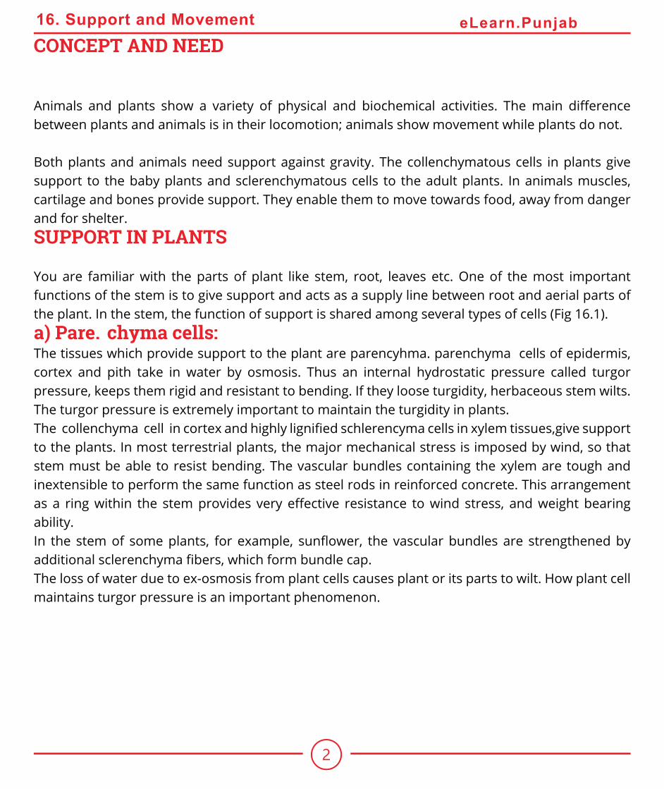
SUPPORT IN PLANTS
You are familiar with the parts of plant like stem, root, leaves etc. One of the most important
functions of the stem is to give support and acts as a supply line between root and aerial parts of
the plant. In the stem, the function of support is shared among several types of cells (Fig 16.1).
a) Parenchyma cells: The tissues which provide support to the plant are parencyhma. parenchyma cells of epidermis,
cortex and pith take in water by osmosis. Thus an internal hydrostatic pressure called turgor
pressure, keeps them rigid and resistant to bending. If they loose turgidity, herbaceous stem wilts.
The turgor pressure is extremely important to maintain the turgidity in plants.
The collenchyma cell in cortex and highly ligniied schlerencyma cells in xylem tissues,give support to the plants. In most terrestrial plants, the major mechanical stress is imposed by wind, so that
stem must be able to resist bending. The vascular bundles containing the xylem are tough and
inextensible to perform the same function as steel rods in reinforced concrete. This arrangement
as a ring within the stem provides very efective resistance to wind stress, and weight bearing ability.
In the stem of some plants, for example, sunlower, the vascular bundles are strengthened by additional sclerenchyma ibers, which form bundle cap.The loss of water due to ex-osmosis from plant cells causes plant or its parts to wilt. How plant cell
maintains turgor pressure is an important phenomenon.

3
16. Support and Movement eLearn.Punjab
Fig. 16.1 Specialized plants cells

4
16. Support and Movement eLearn.Punjab
Turgor pressure is generated by high osmotic pressure of the cell vacuole. The membrane that
bounds vacuole, is called tonoplast which contains a number of active transport systems that pump
ions into the vacuole or vacuolar compartments despite the higher concentration than that of the
extracellular luid. Because of the higher ionic concentration, water enters the vacuole and hence provides turgidity, mechanical support, to soft tissues of plant. The tissues which provide support
to the plants are:
(b) Sclerenchyma CellsThey have thick secondary cell walls usually impregnated with lignin, an organic substance that
makes the walls tough and hard. Most of the sclerenchyma cells are non-living. Their primary
function is to provide support to the plant parts.
There are three types of sclerenchymatous cells.
(i) Fibers (Tracheids): These are long and cylindrical and they may exist as solid bundles in xylem
or as bundle caps.
(ii) Sclereides: These are shorter than ibers and are found in seed coats and nut shells and provide protection.
(iii) Vessels (Tracheae): Long tubular structures, join end to end to form long water conducting
pipe in xylem.
(c) Collenchyma Cells
Collenchyma cells have protoplasts and usually lack secondary walls. They have angular thickening
in their primary walls. They are usually grouped in strands or cylinders. Collenchyma cells provide
support to young herbaceous parts of the plant. Young stems, for instance, often have a cylinder
of collenchyma just below their surface. Collenchyma cells are elastic, elongate with the growth of
stems and leaves.
Signiicance of Secondary Growth
Stem and root often begin to thicken after their apical meristem has produced embryonic or primary
tissue. An increase in plant girth due to vascular cambium and cork cambium is called secondary
growth. The result of secondary growth is most evident in woody perennial plants like trees, shrubs
and vine. Secondary growth occurs due to cell division in : (i) Vascular cambium (ii) Cork cambium.

5
16. Support and Movement eLearn.Punjab
Vascular cambium irst appears as a cylinder of actively dividing cells between primary xylem and primary phloem. Vascular cambium gives rise to two new tissues, one is the secondary xylem next
to the inner surface of the vascular cambium, the other is the secondary phloem appearing outer
to the vascular cambium.
The secondary xylem causes most of the increase in stem thickness. Over the years a woody stem
gets thicker and thicker as its vascular cambium produces layer upon layer of secondary xylem.
These layers are visible as rings. Since one growth ring is formed in one year, a count of the rings
at the base of trunk indicates the age of a tree at the time it was cut.
In most trees, the conduction of water and dissolved substances by secondary xylem become
limited to the outer or younger portion of that tissue. As trees grow older only few annual growth
rings are active in conduction at one time. The active portion is called sap wood. The inactive non-
conducting wood is called heartwood.
In most species, the heartwood accumulates a variety of chemicals such as resins, oils, gums and
tannins. These provide a resistance to decay and insect attack, for example, in red cedar and
conifers.

6
16. Support and Movement eLearn.Punjab
Fig. 16.2 Dicot woody stem

7
16. Support and Movement eLearn.Punjab
Another important function of the cambium is to form callus or wood tissue on or over the wound,
soft parenchymatous tissues are rapidly formed on or below the damaged surface of stems and
roots. Callus also unites the branches during budding and grafting.
MOVEMENTS IN PLANTS
Organisms respond to the external as well as internal stimuli. Animals move in response to
external stimuli; similarly plants also show movements. Animals change their location in response
to stimulus. Plants are ixed therefore, they change their growth pattern.Types of MovementsThere are two types of movements:
1. Autonomic movements 2. Paratonic movements.
Autonomic movements are spontaneous movements due to internal causes whereas paratonic
movements are due to external causes.
The wood from diferent species of trees difers greatly in their suitability for speciic uses. Density, hardness, lexibility, shock resistance, compression strength and texture determine quality and commercial use. The commercial cork is also made from the bark of trees such as
Quercus suber.

8
16. Support and Movement eLearn.Punjab
1. Autonomic movements : Autonomic movements are of three types:
(i) Tactic movements (ii) Turgor movements (iii) Growth movements.
(i) Tactic Movements : These are the movements of an entire cell or organism i.e. locomotion
due to internal stimulus. The tactic movement may be positive if it is towards the stimulus or
negative if it is away from the stimulus. Tactic movements are the movements of locomotion; they
are further classiied on the basis of the nature of the stimulus, (a) Phototactic movement: It is
a movement in response to stimulus of light. The movement may be towards the source of light
(positive) or away from the source of light (negative). The best example of positive tactic movement
is the passive movement of chloroplast due to cyclosis. This movement helps the chloroplast to
absorb maximum light for C02 ixation. The light intensity and direction both afect the intra cellular
distribution of chloroplasts. (b) Chemotactic movement : The movement in response to stimulus
of chemicals is called chemotactic movement. The movements shown by sperms of liver-worts,
mosses, ferns towards archegonia in response to stimulus of nucleic acid released by the ovum is
one such example.
(ii) Turgor Movements : Turgor movement is due to diferential changes in turgor and size of cells as a result of gain or loss of water. Rapid movements of lealets in “touch-me-not” plant and sleep movements of the plants fall under this category of movements, (a) Sleep movements:
Bean plants and some members of legume family lower their leaves in the evening and raise them
in the morning. These are known as sleep movements. These sleeping movements are due to
daily changes in turgor pressure in the pulvinus. The place of attachment of leaf with the shoot,
pulvinus, is swollen portion of the petiole composed of parenchymatous cells with relatively large
inter cellular spaces and central strand of vascular tissues.
When turgor pressure on the lower side of pulvinus increases the leaves rise and become horizontal. When turgor pressure decreases on the lower side of pulvinus, the leaves lower and go to “sleeping” position, (b) Rapid movement of lealets : When the compound leaf of sensitive plant Mimosa
is touched, the lealets fold together. This response takes a second or two resulting from rapid loss of turgor by the cells in pulvinus at the base of each lealet. The investigation has shown that potassium (K+) ions move irst, which causes water to leave the cell by exosmosis. It takes about ten minutes to regain the turgor and restore the internal turgidity of leaf
9
16. Support and Movement eLearn.Punjab
.
(iii) Growth Movements : Growth movements are due to unequal growth on two sides of plant
organs like stem, root, tendrils, buds etc. There are three types of growth movements, (a) Epinasty
: It is shown by leaves, petals etc. The upper surface of leaf in bud condition shows more growth
as compared with the lower surface. This leads to opening of buds, (b) Hyponasty: If growth in
the lower surface of the leaf in bud condition is more than that of the upper surface then the bud
will remain closed, (c) Nutation : The growing tip of young stem moves in a zig-zag fashion due to alternate changes in growth on opposite sides of the apex. This mode of growth is called nutation.
2. Paratonic Movements : These movements are due to external causes. These are of following
types.
(a) Tropic Movements : The word tropic is derived from Greek word ‘Tropos’ meaning ‘turn’. It
is the movement in curvature of whole organ towards or away from stimuli such as light, gravity,
and touch. Following are common tropic movements: (i) Phototropism : It is the movement of
part of plant, in response to stimulus of light and is caused by the diferential growth of part of a plant like stem or root, (ii) Thigmotropism : It is the movement in response to stimulus of touch,
for example climbing vines. When they come in contact with some solid object, the growth on the
opposite side of contact increases and the tendril coils around the support, (iii) Chemotropism :
The movement in response to some chemicals is called chemotropism. The hyphae of fungi are
chemotropic. (iv) Hydrotropism : The movement of plant parts in response to stimulus of water is
called hydrotropism. The growth of roots toward water is due to positive hydrotropism and growth
of shoot away from water is negatively hydrotropic. (v) Geotropism : It is the response to gravity.
Roots display positive geotropism and shoots negative geotropism.
(b) Nastic Movements : These are the non-directional movements of parts of plant in response
to external stimuli. These are of two types: (i) Nyctinasty : The nyctinastic movements are shown by
the organs in response to external stimuli leading to diferential growth. These are due to turgor and growth changes. It may be of two types: (a) Photonasty : The principal stimulus is the photoperiod.
The lowers open and close due to light intensity. (b) Thermonasty : It is due to temperature. The
lowers of tulip close at night because of rapid growth in the lower side by upward and inward bending of the petals.

10
16. Support and Movement eLearn.Punjab
(ii) Haptonastic movements occur in response to contact. Examples include the action of the Venus
ly trap.
Role of Plant Growth Substances In Plant Movement
Plants do not move from one place to other like animals. However, their organs show movements,
which are controlled by hormones. Auxins play major role in phototropism. It is believed that
unequal distribution of auxin indole acetic acid (I A A) in the coleoptiles stumps, produces unequal
cell enlargement, causing a bend in the organ towards source of light.
Auxins are also responsible for positive gravitropism of roots and negative geotropism of stems.
Auxins inhibit the growth of root cells. The cells of the upper surface, therefore elongate and the
root curves downward. Auxins on the other hand, stimulate the growth of the stem cells. The cells
of the lower surface, elongate and stem curves upward. Nastic movements are due to some balance
or ratio between growth inhibitors (abscisins) and growth stimulators (gibberellins). However, it
has been observed that epinasty is due to auxins and hyponasty due to gibberellins.
SUPPORT AND MOVEMENTS IN ANIMALS
The skeleton is tough and rigid framework of the body of animals which provides protection, shape
and support to the body organs. It is composed of inorganic or organic substances or both. In
protozoa it is secreted by a single cell, whereas in multicellular animals it is composed of specialized cells. There are three main types of skeleton in animals, hydrostatic skeleton, exoskeleton and
endoskeleton.
11
16. Support and Movement eLearn.Punjab
1. Hydrostatic Skeleton
In animals that lack a hard skeleton, a luid illed gastrovascular cavity or coelom can act as hydrostatic skeleton. Hydrostatic skeleton provides support and resistance to the contraction of muscles so
that motility results. It is found in cnidarians, annelids and other soft-bodied invertebrates.
The sea anemone has hydrostatic skeleton. Its cavity is illed with sea water to extend its body and tentacles. The sea anemone closes its mouth and constricts its muscle ibers that are arranged in circles around its body. The contraction of these circular muscles puts pressure on the liquid in
body cavity and that pressure forces the body to maintain upright stature.
In earthworm, the hydrostatic skeleton consists of luid-illed compartments separated by septa. Contraction of circular muscle causes compartments to elongate and contraction of longitudinal
muscle causes a compartment to shorten. Alternating waves of elongation and contraction move
the earthworm through the soil, aided by paired setae in each segment.
2. Exoskeleton
An exoskeleton is hardened outer covering to which internal muscles are attached. The exoskeleton
is inert and non-living. It is secreted by the ectoderm in animal cells. It is composed of two layers.
The epicuticle is the outer most layer. Because it is made up of waxy lipoprotein, it is impermeable
to water and serves as a barrier to microorganisms in insects. The bulk of exoskeleton is below the
epicuticle and is called the procuticle.Procuticle consists of an outerlayer exocuticle and inner layer
of endocuticle. The procuticle is composed of chitin, tough, leathery, polysaccharide and several
kinds of protein. It is further hardened by sclerotization and sometimes by impregnation with calcium carbonate.
The simplest example of an exoskeleton is the shell of mollusca, which generally consists of just
one or two pieces. Some marine bivalvia and snail have shell composed of crystals of calcium
carbonate. The shell of land snail generally lacks the hard minerals and are much lighter. Molluscan
shell can grow as the animal grows and growth rings are apparent on the shell. The soft parts of the
molluscan body have a hydrostatic skeleton as well.
The most complex exoskeleton is found among the arthropods. The arthropods have made a
variety of adaptations to allow them to live and grow within their exoskeleton. The invagination of
exoskeleton forms irm ridges and bars for muscle attachment. Another modiication of exoskeleton is the formation of joints. The exoskeleton are thin, soft and lexible at joints, consequently joint move very easily. Other modiications of exoskeleton include sensory receptors called sensilla that are in the form of bristles, and lenses and the modiication of the exoskeleton that permits gaseous exchange.

12
16. Support and Movement eLearn.Punjab
The exoskeleton in arthropoda protects the animals against their enemies and rough environment.
It also protects them from drying.
However, it has one disadvantage and that is animals cannot grow larger. The animal, therefore,
needs to shed its exoskeleton periodically and replace it with one of the larger size. This process is known as “ecdysis or moulting.”
Ecdysis is divided into four stages:
1. Enzymes, secreted from hypodermal glands, begin digesting the old endocuticle. This digestion separates hypodermis and the exoskeleton.
2. The old exoskeleton is split and pores are formed.
3. The digestion of endocuticle is followed by secretion of new procuticle and epicuticle.
4. Finally, the new exoskeleton is hardened by deposition of calcium carbonate. During the
hardening process, the arthropod is vulnerable to predators and remains hidden. All these
changes are controlled by the nervous system and the hormone ecdysone.Some major functions of the skeletal system are as follows:
(i) Support and shape : Bones support soft tissues and serve as attachment sites for most muscles
and provide shape to the body.
(ii) Protection: Bones protect critical internal organs, such as brain, spinal cord, heart and lungs.
(iii) Movement: Skeletal muscles attached to the bones help in moving the body.
(iv) Mineral homeostasis: Bones serve as store for calcium, phosphorus, sodium and potassium.
Through negative feedback mechanisms, bones can release or take up minerals to maintain
homeostasis.
(v) Blood cell production: Red and white blood cells are produced in bone marrow, a connective
tissue found within certain bones.

13
16. Support and Movement eLearn.Punjab
3. EndoskeletonThe endoskeleton is primarily made up of two types of tissues, bones and cartilage. Both bones
and cartilage are types of rigid connective tissue. Both consists of living cells embedded in the
matrix of protein called collagen.
Bone : It is the most rigid form of connective tissue. The collagen ibers of bone are hardened by deposit of calcium phosphate. Bones supporting your arms and legs consist of an outer shell of
compact bone, with spongy bone in the interior. Compact bone is dense and strong and provides
an attachment site for a muscle. Spongy bone is light, rich in blood vessels, and highly porous. The
cavities of spongy bone contain bone marrow where blood cells are formed. There are three types
of cells associated with bone:
Bone-forming cell (osteoblast ), mature bone cell (osteocyte ), and bone dissolving cells (osteoclast
).
Fig. 16.3 Cells of bone

14
16. Support and Movement eLearn.Punjab
Early in development, when bone is replacing cartilage, the osteoclasts invade and dissolve the
cartilage. Then osteoblasts replace it with bone. As bones grow, the matrix of bone is hardened
and the osteoblasts are gradually entrapped within it.
Cartilage : It is much softer than bone. It is a form of connective tissue. It covers ends of the bone
at the joint, and also supports the lexible portion of nose and external ears. The living cells of cartilage are called chondrocytes. These cells secrete lexible, elastic, non-living matrix collagen that surrounds the chondrocytes. No blood vessels penetrate into this cartilage. There are three
main types of cartilage.
(i) Hyaline Cartilage : It is the most abundant type in human body. It is found at the movable
joints.
(ii) Elastic Cartilage: It has matrix containing bundles of collagens ibers. It forms external pinnae of ears and the epiglottis.
(iii) FibroCartilage:
HUMAN SKELETON
Human skeleton can be divided into two parts, axial skeleton and appendicular skeleton.
1) Axial SkeletonThe axial skeleton includes the skull, the vertebrae, ribs and the sternum.
Skull: It is made up of cranium and facial bones. The cranium consists of 8 bones (Fig 16.4), 4
unpaired and 2 paired which protect the brain. Parietal and temporal are paired bones, whereas
frontal, occipital, sphenoid and ethmoid are unpaired bones. Besides that there are 14 facial bones
of which 6 are paired and 2 unpaired. The paired facial bones are maxilla, zygomatic, nasal, lacrimal, palatine and inferior concha. The unpaired facial bones are mandible and vomer.

15
16. Support and Movement eLearn.Punjab
Fig. 16.4 Human skull

16
16. Support and Movement eLearn.Punjab
Vertebral Column : Vertebral column extends from the skull to the pelvis to form backbone,
which protects the spinal cord (Fig 16.5). Normally the vertebral column has 4 curvatures, which
provide more strength than does the straight column. The vertebral column consists of 33 vertebrae.
The vertebrae are named according to their location in the body, viz, cervical, thoracic, lumbar and pelvic.
The cervical vertebrae include seven vertebrae which lie in the neck region. The irst two cervical vertebrae are atlas vertebra and axis vertebra. There are twelve thoracic vertebrae located in the
thoracic region, ive in lumbar region and nine in pelvic region which form two sets, sacrum and
coccyx. Sacrum is formed by the fusion of anterior ive vertebrae, whereas coccyx is formed by the fusion of four posterior vertebrae.
Rib cage : It is composed of twelve pairs of ribs that articulate with the thoracic vertebrae. Ten
of them connect anteriorly with sternum, either directly or through the costal arch. The lower two
pairs of ribs are called “loating ribs” because they do not attach to the sternum. The rib cage provides support to a semi-vaccum chamber called the “chest cavity”.
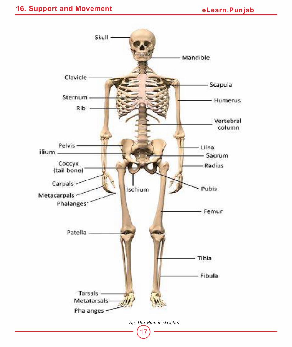
2) Appendicular SkeletonThe appendicular skeleton consists of pectoral girdle and appendages (fore limbs), and pelvic girdle
and appendages (hind limbs). (Fig 16.5)
17
16. Support and Movement eLearn.Punjab
Fig. 16.5 Human skeleton

18
16. Support and Movement eLearn.Punjab
Pectoral Girdle and Fore Limb : Pectoral girdle comprises scapula, suprascapula, and clavicle.
The clavicle connects scapula with sternum.
The fore limb consists of humerus, radius and ulna, 8 carpals, 5 metacarpals and 14 phalanges.
Humerus forms ball and socket joint with scapula, while at distal end humerus forms hinge joint
with radius and ulna. The radius and the ulna at their distal end form multistage joint with eight
wrist bones called carpals. Five metacarpals form the framework of palm of the hand. Five rows of
the phalanges are attached to the metacarpals. They support the ingers.
Pelvic Girdle and Hind Limb : Pelvic girdle attaches the hind limb to the vertebral column (Fig
16.5). It consists of two coxal bones. Each is formed by the fusion of three bones ilium, ischium and
pubis. The pelvic girdle supports the pelvic region.
The hind-limb consists of 1 femur, 2 tibia and ibula, 7 tarsals 5 meta-tarsals and 14 phalanges. Femur is the proximal bone which forms a hip joint with the hipbone, it is a ball and socket joint.
At the distal end, the femur forms knee joint with the proximal end of two parallel bones called
tibia and ibula. The distal end of the tibia and ibula forms a joint with seven tarsals, which are also distally attached to ive metatarsal bones of ankle. Five rows of the fourteen phalanges of the toes are attached to metatarsals (Fig 16.5).
Joints
Joints occur where bones meet. They not only hold our skeleton together, but also give it the mobility.
Joints are classiied on the basis of the amount of movement allowed by them, into three categories:
(i) Immovable joints (ii) Slightly movable joints (iii) Freely movable joints
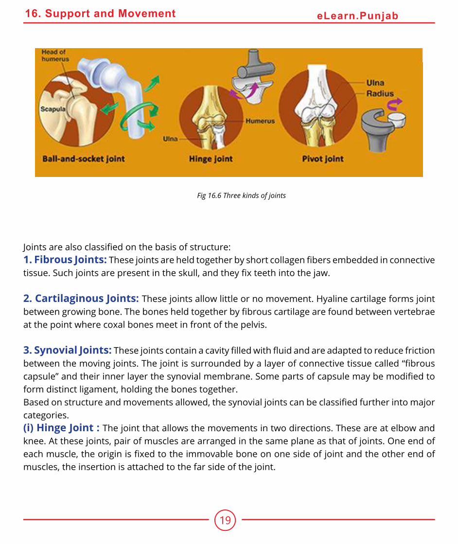
The freely movable joints are of two types viz. hinge joint and ball and socket joint (Fig. 16.6)
19
16. Support and Movement eLearn.Punjab
Joints are also classiied on the basis of structure:1. Fibrous Joints: These joints are held together by short collagen ibers embedded in connective tissue. Such joints are present in the skull, and they ix teeth into the jaw.
2. Cartilaginous Joints: These joints allow little or no movement. Hyaline cartilage forms joint
between growing bone. The bones held together by ibrous cartilage are found between vertebrae at the point where coxal bones meet in front of the pelvis.
3. Synovial Joints: These joints contain a cavity illed with luid and are adapted to reduce friction between the moving joints. The joint is surrounded by a layer of connective tissue called “ibrous capsule” and their inner layer the synovial membrane. Some parts of capsule may be modiied to form distinct ligament, holding the bones together.
Based on structure and movements allowed, the synovial joints can be classiied further into major categories.
(i) Hinge Joint : The joint that allows the movements in two directions. These are at elbow and
knee. At these joints, pair of muscles are arranged in the same plane as that of joints. One end of
each muscle, the origin is ixed to the immovable bone on one side of joint and the other end of muscles, the insertion is attached to the far side of the joint.
Fig 16.6 Three kinds of joints

20
16. Support and Movement eLearn.Punjab
(ii) Ball and Socket Joint: The joint that allows the movement in several directions. Such joints
have at least two pairs of muscles present perpendicular to each other. They provide maximum
lexibility. Hip joint and shoulder joint are the examples of ball and socket joints.
DEFORMITIES OF SKELETON
Human skeleton supports an upright body. Sometimes our skeletal system becomes weak and
results in deformations. The causes of the deformations are variable e.g.
1. Genetic Causes
Cleft palate, a condition in which palatine processes of maxilla and palatine bone fail to fuse. The
persistent opening between the oral and nasal cavity interferes with sucking. It can lead to inhalation
of food into the lungs causing aspiration pneumonia. Microcephaly, the small sized skull is caused by some genetic defect. Arthritis covers over 100 diferent types of inlammatory or degenerative diseases that damage the joints. Osteoarthritis is the most common chronic arthritis, which is a
degenerative joint disease also caused by genetic defect.
2. Hormonal Causes
Osteoporosis is a group of diseases in which bone resorption out paces bone deposit. In this case
bone mass is reduced and chemical composition of the matrix remains normal. Osteoporosis
mostly occurs in aged women, which is related to decreased estrogen level. Other factors which may
contribute include, insuicient exercise, diet poor in calcium and protein, smoking, etc. Estrogen replacement therapy (ERT) ofers the best protection against osteoporotic bone fractures.

21
16. Support and Movement eLearn.Punjab
3. Nutritional CausesOsteomalacia (soft bones) includes a number of disorders in which the bones receive
inadequate minerals. In this disease, calcium salts are not deposited and hence ones
soften and weaken. Weight bearing bones of legs and pelvis bend and deform. The
main symptom is the pain when weight is put on afected bones.
Rickets is another disease in children with bowed legs and deformed pelvis. It is
caused by deiciency of calcium in diet or vitamin ‘D’ deiciency. It is treated by vitamin ‘D’ fortiied milk and exposing skin to sunlight.
Disc - SlipEach intervertebral disc is a cushion - like pad composed of an inner semi luid nucleus
pulposus which acts as rubber ball to give a disc its elasticity and compressibility and
a strong outer ring of ibrocartilage, the annulus ibrosus. The annulus ibrosus holds together successive vertebrae.
The discs act as shock absorber during walking, jumping, running and to lesser extent bend laterally.
Severe or sudden physical trauma to spines for example from bending forward while lifting a heavy
object may result in herniation of one or more discs. The herniated disc (commonly known slipped
disc) usually involves rupture of annulus ibrosus followed by protrusion of the spongy nucleus pulposus. If protrusion presses on spinal cord or on spinal nerves exiting from spinalcord, generate
severe pain or even destruction of these netvous structure. Disc slip is treated with bed rest, traction
and painkiller. If this fails disc may be removed surgically.
SpondylosisIt is the disease, which causes immobility and fusion of vertebral joint.
22
16. Support and Movement eLearn.Punjab
SciaticaIt is characterized by stabbing pain radiating over the course of sciatic nerve. It results due to injury of proximal sciatic nerve, which might follow a fall, a herniated disc or improper administration of
an injection into the buttock. This may result in a number of lower limb impairment depending on
the precise nerve root injured. When sciatic nerve is completely transected, the legs become nearly
useless. They cannot be lexed and all foot-ankle movement is lost. Recovery from sciatic injury is usually slow and incomplete.
ArthritisArthritis is inlammatory or degenerative disease that damages joints. It results in pain, stifness, swelling of the joint. Acute forms of arthritis usually result from bacterial invasion and are treated
with antibiotics. The membrane, lining the joint thickens, luid production is decreased, which consequently leads to increased friction. Chronic arthritis includes osteoarthritis, rheumatoid
arthritis, and gouty arthritis.
REPAIR OF BROKEN BONESDespite remarkable strength, the bones may break. During youth, most fractures result from
trauma that may twist or break the bones such as sports injuries, automobile accidents, falls etc. In
old age, bones become thin and weak and hence fractures occur more frequently.
23
16. Support and Movement eLearn.Punjab
A fracture is treated by reduction which follows realignment of the broken bone ends. There are
two types of reduction: closed and open reduction. In closed reduction the bone ends are coaxed
back to their normal position by physician’s hand. In open reduction surgery is performed and the
bone ends are secured together with pins or wires. After broken bone is reduced, it is immobilized by a cast (or by traction) to allow the healing process to begin. Healing time is 8-12 weeks, but it is
much longer for large weight — bearing bones and for bones of elderly people (because of their
poorer blood circulation).
The repair process of a simple fracture takes place in four phases:
1. Hematoma Formation : When a bone breaks, the blood vessels in the bone itself, and perhaps
in surrounding are torn resulting in hemorrhage. As a result, a hematoma, a mass of clotted blood,
forms at the fracture site. Soon after, bone cell deprived of food begin to die and the tissue at the
fracture site becomes swollen and hence painful.
2. Soft Callus Formation : Next “soft callus” begins to form in 3-4 weeks. Capillaries grow into the hematoma and clear up the debris. Fibroblasts and osteoblasts migrate into the fracture site and
begin to construct bone.
3. Bony Callus Formation : Osteoblasts and osteoclasts continue to migrate inward, multiply
rapidly and gradually convert the soft callus into bony callus. Bone formation begins 3-4 weeks
after injury and continues until a irm bony union is formed within 2-3 months later.
4. Remodeling : After several months bony callus is remodeled by removing the excess material on
the outside of the bone. Final structure of remodeled area resembles that of the original unbroken
bone because it responds to the same set of mechanical stimuli.
MUSCLES
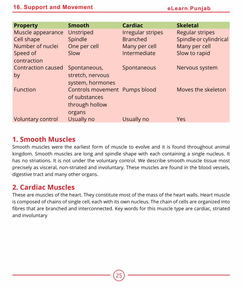
Many multicellular animals have evolved specialized cells for movement. These cells contain numerous ilaments of special protein actin and myosin. The vertebrate possess three kinds of muscle cells, Smooth muscles, skeletal muscles and cardiac muscles (Fig 16.7)

24
16. Support and Movement eLearn.Punjab
Fig. 16.7 Location, characteristics, and functions of the three muscle types.
25
16. Support and Movement eLearn.Punjab
Property Smooth Cardiac SkeletalMuscle appearance Unstriped Irregular stripes Regular stripes
Cell shape Spindle Branched Spindle or cylindrical
Number of nuclei One per cell Many per cell Many per cell
Speed of
contraction
Slow Intermediate Slow to rapid
Contraction caused
by
Spontaneous,
stretch, nervous
system, hormones
Spontaneous Nervous system
Function Controls movement
of substances
through hollow
organs
Pumps blood Moves the skeleton
Voluntary control Usually no Usually no Yes
1. Smooth MusclesSmooth muscles were the earliest form of muscle to evolve and it is found throughout animal
kingdom. Smooth muscles are long and spindle shape with each containing a single nucleus. It
has no striations. It is not under the voluntary control. We describe smooth muscle tissue most
precisely as visceral, non-striated and involuntary. These muscles are found in the blood vessels,
digestive tract and many other organs.
2. Cardiac MusclesThese are muscles of the heart. They constitute most of the mass of the heart walls. Heart muscle
is composed of chains of single cell, each with its own nucleus. The chain of cells are organized into ibres that are branched and interconnected. Key words for this muscle type are cardiac, striated and involuntary

26
16. Support and Movement eLearn.Punjab
3. Skeletal Muscles
The muscles that are attached to the skeleton and are associated with the movement of bones are
called skeletal muscles. The skeletal muscles are consciously controlled and therefore, are called
voluntary muscles. Skeletal muscles are also called striped or striated muscles because they show
alternate light and dark bands, e.g., triceps and biceps. Generally, each end of the entire muscle is
attached to bone by a bundle of collagen, non-elastic ibres, known as tendons.
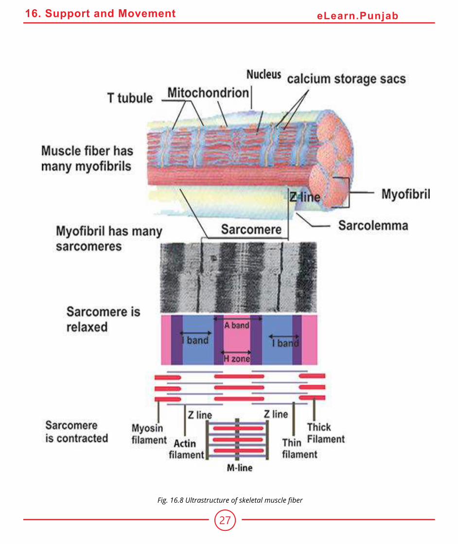
Skeletal Muscle Fibre : Each muscle consists of muscle bundles, which are further composed of
muscle ibres or cells. Each muscle ibre is a long cylindrical cell with multiple oval nuclei arranged just beneath its sarcolemma. Skeletal muscle ibres are huge cells. Their diameter is 10 - 100 mm.
Sarcoplasm of the muscle ibre is similar to the cytoplasm of other cells but it contains usually large amount of stored glycogen and unique oxygen bonding protein myoglobin, a red pigment that
stores oxygen.
When viewed in high magniication, each muscle ibre is seen to contain a large number of myoibrils 1-2 mm in diameter that run in parallel fashion and extend entire length of the cell. Bundles of these
ibrils are enclosed by the muscle cell membrane or sarcolemma. The myoibrils consist of smaller contractile units called sarcomere. In each sarcomere a series of dark and light band are evident
along the length of each myoibril. Each dark band is called A band, because it is anisotropic, i.e it
can polarize visible light. The light band called I band is isotropic or non-polarizing. It gives the cell as a whole its striped appearance. Each A band has a lighter stripe in its mid section called H - zone (H stands for “hele” mean bright). The H-zone is bisected by dark line called M - line. The I bands have mid line called Z - line (Z for zwishen means between).
A sarcomere is the region of a myoibril between two successive Z - lines and is the smallest contractile unit of muscle ibre. The myoibrils contain myoilaments.
27
16. Support and Movement eLearn.Punjab
Fig. 16.8 Ultrastructure of skeletal muscle iber
28
16. Support and Movement eLearn.Punjab
Infrastructure of Myoilament : Myoilament is made up of thick and thin ilaments. The central thick ilaments extend the entire length of the A-band. The thin ilaments extend across the I-band and partly into A band.
The thick ilament which is about 16 nm in diameter is composed of myosin. Each myosin molecule has a tail terminating in two globular heads. Myosin tail consists of two long polypeptide chains
coiled together. The heads are sometimes called cross bridges because they link the thick and the
thin myoilaments together during contraction (Fig. 16.8).
Thin ilaments are 7 - 8 nm thick and are composed chiely of actin molecule. The actin molecules are arranged in two chain which twist around each other like a twisted double strand of pearls.
Twisting around the actin chains are two strands of another protein, tropomyosin. The other major
protein in thin ilament is troponin. It is actually three polypeptide complex, one binds to actin, another binds to tropomyosin while third binds calcium ions. Each myosin ilament is surrounded by six actin ilaments on each end.
Sliding Filament Model
When muscle ibre contracts, the thin and thick ilaments undergo shifting. The I-band reduces in length and Z-line gets closer.
Huxley and A. F. Huxley and their colleagues suggested a hypothesis in 1954 to explain all events
in muscle contraction, this is called “Sliding ilament model” of muscle contraction. According to this theory, the thin ilaments slide past the thick one so that actin and myosin ilaments overlap to greater degree. Thus the Z-line is brought close together, I-band shortens, the H zone disappears. In this process of contraction, the cross bridges of thick ilament become attached to binding sites on the actin ilament. The cross bridges then contract to pull the actin ilament towards the center of the sarcomere.

29
16. Support and Movement eLearn.Punjab
How the bridges are controlled
When the muscle is at rest, the tropomyosin is disposed in such a way that it covers the sites on the
actin chain where the head of myosin becomes attached. When the muscle is required to contract,
calcium ions bind with the troponin molecule and cause them to move slightly. This has the efect of displacing the tropomyosin and exposing the binding sites for the myosin head. Once the myosin
head has become attached to the actin ilament, ATP is hydrolysed and the bridge goes to its cycle. This ATP is provided by the large number of mitochondria present in each muscle cell.
From the above account it is revealed that ATP is needed to break the link between the myosin and
the actin. After death, the amount of ATP in the body falls. Under these circumstances the bridges
can not be broken and so they remain irmly bound. This results in the body becoming stif, a condition known as rigor mortis.
Controlling the Actin - Myosin Interaction By Ca++ ions
Muscle contraction is initiated by nerve impulse arriving at the neuromuscular junction. All the ibres innervated by a single motor neuron are a “motor unit” and contract simultaneously in response to the action potential ired by the motor neurons. The sarcolemma of muscle ibre penetrates deep into the cell to form hollow elongated tube, the transverse tubule, T-tubule the lumen of
which is continuous with the extracellular luid. The thousands of T - tubules of each muscle cell are collectively called T-system. It extends and encircles the myoibril at the level of Z-line or A and I - junction. The T-tubule and the terminal portion of the adjacent envelope of sarcoplasmic reticulum
form triads at regular intervals along the length of the ibril. The nerve impulse is carried through the T-tubule to the adjacent sarcoplasmic reticulum (SR). The calcium gates of the SR open releasing
calcium into the cytosol, thus binding calcium ion to troponin molecules of the thin ilament. The binding sites are exposed and cross bridges with myosin can form, and contraction occurs.
All or None Response : The contraction of each muscle ibre is based on “all or none” principle i.e. all of its ibrils participate in contraction. The degree of contraction depends upon the number of muscle ibers that participate in contraction.

30
16. Support and Movement eLearn.Punjab
Energy For Muscle ContractionEnergy for muscle contraction comes from the ATP. Supply of ATP is. maintained by the aerobic
breakdown of glucose in muscle cell, which comes from stored glycogen in the cell. When more
energy is required due to high metabolism, it is provided by another energy storing substance
called creatine phosphate. Sometime during
oxygen deiciency or very high metabolic activity such as (prolonged or strenuous
muscular activity), ATP requirement is met
by anaerobic breakdown of glucose into
Lactic acid. Lactic acid accumulation causes
muscle fatigue. At rest, 1/5 of the lactic acid
is broken aerobically and its energy is used
to change the remaining 4/5 lactic acid into
glucose.
Muscle Fatigue
Muscle fatigue is a state of physiological
inability to contract. Muscle fatigue results
from relative deicit of ATP. When no ATP is available, contractures or states of
continuous contraction result because the
cross bridges are unable to detach. Excess
accumulation of lactic acid and ionic imbalance also contribute to muscle fatigue. Lactic acid, which
causes muscle pH to drop (and the muscle to ache) causes extreme fatigue by breaking glucose.
Efect of Exercise on MuscleThe amount of work a muscle does is relected in changes in the muscle itself. When muscles are
used actively, they increase in size or strength and become more eicient and fatigue resistant. Aerobic exercises such as swimming, jogging , and
fast walking result in several changes in skeletal
muscles. Capillaries surrounding the muscle ibres, as well as mitochondria within them increase in
number and ibre synthesizes more myoglobin. These changes result in more eicient muscle metabolism and resitance to fatigue. Complete
immobilization of muscle leads to muscle weakness and severe atrophy.
Sarcoplasmic Reticulum (S.R) is continuous system of sarco-tubules extending throughout the
sarcoplasm around each myoibril. It is like endoplasmic reticulum but devoid of ribosomes and exhibits a highly specialized repeating pattern.
31
16. Support and Movement eLearn.Punjab
TetanyTetany is the disease caused by low calcium in the blood. It increases the excitability of neurons
and results in loss of sensations. Muscle twitches and convulsion occur. If untreated the system
progresses to spasm of larynx, respiratory paralysis and ultimately death occurs.
CrampIt is also known as tetanic contraction of the entire muscle: It lasts for just a few seconds to several
hours, causing the muscles to become taut and painful. It is most common in thigh and hip muscles,
it usually occurs at night or after exercise. It relects low blood sugar level, electrolyte depletion, dehydration irritability of spinal cord and neurons.
ARRANGEMENT OF SKELETAL MUSCLES FOR MOVEMENT OF SKELETON
Skeletal muscle has three parts: origin is the end of muscle which remains ixed when muscle contracts, insertion is the end of the muscle that moves the bone, and belly is thick part between
origin and insertion, which contracts.
Connective tissue binds other tissues and helps
to maintain body form by holding the various
organs together. Connective tissue ibrils have two specialized kinds. Ligaments attach bone
to bone and are slightly elastic. Tendons attach
muscles to bones and are non-elastic.
Movement of Bones
The majority of muscle tissue in your body is
skeletal muscle. The skeletal muscles produce
movements by pulling on tendons, cords of
connective tissues that anchor muscle to the
bones. The tendons then pull on bones. Most
muscles pass across a joint and are attached to
the bones that form joints. When such a muscle
contracts, it draws one bone towards or away from the bone with which it articulates.
There are 650 muscles in human body, most of which occur in pairs. At joint, these muscles work
against each other by contraction. This relationship is called antagonism.
Tetanus: The term tetanus is used for an
acute infectious disease caused by anaerobic
bacterium Clostridium tetani resulting in
persistant painful spasms of some skeletal
muscles. Typically begins gradually with
stifness of jaws and neck muscles and progresses to ixed rigidity of jaws (lock jaw) and spasms of trunk and limb muscles, usually
fatal due to respiratory failure. Though rare
in developed countries, the tetanus is the
major killer in developing countries where the
mortality rate is 40 percent.

32
16. Support and Movement eLearn.Punjab
The best example is the movement of elbow joint by biceps and triceps. The biceps bends the arm
at the elbow joint, and triceps straightens it. The biceps brachii muscle arises by the two heads
from scapula and is inserted into the medial surface of the radius bone. The other two muscles
lie below the biceps brachi. The two muscles are brachialis and brachioradialis. The brachialis is
inserted in the ulna, while brachioradialis is inserted in the radius. When these muscles contract
they lift radius and ulna and bend the arm at the elbow. When triceps contracts it straightens arm
at elbow. In the antagonistic pairs one muscle reverses the efect of the other and they do not contract simultaneously.
Fig: 16.9 Working of hinge joint at elbow
Levered movement. Rigid external or internal skeletons are attached to muscles, which move
parts of the skeleton at movable joints. Each bony “lever” is moved by an antagonistic muscle pair. One muscle reverses the action of the other, so that the lever can return to its original
position.

33
16. Support and Movement eLearn.Punjab
LOCOMOTION IN PROCTOCTISTA AND INVERTEBRATES
There is an immense variety of organisms with diferent modes of locomotion.
Locomotion In Euglena
Euglena moves with the help of lagellum. As the lagellum is whipped backwards, the organism moves forward. However, when the lagellum moves forward Euglena does not move backward. Locomotary lagellum is at the anterior end of the body and pulls the organism forward. Waves of activity are generated by the lagellum itself, and they pass in a spiral fashion from its base to its tip. They increase amplitude and velocity. The activity note of the lagellum causes the body of Euglena to rotate forward about its axis. Euglena is able to change its direction by the active contractile
myonemes which run along the length of its body. When they contract the shape of the body as
well as its direction changes. First the body becomes short and wider at the anterior end then in the
middle and later at the posterior end. This characteristic movement is called Euglenoid movement.
Locomotion in Paramecium
Paramecium moves with the help of cilia. This is called ciliary movement. All the cilia do not move
simultaneously, a bunch of cilia moves in a progressive wave-like manner at a time. The wave starts
at the anterior end and progresses backward.
Cilia are short, ine thread-like extensions of the cell membrane. The length of cilia ranges from many microns to many hundred microns and the diameter varies from 0.1 to 0.5. m.
A cilium consists of nine peripheral double ibrils, giving the appearance of 8-shaped igure and two central smaller ibrils. All these ibrils run longitudinally through the cilium. These are covered with the extensions of the membrane.

34
16. Support and Movement eLearn.Punjab
The exact mechanism of movement of cilia is not known. However in 1955 Bradford suggested that
movement of cilia is due to the simultaneous contraction or sliding of double ibrils in two groups one after the other.
1. Five out of nine (5/9) double ibrils contract or slide simultaneously with the result that cilium bend or shorten. It is called efective stroke.
2. The four out of nine(4/9) double ibrils contract and cilium becomes, straight. It is called recovery stroke.
As a result of bending and recovery strokes the Paramecium swims against water the energy for
the movement of cilia is provided from the ATP. The enzyme present in the cilia breaks up ATP to release energy.
The action of the cilia is coordinated and all the cilia beat together in a sequence to propel the
animal in one direction.
Locomotion in Amoeba
In Amoeba movement takes place by means of pseudopodia. The pseudopodia are inger-like projections thrown in the direction of low of the cytoplasm consequently, the body moves in that direction (Fig 16.10). The exact mechanism of formation of pseudopodia is still debatable.

35
16. Support and Movement eLearn.Punjab
Locomotion in Jelly Fish
Jellyish has an umbrella - like body called bell. First of all water enters in the bell then the bell contracts, the water is forced out like a jet and the animal moves forward (Fig 16.11). This movement
is known as jet propulsion.
Fig: 16.10: Amoeba showing amoeboid movement
Fig. 16.11 Jelly ish showing movement by Jet propulsion

36
16. Support and Movement eLearn.Punjab
Locomotion in Earthworm
Earthworm shows accordion like movement, in which setae and muscles both are involved (Fig
16.12). First of all earthworm becomes long and thin. The setae present on the lower side of anterior
end come out, anchor and hold this end irmly. The longitudinal muscles now contract and circular muscles relax and body shortens thus pulling this portion forward. Then the setae of the posterior
end come out and ix the animal on the ground. Now circular muscles contract, longitudinal muscles relax and body becomes thin and long. In this way, earthworm moves from one place to the other.
Fig: 16.12 An earthworm crawling, by peristalsis
37
16. Support and Movement eLearn.Punjab
Locomotion in CockroachThe mode of locomotion in cockroach is swift walking but it also takes to light by its wings. In walking, the legs are used on one side, the foreleg pulls the body forward and the hind leg pushes
it in the same direction. The middle leg of the opposite side acts as a prop. In the mean time, the
remaining three legs begin to move together and the process is repeated.
Out of the two pairs, the posterior pair of wings brings about the light. These beat in air in such a manner that they support the body weight and drive it through the air.
Locomotion in SnailSnails and mussels are mollusks, which crawl or move very slowly by “foot”.Locomotion in Star FishStarish moves with the help of tube feet. The tube feet are present on both sides of radial canal that extends upto the tip of arm. The tube feet extend when water is pumped into them, then they ix themselves by suction cup to some object. Later on they shorten and pull the body in this direction.
In this way, the starish moves in any direction. Arms of the starish also help in swimming.
LOCOMOTION AND SKELETON IN VERTEBRATES
In vertebrates, skeletal muscles and skeleton help in locomotion.
Swimming in FishesSwimming in water presents very diferent problems from walking on land like man or lying in air like birds. The body of most ishes is streamlined, being tapered at both ends. This means that water lows readily over the body surface and friction is reduced to a minimum. Apart from the ins no other structures project from the body of ish and its seems that faster the ish, the more perfect is the stream lined. The dermal denticles of cartilaginous ish and the scales of bony ish are kept moist by slimy exudation from mucus or oil glands and this also considerably reduces
friction between ish and water. Other adaptations proceed by ish for moving eiciently through the water are the ins. The dorsal and ventral, unpaired ins help to stablize the ish, the paired pectoral and pelvic ins are used for steering and balancing the animals, and caudal or tail in, in coordination with paired ins provide forward movement of ish through water. Buoyancy in the water is maintained by a specialized structure in bony ish called swim bladder.Locomotion In AmphibianThe general build of body is essentially ish - like in amphibians. Such forms have two means of locomotion. They wriggle along their belly on the ground with the help of segmentally arranged
muscles as.they “swim on land”, with legs hardly touching the ground when moving deliberately. On the other hand, a few raise up their body on the legs which then propel them along as movable
levers.
38
16. Support and Movement eLearn.Punjab
In the anurans, the entire skeleton and muscular system has become specialized for the peculiar swimming and jumping methods of locomotion; by means of extensor thrusts of both kind of
limbs, acting together.
Frogs and toads also walk and hop on land due to its strong hind limbs.
Locomotion In Reptiles
The life style of reptiles reveals striking adaptations for locomotion. They move better than
amphibians due to the evolution of skeleton. The reptiles use method of walking and running.
The general form of the reptilian skeleton is based on one inherited from ancient amphibians. The
skeleton is highly ossiied to provide greater support.
Reptiles have cervical vertebrae. The irst two cervical vertebrae (atlas and axis) provide greater freedom of movement for head. The axis is modiied for rotational movement. The ribs of reptiles may be highly modiied. The ribs of snakes have muscular connections to large belly scales to aid locomotion. Many prehistoric reptiles were bipedal meaning that they walked on hind limbs. They
had a narrow pelvis and heavy out stretched tail for balance. Bipedal locomotion freed the front
appendages, which became adapted for prey capture or light in some animals.Locomotion in Air
The skeleton of a bird is modiied for light. The most obvious adaptations are the bones with large air spaces which make them lighter.
The fore limbs evolved into wings with very strong pectoral muscles which pull the wings up and
down. The sternum is modiied to form keel. The keel is needed for the attachment of muscles.
The body is covered with feathers which give the wing a large surface area to keep the birds in air.
They also keep their bodies warm, so that they can produce enough energy to ly.
The body is streamlined to cut clearly through the air. The feathers lie smoothly against its body, so
that the air can easily low over them. A bird can ly either passively by gliding or actively by lapping its wings.

39
16. Support and Movement eLearn.Punjab
Passive light; When birds glide, the wings act as aerofoils. An aerofoils is any smooth surface
which moves through the air at an angle,to the airstream. The air lows over the wing in such a way that the bird is given lift; the amount of lift depends on the angle at which the wing is held relative
to the airstream.
Active light: When little or no support can be gained from upward air currents, the same efect can be achieved by lapping the wings. As the birds moves through the air, the air lows more quickly over the curved upper surface than over the lower surface. This reduces the air pressure on
the top of the wing, compared with air pressure below the wing. There is, therefore, a net upward
pressure on the wing which gives lift to the bird.
Locomotion in Mammals
The most eicient way of supporting the body is seen in mammals. The limbs of the mammals have undergone further modiications to produce the following modes of locomotion.
1. Plantigrade : In this mode of locomotion the mammals walk on their soles with palm, wrist,
and digits all resting more or less on ground, such as monkeys, apes, man and bear etc.
2. Digitigrade : Some mammals tend to walk on their digits only. They run faster than plantigrade
animals. In these mammals, irst digit usually reduces or completely lost as in rabbit, rodents etc.
3. Unguligrade : These mammals walk on the tips of toes modiied into hoof as deer, goat. It is the most swift type of locomotion.

40
16. Support and Movement eLearn.Punjab
Evloutionary changes in the arrangement of bonesand related mode of locomotion in major groups of vertebrates
All vertebrates have a common body plan and have skeleton formed of the same basic parts,
but there are many diferences. Some of these can be related to changes in habitat for example, support and locomotion in sea requires adaptations which difer from those needed on land or in air. Most ishes are propelled forward by means of muscle contraction which pass along the body from anterior to posterior producing a characteristic S-band locomotion. Alternate contractions on
both side produce lashing movements which drive the ish forward through the water. This type of motion is seen in cartilaginous ish like dog ish and sharks.
Most land vertebrates are tetrapods. In four footed amphibians and reptiles, the legs emerged
from the sides of the body and the S-wriggle is retained as a part of the body. Girdles and limbs of
tetrapods show clear cut homologies in fundamental structure.
The tetrapod pelvic girdle is united irmly to the sacral region of the vertebral column. It is composed on each side of three cartilaginous bones ilium, ischium, pubis. A depression, the acetabulum
usually located at the point of junction of three bones, furnishes the articular surface for the femur.
The limbs of tetrapods are fundamentally similar, fore and hind-limbs are also alike.
The tetrapod limb is primitively pentadactyle. Reduction and fusion accounts for many variations
from the primitive condition that are encountered. For example in the case of mammalian locomotion
the legs project beneath the body providing more efective support. In running mammal, stride length and power are increased by arching the spine irst upward with the limbs fully extended, in this way the force produced by the back muscle is transmitted to ground.
Flight has evolved in three types of vertebrates namely in pterodactyls, birds and bats. It involves
far more muscular efort than swimming and walking or running.

41
16. Support and Movement eLearn.Punjab
To generate suicient lift to remain air-borne a lying organism must have wings with a large surface area in contact with the air and must beat its wings powerfully. The skeleton of birds
is highly modiied for light. Among the more obvious adaptations are the enlargements of the pectoral girdle and the development of sternum to form a massive keel for the attachment of
light muscles. The supra-coracoid muscles provide power for tlhe upward stroke. The lifting action is possible because the tendon of the supra- coracoid muscles passes through an opening the
formen triosseum formed between the scapula coracoid and clavicle bones and is attached to the
upper surface of the humerus. The number of bones is reduced as compared to those in the limbs
of other vertebrates and many bones are fused together to increase strength.
The shape of the wings greatly inluences the speed and the type of light which can be achieved. For example long narrow wings like those of gulls and other sea birds are ideal for gliding into wind.
While short broad wings like those of many garden birds are efective for slow lapping light. Bats have a quite diferent arrangement of wing bones but show a parallel range of adaptation for light.

42
16. Support and Movement eLearn.Punjab
Exercise
Q.1 Fill in the blanks:
(i) Each muscle is enclosed by a membrane known as _____________________.
(ii) Osteoporois in caused by the decrease in the level of _________________________.
(iii) The “molting” is controlled by a hormone _______________________.
(iv) __________________ is stored in the muscle cell as reserve food.
(V) Collenchymatous cells lack _________________ in their primary wall.
(vi) There are _______________________ vertebrae in the neck region of mammals.
(vii) The most abundant proteins in the muscle are ________________________.
(viii) _____________________________ connect a muscle to a bone.
(ix) Thick muscle ilament is composed of ________________________.
Q.2 Write whether the statement is true or false and write the correct statement if false.
(i) The shoulder joint is a hinge joint.
(ii) Tendons connect bones together at joints.
(iii) Arthritis often accompanies aging.
(iv) Calcium provides energy to the muscle contraction.
(v) Most of the sclerenchymatous cells are non-living.
(vi) Visceral muscle are striated, involuntary and smooth.
43
16. Support and Movement eLearn.Punjab
Q.4 Short questions?
(i) What is the cause of cramps?
(ii) What is the diference between tetanus and muscle tetany?
(iii) What is a ligament?
(iv) What is “nutation”?
(v) How many ribs do not attach with the sternum?
(vi) How is rickets produced?
(vii) What is the cause of tetanus?
(viii) How is muscle fatigue produced?
(ix) Distinguish between the following.
(a) Axial skeleton and appendicular skeleton.
(b) Phototactic and chemotactic stimulus.
(c) Osteocytes and osteoblast.
(d) Brachialis and brachioradialis.
(e) Origin and insertion of muscles.
(f) Bone and cartilage.
(g) Troponin and tropomyosin.
44
16. Support and Movement eLearn.Punjab
Q.5. Extensive questions.
(i) What are the disadvantages of exoskeleton?
(ii) What is the sliding ilament model? What does it explain?(iii) Describe a hinge joint and explain how it is moved by antagonistic muscle.
(iv) Deine joints. How are they classiied? Explain.(v) Explain appendicular skeleton with the help of a diagram.
(vi) Draw and label the human skull.
(vii) Write the major evolutionary adaptation in the lines of tetrapod. .
(viii) Deine secondary growth. Explain.(ix) What are the main diferences between exoskeleton and endoskeleton.(x) List the functions of skeleton.
(xi) Explain the role of osteoclasts in remodeling of bone and describe the structure of
compact bone.
(xii) List the main parts of axial skeleton.
(xiii) Distinguish between ibrous, cartilaginous and synovial joints.(xiv) Discuss methods of locomotion in ish, land vertebrates and birds.