Chapter 16 Hyaluronan - University of California,...
52
Chapter 16 Hyaluronan Essentials of Glycobiology, 3 rd edition
Transcript of Chapter 16 Hyaluronan - University of California,...
Chapter 16 Hyaluronan
Essentials of Glycobiology, 3rd edition
Chapter16Hyaluronan
Authors:VincentHascallandJeffreyD.Esko
Old#
New#
TextFileName
Original
(Words)*
Current
(Words)*
%Cut
Original
Figures
Current
Figures
Original
Tables
Current
Tables
15 16 3edch16PF
3206 3325 - 5 6 0 0
PrimaryWordfilenamestyle:3edch16PF.docxFigureFiles3edch16f1NC.jpg3edch16f2NC.jpg3edch16f3NC.jpg3edch16f4NEW.pptxNew,seenotes3edch16f5NC.jpg 3edch16f6NC.jpg
Animalcellsandsomebacteriaproducehyaluronan,ahighmolecularweight,non-sulfatedglycosaminoglycansynthesizedatthecellsurfaceandextrudedintotheextracellularenvironment.Itsbiologicalactivitydependsonsizeandbindingtoanumberofhyaluronan-bindingproteins.Thischapterdescribesthestructureandmetabolismofhyaluronan,itschemicalandbiophysicalattributes,anditshighlydiverseandversatilebiologicalfunctions.
HISTORICALANDEVOLUTIONARYPERSPECTIVES
Sulfatedglycosaminoglycanswerefirstisolatedinthelate1800s,andtheisolationofhyaluronicacid(nowcalledhyaluronan)followedintheearly1930s.Intheirclassicpaper,KarlMeyerandJohnPalmernamedthe“polysaccharideacidofhighmolecularweight”thattheypurifiedfrombovinevitreoushumoras“hyaluronicacid”(fromhyaloid,meaningvitreous),andtheyshowedthatitcontained“uronicacid(and)anaminosugar.”Ittookalmost20yearstodeterminetheactualstructureoftherepeatingdisaccharidemotifofhyaluronan(Figure16.1).Incontrasttotheotherclassesofglycosaminoglycans,hyaluronanisnotfurthermodifiedbysulfationorbyepimerizationoftheglucuronicacidmoietytoiduronicacid.Thus,thechemicalstructureshowninFigure16.1isfaithfullyreproducedbyanycellthatsynthesizeshyaluronan,includinganimalcellsandbacteria.Atfirstglance,thesimplicityofhyaluronanmightsuggestthatitaroseearlyinevolutionrelativetootherglycosaminoglycans.However,thisisnotthecase,becauseDrosophilamelanogasterandCaenorhabditiselegansdonotcontainthenecessarysynthasesforitsassembly.Instead,itappearsthathyaluronanaroseduringtheevolutionofthenotocordshortlybeforeorconcurrentwiththeadventofcartilageandappendicularskeletons.Virtuallyallcellsfromvertebratespeciescanproducehyaluronan,anditsexpressioncorrelateswithtissueexpansionandcellmotility.Asdiscussedbelow,hyaluronanhasessentialrolesindevelopment,tissuearchitecture,cellproliferation,signalingreactionsacrosstheplasmamembrane,inflammationandmicrobialvirulence.
STRUCTUREANDBIOPHYSICALPROPERTIES
Hyaluronanhasanindefiniteandveryhighdegreeofpolymerization,typicallyintherangeof104disaccharides,withanend-to-endlengthofapproximately10μm(~1nm/disaccharide).Thus,asinglemoleculeofhyaluronancouldstretchabouthalfwayaroundthecircumferenceofatypicalmammaliancell.Thecarboxylgroupsontheglucuronicacidresidues(pKa4–5)arenegativelychargedatphysiologicalpHandionicstrength,makinghyaluronanpolyanionic.Theanionicnatureofhyaluronantogetherwithspatialrestrictionsaroundtheglycosidicbondsconferarelativelystiff,randomcoilstructuretoindividualhyaluronanmoleculesinmostbiologicalsettings.Hyaluronanchainsoccupyalargehydrodynamicvolumesuch
thatinasolutioncontaining3–5mg/mlhyaluronan,individualmoleculesoccupyessentiallyallofthesolvent.Thisarrangementcreatesasize-selectivebarrierinwhichsmallmoleculescandiffusefreely,whereaslargermoleculesarepartiallyorcompletelyexcluded.Additionally,thissolutionwouldexhibithighviscositywithviscoelasticproperties,conditionsfoundinthevitreoushumorofthehumaneyeandinsynovialfluidofjoints.Hyaluronaninsynovialfluidsofarticularjointsisessentialfordistributingloadduringjointmotionandforprotectingthecartilaginoussurfaces.Thus,inbotheyeandjointtissues,thephysicalpropertiesofhyaluronanrelatedirectlytotissuefunction.
BIOSYNTHESIS
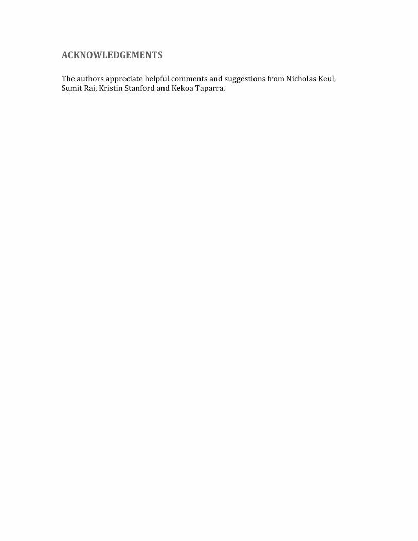
Hyaluronanbiosynthesisiscatalyzedbyhyaluronansynthases(HAS)(Figure16.2).ThefirstbonafideHASgene(spHas)wasclonedfromStreptococcus,andtheproteinexpressedinEscherichiacoliwasshowntosynthesizehigh-molecular-weighthyaluronanfromtheUDP-sugarsubstrates.ThegeneshowshomologywithaXenopusgene,DG42(nowknownasxlHAS1;Chapter27).ThehomologywasinstrumentalinthesubsequentidentificationofthethreemembersofthemammalianHasgenefamily,Has1–3.Thesegenescodeforhomologousproteinspredictedtocontainfivetosixmembrane-spanningsegmentsandacentralcytoplasmicdomain.AsdescribedinChapter17,cellssynthesizesulfatedglycosaminoglycans(heparansulfate,chondroitinsulfate,andkeratansulfate)oncoreproteinsofproteoglycansastheytransitthroughtheGolgi,andelongationofthechainsoccursattheirnonreducingends.Incontrast,hyaluronansynthesisnormallyoccursattheinnersurfaceoftheplasmamembraneineukaryoticcellsandatthecytoplasmicmembraneofbacteriathatproducehyaluronancapsules.ThesynthasesusethecytosolicsubstratesUDP-glucuronicacid(UDP-GlcA)andUDP-N-acetylglucosamine(UDP-GlcNAc)andextrudethegrowingpolymerthroughthemembranetoformextracellularmatrices(Figure16.2).Accordingtothemodel,thereducingendofthegrowingchainwouldhaveaUDPmoietythatisdisplacedwhenthenextnucleotidesugarisadded.Inmammaliancells,divisionunderconditionsofhyperglycemia(2-3timesnormalglucoselevel)resultsinhyaluronansynthesisintheendoplasmicreticulum(ER),Golgiandtransportvesicles.Undertheseconditions,theelongatinghyaluronanchainsareinsertedinappropriatelyintothesecompartments,inducingabnormalitiesincellularfunctions,e.g.kidneynephropathyandproteinurea.TheactivityoftheHASenzymesalsocanberegulatedbyphosphorylationandadditionofO-GlcNAc(Chapter19).Hyaluronanbiosynthesisinbacteriainvolvestheexpressionofmultipleenzymes,usuallyasanoperon.Forexample,inStreptococcus,hasCencodesanenzymethat

makesUDP-GlcfromUTPandglucose-1-P;hasBencodesthedehydrogenasethatconvertsUDP-GlctoUDP-GlcA;hasDgeneratesUDP-GlcNAcfromglucosamine-1-P,acetylCoA,andUTP;andhasA(spHas)encodesthehyaluronansynthase.TheStreptococcushasAgeneencodesabifunctionalproteinthatcontainsbothtransferaseactivities.Thus,spHasassemblesthepolysaccharidefromthereducingend.Thesynthasespansthemembranemultipletimes,presumablyformingaporeforhyaluronanextrusionduringcapsuleformation.Incontrast,PasteurellasynthesizeshyaluronanbyanenzymethatisunrelatedtohasAandthemammalianHasgenefamily.Inthiscase,theenzymehastwoseparabledomainswithindependentglycosyltransferaseactivities—oneforUDP-GlcNAcandtheotherforUDP-GlcA,andtheelongationisonthenon-reducingend.
THEHYALURONIDASESANDHYALURONANTURNOVER
Animalcellsexpressasetofcatabolicenzymesthatdegradehyaluronan.Thehumanhyaluronidasegene(HYAL)familyiscomplex,withtwosetsofthreecontiguousgeneslocatedontwochromosomes,apatternthatsuggeststwoancientgeneduplicationsfollowedbyablockduplication.Inhumans,theclusteronchromosome3p21.3(HYAL1,2,and3)appearstohavemajorrolesinsomatictissues.HYAL4intheclusteronchromosome7q31.3codesforaproteinthatappearstohavechondroitinase,butnothyaluronidase,activity;PHYAL1isapseudogene;andSPAM1(spermadhesionmolecule1,PH-20)isrestrictedtotestes.TheroleofSPAM1infertilizationisdiscussedbelow.Theturnoverofhyaluronaninmosttissuesisrapid(e.g.,ahalf-lifeofapproximately1dayinepidermaltissues),butitsresidencetimeinsometissuescanbequitelonganddependentonlocation(e.g.,incartilage).Ithasbeenestimatedthatanadulthumancontainsapproximately15gofhyaluronanandthataboutone-thirdturnsoverdaily.Turnoverappearstooccurbyreceptor-mediatedendocytosisandlysosomaldegradationeitherlocallyoraftertransportbylymphtolymphnodesorbybloodtoliver.TheendothelialcellsofthelymphnodeandliversinusoidsremovehyaluronanviaspecificreceptorssuchasLYVE-1(ahomologofCD44)andHARE(hyaluronanreceptorforendocytosis).HAREappearstobethemajorclearancereceptorforhyaluronandeliveredsystemicallybylymphandblood.Thecurrentunderstandingofthiscatabolicprocessisthathyaluronidasesatthecellsurfaceandinthelysosomecooperatetodegradethechains.Largehyaluronanmoleculesintheextracellularspaceinteractwithcell-surfacereceptorsthatinternalizefragmentsproducedbyamembrane-associated,GPI-anchoredhyaluronidase,mostlikelyHYAL2.Thefragmentsaretransportedintoauniquevesicularendosomalcompartmentandeventuallyenterapathwaytolysosomesforcompletedegradationtomonosaccharides,probablyinvolvingHYAL1andthetwoexoglycosidasesβ-glucuronidaseandβ-N-acetylglucosaminidase.TheimportanceofthisprocessisdemonstratedbythefactthatHyal2-nullmiceareembryoniclethal
andbytheidentificationofalysosomalstoragedisorderinapersonwithamutationinHYAL1.Hyaluronanfragmentshavebeensuggestedtoactasanendogenoussignalofinjury,forexamplefrominfectionbyGroupAStreptococcus.ThesignalingactivityofHAfragmentsismediatedthroughbindingofcellsurfacereceptors,suchasCD44,whichinturnmodulatesresponsethroughtoll-likereceptors.Signalingthroughtheseandotherreceptorsareaffectedbythesizeofthehyaluronanfragments,butthemechanismunderlyingthedependenceofactivityonthesizeofthefragmentsremainsanareaofactiveresearch.
HYALURONANFUNCTIONINTHEEXTRACELLULARMATRIX
Hyaluronanhasmultiplerolesinearlydevelopment,tissueorganization,andcellproliferation.TheHas2-nullmouseexhibitsanembryoniclethalphenotypeatthetimeofheartformation,whereasHas1-,Has3-nullandHas1/3compoundmutantmiceshownoobviousdevelopmentalphenotype.Interestingly,explantedcellsfromtheHas2-nullembryonicheartdonotsynthesizehyaluronanorundergoepithelial-mesenchymaltransformationandmigrationunlesssmallamountsofhyaluronanareaddedtotheculturemedium.Thisfindingindicatesthattheproductionofhyaluronanatkeypointsmaybeessentialformanytissuemorphogenetictransformations—inthiscase,formationofthetricuspidandmitralvalves.Manyoftheactivitiesofhyaluronandependonbindingproteinspresentonthecellsurfaceand/orsecretedintotheextracellularmatrix.Aclassofproteinsthatbindselectivelytohyaluronanwasfirstdiscoveredincartilage.Thisclassisnowreferredtoasthelinkmodulefamilyofhyaladherins(Figure16.3).Proteoglycanswereefficientlyextractedfromthistissuewithdenaturingsolventsandwereshowntoreaggregatewhenrestoredtorenaturingconditions.Anessentialprotein,referredtoasthelinkprotein(HAPLN-1),wasshowntobenecessaryforstabilizingtheproteoglycanaggregates,andsubsequently,thestructureoftheaggregatewasdefined(Figure16.4).Thelinkproteincontainstwohomologousrepeatsofasequencemotif,nownamedthelinkmodule.Proteinsthathavealinkmodule,includinglinkproteins(HAPLN-1through4inhumans),severalproteoglycans,andotherextracellularmatrixproteins,caninteractspecificallywithhyaluronan.Themajorcartilageproteoglycan,nownamedaggrecan,alsocontainsaglobulardomain,theG1domain,withtwohomologouslinkmodulesthatinteractwithhyaluronan.Anadditionaldomaininthelinkprotein(HAPLN-1)cooperativelyinteractswithahomologousdomaininG1,whichlockstheproteoglycanonthehyaluronanchain.IntheabsenceoftheHAPLN-1,aggrecanfailstoanchortohyaluronan.MicedeficientinHAPLN-1showdefectsincartilagedevelopmentanddelayedboneformation(shortlimbsandcraniofacialanomalies).Mostmutantmicedieshortlyafterbirthas

aresultofrespiratoryfailure,andthefewsurvivorsdevelopprogressiveskeletaldeformities.Interestingly,therearefourproteoglycangeneswithhomologousG1domainsthatinteractwithhyaluronan(versican,neurocan,brevican,andaggrecan)(Figure16.3).Versicanisamajorcomponentofmanysofttissuesandisespeciallyimportantinvascularbiology.Neurocanandbrevicanareexpressedpredominantlyinbraintissue.Versicanandaggrecanareanchoredtohyaluronanintissuesbysimilarlinkprotein-dependentmechanisms,anditislikelythatneurocanandbrevicanareorganizedsimilarly.Thus,hyaluronanactsasascaffoldonwhichtobuildproteoglycanaggregatestructuresadaptedtodiversetissuefunctions.Animpressiveexampleoftherequirementforahyaluronan-basedmatrixoccursduringtheprocessofcumulusoophorusexpansioninthemammalianpreovulatoryfollicle.Atthebeginningofthisprocess,theoocyteissurroundedbyabout1000cumuluscellstightlycompactedandingap-junctioncontactwiththeoocyte.Inresponsetohormonalstimuli,thecumuluscellsup-regulateHAS2andalinkmodulefamilyhyaladherinencodedbytumornecrosisfactor-stimulatedgene6(TSG-6).Theexpressionoftheseproteinsinitiatesproductionofhyaluronananditsorganizationintoanexpandingmatrixaroundthecumuluscells.Concurrently,thefolliclebecomespermeabletoserum,whichintroducesanunusualmoleculecalledinter-α-trypsininhibitor(ITI),composedofthetrypsininhibitorbikuninandtwoheavychainsallcovalentlyboundtoachondroitinsulfatechain.Inacomplexprocess,TSG-6catalyzesthetransferofheavychainsthatarecovalentlylinkedtochondroitinsulfate(viaanesterbondbetweentheC-terminalaspartateresidueoftheheavychainsandtheC-6ofN-acetylgalactosamineinchondroitin-4-sulfate)ontothenewlysynthesizedhyaluronanbytransesterificationtoC6ofanN-acetylglucosamineresidue.IntheabsenceofeitherTSG-6orITI,thematrixdoesnotform,andthephenotypeofmicenullforeitherofthesemoleculesisfemaleinfertility.Atthetimeofovulation,hyaluronansynthesisceases,andovulationoftheexpandedcumuluscell–oocytecomplexoccurs.Priortofertilization,individualspermundergocapacitationenablingthemtopenetrateandfertilizeanovum.Duringthisprocess,SPAM1/PH20,aGPI-anchoredhyaluronidase,redistributesandaccumulatesinthespermhead.SPAM1bindshyaluronaninthecumulus,causinganincreaseinCa++fluxandspermmotility.Italsohelpsdissolvethecumulusmatrixasthespermmovesthroughthehyaluronanvestment.AsolubleformofSPAM1issecretedduringtheacrosomereaction.Thereleaseofacrosomalhyaluronidaseandproteasesrendersthespermcapableoffusingwiththeeggandeventuallydestroystheentirematrixtoallowthefertilizedoocytetoimplantanddevelop.
HYALURONAN-BINDINGPROTEINSWITHLINKMODULES
Thereareseveralhyaluronan-bindingproteinswithhomologouslinkmodules(Figure16.3).Thefourhomologouslinkproteinsbelongtoasubfamilycalledthe“hyaluronanandproteoglycanlinkproteins”(HAPLN);theseareexpressedinmanytissues.Fourcell-surfacereceptorshaveextracellulardomainswithonelinkmodule:CD44,LYVE-1(lymphaticvesselendothelialhyaluronanreceptor),HARE/STABILIN-2(hepatichyaluronanclearancereceptor),andSTABILIN-1,whichareexpressedondiscontinuousendothelialcellsandsomeactivatedmacrophages.Otherhyaluronan-bindingproteinsaresecretedandincludethechondroitinsulfateproteoglycansthatcomprisetheaggrecansuperfamily(aggrecan,versican,brevican,andneurocan)andTNFα-stimulatedgene6(TSG-6),whichhasonelinkmodule.Thethree-dimensionalstructureofthelinkmodulefoldinTSG-6hasbeendeterminedbynuclearmagneticresonanceanddefinesaconsensusfoldofthetwoα-helicesandtwotriple-strandedantiparallelβ-sheets(Figure16.5).Thefoldconsistsofabout100aminoacidsandcontainsfourcysteinesdisulfide-bondedinthepatternCys1-Cys4andCys2-Cys3.Thisfoldhasonlybeenfoundinvertebrates,consistentwiththefactthathyaluronanisarelativelyrecentevolutionaryinvention.ThelinkmodulefoldisrelatedtothatfoundintheC-typelectins,butitlackstheCa++bindingmotif(Chapter34).InthecaseofTSG-6,theinteractionofhyaluronanwiththeproteininvolves1)ionicinteractionsbetweenpositivelychargedaminoacidresiduesandthecarboxylgroupsoftheuronicacids,and2)hydrophobicinteractionsbetweentheacetamidosidechainsoftwoN-acetylglucosamineresiduesandhydrophobicpocketsoneithersideofadjacenttyrosines(Figure16.5).Manyofthesefeaturesareconservedinothermembersofthehyaluronan-bindingproteins.Subgroups,however,differinthepreferredsizeandlengthofhyaluronanforbinding(e.g.,hexasaccharidestodecasaccharides).Somehyaluronan-bindingproteinsdonotcontainalinkmodule(RHAMM,ITI,SPACR,SPACRCAN,CD38,CDC37,HABP1/P-32,andIHABP4),andmostoftheseareunrelatedtooneanotherbyprimarysequence.Someoftheseproteinscontainclustersofbasicaminoacids,referredtoasBX7Bmotifs(whereBiseitherlysineorarginineandXcanbeanyaminoacidotherthanacidicresidues),buttheactualhyaluronandockingsiteofthechainwiththismotifhasnotbeenestablished.Thus,thepresenceoftheBX7Bmotifshouldnotbetakenasproofthattheproteininteractswithhyaluronan.
HYALURONANANDCELLSIGNALING
Hyaluronanexpressionhaslongbeenimplicatedinenhancedcelladhesionandlocomotionbecauseitisexpressedabundantlyduringmorphogenesisandinbothphysiologicalandpathologicalinvasiveprocesses.Asearchforcell-surfacereceptorsrevealedtwomajorhyaluronan-bindingproteins,CD44andRHAMM(receptorforhyaluronan-mediatedmotility).CD44isatransmembranereceptorexpressedbymanycelltypes,anditvariesmarkedlyinglycosylation,oligomerization,andproteinsequencebecauseofdifferentialmRNAsplicing.CD44H(theisoformexpressedbyhematopoieticcells)bindstohyaluronan,andtheinteractioncanmediateleukocyterollingandextravasationinsometissues.ChangesinCD44expression,notablyexpressionofCD44variants,areassociatedwithawidevarietyoftumorsandthemetastaticspreadofcancer.ManycellsalsoexpressthereceptorRHAMM,whichisinvolvedincellmotilityandcelltransformation.TheRHAMMpathwayisthoughttoinducefocaladhesionstosignalthecytoskeletalchangesrequiredforelevatedcellmotilityseenintumorprogression,invasion,andmetastasis.LikeCD44,RHAMMsplicevariantsexist,someofwhichmaybeintracellular.CD44containsacytoplasmicdomain,atransmembranesegment,andanectodomainwithasinglelinkmodulethatcanbindhyaluronan.WhenhyaluronanbindstoCD44,thecytoplasmictailcaninteractwithregulatoryandadaptormolecules,suchasSRCkinases,RHO(rashomolog)GTPases,VAV2(ahumanproto-oncogene),GAB1(aGRB2-associatedbindingprotein),andankyrinandezrin(whichregulatecytoskeletalassembly/disassemblyandcellmigration).HyaluronanbindingtoRHAMMalsotransducessignalsthatinfluencegrowthandmotility,forexample,byactivatingSRC,FAK(focaladhesionkinase),ERK(extracellularmitogen-regulatedproteinkinase),andPKC(proteintyrosinekinaseC)(Chapter40).InteractionofhyaluronanwithCD44canalsoregulateERBB-family(epithelialgrowthfactorreceptor)signaling,therebyactivatingthePI3K(phophoinositide-3-kinase)–PKB/AKT(proteinkinaseB)signalingpathwayandphosphorylationofFAKandBAD(BCL2-antagonistofcelldeath),whichpromotecellsurvival.RHAMMcaninteractwithandactivateERK1,whichcanalsophosphorylateBAD.Thus,bothCD44andRHAMMinteractionswithhyaluronancaninfluencecellsurvival.Thesepathwaysarerelevanttotumorcellsurvivalandinvasion;theirinhibitionbyhyaluronanoligomersandsolublehyaluronan-bindingproteinssuggestsnoveltherapeuticapproachesfortreatingcancer(Chapter47).
HYALURONANCAPSULESINBACTERIA
Somepathogenicbacteria(e.g.,certainstrainsofStreptococcusandPasteurella)producehyaluronananddeposititasanextracellularcapsule(Figure16.6;alsosee
Chapter21).Capsularhyaluronan,likeothercapsularpolysaccharides,increasesvirulencebyhelpingtoshieldthemicrobefromhostdefenses.Forexample,thecapsuleblocksphagocytosisandprotectsagainstcomplement-mediatedkilling.Becausebacterialhyaluronanisidenticalinstructuretohosthyaluronan,thecapsulecanalsopreventtheformationofprotectiveantibodies.Thus,theformationofhyaluronancapsulesbybacteriaisaformofmolecularmimicry.Thecapsulealsocanaidinbacterialadhesiontohosttissue,facilitatingcolonization(Chapter37).Finally,theproductionofhyaluronanbyinvadingbacteriacanalsoinduceanumberofsignalingeventsthroughhyaluronan-bindingproteinsthatmodulatethehostphysiology,i.e.cytokineproduction(Chapter42).Inadditiontobacteria,analgalvirus(Chlorella)encodesahyaluronansynthase.Thefunctionalsignificanceofviralhyaluronanproductionisunknown,butcouldberelatedtopreventionofsecondaryviralinfection,increaseinhostcapacitytoproducevirus,orviralburstsize.TheoriginofviralHASisunknown,butbasedonsequencehomologyitmostlikelyarosefromavertebrate.
HYALURONANASATHERAPEUTICAGENT
Hyaluronanhasbeenusedtherapeuticallyforanumberofyears.Insomecountries,patientswithosteoarthritisaresuccessfullytreatedbydirectinjectionofhigh-molecular-weighthyaluronanintothesynovialspaceofanaffectedjoint.Themechanismofactioniscomplexandprobablyinvolvesboththeviscoelasticpropertiesofthepolymeraswellaseffectsonthegrowthofsynovialcellsinthejointcapsule.Hyaluronansuppressescartilagedegeneration,actsasalubricant(therebyprotectingthesurfaceofarticularcartilage),andreducespainperception.Theapplicationofhyaluronaninophthalmologyiswidespread.Duringsurgeryforlensreplacementduetocataracts,ahighpotentialforinjuryoffragileintraoculartissuesexists,especiallyfortheendotheliallayerofthecornea.High-molecular-weighthyaluronanisinjectedtomaintainoperativespaceandstructureandtoprotecttheendotheliallayerfromphysicaldamage.Hyaluronanalsohasbeenapprovedforcosmeticuse(e.g.,bysubdermalinjectiontofillwrinklesorpocketsundertheskin).Low-molecular-weighthyaluronanoligosaccharides(~103–104D)alsohavepotentbiologicalactivitiesbyalteringselectivesignalingpathways.Incancercells,hyaluronanoligosaccharidesinduceapoptosisandinhibittumorgrowthinvivo.Thus,shorthyaluronanchainsmayproveusefulforpreventingcancermetastasisbyboostingcertainimmuneresponsesoralteringnewbloodvesselgrowth.Recently,recombinantformsofthePasteurellasynthase(pmHas)havebeenengineeredtoproducehyaluronanoligosaccharidesofdefinedsize.Thisstrategyhasgreatpromiseforexploringtherelationshipofhyaluronansizetofunction,whichmayinturnyieldnewtherapeuticagentswithselectiveactivities.
ACKNOWLEDGEMENTS
TheauthorsappreciatehelpfulcommentsandsuggestionsfromNicholasKeul,SumitRai,KristinStanfordandKekoaTaparra.
FURTHERREADING
HascallVC,WangA,TammiM,OikariS,TammiR,PassiA,VigettiD,HansonRW,HartGW.2014.ThedynamicmetabolismofhyaluronanregulatesthecytosolicconcentrationofUDP-GlcNAc.MatrixBiol35:14-17.
JiangD,LiangJ,NoblePW.2007.Hyaluronanintissueinjuryandrepair.AnnuRevCellDevBiol23:435-461.
LiangJ,JiangD,NoblePW.2016.Hyaluronanasatherapeutictargetinhumandiseases.AdvDrugDelivRev97:186-203.
MaytinEV.2016.Hyaluronan:MoreThanJustAWrinkleFiller.Glycobiology.Inpress.
PetreyAC,delaMotteCA.2014.Hyaluronan,aCrucialRegulatorofInflammation.Frontiersinimmunology5:101.
SmithPD,Coulson-ThomasVJ,FoscarinS,KwokJC,FawcettJW.2015."GAG-ingwiththeneuron":Theroleofglycosaminoglycanpatterninginthecentralnervoussystem.ExpNeurol274:100-114.
WangA,delaMotteC,LauerM,HascallV.2011.Hyaluronanmatricesinpathobiologicalprocesses.TheFEBSjournal278:1412-1418.
WeigelPH.2015.HyaluronanSynthase:Themechanismofinitiationatthereducingendandapendulummodelforpolysaccharidetranslocationtothecellexterior.IntJCellBiol2015:367579.
WeigelPH,DeangelisPL.2007.Hyaluronansynthases:Adecade-plusofnovelglycosyltransferases.JBiolChem282:36777-36781.
ZhuoL,KimataK.2008.Structureandfunctionofinter-alpha-trypsininhibitorheavychains.ConnectTissueRes49:311-320.
Figure Legends
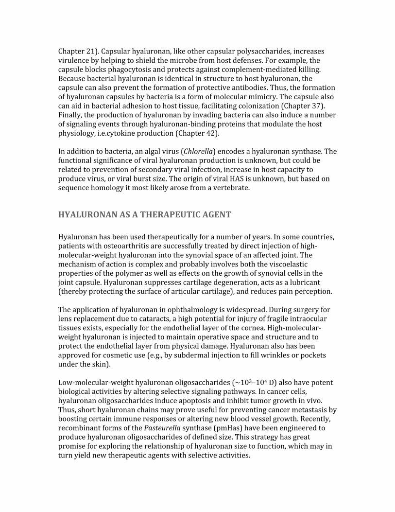
FIGURE16.1.HyaluronanconsistsofrepeatingdisaccharidescomposedofN-acetylglucosamine(GlcNAc)andglucuronicacid(GlcA).Itisthelargestpolysaccharidefoundinvertebrates,anditformshydratedmatrices.(ElectronmicrographprovidedbyDrs.RichardMayneandRandolphBrewton,UniversityofAlabamaatBirmingham.)
FIGURE16.2.Hyaluronanbiosynthesisbyhyaluronansynthase(Has)occursbyadditionofUDP-sugars(UDP-N-acetyl-glucosamineandUDP-glucuronicacid)tothereducingendofthepolymerwithreleaseoftheanchoringUDP.M++referstoametalioncofactor.
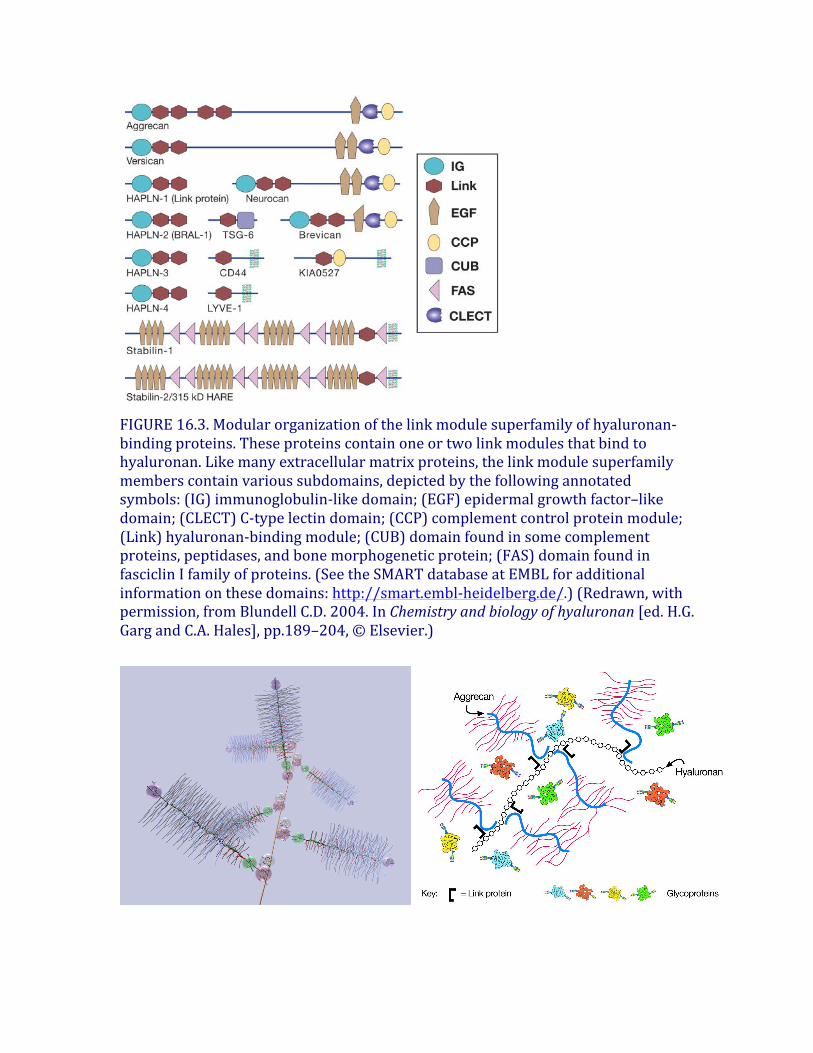
FIGURE16.3.Modularorganizationofthelinkmodulesuperfamilyofhyaluronan-bindingproteins.Theseproteinscontainoneortwolinkmodulesthatbindtohyaluronan.Likemanyextracellularmatrixproteins,thelinkmodulesuperfamilymemberscontainvarioussubdomains,depictedbythefollowingannotatedsymbols:(IG)immunoglobulin-likedomain;(EGF)epidermalgrowthfactor–likedomain;(CLECT)C-typelectindomain;(CCP)complementcontrolproteinmodule;(Link)hyaluronan-bindingmodule;(CUB)domainfoundinsomecomplementproteins,peptidases,andbonemorphogeneticprotein;(FAS)domainfoundinfasciclinIfamilyofproteins.(SeetheSMARTdatabaseatEMBLforadditionalinformationonthesedomains:http://smart.embl-heidelberg.de/.)(Redrawn,withpermission,fromBlundellC.D.2004.InChemistryandbiologyofhyaluronan[ed.H.G.GargandC.A.Hales],pp.189–204,©Elsevier.)
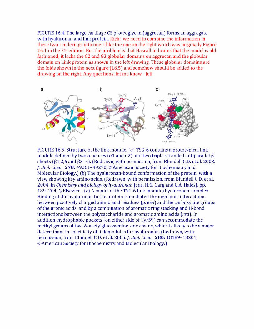
FIGURE16.4.ThelargecartilageCSproteoglycan(aggrecan)formsanaggregatewithhyaluronanandlinkprotein.Rick:weneedtocombinetheinformationinthesetworenderingsintoone.IliketheoneontherightwhichwasoriginallyFigure16.1inthe2ndedition.ButtheproblemisthatHascallindicatesthatthemodelisoldfashioned;itlackstheG2andG3globulardomainsonaggrecanandtheglobulardomainonLinkproteinasshownintheleftdrawing.Theseglobulardomainsarethefoldsshowninthenextfigure(16.5)andsomehowshouldbeaddedtothedrawingontheright.Anyquestions,letmeknow.-Jeff
FIGURE16.5.Structureofthelinkmodule.(a)TSG-6containsaprototypicallinkmoduledefinedbytwoαhelices(α1andα2)andtwotriple-strandedantiparallelβsheets(β1,2,6andβ3–5).(Redrawn,withpermission,fromBlundellC.D.etal.2003.J.Biol.Chem.278:49261–49270,©AmericanSocietyforBiochemistryandMolecularBiology.)(b)Thehyaluronan-boundconformationoftheprotein,withaviewshowingkeyaminoacids.(Redrawn,withpermission,fromBlundellC.D.etal.2004.InChemistryandbiologyofhyaluronan[eds.H.G.GargandC.A.Hales],pp.189–204,©Elsevier.)(c)AmodeloftheTSG-6linkmodule/hyaluronancomplex.Bindingofthehyaluronantotheproteinismediatedthroughionicinteractionsbetweenpositivelychargedaminoacidresidues(green)andthecarboxylategroupsoftheuronicacids,andbyacombinationofaromaticringstackingandH-bondinteractionsbetweenthepolysaccharideandaromaticaminoacids(red).Inaddition,hydrophobicpockets(oneithersideofTyr59)canaccommodatethemethylgroupsoftwoN-acetylglucosaminesidechains,whichislikelytobeamajordeterminantinspecificityoflinkmodulesforhyaluronan.(Redrawn,withpermission,fromBlundellC.D.etal.2005.J.Biol.Chem.280:18189–18201,©AmericanSocietyforBiochemistryandMolecularBiology.)
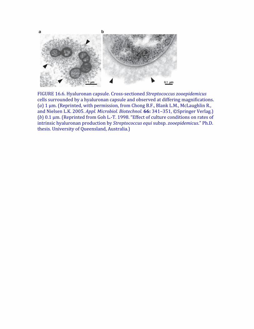
FIGURE16.6.Hyaluronancapsule.Cross-sectionedStreptococcuszooepidemicuscellssurroundedbyahyaluronancapsuleandobservedatdifferingmagnifications.(a)1μm.(Reprinted,withpermission,fromChongB.F.,BlankL.M.,McLaughlinR.,andNielsenL.K.2005.Appl.Microbiol.Biotechnol.66:341–351,©SpringerVerlag.)(b)0.1μm.(ReprintedfromGohL.-T.1998.“EffectofcultureconditionsonratesofintrinsichyaluronanproductionbyStreptococcusequisubsp.zooepidemicus.”Ph.D.thesis.UniversityofQueensland,Australia.)
Chapter 17 Proteoglycans and Sulfated Glycosaminoglycans
Essentials of Glycobiology, 3rd edition
Title:ProteoglycansandSulfatedGlycosaminoglycans
Authors:UlfLindahl,JohnCouchman,KojiKimata,andJeffreyD.Esko
Old#
New#
TextFileName
Original
(Words)*
Current
(Words)*
%Cut
Original
FiguresCurren
tFigures
Original
TablesCurren
tTables
16 17 3edch17PF
5220 3946 30 10 5 8 3
PrimaryWordfilenamestyle:3edch17PF.docx3edch17Table17.1FigureFiles3edch17f1MOD.zip Fig.16.22ndedition,Seenotes3edch17f2MOD.pptxFig.16.32ndedition,updatesymbols3edch17f3MOD.pptxFig.16.42ndedition3edch17f4MOD.jpg Fig.16.52ndedition,seecallouts3edch17f5NEW.pptxNew
Thischapterfocusesonthestructure,biosynthesis,andgeneralbiologyofproteoglycans.Topicsincludeadescriptionofthemajorfamiliesofproteoglycans,theircharacteristicpolysaccharidechains(glycosaminoglycans),biosyntheticpathways,andgeneralconceptsaboutproteoglycanfunction.Proteoglycans,likeotherglycoconjugates,havemanyessentialrolesinbiology.
HISTORICALPERSPECTIVE
Thestudyofproteoglycansdatesbacktothebeginningofthe20thcenturywithinvestigationsof“chondromucoid”fromcartilageandanticoagulantpreparationsfromliver(heparin).From1930to1960,greatstridesweremadeinanalyzingthechemistryofthepolysaccharidesofthesepreparations(alsoknownas“mucopolysaccharides”),yieldingthestructureofhyaluronan(Chapter16),dermatansulfate,keratansulfate,differentisomericformsofchondroitinsulfate,heparin,andheparansulfate.Together,thesepolysaccharidescametobeknownasglycosaminoglycans(sometimesabbreviatedasGAGs)toindicatethepresenceofaminosugarsandothersugarsinapolymericform.Subsequentstudiesprovidedinsightsintothelinkageofthechainstocoreproteins.Thesestructuralstudiespavedthewayforbiosyntheticstudies.The1970smarkedaturningpointinthefield,whenimprovedisolationandchromatographicproceduresweredevelopedforthepurificationandanalysisoftissueproteoglycansandglycosaminoglycans.Density-gradientultracentrifugationallowedseparationofthelargeaggregatingproteoglycansfromcartilage,revealingacomplexofproteoglycan,hyaluronan,andlinkprotein.Alsoduringthisperiod,itwasrealizedthattheproductionofproteoglycanswasageneralpropertyofanimalcellsandthatproteoglycansandglycosaminoglycanswerepresentonthecellsurface,insidethecell,andintheextracellularmatrix(ECM).Thisobservationledtoarapidexpansionofthefieldandtheeventualappreciationofproteoglycanfunctionincelladhesion,signaling,andotherbiologicalactivities(seeChapter38).Today,studieswithsomaticcellmutants(Chapter49)aswellasexperimentsusinggeneknockoutandsilencingtechniquesinavarietyofmodelorganisms,suchasnematodeworms(Caenorhabditiselegans),fruitflies(Drosophilamelanogaster),Africanclawedfrogs(Xenopuslaevis),zebrafish(Daniorerio),andmice(Musmusculus),areaimedatextendingourunderstandingoftheroleofproteoglycansindevelopmentandphysiology(Chapters25-27)andhumandiseases(Chapters41-47).Theapplicationofavarietyofnewlydevelopedanalyticaltools,includingmassspectrometry(Chapter50)andglycanarrays(Chapter48),areleadingtoabetterunderstandingofproteoglycanstructureandfunction.
PROTEOGLYCANANDGLYCOSAMINOGLYCANCOMPOSITION
Proteoglycansconsistofacoreproteinandoneormorecovalentlyattachedglycosaminoglycanchains(Figure17.1).Glycosaminoglycansarelinearpolysaccharides,whosedisaccharidebuildingblocksconsistofanaminosugar(glucosaminethatisN-acetylatedorN-sulfatedorN-acetylgalactosamine)andauronicacid(glucuronicacidoriduronicacid)orgalactose.Figure17.2depictsshortsegmentsofglycosaminoglycansandtheircharacteristicfeatures.Hyaluronandoesnotoccurcovalentlylinkedtoproteoglycans,butinsteadinteractsnoncovalentlywithsomeproteoglycansviahyaluronan-bindingmotifs(Chapter16).Generally,invertebratesproducethesametypesofglycosaminoglycansasvertebrates,exceptthathyaluronanisnotpresentandthechondroitinchainstendtobenonsulfated.MostproteoglycansalsocontainN-andO-glycanstypicallyfoundonglycoproteins(seeChapters9and10).Theglycosaminoglycanchainsaremuchlargerthantheseothertypesofglycans(e.g.,a20kDglycosaminoglycanchaincontains~80sugarresidues,whereasatypicalbiantennaryN-glycancontains10–12residues).Boththecompositionoftheglycosaminoglycanchain,thestructureoftheproteincore,andthedistributionoftheproteoglycandeterminethebiologicalactivitiesassociatedwithproteoglycans.
PROTEOGLYCANSAREDIVERSEINSTRUCTUREANDFUNCTION
VirtuallyallmammaliancellsproduceproteoglycansandsecretethemintotheECM,insertthemintotheplasmamembrane,orstoretheminsecretorygranules.TheECM,anessentialcomponentofallmulticellularanimals,determinesthephysicalcharacteristicsoftissuesandmanyofthebiologicalpropertiesofthecellsembeddedinit.ThemajorcomponentsoftheECMarefibrillarproteinsthatprovidetensilestrengthandelasticity(e.g.,variouscollagensandelastins),adhesiveglycoproteins(e.g.,fibronectin,laminins,andtenascins),andproteoglycansthatinteractwithotherECMcomponentstopromoteECMassembly,governitsphysicalpropertiesandserveasareservoirofbiologicallyactivesmallproteinssuchasgrowthfactors.Asinglecelltypecanexpressmultipleproteoglycans.Vascularendothelialcells,forexample,synthesizeseveraldifferentcellsurfaceproteoglycans,secretorygranuleproteoglycans,aswellasseveralECMproteoglycans.Comparedtothehundreds,perhapsthousands,ofglycoproteinsthatcarryN-andO-linkedglycans,relativelyfewproteinshavebeenidentifiedthatcarryglycosaminoglycans(~17withheparansulfate,approximately~20withchondroitin/dermatansulfate,and~8withkeratansulfate).Nevertheless,tremendousstructuralvariationofproteoglycansexistsduetoanumberoffactors.First,manyproteoglycanscanbesubstitutedwithoneortwotypesofglycosaminoglycanchains,forexampleglypicanscontainheparansulfatewhereas
syndecan-1containsbothheparansulfateandchondroitinsulfatechains.Someproteoglycanscontainonlyoneglycosaminoglycanchain(e.g.,decorin),whereasothershavemorethan100chains(e.g.,aggrecan).Anothersourceofvariabilityliesinthestoichiometryofglycosaminoglycanchainsubstitution.Forexample,syndecan-1hasfiveattachmentsitesforglycosaminoglycans,butnotallofthesitesareusedequally.Otherproteoglycanscanbe“parttime,”thatis,theymayexistwithorwithoutaglycosaminoglycanchainorwithonlyatruncatedoligosaccharide.Agivenproteoglycanpresentindifferentcelltypesoftenexhibitsdifferencesinthenumberofglycosaminoglycanchains,theirlengths,andthearrangementofsulfatedresiduesalongthechains.Thus,apreparationofanyoneproteoglycan(definedbyitscoreprotein)representsadiversepopulationofmolecules,eachpotentiallyrepresentingauniquestructuralentity.Thesecharacteristics,typicalofallproteoglycans,createenormousdiversityandpotentialbiologicalvariationinactivity.
Mammalianproteoglycans–formandfunction
Themajorclassesofproteoglycanscanbeclassifiedbytheirdistribution,homologies,andfunction.Table17.1providesanoverviewofmanyoftheknownproteoglycans.TheaggrecanfamilyofECMproteoglycans(alsoknownaslecticans)consistsofaggrecan,versican,brevican,andneurocan.Inallfourmembers,theproteinmoietycontainsanamino-terminaldomaincapableofbindinghyaluronan,acentralregionthatcontainscovalentlyboundchondroitinsulfatechains,andacarboxy-terminaldomaincontainingaC-typelectindomain(Chapter34).Aggrecanisthebest-studiedmemberofthisfamily,becauseitrepresentsthemajorproteoglycanincartilagewhereitformsastablematrixcapableofwithstandingcompressiveforcesbywaterdesorptionandresorption.Versican,whichisproducedpredominantlybyconnectivetissuecells,undergoesalternativesplicingeventsthatgenerateafamilyofproteins.Neurocanisexpressedinthelateembryoniccentralnervoussystem(CNS)andcaninhibitneuriteoutgrowth.BrevicanisexpressedintheterminallydifferentiatedCNS,particularlyinperineuronalnets.Thesmallleucine-richproteoglycans(SLRPs)containleucine-richrepeatsflankedbycysteinesintheircentraldomain.Atleastninemembersofthisfamilyareknownandsomecarrychondroitinsulfate,dermatansulfate,orkeratansulfatechains.Theseproteoglycanshelptostabilizeandorganizecollagenfibers,buthaveotherrolesininnateimmunityandregulationofgrowthfactorsignaling.Theinterstitialproteoglycansandaggrecanfamilyofproteoglycansappeartobeuniquetovertebrates.C.elegansandD.melanogasterexpressotherproteoglycans,suggestingthatthecoreproteinshaveundergoneenormousdiversificationduringevolution,presumablytoaccommodatedifferentneedsoftheorganism.Incontrast,
thebiosyntheticmachineryhasbeenevolutionarilyconserved,demonstratingconservationoffunctionfortheglycosaminoglycanchains.BasementmembranesarehighlyspecializedthinlayersoftheECMthatlieflushagainstepithelialcellsandsurroundmuscleandfatcells.Majorcomponentsarelaminins,nidogens,andcollagens,aswellasthreeunrelatedbasementmembraneproteoglycans,oneofwhichisacollagen(perlecan,agrin,andTypeXVIIIcollagen).Theseproteoglycansinteractwithotherbasementmembranecomponentsandcellsurfaceadhesionreceptors,butmayalsobeimportantreservoirsofheparansulfate-bindinggrowthfactors.Themembrane-boundproteoglycansarediverse.Thesyndecanfamilyconsistsoffourmembers,eachwithashorthydrophobicdomainthatspansthemembrane,linkingthelargerextracellulardomaincontainingtheglycosaminoglycanattachmentsitestoasmallerintracellularcytoplasmicdomain.Thesyndecansareexpressedinatissue-specificmannerandfacilitatecellularinteractionswithawiderangeofextracellularligands,suchasgrowthfactorsandmatrixmolecules.Becauseoftheirmembrane-spanningproperties,thesyndecanscantransmitsignalsfromtheextracellularenvironmenttotheintracellularcytoskeletonviatheircytoplasmictails.Syndecansareverysensitivetoproteolyticcleavagebymatrixmetalloproteases,resultinginsheddingoftheectodomainsbearingtheglycosaminoglycanchainsthatretainpotentbiologicalactivity(Chapter38).C.elegansandD.melanogasterexpressonlyonesyndecan(Chapter26).Glypicanscarryonlyheparansulfate chains,whichcanbindawidearrayoffactorsessentialfordevelopmentandmorphogenesis.Sixglypicanfamilymembersexistinmammals,onlytwoareexpressedinD.melanogasterandC.elegans.Eachmemberoftheglypicanfamilyofcell-surfaceproteoglycanshasaglycosylphosphatidylinositolanchorattachedatthecarboxylterminus,whichembedsthemintheouterleafletoftheplasmamembrane(Chapter12).Theamino-terminalportionoftheproteinhasmultiplecysteineresiduesandaglobularshapethatdistinguishestheglypicansfromthesyndecanectodomains,whichtendtobeextendedstructures(Figure17.1).AnumberofothermembraneproteoglycansareexpressedonthesurfaceofmanydifferentcelltypesincludingthewidespreadCD44,NG2(alsoknownasCSPG4),phosphacan(PTPζ),thrombomodulinandinvariantchainoftheMHCclassIIsystem.Serglycinisthemajorcytoplasmicsecretorygranuleproteoglycanthatispresentinendothelial,endocrine,andhematopoieticcells.Dependingonthespecies,ithasavariablenumberofglycosaminoglycanattachmentsitesthatcancarrychondroitinsulfate orheparinchains.Heparinisahighlysulfatedformofheparansulfate (discussedbelow).Toalargeextent,thebiologicalfunctionsofproteoglycansdependontheinteractionoftheglycosaminoglycanchainswithdifferentproteinligands.Table17.2listsexamplesofproteinsknowntointeractwithglycosaminoglycans.Proteins
thatbindtothesulfatedglycosaminoglycanchainsappeartohaveevolvedbyconvergentevolution(i.e.,theydonotcontainaspecificfoldpresentinallglycosaminoglycanbindingproteins,incontrasttoothergroupsofglycanbindingproteins).TheseinteractionshaveprofoundphysiologicaleffectsandarediscussedfurtherinChapter38.
LINKAGESOFGLYCOSAMINOGLYCANSTOPROTEINS
Differentsubtypesofsulfatedglycosaminoglycansareattachedtotheircoreproteinsbyuniquelinkages.Therearetwotypesofkeratansulfate,distinguishedbythenatureoftheirlinkagetoprotein(Figure17.3).KSI,originallydescribedincornea,isfoundonanN-glycanlinkedtoproteinthroughanasparagineresidue(Chapter9).KSII(skeletalkeratansulfate)isfoundonanO-glycancore2structureandisthuslinkedthroughN-acetylgalactosamine(GalNAc)toserineorthreonine(Chapter10).Thestructuralfeaturesincontrolofkeratansulfate substitutionremainunclear.Notably,inhumansandbovine,thelargechondroitinsulfate proteoglycanfoundincartilage(aggrecan)containsasegmentof4–23hexapeptiderepeats(E-E/L-P-F-P-S)wherethekeratansulfate chainsarelocated,whereasaggrecaninratsandotherrodentslacksthismotifanddoesnotcontainkeratansulfate .Twoclassesofglycosaminoglycanchains,chondroitinsulfate/dermatansulfate andheparansulfate/heparin,arelinkedtoserineresiduesincoreproteinsbywayofxylose(Figure17.4).XylosyltransferaseinitiatestheprocessusingUDP-xyloseasdonor.Twoisoformsoftheenzymeareknowninvertebrates(Xylt1andXylt2),butonlyoneisozymeexistsinC.elegansandD.melanogaster.Aglycineresidueinvariablyliestothecarboxy-terminalsideoftheserineattachmentsite,butaperfectconsensussequenceforxylosylationdoesnotexist.Atleasttwoacidicaminoacidresiduesareusuallypresent,andtheycanbelocatedononeorbothsidesoftheserine,usuallywithinafewresidues.Severalproteoglycanscontainclusteredglycosaminoglycanattachmentsites,raisingthepossibilitythatxylosyltransferasecouldactinaprocessivemanner.Xylosylationisanincompleteprocessinsomeproteoglycans,whichmayexplainwhyproteoglycanswithmultiplepotentialattachmentsitescontaindifferentnumbersofchainsindifferentcells.Afterxyloseaddition,alinkagetetrasaccharideassemblesbythetransferoftwogalactoseresiduescatalyzedbyuniquemembersoftheβ4galactosyl-,β3galactosyl-andβ3glucuronosyltransferasefamiliesofenzymes(Figure17.4).ThisintermediatecanundergophosphorylationattheC-2positionofxyloseandinthecaseofchondroitinsulfate,sulfationofthegalactoseresidues.Ingeneral,phosphorylationandsulfationoccursubstoichiometrically,butphosphorylationmaybetransient.PhosphorylationoccursearlyintheassemblyprocessandcreatesthepreferredsubstrateforB4GALT7;aphosphataseremovesthephosphateata
laterstageofbiosynthesis.Galactosesulfationisfoundonlyinchondroitinsulfate,butitsfunctionremainsunclear.Thelinkagetetrasaccharideliesatabifurcationinthebiosyntheticpathway.Twotypesofreactionsoccur:additionofβ4-linkedN-acetylgalactosamine(GalNAc),whichinitiateschondroitinsulfateassembly,oradditionofα4-linkedN-acetylglucosamine(GlcNAc),whichinitiatesheparansulfateassembly(Figure17.4).GeneticevidencefromstudiesofC.eleganssuggeststhatGalNAcadditionduringchondroitinassemblyismediatedbythesameenzymethatisinvolvedinchainpolymerization(SQV5),butbiochemicalevidencesuggeststhatmorethanoneenzymemayexistinvertebrates.Inheparin/heparansulfate formation,theadditionofthefirstGlcNAcresidueiscatalyzedbyanenzymecalledEXTL3,whichdiffersfromthetransferasesinvolvedinheparanpolymerization(calledEXT1andEXT2).Theseenzymesareimportantcontrolpointsbecausetheyultimatelyregulatethetypeofglycosaminoglycanchainthatwillassemble.Controloftheadditionofβ4GalNAcorα4GlcNAcappearstobemanifestedatthelevelofenzymerecognitionofthepolypeptidesubstrate.
GLYCOSAMINOGLYCANBIOSYNTHESIS
Keratansulfate
Keratansulfatechainscontainamixtureofnonsulfated(Galβ4GlcNAcβ3),monosulfated(Galβ4GlcNAc6Sβ3),anddisulfated(Gal6Sβ4GlcNAc6Sβ3)disaccharideunits(Figure17.2).Thebiosynthesisofthepoly-N-acetyllactosaminebackboneisdescribedinChapter10.Atleasttwoclassesofsulfotransferases,oneormoreGlcNAc6-O-sulfotransferases(e.g.,Chst6),andoneGal6-O-sulfotransferase(Chst1)catalyzethesulfationreactions.Theseenzymes,likeothersulfotransferases,useactivatedsulfate(PAPS[3′-phosphoadenyl-5′-phosphosulfate])asahigh-energydonor(Chapter5).GlcNAc6-O-sulfationoccursonthenonreducingterminalGlcNAcresidue,asaprerequisitetofurtherchainelongation,whereassulfationofgalactoseresiduestakesplaceonnonreducingterminalandinternalgalactoseresidues,withapreferenceforgalactoseunitsadjacenttoasulfatedGlcNAc.Sulfationofanonreducingterminalgalactoseresidueblocksfurtherelongationofthechain,providingapotentialmechanismforcontrollingchainlength.Thepoly-N-acetyllactosaminechainsofKSIaregenerallylongerthanthoseofKSII,andmaycontainupto50disaccharideunits(20–25kD).TherelationshipofenzymesinvolvedinKSIandKSIIsulfationisunclear.Thechainscanbefucosylatedandsialylatedaswell(Chapter14).
Chondroitinsulfate
Vertebratechondroitinsulfateconsistsofrepeatingsulfate-substitutedGalNAcβ4GlcAβ3disaccharideunitspolymerizedintolongchains(Figure17.2).Incontrast,invertebratessuchasC.elegansandD.melanogastermakeeithernonsulfatedorlowsulfatedchains,respectively.Theassemblyprocessforthebackboneappearstobehighlyconserved,basedonthepresenceofhomologousgenesforallofthereactions(Chapters25and26).Asdescribedabove,theassemblyprocessisinitiatedbythetransferofGalNAcβ3tothelinkagetetrasaccharide(Figure17.4).Inbothvertebratesandinvertebrates,thepolymerizationstepiscatalyzedbyoneormorebifunctionalenzymes(chondroitinsynthases)thathavebothβ3glucuronosyltransferaseandβ4N-acetylgalactosaminyltransferaseactivities.Vertebratesalsoexpresshomologsthatcantransferindividualsugarstothechain.Chondroitinpolymerizationalsorequirestheactionofthechondroitinpolymerizingfactor(Chpf),aproteinthatlacksindependentactivitybutcollaborateswiththepolymerasestoenhancetheformationofpolymers.Sulfationofchondroitininvertebratesisacomplexprocess,withmultiplesulfotransferasesinvolvedin4-O-sulfationand6-O-sulfationofGalNAcresidues(Figure17.5). Additionalenzymesexistforepimerizationofglucuronicacid(GlcA)toiduronicacid(IdoA)indermatansulfate(Dse1-2),sulfationattheC-2positionoftheuronicacids,andotherpatternsofsulfationfoundinunusualspeciesofchondroitin(Table17.2).Thelocationofsulfategroupsiseasilyassessedusingbacterialchondroitinases(ABC,B,andACII)thatcleavethechainsintodisaccharides.Manychainsarehybridstructurescontainingmorethanonetypeofchondroitindisaccharideunit.Forexample,dermatansulfate isdefinedashavingoneormoreIdoA-containingdisaccharideunits(chondroitinsulfate B)aswellasGlcA-containingdisaccharides(chondroitinsulfate AandC).Animalcellsalsodegradechondroitinsulfate inlysosomesusingaseriesofexolyticactivities(Chapter44).
Heparansulfate
HeparansulfateassemblesasacopolymerofGlcNAcα4GlcAβ4(Figure17.5),whichthenundergoesextensivemodificationreactions,catalyzedbyatleastfourfamiliesofsulfotransferasesandoneepimerase.N-acetylglucosamineN-deacetylase/N-sulfotransferases(Ndst1-4)actonasubsetofGlcNAcresiduestogenerateN-sulfatedglucosamine(GlcNSO3)units,manyofwhichoccurinclustersalongthechain.Generally,theenzymedeacetylatesGlcNAcandrapidlyaddssulfatetothefreeaminogrouptoformGlcNSO3,butasmallnumberofglucosamineresidueswithunsubstitutedaminogroupsmayarisefromincompleteN-sulfation.Anepimerase(Glce),differentfromtheoneinvolvedindermatansulfate synthesis,thenactsonsomeGlcAresidues,followedby2-O-sulfationofsomeoftheIdoAunits(catalyzedbyHs2st).Someglucuronicunitsalsoundergo2-O-sulfationbythesameenzyme.Theadditionof2-O-sulfategroupstoGlcAorIdoAblockstheepimerizationreaction.Next,6-O-sulfotransferases(Hs6st1-3)addsulfategroupstoselected
glucosamineresidues.Finally,certainsubsequencesofsulfatedsugarresiduesanduronicacidepimersprovidetargetsfor3-O-sulfotransferases(Hs3st1-6).Incontrasttochondroitinchains,whichtendtohavelongtractsoffullymodifieddisaccharides,themodificationreactionsinheparansulfate biosynthesisoccurinclustersalongthechain,withregionsdevoidofsulfateseparatingthemodifiedtracts.Ingeneral,thereactionsproceedintheorderindicated,buttheyoftenfailtogotocompletion,resultingintremendouschemicalheterogeneity.Thedisaccharidecompositionofthechainscanbereadilyassessedusingbacterialheparinlyasesorchemicaldegradationmethods(whicharemoreusefulfordifferentiatingGlcA/IdoA)butdirectsequencingofthechainshasprovendifficultbecauseoftheirheterogeneity.Newmassspectrometrymethodsaremakingsignificantinroadsintosequencingofglycosaminoglycans(Chapter50).
Regulationofglycosaminoglycanassembly
Thespecificarrangementofsulfatedresiduesanduronicacidepimersinheparin/heparansulfate anddermatansulfate givesrisetobindingsequencesforligands.ThethreeexamplesshowninFigure17.5demonstrateminimalsequencesthatcaninteractwithfibroblastgrowthfactors(FGFs),antithrombinandheparincofactorII.Moremodifiedsequencescaninteractaswell,andthebindingofmostFGFsisactuallymoresensitivetooverallsulfationthantothespecificpositionofthesulfategroups.BindingofglycosaminoglycanstoproteinsisdescribedingreaterdetailinChapter38.Amajorquestionremainsregardinghowtheenzymesandbiosyntheticpathwaysareregulatedtoachievetissue-specificexpressionofligand-bindingsequences.Duringthelastdecade,mostifnotalloftheenzymesinvolvedinglycosaminoglycansynthesishavebeenpurifiedandmolecularlyclonedfrommammalsandmodelorganisms.Severalimportantfeatureshaveemergedfromthesestudies,whichmayshedlightonhowdifferentproteinbindingsequencesarise.• Severaloftheenzymesappeartohavedualcatalyticactivities.Thus,asingleproteinbearingtwocatalyticdomainscatalyzesN-deacetylationofGlcNAcresiduesandsubsequentN-sulfation(Ndsts)inheparansulfateformation.Thesameistrueofthecopolymerases,whichtransferGlcNAcandGlcA(heparansulfate)andGalNAcandGlcA(chondroitinsulfate)fromthecorrespondingUDP-sugarstothegrowingpolymer.Incontrast,theepimerasesandO-sulfotransferaseactivitiesappeartobeuniquepropertiesofindependentenzymes.
• Inseveralcases,multipleisozymesexistthatcancatalyzeeitherasingleorapairofreactions.Thus,fourN-deacetylase/N-sulfotransferases,three6-O-sulfotransferases,andseven3-O-sulfotransferaseshavebeenidentifiedinheparansulfatebiosynthesis.Theirtissuedistributionvariesanddifferencesexistinsubstratepreference,whichmaycausedifferencesinthepatternofsulfation.
However,someoverlapinexpressionandinsubstrateutilizationoccursaswell.Multipleisozymesof4-O-and6-O-sulfotransferasesalsocanparticipateinchondroitinsulfateformation.
• ThepolymerizationandpolymermodificationreactionsprobablycolocalizeinthesamestacksoftheGolgicomplex.Thus,theenzymesmayformsupramolecularcomplexesthatcoordinatethesereactions.Thecompositionofthesecomplexesmayplayapartinregulatingthefinestructureofthechains.
• Ingeneral,thecompositionofheparansulfate,andlikelychondroitinanddermatansulfate,onagivenproteoglycanvariesmorebetweencelltypesthanthatofheparansulfate ondifferentcoreproteinsexpressedinthesamecell.Thisobservationsuggeststhateachcelltypemayexpressauniquearrayofenzymesandpotentialregulatoryfactors.Themechanismsbehindthegenerationofapparentlycell-specificglycosaminoglycan chainsthroughregulated,yetpartlystochasticmodificationreactionsremainpoorlyunderstood.
• Recombinantenzymesandnewsyntheticschemesareincreasinglyusedtogeneratedefinedglycosaminoglycanoligosaccharides,whichcanbeusedtoprobeligand-bindingaffinitiesandspecificities.
HEPARINVERSUSHEPARANSULFATE
Considerableconfusionexistsregardingthedefinitionofheparinandheparansulfate .Heparinisproducedbyalimitednumberofcells,notablyconnectivetissue-typemastcellsandbipotentialglialprogenitorcells,whereasheparansulfate ismadebyvirtuallyalltypesofcells.Duringbiosynthesis,heparinundergoesmoreextensivesulfationanduronicacidepimerization,suchthatmorethan80%oftheGlcNAcresiduesareN-deacetylatedandN-sulfatedandmorethan70%oftheglucuronicacidsundergoepimerizationtoIdoA.Heparinderivedfromporcineandbovineentrailsispreparedcommerciallybyselectiveprecipitationandissoldbypharmaceuticalcompaniesasananticoagulantduetoitscapacitytobindtoantithrombin.TheactivesequenceisapentasaccharideshowninFigure17.5,whichisnowsoldasapurelysyntheticanticoagulant(Arixtra).Low-molecular-weightheparinsarederivedfromcommercialunfractionatedheparinbychemicalorenzymaticcleavage,dependingonthebrand.Selectivelydesulfatedformsofheparinarealsoavailablecommercially,someofwhichlackanticoagulantactivity,butstillretainotherpotentiallyusefulproperties(e.g.,inhibitionofinflammationandcellproliferation,andantimetastaticactivity).Heparansulfatealsocancontainanticoagulantactivity,buttypicalpreparationsfromcellsortissuesaremuchlessactivethanheparin.Careshouldbetakeninextrapolatingdataobtainedwithheparin(e.g.bindingtoheparin-Sepharose)versusbindingtoheparansulfateandheparansulfateproteoglycans;bindingtoheparincanoccurduetothehighchargecontentofthepolysaccharide,whereasthesamefactormightbindtoheparansulfatewithloweraffinityornotatall.Ontheotherhand,specificprotein-bindingmotifsexpressedinsubspeciesofheparansulfatemayoccuralsoinheparin,althoughconcealedbyadditional,redundant,sulfateresidues.
PROTEOGLYCANPROCESSINGANDTURNOVER
Cellssecretematrixproteoglycansdirectlyintotheextracellularenvironment(e.g.,membersoftheaggrecanfamily,thebasementmembraneproteoglycans,SLRPs,andserglycin).However,othersareshedfromthecellsurfacethroughproteolyticcleavageofthecoreproteinthroughmatrixmetalloproteases(e.g.,thesyndecans).Cellsalsointernalizealargefractionofcell-surfaceheparansulfateproteoglycansbyendocytosis.Theseinternalizedproteoglycansfirstencounterproteasesandheparanase,anendo-ß-glucuronidase,andthecoreproteinandtheheparansulfatechainsarecleavedendolytically.Theresultingsmallerheparansulfate fragmentseventuallyappearinthelysosomeandundergocompletedegradationbywayofaseriesofexoglycosidasesandsulfatases(Chapter44).Chondroitinsulfate anddermatansulfate proteoglycansfollowasimilarendocyticroute.Oneofhumanhyaluronidases(Hyal-4)hasbeenfoundtobeinvolvedinendolyticdegradationofchondroitinsulfate.Cellssecreteheparanaseaswell.Extracellularheparanasecancleaveheparansulfate chainsatrestrictedsites,resultinginreleaseofgrowthfactorsorchemokinesimmobilizedonheparansulfate proteoglycansatcellsurfacesorintheECM.Inparticular,invadingcellssecreteheparanase.Thus,heparanasemayactwithmatrixmetalloproteasestoremodeltheECM.Afamilyofplasmamembraneendosulfatases(Sulfs)canremovesulfategroupsfrominternal6-O-sulfatedglucosamineresidues.Thispost-assemblyprocessingofthechainsatthecellsurfaceresultsinalteredresponseofcellstogrowthfactorsandmorphogens.Themammaliangenomecontainsothersulfatasesofunknownfunction,raisingthepossibilitythatotherpost-assemblyprocessingreactionsofglycosaminoglycansmayoccur.
Acknowlegements
TheauthorsappreciatehelpfulcommentsandsuggestionsfromAndreasGeissner,MarissaMartinezandYukoNaito-Matsui.

FURTHERREADING
FilmusJ,CapurroM.2014.TheroleofglypicansinHedgehogsignaling.MatrixBiol
35:248-252.FriandV,DavidG,ZimmermannP.2015.Synteninandsyndecaninthebiogenesisof
exosomes.BiolCell107:331-341.GallagherJ.2015.Fell-MuirLecture:Heparansulphateandtheartofcellregulation:
apolymerchainconductstheproteinorchestra.IntJExpPathol96:203-231.Hultgardh-NilssonA,BorenJ,ChakravartiS.2015.Thesmallleucine-richrepeat
proteoglycansintissuerepairandatherosclerosis.JInternMed278:447-461.
MizumotoS,YamadaS,SugaharaK.2015.Molecularinteractionsbetweenchondroitin-dermatansulfateandgrowthfactors/receptors/matrixproteins.CurrOpinStructBiol34:35-42.
NeillT,SchaeferL,IozzoRV.2015.Decodingthematrix:instructiverolesofproteoglycanreceptors.Biochemistry54:4583-4598.
ThackerBE,XuD,LawrenceR,EskoJD.2013.Heparansulfate3-O-sulfation:Araremodificationinsearchofafunction.MatrixBiol35:60-72.
VlodavskyI,IozzoRV,SandersonRD.2013.Heparanase:multiplefunctionsininflammation,diabetesandatherosclerosis.MatrixBiol32:220-222.
WightTN,KinsellaMG,EvankoSP,Potter-PerigoS,MerrileesMJ.2014.Versicanandtheregulationofcellphenotypeindisease.BiochimBiophysActa1840:2441-2451.
XuD,EskoJD.2014.Demystifyingheparansulfate-proteininteractions.AnnuRevBiochem83:129-157.
YurchencoPD.2011.Basementmembranes:cellscaffoldingsandsignalingplatforms.ColdSpringHarbPerspectBiol3.
Figure Legends
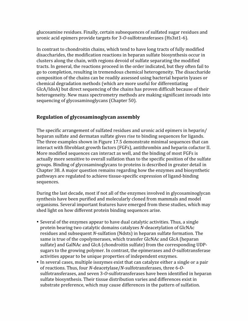
FIGURE17.1.Proteoglycansconsistofaproteincore(brown)andoneormorecovalentlyattachedglycosaminoglycanchains([blue]heparansulfate ;[yellow]chondroitinsulfate /dermatansulfate ).Membraneproteoglycanseitherspantheplasmamembrane(typeImembraneproteins)orarelinkedbyaGPIanchor.ECMproteoglycansareusuallysecreted,butsomeproteoglycanscanbeproteolyticallycleavedandshedfromthecellsurface(notshown).
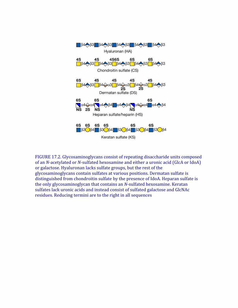
FIGURE17.2.GlycosaminoglycansconsistofrepeatingdisaccharideunitscomposedofanN-acetylatedorN-sulfatedhexosamineandeitherauronicacid(GlcAorIdoA)orgalactose.Hyaluronanlackssulfategroups,buttherestoftheglycosaminoglycanscontainsulfatesatvariouspositions.Dermatansulfateisdistinguishedfromchondroitinsulfate bythepresenceofIdoA.HeparansulfateistheonlyglycosaminoglycanthatcontainsanN-sulfatedhexosamine.KeratansulfateslackuronicacidsandinsteadconsistofsulfatedgalactoseandGlcNAcresidues.Reducingterminiaretotherightinallsequences
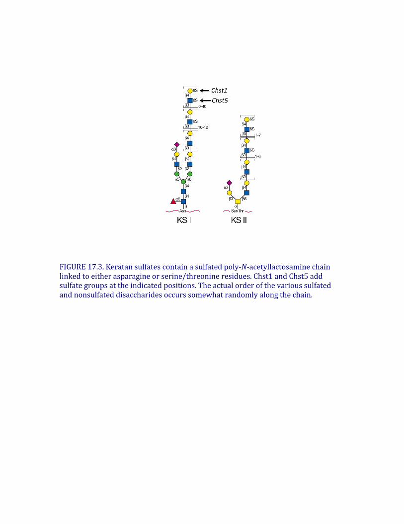
FIGURE17.3.Keratansulfatescontainasulfatedpoly-N-acetyllactosaminechainlinkedtoeitherasparagineorserine/threonineresidues.Chst1andChst5addsulfategroupsattheindicatedpositions.Theactualorderofthevarioussulfatedandnonsulfateddisaccharidesoccurssomewhatrandomlyalongthechain.
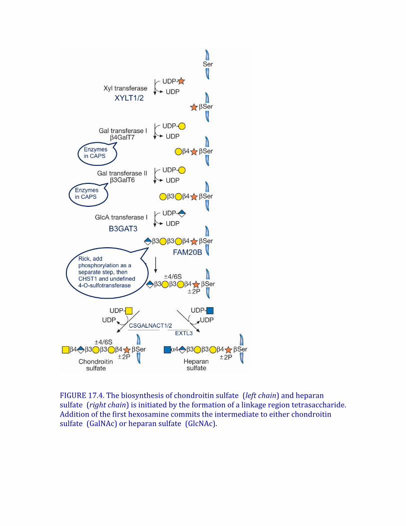
FIGURE17.4.Thebiosynthesisofchondroitinsulfate (leftchain)andheparansulfate (rightchain)isinitiatedbytheformationofalinkageregiontetrasaccharide.Additionofthefirsthexosaminecommitstheintermediatetoeitherchondroitinsulfate (GalNAc)orheparansulfate (GlcNAc).
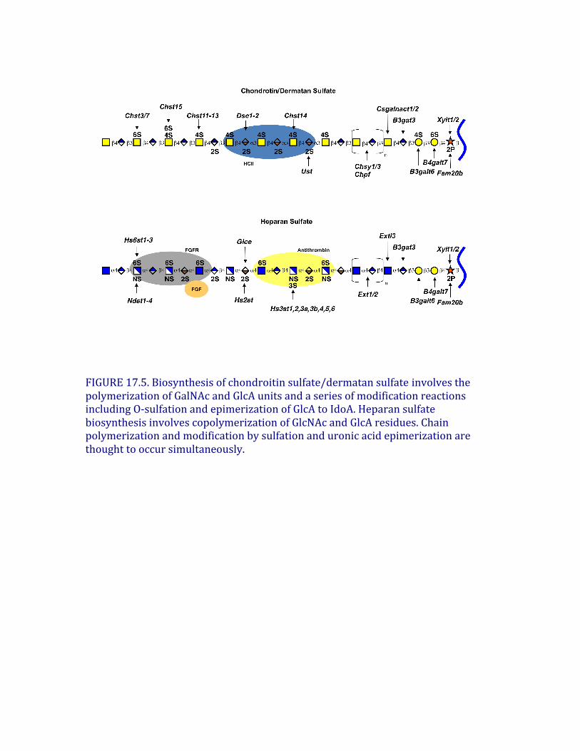
FIGURE17.5.Biosynthesisofchondroitinsulfate/dermatansulfate involvesthepolymerizationofGalNAcandGlcAunitsandaseriesofmodificationreactionsincludingO-sulfationandepimerizationofGlcAtoIdoA.HeparansulfatebiosynthesisinvolvescopolymerizationofGlcNAcandGlcAresidues.Chainpolymerizationandmodificationbysulfationanduronicacidepimerizationarethoughttooccursimultaneously.
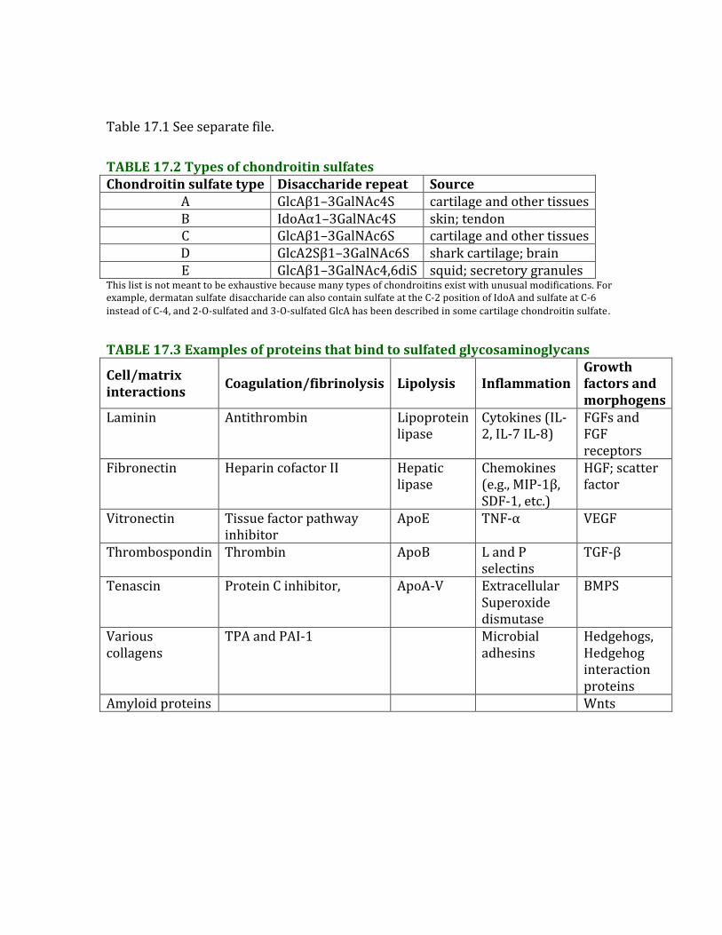
Table17.1Seeseparatefile.TABLE17.2TypesofchondroitinsulfatesChondroitinsulfatetype Disacchariderepeat Source
A GlcAβ1–3GalNAc4S cartilageandothertissuesB IdoAα1–3GalNAc4S skin;tendonC GlcAβ1–3GalNAc6S cartilageandothertissuesD GlcA2Sβ1–3GalNAc6S sharkcartilage;brainE GlcAβ1–3GalNAc4,6diS squid;secretorygranules
Thislistisnotmeanttobeexhaustivebecausemanytypesofchondroitinsexistwithunusualmodifications.Forexample,dermatansulfate disaccharidecanalsocontainsulfateattheC-2positionofIdoAandsulfateatC-6insteadofC-4,and2-O-sulfatedand3-O-sulfatedGlcAhasbeendescribedinsomecartilagechondroitinsulfate.
TABLE17.3Examplesofproteinsthatbindtosulfatedglycosaminoglycans
Cell/matrixinteractions Coagulation/fibrinolysis Lipolysis Inflammation
Growthfactorsandmorphogens
Laminin Antithrombin Lipoproteinlipase
Cytokines(IL-2,IL-7IL-8)
FGFsandFGFreceptors
Fibronectin HeparincofactorII Hepaticlipase
Chemokines(e.g.,MIP-1β,SDF-1,etc.)
HGF;scatterfactor
Vitronectin Tissuefactorpathwayinhibitor
ApoE TNF-α VEGF
Thrombospondin Thrombin ApoB LandPselectins
TGF-β
Tenascin ProteinCinhibitor, ApoA-V ExtracellularSuperoxidedismutase
BMPS
Variouscollagens
TPAandPAI-1 Microbialadhesins
Hedgehogs,Hedgehoginteractionproteins
Amyloidproteins Wnts
Review Article
Hyaluronan Synthase: The Mechanism of Initiation at the Reducing End and a Pendulum Model for
Polysaccharide Translocation to the Cell Exterior
Review ArticleHyaluronan Synthase: The Mechanism ofInitiation at the Reducing End and a Pendulum Model forPolysaccharide Translocation to the Cell Exterior
Paul H. Weigel
Department of Biochemistry & Molecular Biology,The Oklahoma Center for Medical Glycobiology,University of Oklahoma Health Sciences Center, Oklahoma City, OK 73190, USA
Correspondence should be addressed to Paul H. Weigel; [email protected]
Received 2 October 2014; Accepted 14 January 2015
Academic Editor: Howard Beverley Osborne
Copyright © 2015 Paul H. Weigel. This is an open access article distributed under the Creative Commons Attribution License,which permits unrestricted use, distribution, and reproduction in any medium, provided the original work is properly cited.
Hyaluronan (HA) biosynthesis has been studied for over six decades, but our understanding of the biochemical details of how HAsynthase (HAS) assembles HA is still incomplete. Class I family members include mammalian and streptococcal HASs, the focusof this review, which add new intracellular sugar-UDPs at the reducing end of growing hyaluronyl-UDP chains. HA-producingcells typically create extracellular HA coats (capsules) and also secrete HA into the surrounding space. Since HAS contains multipletransmembrane domains and is lipid-dependent, we proposed in 1999 that it creates an intraprotein HAS-lipid pore through whicha growing HA-UDP chain is translocated continuously across the cell membrane to the exterior. We review here the evidence fora synthase pore-mediated polysaccharide translocation process and describe a possible mechanism (the Pendulum Model) andpotential energy sources to drive this ATP-independent process. HA synthases also synthesize chitin oligosaccharides, which arecreated by cleavage of novel oligo-chitosyl-UDP products. The synthesis of chitin-UDP oligomers by HAS confirms the reducingend mechanism for sugar addition during HA assembly by streptococcal and mammalian Class I enzymes. These new findingsindicate the possibility that HA biosynthesis is initiated by the ability of HAS to use chitin-UDP oligomers as self-primers.
1. Introduction and Overview ofHA Biosynthesis
Cell-free biosynthesis of HA was demonstrated in 1959 usingStreptococcus membranes [1]. The enzyme responsible, HAsynthase (HAS), is a membrane protein that requires onlyMg+2 and two sugar-UDP substrates (GlcUA-UDP andGlcNAc-UDP) to polymerize HA chains. (To be consistentin using the standard convention of showing the reducingend of any glycan or saccharide to the right, we do notuse the normal convention for nucleotide-sugars (e.g., UDP-GlcNAc); instead HA-UDP, GlcNAc-UDP, and GlcUA-UDPare abbreviated to show their reducing ends to the right.) Noone was able to identify any streptococcal or eukaryotic HAsynthase gene until 1993 when the hasA gene was identifiedand cloned, and the S. pyogenes HAS protein was expressed[2–4]. Identification of the hasA gene and the biochemical
demonstration that only the HAS protein was required tosynthesize HA [5] then led to the identification of hasAgenes in S. equisimilis [6] and S. uberis [7] and vertebratehomologues of these HAS genes in many species [8–10].Thefirst active HAS was purified when the recombinant enzymesfrom Group A (SpHAS) and Group C (SeHAS) Streptococcuswere overexpressed in E. coli SURE cells [11].
Mammalian genomes have three different HAS genes(HAS1, HAS2, and HAS3) that are expressed at specific timesand specific tissues during development, aging, wound heal-ing, and under normal or pathologic conditions or in diseasessuch as cancer [12, 13]. HA, which is found in only someprokaryotes but is a general ubiquitous extracellular matrixcomponent in vertebrates [14, 15], is a linear heteropolysac-charide composed of the repeating disaccharide: (-3)-!-D-N-acetylglucosamine-!(1,4)-D-glucuronic acid-!(1-). Thisunsulfated glycosaminoglycan is a major component in
Hindawi Publishing Corporation
International Journal of Cell Biology
Volume 2015, Article ID 367579, 15 pages
http://dx.doi.org/10.1155/2015/367579
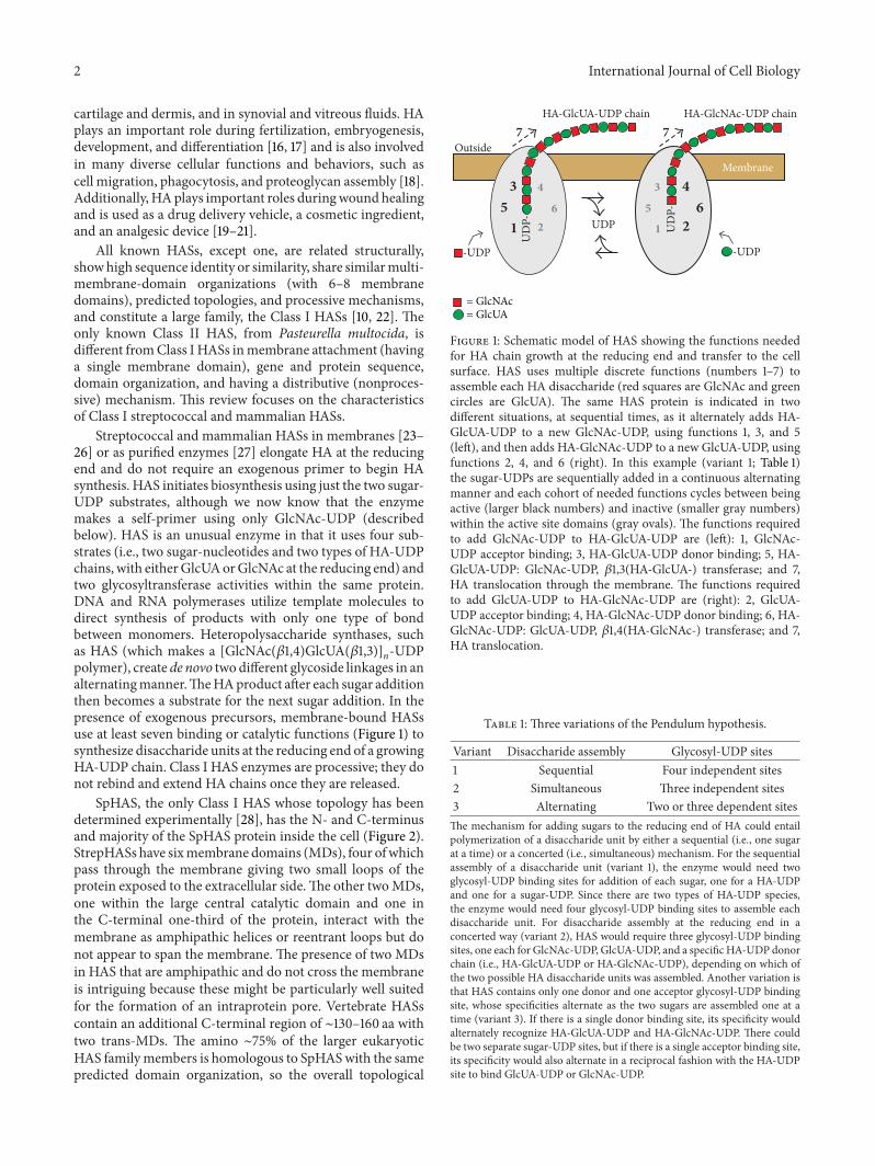
2 International Journal of Cell Biology
cartilage and dermis, and in synovial and vitreous fluids. HAplays an important role during fertilization, embryogenesis,development, and differentiation [16, 17] and is also involvedin many diverse cellular functions and behaviors, such ascell migration, phagocytosis, and proteoglycan assembly [18].Additionally, HAplays important roles duringwound healingand is used as a drug delivery vehicle, a cosmetic ingredient,and an analgesic device [19–21].
All known HASs, except one, are related structurally,showhigh sequence identity or similarity, share similarmulti-membrane-domain organizations (with 6–8 membranedomains), predicted topologies, and processive mechanisms,and constitute a large family, the Class I HASs [10, 22]. Theonly known Class II HAS, from Pasteurella multocida, isdifferent fromClass I HASs inmembrane attachment (havinga single membrane domain), gene and protein sequence,domain organization, and having a distributive (nonproces-sive) mechanism. This review focuses on the characteristicsof Class I streptococcal and mammalian HASs.
Streptococcal and mammalian HASs in membranes [23–26] or as purified enzymes [27] elongate HA at the reducingend and do not require an exogenous primer to begin HAsynthesis. HAS initiates biosynthesis using just the two sugar-UDP substrates, although we now know that the enzymemakes a self-primer using only GlcNAc-UDP (describedbelow). HAS is an unusual enzyme in that it uses four sub-strates (i.e., two sugar-nucleotides and two types of HA-UDPchains, with eitherGlcUAorGlcNAc at the reducing end) andtwo glycosyltransferase activities within the same protein.DNA and RNA polymerases utilize template molecules todirect synthesis of products with only one type of bondbetween monomers. Heteropolysaccharide synthases, suchas HAS (which makes a [GlcNAc(!1,4)GlcUA(!1,3)]n-UDPpolymer), create de novo two different glycoside linkages in analternatingmanner.TheHAproduct after each sugar additionthen becomes a substrate for the next sugar addition. In thepresence of exogenous precursors, membrane-bound HASsuse at least seven binding or catalytic functions (Figure 1) tosynthesize disaccharide units at the reducing end of a growingHA-UDP chain. Class I HAS enzymes are processive; they donot rebind and extend HA chains once they are released.
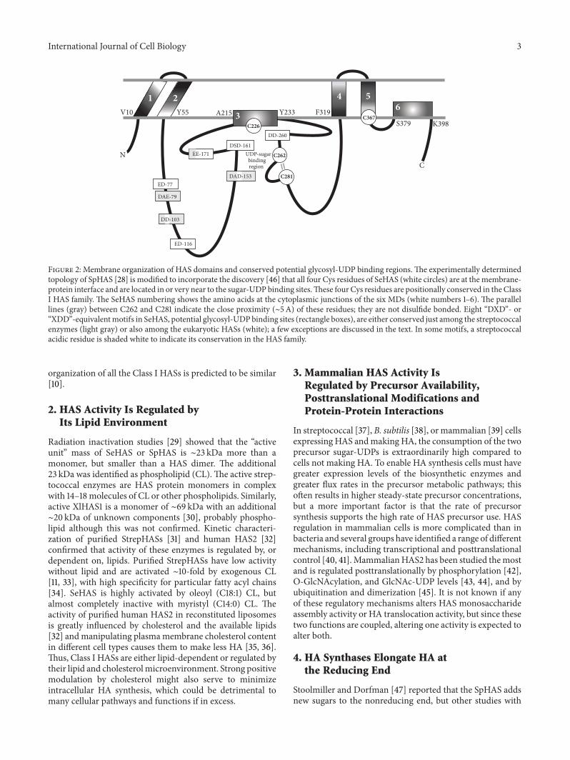
SpHAS, the only Class I HAS whose topology has beendetermined experimentally [28], has the N- and C-terminusand majority of the SpHAS protein inside the cell (Figure 2).StrepHASs have sixmembrane domains (MDs), four ofwhichpass through the membrane giving two small loops of theprotein exposed to the extracellular side.The other twoMDs,one within the large central catalytic domain and one inthe C-terminal one-third of the protein, interact with themembrane as amphipathic helices or reentrant loops but donot appear to span the membrane.The presence of two MDsin HAS that are amphipathic and do not cross the membraneis intriguing because these might be particularly well suitedfor the formation of an intraprotein pore. Vertebrate HASscontain an additional C-terminal region of ∼130–160 aa withtwo trans-MDs. The amino ∼75% of the larger eukaryoticHAS familymembers is homologous to SpHASwith the samepredicted domain organization, so the overall topological
51
3
62
451
36
2
4Membrane
Outside
UD
P-
7
UDP
UD
P-
HA-GlcNAc-UDP chainHA-GlcUA-UDP chain
-UDP -UDP
7
= GlcNAc= GlcUA
Figure 1: Schematic model of HAS showing the functions neededfor HA chain growth at the reducing end and transfer to the cellsurface. HAS uses multiple discrete functions (numbers 1–7) toassemble each HA disaccharide (red squares are GlcNAc and greencircles are GlcUA). The same HAS protein is indicated in twodifferent situations, at sequential times, as it alternately adds HA-GlcUA-UDP to a new GlcNAc-UDP, using functions 1, 3, and 5(left), and then adds HA-GlcNAc-UDP to a new GlcUA-UDP, usingfunctions 2, 4, and 6 (right). In this example (variant 1; Table 1)the sugar-UDPs are sequentially added in a continuous alternatingmanner and each cohort of needed functions cycles between beingactive (larger black numbers) and inactive (smaller gray numbers)within the active site domains (gray ovals). The functions requiredto add GlcNAc-UDP to HA-GlcUA-UDP are (left): 1, GlcNAc-UDP acceptor binding; 3, HA-GlcUA-UDP donor binding; 5, HA-GlcUA-UDP: GlcNAc-UDP, !1,3(HA-GlcUA-) transferase; and 7,HA translocation through the membrane. The functions requiredto add GlcUA-UDP to HA-GlcNAc-UDP are (right): 2, GlcUA-UDP acceptor binding; 4, HA-GlcNAc-UDP donor binding; 6, HA-GlcNAc-UDP: GlcUA-UDP, !1,4(HA-GlcNAc-) transferase; and 7,HA translocation.
Table 1:Three variations of the Pendulum hypothesis.
Variant Disaccharide assembly Glycosyl-UDP sites1 Sequential Four independent sites2 Simultaneous Three independent sites3 Alternating Two or three dependent sitesThe mechanism for adding sugars to the reducing end of HA could entailpolymerization of a disaccharide unit by either a sequential (i.e., one sugarat a time) or a concerted (i.e., simultaneous) mechanism. For the sequentialassembly of a disaccharide unit (variant 1), the enzyme would need twoglycosyl-UDP binding sites for addition of each sugar, one for a HA-UDPand one for a sugar-UDP. Since there are two types of HA-UDP species,the enzyme would need four glycosyl-UDP binding sites to assemble eachdisaccharide unit. For disaccharide assembly at the reducing end in aconcerted way (variant 2), HAS would require three glycosyl-UDP bindingsites, one each for GlcNAc-UDP, GlcUA-UDP, and a specific HA-UDP donorchain (i.e., HA-GlcUA-UDP or HA-GlcNAc-UDP), depending on which ofthe two possible HA disaccharide units was assembled. Another variation isthat HAS contains only one donor and one acceptor glycosyl-UDP bindingsite, whose specificities alternate as the two sugars are assembled one at atime (variant 3). If there is a single donor binding site, its specificity wouldalternately recognize HA-GlcUA-UDP and HA-GlcNAc-UDP. There couldbe two separate sugar-UDP sites, but if there is a single acceptor binding site,its specificity would also alternate in a reciprocal fashion with the HA-UDPsite to bind GlcUA-UDP or GlcNAc-UDP.
International Journal of Cell Biology 3
NC
1 2 4 56
DSD-161
A215 Y233 F319Y55V10K398S379
UDP-sugarbindingregion
C262
C281DAD-153
ED-116
DD-260
C3673C226
DAE-79
ED-77
EE-171
DD-103
Figure 2: Membrane organization of HAS domains and conserved potential glycosyl-UDP binding regions.The experimentally determinedtopology of SpHAS [28] is modified to incorporate the discovery [46] that all four Cys residues of SeHAS (white circles) are at the membrane-protein interface and are located in or very near to the sugar-UDP binding sites.These four Cys residues are positionally conserved in the ClassI HAS family. The SeHAS numbering shows the amino acids at the cytoplasmic junctions of the six MDs (white numbers 1–6).The parallellines (gray) between C262 and C281 indicate the close proximity (∼5A) of these residues; they are not disulfide bonded. Eight “DXD”- or“XDD”-equivalentmotifs in SeHAS, potential glycosyl-UDPbinding sites (rectangle boxes), are either conserved just among the streptococcalenzymes (light gray) or also among the eukaryotic HASs (white); a few exceptions are discussed in the text. In some motifs, a streptococcalacidic residue is shaded white to indicate its conservation in the HAS family.
organization of all the Class I HASs is predicted to be similar[10].
2. HAS Activity Is Regulated byIts Lipid Environment
Radiation inactivation studies [29] showed that the “activeunit” mass of SeHAS or SpHAS is ∼23 kDa more than amonomer, but smaller than a HAS dimer. The additional23 kDa was identified as phospholipid (CL).The active strep-tococcal enzymes are HAS protein monomers in complexwith 14–18molecules of CL or other phospholipids. Similarly,active XlHAS1 is a monomer of ∼69 kDa with an additional∼20 kDa of unknown components [30], probably phospho-lipid although this was not confirmed. Kinetic characteri-zation of purified StrepHASs [31] and human HAS2 [32]confirmed that activity of these enzymes is regulated by, ordependent on, lipids. Purified StrepHASs have low activitywithout lipid and are activated ∼10-fold by exogenous CL[11, 33], with high specificity for particular fatty acyl chains[34]. SeHAS is highly activated by oleoyl (C18:1) CL, butalmost completely inactive with myristyl (C14:0) CL. Theactivity of purified human HAS2 in reconstituted liposomesis greatly influenced by cholesterol and the available lipids[32] andmanipulating plasmamembrane cholesterol contentin different cell types causes them to make less HA [35, 36].Thus, Class I HASs are either lipid-dependent or regulated bytheir lipid and cholesterolmicroenvironment. Strong positivemodulation by cholesterol might also serve to minimizeintracellular HA synthesis, which could be detrimental tomany cellular pathways and functions if in excess.
3. Mammalian HAS Activity IsRegulated by Precursor Availability,Posttranslational Modifications andProtein-Protein Interactions
In streptococcal [37], B. subtilis [38], or mammalian [39] cellsexpressing HAS andmaking HA, the consumption of the twoprecursor sugar-UDPs is extraordinarily high compared tocells not making HA. To enable HA synthesis cells must havegreater expression levels of the biosynthetic enzymes andgreater flux rates in the precursor metabolic pathways; thisoften results in higher steady-state precursor concentrations,but a more important factor is that the rate of precursorsynthesis supports the high rate of HAS precursor use. HASregulation in mammalian cells is more complicated than inbacteria and several groups have identified a range of differentmechanisms, including transcriptional and posttranslationalcontrol [40, 41].MammalianHAS2 has been studied themostand is regulated posttranslationally by phosphorylation [42],O-GlcNAcylation, and GlcNAc-UDP levels [43, 44], and byubiquitination and dimerization [45]. It is not known if anyof these regulatory mechanisms alters HAS monosaccharideassembly activity or HA translocation activity, but since thesetwo functions are coupled, altering one activity is expected toalter both.
4. HA Synthases Elongate HA atthe Reducing End
Stoolmiller and Dorfman [47] reported that the SpHAS addsnew sugars to the nonreducing end, but other studies with
4 International Journal of Cell Biology
membranes from streptococci [24] or eukaryotic cells [23, 26]show that HA synthesis occurs at the reducing end. PurifiedSeHAS and SpHAS [27] or SpHAS in crude membranes [25]also add sugar-UDP units at the reducing end. The mech-anism for polysaccharide biosynthesis is different if chaingrowth is from the reducing or nonreducing end. When asugar is added from a sugar-nucleotide (making it the donor)to the nonreducing end of a polysaccharide (the acceptor),the nucleotide (e.g., UDP) is released. However, for reducingend elongation, the growing polymer chain is always attachedto UDP. Reaction (1) shows the reaction for HA disaccharideassembly (D = disaccharide units). During HA synthesis, theUDP released at each transfer step comes from the HA-UDPintermediate formed by addition of the previous sugar. Ineach cycle of monosaccharide addition, the released UDP isderived from the last monosaccharide added:(HAD)-UDP + GlcUA-%&'!→ UDP + (HAD)-GlcUA-%&'↓ GlcNAc-UDP%&' + (HAD)-GlcUA-GlcNAc-UDP
(1)
The donor HA-UDP transfers a hyaluronyl- (HA-) chain tothe new sugar-UDP (acceptor) without cleavage of the latterhigh-energy linkage to UDP; the UDP released is from theHA-UDPdonor.This situation is analogous to that for proteinand fatty acid synthesis [48].
The IUBMB nomenclature for HAS glycosyltransferaseactivities (EC 2.4.1.212) is different compared to that for typ-ical glycosyltransferases (Figure 1). An enzyme that utilizesGlcNAc-UDP to add to the nonreducing end of GlcUAwouldcreate a GlcNAc(!1,4)GlcUA linkage, whereas the HAStransferase activity adding GlcNAc-UDP at the reducing endcreates the GlcUA(!1,3)GlcNAc linkage. Systematic namingof a transferase activity specifies the donor: acceptor, grouptransferred. Thus, addition of a GlcUA residue to a GlcNAcat the reducing end of the growing HA chain is catalyzed byan activity that adds a hyaluronyl chain from HA-GlcNAc-UDP to GlcUA-UDP. This is a HA-GlcNAc()1→)UDP:GlcUA()1→)UDP, !(1,4) hyaluronyltransferase. Similarly,an activity adding GlcNAc-UDP to a HA-GlcUA-UDP chainis a HA-GlcUA()1→)UDP: GlcNAc()1→)UDP, !(1,3) hya-luronyltransferase.
5. HA Translocation to the Cell Exterior IsMediated by the HAS Protein Itself
The active sites of HAS and the sugar-UDP substrates areinside cells [28], so how do the large HA products (e.g.,>40,000 sugars long; >8MDa) reach the surface or extra-cellular space? Only the exogenous HAS protein (gene) andsufficientGlcNAc-UDP andGlcUA-UDP are required forHAbiosynthesis and secretion by heterologous cells that cannotnormally make HA, including E. faecalis [3], B. subtilis [38]and D. melanogaster [49]. Based on these findings and thelipid dependence and the topology of HAS, we proposed thatHAS must have the ability to translocate the growing HA
chain across the cell membrane into the extracellular space[11]. An alternative proposal was that an ABC transportsystem is required for the appearance of extracellular HA [50,51], as for many bacterial polysaccharides [52]; HAS wouldsynthesize intracellular HA that, while still being assembled,would be exported by a nearby membrane-bound ABCtransport system.
It seemed unlikely that an ABC transport system wasinvolved inHA translocation for several reasons: (i) It is unex-pected that ABC polysaccharide transporters in E. faecalis, B.subtilis, and fruit flies would have such low specificity for theirnormal substrate that they would effectively transport HA.(ii) Since multiple MDs are not needed for just HA synthesis(e.g., the Class II P. multocida HAS contains one membraneanchor and, unlike Class IHASs, can be expressed as an activesoluble truncated protein [53]), the topological organizationofHAS enzymes, containing 6–8membrane domains, ismoreconsistent with a translocation function [54]. (iii) HAS activ-ity is lipid-dependent or modulated by its lipid environment,consistent with an inherently intimate organization withinthe membrane bilayer, as expected for an HA translocationfunction, but not the independent ABC transport model. (iv)The sugar-UDP binding sites of Strep and mammalian HASsare at the inner membrane surface [46], which better fits amodel inwhich theHA-UDPchain is extendednear orwithinthe membrane and translocated through the enzyme to theexterior (Figure 2). (v) Class I HASs are processive enzymes,meaning they do not release their HA-UDP chains duringsynthesis; dissociation of HA from HAS does not occur [5,55]. This characteristic strongly supports a Pore model. HA-UDP that is bound by weak HA-HAS interactions wouldbe released, moved, and elongated continuously within theenzyme, while still being retained by the topological con-straint of being within a pore. In the ABC model, HA-HASinteractions are reversible, as for the nonprocessive PmHAS,which dissociates from and then rebindsHA after every sugaraddition [53].
The strong biochemical logic supporting a Pore Translo-cation Model was confirmed by multiple studies showingthat an ABC transporter Model for HA translocation is notcorrect.Thomas andBrown [56] found thatABC transportersare not involved in HA translocation by breast cancer cellsand Medina et al. [57] showed that purified SeHAS mediatesluminal dye efflux when added to liposomes, demonstratingthe presence of an intraprotein pore. Hubbard et al. [58]found that SeHAS, incorporated into liposomes, delivers HAdirectly to the internal lumen, demonstrating that HAS pos-sesses the predicted HA translocation function.
Misra et al. [59] showed that ABC transporter MDR1expression is regulated by changes in the pericellularHA coat.Coordinately regulated expression of ABC transporters andHAS provides an alternative interpretation of studies impli-cating a role for transporters inHA transfer. Two independentcellular protective mechanisms (provided by pericellular HAcoats and ABC multidrug transporters) may have coevolvedin vertebrates to be coordinately regulated in a complexman-ner in response to environmental cues; this could explainwhyHAS and extracellularHA levels are lower in cells treatedwithinhibitors of multidrug transporter function [50]. Another
International Journal of Cell Biology 5
explanation for inhibition of HA translocation by ABCtransporter inhibitors is that these inhibitors alter uridineuptake or salvage pathways and change uridine nucleotidepools, which inhibits HAS (e.g., controls were not preformedto verify that substrate sugar-UDP levels did not decrease orthat the potent HAS inhibitor UDP did not increase).
Finally, the finding that a bacterial cellulose synthasecreates an intraprotein pore, in which the product celluloseis synthesized and translocated [60], confirms the principlewe proposed in 1999 [11] that glycosyltransferases such as HAand cellulose synthases can mediate both polysaccharidesynthesis and translocation.
6. HAS Synthesizes Chitin andChitosyl-UDP Oligosaccharides
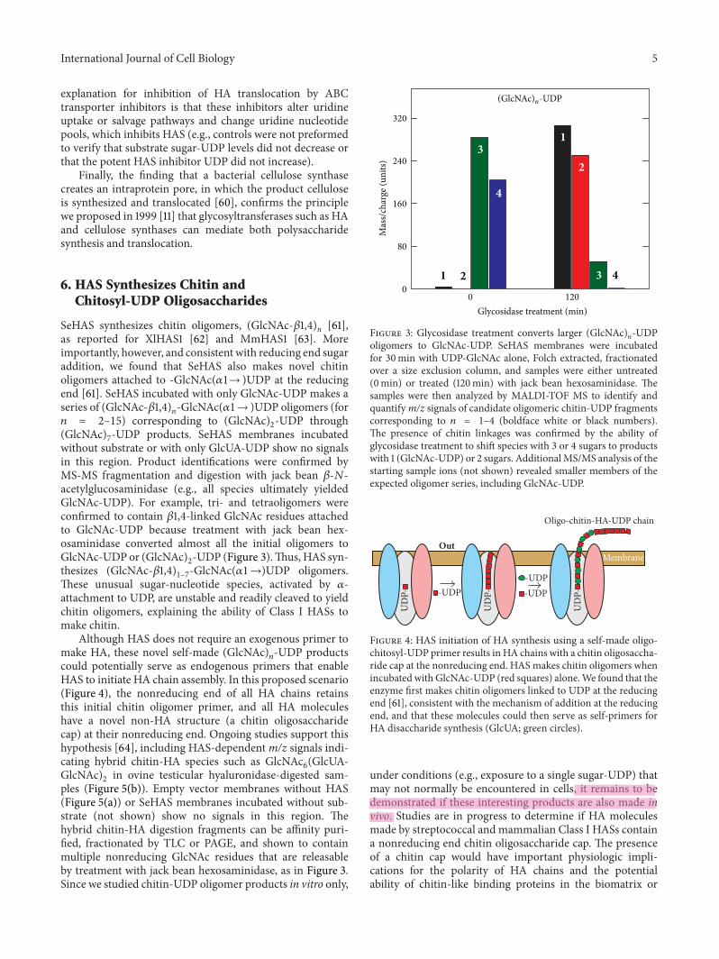
SeHAS synthesizes chitin oligomers, (GlcNAc-!1,4)n [61],as reported for XlHAS1 [62] and MmHAS1 [63]. Moreimportantly, however, and consistent with reducing end sugaraddition, we found that SeHAS also makes novel chitinoligomers attached to -GlcNAc()1→ )UDP at the reducingend [61]. SeHAS incubated with only GlcNAc-UDP makes aseries of (GlcNAc-!1,4)n-GlcNAc()1→ )UDP oligomers (for+ = 2–15) corresponding to (GlcNAc)2-UDP through(GlcNAc)7-UDP products. SeHAS membranes incubatedwithout substrate or with only GlcUA-UDP show no signalsin this region. Product identifications were confirmed byMS-MS fragmentation and digestion with jack bean !-,-acetylglucosaminidase (e.g., all species ultimately yieldedGlcNAc-UDP). For example, tri- and tetraoligomers wereconfirmed to contain !1,4-linked GlcNAc residues attachedto GlcNAc-UDP because treatment with jack bean hex-osaminidase converted almost all the initial oligomers toGlcNAc-UDP or (GlcNAc)2-UDP (Figure 3).Thus, HAS syn-thesizes (GlcNAc-!1,4)1–7-GlcNAc()1→)UDP oligomers.These unusual sugar-nucleotide species, activated by )-attachment to UDP, are unstable and readily cleaved to yieldchitin oligomers, explaining the ability of Class I HASs tomake chitin.
Although HAS does not require an exogenous primer tomake HA, these novel self-made (GlcNAc)!-UDP productscould potentially serve as endogenous primers that enableHAS to initiate HA chain assembly. In this proposed scenario(Figure 4), the nonreducing end of all HA chains retainsthis initial chitin oligomer primer, and all HA moleculeshave a novel non-HA structure (a chitin oligosaccharidecap) at their nonreducing end. Ongoing studies support thishypothesis [64], including HAS-dependentm/z signals indi-cating hybrid chitin-HA species such as GlcNAc6(GlcUA-GlcNAc)2 in ovine testicular hyaluronidase-digested sam-ples (Figure 5(b)). Empty vector membranes without HAS(Figure 5(a)) or SeHAS membranes incubated without sub-strate (not shown) show no signals in this region. Thehybrid chitin-HA digestion fragments can be affinity puri-fied, fractionated by TLC or PAGE, and shown to containmultiple nonreducing GlcNAc residues that are releasableby treatment with jack bean hexosaminidase, as in Figure 3.Since we studied chitin-UDP oligomer products in vitro only,
Mas
s/cha
rge (
units
)
320
240
160
80
0
Glycosidase treatment (min)
1
1
2
23
3
4
4
0 120
(GlcNAc)n-UDP
Figure 3: Glycosidase treatment converts larger (GlcNAc)!-UDPoligomers to GlcNAc-UDP. SeHAS membranes were incubatedfor 30min with UDP-GlcNAc alone, Folch extracted, fractionatedover a size exclusion column, and samples were either untreated(0min) or treated (120min) with jack bean hexosaminidase. Thesamples were then analyzed by MALDI-TOF MS to identify andquantifym/z signals of candidate oligomeric chitin-UDP fragmentscorresponding to + = 1–4 (boldface white or black numbers).The presence of chitin linkages was confirmed by the ability ofglycosidase treatment to shift species with 3 or 4 sugars to productswith 1 (GlcNAc-UDP) or 2 sugars. AdditionalMS/MS analysis of thestarting sample ions (not shown) revealed smaller members of theexpected oligomer series, including GlcNAc-UDP.
MembraneOut
Oligo-chitin-HA-UDP chain
UD
P-
UD
P- -UDP
UD
P- -UDP-UDP
Figure 4: HAS initiation of HA synthesis using a self-made oligo-chitosyl-UDP primer results inHA chains with a chitin oligosaccha-ride cap at the nonreducing end. HAS makes chitin oligomers whenincubated with GlcNAc-UDP (red squares) alone.We found that theenzyme first makes chitin oligomers linked to UDP at the reducingend [61], consistent with the mechanism of addition at the reducingend, and that these molecules could then serve as self-primers forHA disaccharide synthesis (GlcUA; green circles).
under conditions (e.g., exposure to a single sugar-UDP) thatmay not normally be encountered in cells, it remains to bedemonstrated if these interesting products are also made invivo. Studies are in progress to determine if HA moleculesmade by streptococcal and mammalian Class I HASs containa nonreducing end chitin oligosaccharide cap. The presenceof a chitin cap would have important physiologic impli-cations for the polarity of HA chains and the potentialability of chitin-like binding proteins in the biomatrix or
jesko
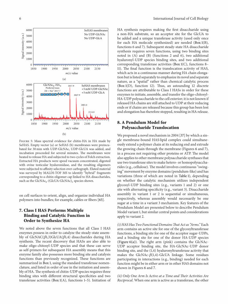
6 International Journal of Cell BiologySi
gnal
inte
nsity
(a.u
.)
6000
5000
4000
3000
2000
1000
01850 1900 1950 2000 2050 2100 2150
m/z value
SeHAS membranesNo UDP-GlcNAcNo UDP-GlcA
(a)
Sign
al in
tens
ity (a
.u.)
6000
5000
4000
3000
2000
1000
01850 1900 1950 2000 2050 2100 2150
m/z value
SeHAS membranes+5mM UDP-GlcNAc+5mM UDP-GlcA
GlcNAc6-HA4Predicted mass
[M + Na]+2017.7
2017.9
2047.9
2081.0
2114.0
(b)
Figure 5: Mass spectral evidence for chitin-HA in HA made bySeHAS. Empty vector (a) or SeHAS (b) membranes were preincu-bated for 30min with UDP-GlcNAc, UDP-GlcUA was added, andincubation proceeded for several minutes. The membranes wereheated to release HA and subjected to two cycles of Folch extraction.Extracted HA products were speed vacuum concentrated, digestedwith ovine testicular hyaluronidase, and the resulting oligomerswere subjected to affinity selection over carbograph. Eluted materialwas surveyed by MALDI-TOF MS to identify “hybrid” fragmentscorresponding to a chitin oligomer cap linked to HA disaccharides,such as the GlcNAc6-(GlcUA-GlcNAc)2 species shown.on cell surfaces to orient, align, and organize individual HApolymers into bundles; for example, cables or fibers [65].
7. Class I HAS Performs MultipleBinding and Catalytic Function inOrder to Synthesize HA
We noted above the seven functions that all Class I HASenzymes possess in order to catalyze the steady-state assem-bly of GlcNAC(!1,3)GlcUA(!1,4) disaccharides during HAsynthesis. The recent discovery that HASs are also able tomake oligo-chitosyl-UDP species and that these can serveas self-primers for subsequent HA assembly means that thisenzyme family also possesses more binding site and catalysisfunctions than previously recognized. These functions aresummarized in Box 1, using the standard transferase nomen-clature, and listed in order of use in the initiation and assem-bly of HA.The synthesis of chitin-UDP species requires threebinding sites with different structural specificities and twotransferase activities (Box 1(A), functions 1–5). Initiation of
HA synthesis requires making the first disaccharide usinga non-HA substrate, so an acceptor site for the GlcUA tobe added and a unique transferase activity (used only oncefor each HA molecule synthesized) are needed (Box 1(B),functions 6 and 7). Subsequent steady-state HA disaccharidesynthesis requires seven functions, using two binding sitesnoted in (A) and (B) (functions 2 and 6), two additionalhyaluronyl-UDP species binding sites, and two additionalcorresponding transferase activities (Box 1(C), functions 8–11). The final function is the translocation activity of HAS,which acts in a continuous manner during HA chain elonga-tion but is listed separately to emphasize its novel and separatenature, as a “spatial” rather than chemical catalytic process(Box 1(D), function 12). Thus, an astounding 12 discretefunctions are attributable to Class I HASs in order for theseenzymes to initiate, assemble, and transfer the oligo-chitosyl-HA-UDPpolysaccharide to the cell exterior; it is not known ifreleased HA chains are still attached to UDP at their reducingends or if chains are released because this group has been lostand elongation has therefore stopped, resulting inHA release.
8. A Pendulum Model forPolysaccharide Translocation
We proposed a novel mechanism in 2004 [37] by which a sin-gle membrane-bound HAS⋅lipid complex could simultane-ously extend a polymer chain at its reducing end and extrudethe growing chain through the membrane (Figures 6 and 7),in a process not requiring other proteins or ATP. The modelalso applies to othermembrane polysaccharide synthases thatuse two transferase sites tomake hetero- or homopolysaccha-rides (e.g., cellulose).The model involves continuous “swing-ing”movement by enzyme domains (pendulum-like) and hasvariations (three of which are noted in Table 1), dependingon whether the catalytic mechanism utilizes independentglycosyl-UDP binding sites (e.g., variants 1 and 2) or onesite with alternating specificity (e.g., variant 3). Disaccharideassembly in variant 1 or 2 is sequential or simultaneous,respectively, whereas assembly would necessarily be onesugar at a time in a variant 3mechanism. Key features of thePendulum Model are presented below to describe PendulumModel variant 1, but similar central points and considerationsapply to variant 2.
(i)HASHasTwoFunctionalDomainsThatAct as “Arms.” Eacharm contains an active site for one of the glycosyltransferasefunctions, a binding site for one of the acceptor sugar-UDPs,and a binding site for one of the donor HA-UDP species(Figure 6(a)). The right arm (pink) contains the GlcNAc-UDP acceptor binding site, the HA-GlcNAc-UDP donorbinding site, and the (1,4) hyaluronyltransferase activity thatmakes the GlcNAc-!(1,4)-GlcUA linkage. Some residuesparticipating in interactions (e.g., binding) needed for eachfunctionmight be in either arm or in other HAS domains notshown in Figures 6 and 7.
(ii) Only One Arm Is Active at a Time andTheir Activities AreReciprocal. When one arm is active as a transferase, the other
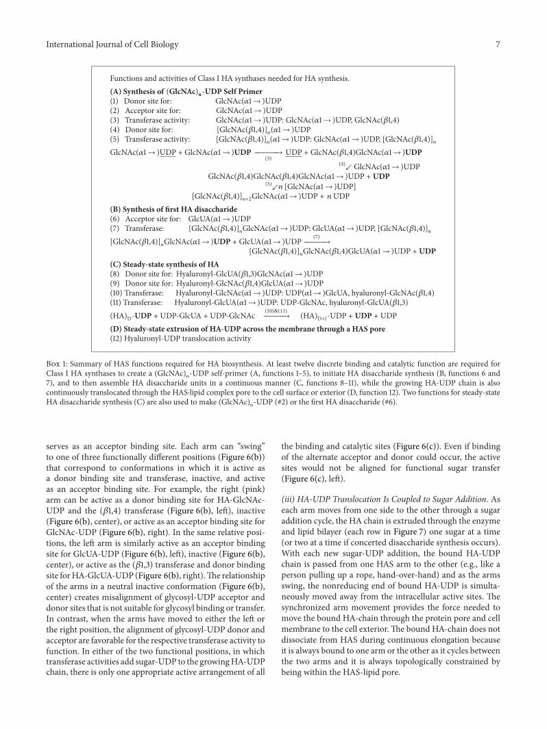
International Journal of Cell Biology 7
Functions and activities of Class I HA synthases needed for HA synthesis.(A) Synthesis of (GlcNAc)n-UDP Self Primer(1) Donor site for: GlcNAc()1→ )UDP(2) Acceptor site for: GlcNAc()1→ )UDP(3) Transferase activity: GlcNAc()1→ )UDP: GlcNAc()1→ )UDP, GlcNAc(!1,4)(4) Donor site for: [GlcNAc(!1,4)]!()1→ )UDP(5) Transferase activity: [GlcNAc(!1,4)]!()1→ )UDP: GlcNAc()1→ )UDP, [GlcNAc(!1,4)]!GlcNAc()1→ )UDP + GlcNAc()1→ )UDP .......→(3) UDP + GlcNAc(!1,4)GlcNAc()1→ )UDP(5)↙ GlcNAc()1→ )UDP
GlcNAc(!1,4)GlcNAc(!1,4)GlcNAc()1→ )UDP + UDP(5)↙+ [GlcNAc()1→ )UDP][GlcNAc(!1,4)]!+2GlcNAc()1→ )UDP + + UDP
(B) Synthesis of first HA disaccharide(6) Acceptor site for: GlcUA()1→ )UDP(7) Transferase: [GlcNAc(!1,4)]!GlcNAc()1→ )UDP: GlcUA()1→ )UDP, [GlcNAc(!1,4)]![GlcNAc(!1,4)]!GlcNAc()1→ )UDP + GlcUA()1→ )UDP (7).......→
[GlcNAc(!1,4)]!GlcNAc(!1,4)GlcUA()1→ )UDP + UDP(C) Steady-state synthesis of HA(8) Donor site for: Hyaluronyl-GlcUA(!1,3)GlcNAc()1→ )UDP(9) Donor site for: Hyaluronyl-GlcNAc(!1,4)GlcUA()1→ )UDP(10) Transferase: Hyaluronyl-GlcNAc()1→ )UDP: UDP()1→ )GlcUA, hyaluronyl-GlcNAc(!1,4)(11) Transferase: Hyaluronyl-GlcUA()1→ )UDP: UDP-GlcNAc, hyaluronyl-GlcUA(!1,3)(HA)D-UDP + UDP-GlcUA + UDP-GlcNAc (10)&(11)......→ (HA)D+1-UDP + UDP + UDP(D) Steady-state extrusion of HA-UDP across the membrane through a HAS pore(12) Hyaluronyl-UDP translocation activity
Box 1: Summary of HAS functions required for HA biosynthesis. At least twelve discrete binding and catalytic function are required forClass I HA synthases to create a (GlcNAc)!-UDP self-primer (A, functions 1–5), to initiate HA disaccharide synthesis (B, functions 6 and7), and to then assemble HA disaccharide units in a continuous manner (C, functions 8–11), while the growing HA-UDP chain is alsocontinuously translocated through the HAS⋅lipid complex pore to the cell surface or exterior (D, function 12). Two functions for steady-stateHA disaccharide synthesis (C) are also used to make (GlcNAc)!-UDP (#2) or the first HA disaccharide (#6).
serves as an acceptor binding site. Each arm can “swing”to one of three functionally different positions (Figure 6(b))that correspond to conformations in which it is active asa donor binding site and transferase, inactive, and activeas an acceptor binding site. For example, the right (pink)arm can be active as a donor binding site for HA-GlcNAc-UDP and the (!1,4) transferase (Figure 6(b), left), inactive(Figure 6(b), center), or active as an acceptor binding site forGlcNAc-UDP (Figure 6(b), right). In the same relative posi-tions, the left arm is similarly active as an acceptor bindingsite for GlcUA-UDP (Figure 6(b), left), inactive (Figure 6(b),center), or active as the (!1,3) transferase and donor bindingsite forHA-GlcUA-UDP (Figure 6(b), right).The relationshipof the arms in a neutral inactive conformation (Figure 6(b),center) creates misalignment of glycosyl-UDP acceptor anddonor sites that is not suitable for glycosyl binding or transfer.In contrast, when the arms have moved to either the left orthe right position, the alignment of glycosyl-UDP donor andacceptor are favorable for the respective transferase activity tofunction. In either of the two functional positions, in whichtransferase activities add sugar-UDP to the growingHA-UDPchain, there is only one appropriate active arrangement of all
the binding and catalytic sites (Figure 6(c)). Even if bindingof the alternate acceptor and donor could occur, the activesites would not be aligned for functional sugar transfer(Figure 6(c), left).
(iii) HA-UDP Translocation Is Coupled to Sugar Addition. Aseach arm moves from one side to the other through a sugaraddition cycle, the HA chain is extruded through the enzymeand lipid bilayer (each row in Figure 7) one sugar at a time(or two at a time if concerted disaccharide synthesis occurs).With each new sugar-UDP addition, the bound HA-UDPchain is passed from one HAS arm to the other (e.g., like aperson pulling up a rope, hand-over-hand) and as the armsswing, the nonreducing end of bound HA-UDP is simulta-neously moved away from the intracellular active sites. Thesynchronized arm movement provides the force needed tomove the bound HA-chain through the protein pore and cellmembrane to the cell exterior.The bound HA-chain does notdissociate from HAS during continuous elongation becauseit is always bound to one arm or the other as it cycles betweenthe two arms and it is always topologically constrained bybeing within the HAS-lipid pore.
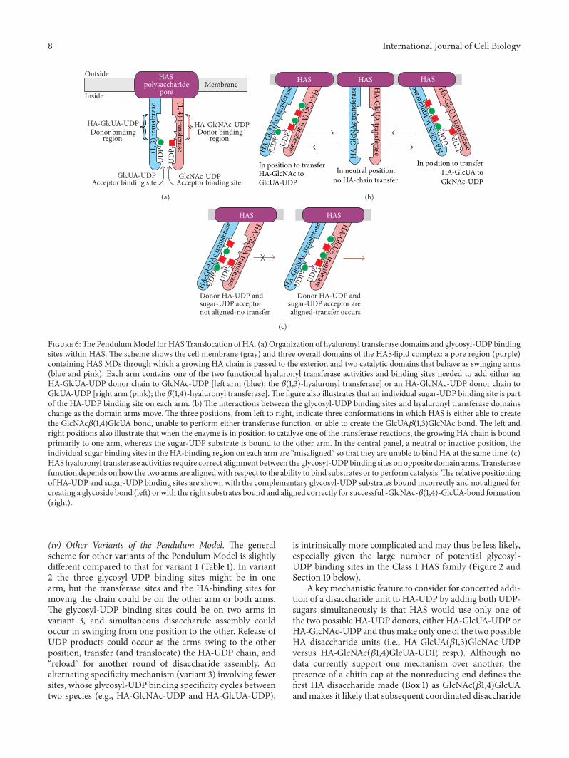
8 International Journal of Cell Biology
UD
P-U
DP-
GlcUA-UDPAcceptor binding site Acceptor binding site
GlcNAc-UDP
HA-GlcUA-UDPDonor binding
regionDonor binding
region
HA-GlcNAc-UDP
HASpolysaccharide
poreMembrane
Outside
Inside (1,4)transferase(1,3)t
rans
fera
se
(a)
In position to transferHA-GlcNAc toGlcUA-UDP
In position to transferHA-GlcUA toGlcNAc-UDP
In neutral position:no HA-chain transfer
HASHAS HAS
UDP-UD
P-
UDP-
UDP-
HA-G
lcNAc
tran
sfera
se HA-GlcUA transferase
HA-
GlcN
Ac tr
ansfe
rase
HA-G
lcUA transferase HA-G
lcNAc
tran
sfera
se HA-GlcUA transferase
(b)
HASHAS
Donor HA-UDP andsugar-UDP acceptor arealigned-transfer occurs
UDP-
UDP-
UDP-
UDP-
Donor HA-UDP andsugar-UDP acceptornot aligned-no transfer
HA-G
lcNAc
tran
sfera
se
HA-G
lcNAc
tran
sfera
seHA-GlcUA transferase
HA-GlcUA transferase
(c)
Figure 6:The PendulumModel for HAS Translocation of HA. (a) Organization of hyaluronyl transferase domains and glycosyl-UDP bindingsites within HAS. The scheme shows the cell membrane (gray) and three overall domains of the HAS⋅lipid complex: a pore region (purple)containing HAS MDs through which a growing HA chain is passed to the exterior, and two catalytic domains that behave as swinging arms(blue and pink). Each arm contains one of the two functional hyaluronyl transferase activities and binding sites needed to add either anHA-GlcUA-UDP donor chain to GlcNAc-UDP [left arm (blue); the !(1,3)-hyaluronyl transferase] or an HA-GlcNAc-UDP donor chain toGlcUA-UDP [right arm (pink); the !(1,4)-hyaluronyl transferase].The figure also illustrates that an individual sugar-UDP binding site is partof the HA-UDP binding site on each arm. (b)The interactions between the glycosyl-UDP binding sites and hyaluronyl transferase domainschange as the domain arms move.The three positions, from left to right, indicate three conformations in which HAS is either able to createthe GlcNAc!(1,4)GlcUA bond, unable to perform either transferase function, or able to create the GlcUA!(1,3)GlcNAc bond. The left andright positions also illustrate that when the enzyme is in position to catalyze one of the transferase reactions, the growing HA chain is boundprimarily to one arm, whereas the sugar-UDP substrate is bound to the other arm. In the central panel, a neutral or inactive position, theindividual sugar binding sites in the HA-binding region on each arm are “misaligned” so that they are unable to bind HA at the same time. (c)HAShyaluronyl transferase activities require correct alignment between the glycosyl-UDPbinding sites on opposite domain arms. Transferasefunction depends on how the two arms are alignedwith respect to the ability to bind substrates or to perform catalysis.The relative positioningof HA-UDP and sugar-UDP binding sites are shown with the complementary glycosyl-UDP substrates bound incorrectly and not aligned forcreating a glycoside bond (left) or with the right substrates bound and aligned correctly for successful -GlcNAc-!(1,4)-GlcUA-bond formation(right).
(iv) Other Variants of the Pendulum Model. The generalscheme for other variants of the Pendulum Model is slightlydifferent compared to that for variant 1 (Table 1). In variant2 the three glycosyl-UDP binding sites might be in onearm, but the transferase sites and the HA-binding sites formoving the chain could be on the other arm or both arms.The glycosyl-UDP binding sites could be on two arms invariant 3, and simultaneous disaccharide assembly couldoccur in swinging from one position to the other. Release ofUDP products could occur as the arms swing to the otherposition, transfer (and translocate) the HA-UDP chain, and“reload” for another round of disaccharide assembly. Analternating specificity mechanism (variant 3) involving fewersites, whose glycosyl-UDP binding specificity cycles betweentwo species (e.g., HA-GlcNAc-UDP and HA-GlcUA-UDP),
is intrinsically more complicated and may thus be less likely,especially given the large number of potential glycosyl-UDP binding sites in the Class I HAS family (Figure 2 andSection 10 below).
A keymechanistic feature to consider for concerted addi-tion of a disaccharide unit to HA-UDP by adding both UDP-sugars simultaneously is that HAS would use only one ofthe two possible HA-UDP donors, either HA-GlcUA-UDP orHA-GlcNAc-UDP and thusmake only one of the two possibleHA disaccharide units (i.e., HA-GlcUA(!1,3)GlcNAc-UDPversus HA-GlcNAc(!1,4)GlcUA-UDP, resp.). Although nodata currently support one mechanism over another, thepresence of a chitin cap at the nonreducing end defines thefirst HA disaccharide made (Box 1) as GlcNAc(!1,4)GlcUAand makes it likely that subsequent coordinated disaccharide
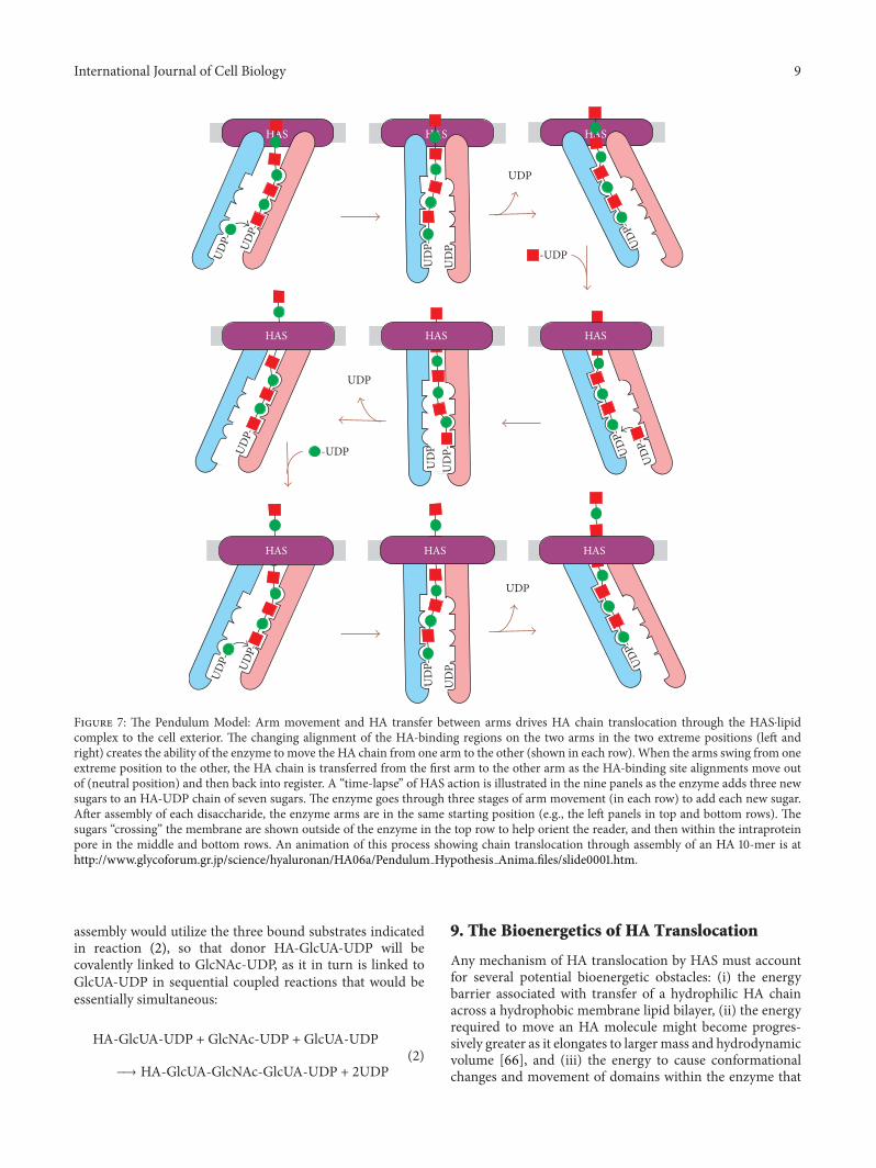
International Journal of Cell Biology 9
HAS HAS HAS
UDP-
UD
PU
DP-
UD
PU
DP-
UD
PU
DP-
UDP
UDP- UD
P--UDP
-UDPUDP-
UDP-
UDP-
UDP
UDP- UD
P-
UDP
UDP-
HAS HAS HAS
HAS HAS HAS
Figure 7: The Pendulum Model: Arm movement and HA transfer between arms drives HA chain translocation through the HAS⋅lipidcomplex to the cell exterior. The changing alignment of the HA-binding regions on the two arms in the two extreme positions (left andright) creates the ability of the enzyme to move the HA chain from one arm to the other (shown in each row).When the arms swing from oneextreme position to the other, the HA chain is transferred from the first arm to the other arm as the HA-binding site alignments move outof (neutral position) and then back into register. A “time-lapse” of HAS action is illustrated in the nine panels as the enzyme adds three newsugars to an HA-UDP chain of seven sugars.The enzyme goes through three stages of arm movement (in each row) to add each new sugar.After assembly of each disaccharide, the enzyme arms are in the same starting position (e.g., the left panels in top and bottom rows). Thesugars “crossing” the membrane are shown outside of the enzyme in the top row to help orient the reader, and then within the intraproteinpore in the middle and bottom rows. An animation of this process showing chain translocation through assembly of an HA 10-mer is athttp://www.glycoforum.gr.jp/science/hyaluronan/HA06a/Pendulum Hypothesis Anima.files/slide0001.htm.
assembly would utilize the three bound substrates indicatedin reaction (2), so that donor HA-GlcUA-UDP will becovalently linked to GlcNAc-UDP, as it in turn is linked toGlcUA-UDP in sequential coupled reactions that would beessentially simultaneous:
HA-GlcUA-UDP + GlcNAc-UDP + GlcUA-UDP.→ HA-GlcUA-GlcNAc-GlcUA-UDP + 2UDP (2)
9. The Bioenergetics of HA Translocation
Any mechanism of HA translocation by HAS must accountfor several potential bioenergetic obstacles: (i) the energybarrier associated with transfer of a hydrophilic HA chainacross a hydrophobic membrane lipid bilayer, (ii) the energyrequired to move an HA molecule might become progres-sively greater as it elongates to largermass and hydrodynamicvolume [66], and (iii) the energy to cause conformationalchanges and movement of domains within the enzyme that
10 International Journal of Cell Biology
could be physically responsible for rapid chain movementacross the membrane without releasing the chain. For per-spective, an 8MDa HA chain proportionally enlarged tohuman scale would be equivalent to a 1mm wide thread>30m long.
(i) How Does HAS Overcome the Hydrophobic MembraneBarrier? Membrane transporters, such as lac permease with12MDs [67], mediate sugar transfer through an intraproteinpore created by interactions amongmanyMDs. HASs appearto compensate for not having enough MDs to form such anintraprotein pore by their interactions with phospholipids[11, 32]. HAS⋅lipid interactions could allow the enzyme tocreate a larger pore-like passage within the enzyme, throughwhich the growingHA chain could pass [11, 57, 68]. HAS⋅lipidcomplexes would present exterior hydrophobic interactionswith fatty acyl groups in the bilayer, while engagingHAon theinterior pore surface with hydrophilic interactions; thus, anHA chain moving through a HAS⋅lipid pore could effectivelybypass the hydrophobic membrane barrier.
(ii) How Could HAS Move HA Chains with Masses That GetProgressively Greater? Intuitively, one might expect that theenergy needed to overcome the inertia of, and move, a longHA-UDP chain (e.g., 4MDa)would bemuch greater than theenergy needed tomove a shortHAoligosaccharyl-UDP chain(e.g., 4 kDa). However, two factors may make this apparent“molecular weight-lifting” less difficult than it appears.
(a) HA Segmentation.The segmented, semi-independentmovement of discrete portions (roughly 100 sugarslong) of a large HA chain [69–71] could create anupper limit for the “apparent” mass of HA thatHAS would need to “move” to translocate a discretesection. If segments of ∼100 sugars behave semi-independently for brief periods (e.g., msec), duringwhich translocation is favored, then the apparentmass of HA against which HAS needs to generate atranslocation force would only be∼20 kDa. As a chainwas initiated and elongated, HAS might initiallytranslocate it rapidly and then progressively moreslowly until the segmental length was approached,and a steady-state translocation rate was thenreached. The observed kinetic profile for how HAmass increases during synthesis begins very rapidlyand then becomes slower [55].
(b) Increased Release Forces. The net forces generated byBrownianmotionmay tend to pull theHA chain awayfrom the enzyme and thus facilitate the translocationaction of HAS. A possible chain release mechanism,in fact, could be the balance between the Brownianand other forces acting to release the HA and the HA-binding forces mediated by sites within the enzymeacting to retain the HA [66]. In contrast, cells witha pericellular HA coat might effectively stabilize andstop HA synthesis, because the presence of nearbyHAS-HA complexes could augment and increase theforces favoring HA retention. As HA chain density
increased at the cell surface, the network of interact-ing chains would diminish the effects of Brownian-generated forces acting to release the HA, thusresulting in a stabilized gel-like cell surface coat stillanchored to multiple HAS molecules. In the extremeof this scenario, HA synthesis would slow or stop asforces generated by the HA coat network increased,finally providing too much resistance for HAS totranslocate HA.
(iii) What Are the Energy Sources for HA Translocation? Atleast three possible energy sources could contribute to anHA translocation mechanism (Figure 8), as estimated below:glycosyl-UDP hydrolysis [4–8 kJ/mol], H-bond formation[12–24 kJ/mol], and ion-pair reactions and the potentialenergy of electrochemical gradients [4–8 kJ/mol]. If theseenergy sources contribute to the HA translocation mecha-nism, a conservative estimate of the total free energy changefor HA translocation is ∼30 kJ/mol or ∼7 kcal/mol (1 kcal =4.18 kJ) of HA disaccharide units moved across the mem-brane. This is equivalent to 1 ATP and would be a favorablebioenergetic situation, explaining how an ATP-independenttranslocation process is feasible.
(a) UDP-Sugar Hydrolysis. The free energy for Glc-UDPhydrolysis is 7.3–7.6 kcal/mol and the differencebetween Glc-UDP hydrolysis and glucoside bond for-mation is ∼0.6–1.0 kcal/mol [72]. Values for hydroly-sis of GlcNAc-UDP and GlcUA-UDP are not avail-able but are presumably somewhat greater, due todestabilizing repulsive and steric factors. Free energydifferences between sugar-UDP hydrolysis and glyco-side bond formation for GlcNAc andGlcUA are likelysimilar to Glc, ∼0.6–1.0 kcal/mol. Thus, the available“excess” free energy to perform additional workbeyond creating two glycoside bonds could be ∼1-2 kcal/mol (∼4–8 kJ/mol) of HA disaccharide units(Figure 8, #1).
(b) Electrochemical Gradients or External Ion ReactionsMay Provide Additional Energy. HA translocation tothe cell exterior must necessarily be associated withone ormore electrochemical gradients that could pro-vide additional energy for the translocation process.HAS might recognize only one of ≥4 possible UDP-GlcUA species as the correct substrate during cataly-sis, depending on the state of the carboxyl group.Thecarboxyl group could be dissociated and free (i.e., ananion) or neutral and bound to either H+, Na+, or K+(Mg+2 is also a possibility). In most of these cases, afavorable energetic situationwould occurwhen newlyadded GlcUA is transferred to the exterior: proto-nated carboxyl groupswould readily dissociate releas-ing H+, negatively charged carboxyl groups wouldbind to extracellular cations (e.g., Na+), and boundK+ ions would exchange with Na+ ions, which arein excess. Such ion-pair reactions would yield energy(that is difficult to estimate) and tend to increase(pull) the equilibrium for translocation, in a mannersimilar to removal of a reaction product.
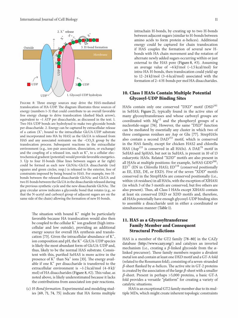
International Journal of Cell Biology 11
Outside 3. H-bond formation
1. Glycosyl-UDP hydrolysis
-UDP -UDP
2. X+ release
X+ X + X
+
MembraneU
DP-
UDP
X
X
X
X
X
X
Figure 8: Three energy sources may drive the HAS-mediatedtranslocation of HA-UDP. The diagram illustrates three sources ofenergy (numbers 1–3) that could contribute to an overall favorablefree energy change to drive translocation (dashed black arrow),equivalent to ∼1 ATP per disaccharide, as discussed in the text. 1.Two HA-UDP bonds are hydrolyzed to make two glycoside bondsper disaccharide. 2. Energy can be captured by extracellular releaseof a cation (X+; bound to the intracellular GlcUA-UDP substrateand incorporated into HA by HAS) as the GlcUA is released fromHAS and any associated restraints on the –CO2X group by thetranslocation process. Subsequent reactions in the extracellularenvironment (e.g., ion-pair association, dissociation, or exchange)and the coupling of a released ion, such as K+, to a cellular elec-trochemical gradient (potential) would provide favorable energetics.3. Up to four H-bonds (blue lines between sugars at far right)could be formed as each new GlcNAc-GlcUA disaccharide (redsquares and green circles, resp.) is released to the exterior, free ofconstraints imposed by being bound to HAS. For example, two H-bonds between the released disaccharide GlcNAc and GlcUA andtwoH-bonds between theGlcUA in the disaccharide released duringthe previous synthetic cycle and the new disaccharide GlcNAc.Thegray circular arrow indicates a glycosidic bond that rotates (e.g., sothat the N-acetyl and carboxyl group of adjacent sugars are on thesame side of the chain) allowing the formation of new H-bonds.
The situation with bound K+ might be particularlyfavorable because HA translocation would also thenbe coupled to the cellular K+ ion gradient (high intra-cellular and low outside), providing an additionalenergy source for overall HA synthesis and translo-cation [73]. Given the intracellular abundance of K+,ion composition and pH, the K+-GlcUA-UDP speciesis likely the most abundant form of GlcUA-UDP and,thus, likely to be the normal HAS substrate. Consis-tent with this, purified SeHAS is more active in thepresence of K+ than Na+ ions [33]. The energy avail-able if one K+ per disaccharide is transferred to theextracellular environment is ∼1-2 kcal/mol (4–8 kJ/mol) of HA disaccharides (Figure 8, #2).This value, asnoted above, is likely underestimated because it lacksthe contributions from associated ion-pair reactions.
(c) H-Bond formation. Experimental and modeling stud-ies [69, 71, 74, 75] indicate that HA forms multiple
intrachain H-bonds, by creating up to two H-bondsbetween adjacent sugars (similar to H-bonds betweenamino acids to form protein )-helices). Additionalenergy could be captured for chain translocationif HAS couples the formation of several new H-bonds with HA chain movement and the rotation ofalternate newly added sugars occurring within or justexternal to the HAS pore (Figure 8, #3). Assumingan average value of ∼6 kJ/mol (∼1.5 kcal/mol) forintra-HA H-bonds, then translocation could yield upto 12–24 kJ/mol (3–6 kcal/mol) associated with theformation of 2–4H-bonds per mol HA disaccharides.
10. Class I HASs Contain Multiple PotentialGlycosyl-UDP Binding Sites
HASs contain only one conserved “DXD” motif (DSD161in SeHAS; Figure 2), typically found in the active sites ofmany glycosyltransferases and whose carboxyl groups arecoordinated with Mg+2 and the phosphoryl groups of anucleotide-sugar [76]. However, the same “DXD” functioncan be mediated by essentially any cluster in which two ofthree contiguous residues are Asp or Glu [77]. StrepHASsalso contain a second DAD153 motif, which is conservedin the HAS family, except for chicken HAS2 and chlorellaHAS (Asp153 is conserved in all HASs). A DAE79 motif inSeHAS and SpHAS, but not in SuHAS, is present in 10 of 13eukaryotic HASs. Related “XDD” motifs are also present inall HASs at multiple positions: for example, SeHAS GDD260;ED77 (EN in Chlorella HAS); ED116 (conserved positionallyas EE, EXE, DE, or EXD). Five of the seven “XDD” motifsconserved in the StrepHASs are conserved positionally (i.e.,within≤6 residues) in all HASs, with the exception of XlHAS1(in which 3 of the 5 motifs are conserved, but five others arealso present). Thus, all Class I HASs except XlHAS1 containat least six conserved DXD or XDD motifs and, therefore,all HASs potentially have enough glycosyl-UDP binding sitesto assemble a disaccharide unit in either a coordinated orsimultaneous manner (Table 1).
11. HAS as a GlycosyltransferaseFamily Member and ConsequentStructural Predictions
HAS is a member of the GT2 family [78–80] in the CAZydatabase (http://www.cazy.org/) and catalyzes an invertedmechanism (i.e., creating a !-linked glycoside from the )-linked precursor). These family members require a divalentmetal ion and contain at least oneDXDmotif and aGT-A fold(related to theRossmann fold), consisting of a seven-stranded!-sheet flanked by )-helices.The active site in GT-2 proteinsis created by the association of the large!-sheet with a smaller!-sheet. Present in perhaps >5,000 proteins, a basic GT-Afold provides a versatile “platform” for creating a variety ofcatalytic situations.
HAS is an exceptional GT2 familymember due to itsmul-tiple MDs, which might create inherent topologic constraints
12 International Journal of Cell Biology
in regions of homology to the GT-A fold (making modelingessentially impossible). Based on extensive sequence sim-ilarity through a ∼260 aa region between MD2 and MD4(Figure 2), the HAS catalytic domain might adopt an overallfold similar to otherGT2 familymembers, including cellulosesynthase [81]. Similarities between HA and cellulose syn-thases include N-terminal nucleotide binding motifs, a puta-tive catalytic base, a QxxRWmotif associated with processiv-ity, and that both proteins create an intraprotein pore for theirproducts [58, 60].The GT2 family model predicts that the N-terminal subdomain binds the sugar-UDP donor and the C-terminal subdomain binds the acceptor. For enzymes such asHAS that add to the reducing end the normal designation ofdonor and acceptor binding sites may be switched. Thus, inHAS an N-terminal subdomain binding site for sugar-UDPcould be an acceptor site. Alternatively, if this was conservedas a donor site, then it would accommodate an HA-UDPsubstrate.The ultimate answers to many of themolecular andmechanistic questions posed here will likely only be revealedwhen the structure of HAS is ultimately determined atsufficient atomic resolution.
12. Conclusions
Since the first HAS gene was cloned in 1993, the field hasprogressed and answered important questions about themolecular basis ofHA synthesis. In particular, there is a grow-ing consensus that membrane-bound Class I HAS enzymesare lipid-dependent and active as both protein monomers orHAS multimers associated with specific lipids (and choles-terol) and that the Strep and mammalian HASs assembleHA at the reducing end. It is now resolved that HAS usesintracellular precursors and couples HA synthesis with HAtranslocation to the cell surface or biomatrix. Elucidation ofthis multifunctional process will surely reveal more surprisesand unexpected intricacies about the complicated orchestra-tion of molecular processes employed by a single enzymeprotein to assemble and simultaneously translocate one of thelargest single biomolecules made in the animal or microbialkingdoms. Future structural studies will reveal whether thegeneral principles of the PendulumModel are able to explainthe mechanism of how HAS couples HA synthesis withtranslocation. Understanding how these enzymes work tosynthesize and translocate HA will likely provide opportu-nities to identify therapeutics and to better understand howHASs are involved in normal human health and in tumorige-nesis, metastasis, and inflammatory diseases such as arthritis.
Abbreviations
CL: CardiolipinGlcNAc-UDP: The nucleotide-sugar with the reducing
end at the rightGlcUA-UDP: The nucleotide-sugar with the reducing
end at the rightHA: Hyaluronic acid, hyaluronate, and
hyaluronanHA-: A hyaluronyl group
HA-UDP: A hyaluronyl group with )-linked UDP at thereducing end
HAS: HA synthaseMD: Membrane domainSeHAS: Streptococcus equisimilisHASSpHAS: Streptococcus pyogenesHASStrepHAS: Streptococcal HA synthasesSuHAS: Streptococcus uberisHASXlHAS: Xenopus laevis.
Conflict of Interests
The author declares that there is no conflict of interestsregarding the publication of this paper.
Acknowledgments
Research from the author’s laboratory was supported bythe National Institutes of Health Grant GM35978 from theInstitute of General Medical Sciences.The author also thanksDr. Christopher West for very helpful discussions and input.
References
[1] A.Markovitz, J. A. Cifonelli, andA.Dorfman, “The biosynthesisof hyaluronic acid by group A Streptococcus. VI. Biosynthesisfrom uridine nucleotides in cell-free extracts,” The Journal ofBiological Chemistry, vol. 234, pp. 2343–2350, 1959.
[2] P. L. DeAngelis, J. Papaconstantinou, and P. H. Weigel, “Molec-ular cloning, identification, and sequence of the hyaluronansynthase gene from group a Streptococcus pyogenes,”The Journalof Biological Chemistry, vol. 268, no. 26, pp. 19181–19184, 1993.
[3] P. L. DeAngelis, J. Papaconstantinou, and P. H. Weigel, “Isola-tion of a Streptococcus pyogenes gene locus that directs hyaluro-nan biosynthesis in acapsular mutants and in heterologousbacteria,” Journal of Biological Chemistry, vol. 268, no. 20, pp.14568–14571, 1993.
[4] B. A. Dougherty and I. van de Rijn, “Molecular characterizationof hasA from an operon required for hyaluronic acid synthesisin group A streptococci,” Journal of Biological Chemistry, vol.269, no. 1, pp. 169–175, 1994.
[5] P. L. DeAngelis and P. H. Weigel, “Immunochemical confir-mation of the primary structure of streptococcal hyaluronansynthase and synthesis of high molecular weight product by therecombinant enzyme,” Biochemistry, vol. 33, no. 31, pp. 9033–9039, 1994.
[6] K. Kumari and P. H. Weigel, “Molecular cloning, expression,and characterization of the authentic hyaluronan synthase fromgroup C Streptococcus equisimilis,” The Journal of BiologicalChemistry, vol. 272, no. 51, pp. 32539–32546, 1997.
[7] P. N. Ward, T. R. Field, W. G. F. Ditcham, E. Maguin, and J.A. Leigh, “Identification and disruption of two discrete lociencoding hyaluronic acid capsule biosynthesis genes hasA,hasB, and hasC in Streptococcus uberis,” Infection and Immunity,vol. 69, no. 1, pp. 392–399, 2001.
[8] P. L. DeAngelis, “Hyaluronan synthases: fascinating glycosyl-transferases from vertebrates, bacterial pathogens, and algalviruses,” Cellular and Molecular Life Sciences, vol. 56, no. 7-8,pp. 670–682, 1999.
[9] A. P. Spicer and J. A. McDonald, “Characterization and molec-ular evolution of a vertebrate hyaluronan synthase gene family,”
International Journal of Cell Biology 13
Journal of Biological Chemistry, vol. 273, no. 4, pp. 1923–1932,1998.
[10] P. H. Weigel and P. L. DeAngelis, “Hyaluronan synthases: adecade-plus of novel glycosyltransferases,” Journal of BiologicalChemistry, vol. 282, no. 51, pp. 36777–36781, 2007.
[11] V. L. Tlapak-Simmons, B. A. Baggenstoss, T. Clyne, and P. H.Weigel, “Purification and lipid dependence of the recombinanthyaluronan synthases from Streptococcus pyogenes and Strep-tococcus equisimilis,” The Journal of Biological Chemistry, vol.274, no. 7, pp. 4239–4245, 1999.
[12] J. Y. L. Tien and A. P. Spicer, “Three vertebrate hyaluronansynthases are expressed during mouse development in distinctspatial and temporal patterns,” Developmental Dynamics, vol.233, no. 1, pp. 130–141, 2005.
[13] K. Torronen, K. Nikunen, R. Karna, M. Tammi, R. Tammi,and K. Rilla, “Tissue distribution and subcellular localizationof hyaluronan synthase isoenzymes,” Histochemistry and CellBiology, vol. 141, no. 1, pp. 17–31, 2014.
[14] C. B. Knudson andW. Knudson, “Hyaluronan-binding proteinsin development, tissue homeostasis, and disease,” FASEB Jour-nal, vol. 7, no. 13, pp. 1233–1241, 1993.
[15] J. R. E. Fraser, T. C. Laurent, and U. B. G. Laurent, “Hyaluronan:its nature, distribution, functions and turnover,” Journal ofInternal Medicine, vol. 242, no. 1, pp. 27–33, 1997.
[16] B. A. Fenderson, I. Stamenkovic, and A. Aruffo, “Localizationof hyaluronan in mouse embryos during implantation, gastru-lation and organogenesis,”Differentiation, vol. 54, no. 2, pp. 85–98, 1993.
[17] B. P. Toole, “Hyaluronan in morphogenesis,” Journal of InternalMedicine, vol. 242, no. 1, pp. 35–40, 1997.
[18] E. A. Turley, P. Bowman, and M. A. Kytryk, “Effects of hyalu-ronate and hyaluronate binding proteins on cell motile andcontact behaviour,” Journal of Cell Science, vol. 78, pp. 133–145,1985.
[19] E. A. Balazs, “Analgesic effect of elastoviscous hyaluronansolutions and the treatment of arthritic pain,” Cells TissuesOrgans, vol. 174, no. 1-2, pp. 49–62, 2003.
[20] E. A. Balazs and J. L. Denlinger, “Viscosupplementation: a newconcept in the treatment of osteoarthritis,” Journal of Rheuma-tology, vol. 20, no. 39, pp. 3–9, 1993.
[21] K. L. Goa and P. Benfield, “Hyaluronic acid—a review of itspharmacology and use as a surgical aid in ophthalmology, andits therapeutic potential in joint disease and wound healing,”Drugs, vol. 47, no. 3, pp. 536–566, 1994.
[22] N. Itano and K. Kimata, “Mammalian hyaluronan synthases,”IUBMB Life, vol. 54, no. 4, pp. 195–199, 2002.
[23] P. Prehm, “Biosynthesis of hyaluronan: direction of chainelongation,” Biochemical Journal, vol. 398, no. 3, pp. 469–473,2006.
[24] P. Prehm, “Synthesis of hyaluronate in differentiated teratocar-cinoma cells.Mechanism of chain growth,”Biochemical Journal,vol. 211, no. 1, pp. 191–198, 1983.
[25] S. Bodevin-Authelet, M. Kusche-Gullberg, P. E. Pummill, P. L.DeAngelis, and U. Lindahl, “Biosynthesis of hyaluronan: direc-tion of chain elongation,” The Journal of Biological Chemistry,vol. 280, no. 10, pp. 8813–8818, 2005.
[26] T. Asplund, J. Brinck, M. Suzuki, M. J. Briskin, and P. Heldin,“Characterization of hyaluronan synthase from a human gliomacell line,” Biochimica et Biophysica Acta—General Subjects, vol.1380, no. 3, pp. 377–388, 1998.
[27] V. L. Tlapak-Simmons, C. A. Baron, R. Gotschall, D. Haque,W. M. Canfield, and P. H. Weigel, “Hyaluronan biosynthesis byclass I streptococcal hyaluronan synthases occurs at the reduc-ing end,”The Journal of Biological Chemistry, vol. 280, no. 13, pp.13012–13018, 2005.
[28] C. Heldermon, P. L. DeAngelis, and P. H. Weigel, “Topologicalorganization of the hyaluronan synthase from Streptococcuspyogenes,” The Journal of Biological Chemistry, vol. 276, no. 3,pp. 2037–2046, 2001.
[29] V. L. Tlapak-Simmons, E. S. Kempner, B. A. Baggenstoss, andP. H. Weigel, “The active streptococcal hyaluronan synthases(HASS) contain a single HAS monomer and multiple cardi-olipin molecules,”The Journal of Biological Chemistry, vol. 273,no. 40, pp. 26100–26109, 1998.
[30] P. E. Pummill, E. S. Kempner, and P. L. DeAngelis, “Functionalmolecular mass of a vertebrate hyaluronan synthase as deter-mined by radiation inactivation analysis,” Journal of BiologicalChemistry, vol. 276, no. 43, pp. 39832–39835, 2001.
[31] V. L. Tlapak-Simmons, B. A. Baggenstoss, K. Kumari, C.Heldermon, and P. H. Weigel, “Kinetic characterization of therecombinant hyaluronan synthases from Streptococcus pyogenesand Streptococcus equisimilis,” Journal of Biological Chemistry,vol. 274, no. 7, pp. 4246–4253, 1999.
[32] P. Ontong, Y. Hatada, S. Taniguchi, I. Kakizaki, and N. Itano,“Effect of a cholesterol-rich lipid environment on the enzymaticactivity of reconstituted hyaluronan synthase,” Biochemical andBiophysical Research Communications, vol. 443, no. 2, pp. 666–671, 2014.
[33] V. L. Tlapak-Simmons, C. A. Baron, and P. H. Weigel, “Char-acterization of the purified hyaluronan synthase from Strepto-coccus equisimilis,” Biochemistry, vol. 43, no. 28, pp. 9234–9242,2004.
[34] P. H.Weigel, Z. Kyossev, and L. C. Torres, “Phospholipid depen-dence and liposome reconstitution of purified hyaluronansynthase,” Journal of Biological Chemistry, vol. 281, no. 48, pp.36542–36551, 2006.
[35] S. W. Sakr, S. Potter-Perigo, M. G. Kinsella et al., “Hyaluronanaccumulation is elevated in cultures of low density lipoproteinreceptor-deficient cells and is altered by manipulation of cellcholesterol content,” The Journal of Biological Chemistry, vol.283, no. 52, pp. 36195–36204, 2008.
[36] A. Kultti, R. Karna, K. Rilla et al., “Methyl-beta-cyclodextrinsuppresses hyaluronan synthesis by down-regulation of hyalu-ronan synthase 2 through inhibition of Akt,” The Journal ofBiological Chemistry, vol. 285, no. 30, pp. 22901–22910, 2010.
[37] P. H. Weigel, “The bacterial hyaluronan synthases—an update,”in Science of Hyaluronan Today, V. C. Hascall and M. Yanag-ishita, Eds., 2004.
[38] B. Widner, R. Behr, S. von Dollen et al., “Hyaluronic acid pro-duction in Bacillus subtilis,” Applied and Environmental Micro-biology, vol. 71, no. 7, pp. 3747–3752, 2005.
[39] M. Viola, D. Vigetti, A. Genasetti et al., “Molecular control ofthe hyaluronan biosynthesis,” Connective Tissue Research, vol.49, no. 3-4, pp. 111–114, 2008.
[40] R. H. Tammi, A. G. Passi, K. Rilla et al., “Transcriptional andpost-translational regulation of hyaluronan synthesis,” FEBSJournal, vol. 278, no. 9, pp. 1419–1428, 2011.
[41] D.Vigetti andA. Passi, “Hyaluronan synthases posttranslationalregulation in cancer,” in Hyaluronan Signaling and Turnover,vol. 123 of Advances in Cancer Research, chapter 4, pp. 95–119,Elsevier, New York, NY, USA, 2014.
14 International Journal of Cell Biology
[42] D. Vigetti, M. Clerici, S. Deleonibus et al., “Hyaluronan synthe-sis is inhibited by adenosine monophosphate-activated proteinkinase through the regulation of HAS2 activity in human aorticsmooth muscle cells,” The Journal of Biological Chemistry, vol.286, no. 10, pp. 7917–7924, 2011.
[43] V. C. Hascall, A. Wang, M. Tammi et al., “The dynamicmetabolism of hyaluronan regulates the cytosolic concentrationof UDP-GlcNAc,”Matrix Biology, vol. 35, pp. 14–17, 2014.
[44] T. A. Jokela, K. M. Makkonen, S. Oikari et al., “Cellular contentof UDP-N-acetylhexosamines controls hyaluronan synthase 2expression and correlates with O-linked N-acetylglucosaminemodification of transcription factors YY1 and SP1,”The Journalof Biological Chemistry, vol. 286, no. 38, pp. 33632–33640, 2011.
[45] E. Karousou, M. Kamiryo, S. S. Skandalis et al., “The activityof hyaluronan synthase 2 is regulated by dimerization andubiquitination,”The Journal of Biological Chemistry, vol. 285, no.31, pp. 23647–23654, 2010.
[46] K. Kumari and P. H. Weigel, “Identification of a membrane-localized cysteine cluster near the substrate-binding sites of theStreptococcus equisimilis hyaluronan synthase,” Glycobiology,vol. 15, no. 5, pp. 529–539, 2005.
[47] A. C. Stoolmiller and A. Dorfman, “The biosynthesis of hyalu-ronic acid by Streptococcus,” Journal of Biological Chemistry, vol.244, no. 2, pp. 236–246, 1969.
[48] P. W. Robbins, D. Bray, M. Dankert, and A. Wright, “Directionof chain growth in polysaccharide synthesis,” Science, vol. 158,no. 3808, pp. 1536–1542, 1967.
[49] S. Takeo, M. Fujise, T. Akiyama et al., “In vivo hyaluronan syn-thesis upon expression of the mammalian hyaluronan synthasegene in drosophila,” Journal of Biological Chemistry, vol. 279, no.18, pp. 18920–18925, 2004.
[50] P. Prehm and U. Schumacher, “Inhibition of hyaluronan exportfrom human fibroblasts by inhibitors of multidrug resistancetransporters,”Biochemical Pharmacology, vol. 68, no. 7, pp. 1401–1410, 2004.
[51] G. Ouskova, B. Spellerberg, and P. Prehm, “Hyaluronan releasefrom Streptococcus pyogenes: export by an ABC transporter,”Glycobiology, vol. 14, no. 10, pp. 931–938, 2004.
[52] M. J. Fath and R. Kolter, “ABC transporters: bacterial exporters,”Microbiological Reviews, vol. 57, no. 4, pp. 995–1017, 1993.
[53] W. Jing and P. L. de Angelis, “Dissection of the two transferaseactivities of the Pasteurella multocida hyaluronan synthase: twoactive sites exist in one polypeptide,”Glycobiology, vol. 10, no. 9,pp. 883–889, 2000.
[54] K. Kumari, B. A. Baggenstoss, A. L. Parker, and P. H. Weigel,“Mutation of two intramembrane polar residues conservedwithin the hyaluronan synthase family alters hyaluronan prod-uct size,”The Journal of Biological Chemistry, vol. 281, no. 17, pp.11755–11760, 2006.
[55] B. A. Baggenstoss and P. H. Weigel, “Size exclusion chromatog-raphy-multiangle laser light scattering analysis of hyaluronansize distributions made by membrane-bound hyaluronan syn-thase,” Analytical Biochemistry, vol. 352, no. 2, pp. 243–251,2006.
[56] N. K. Thomas and T. J. Brown, “ABC transporters do not con-tribute to extracellular translocation of hyaluronan in humanbreast cancer in vitro,” Experimental Cell Research, vol. 316, no.7, pp. 1241–1253, 2010.
[57] A. P. Medina, J. Lin, and P. H. Weigel, “Hyaluronan synthasemediates dye translocation across liposomal membranes,” BMCBiochemistry, vol. 13, no. 1, article 2, 2012.
[58] C. Hubbard, J. T. McNamara, C. Azumaya, M. S. Patel, and J.Zimmer, “The hyaluronan synthase catalyzes the synthesis andmembrane translocation of hyaluronan,” Journal of MolecularBiology, vol. 418, no. 1-2, pp. 21–31, 2012.
[59] S. Misra, S. Ghatak, and B. P. Toole, “Regulation of MDR1expression and drug resistance by a positive feedback loopinvolving hyaluronan, phosphoinositide 3-kinase, and ErbB2,”The Journal of Biological Chemistry, vol. 280, no. 21, pp. 20310–20315, 2005.
[60] J. L. W. Morgan, J. Strumillo, and J. Zimmer, “Crystallographicsnapshot of cellulose synthesis and membrane translocation,”Nature, vol. 493, no. 7431, pp. 181–186, 2013.
[61] P. H. Weigel, C. M. West, P. Zhao, L. Wells, B. A. Baggenstoss,and J. L. Washburn, “Hyaluronan synthase assembles chitinoligomers with -GlcNAc()1→ )UDP at the reducing end,”Glycobiology, vol. 25, no. 6, pp. 632–643, 2015.
[62] C. E. Semino and P. W. Robbins, “Synthesis of ‘Nod’-like chitinoligosaccharides by theXenopus developmental proteinDG42,”Proceedings of the National Academy of Sciences of the UnitedStates of America, vol. 92, no. 8, pp. 3498–3501, 1995.
[63] M. Yoshida, N. Itano, Y. Yamada, and K. Kimata, “In vitrosynthesis of hyaluronan by a single protein derived frommouseHAS1 gene and characterization of amino acid residues essentialfor the activity,”The Journal of Biological Chemistry, vol. 275, no.1, pp. 497–506, 2000.
[64] C. L. Feasley, B. A. Baggenstoss, and P. H. Weigel, “(GlcNAc)n-UDP oligomer synthesis by class I hyaluronan synthase andits role as a self priming initiator for hyaluronan synthesis,” inProceedings of the 9th International Conference on Hyaluronan(Hyaluronan ’13), p. 21, Oklahoma City, Okla, USA, June 2013,http://www.ishas.org/images/2013 Conference/ISHAS 2013Book of Abstracts.pdf.
[65] C. A. De laMotte, V. C. Hascall, J. Drazba, S. K. Bandyopadhyay,and S. A. Strong, “Mononuclear leukocytes bind to specifichyaluronan structures on colon mucosal smooth muscle cellstreated with polyinosinic acid: polycytidylic acid. Inter-)-trypsin inhibitor is crucial to structure and function,”AmericanJournal of Pathology, vol. 163, no. 1, pp. 121–133, 2003.
[66] P. H. Weigel and B. A. Baggenstoss, “Hyaluronan synthasepolymerizing activity and control of product size are discreteenzyme functions that can be uncoupled by mutagenesis ofconserved cysteines,”Glycobiology, vol. 22, no. 10, pp. 1302–1310,2012.
[67] J. Abramson, I. Smirnova, V. Kasho, G. Verner, H. R. Kaback,and S. Iwata, “Structure andmechanism of the lactose permeaseof Escherichia coli,” Science, vol. 301, no. 5633, pp. 610–615, 2003.
[68] H. Palsdottir and C. Hunte, “Lipids in membrane proteinstructures,” Biochimica et Biophysica Acta—Biomembranes, vol.1666, no. 1-2, pp. 2–18, 2004.
[69] J. E. Scott and F. Heatley, “Biological properties of hyaluronanin aqueous solution are controlled and sequestered by reversibletertiary structures, defined by NMR spectroscopy,” Biomacro-molecules, vol. 3, no. 3, pp. 547–553, 2002.
[70] D. A. Gabriel and M. E. Carr Jr., “Calcium destabilizes andcauses conformational changes in hyaluronic acid,”The Ameri-can Journal of theMedical Sciences, vol. 298, no. 1, pp. 8–14, 1989.
[71] P. Dais, E. Tylianakis, J. Kanetakis, and F. R. Taravel, “13Cnuclear magnetic relaxation study of segmental dynamics ofhyaluronan in aqueous solutions,”Biomacromolecules, vol. 6, no.3, pp. 1397–1404, 2005.
International Journal of Cell Biology 15
[72] G. Avigad, “Sucrose-uridine diphosphate glucosyltransferasefrom Jerusalem artichoke tubers,” The Journal of BiologicalChemistry, vol. 239, pp. 3613–3618, 1964.
[73] D. Hagenfeld, B. Borkenhagen, T. Schulz, H. Schillers, U. Schu-macher, and P. Prehm, “Hyaluronan export through plasmamembranes depends on concurrent K+ efflux by K"# channels,”PLoS ONE, vol. 7, no. 6, Article ID e39096, 2012.
[74] J. E. Scott and F. Heatley, “Hyaluronan forms specific stabletertiary structures in aqueous solution: A 13CNMR study,” Pro-ceedings of the National Academy of Sciences of the United Statesof America, vol. 96, no. 9, pp. 4850–4855, 1999.
[75] A. Almond, J. K. Sheehan, and A. Brass, “Molecular dynamicssimulations of the two disaccharides of hyaluronan in aqueoussolution,” Glycobiology, vol. 7, no. 5, pp. 597–604, 1997.
[76] C. Breton and A. Imberty, “Structure/function studies of gly-cosyltransferases,” Current Opinion in Structural Biology, vol. 9,no. 5, pp. 563–571, 1999.
[77] S. Gulberti, S. Fournel-Gigleux, G. Mulliert et al., “The func-tional glycosyltransferase signature sequence of the human !1,3-glucuronosyltransferase is a XDDmotif,” Journal of Biologi-cal Chemistry, vol. 278, no. 34, pp. 32219–32226, 2003.
[78] I. M. Saxena, R. M. Brown Jr., M. Fevre, R. A. Geremia, andB. Henrissat, “Multidomain architecture of !-glycosyl trans-ferases: implications for mechanism of action,” Journal of Bacte-riology, vol. 177, no. 6, pp. 1419–1424, 1995.
[79] J. Liu and A. Mushegian, “Three monophyletic superfamiliesaccount for the majority of the known glycosyltransferases,”Protein Science, vol. 12, no. 7, pp. 1418–1431, 2003.
[80] C. Breton, L. Snajdrova, C. Jeanneau, J. Koca, and A. Imberty,“Structures and mechanisms of glycosyltransferases,” Glycobi-ology, vol. 16, no. 2, pp. 29R–37R, 2006.
[81] I. M. Saxena, R. M. Brown Jr., and T. Dandekar, “Structure—function characterization of cellulose synthase: relationship toother glycosyltransferases,” Phytochemistry, vol. 57, no. 7, pp.1135–1148, 2001.

Submit your manuscripts athttp://www.hindawi.com
Hindawi Publishing Corporationhttp://www.hindawi.com Volume 2014
Anatomy Research International
PeptidesInternational Journal of
Hindawi Publishing Corporationhttp://www.hindawi.com Volume 201
Hindawi Publishing Corporation http://www.hindawi.com
International Journal of
Volume 201�
=RRORJ\
Hindawi Publishing Corporationhttp://www.hindawi.com Volume 2014
Molecular Biology International
GenomicsInternational Journal of
Hindawi Publishing Corporationhttp://www.hindawi.com Volume 2014
The Scientific World JournalHindawi Publishing Corporation http://www.hindawi.com Volume 2014
Hindawi Publishing Corporationhttp://www.hindawi.com Volume 2014
BioinformaticsAdvances in
Marine BiologyJournal of
Hindawi Publishing Corporationhttp://www.hindawi.com Volume 201
Hindawi Publishing Corporationhttp://www.hindawi.com Volume 2014
Signal TransductionJournal of
Hindawi Publishing Corporationhttp://www.hindawi.com Volume 2014
BioMed Research International
Evolutionary BiologyInternational Journal of
Hindawi Publishing Corporationhttp://www.hindawi.com Volume 201
Hindawi Publishing Corporationhttp://www.hindawi.com Volume 201
Biochemistry Research International
ArchaeaHindawi Publishing Corporationhttp://www.hindawi.com Volume 2014
Hindawi Publishing Corporationhttp://www.hindawi.com Volume 2014
Genetics Research International
Hindawi Publishing Corporationhttp://www.hindawi.com Volume
Advances in
Virolog y
Hindawi Publishing Corporationhttp://www.hindawi.com
Nucleic AcidsJournal of
Volume 201
Stem CellsInternational
Hindawi Publishing Corporationhttp://www.hindawi.com Volume 201
Hindawi Publishing Corporationhttp://www.hindawi.com Volume 201
Enzyme Research
Hindawi Publishing Corporationhttp://www.hindawi.com Volume 2014
International Journal of
Microbiology


![Noninvasive Transdermal Vaccination Using Hyaluronan ... · intradermal injection, [ 22 ] to deliver vaccine deep into the skin. Hyaluronan (HA), a natural macromolecule with intrinsically](https://static.fdocuments.net/doc/165x107/5f0f07747e708231d44222c7/noninvasive-transdermal-vaccination-using-hyaluronan-intradermal-injection.jpg)