Chapter 14 The Digestive System - Anatomy and...
82
Chapter 14 The Digestive System
Transcript of Chapter 14 The Digestive System - Anatomy and...

Chapter 14
The Digestive System

Foods are used as metabolic fuel (energy) ◦ Foods are oxidized and transformed into ATP
◦ ATP = Chemical energy the drives cellular activities
Energy value of food is measured in Calories (C)

Major nutrients ◦ carbohydrates, fats, proteins AND water
Minor nutrients – ◦ Vitamins and minerals

Vitamins - mostly found in fruits and vegetables ◦ Used as coenzymes ◦ Fat soluble: Vitamin A,D,E and K
◦ Water soluble: Vitamin B, C
Minerals - foods rich in minerals = vegetables legumes, milk and some meats ◦ Important for enzyme activity
◦ Iron (making hemoglobin), calcium (building bones, blood clotting)
Vitamin and mineral deficiencies can cause severe disorders in the body

Food must be processed to obtain macromolecules and nutrients
The 4 main mechanisms of food processing are: ◦ Ingestion: eating (taking in food)
◦ Digestion: physical and chemical breakdown of food into the nutrient molecules
◦ Absorption: movement of nutrients into the bloodstream
◦ Defecation: elimination of indigestible waste

Extracellular Digestion: ◦ In a gastrointestinal cavity in simple animals
Coiled, hollow tube
Intracellular Digestion: ◦ Occurs inside the cell, once the cell engulfs food
particles
◦ Is performed by the cell’s lysosomes

Alimentary canals are also called complete digestive tracts
“one way movement” of food: a specific opening for ingestion and another for elimination ◦ Makes for orderly progression of breakdown
◦ Allows for more than 1 meal to enter the tract a time

The organs and functions of that make up the human alimentary canal and accessory structures.

Is bounded externally by lips and cheeks ◦ Vestibule-the space between the lips, cheeks, & gums
The tongue-composed of skeletal muscle
◦ Papillae-rough projections on the tongue
Allow food handling
Contain sensory receptors (taste buds)
Lingual frenulum-fold of mucous membrane underside the tongue
Difficulty speaking if too short (“tongue-tied”)
Roof of the Mouth ◦ Anterior hard palate-maxilla and palatine bones
◦ Posterior soft palate-muscle and glandular tissue Uvula-finger-shaped projection


Teeth-Begin the mechanical digestion by chewing food – increasing surface area.
Salivary glands- produce saliva (begins carbohydrate breakdown)
Tonsils-immune function
The Oral Cavity (Mouth)

Parotid-anterior and inferior to the ears
Send saliva to the mouth via ducts
Sublingual-underneath the tongue
Submandibular-floor of the mouth, inside the surface of the lower jaw ◦ Ducts for both open
underneath the tongue

Functions: ◦ Mastication (chewing) of food
◦ Tongue mixes masticated food with saliva
◦ Tongue initiates swallowing
◦ Taste buds on the tongue allow for taste

Mucus, water, bicarbonate Functions 1. protects mouth while chewing 2. lubricates food for swallowing 3. helps prevent tooth decay 4. kills many bacteria before swallowing 5. Chemical digestion of carbohydrates by
Salivary amylase (starch maltose)

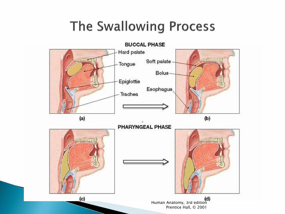
The connection between our oral cavity and the esophagus ◦ Nasopharynx, Oropharynx, Laryngopharynx
The windpipe (trachea) also is located anterior to the pharynx and the esophagus
A flap of cartilage called the epiglottis closes the opening of the trachea when we swallow, which prevents things from entering the lungs or choking.
Swallowing-2 phases (peristalsis) Voluntary
Reflex- once food is pushed into the oropharynx
The Pharynx – passageway for food and air

Nasopharynx – respiratory function
Oropharynx – respiratory and digestive function
common pathway for food and air
Laryngopharynx – respiratory and digestive function
Human Anatomy, 3rd edition Prentice Hall, © 2001

Human Anatomy, 3rd edition Prentice Hall, © 2001

Human Anatomy, 3rd edition Prentice Hall, © 2001
◦ 2 sets Deciduous (20) Ages 6 mo. - 2 ½ years
Permanent (32) Ages 6 years – 25 years (3rd molars)
◦ Held in sockets ◦ Gingiva surrounds the base of the teeth

Human Anatomy, 3rd edition Prentice Hall, © 2001


◦ Crown Part you see above the gum
◦ Root 1 – 3 projections embedded in socket
◦ Neck
Junction between crown and root
Human Anatomy, 3rd edition Prentice Hall, © 2001
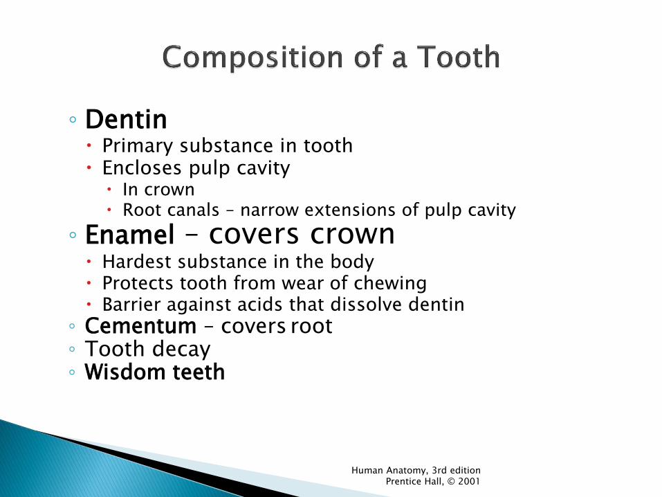
◦ Dentin Primary substance in tooth Encloses pulp cavity
In crown Root canals – narrow extensions of pulp cavity
◦ Enamel – covers crown Hardest substance in the body Protects tooth from wear of chewing Barrier against acids that dissolve dentin
◦ Cementum – covers root ◦ Tooth decay ◦ Wisdom teeth

Human Anatomy, 3rd edition Prentice Hall, © 2001

Anatomy: ~10 inches long (pharynx to stomach Stratified squamous epithelium Rhythmic muscular contractions, known as
peristalsis push the food in one direction…Down.
No digestion occurs here.
The Esophagus (food pipe)


Lower Esophageal Sphincter (gastroesophageal) constriction between the esophagus and the stomach ◦ Sphincters-muscles that encircle tubes
◦ Relaxation-allows bolus to enter the stomach
◦ Constriction-prevents acid backup from the stomach
Heartburn-acid reflux from the stomach
Vomiting-propelling of stomach contents by contraction of abdominal and stomach muscles

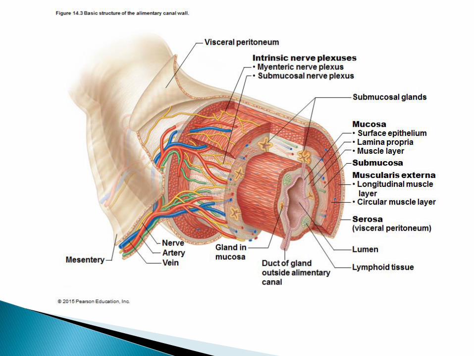
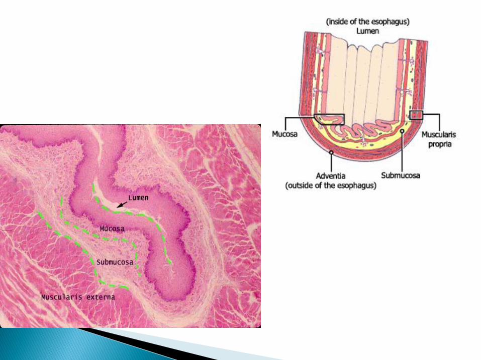
1. Mucosa (mucous membrane layer): A layer of epithelium Glandular cells (enzyme secretion) Goblet cells (mucous secretion)
2. Submucosa : Loose connective tissue with blood vessels, lymphatics, and
nerves beneath it Peyer’s patches-lymph nodules-immune function
3. Muscularis (smooth muscle layer): Inner circular and outer longitudinal layer Oblique-only in the stomach
4. Serosa (serous membrane layer): Very thin outermost layer of squamous epi Secretes serous fluid Adventitia- only in the esophagus, loose CT

Human Anatomy, 3rd edition Prentice Hall, © 2001

Parietal peritoneum: lines the abdominal wall
Visceral peritoneum: covers the organs ◦ Mesentary: double layer of visceral peritoneum in
between the organs
◦ Greater omentum: hangs down anteriorly
Fat cushion for insulation
Contains macrophages
Contains infections from spreading
◦ Lesser omnetum: between the stomach and the liver
Human Anatomy, 3rd edition Prentice Hall, © 2001

Human Anatomy, 3rd edition Prentice Hall, © 2001

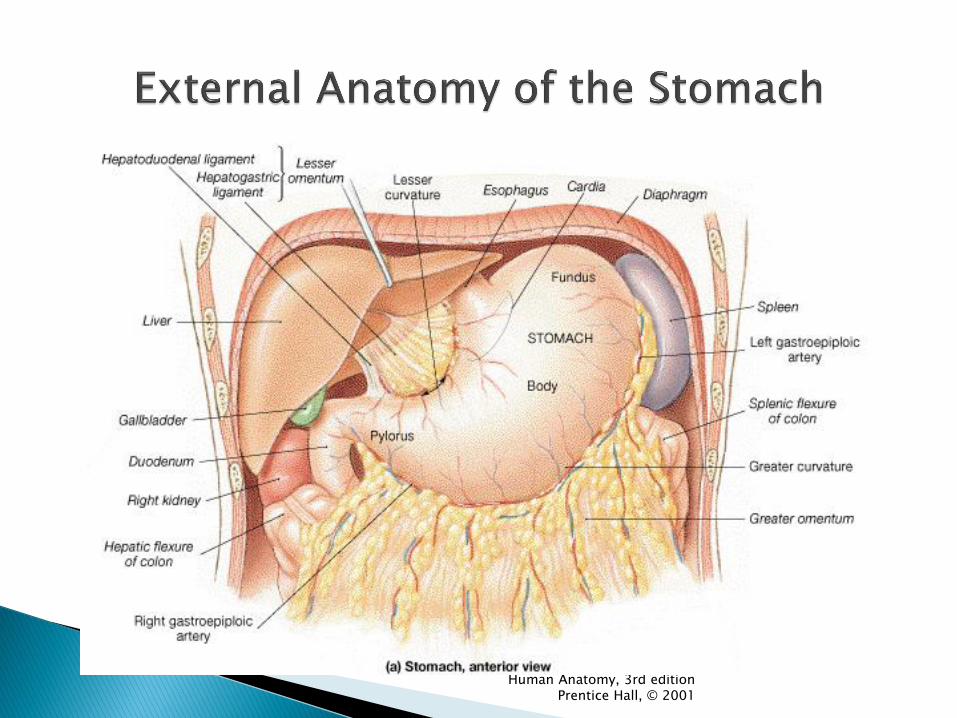
The Stomach

Performs 2 major functions: Storage and Digestion
Can hold up to 4 liters (1 gallon) of food (entire meal)
Creates Gastric Juice, which begins protein digestion.
Stomach walls churn the food to further increase surface area and to mix the gastric juice in with the food.
The Stomach

4 Regions:
◦ Cardiac-near the heart, surrounds the lower esophageal sphincter
◦ Fundic-superior expansion, temporarily holds food
◦ Body-main region
◦ Pyloric-funnel-shaped terminal end - leads to the pyloric sphincter
Rugae-stomach folds with allow the diameter of the stomach to expand


Smooth Muscle Layers of the Stomach
Three muscle layers:
•longitudinal
•circular
•oblique
Functions:
•move food along
•churn and mix food
with gasric juices
•Break food down
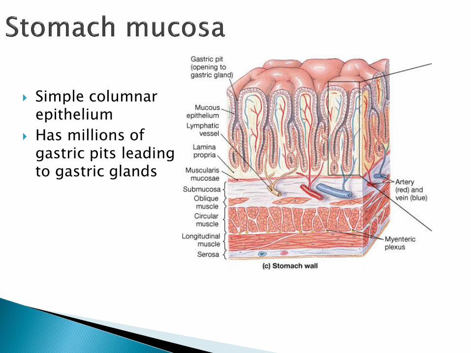
Simple columnar epithelium
Has millions of gastric pits leading to gastric glands

4 Cell types in gastric glands. Gastric juice is made from its 3 types of cells:
◦ Mucus Cells
◦ Chief Cells
◦ Parietal Cells
Enteroendocrine cells
Secrete mucus to line the
stomach wall
Secrete Pepsinogen
Secrete HCl and Intrinsic factor * needed for absorption of Vitamin B12
Secrete gastrin

Mucous cells: produce mucus ◦ Protects stomach from digesting itself ◦ Stomach pH=2 ◦ Ulcer-open sore (most common cause H. Pylori)
Chief cells: secrete pepsinogen (inactive enzyme) Parietal cells: produce HCL + Intrisic factor (IF)
◦ HCl + Pepsinogen Pepsin (active)
Pepsin chemically digests proteins into smaller proteins
(polypeptides) ◦ IF binds to VitB12 preventing pernicious anemia (failure of RBC development)

Enteroendocrine Cells: produce gastrin ◦ Gastrin (hormone) regulate stomach wall
contractions and secretions
The food (bolus) mixed with the stomach secretions is called Chyme.
◦ (Acidic) Chyme is released into the small intestine in small amounts through the Pyloric Sphincter
◦ Alcohol and water get absorbed in the stomach, but food does not
◦ Gastric emptying takes 2-6 hours


Major Digestive organ
7-13 feet long
Nearly all digestion and nutrient absorption occurs here.
3 main sections: ◦ Duodenum
◦ Jejunum
◦ Ileum

Attaches to the stomach
Curves around pancreas
The acid chyme from the stomach mixes with the digestive juices from the liver, pancreas, gall bladder and the small intestine walls.
Most of the digestion (breakdown) of the chyme is completed by the time it reaches the end of the Duodenum

Remaining sections of the S.I.
Majority of food molecule absorption
Lined with finger-like projections called Villi.
Villi are further lined themselves with Microvilli

Mucosa-simple columnar epi
Serve to increase the rate of absorption by vastly increasing the surface area.
Each Villi has surrounding capillaries and a central lacteal ◦ a small lymphatic
vessel

The capillaries absorb simple
sugars, which are transported
to the liver through the
Hepatic Portal Vein
Amino acids are also absorbed & transported through the body via the capillaries.
Fats are absorbed through the lacteals and travel through the lymphatic system

Human Anatomy, 3rd edition Prentice Hall, © 2001

A.k.a. Colon.
Larger in diameter
Extends from ileocecal valve to anus
Involved in water recovery from the digestive juices left behind.
Houses beneficial Bacteria that produce several vitamins

Human Anatomy, 3rd edition Prentice Hall, © 2001

The useless end product of the digestive process is feces.
Feces contains masses of bacteria, cellulose and undigested food materials.
Feces is stored at the terminal portion of the L.I. called the rectum.
Two rings of muscle called Sphincters control the elimination of feces from the body via the anus.

There are occasional interruptions in proper Large Intestine function:
◦ Constipation occurs when peristalsis slows and
most of the water is removed from the feces, which becomes impacted.
◦ Diarrhea occurs when water is not reabsorbed, either due to bacterial infection or some other irritatant.

Accessory organs of the digestive system: ◦ Teeth, tongue, salivary glands, liver, gallbladder,
pancreas
Accessory organs are essential to digestion, yet they are not actually part of the Alimentary Canal
Three accessory organs associated with the small intestine: ◦ Pancreas
◦ Liver
◦ Gall Bladder

Gastrin – produced by the enteroendocrine cells in the stomach when you eat a meal high in protein. ◦ Causes stomach wall to contract and gastric juices to be
secreted.
Secretin – produced by cells of duodenum when it receives acidic chyme from stomach oCauses pancreas to release bicarbonate (strong base) into
the duodenum (to raise the pH).
CCK (cholecystokinin) – produced by cells of duodenum when it receives partially digested fats and protein from the stomach oCauses pancreas to release pancreatic juice into the
duodenum (to complete breakdown of fat & protein)

Human Anatomy, 3rd edition Prentice Hall, © 2001
Hepatocytes – liver cells produce bile ◦ Composition
Water
Bile salts
Cholesterol
Pigments
Bilirubin – principal pigment.
Jaundice – caused by excessive amounts of bilirubin/blocked ducts – causes yellowing of skin/eyes.
Digestive function
Emulsification of fats – makes them more manageable for lipase to completely break them down.
o Stores carbohydrates as glycogen
o Filtering unit of the body

Large & triangle shaped
Right side of the body
Below the diaphragm
4 lobes

Human Anatomy, 3rd edition Prentice Hall, © 2001

Human Anatomy, 3rd edition Prentice Hall, © 2001

Human Anatomy, 3rd edition Prentice Hall, © 2001

Stores and concentrates bile made by the liver
Contracts and squeezes bile into the Duodenum of the small intestine, as needed.

Human Anatomy, 3rd edition Prentice Hall, © 2001

1. Cystic duct from
gall bladder
2. Hepatic ducts
from Liver
These 2 ducts
form the common
bile duct that lead
to the duodenum
(pathway for bile)

Head: the broad end that fits into the C-shaped duodenum
Tail: the narrow end
Pancreatic duct: carries pancreatic enzymes to the duodenum Opening of the duct:
hepatopancreatic ampulla

Human Anatomy, 3rd edition Prentice Hall, © 2001
The pancreatic duct is where the pancreatic juice leaves pancreas into the duodenum

Acinus (Acini)-secrete pancreatic enzymes – trypsin, nuclease, lipase, amylase
Islet cells-secrete endocrine hormones (insulin and glucoagon to regulate blood sugar

Digestive enzymes: help with the chemical digestion of food in the small intestine
Pancreatic Amylase: disaccharides into monosaccharides
Proteases & Trypsin: small polypeptides into amino acids
Pancreatic Lipase: fat droplets (emulsified in bile salts) glycerol & fatty acids
Nuclease: Nucleic acids into nucleotides
Bicarbonate: a base that neutralizes the highly acidic chyme
All of these tiny molecules will be absorbed into the capillaries & lacteal inside the villi
http://www.zerobio.com/secretin_flash.htm

Salivary Amylase- made by salivary glands and breaks up carbs into disaccharides
Pepsin – made in stomach and breaks up proteins into small polypeptides
Trypsin, Amylase, Nuclease, Lipase – made in Pancreas and delivered to Small intestine. ◦ Trypsin – breaks polypeptides into amino acids
◦ Amylase- breaks disaccharides into monosaccharides
◦ Nuclease – breaks nucleic acids into nucleotides
◦ Lipase – breaks emulsified fats into fatty acids and glycerol
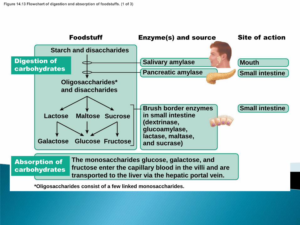
Digestion of
carbohydrates
Foodstuff Enzyme(s) and source Site of action
Absorption of
carbohydrates
The monosaccharides glucose, galactose, and
fructose enter the capillary blood in the villi and are
transported to the liver via the hepatic portal vein.
Brush border enzymes in small intestine (dextrinase, glucoamylase, lactase, maltase, and sucrase)
Small intestine
Galactose Glucose Fructose
Lactose Maltose Sucrose
Oligosaccharides*
and disaccharides
Starch and disaccharides
Salivary amylase
Pancreatic amylase
Mouth
Small intestine
*Oligosaccharides consist of a few linked monosaccharides.

Digestion
of proteins
Absorption
of proteins
Foodstuff Enzyme(s) and source Site of action
Brush border enzymes
(aminopeptidase,
carboxypeptidase,
and dipeptidase)
Amino acids
(some dipeptides
and tripeptides)
Small polypeptides,
small peptides
Large polypeptides
Protein Pepsin
(stomach glands) in
the presence of HCI
Stomach
Small intestine
Small intestine
Pancreatic enzymes
(trypsin, chymotrypsin,
carboxypeptidase)
Amino acids enter the capillary blood in the villi and are
transported to the liver via the hepatic portal vein.

Digestion
of fats
Absorption
of fats
Foodstuff Enzyme(s) and source Site of action
Fatty acids and monoglycerides enter the lacteals of the villi
and are transported to the systemic circulation via the lymph
in the thoracic duct. (Glycerol and short-chain fatty acids are
absorbed into the capillary blood in the villi and transported
to the liver via the hepatic portal vein.)
Monoglycerides
and fatty acids
Glycerol and
fatty acids
Pancreatic lipase
Small intestine
Small intestine
Emulsified by the
detergent action of
bile salts from the liver
Unemulsified fats

Digestion
Defecation
Mechanical
breakdown
Ingestion
Propulsion
Absorption
Food
Pharynx
Esophagus • Chewing (mouth) • Churning (stomach) • Segmentation
(small intestine) • Swallowing
(oropharynx) • Peristalsis
(esophagus, stomach,
small intestine, large
intestine)
Stomach
Lymph vessel
Blood vessel
Mainly H2O
Feces
Anus
Small intestine
Large intestine

Essential nutrients are minerals that are needed by the body, yet cannot be made by the organism.
Missing out on one or more essential nutrient is known as malnourished.
Undernourishment is caused by insufficient calories.

Vitamin and mineral deficiencies can cause severe disorders in the body
Examples include: Kwashiorkor, Scurvy, Rickets, and Goiter.

Kwashiorkor
Caused by inadequate Protein intake
The protuberant abdomen is caused by the body’s inability to absorb fluids, due to depleted necessary blood proteins
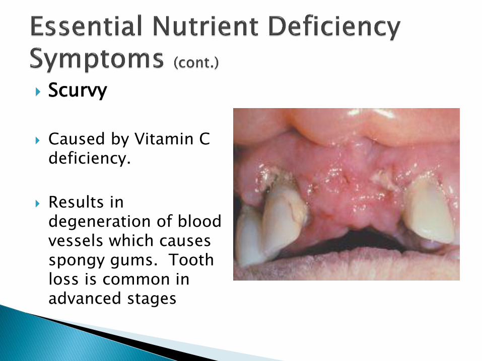
Scurvy
Caused by Vitamin C
deficiency.
Results in degeneration of blood vessels which causes spongy gums. Tooth loss is common in advanced stages

Rickets
Caused by Vitamin D deficiency
Causes bone softening and deformity

Goiter - thyroid gland that has grown to an abnormally large size
Iodine deficiency


![Digestive System Anatomy Practical [PHL 212]. The digestive system is made up of the digestive tract & accessory digestive organs: a series of hollow.](https://static.fdocuments.net/doc/165x107/56649ce35503460f949aef0e/digestive-system-anatomy-practical-phl-212-the-digestive-system-is-made.jpg)