Chapter 14 Lipid Metabolism, Lipid Signalling and...
23
307 © Springer International Publishing Switzerland 2016 A. Olsen, M.S. Gill (eds.), Ageing: Lessons from C. elegans, Healthy Ageing and Longevity 5, DOI 10.1007/978-3-319-44703-2_14 Chapter 14 Lipid Metabolism, Lipid Signalling and Longevity Jonathon Duffy, Ayse Sena Mutlu, and Meng C. Wang Abstract Ageing research gains more attention as the aged population increases worldwide and ageing-related diseases become more prevalent. Model organism research in the last three decades has shown that ageing is regulated via several genetic pathways and environmental interventions, most of which are evolutionarily conserved. C. elegans has been the powerhouse of ageing research since the discov- ery of mutant strains with doubled lifespan. Interestingly, the pathways that regulate C. elegans ageing often affect lipid biology as well. This chapter will focus on the interaction between lipid biology and ageing by introducing well-known pathways that regulate ageing and how lipid levels, composition or distribution change when these pathways are defective. Last but not least, the signalling role for lipids in age- ing will be discussed. Keywords Lipid molecules • Lipid signalling • Longevity 14.1 Introduction Ageing is an inevitable part of life, and until recently, it was thought to be a passive phenomenon that leads to decrease in organismal functions and fitness. However, elaborate research in the last three decades has shown that ageing is a complex pro- cess that is regulated by both intrinsic signalling pathways and extrinsic J. Duffy • A.S. Mutlu Graduate Program in Developmental Biology, Baylor College of Medicine, Houston, TX 77030, USA M.C. Wang (*) Graduate Program in Developmental Biology, Baylor College of Medicine, Houston, TX 77030, USA Department of Molecular and Human Genetics, Baylor College of Medicine, Houston, TX 77030, USA Huffington Center on Aging, Baylor College of Medicine, Houston, TX 77030, USA e-mail: [email protected]
Transcript of Chapter 14 Lipid Metabolism, Lipid Signalling and...

307© Springer International Publishing Switzerland 2016 A. Olsen, M.S. Gill (eds.), Ageing: Lessons from C. elegans, Healthy Ageing and Longevity 5, DOI 10.1007/978-3-319-44703-2_14
Chapter 14 Lipid Metabolism, Lipid Signalling and Longevity
Jonathon Duffy , Ayse Sena Mutlu , and Meng C. Wang
Abstract Ageing research gains more attention as the aged population increases worldwide and ageing-related diseases become more prevalent. Model organism research in the last three decades has shown that ageing is regulated via several genetic pathways and environmental interventions, most of which are evolutionarily conserved. C. elegans has been the powerhouse of ageing research since the discov-ery of mutant strains with doubled lifespan. Interestingly, the pathways that regulate C. elegans ageing often affect lipid biology as well. This chapter will focus on the interaction between lipid biology and ageing by introducing well- known pathways that regulate ageing and how lipid levels, composition or distribution change when these pathways are defective. Last but not least, the signalling role for lipids in age-ing will be discussed.
Keywords Lipid molecules • Lipid signalling • Longevity
14.1 Introduction
Ageing is an inevitable part of life, and until recently, it was thought to be a passive phenomenon that leads to decrease in organismal functions and fi tness. However, elaborate research in the last three decades has shown that ageing is a complex pro-cess that is regulated by both intrinsic signalling pathways and extrinsic
J. Duffy • A. S. Mutlu Graduate Program in Developmental Biology , Baylor College of Medicine , Houston , TX 77030 , USA
M. C. Wang (*) Graduate Program in Developmental Biology , Baylor College of Medicine , Houston , TX 77030 , USA
Department of Molecular and Human Genetics , Baylor College of Medicine , Houston , TX 77030 , USA
Huffi ngton Center on Aging , Baylor College of Medicine , Houston , TX 77030 , USA e-mail: [email protected]
308
environmental stimuli [ 1 ]. Studies using model organisms such as yeast, nematodes, fruit fl ies and mice discovered that several genetic pathways and environmental interventions, which regulate ageing, are evolutionarily conserved and hold great promise for human ageing research.
Since the discovery that mutations in the C. elegans insulin receptor, DAF-2 , can lead to doubling of lifespan , worms have become the prominent force in ageing research [ 1 ]. In addition to their short lifespan, C. elegans are transparent which renders them benefi cial for staining techniques and microscopy imaging. Last but not least, the vast number of tools available for genetic manipulation as well as the ease of performing high-throughput genetic screens has enabled researchers to fi nd several key players of signalling pathways regulating longevity and their detailed epistasis analysis.
Interestingly, many of the pathways that regulate longevity affect lipid biology as well. For example, C. elegans mutants that lack normal DAF-2 activity also have increased lipid levels [ 2 , 3 ]. However, there is no simple correlation between pro- longevity pathways and increased lipid storage levels. eat-2 mutants, genetic mod-els of dietary restriction in C. elegans , are long-lived, but have decreased lipid storage [ 4 ]. Thus, the involvement of lipids in ageing is more complicated than expected. Apart from their role in energy storage, lipids are also important signal-ling molecules. Examples include, ceramides and certain fatty acids, which have been shown to be important for ageing [ 5 , 6 ]. More studies on the characterization of the role of lipid biology in longevity will advance our knowledge in the biology of ageing and improve strategies for therapeutic interventions for healthy ageing.
In this chapter, we will focus on the general concepts of lipid biology and lifespan- regulating pathways with an emphasis on C. elegans longevity mutants and their lipid metabolism. We will also provide an overview of lipid analysis method-ologies. Finally, we will mention more recent research on the role of lipids as sig-nalling molecules.
14.2 Lipids
Lipids are a diverse class of small, organic molecules that are either amphipathic or hydrophobic [ 7 ]. Lipids are perhaps most associated with their roles as a source of energy storage and accumulation during obesity. Other than storing energy, lipids play major roles in forming membranes to mark the boundaries of a cell and to sepa-rate cellular compartments. Besides their well-known structural functions, lipids are also biologically active molecules providing communication within and between cells [ 8 ]. These signalling roles have implications in several diseases, such as vari-ous types of cancer and metabolic syndromes [ 9 ], as well as regulating healthy age-ing [ 10 ].
J. Duffy et al.
309
14.2.1 Structure and Classifi cation
Because lipids are such a broad grouping of molecules, they have been classifi ed into categories, each of which has multiple subclasses [ 7 ]. These categories are: fatty acyls, glycerolipids, glycerophospholipids, sphingolipids, sterol lipids, prenol lipids, saccharolipids, and polyketides [ 7 ]. Within these groups, different lipids are distinguished from each other by a variety of criteria, including their length, the number and location of double bonds in their hydrocarbon tail(s), and the attached structure(s), such as phosphates or glycerol. Four groups of lipids are focused in this chapter to demonstrate the diversity between different lipid categories (Fig. 14.1 ).
14.2.1.1 Fatty Acyls
Fatty acyls are a diverse group of lipids that include the major sub-grouping of fatty acids, which are carboxylic acids with a hydrocarbon tail [ 11 ]. Based upon the num-ber of double bonds in the hydrocarbon tail, fatty acids can be further divided into saturated (no double bonds), monounsaturated (a single carbon-carbon double bond), and polyunsaturated fatty acids (more than one carbon-carbon double bond).
The saturated fatty acids usually contain 14–22 carbon atoms. The monounsatu-rated fatty acids are also similar length, but they have a carbon-carbon double bond, commonly in cis -confi guration, which means the hydrogen bonds next to the double bond are positioned in the same direction. The presence of the double bond gives the molecule a “kink” in its shape, which changes its biochemical properties. Polyunsaturated fatty acids (PUFAs) contain several carbon-carbon double bonds, and they are named depending on the location of the fi rst double bond: they are called ω-3 fatty acids if the bond is between the third and the fourth carbon after the ω-carbon and ω-6 if the bond is between the sixth and seventh. Two PUFAs, linoleic acid and alpha-linolenic acid are essential nutrients for mammals, but unlike mam-mals, C. elegans express the desaturases ( fat-1 and fat-2 ) that are necessary to syn-thesize these PUFAs de novo [ 12 ].
Fatty acids are important energy fuels for the cell, which can be degraded via β–oxidation to generate acetyl-CoA and subsequently used to generate ATP via the citric acid cycle [ 13 ]. Fatty acids and their derivatives are crucial for cellular homeo-stasis and organism fi tness, and they can be utilized in both intracellular and extra-cellular signalling. Research in the last decade showed that dietary ω-3 fatty acids are involved in both neurotransmission and neurogenesis, and may also be impor-tant for preventing age-related brain damage and neurodegenerative diseases, such as Alzheimer’s disease [ 14 ]. Even though the complete mechanism is unclear, these studies showed that ω-3 fatty acids regulate microglia and astrocyte activity, improve mitochondrial functions, and reduce oxidative damage [ 14 ]. More recently, a spe-cifi c monounsaturated fatty acid derivative, oleoylethanolamide (OEA), was shown to be involved in longevity regulation at the organismal level. This C. elegans study showed that OEA acts as a signalling molecule between the lysosomes and the
14 Lipid Metabolism, Lipid Signalling and Longevity

310
Fig. 14.1 Different classes of lipid species . Fatty acids, which are carboxylic acids with a hydrocarbon tail, are building blocks of many other lipid species. Depending on the number of double bonds in the hydrocarbon tail, they are classifi ed into three groups: saturated, mono-unsaturated ( MUFA ) and poly-unsatu-rated ( PUFA ). Glycerolipids consist of a glycerol backbone attached to one-to-three fatty acids. For example, triacylglycerols have glycerol attached to three fatty acids, which can have hydrocarbon tails of different sizes and saturation. Triacylglycerols are the major source of energy storage in cells. Glycerophospholipids are lipids with a polar group attached to one of the positions on the glycerol backbone and fatty acids attached to the other two. For example, phosphatidylcholine has a choline attached to the third position on the glycerol. Sphingolipids are membrane-associated lipids with a sphingoid base attached to one fatty acid. For example, ceramides have sphingosine attached to a fatty acid
J. Duffy et al.
No period
311
nucleus where it will activate specifi c nuclear hormone receptors to induce longev-ity [ 10 ]. OEA is part of a larger group of fatty acid derivatives known as N-acylethanolamines (NAEs). Another NAE, eicosapentaenoyl ethanolamide (EPEA), has been shown to be able to modulate organism lifespan via dietary- restriction [ 15 ]. Additionally, fatty acids have roles in extracellular signalling. Several free fatty acids (FFA) have been shown to regulate insulin secretion in mammalian cell lines via G-protein coupled receptor (GPCR) signalling [ 16 ]. More specifi cally, palmitoleate (C16;1n7) acts as a lipokine derived from the adipose tis-sue to improve insulin and glucose metabolic homeostasis in the muscle and liver systematically [ 17 ].
14.2.1.2 Glycerolipids
Glycerolipids are lipids with a glycerol backbone with one to three attached fatty acids [ 11 ]. There are mono-, di- or tri-acylglycerols depending on the number of attached fatty acids. Each of the fatty acids in diacylglycerols (DAGs) or in triacyl-glycerols (TAGs) can be different.
TAGs are the major intracellular source of energy storage, and they can be degraded by lipases in the presence of the proper signals, such as a demand for energy, resulting in the release of free fatty acids (FFAs). Homeostasis in lipid stor-age, especially the level of TAGs, is essential for healthy ageing since obesity is associated with age- related diseases such as cardiovascular disease, type II diabetes and certain types of cancer [ 18 ]. However, as mentioned in the introduction, there may not be a simple correlation between overall lipid levels and organism longevity. Worms under dietary restriction have lower lipid storage [ 19 ] whereas insulin- receptor defi cient worms, daf-2 mutants, have more [ 20 ], but yet both worm strains are long-lived. Therefore, future studies should investigate whether differential dis-tribution of TAGs in certain tissues affect ageing. It is also possible that the compo-sition and not the overall level of TAGs affect longevity.
DAGs, on the other hand, have a much more diverse set of roles. Many intracel-lular pathways use DAGs as a second messenger by binding to a group of proteins with a C1 domain such as protein kinase C [ 21 ] and indirectly regulate the activities of G proteins [ 22 ]. Therefore, DAGs have important roles in processes such as pro-liferation, apoptosis, differentiation, and cellular migration [ 23 ]. DAGs also affect the physical aspects of membranes, such as their structure and dynamics, as well as functioning as a component in lipid metabolism by either being degraded to gener-ate FFAs or added to other lipids in order to generate more complex lipids [ 21 ]. DAG metabolism is also involved in ageing . In fl ies and worms, knockdown of diacylglycerol lipase or overexpression of the diacylglycerol kinase extends lifes-pan [ 24 ]. The same study suggested that DAG metabolism interacts with TOR sig-nalling to regulate longevity.
14 Lipid Metabolism, Lipid Signalling and Longevity

312
14.2.1.3 Glycerophospholipids
Glycerophospholipids are lipids that have a phosphate group connecting a polar group to a glycerol backbone, with fatty acids attached to the other two positions on the glycerol [ 11 ]. Similar to the glycerolipids, the fatty acids attached to the glycerol can be of any kind. But mostly they contain a saturated fatty acid in the fi rst carbon and an unsaturated fatty acid in the second carbon. The third carbon is joined to a polar alcohol with a phosphodiester bond. Like all of the lipid molecules in the cell membrane, glycerophospholipids are also amphipathic.
Glycerophospholipids are the main component of the membrane bilayer. In addi-tion to this commonly known role, they play an important role in signalling. One of the best-known phospholipid signalling molecules is the phosphotidylinositols. Phosphotidylinositols are a subclass of phospholipids that are partially located within the membrane. Certain cell-surface G-protein coupled receptors (GPCRs) activate phospholipase C (PLC), which then cleaves phosphotidylinositols to pro-duce diacylglycerol (DAG) and inositol phosphates [ 25 ]. Both DAG and inositol phosphates have signalling capabilities and affect a plethora of aspects of the cell, including Ca 2+ release, lipid transport and membrane dynamics [ 26 ].
Ageing increases the cholesterol/ phospholipid ratio in cell membranes, thus decreasing the membrane fl uidity. Lipofuscins, sometimes called age pigments, are derived from the peroxidation of subcellular membrane lipids containing PUFAs and precipitate in the lysosomes. Lipofuscin accumulation is also implicated in neu-rodegenerative diseases such as Alzheimer’s disease and Parkinson’s disease. Phosphoinositide signalling cascade is also affected at many levels during the age-ing process, such as receptor availability and kinase activity [ 27 ].
14.2.1.4 Sphingolipids
Sphingolipids are membrane-associated lipids that have a sphingoid base . These are then built upon and modifi ed to become more complex lipids, such as ceramides [ 11 ]. Ceramide is important in multiple aspects of programmed cell death (PCD) [ 28 , 29 ]. It can affect intrinsic and extrinsic PCD-related signalling pathways as well as both caspase-dependent and caspase–independent mechanisms [ 30 ]. Ceramide also plays a crucial role in other developmental processes such as differ-entiation of the primitive ectoderm in embryos and asymmetric cell division [ 31 ]. As worms develop and age, sphingolipids naturally accumulate, and so inhibition of the synthesis and accumulation of sphingolipids leads to a delay in the development and ageing of C. elegans [ 32 ]. Interestingly, loss-of-function mutations in genes encoding the ceramide-synthesis enzymes, lagr-1 and sphk-1 , results in increased autophagy and extension in lifespan [ 33 ].
J. Duffy et al.

313
14.2.2 Synthesis, Storage, and Degradation of Lipids
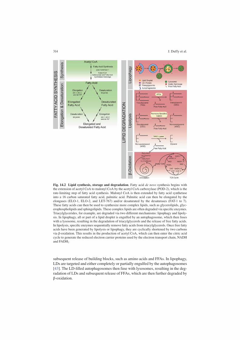
Lipids can be either synthesized de novo or absorbed from the diet (Fig. 14.2 ). The starting point of de novo synthesis is acetyl CoA , which will be extended into malonyl- CoA by acetyl-CoA carboxylase and further into palmitic acid by fatty acid synthase [ 34 ]. Then, palmitic acid, a 16 carbon saturated fatty acid, can be further elongated by the enzymes ELO-1, ELO-2, and LET-767, and/or desaturated by the enzymes FAT-1 to FAT-7. Desaturation is possible in C. elegans , but possible only to a limited extent in mammals [ 12 , 34 ]. These fatty acids then serve as the base for many lipids. For example, coenzyme A-bound fatty acids (acyl-CoA) can be combined with glycerol-3-phosphate, a phosphorylated glycerol, to generate lysophosphatidic acid (LPA). A second acyl-CoA can be added to LPA to generate phosphatidic acid, which can have its phosphate removed to generate DAG. A third acyl-CoA can be incorporated to then generate TAG [ 35 ].
While de novo synthesis is extremely important, not every organism can synthe-size every lipid. This makes the dietary intake of lipids a key aspect in maintaining lipid homeostasis and general organismal functions. One notable example of the importance of dietary intake of lipids in C. elegans is cholesterol, which is a key component of membrane structures and signalling pathways [ 36 – 38 ]. Unlike mam-mals, C. elegans cannot synthesize cholesterol and relies on dietary cholesterol for its normal development and functions. Mammals on the other hand require the dietary intake of two polyunsaturated fatty acids, linoleic acid and linolenic acid, in order to synthesize more complex lipids, such as arachidonic acid [ 39 ].
Since lipids are a great source of energy, they often need to be stored for an extended period of time. Neutral lipids, such as TAG, are predominantly stored in a conserved organelle called a lipid droplet (LD). LDs are formed when there is a localized accumulation of TAGs within the lipid bilayer of the endoplasmic reticu-lum (ER), leading to the eventual budding off of a LD [ 40 ]. They are surrounded by a phospholipid monolayer, structural proteins called perilipins that protect the LD from cytoplasmic lipases , and other proteins involved in multiple aspects of LD biology, including lipases for lipid degradation/mobilization [ 41 ]. Interestingly, C. elegans lack perilipins, but still maintain a tight control over LD degradation [ 42 ]. Active on-going researches in different laboratories are addressing the funda-mental mechanisms underlying LD maintenance and dynamics in C. elegans .
In order to metabolize the lipids stored within LDs, two pathways are used, lipol-ysis and lipophagy (Fig. 14.2 ). In lipolysis, cytoplasmic and LD-associated lipases degrade the neutral lipids within the LDs. First, ATGL cleaves TAG to generate DAG and a FFA. The DAG can then be degraded by hormone sensitive lipase to generate monoacylglycerol (MAG) and another FFA. MAG can then be degraded by MAG lipase to generate a FFA and glycerol [ 40 ]. In C. elegans , ATGL-1, which is localized to LDs, is the lipase necessary for lipolysis [ 42 ]. In lipophagy, a branch of autophagy , autophagosomes are used to mobilize the lipids stored within LDs. In normal autophagy, cellular contents are engulfed by the autophagosomes that will fuse with lysosomes, resulting in the degradation of the autolysosomal contents and
14 Lipid Metabolism, Lipid Signalling and Longevity
314
Fig. 14.2 Lipid synthesis, storage and degradation . Fatty acid de novo synthesis begins with the extension of acetyl CoA to malonyl CoA by the acetyl CoA carboxylase (POD-2), which is the rate-limiting step of fatty acid synthesis. Malonyl CoA is then extended by fatty acid synthetase into a 16 carbon saturated fatty acid, palmitic acid. Palmitic acid can then be elongated by the elongases (ELO-1, ELO-2, and LET-767) and/or desaturated by the desaturases (FAT-1 to 7). These fatty acids can then be used to synthesize more complex lipids, such as glycerolipids, glyc-erophospholipids and sphingolipids. These complex lipids are often degraded via specifi c enzymes. Triacylglycerides, for example, are degraded via two different mechanisms: lipophagy and lipoly-sis. In lipophagy, all or part of a lipid droplet is engulfed by an autophagosome, which then fuses with a lysosome, resulting in the degradation of triacylglycerols and the release of free fatty acids. In lipolysis, specifi c enzymes sequentially remove fatty acids from triacylglycerols. Once free fatty acids have been generated by lipolysis or lipophagy, they are cyclically shortened by two carbons via β-oxidation. This results in the production of acetyl CoA, which can then enter the citric acid cycle to generate the reduced electron carrier proteins used by the electron transport chain, NADH and FADH 2
subsequent release of building blocks, such as amino acids and FFAs. In lipophagy, LDs are targeted and either completely or partially engulfed by the autophagosomes [ 43 ]. The LD-fi lled autophagosomes then fuse with lysosomes, resulting in the deg-radation of LDs and subsequent release of FFAs, which are then further degraded by β-oxidation.
J. Duffy et al.

315
β-oxidation occurs via the same reactions in the both the mitochondria and per-oxisomes (Fig. 14.2 ) [ 44 ], but the identity of the enzymes used in these reactions are different between the two organelles [ 45 ]. Accordingly, while most fatty acids can be degraded by both organelles, certain fatty acids, such as very long chain fatty acids, prefer peroxisomal degradation [ 45 ]. In β-oxidation, the hydrocarbon tails of saturated FFAs are cyclically degraded by two carbons at a time to generate acetyl CoA [ 44 ], which is utilized to generate energy via the TCA cycle and oxidized electron carriers, which can be used in the electron transport chain (ETC). Unsaturated fatty acids require additional enzymes, such as isomerases and dehy-drogenases, to process the double bonds before the β-oxidation pathway can degrade the fatty acids [ 46 ].
14.2.3 Methods of Studying Lipid Metabolism
Since lipids play such a variety of vital roles in cellular homeostasis and organismal fi tness, it is important that we have effective methods to study their storage, compo-sition and distribution. In C. elegans , lipids are stored in the intestine , the hypoder-mis and oocytes. The intestine of C. elegans provides the function of multiple organs/tissues, such as digestion like the mammalian intestine, detoxifi cation like the liver, and fat storage like the adipose tissue [ 47 ]. Additionally, lipids are synthe-sized in the intestine and transported to oocytes by vitellogenin proteins, where they play major roles in oocyte and embryo development [ 38 , 48 , 49 ].
Biochemical assays are powerful methods to study lipid levels and composition. These can provide knowledge about the relative amounts of different lipid species within a sample, which can be important when examining lipid metabolism at the molecular level. There are two methods that are commonly used to analyse lipids biochemically, mass spectrometry (MS) [ 3 ] and nuclear magnetic resonance (NMR) spectroscopy [ 50 ]. MS analyses the mass/charge ratio of the molecules in a sample which have to be ionized prior to analysis [ 51 ]. Before the samples are analysed via MS, a separation technique, such as gas chromatography, liquid chromatography or capillary electrophoresis, is often performed. The samples are then ionized through one of several techniques. Two of the more common techniques are electron-spray ionization (ESI) and matrix-assisted laser desorption/ionization (MALDI). These charged molecules will then be separated based upon their mass-to-charge ratio via techniques such as time-of-fl ight or ion traps, before reaching a detector. Each lipid molecule will give specifi c peaks in the MS spectrum, which can be detected and analysed.
NMR is based upon the magnetic properties of hydrogens in the compounds, which can be affected by the bonds and connected structures near the hydrogens. This can help to provide physical and chemical details about the lipids analysed [ 52 ]. As with every analysis, there are pros and cons for both NMR and MS. Even though MS is more sensitive than NMR [ 51 ], NMR is quantitative and does not require extra sample preparation steps. NMR is also a non-destructive technique
14 Lipid Metabolism, Lipid Signalling and Longevity
“in both mitochondria and peroxisomes”

316
where the sample can be recovered and used for further analyses. MS is a destruc-tive technique but requires a much smaller amount of sample than NMR needs [ 53 ]. Even though these techniques are useful for detecting different kinds of lipid spe-cies at the molecular level, they lack spatial information of lipid distribution.
The cellular/tissue distribution of lipids and their transportation between cells/tissues are very crucial for their functions. The transparent nature of C. elegans makes it an ideal model for visualizing lipid storage with subcellular resolution at the whole organism level. There are several stains that are commonly used to visual-ize the lipid stores of C. elegans . Two of these, BODIPY-labelled fatty acids and Nile Red can emit fl uorescence when labelling LDs; while two other stains, Sudan Black and Oil Red O, appear blue-black and red colour, respectively when enriched in LDs [ 54 ]. However, both fl uorescent and non-fl uorescent-based methods require fi xation, and are commonly associated with a higher degree of variability.
Alternative to the staining techniques, chemical imaging methods are established in several laboratories for visualizing lipid species and different metabolites [ 20 , 55 – 59 ]. These two, relatively new methods are coherent anti-Stokes Raman scatter-ing (CARS) and stimulated Raman scattering (SRS) microscopy, both of which are based upon stimulated Raman scattering. In both of these methods, chemical bonds are stimulated with two lasers, one of which is fi xed at a certain wavelength, while the wavelength of the other can be adjusted accordingly to the vibrational frequency of the chemical bond of interest. If the frequency difference of the laser beams matches the vibrational frequency of the chemical bond, the molecular vibration transitions to an excited state. As a result, anti-Stokes signals are emitted and the beam intensities will change, which can be detected and quantifi ed as a measure for the level of the chemical bond of interest [ 60 ]. CARS was fi rst demonstrated in 1982 as a viable microscopy method, but was not really used until 1999 [ 61 ]. Later, in 2008, SRS was shown to be an improvement over CARS by reducing the non- resonant background, providing easier quantifi cation [ 20 ], and quicker, more sensi-tive imaging [ 60 ]. SRS microscopy can visualize lipid storage at diffraction-limited spatial resolution and with 3D imaging capacity in living cells and organisms [ 62 ]. More recently, by administering deuterium-labelled [ 63 ] or alkyne-labelled [ 64 ] lip-ids to C. elegans or mammalian cells and using SRS microscopy to detect the spe-cifi c signals from these labels, this technique was proven to be useful also for analysing the incorporation, synthesis and degradation of lipids .
14.3 Pathways Regulating Ageing
In 1993, the discovery that loss-of-function mutations in daf-2 could double the lifespan of C. elegans accelerated the fi eld of ageing research. Since then, consider-able effort has been put into elucidating the genetic pathways involved in the regula-tion of organism ageing and longevity. These pathways often have common components and crosstalk. For example, nhr-80 is a downstream effector for both glp-1 and lipl-4 . In this part, we are going to discuss the important longevity regulat-ing signalling pathways in C. elegans and their effects on lipid metabolism.
J. Duffy et al.

317
14.3.1 Insulin/IGF-1 Signalling (IIS)
daf-2 is a key player in the insulin/IGF-1 signalling (IIS) pathway and its role in ageing is discussed in Chap. 4 . It encodes the C. elegans homologue of the insulin/IGF-1 receptor [ 2 ], which is a receptor tyrosine kinase. When bound to an activating ligand, the DAF-2 receptor activates AGE-1, the C. elegans homologue of phos-phoinositide 3-kinase (PI3K). AGE-1/PI3K then phosphorylates phosphatidylinosi-tol 4,5-bisphosphate to generate phosphatidylinositol 3,4,5-trisphosphate, which then activates a kinase cascade culminating in the phosphorylation of multiple pro-teins, including DAF-16/ FoxO [ 65 ]. DAF-16/FoxO is a transcription factor and when phosphorylated as a result of the active IIS, it is sequestered in the cytoplasm along with several other transcription factors [ 65 ]. When IIS is low, DAF-16/FoxO translocates to the nucleus and promotes longevity by regulating the expression of genes involved in biological processes including both fat metabolism and ageing [ 66 , 67 ].
There are a number of daf-2 mutant alleles that lead to lifespan extension [ 68 ]. In addition to the longevity phenotype, mutants with the hypomorphic daf-2(e1370) allele, have increased lipid storage as shown by Nile Red, Oil Red O staining [ 69 ] and CARS/SRS microscopy analyses [ 20 ], and elevated de novo lipid synthesis assayed by 13 C isotope labelling strategy [ 3 ]. However, different lifespan-extending alleles of daf-2 can have different effects on lipid synthesis and storage. For exam-ple, the m577 and e1368 alleles showed no increase in de novo lipid synthesis or total lipid storage [ 3 ]. This has lead to more detailed analyses of daf-2 mutants at the transcriptional, metabolic and protein levels. The daf-2(m21) mutant was found to downregulate the expression of several of vit/ lipid transport genes and upregulate several fat/ fatty acid desaturase genes [ 70 ]. Proteomics analysis of the daf-2(e1370) mutant also revealed that intermediary metabolism is reorganized, and some of these changes might be related to increased lipid storage and longevity in this mutant allele [ 71 ]. Furthermore, several daf-2 alleles were also subjected to metabo-lite profi ling, and amongst a variety of metabolite changes, choline metabolism was specifi cally reprogrammed, possibly due to altered phospholipid metabolism [ 72 ].
14.3.2 Dietary Restriction
eat-2 encodes a non-α-nicotinic acetylcholine receptor subunit [ 73 ]. It is expressed in the pharyngeal muscle and required for the proper neuromuscular junction (NMJ) activity, specifi cally the NMJ with the MC neuron [ 74 ]. Because of irregular and slow pharyngeal pumping in eat-2 mutants [ 74 ], they have been utilized to model caloric restriction, show a lifespan extension of 29–57 % [ 75 ], and display decreased lipid storage as shown by several visualization methods [ 19 ] and enzymatic assays [ 76 , 77 ]. eat-2 longevity, decreased lipid storage, and increased autophagy are all dependent upon nhr-62 , which encodes a nuclear hormone receptor [ 4 ]. In addition
14 Lipid Metabolism, Lipid Signalling and Longevity

318
to being required for eat-2 longevity, NHR-62 has been shown to play a role in other forms of dietary restriction, such as using a diluted bacterial diet [ 4 ]. A more detailed discussion of dietary restriction can be found in Chap. 16 .
14.3.3 Germline Loss
In various species, germline signals have been linked with longevity regulation [ 78 – 80 ]. In C. elegans , ablation of germline precursor cells leads to more than 50 % lifespan extension [ 81 ], and is discussed in detail in Chap. 6 . Similar longevity phe-notypes were also observed in the loss-of-function mutant of glp-1 , which lacks germline stem cells [ 82 ]. glp-1 encodes one of the two DSL-family Notch receptors, is expressed in germline stem cells, perceives signals from their niche provided by the distal tip cells, and is required to maintain the germline stem cell pool [ 82 , 83 ]. Beside its longevity phenotype, the glp-1 mutant also displays increased lipid stor-age, as shown by Oil Red O staining, MS analysis [ 54 ], and CARS microscopy [ 19 ]. This intestinal lipid accumulation is thought to be largely due to absence of lipid transfer from the intestine to oocytes when germline development is arrested. Several factors have been implicated in the regulation of the longevity conferred by germline defi ciency, including the nuclear receptors, daf-12, nhr-49 and nhr-80 , the transcription factor , daf-16, and the lipase, lipl-4.
1. daf-12 encodes a nuclear receptor that is required for the longevity conferred by removal of germline stem cells. DAF-12 binds to the endogenous cholesterol derivatives Δ 1,7 -dafachronic acid (DA), Δ 7 -DA and 3α-OH-Δ 7 -DA [ 50 ], and reg-ulates the expression of a variety of genes involved in development and metabo-lism [ 84 , 85 ]. Gain-of-function alleles of daf-12 occur in the ligand-binding domain of the protein, resulting in increased activity and lifespan extension [ 86 ]. On the other hand, daf-12 loss-of-function alleles are mostly in the DNA binding domain, and show shortened lifespans. Knockdown of daf-12 results in a slight decrease in lipid levels [ 87 ], but gain-of-function alleles of daf-12 display increased fat content (unpublished results). DAF-12 functions with the corepres-sor DIN-1S, which regulates lipid storage [ 88 ]. daf-12 knockdown affects the expression of several lipid metabolic genes , such as lipl-4 , lips-17 , and fard-1 , and these genes are required for the longevity phenotype of the glp-1 mutant [ 89 ].
2. nhr-80 encodes a nuclear hormone receptor that is also required for the longev-ity phenotype of glp-1 mutants [ 90 ]. Once bound to its ligand, NHR-80 can use other nuclear receptors, such as NHR-49 and DAF-12, as cofactors to regulate the expression of its target genes, which include lipid metabolic genes such as acs-2 and fat-6 [ 91 , 92 ]. Loss of nhr-80 function does not affect the lifespan of wild type animals, but completely abrogates the lifespan extension of the glp-1 mutant [ 90 ]. nhr-80 mutants have no changes in their total amount of lipids stores, but do display changes in the relative lipid composition of their lipid stores [ 91 , 92 ].
J. Duffy et al.

319
3. nhr-49 encodes a nuclear hormone receptor necessary for β- oxidation gene expression [ 93 ]. nhr-49 is required for the increased lipid accumulation and lifespan extension in the glp-1 mutant [ 93 ], and also plays a crucial role in exe-cuting adult reproductive diapause (ARD) in which adults halt reproduction and survive for an exceptionally long time [ 94 ]. Mutations in nhr-49 affect de novo lipid synthesis and consequently lipid storage, at least in part via altering expres-sion of several fatty acid desaturase genes [ 93 ].
4. lipl-4 encodes a lysosomal triglyceride lipase that is induced in the glp-1 mutant and required for its longevity [ 87 ]. When constitutively over expressed in the intestine , lipl-4 is suffi cient to extend lifespan on its own [ 10 , 87 ]. Interestingly, the lipl- 4-induced longevity effect requires both NHR-49 and NHR-80. Metabolite profi ling of the long-lived lipl-4 transgenic worms revealed the induction of several lipid molecules ; amongst them, a specifi c fatty acid deriva-tive OEA acts as an agonist of the nuclear receptor NHR-80, and is suffi cient to prolong lifespan when supplemented to wild type worms [ 10 , 87 ]. The transduc-tion of lysosome- to- nucleus lipid messenger signalling requires a specifi c fatty acid binding protein LBP-8 [ 10 ].
14.4 Lipids as Signalling Molecules
Beside their well-known functions as energy fuels and structural building blocks, lipids play important roles in both intracellular and extracellular signalling. While some of these signalling functions have been thoroughly established, others are still being discovered and elucidated. Emerging studies have revealed the signifi cance of signalling lipid molecules in the regulation of organism longevity, and have discov-ered the involvement of protein chaperones, transporters and receptors in shuttling lipid molecules between compartments, as well as recognizing and transducing lipid signals.
14.4.1 Lipid Messengers
In C. elegans , a variety of lipids have signalling capabilities. One group of external lipid-derived signals are the ascosaride dauer -inducing pheromones [ 95 ]. These “daumones” are secreted by C. elegans into the environment and induce dauer for-mation in nearby larvae by binding to and activating cell surface receptors on cili-ated chemosensory neurons. Each daumone has a different ability to induce dauer formation [ 96 ], and they often function together [ 97 ].
Beyond external lipid signals like daumones, C. elegans uses several internally generated lipid signals to communicate within and between cells . For example, the inositol-signalling pathway, a well-conserved intracellular signalling pathway, is involved in a variety of processes, including embryonic development [ 98 ] and lifes-
14 Lipid Metabolism, Lipid Signalling and Longevity

320
pan [ 99 ]. Additionally, free fatty acids and their derivatives play roles in proper neurotransmission [ 100 ] and lifespan [ 10 ], amongst other processes.
Last but not least, sterols play an important role in biological signalling. Sterols, which include cholesterol and its derivatives, are lipids with four carbon rings and auxiliary components. Cholesterol is the basis for many of the hormones used in mammals, and plays a major role in C. elegans biology. Sterols cannot be generated de novo in C. elegans , so their dietary inputs play a key role in the evaluation of environmental quality. A key transcription factor , SBP-1, the conserved homologue of the mammalian sterol regulatory element binding protein, SREBP-1, controls the expression of several of the fat /fatty acid desaturase genes, along with other fatty acid synthesis genes [ 34 , 101 ]. However, it is not known whether SBP-1 also regu-lates sterol metabolism in C. elegans as SREBP-1 does in mammals.
14.4.2 Proteins as Lipid Signalling Chaperones
There are several lipid-binding chaperones in C. elegans , and two major families are lipid-binding proteins (LBPs) and vitellogenins. There are 9 lbp genes in C. elegans , which have varied tissue and developmental expression patterns. Three of these, lbp-1 , lbp-2 , and lbp-3 , have secretory signals suggesting a role in extracellular signalling [ 102 ]. Other LBPs play important roles in intracellular signalling. For example, LBP-5 is involved in multiple aspects of metabolism, such as β– oxidation , fat storage, and glycolysis [ 103 ], and LBP-8 mediates lysosome- to-nucleus com-munication [ 10 ]. The other family of lipid-binding chaperones is the yolk proteins encoded by 6 vitellogenin, or vit genes [ 104 ]. These yolk proteins are produced exclusively in the intestine of hermaphrodites [ 105 ] and function to transport lipids from the intestine to oocytes.
Both families of lipid-binding chaperones have been linked to lifespan regula-tion. For example, LBP-8 was recently shown to shuttle OEA from the lysosome to nucleus where OEA binds to and activates NHR-80. When lbp-8 is overexpressed, the increased shuttling of OEA and subsequent activation of NHR-80 results in a longer lifespan [ 10 ]. Additionally, when knocked down, vit-5 results in lifespan extension [ 106 ], and in long-lived daf-2 mutants, all six of the vit genes are down regulated [ 70 ].
14.4.3 Lipid Signalling Receptors
As signalling molecules, lipids can bind to and activate G-protein coupled receptors (GPCRs) and nuclear hormone receptors (NHRs). There are almost 2000 GPCRs in C. elegans most of which are expressed in individual ciliated chemosensory neurons to sense their environmental cues [ 107 ], which includes the lipid derivatives
J. Duffy et al.

321
daumones [ 108 ]. Several GPCRs are required for the daumone-induced dauer- formation response, such as SRBC-64/SRBC-66 [ 109 ], SRG-36/SRG-37 [ 110 ], and DAF-37/DAF-38 [ 111 ]. One of these receptors, DAF-37, is specifi c for the ascaro-side#2, but can mediate different responses to ascaroside#2 depending upon if DAF-37 is activated in the ASK or ASI chemosensory neurons [ 111 ].
C. elegans have 284 NHRs, which are transcription factors with a DNA-binding domain and a ligand-binding domain that is often activated by small hydrophobic molecules such as lipids and lipid derivatives [ 50 , 112 ]. Several NHRs regulate the expression of genes important in lipid metabolism and/or longevity pathways, such as DAF-12 in the germline regulation of lifespan [ 81 ], NHR-49 in lipid metabolism, lipid storage, and lifespan [ 113 ], NHR-80 in germline-mediated longevity and the associated lipid metabolism and storage changes [ 90 ], and NHR-62 in caloric restriction-induced longevity [ 4 ]. These receptors respond to signals that are usually generated within the organism. For example, DAF-12 is activated by three endoge-nous derivatives of dafachronic acid (DA), Δ 1,7 -DA, Δ 7 -DA and 3α-OH-Δ 7 -DA [ 50 ], and NHR-80 is activated by the endogenous fatty acid derivative OEA ( 10 ).
14.5 Relevance to Humans
Since C. elegans is a eutelic nematode roughly the size of a comma in a sentence, it is often diffi cult to realize how studies in C. elegans can be relevant for human biol-ogy. C. elegans and mammals appear vastly different, in no small part due to their anatomical and physiological differences. Despite these glaring differences, most of the proteins and genes mentioned throughout this chapter are not only functionally conserved, but are often structurally conserved as well [ 114 , 115 ].
When studying human ageing , some populations have been especially valuable when looking for insights into healthy ageing. One especially important population are centenarians. Multiple studies have been performed to look for the genes that may explain their longer life. Not surprisingly, hypomorphic mutations in the gene IGF1R, which encodes the insulin-like growth factor 1 receptor, has been impli-cated in the longevity displayed by some centenarians [ 116 ]. Variants in other com-ponents of the IIS pathways, such as INSR [ 117 ], PI3K [ 117 ], AKT1 [ 118 ] and FOXO3A [ 118 – 122 ] have been examined in several populations, and are associated with longevity as well.
Additionally, multiple studies have been performed to study the centenarian peo-ple from Okinawa, Japan since it was fi rst noticed in the 1960s that they were unin-tentionally calorically restricting themselves [ 123 , 124 ]. Although there have been many studies on the effects of relatively short-term caloric restriction on a variety of ageing hallmarks [ 125 – 127 ], this population has provided the best evidence for the conservation of many benefi ts of caloric restriction.
14 Lipid Metabolism, Lipid Signalling and Longevity
322
14.6 Conclusion and Perspectives
As we age, changes in our metabolism occur. One noticeable change is in our lipid metabolism, especially the localization and quantity of lipid storage. These stor-age locations respond to ageing and disease states differently. As we age, the sub-cutaneous storage of fat tends to decrease, but visceral fat tends to remain and increase [ 128 ]. The accumulation of visceral fat is associated with several disease states, such as insulin resistance and cardiovascular disease [ 129 ]. In addition to the proportion of fat stored in visceral fat in aged individuals being important for healthfulness, it is becoming more evident that the profi le of the lipids stored is important as well. In some neurodegenerative diseases, such as Alzheimer’s, the metabolism of certain lipid species, such as arachidonic acid, has been shown to be altered, which may play a role in the progression of the disease [ 130 ]. In obese people, the rate of fat breakdown is decreased. Relatedly, lipid turnover is also decreased and inversely correlated with insulin resistance in both obese people and people with familial combined hyperlipidemia [ 131 ]. This points to the impor-tance not just of the composition and tissue distribution of lipids, but also how long they are stored.
Hopefully key insights will be gained into the role of individual lipids/lipid spe-cies in modulating ageing, the differences between subcutaneous and visceral fat that leads to both decreased subcutaneous fat storage and increased insulin resis-tance, the molecular mechanism behind lipid turnover’s relationship with insulin resistance, and more. Using animal models such as C. elegans , which have multiple lipid storage tissues, will end up being a critical aspect of this future knowledge .
References
1. Kenyon CJ (2010) The genetics of ageing. Nature 464(7288):504–512. doi: 10.1038/nature08980
2. Kimura KD, Tissenbaum HA, Liu Y, Ruvkun G (1997) daf-2, an insulin receptor-like gene that regulates longevity and diapause in C. elegans . Science 277(5328):942–946
3. Perez CL, Van Gilst MR (2008) A 13C isotope labeling strategy reveals the infl uence of insulin signaling on lipogenesis in C. elegans . Cell Metab 8(3):266–274. doi:S1550- 4131(08)00248-9 [pii] 10.1016/j.cmet.2008.08.007
4. Heestand BN, Shen Y, Liu W, Magner DB, Storm N, Meharg C, Habermann B, Antebi A (2013) Dietary restriction induced longevity is mediated by nuclear receptor NHR-62 in C. elegans . PLoS Genet 9(7), e1003651. doi: 10.1371/journal.pgen.1003651
5. Cutler RG, Mattson MP (2001) Sphingomyelin and ceramide as regulators of development and lifespan. Mech Ageing Dev 122(9):895–908
6. Yehuda S, Rabinovitz S, Mostofsky DI (2005) Essential fatty acids and the brain: from infancy to aging. Neurobiol Aging 26(Suppl 1):98–102. doi: 10.1016/j.neurobiolaging.2005.09.013
7. Fahy E, Subramaniam S, Murphy RC, Nishijima M, Raetz CR, Shimizu T, Spener F, van Meer G, Wakelam MJ, Dennis EA (2009) Update of the LIPID MAPS comprehensive clas-
J. Duffy et al.

323
sifi cation system for lipids. J Lipid Res 50(Suppl):S9–S14. doi: 10.1194/jlr.R800095-JLR200
8. Khan WA, Blobe GC, Hannun YA (1995) Arachidonic acid and free fatty acids as second messengers and the role of protein kinase C. Cell Signal 7(3):171–184
9. Wymann MP, Schneiter R (2008) Lipid signalling in disease. Nat Rev Mol Cell Biol 9(2):162–176. doi: 10.1038/nrm2335
10. Folick A, Oakley HD, Yu Y, Armstrong EH, Kumari M, Sanor L, Moore DD, Ortlund EA, Zechner R, Wang MC (2015) Aging. Lysosomal signaling molecules regulate longevity in C. elegans . Science 347(6217):83–86. doi: 10.1126/science.1258857
11. Fahy E, Sud M, Cotter D, Subramaniam S (2007) LIPID MAPS online tools for lipid research. Nucleic Acids Res 35 (Web Server issue):W606-612. doi: 10.1093/nar/gkm324
12. Rustan AC, Drevon CA (2005) Fatty acids: structures and properties. doi: 10.1038/npg.els.0003894
13. Houten SM, Wanders RJ (2010) A general introduction to the biochemistry of mitochondrial fatty acid beta-oxidation. J Inherit Metab Dis 33(5):469–477. doi: 10.1007/s10545-010-9061-2
14. Denis I, Potier B, Heberden C, Vancassel S (2015) Omega-3 polyunsaturated fatty acids and brain aging. Curr Opin Clin Nutr Metab Care 18(2):139–146. doi: 10.1097/MCO.0000000000000141
15. Lucanic M, Held JM, Vantipalli MC, Klang IM, Graham JB, Gibson BW, Lithgow GJ, Gill MS (2011) N-acylethanolamine signalling mediates the effect of diet on lifespan in C. elegans . Nature 473(7346):226–229. doi: 10.1038/nature10007
16. Itoh Y, Kawamata Y, Harada M, Kobayashi M, Fujii R, Fukusumi S, Ogi K, Hosoya M, Tanaka Y, Uejima H, Tanaka H, Maruyama M, Satoh R, Okubo S, Kizawa H, Komatsu H, Matsumura F, Noguchi Y, Shinohara T, Hinuma S, Fujisawa Y, Fujino M (2003) Free fatty acids regulate insulin secretion from pancreatic beta cells through GPR40. Nature 422(6928):173–176. doi: 10.1038/nature01478
17. Cao H, Gerhold K, Mayers JR, Wiest MM, Watkins SM, Hotamisligil GS (2008) Identifi cation of a lipokine, a lipid hormone linking adipose tissue to systemic metabolism. Cell 134(6):933–944. doi: 10.1016/j.cell.2008.07.048
18. Lawrence VJ, Kopelman PG (2004) Medical consequences of obesity. Clin Dermatol 22(4):296–302. doi: 10.1016/j.clindermatol.2004.01.012
19. Klapper M, Ehmke M, Palgunow D, Bohme M, Matthaus C, Bergner G, Dietzek B, Popp J, Doring F (2011) Fluorescence-based fi xative and vital staining of lipid droplets in C. elegans reveal fat stores using microscopy and fl ow cytometry approaches. J Lipid Res 52(6):1281–1293. doi: 10.1194/jlr.D011940
20. Wang MC, Min W, Freudiger CW, Ruvkun G, Xie XS (2011) RNAi screening for fat regula-tory genes with SRS microscopy. Nat Methods 8(2):135–138. doi: 10.1038/nmeth.1556
21. Carrasco S, Merida I (2007) Diacylglycerol, when simplicity becomes complex. Trends Biochem Sci 32(1):27–36. doi: 10.1016/j.tibs.2006.11.004
22. Topham MK, Prescott SM (1999) Mammalian diacylglycerol kinases, a family of lipid kinases with signaling functions. J Biol Chem 274(17):11447–11450
23. Griner EM, Kazanietz MG (2007) Protein kinase C and other diacylglycerol effectors in cancer. Nat Rev Cancer 7(4):281–294. doi: 10.1038/nrc2110
24. Lin YH, Chen YC, Kao TY, Lin YC, Hsu TE, Wu YC, Ja WW, Brummel TJ, Kapahi P, Yuh CH, Yu LK, Lin ZH, You RJ, Jhong YT, Wang HD (2014) Diacylglycerol lipase regulates lifespan and oxidative stress response by inversely modulating TOR signaling in Drosophila and C. elegans . Aging Cell 13(4):755–764. doi: 10.1111/acel.12232
25. Nishizuka Y (1995) Protein kinase C and lipid signaling for sustained cellular responses. FASEB J 9(7):484–496
26. Balla T, Szentpetery Z, Kim YJ (2009) Phosphoinositide signaling: new tools and insights. Physiology (Bethesda) 24:231–244. doi: 10.1152/physiol.00014.2009
14 Lipid Metabolism, Lipid Signalling and Longevity

324
27. Bothmer J, Jolles J (1994) Phosphoinositide metabolism, aging and Alzheimer’s disease. Biochim Biophys Acta 1225(2):111–124
28. Thon L, Mohlig H, Mathieu S, Lange A, Bulanova E, Winoto-Morbach S, Schutze S, Bulfone-Paus S, Adam D (2005) Ceramide mediates caspase-independent programmed cell death. FASEB J 19(14):1945–1956. doi: 10.1096/fj.05-3726com
29. Bieberich E (2008) Ceramide signaling in cancer and stem cells. Futur Lipidol 3(3):273–300. doi: 10.2217/17460875.3.3.273
30. Morad SA, Cabot MC (2013) Ceramide-orchestrated signalling in cancer cells. Nat Rev Cancer 13(1):51–65. doi: 10.1038/nrc3398
31. Bieberich E (2011) Ceramide in stem cell differentiation and embryo development: novel functions of a topological cell-signaling lipid and the concept of ceramide compartments. J Lipids 2011:610306. doi: 10.1155/2011/610306
32. Cutler RG, Thompson KW, Camandola S, Mack KT, Mattson MP (2014) Sphingolipid metabolism regulates development and lifespan in C. elegans . Mech Ageing Dev 143–144:9–18. doi: 10.1016/j.mad.2014.11.002
33. Mosbech MB, Kruse R, Harvald EB, Olsen AS, Gallego SF, Hannibal-Bach HK, Ejsing CS, Faergeman NJ (2013) Functional loss of two ceramide synthases elicits autophagy-dependent lifespan extension in C. elegans . PLoS One 8(7), e70087. doi: 10.1371/journal.pone.0070087
34. Watts JL (2009) Fat synthesis and adiposity regulation in C. elegans . Trends Endocrinol Metab 20(2):58–65. doi: 10.1016/j.tem.2008.11.002
35. Coleman RA, Lewin TM, Muoio DM (2000) Physiological and nutritional regulation of enzymes of triacylglycerol synthesis. Annu Rev Nutr 20:77–103. doi: 10.1146/annurev.nutr.20.1.77
36. Maxfi eld FR, Tabas I (2005) Role of cholesterol and lipid organization in disease. Nature 438(7068):612–621. doi: 10.1038/nature04399
37. Sheng R, Chen Y, Yung Gee H, Stec E, Melowic HR, Blatner NR, Tun MP, Kim Y, Kallberg M, Fujiwara TK, Hye Hong J, Pyo Kim K, Lu H, Kusumi A, Goo Lee M, Cho W (2012) Cholesterol modulates cell signaling and protein networking by specifi cally interacting with PDZ domain-containing scaffold proteins. Nat Commun 3:1249. doi: 10.1038/ncomms2221
38. Matyash V, Geier C, Henske A, Mukherjee S, Hirsh D, Thiele C, Grant B, Maxfi eld FR, Kurzchalia TV (2001) Distribution and transport of cholesterol in C. elegans . Mol Biol Cell 12(6):1725–1736
39. Nakamura MT, Nara TY (2003) Essential fatty acid synthesis and its regulation in mammals. Prostaglandins Leukot Essent Fatty Acids 68(2):145–150
40. Thiam AR, Farese RV Jr, Walther TC (2013) The biophysics and cell biology of lipid drop-lets. Nat Rev Mol Cell Biol 14(12):775–786. doi: 10.1038/nrm3699
41. Singh R, Cuervo AM (2012) Lipophagy: connecting autophagy and lipid metabolism. Int J Cell Biol 2012:282041. doi: 10.1155/2012/282041
42. Lee JH, Kong J, Jang JY, Han JS, Ji Y, Lee J, Kim JB (2014) Lipid droplet protein LID-1 mediates ATGL-1-dependent lipolysis during fasting in C. elegans . Mol Cell Biol 34(22):4165–4176. doi: 10.1128/MCB.00722-14
43. Liu K, Czaja MJ (2013) Regulation of lipid stores and metabolism by lipophagy. Cell Death Differ 20(1):3–11. doi: 10.1038/cdd.2012.63
44. Kunau WH, Dommes V, Schulz H (1995) beta-oxidation of fatty acids in mitochondria, per-oxisomes, and bacteria: a century of continued progress. Prog Lipid Res 34(4):267–342
45. Wanders RJ, Waterham HR (2006) Peroxisomal disorders: the single peroxisomal enzyme defi ciencies. Biochim Biophys Acta 1763(12):1707–1720. doi: 10.1016/j.bbamcr.2006.08.010
46. Schulz H, Kunau W-H (1987) Beta-oxidation of unsaturated fatty acids: a revised pathway. Trends Biochem Sci 12:403–406. doi: 10.1016/0968-0004(87)90196-4
47. McGhee JD (2013) The C. elegans intestine. Wiley Interdiscip Rev Dev Biol 2(3):347–367. doi: 10.1002/wdev.93
J. Duffy et al.

325
48. Watts JL, Browse J (2006) Dietary manipulation implicates lipid signaling in the regulation of germ cell maintenance in C. elegans . Dev Biol 292(2):381–392. doi: 10.1016/j.ydbio.2006.01.013
49. Kubagawa HM, Watts JL, Corrigan C, Edmonds JW, Sztul E, Browse J, Miller MA (2006) Oocyte signals derived from polyunsaturated fatty acids control sperm recruitment in vivo. Nat Cell Biol 8(10):1143–1148. doi: 10.1038/ncb1476
50. Mahanti P, Bose N, Bethke A, Judkins JC, Wollam J, Dumas KJ, Zimmerman AM, Campbell SL, Hu PJ, Antebi A, Schroeder FC (2014) Comparative metabolomics reveals endogenous ligands of DAF-12, a nuclear hormone receptor, regulating C. elegans development and lifes-pan. Cell Metab 19(1):73–83. doi: 10.1016/j.cmet.2013.11.024
51. Oresic M (2009) Metabolomics, a novel tool for studies of nutrition, metabolism and lipid dysfunction. Nutr Metab Cardiovasc Dis 19(11):816–824. doi: 10.1016/j.numecd.2009.04.018
52. Forseth RR, Schroeder FC (2011) NMR-spectroscopic analysis of mixtures: from structure to function. Curr Opin Chem Biol 15(1):38–47. doi: 10.1016/j.cbpa.2010.10.010
53. Emwas AH (2015) The strengths and weaknesses of NMR spectroscopy and mass spectrom-etry with particular focus on metabolomics research. Methods Mol Biol 1277:161–193. doi: 10.1007/978-1-4939-2377-9_13
54. O’Rourke EJ, Soukas AA, Carr CE, Ruvkun G (2009) C. elegans major fats are stored in vesicles distinct from lysosome-related organelles. Cell Metab 10(5):430–435. doi: 10.1016/j.cmet.2009.10.002
55. Freudiger CW, Min W, Saar BG, Lu S, Holtom GR, He C, Tsai JC, Kang JX, Xie XS (2008) Label-free biomedical imaging with high sensitivity by stimulated Raman scattering micros-copy. Science 322(5909):1857–1861. doi: 10.1126/science.1165758
56. Nan X, Potma EO, Xie XS (2006) Nonperturbative chemical imaging of organelle transport in living cells with coherent anti-stokes Raman scattering microscopy. Biophys J 91(2):728–735. doi: 10.1529/biophysj.105.074534
57. Wei L, Yu Y, Shen Y, Wang MC, Min W (2013) Vibrational imaging of newly synthesized proteins in live cells by stimulated Raman scattering microscopy. Proc Natl Acad Sci U S A 110(28):11226–11231. doi: 10.1073/pnas.1303768110
58. Yu Y, Ramachandran PV, Wang MC (2014) Shedding new light on lipid functions with CARS and SRS microscopy. Biochim Biophys Acta 1841(8):1120–1129. doi: 10.1016/j.bbalip.2014.02.003
59. Ramachandran PV, Mutlu AS, Wang MC (2015) Label-free biomedical imaging of lipids by stimulated Raman scattering microscopy. Curr Protoc Mol Biol 109:30-33–31-17. doi: 10.1002/0471142727.mb3003s109
60. Folick A, Min W, Wang MC (2011) Label-free imaging of lipid dynamics using Coherent Anti-stokes Raman Scattering (CARS) and Stimulated Raman Scattering (SRS) microscopy. Curr Opin Genet Dev 21(5):585–590. doi: 10.1016/j.gde.2011.09.003
61. Evans CL, Xie XS (2008) Coherent anti-stokes Raman scattering microscopy: chemical imaging for biology and medicine. Annu Rev Anal Chem (Palo Alto Calif) 1:883–909. doi: 10.1146/annurev.anchem.1.031207.112754
62. Cheng JX, Xie XS (2015) Vibrational spectroscopic imaging of living systems: an emerging platform for biology and medicine. Science 350(6264):aaa8870. doi: 10.1126/science.aaa8870
63. Fu D, Yu Y, Folick A, Currie E, Farese RV Jr, Tsai TH, Xie XS, Wang MC (2014) In vivo metabolic fi ngerprinting of neutral lipids with hyperspectral stimulated Raman scattering microscopy. J Am Chem Soc 136(24):8820–8828. doi: 10.1021/ja504199s
64. Wei L, Hu F, Shen Y, Chen Z, Yu Y, Lin CC, Wang MC, Min W (2014) Live-cell imaging of alkyne-tagged small biomolecules by stimulated Raman scattering. Nat Methods 11(4):410–412. doi: 10.1038/nmeth.2878
14 Lipid Metabolism, Lipid Signalling and Longevity

326
65. Murphy CT, Hu PJ (2013) Insulin/insulin-like growth factor signaling in C. elegans . WormBook:1–43. doi: 10.1895/wormbook.1.164.1
66. Tullet JM (2015) DAF-16 target identifi cation in C. elegans : past, present and future. Biogerontology 16(2):221–234. doi: 10.1007/s10522-014-9527-y
67. Oh SW, Mukhopadhyay A, Dixit BL, Raha T, Green MR, Tissenbaum HA (2006) Identifi cation of direct DAF-16 targets controlling longevity, metabolism and diapause by chromatin immunoprecipitation. Nat Genet 38(2):251–257
68. Kenyon C, Chang J, Gensch E, Rudner A, Tabtiang R (1993) A C. elegans mutant that lives twice as long as wild type. Nature 366(6454):461–464
69. Yen K, Le TT, Bansal A, Narasimhan SD, Cheng JX, Tissenbaum HA (2010) A comparative study of fat storage quantitation in nematode C. elegans using label and label- free methods. PLoS One 5(9). doi: 10.1371/journal.pone.0012810
70. Halaschek-Wiener J, Khattra JS, McKay S, Pouzyrev A, Stott JM, Yang GS, Holt RA, Jones SJ, Marra MA, Brooks-Wilson AR, Riddle DL (2005) Analysis of long-lived C. elegans daf-2 mutants using serial analysis of gene expression. Genome Res 15(5):603–615
71. Depuydt G, Xie F, Petyuk VA, Smolders A, Brewer HM, Camp DG 2nd, Smith RD, Braeckman BP (2014) LC-MS proteomics analysis of the insulin/IGF-1-defi cient C. elegans daf-2(e1370) mutant reveals extensive restructuring of intermediary metabolism. J Proteome Res 13(4):1938–1956. doi: 10.1021/pr401081b
72. Fuchs S, Bundy JG, Davies SK, Viney JM, Swire JS, Leroi AM (2010) A metabolic signature of long life in C. elegans . BMC Biol 8:14. doi: 10.1186/1741-7007-8-14
73. McKay JP, Raizen DM, Gottschalk A, Schafer WR, Avery L (2004) eat-2 and eat-18 are required for nicotinic neurotransmission in the C. elegans pharynx. Genetics 166(1):161–169
74. Raizen DM, Lee RY, Avery L (1995) Interacting genes required for pharyngeal excitation by motor neuron MC in C. elegans . Genetics 141(4):1365–1382
75. Lakowski B, Hekimi S (1998) The genetics of caloric restriction in C. elegans . Proc Natl Acad Sci U S A 95(22):13091–13096
76. Jia K, Levine B (2007) Autophagy is required for dietary restriction-mediated life span exten-sion in C. elegans . Autophagy 3(6):597–599
77. Morck C, Pilon M (2006) C. elegans feeding defective mutants have shorter body lengths and increased autophagy. BMC Dev Biol 6:39. doi: 10.1186/1471-213X-6-39
78. Flatt T, Min KJ, D’Alterio C, Villa-Cuesta E, Cumbers J, Lehmann R, Jones DL, Tatar M (2008) Drosophila germ-line modulation of insulin signaling and lifespan. Proc Natl Acad Sci U S A 105(17):6368–6373. doi: 10.1073/pnas.0709128105
79. Min KJ, Lee CK, Park HN (2012) The lifespan of Korean eunuchs. Curr Biol 22(18):R792–R793. doi: 10.1016/j.cub.2012.06.036
80. Parker WH, Broder MS, Chang E, Feskanich D, Farquhar C, Liu Z, Shoupe D, Berek JS, Hankinson S, Manson JE (2009) Ovarian conservation at the time of hysterectomy and long- term health outcomes in the nurses’ health study. Obstet Gynecol 113(5):1027–1037. doi: 10.1097/AOG.0b013e3181a11c64
81. Hsin H, Kenyon C (1999) Signals from the reproductive system regulate the lifespan of C. elegans . Nature 399(6734):362–366
82. Arantes-Oliveira N, Apfeld J, Dillin A, Kenyon C (2002) Regulation of life-span by germ- line stem cells in C. elegans . Science 295(5554):502–505
83. Austin J, Kimble J (1987) glp-1 is required in the germ line for regulation of the decision between mitosis and meiosis in C. elegans . Cell 51(4):589–599
84. Hochbaum D, Zhang Y, Stuckenholz C, Labhart P, Alexiadis V, Martin R, Knolker HJ, Fisher AL (2011) DAF-12 regulates a connected network of genes to ensure robust developmental decisions. PLoS Genet 7(7), e1002179. doi: 10.1371/journal.pgen.1002179
85. Shostak Y, Van Gilst MR, Antebi A, Yamamoto KR (2004) Identifi cation of C. elegans DAF- 12- binding sites, response elements, and target genes. Genes Dev 18(20):2529–2544. doi: 10.1101/gad.1218504
J. Duffy et al.

327
86. Fisher AL, Lithgow GJ (2006) The nuclear hormone receptor DAF-12 has opposing effects on C. elegans lifespan and regulates genes repressed in multiple long-lived worms. Aging Cell 5(2):127–138
87. Wang MC, O’Rourke EJ, Ruvkun G (2008) Fat metabolism links germline stem cells and longevity in C. elegans . Science 322(5903):957–960. doi:322/5903/957 [pii] 10.1126/science.1162011
88. Ludewig AH, Kober-Eisermann C, Weitzel C, Bethke A, Neubert K, Gerisch B, Hutter H, Antebi A (2004) A novel nuclear receptor/coregulator complex controls C. elegans lipid metabolism, larval development, and aging. Genes Dev 18(17):2120–2133. doi: 10.1101/gad.312604
89. McCormick M, Chen K, Ramaswamy P, Kenyon C (2012) New genes that extend C. elegans ’ lifespan in response to reproductive signals. Aging Cell 11(2):192–202. doi: 10.1111/j.1474-9726.2011.00768.x
90. Goudeau J, Bellemin S, Toselli-Mollereau E, Shamalnasab M, Chen Y, Aguilaniu H (2011) Fatty acid desaturation links germ cell loss to longevity through NHR-80/HNF4 in C. ele-gans . PLoS Biol 9(3), e1000599. doi: 10.1371/journal.pbio.1000599
91. Brock TJ, Browse J, Watts JL (2006) Genetic regulation of unsaturated fatty acid composi-tion in C. elegans . PLoS Genet 2(7):e108
92. Pathare PP, Lin A, Bornfeldt KE, Taubert S, Van Gilst MR (2012) Coordinate regulation of lipid metabolism by novel nuclear receptor partnerships. PLoS Genet 8(4), e1002645. doi: 10.1371/journal.pgen.1002645
93. Ratnappan R, Amrit FR, Chen SW, Gill H, Holden K, Ward J, Yamamoto KR, Olsen CP, Ghazi A (2014) Germline signals deploy NHR-49 to modulate fatty-acid beta-oxidation and desaturation in somatic tissues of C. elegans . PLoS Genet 10(12), e1004829. doi: 10.1371/journal.pgen.1004829
94. Angelo G, Van Gilst MR (2009) Starvation protects germline stem cells and extends repro-ductive longevity in C. elegans . Science 326(5955):954–958. doi:1178343 [pii] 10.1126/science.1178343
95. Butcher RA, Ragains JR, Li W, Ruvkun G, Clardy J, Mak HY (2009) Biosynthesis of the C. elegans dauer pheromone. Proc Natl Acad Sci U S A 106(6):1875–1879. doi: 10.1073/pnas.0810338106
96. Butcher RA, Fujita M, Schroeder FC, Clardy J (2007) Small-molecule pheromones that con-trol dauer development in C. elegans . Nat Chem Biol 3(7):420–422. doi: 10.1038/nchembio.2007.3
97. Butcher RA, Ragains JR, Kim E, Clardy J (2008) A potent dauer pheromone component in C. elegans that acts synergistically with other components. Proc Natl Acad Sci U S A 105(38):14288–14292. doi: 10.1073/pnas.0806676105
98. Walker DS, Gower NJ, Ly S, Bradley GL, Baylis HA (2002) Regulated disruption of inositol 1,4,5-trisphosphate signaling in C. elegans reveals new functions in feeding and embryogen-esis. Mol Biol Cell 13(4):1329–1337. doi: 10.1091/mbc.01-08-0422
99. Iwasa H, Yu S, Xue J, Driscoll M (2010) Novel EGF pathway regulators modulate C. elegans healthspan and lifespan via EGF receptor, PLC-gamma, and IP3R activation. Aging Cell 9(4):490–505. doi: 10.1111/j.1474-9726.2010.00575.x
100. Lesa GM, Palfreyman M, Hall DH, Clandinin MT, Rudolph C, Jorgensen EM, Schiavo G (2003) Long chain polyunsaturated fatty acids are required for effi cient neurotransmission in C. elegans . J Cell Sci 116(Pt 24):4965–4975. doi: 10.1242/jcs.00918
101. Nomura T, Horikawa M, Shimamura S, Hashimoto T, Sakamoto K (2010) Fat accumulation in C. elegans is mediated by SREBP homolog SBP-1. Genes Nutr 5(1):17–27. doi: 10.1007/s12263-009-0157-y
102. Plenefi sch J, Xiao H, Mei B, Geng J, Komuniecki PR, Komuniecki R (2000) Secretion of a novel class of iFABPs in nematodes: coordinate use of the Ascaris/Caenorhabditis model systems. Mol Biochem Parasitol 105(2):223–236
14 Lipid Metabolism, Lipid Signalling and Longevity

328
103. Xu M, Choi EY, Paik YK (2014) Mutation of the lbp-5 gene alters metabolic output in C. elegans . BMB Rep 47(1):15–20
104. Spieth J, Blumenthal T (1985) The C. elegans vitellogenin gene family includes a gene encoding a distantly related protein. Mol Cell Biol 5(10):2495–2501
105. Kimble J, Sharrock WJ (1983) Tissue-specifi c synthesis of yolk proteins in C. elegans . Dev Biol 96(1):189–196
106. Yuan Y, Kadiyala CS, Ching TT, Hakimi P, Saha S, Xu H, Yuan C, Mullangi V, Wang L, Fivenson E, Hanson RW, Ewing R, Hsu AL, Miyagi M, Feng Z (2012) Enhanced energy metabolism contributes to the extended life span of calorie-restricted C. elegans . J Biol Chem 287(37):31414–31426. doi: 10.1074/jbc.M112.377275
107. Bargmann CI (2006) Chemosensation in C. elegans . WormBook:1–29 108. Ludewig AH, Schroeder FC (2013) Ascaroside signaling in C. elegans . WormBook:1–22.
doi: 10.1895/wormbook.1.155.1 109. Kim K, Sato K, Shibuya M, Zeiger DM, Butcher RA, Ragains JR, Clardy J, Touhara K,
Sengupta P (2009) Two chemoreceptors mediate developmental effects of dauer pheromone in C. elegans . Science 326(5955):994–998. doi: 10.1126/science.1176331
110. McGrath PT, Xu Y, Ailion M, Garrison JL, Butcher RA, Bargmann CI (2011) Parallel evolu-tion of domesticated Caenorhabditis species targets pheromone receptor genes. Nature 477(7364):321–325. doi: 10.1038/nature10378
111. Park D, O’Doherty I, Somvanshi RK, Bethke A, Schroeder FC, Kumar U, Riddle DL (2012) Interaction of structure-specifi c and promiscuous G-protein-coupled receptors mediates small-molecule signaling in C. elegans . Proc Natl Acad Sci U S A 109(25):9917–9922. doi: 10.1073/pnas.1202216109
112. Antebi A (2015) Nuclear receptor signal transduction in C. elegans . WormBook:1–49. doi: 10.1895/wormbook.1.64.2
113. Van Gilst MR, Hadjivassiliou H, Jolly A, Yamamoto KR (2005) Nuclear hormone receptor NHR-49 controls fat consumption and fatty acid composition in C. elegans . PLoS Biol 3(2), e53. doi: 10.1371/journal.pbio.0030053
114. Guarente L, Kenyon C (2000) Genetic pathways that regulate ageing in model organisms. Nature 408(6809):255–262
115. Kappeler L, De Magalhaes FC, Le Bouc Y, Holzenberger M (2006) Ageing, genetics and the somatotropic axis. Med Sci (Paris) 22(3):259–265. doi: 10.1051/medsci/2006223259
116. Suh Y, Atzmon G, Cho MO, Hwang D, Liu B, Leahy DJ, Barzilai N, Cohen P (2008) Functionally signifi cant insulin-like growth factor I receptor mutations in centenarians. Proc Natl Acad Sci U S A 105(9):3438–3442
117. Kojima T, Kamei H, Aizu T, Arai Y, Takayama M, Nakazawa S, Ebihara Y, Inagaki H, Masui Y, Gondo Y, Sakaki Y, Hirose N (2004) Association analysis between longevity in the Japanese population and polymorphic variants of genes involved in insulin and insulin-like growth factor 1 signaling pathways. Exp Gerontol 39(11–12):1595–1598. doi: 10.1016/j.exger.2004.05.007
118. Pawlikowska L, Hu D, Huntsman S, Sung A, Chu C, Chen J, Joyner AH, Schork NJ, Hsueh WC, Reiner AP, Psaty BM, Atzmon G, Barzilai N, Cummings SR, Browner WS, Kwok PY, Ziv E, Study of Osteoporotic F (2009) Association of common genetic variation in the insu-lin/IGF1 signaling pathway with human longevity. Aging Cell 8(4):460–472. doi: 10.1111/j.1474-9726.2009.00493.x
119. Willcox BJ, Donlon TA, He Q, Chen R, Grove JS, Yano K, Masaki KH, Willcox DC, Rodriguez B, Curb JD (2008) FOXO3A genotype is strongly associated with human longev-ity. Proc Natl Acad Sci U S A 105(37):13987–13992. doi: 10.1073/pnas.0801030105
120. Flachsbart F, Caliebe A, Kleindorp R, Blanche H, von Eller-Eberstein H, Nikolaus S, Schreiber S, Nebel A (2009) Association of FOXO3A variation with human longevity con-fi rmed in German centenarians. Proc Natl Acad Sci U S A 106(8):2700–2705. doi: 10.1073/pnas.0809594106
J. Duffy et al.

329
121. Li Y, Wang WJ, Cao H, Lu J, Wu C, Hu FY, Guo J, Zhao L, Yang F, Zhang YX, Li W, Zheng GY, Cui H, Chen X, Zhu Z, He H, Dong B, Mo X, Zeng Y, Tian XL (2009) Genetic associa-tion of FOXO1A and FOXO3A with longevity trait in Han Chinese populations. Hum Mol Genet 18(24):4897–4904. doi: 10.1093/hmg/ddp459
122. Anselmi CV, Malovini A, Roncarati R, Novelli V, Villa F, Condorelli G, Bellazzi R, Puca AA (2009) Association of the FOXO3A locus with extreme longevity in a southern Italian cente-narian study. Rejuvenation Res 12(2):95–104. doi: 10.1089/rej.2008.0827
123. Willcox DC, Willcox BJ, Todoriki H, Curb JD, Suzuki M (2006) Caloric restriction and human longevity: what can we learn from the Okinawans? Biogerontology 7(3):173–177. doi: 10.1007/s10522-006-9008-z
124. Davinelli S, Willcox DC, Scapagnini G (2012) Extending healthy ageing: nutrient sensitive pathway and centenarian population. Immunol Ageing 9:9. doi: 10.1186/1742-4933-9-9
125. Witte AV, Fobker M, Gellner R, Knecht S, Floel A (2009) Caloric restriction improves mem-ory in elderly humans. Proc Natl Acad Sci U S A 106(4):1255–1260. doi: 10.1073/pnas.0808587106
126. Holloszy JO, Fontana L (2007) Caloric restriction in humans. Exp Gerontol 42(8):709–712. doi: 10.1016/j.exger.2007.03.009
127. Ravussin E, Redman LM, Rochon J, Das SK, Fontana L, Kraus WE, Romashkan S, Williamson DA, Meydani SN, Villareal DT, Smith SR, Stein RI, Scott TM, Stewart TM, Saltzman E, Klein S, Bhapkar M, Martin CK, Gilhooly CH, Holloszy JO, Hadley EC, Roberts SB, Group CS (2015) A 2-year randomized controlled trial of human caloric restriction: feasibility and effects on predictors of health span and longevity. J Gerontol A Biol Sci Med Sci 70(9):1097–1104. doi: 10.1093/gerona/glv057
128. Cartwright MJ, Tchkonia T, Kirkland JL (2007) Aging in adipocytes: potential impact of inherent, depot-specifi c mechanisms. Exp Gerontol 42(6):463–471. doi: 10.1016/j.exger.2007.03.003
129. Despres JP (2012) Body fat distribution and risk of cardiovascular disease: an update. Circulation 126(10):1301–1313. doi: 10.1161/CIRCULATIONAHA.111.067264
130. Esposito G, Giovacchini G, Liow JS, Bhattacharjee AK, Greenstein D, Schapiro M, Hallett M, Herscovitch P, Eckelman WC, Carson RE, Rapoport SI (2008) Imaging neuroinfl amma-tion in Alzheimer’s disease with radiolabeled arachidonic acid and PET. J Nucl Med 49(9):1414–1421. doi: 10.2967/jnumed.107.049619
131. Arner P, Bernard S, Salehpour M, Possnert G, Liebl J, Steier P, Buchholz BA, Eriksson M, Arner E, Hauner H, Skurk T, Ryden M, Frayn KN, Spalding KL (2011) Dynamics of human adipose lipid turnover in health and metabolic disease. Nature 478(7367):110–113. doi: 10.1038/nature10426
14 Lipid Metabolism, Lipid Signalling and Longevity