Chapter 13 218/Martini PPT... · Chapter 13 The Nervous System Neural Tissue ... Cellular...
62
Lecture Presentation by Steven Bassett Southeast Community College Chapter 13 The Nervous System Neural Tissue © 2015 Pearson Education, Inc.
Transcript of Chapter 13 218/Martini PPT... · Chapter 13 The Nervous System Neural Tissue ... Cellular...
Lecture Presentation by
Steven Bassett
Southeast Community College
Chapter 13
The Nervous
System
Neural Tissue
© 2015 Pearson Education, Inc.

Introduction
• Nervous System Characteristics
• Controls and adjust the activity of the body
• Provides swift but brief responses
© 2015 Pearson Education, Inc.
An Overview of the Nervous System
• The nervous system includes:
• Central Nervous System (CNS)
• Associated with the brain and the spinal cord
• Peripheral Nervous System (PNS)
• Associated with the tissue outside the CNS
© 2015 Pearson Education, Inc.

An Overview of the Nervous System
• Central Nervous System (CNS)
• Responsible for integrating, processing, and
coordinating sensory input
• Responsible for integrating, processing, and
coordinating motor output
• It is the seat of intelligence, memory, learning, and
emotion
© 2015 Pearson Education, Inc.

An Overview of the Nervous System
• Peripheral Nervous System (PNS)
• Provides sensory information to the CNS and
carries motor commands away from the CNS
• Can be divided into:
• Afferent division
• Brings sensory information to the CNS
• Efferent division
• Carries motor commands to muscles and glands
© 2015 Pearson Education, Inc.

Figure 13.1 The Nervous System
© 2015 Pearson Education, Inc.
CENTRAL NERVOUS
SYSTEM
Brain
Spinal cord
PERIPHERAL NERVOUS
SYSTEM
Peripheral nerves
An Overview of the Nervous System
• The Peripheral Nervous System (PNS) can be
subdivided into:
• Afferent: Brings sensory information toward the
CNS
• Can be further subdivided into somatic and
visceral
• Efferent: Carries motor commands away from the
CNS
• Can be further subdivided into somatic nerves
and autonomic nerves
© 2015 Pearson Education, Inc.

An Overview of the Nervous System
• The Peripheral Nervous System (PNS) can be
further subdivided into:
• Afferent:
• Can be further subdivided into somatic and
visceral
• Efferent:
• Can be further subdivided into somatic nerves
and autonomic nerves
• Can be further subdivided into sympathetic and
parasympathetic nerves
© 2015 Pearson Education, Inc.

An Overview of the Nervous System
• The Peripheral Nervous System (PNS)
• Afferent
• Somatic portion: monitors skeletal muscles and
joints
• Visceral portion: monitors smooth muscles, cardiac
muscle, and other internal organs
• Efferent
• Somatic nerves: control skeletal muscle
contraction
• Autonomic nerves: control internal organ activities
© 2015 Pearson Education, Inc.

An Overview of the Nervous System
• The Peripheral Nervous System (PNS)
• Parasympathetic nerves
• Cause pupil constriction, decrease heart rate, and
tense the urinary bladder (for example)
• Sympathetic nerves
• Cause pupil dilation, increase heart rate, and relax
urinary bladder (for example)
© 2015 Pearson Education, Inc.
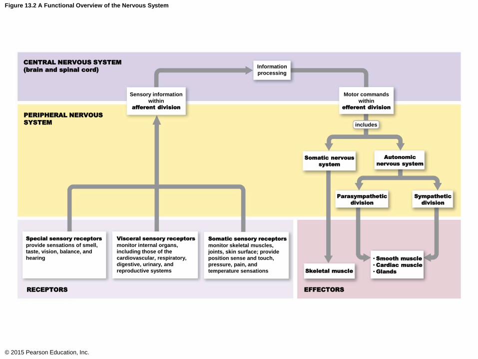
Figure 13.2 A Functional Overview of the Nervous System
© 2015 Pearson Education, Inc.
CENTRAL NERVOUS SYSTEM
(brain and spinal cord)
Sensory information
within
afferent division
PERIPHERAL NERVOUS
SYSTEM
Information
processing
Motor commands
within
efferent division
includes
Somatic nervous
system
Autonomic
nervous system
Parasympathetic
division
Sympathetic
division
Skeletal muscle
• Smooth muscle
• Cardiac muscle
• Glands
EFFECTORS
Somatic sensory receptors
monitor skeletal muscles,
joints, skin surface; provide
position sense and touch,
pressure, pain, and
temperature sensations
Visceral sensory receptors
monitor internal organs,
including those of the
cardiovascular, respiratory,
digestive, urinary, and
reproductive systems
Special sensory receptors
provide sensations of smell,
taste, vision, balance, and
hearing
RECEPTORS
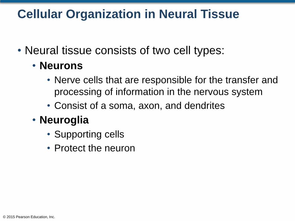
Cellular Organization in Neural Tissue
• Neural tissue consists of two cell types:
• Neurons
• Nerve cells that are responsible for the transfer and
processing of information in the nervous system
• Consist of a soma, axon, and dendrites
• Neuroglia
• Supporting cells
• Protect the neuron
© 2015 Pearson Education, Inc.

Figure 13.4 A Review of Neuron Structure
© 2015 Pearson Education, Inc.
Dendrites Cell body Axon Terminal boutons
Stimulated byenvironmental changesor the activities ofother cells
Contains the nucleus,mitochondria, ribosomes,and other organelles andinclusions
Conducts nerveimpulse (actionpotential) towardsynaptic terminals
Affect anotherneuron or effectororgan (muscle orgland)
AxonhillockMitochondrion
Nucleus
Nucleolus
Nissl bodies(clusters of RER
and free ribosomes)
Dendriticspines

Cellular Organization in Neural Tissue
• Functions of Neuroglia
• Provide the framework for the neural tissue
• Maintain the intercellular environment
• Act as phagocytes
• Have the ability to reproduce
© 2015 Pearson Education, Inc.

Neuroglia
• Neuroglia Cells of the CNS
• Astrocytes
• Oligodendrocytes
• Microglia
• Ependymal cells
• Neuroglia Cells of the PNS
• Satellite cells
• Schwann cells
© 2015 Pearson Education, Inc.

Figure 13.5 The Classification of Neuroglia
© 2015 Pearson Education, Inc.
Neuroglia
Peripheral Nervous System Central Nervous System
are found in
containscontains
Satellite cells Schwann cells Oligodendrocytes Astrocytes Microglia Ependymal cells
Surround neuron cellbodies in ganglia;regulate O2, CO2,nutrient, andneurotransmitter levelsaround neurons inganglia
Surround all axons inPNS; responsible formyelination ofperipheral axons;participate in repairprocess after injury
Myelinate CNSaxons; providestructuralframework
Maintain blood-brain barrier;provide structural support;regulate ion, nutrient, anddissolved-gas concentra-tions; absorb and recycleneurotransmitters; form scartissue after injury
Remove celldebris, wastes,and pathogensby phagocytosis
Line ventricles(brain) and centralcanal (spinal cord);assist in producing,circulating, andmonitoringcerebrospinal fluid

Neuroglia
• Neuroglia of the CNS
• Astrocytes
• Have a large number of cytoplasmic processes
• Control the chemical content of the interstitial
environment
• Maintain the blood–brain barrier
• Isolate the neurons from general circulation
© 2015 Pearson Education, Inc.
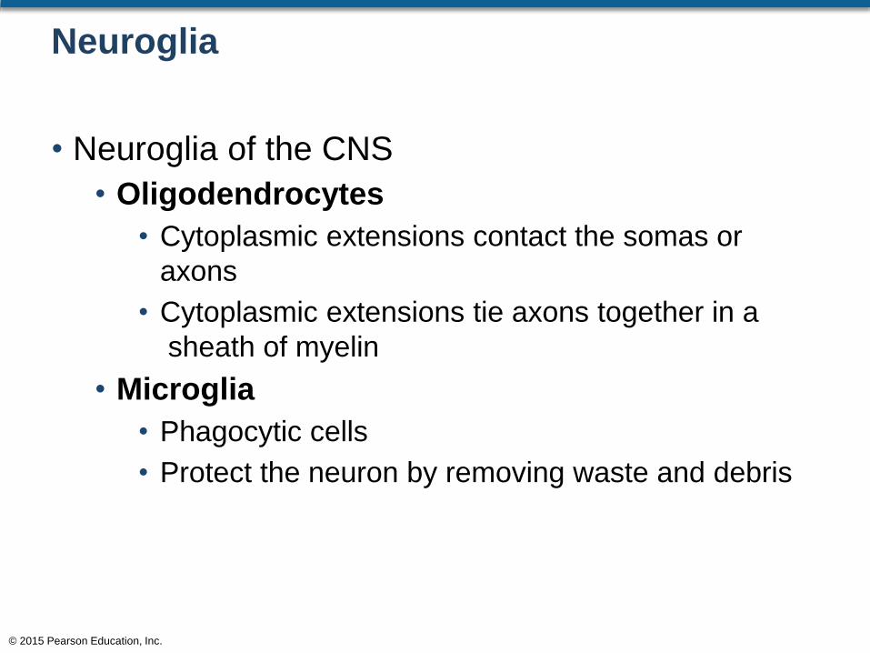
Neuroglia
• Neuroglia of the CNS
• Oligodendrocytes
• Cytoplasmic extensions contact the somas or
axons
• Cytoplasmic extensions tie axons together in a
sheath of myelin
• Microglia
• Phagocytic cells
• Protect the neuron by removing waste and debris
© 2015 Pearson Education, Inc.
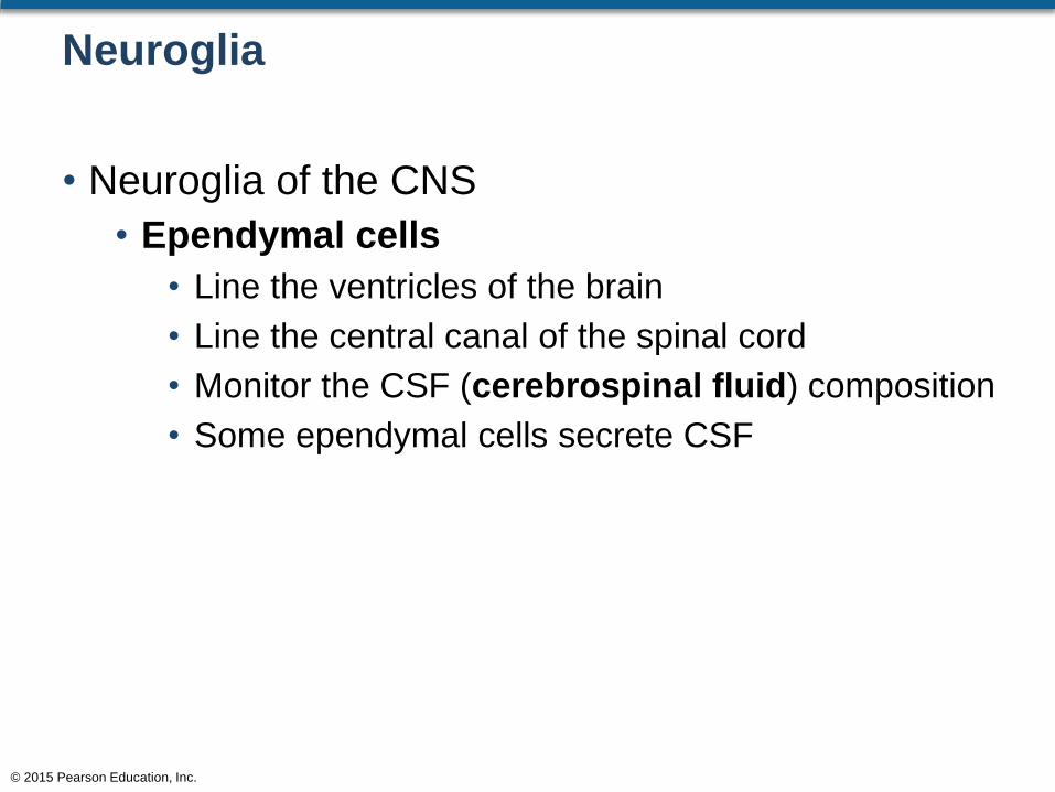
Neuroglia
• Neuroglia of the CNS
• Ependymal cells
• Line the ventricles of the brain
• Line the central canal of the spinal cord
• Monitor the CSF (cerebrospinal fluid) composition
• Some ependymal cells secrete CSF
© 2015 Pearson Education, Inc.
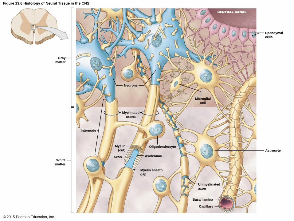
Figure 13.6 Histology of Neural Tissue in the CNS
© 2015 Pearson Education, Inc.
Gray
matter
Neurons
Internode
Myelinated
axons
Myelin
(cut)
White
matter
Oligodendrocyte
Axon Axolemma
Myelin sheath
gap
Unmyelinated
axon
Astrocyte
Basal lamina
Capillary
Microglial
cell
Ependymal
cells
CENTRAL CANAL
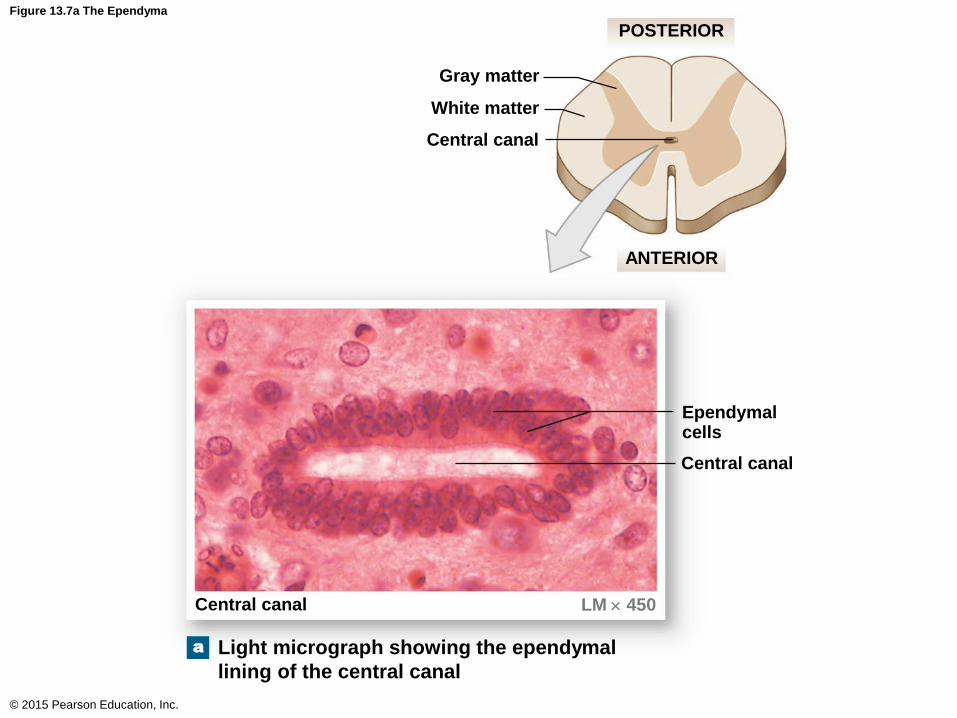
Figure 13.7a The Ependyma
© 2015 Pearson Education, Inc.
Ependymalcells
Gray matter
White matter
Central canal
POSTERIOR
ANTERIOR
Central canal
Central canal LM 450
Light micrograph showing the ependymal
lining of the central canal
a

Neuroglia
• Neuroglia of the PNS
• Satellite cells
• Regulate the exchange of material between the cell
body and the environment
• Schwann cells
• Also called neurolemmocytes
• Form a myelin sheath
© 2015 Pearson Education, Inc.

Figure 13.8 Satellite Cells and Peripheral Neurons
© 2015 Pearson Education, Inc.
Nerve cell body
Nucleus
Satellite cells
Connective tissue
Peripheral ganglion LM 25

Figure 13.9 Myelination (Part 5 of 6)
© 2015 Pearson Education, Inc.
A Myelinated Axon in the PNS
Because each Schwann cell can
myelinate only about 1 mm of an
axon, it takes many Schwann cells to
myelinate an entire axon. The narrow
open region between two adjacent
Schwann cells is called a node. The
internodes are the areas myelinated
by individual Schwann cells.
Nucleus
Dendrite
Myelinated axon
Myelin sheath
Axon
Neurolemma
Schwann cell
nucleus
In this cross section of a myelinated axon,
the myelin sheath appears as concentric
dense lines around the axon.
TEM x 20,600
Myelin sheath of
internode
Axon
Axolemma
Nodes
Initial segment
(unmyelinated)
Myelinated
internode
Axon hillock
Axon

Neurons
• Neuron Structure
• Neurons consist of:
• Axons
• Soma (cell body)
• Dendrites
• Terminal aborizations
• Terminal boutons
• Axon hillock
• Axoplasm
© 2015 Pearson Education, Inc.

Figure 13.10a Anatomy of a Representative Neuron
© 2015 Pearson Education, Inc.
Dendritic spines
Dendrite
Nucleolus
Nucleus
Golgi apparatus
Chromatophilic
susbtance
Neurofilament
Mitochondrion
Axon hillock
Initial segment
of axon
Nerve cell body LM 1600
Nerve cell body
Axon hillock
Initial segment
of axon
Axon (may be
myelinated)
Terminal
boutons
Postsynaptic cell
Multipolar neuron.a
Nerve cell
body

Neurons
• Details of Neuron Structure
• Soma consists of:
• Nucleus
• Nucleolus
• Ribosomes (clusters are called chromatophilic
substance—create gray matter)
• Mitochondria
• Golgi apparatus
• Lack centrosomes—cannot reproduce
© 2015 Pearson Education, Inc.

Figure 13.10a Anatomy of a Representative Neuron
© 2015 Pearson Education, Inc.
Dendritic spines
Dendrite
Nucleolus
Nucleus
Golgi apparatus
Chromatophilic
susbtance
Neurofilament
Mitochondrion
Axon hillock
Initial segment
of axon
Nerve cell body LM 1600
Nerve cell body
Axon hillock
Initial segment
of axon
Axon (may be
myelinated)
Terminal
boutons
Postsynaptic cell
Multipolar neuron.a
Nerve cell
body

Neurons
• Details of Neuron Structure
• Axon (nerve fiber) consists of:
• Axon hillock area
• Axoplasm
• Axon vesicles
• Contain neurotransmitters
© 2015 Pearson Education, Inc.

Neurons
• Neuron Classification
• Can be classified based on structure or function
• Structural classification
• Based on the number of processes extending from
the cell body
• Functional classification
• Sensory
• Motor
• Interneuron (involved with both sensory and motor)
© 2015 Pearson Education, Inc.

Neurons
• Structural Classification of Neurons
• Anaxonic
• Has many processes but cannot differentiate
between axons and dendrites
• Found only in the CNS
• Bipolar
• The cell body is between the dendrite and axon
• Axons are not myelinated
© 2015 Pearson Education, Inc.
Neurons
• Structural Classification of Neurons
• Pseudounipolar
• The cell body is off to one side of the axon
• Multipolar
• Typically has a single axon and multiple dendrites
• Most common type in the CNS
© 2015 Pearson Education, Inc.

Figure 13.10b Anatomy of a Representative Neuron
© 2015 Pearson Education, Inc.
Collateral branch
AxonNeuron
Dendrites
NeuronSynapses with
another neuron
Neuromuscular synapses
Terminal arborization
Terminal boutons Skeletal
muscle
A neuron may innervate (1) other neurons, (2) skeletal muscle fibers,
or (3) gland cells. Synapses are shown in boxes for each example.
A single neuron would not innervate all three.
Neuroglandular
synapses
Gland cells
1. Synapses with another neuron
2. Neuromuscular synapses
3. Neuroglandular synapses
b
Neurons
• Functional Classification of Neurons
• Sensory (afferent division)
• Almost all are pseudounipolar neurons
• Sends information from the PNS to the CNS
• There are:
• Somatic sensory and visceral sensory
• Motor (efferent division)
• Sends information from the CNS to the periphery
• Consists of:
• Somatic nerves and autonomic nerves
© 2015 Pearson Education, Inc.
Neurons
• Functional Classification of Neurons
• Interneurons
• Located entirely in the CNS
• Situated between the motor and sensory neurons
• Analyze sensory input and coordinate motor
outputs
• Can be excitatory or inhibitory
© 2015 Pearson Education, Inc.
Neurons
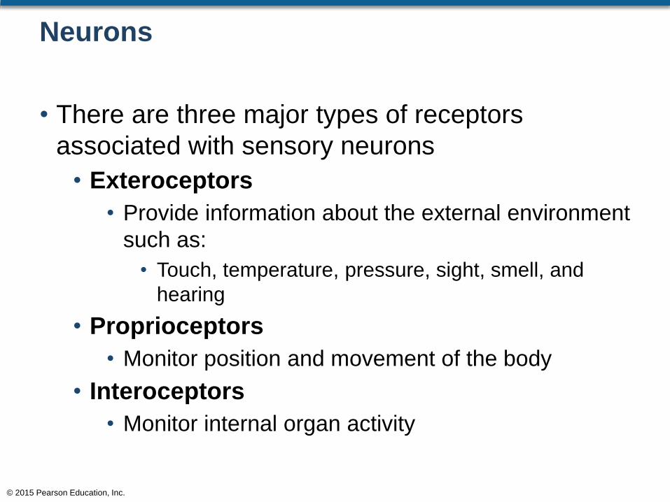
• There are three major types of receptors
associated with sensory neurons
• Exteroceptors
• Provide information about the external environment
such as:
• Touch, temperature, pressure, sight, smell, and
hearing
• Proprioceptors
• Monitor position and movement of the body
• Interoceptors
• Monitor internal organ activity
© 2015 Pearson Education, Inc.

Figure 13.12 A Functional Classification of Neurons
© 2015 Pearson Education, Inc.
RECEPTORS
Interoceptors
EFFECTORS
PERIPHERAL NERVOUS SYSTEM CENTRAL NERVOUS SYSTEM
Exteroceptors
Proprioceptors
Skeletal muscles
Skeletalmusclefibers
Smoothmuscles
Visceral effectors
Glands
Cardiacmuscle
Adiposetissue
Postganglionicfibers
Preganglionicfibers
Visceral
motor neurons
in peripheral
motor ganglia
Visceral
motor
neurons
in CNS
= Somatic (sensory & motor)
= Visceral (sensory & motor)
Afferent fibers
Efferent fibers
Sensory neurons
in peripheral ganglia
Somatic
motor
neurons
Interneurons

Neural Regeneration
• Neural Regeneration
• Steps involved in the limited ability to repair
• Schwann cells grow into the cut area
• Axon sends buds into network of Schwann cells
• Axons begin to grow into the Schwann cells
© 2015 Pearson Education, Inc.

Figure 13.13 Nerve Regeneration
© 2015 Pearson Education, Inc.
Site of injury
Fragmentation of axon and
myelin occurs in distal stump.
Axon Myelin Proximal stump Distal stump
Schwann cells form cord, grow into cut, and unite stumps.
Macrophages engulf degenerating axon and myelin.
Schwann cell Macrophage
Axon sends buds into network of Schwann cells
and then starts growing along cord of Schwann cells.
Axon continues to grow into distal stump and is enfolded
by Schwann cells.
1
2
3
4

The Nerve Impulse
• A nerve impulse is the action potential of a
nerve
• The action potential is due to the exchange of
ions across the membrane
• The ability to conduct the impulse is known as
excitability
© 2015 Pearson Education, Inc.

The Nerve Impulse
• A stimulus is anything that causes an action
potential to occur
• The stimulus has to overcome the threshold
level of that particular neuron
• The threshold level is the amount of stimuli
required to create the action potential
• Once an impulse starts, it is propagated along the
length of the axon
© 2015 Pearson Education, Inc.
The Nerve Impulse
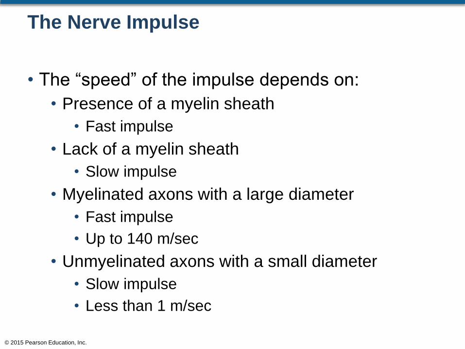
• The “speed” of the impulse depends on:
• Presence of a myelin sheath
• Fast impulse
• Lack of a myelin sheath
• Slow impulse
• Myelinated axons with a large diameter
• Fast impulse
• Up to 140 m/sec
• Unmyelinated axons with a small diameter
• Slow impulse
• Less than 1 m/sec
© 2015 Pearson Education, Inc.

Synaptic Communication
• Vesicular Synapses
• A synapse is the junction between:
• Axodendritic
• The axon of one neuron and the dendrite of
another neuron
• Axosomic
• The axon of one neuron and the soma of another
neuron
• Axoaxonic
• The axon of one neuron and the axon of another
neuron
© 2015 Pearson Education, Inc.

Synaptic Communication
• Vesicular Synapses
• A synapse is the junction between:
• Neuromuscular
• The axon of a neuron and a muscle
• Neuroglandular
• The axon of a neuron and a gland
© 2015 Pearson Education, Inc.
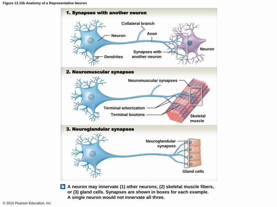
Figure 13.10b Anatomy of a Representative Neuron
© 2015 Pearson Education, Inc.
Collateral branch
AxonNeuron
Dendrites
NeuronSynapses with
another neuron
Neuromuscular synapses
Terminal arborization
Terminal boutons Skeletal
muscle
A neuron may innervate (1) other neurons, (2) skeletal muscle fibers,
or (3) gland cells. Synapses are shown in boxes for each example.
A single neuron would not innervate all three.
Neuroglandular
synapses
Gland cells
1. Synapses with another neuron
2. Neuromuscular synapses
3. Neuroglandular synapses
b
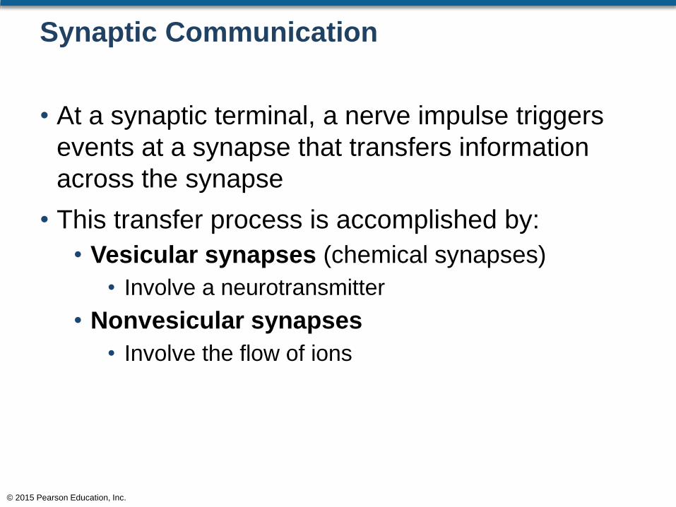
Synaptic Communication
• At a synaptic terminal, a nerve impulse triggers
events at a synapse that transfers information
across the synapse
• This transfer process is accomplished by:
• Vesicular synapses (chemical synapses)
• Involve a neurotransmitter
• Nonvesicular synapses
• Involve the flow of ions
© 2015 Pearson Education, Inc.

Synaptic Communication
• Vesicular Synapse Events
• Impulses are conveyed in one direction only
• Sequence of events:
• An action potential arrives at the presynaptic membrane
• This triggers the release of a neurotransmitter from the
axon vesicles
• The neurotransmitter diffuses across the synapse
• The neurotransmitter binds to the postsynaptic
membrane
• This binding action causes a change in the permeability
of the postsynaptic membrane
• This change in permeability results in an action potential
of the next neuron
© 2015 Pearson Education, Inc.
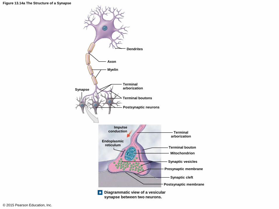
Figure 13.14a The Structure of a Synapse
© 2015 Pearson Education, Inc.
Dendrites
Terminalarborization
Axon
Myelin
Synapse
Terminal boutons
Postsynaptic neurons
Terminalarborization
Impulseconduction
Endoplasmicreticulum
Terminal bouton
Mitochondrion
Synaptic vesicles
Presynaptic membrane
Synaptic cleft
Postsynaptic membrane
Diagrammatic view of a vesicular
synapse between two neurons.
a

Synaptic Communication
• Nonvesicular Synapse Events
• Impulses can be conveyed in any direction
• Sequence of events:
• The presynaptic membrane of one neuron is tightly
bound to the postsynaptic membrane of another
neuron
• This binding permits the passage of ions from one
neuron to the next
© 2015 Pearson Education, Inc.
Neuron Organization and Processing
• Neurons can be organized into smaller organized
groups called neuronal pools
• The neuronal pools are identified by their neural
circuitry such as:
• Divergence
• Convergence
• Serial processing
• Parallel processing
• Reverberation
© 2015 Pearson Education, Inc.
Neuron Organization and Processing
• Divergence
• The spread of information from one neuron to
several neurons
• Permits broad distribution of a specific input
• Information enters the CNS and then spreads to
the brain and spinal cord at the same time
• Convergence
• Information going from several neurons to a single
neuron
• Movements of the diaphragm muscle are
involuntary, but yet at times we can move the
diaphragm muscle voluntarily© 2015 Pearson Education, Inc.
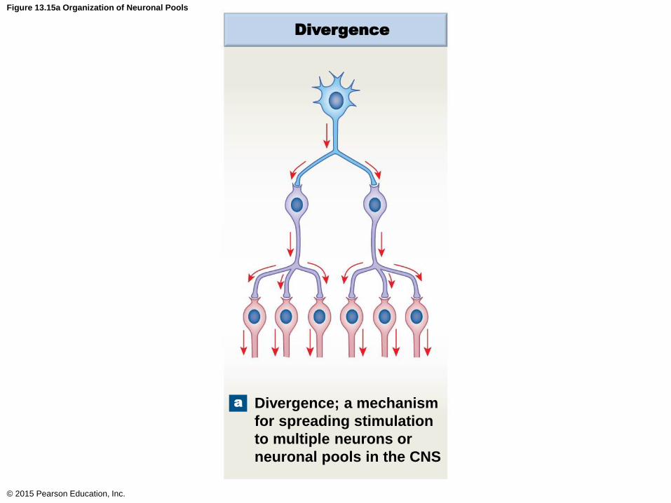
Figure 13.15a Organization of Neuronal Pools
© 2015 Pearson Education, Inc.
Divergence
Divergence; a mechanism
for spreading stimulation
to multiple neurons or
neuronal pools in the CNS
a
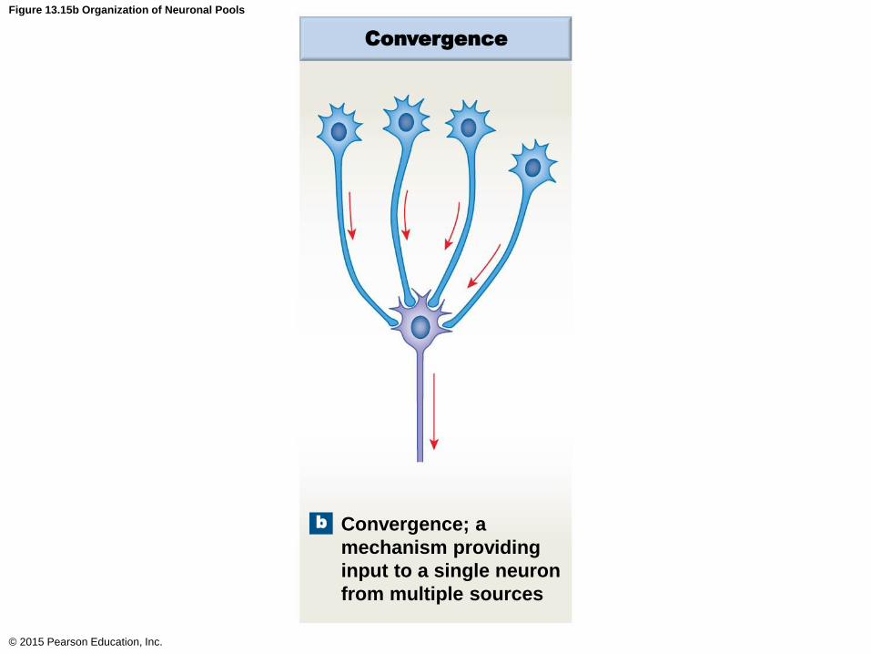
Figure 13.15b Organization of Neuronal Pools
© 2015 Pearson Education, Inc.
Convergence
Convergence; a
mechanism providing
input to a single neuron
from multiple sources
b

Neuron Organization and Processing
• Serial Processing
• Information going from one neuron to the next in a
sequence
• Information going to one part of the brain, then to
another part, and then to another part, etc.
• Parallel Processing
• Several neurons are processing the information at
the same time
• If you step on a nail, you typically move your foot,
shout “ouch,” and dance a bit, all at the same time
© 2015 Pearson Education, Inc.
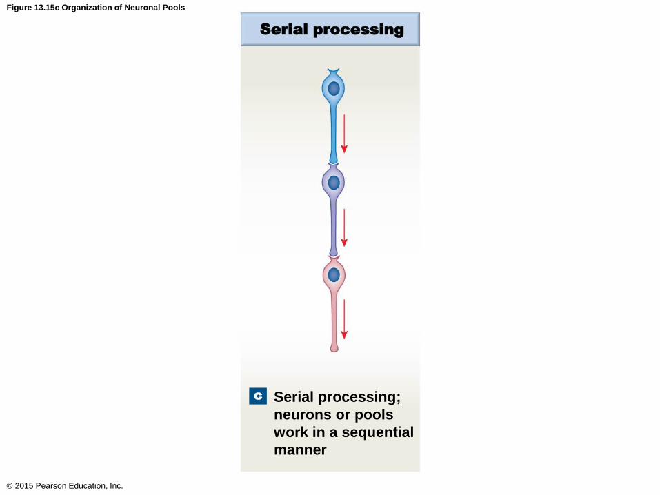
Figure 13.15c Organization of Neuronal Pools
© 2015 Pearson Education, Inc.
Serial processing
Serial processing;
neurons or pools
work in a sequential
manner
c
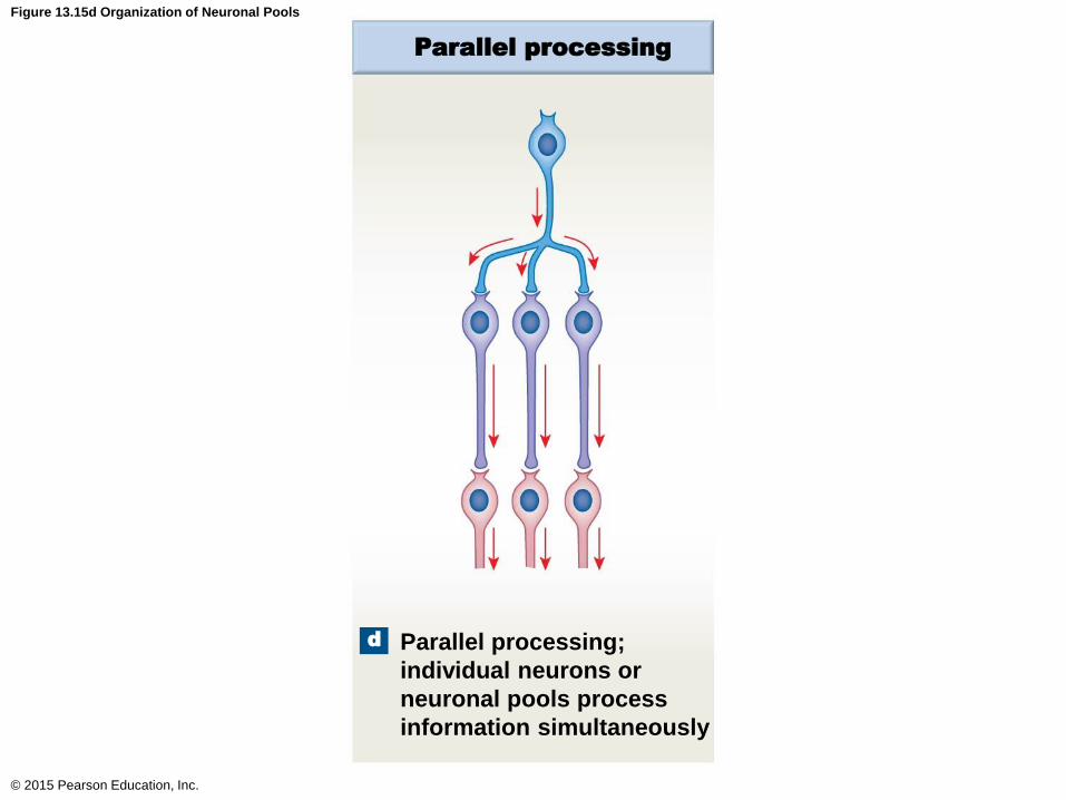
Figure 13.15d Organization of Neuronal Pools
© 2015 Pearson Education, Inc.
Parallel processing
Parallel processing;
individual neurons or
neuronal pools process
information simultaneously
d

Neuron Organization and Processing
• Reverberation
• Collateral axons extend back toward the origin of
the impulse to cause an enhancement or a
continuation of the impulse
© 2015 Pearson Education, Inc.
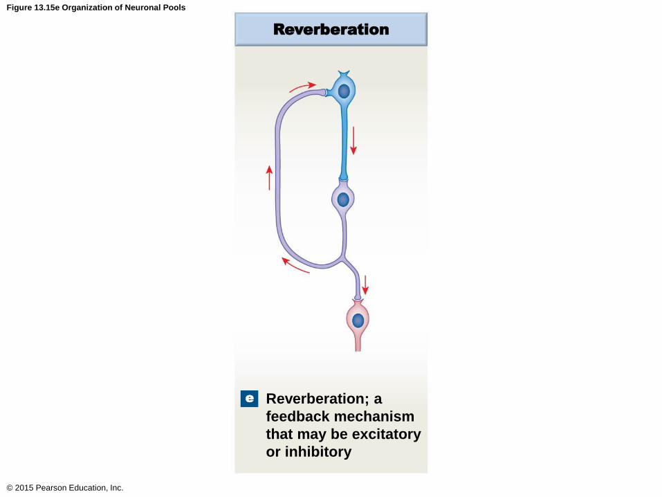
Figure 13.15e Organization of Neuronal Pools
© 2015 Pearson Education, Inc.
Reverberation
Reverberation; a
feedback mechanism
that may be excitatory
or inhibitory
e
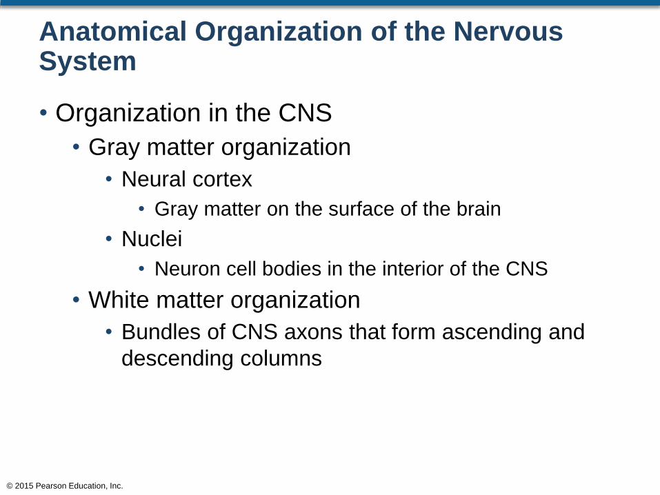
Anatomical Organization of the Nervous System
• Organization in the CNS
• Gray matter organization
• Neural cortex
• Gray matter on the surface of the brain
• Nuclei
• Neuron cell bodies in the interior of the CNS
• White matter organization
• Bundles of CNS axons that form ascending and
descending columns
© 2015 Pearson Education, Inc.
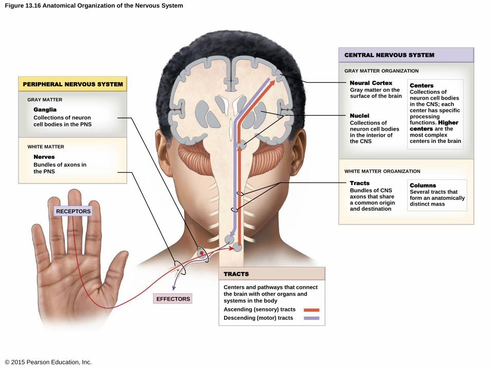
Figure 13.16 Anatomical Organization of the Nervous System
© 2015 Pearson Education, Inc.
Centers and pathways that connect
the brain with other organs and
systems in the body
Ascending (sensory) tracts
Descending (motor) tracts
EFFECTORS
TRACTS
RECEPTORS
PERIPHERAL NERVOUS SYSTEM
GRAY MATTER
WHITE MATTER
Ganglia
Nerves
Collections of neuron
cell bodies in the PNS
Bundles of axons in
the PNS
CENTRAL NERVOUS SYSTEM
GRAY MATTER ORGANIZATION
Neural Cortex
Nuclei
Centers
Collections ofneuron cell bodiesin the CNS; eachcenter has specificprocessingfunctions. Higher
centers are themost complexcenters in the brain
WHITE MATTER ORGANIZATION
TractsColumns
Gray matter on thesurface of the brain
Collections ofneuron cell bodiesin the interior ofthe CNS
Bundles of CNSaxons that sharea common originand destination
Several tracts thatform an anatomicallydistinct mass
Anatomical Organization of the Nervous System
• Organization in the PNS
• Gray matter
• Ganglia are collections of neuron cell bodies in the
PNS
• White matter
• Bundles of axons in the PNS
© 2015 Pearson Education, Inc.

Figure 13.16 Anatomical Organization of the Nervous System
© 2015 Pearson Education, Inc.
Centers and pathways that connect
the brain with other organs and
systems in the body
Ascending (sensory) tracts
Descending (motor) tracts
EFFECTORS
TRACTS
RECEPTORS
PERIPHERAL NERVOUS SYSTEM
GRAY MATTER
WHITE MATTER
Ganglia
Nerves
Collections of neuron
cell bodies in the PNS
Bundles of axons in
the PNS
CENTRAL NERVOUS SYSTEM
GRAY MATTER ORGANIZATION
Neural Cortex
Nuclei
Centers
Collections ofneuron cell bodiesin the CNS; eachcenter has specificprocessingfunctions. Higher
centers are themost complexcenters in the brain
WHITE MATTER ORGANIZATION
TractsColumns
Gray matter on thesurface of the brain
Collections ofneuron cell bodiesin the interior ofthe CNS
Bundles of CNSaxons that sharea common originand destination
Several tracts thatform an anatomicallydistinct mass