Chapter 12: Cardiovascular Physiology Excitation-Contraction …€¦ · Chapter 12: Cardiovascular...
51
Chapter 12: Cardiovascular Physiology The mechanism that couples excitation – an action potential in the plasma membrane of the muscle cell – and contraction of heart muscle Excitation-Contraction Coupling
Transcript of Chapter 12: Cardiovascular Physiology Excitation-Contraction …€¦ · Chapter 12: Cardiovascular...

Chapter 12:Cardiovascular Physiology
The mechanism that couples excitation – an
action potential in the plasma membrane of
the muscle cell – and contraction of heart
muscle
Excitation-Contraction Coupling

Figure 9-12
Passage of an action potential along the transverse tubule opens nearby voltage-gated calcium channels, the “ryanodine receptor,”located on the sarcoplasmic reticulum, andcalcium ions released into the cytosol bind to troponin.The calcium-troponin complex “pulls” tropomyosin off the myosin-binding site of actin, thus allowing the binding of the cross-bridge, followed by its flexing to slide the actin filament.

Calcium ions regulate thecontraction of cardiac muscle:
the entry of extracellular calcium ions causes the release of calcium from the sarcoplasmic reticulum (calcium-induced calcium release), the source of about 95% of the calcium in the cytosol.
Figure 12-17
Excitation-contraction coupling in cardiac muscle

Click here to play theCardiac EC Coupling
Flash Animation

Cardiac cycle
• The cardiac events that occur from beginning of one
heartbeat to the beginning of the next are called the
cardiac cycle
Click here to play theMechanical Eventsof the Cardiac Cycle
Flash Animation
Mechanical Events of the Cardiac Cycle

• Pressure
• Volume
• Valves
• Blood flow
What happens in the heart during each cardiac cycle?

Systole:ventricles contracting
Diastole:ventricles relaxed
Figure 12-19

Summary of events in the left atrium, left ventricle, and aorta during the cardiac cycle
Figure 12-20


Pressure changes in the right heart during a contraction cycle.
Figure 12-21

• Atria──primer pump
• Ventricles──major source of power
Role of atria and ventricles during each cardiac cycle

Heart Sounds
• 1st sound– soft low-pitched lub– associated with closure of the AV valves– Marks the onset of systole
• 2nd sound– louder dup– associated with closure of the PA and aortic valves– Occurs at the onset of diastole

Chest surface areas for auscultation of normal heart sounds
Four traditional value areas – Aortic space: 2RIS – Pulmonic valve: 2LIS – Tricuspid valve: 4ICS LLSB– Mitral valve: Apex
RIS--right intercostal spaceLIS—left intercostal space ICS--intercostal space LLSB--left lower sternal border

Phonocardiogram

Heart valve defects causing turbulent blood flow and murmurs

Acute rheumatic fever
Mitral stenosis -- Accentuated first sound
Mitral stenosis – Presystolic murmur
Mitral regurgitation -- systolic murmur
Aortic insufficiency -- Loud systolic ejection murmur,third sound

Evaluation of Heart Pumping
1. Stroke volume (SV): volume
of blood pumped per beat
SV = EDV - ESV
EDV: end-diastolic volume
ESV: end-systolic volume
~70ml (60~80ml)

heart enlargement
Stroke volume for evaluation?

2. Ejection fraction (EF)
EF=(SV/EDV) x 100%
55~65%

3. Cardiac output (CO): the total volume of blood
pumped by each ventricle per minute
CO=SV x heart rate (HR)
5 L/min (4.5~6.0 L/min)

What parameters for comparison of people in different size?

4. Cardiac index (CI):
cardiac output per
square meter of body
surface area
3.0~ 3.5 L/min•m2

5. Cardiac reserve: the maximum percentage that the
cardiac output can increase above the normal level
In the normal young adult the cardiac reserve is 300
to 400 percent
Achieved by an increase in either stroke volume (SV)
or heart rate (HR) or both
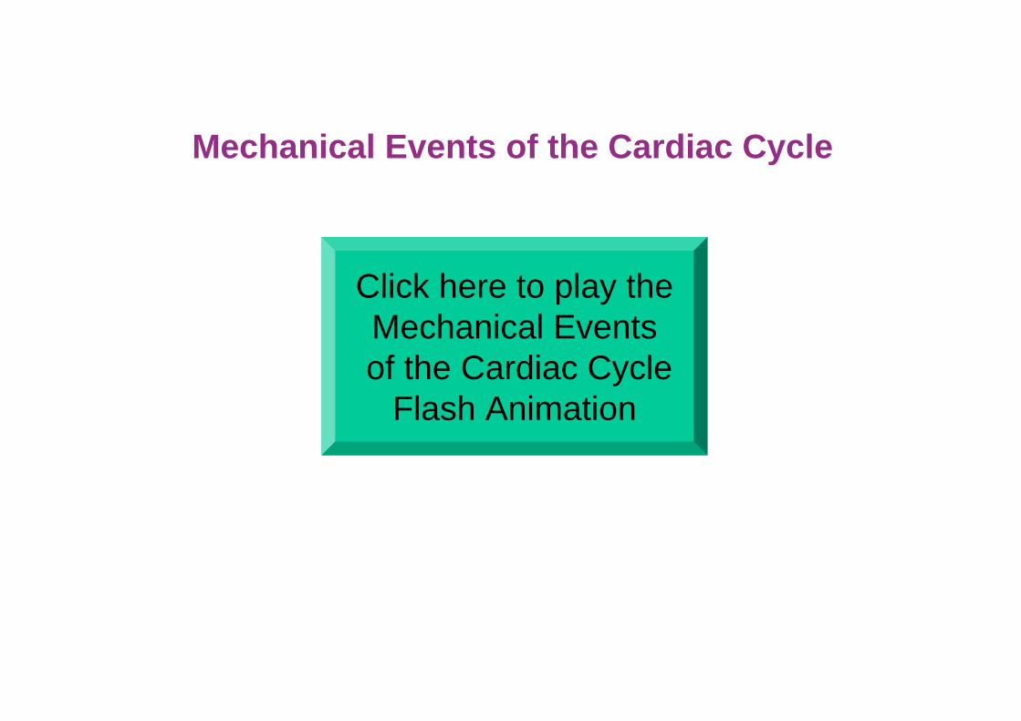
Measurement of Cardiac Function
• Echocardiography

• Cardiac angiography
Coronary Angiography from a 56-year-old man presented with unstable angina and acute pulmonary edema
Rerkpattanapipat P, et al. Circulation. 1999;99:2965

Regulation of heart pumping

Regulation of stroke volume
1. Preload – Frank-Starling mechanism
Preload of ventricles:end-diastolic volume (EDV)end-diastolic pressure (EDP)

Frank-Starling mechanism(Intrinsic regulation or heterometric regulation)
The fundamental principle of cardiac behavior which states that the force of contraction of the cardiac muscle is proportional to its initial length
Significance:Precise regulation of SV

To increase the heart’s stroke volume:fill it more fully with blood. The increased stretch of the ventricle will align its actin and myosin in a more optimal pattern of overlap.
Figure 12-25 Control of stroke volume
Frank-Starling mechanism

Ventricular function curve (Frank-Starling curve)

Ventricular function curve (Frank-Starling curve)

Factors affecting preload (EDV)
• (1) Venous return
• Filling time
• Venous return rate
• Compliance
• (2) Residual blood in ventricles after ejection

2. Afterload (Usually measured as arterial pressure)


Afterload has very little effect on the normal ventricleHowever, as systolic failure develops even small increases in afterload have significant effects on compromised ventricular systolic functionConversely, small reductions in afterload in a failing ventricle can have significant beneficial effects on impaired contractility
Congestive heart failure (CHF)

3. Myocardial contractility (Inotropic state)
Homometric regulation

To further increase the stroke volume:fill it more fully with blood
ANDdeliver sympathetic signals (norepinephrine and epinephrine);it will also relax more rapidly, allowing more time to refill.
Figure 12-26

Sympathetic signals (norepinephrine and epinephrine) cause a stronger and more rapid contraction and a more rapid relaxation.
Figure 12-27


Factors regulating contractility


• HR↑→CO↑ (CO = SV x HR)
• HR↑→Contractility↑ (Treppe effect)
• HR↑→ diastolic filling time ↓
Regulation of heart rate
40~180 /min,HR↑→CO↑>180 /min,or <40/min,CO↓


Figure 12-23
Control of heart rate

To speed up the heart rate:• deliver the sympathetic hormone, epinephrine, and/or• release more sympathetic neurotransmitter (norepinephrine), and/or • reduce release of parasympathetic neurotransmitter (acetylcholine).
Figure 12-24
T, ions, metabolites,
other hormones

Staircase phenomenon (Treppe effect , Force-frequency relationship)
Increase in rate of contraction (heart rate) causes increase in contractility


Figure 12-28
To increase SV, increase:end-diastolic volume,norepinephrine delivery from sympathetic neurons, andepinephrine delivery from the adrenal medulla.
To increase HR, increase:norepinephrine delivery from
sympathetic neurons, andepinephrine
delivery from adrenal medulla
(reduce parasympathetic).
It is not possible, under normal circumstances, to increase one but not the other of these determinants of cardiac output.

The End.