Chapter 11: The Muscular System Biol 141 A&P. The Muscular System Consists only of skeletal muscles...
155
Chapter 11: The Muscular System Biol 141 A&P
-
Upload
jordan-bishop -
Category
Documents
-
view
217 -
download
3
Transcript of Chapter 11: The Muscular System Biol 141 A&P. The Muscular System Consists only of skeletal muscles...

Chapter 11:The Muscular System
Biol 141 A&P

The Muscular System
• Consists only of skeletal muscles
• How are fascicles arranged in the various types of muscles, and what are the resulting functional differences?

Muscle Organization and Function
• Muscle organization affects power, range, and speed of muscle movement
Fascicles• Muscle cells (fibers) are organized in
bundles (fascicles)
Classification of Skeletal Muscles
• By the way fascicles are organized• By relationships of fascicles to tendons

Organization of Skeletal Muscle Fibers
• 4 patterns of fascicle organization:• 1) Parallel-
– Fibers parallel to the long axis of muscle
- e.g., biceps brachii• 2) Convergent-• 3) Pennate-• 4) Circular-
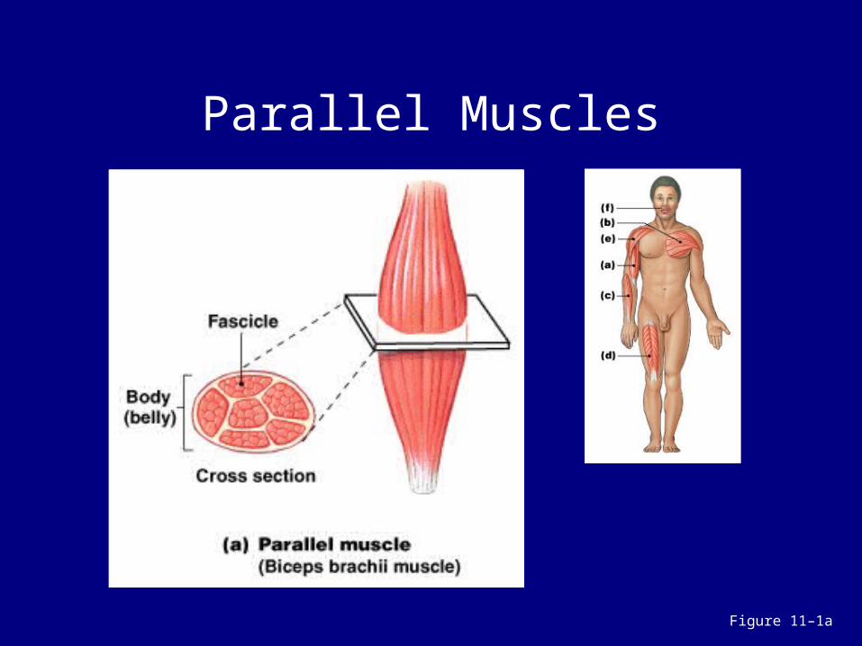
Parallel Muscles
Figure 11–1a

Parallel Muscle Body• The center or body of the muscle
thickens when parallel muscle contracts• Parallel muscles contract about 30%
Parallel Muscle Tension • Depends on total number of myofibrils• Directly relates to cross section of
muscle• 1 in.2 (6.45 cm2) of cross section
develops 50 lb (23 kg) of tension

Convergent Muscles
Figure 11–1b

Convergent Muscles
• A broad area converges on attachment site (tendon, aponeurosis, or raphe)
• Muscle fibers pull in different directions, depending on stimulation
• e.g., pectoralis muscles

Pennate Muscles
Figure 11–1c, d, e

Pennate Muscles • Unipennate:
– fibers on 1 side of tendon – e.g., extensor digitorum
• Bipennate:– fibers on both sides of tendon– e.g., rectus femoris
• Multipennate:– tendon branches within muscle– e.g., deltoid

Pennate Muscles
• Form an angle with the tendon• Do not move as far as parallel
muscles• Contain more myofibrils than
parallel muscles• Develop more tension than parallel
muscles

Circular Muscles
Figure 11–1f

Circular Muscles
• Also called sphincters • Open and close to guard entrances
of body• e.g., obicularis oris

Skeletal Motion
• Skeletal muscles attach to skeleton, produce motion
• Type of muscle attachment affects power, range, and speed of muscle movement

What are the classes of levers, and how do they
make muscles more efficient?
Levers• Mechanically, each bone is a lever
(a rigid, moving structure):– and each joint a fulcrum (a fixed point)
• Muscles provide applied force (AF):– required to overcome resistance (R)

Functions of a Lever
• To change:– direction of an AF– distance and speed of movement
produced by an AF– effective strength of an AF
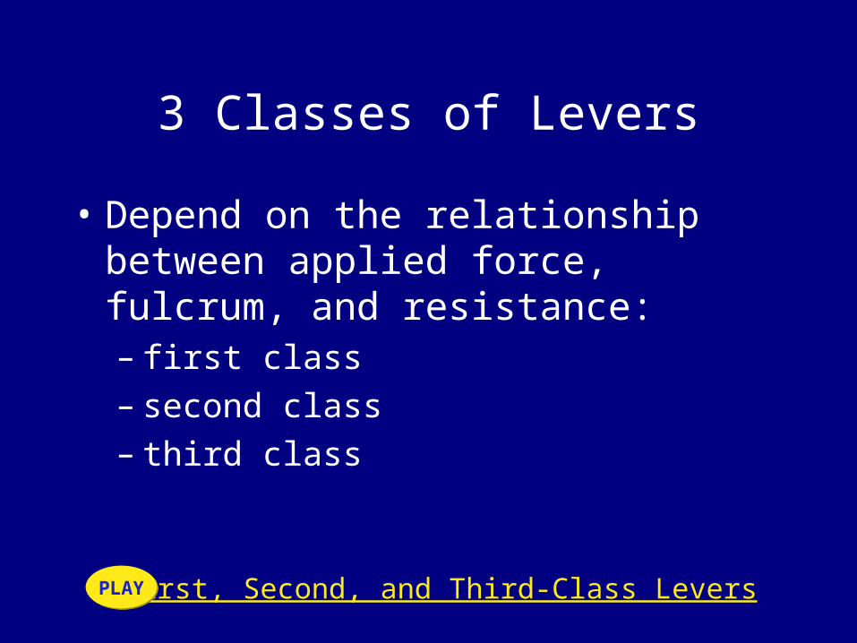
3 Classes of Levers
• Depend on the relationship between applied force, fulcrum, and resistance:– first class– second class– third class
First, Second, and Third-Class LeversPLAYPLAY

First-Class Levers
Figure 11–2a

First-Class Levers
• Seesaw is an example• Center fulcrum between applied
force and resistance• Force and resistance are balanced

Second–Class Levers
Figure 11–2b

Second-Class Levers
• Wheelbarrow is an example• Center resistance between applied
force and fulcrum• A small force moves a large weight

Third-Class Levers
Figure 11–2c

Third-Class Levers
• Most common levers in the body• Center applied force between
resistance and fulcrum• Greater force moves smaller
resistance• Maximizes speed and distance
traveled

KEY CONCEPT
• Skeletal muscles can shorten to 70% of resting length
• Power, speed, and range of movement
• Depend on positions of muscle attachment
• Relative to joints

How are actions of a muscle based on the
relative positions of its origin and insertion?

Origins and Insertions
• Muscles have 1 fixed point of attachment (origin) and 1 moving point of attachment (insertion)
• Most muscles originate or insert on the skeleton
• Origin is usually proximal to insertion

Actions
• Movements produced by muscle contraction
• Body movements – e.g., flexion, extension, adduction,
etc.
• Described in terms of bone, joint, or region

How do muscles interact to produce or oppose
movements?
Muscle Interactions
• Muscles work in groups to maximize efficiency
• Smaller muscles reach maximum tension first, followed by larger, primary muscles

Muscle Terminology Based on Function
• Agonist (prime mover):– produces a particular movement
• Antagonist:– opposes movement of a particular agonist
• Synergist:– a smaller muscle that assists a larger
agonist– helps start motion or stabilize origin of
agonist (fixator)

Muscle Opposition
• Agonists and antagonists work in pairs:– when 1 contracts, the other stretches– i.e., flexors–extensors, abductors–
adductors, etc.

How does the name of a muscle help identify its location, appearance, or
function?

Names of Skeletal Muscles
• Correct names of muscles include the term muscle
• Exceptions: – platysma– diaphragm

Naming Skeletal Muscles
Table 11–1 (1 of 2)

Naming Skeletal Muscles
Table 11–1 (2 of 2)

Descriptive Names for Skeletal Muscles
1. Location in the body-Identifies body regions: e.g.- temporalis muscle
2. Origin and insertion- First part of name indicates origin, Second part of name indicates insertion: e.g. genioglossus muscle
3. Fascicle organization-Describes fascicle orientation within muscle:
– i.e., rectus (straight), transversus, oblique

Descriptive Names for Skeletal Muscles
4. Relative position- Externus (superficialis):– visible at body surface
• Internus (profundus):deep muscles• Extrinsic:muscles outside an organ• Intrinsic:muscles inside an organ5. Structural characteristics- Number of tendons:
– bi = 2, tri = 3
• Shape: trapezius, deltoid, rhomboid• Size-6. Action- Movements: e.g., flexor, extensor,
retractor• Occupations or habits: e.g., risor = laughter

Names for Muscle Size (1 of 2)
• Longus = long• Longissimus = longest• Teres = long and round• Brevis = short• Magnus = large• Major = larger• Maximus = largest• Minor = small• Minimus = smallest

Axial and Appendicular Muscles
Figure 11–3a
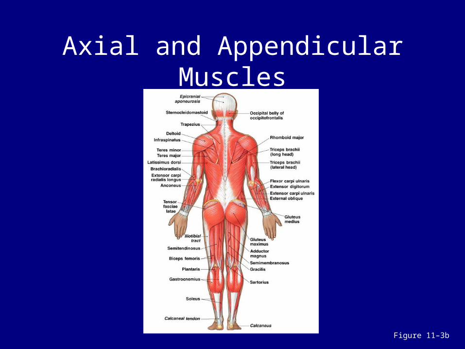
Axial and Appendicular Muscles
Figure 11–3b

Divisions of the Muscular System
1. Axial muscles:– position head and spinal column– move rib cage– 60% of skeletal muscles
2. Appendicular muscles:– support pectoral and pelvic girdles– support limbs– 40% of skeletal muscles

What are the principle axial muscles of the body, their origins, insertions, actions, and innervation?
The Axial Muscles• Divisions based on location and
function:– muscles of head and neck– muscles of vertebral column– oblique and rectus muscles– muscles of pelvic floor

3D Peel-Away of Muscles of the Head and NeckPLAYPLAY
6 Muscle Groups of the Head and Neck
• Muscles of facial expression:– originate on skull
• Extrinsic eye muscles:– originate on surface of orbit– control position of eye

6 Muscle Groups of the Head and Neck
• Muscles of mastication:– move the mandible
• Muscles of the tongue:– names end in glossus
• Muscles of the pharynx:– begin swallowing process

6 Muscle Groups of the Head and Neck
• Anterior muscles of the neck:– control position of larynx– depress the mandible– support tongue and pharynx

Muscles of Facial Expression
Figure 11–4a

Muscles of Facial Expression
Figure 11–4b

Muscles of Facial Expression
• Orbicularis oris:– constricts the mouth opening
• Buccinator:– moves food around the cheeks
• Muscles of the epicranium (scalp)

Muscles of the Epicranium (Scalp)
• Temporoparietalis• Occipitofrontalis:
– frontal and occipital bellies– separated by epicranial aponeurosis
• Platysma:– covers anterior surface of neck

Summary: Muscles of Facial Expression
Table 11–2 (1 of 2)

Summary: Muscles of Facial Expression
Table 11–2 (2 of 2)

Extrinsic Eye Muscles
• Also called extra-ocular muscles
Figure 11–5a, b

Extrinsic Eye Muscles
Figure 11–5c
Inferior rectusMedial rectusSuperior rectusLateral rectusInferior obliqueSuperior oblique

Summary: Extrinsic Eye Muscles
Table 11–3

Muscles of Mastication
Figure 11–6

3 Muscles of Mastication
• Masseter:– the strongest jaw muscle
• Temporalis:– helps lift the mandible
• Pterygoid muscles:– position mandible for chewing

Summary: Muscles of Mastication
Table 11–4

Muscles of the Tongue
Figure 11–7

4 Muscles of the Tongue
• Palatoglossus:– originates at palate
• Styloglossus:– originates at styloid process
• Genioglossus:– originates at chin
• Hypoglossus:– originates at hyoid bone

Summary: Muscles of the Tongue
Table 11–5

Muscles of the Pharynx
Figure 11–8

3 Muscles of the Pharynx
• Pharyngeal constrictor muscles:– move food into esophagus
• Laryngeal elevator muscles:– elevate the larynx
• Palatal muscles:– lift the soft palate

Summary: Muscles of the Pharynx
Table 11–6

Anterior Muscles of the Neck
Figure 11–9

6 Anterior Muscles of the Neck • Digastric:
– from chin to hyoid– and hyoid to mastoid
• Mylohyoid:– floor of the mouth
• Geniohyoid:– between hyoid and chin
• Stylohyoid:– between hyloid and styloid
• Sternocleidomastoid:– from clavicle and sternum to mastoid
• Omohyoid:– attaches scapula, clavicle, first rib, and hyoid

Summary: Anterior Muscles of the Neck
Table 11–7

Muscles of the Vertebral Column
Figure 11–10a

Muscles of the Vertebral Column
• Spinal extensors or erector spinae muscles (superficial and deep)
• Spinal flexors (transversospinalis)

Superficial Spinal Extensors• Spinalis group• Longissimus group• Iliocostalis group
Deep Spinal Extensors• Semispinalis group• Multifidus muscle• Interspinalis muscles• Intertransversarii muscles• Rotatores muscles

Spinal Flexors
• Neck:– longus capitis and longus colli – rotate and flex the neck
• Lumbar:– quadratus lumborum muscles – flex spine and depress ribs

Summary: Muscles of the Vertebral Column
Table 11–8 (1 of 2)

Summary: Muscles of the Vertebral Column
Table 11–8 (2 of 2)

Oblique and Rectus Muscles
• Lie within the body wall
Figure 11–11a, b

Oblique and Rectus Muscles
Figure 11–11a, c

Oblique and Rectus Muscles
• Oblique muscles:– compress underlying structures– rotate vertebral column
• Rectus muscles:– flex vertebral column– oppose erector spinae
Oblique MusclesCervical region:– scalene muscles– flex the neck
Thoracic region:– intercostal muscles (external and
internal intercostals):• respiratory movements of ribs
– transversus thoracis:• cross inner surface of ribs

Oblique Muscles
• Abdominopelvic region (same pattern as thoracic):– external oblique muscles – internal oblique muscles
• Transversus abdominis
Rectus Group• Rectus abdominis:
– between xiphoid process and pubic symphysis
– divided longitudinally by linea alba– divided transversely by tendinous
inscriptions
• Diaphragmatic muscle or diaphragm:– divides thoracic and abdominal cavities– performs respiration

Summary: Oblique and Rectus Muscles
Table 11–9 (1 of 2)

Summary: Oblique and Rectus Muscles
Table 11–9 (2 of 2)

Muscles of the Pelvic Floor
Figure 11–12a

Muscles of the Pelvic Floor
Figure 11–12b

Functions of Pelvic Floor Muscles
1. Support organs of pelvic cavity2. Flex sacrum and coccyx3. Control movement of materials
through urethra and anus

Perineum• Muscular sheet forming the pelvic
floor, divided into:– anterior urogenital triangle – posterior anal triangle
Pelvic Diaphragm– Deep muscular layer extending to
pubis:– supports anal triangle
Urogenital Diaphragm
• Deep muscular layer between pubic bones:– supports the pelvic floor – and muscles of the urethra
• Superficial muscles of the urogenital triangle:– support external genitalia
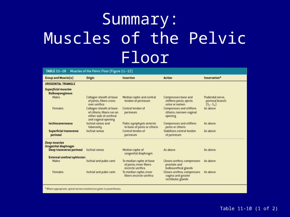
Summary: Muscles of the Pelvic Floor
Table 11-10 (1 of 2)

Summary: Muscles of the Pelvic Floor
Table 11-10 (2 of 2)
What are the principal appendicular muscles of
the body and their origins, insertions,
actions, and innervations?

The Appendicular Muscles
Figure 11–13a

The Appendicular Muscles
Figure 11–13b

The Appendicular Muscles
• Position and stabilize pectoral and pelvic girdles
• Move upper and lower limbs

Divisions of Appendicular Muscles
1) Muscles of the shoulders and upper limbs:
• Position the pectoral girdle• Move the arm• Move the forearm and hand• Move the hand and fingers
2) Muscles of the pelvis and lower limbs

3D Peel-Away of Muscles of the Pectoral GirdlePLAYPLAY
Muscles that Position the Pectoral Girdle
Figure 11–14a

Muscles that Position the Pectoral Girdle
Figure 11–14b
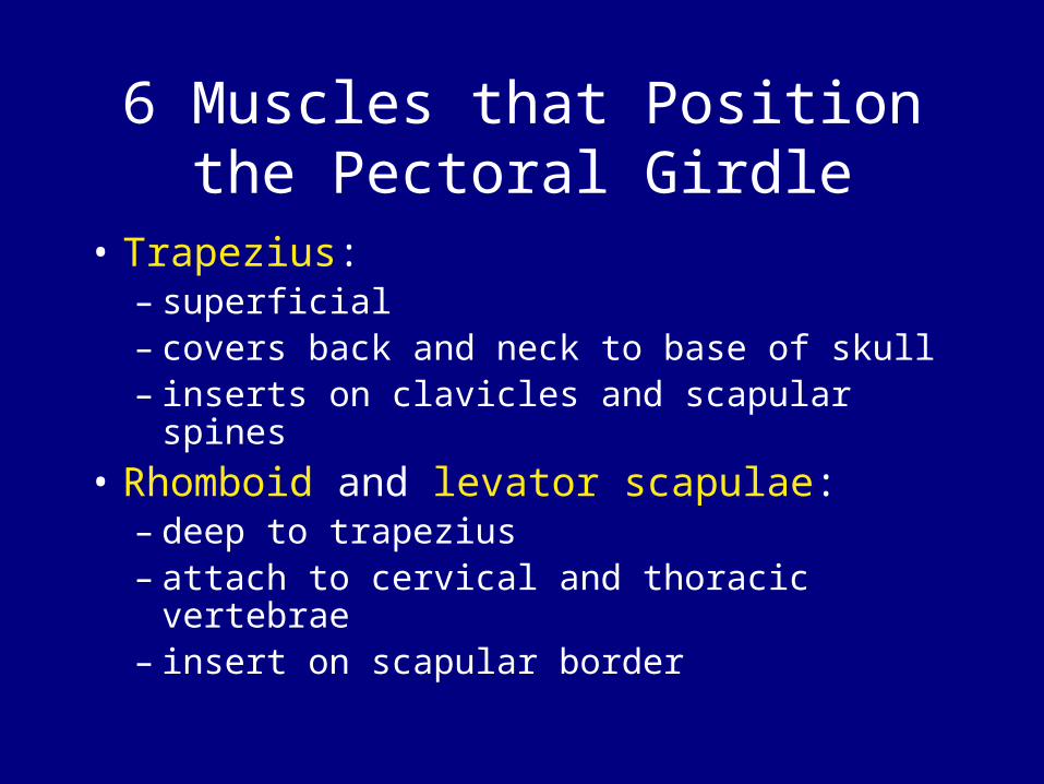
6 Muscles that Position the Pectoral Girdle
• Trapezius:– superficial– covers back and neck to base of skull– inserts on clavicles and scapular spines
• Rhomboid and levator scapulae:– deep to trapezius– attach to cervical and thoracic vertebrae– insert on scapular border

6 Muscles that Position the Pectoral Girdle
• Serratus anterior:– on the chest– originates along ribs– inserts on anterior scapular margin
• Subclavius:– originates on ribs– inserts on clavicle
• Pectoralis minor:– attaches to scapula

Summary: Muscles that Position the Pectoral Girdle
Tables 11–11

Muscles that Move the Arm
Figure 11–15a

Muscles that Move the Arm
Figure 11–15b

9 Muscles that Move the Arm (1 of 3)
• Deltoid: – the major abductor
• Supraspinatus: – assists deltoid
• Subscapularis and teres major: – produce medial rotation at shoulder
3D Rotation: Muscles of the ArmPLAYPLAY

9 Muscles that Move the Arm (2 of 3)
• Infraspinatus and teres minor: – produce lateral rotation at shoulder
• Coracobrachialis: – attaches to scapula– produces flexion and adduction at
shoulder

9 Muscles that Move the Arm (3 of 3)
• Pectoralis major: – between anterior chest and greater
tubercle of humerus– produces flexion at shoulder joint
• Latissimus dorsi:– between thoracic vertebrae and
humerus– produces extension at shoulder joint

The Rotator Cuff
• Muscles involved in shoulder rotation– supraspinatus, subscapularis,
infraspinatus, teres minor,and their tendons

Summary: Muscles that Move the Arm
Table 11–12

Muscles that Move the Forearm and Hand
Figure 11–16a

Muscles that Move the Forearm and Hand
Figure 11–16b
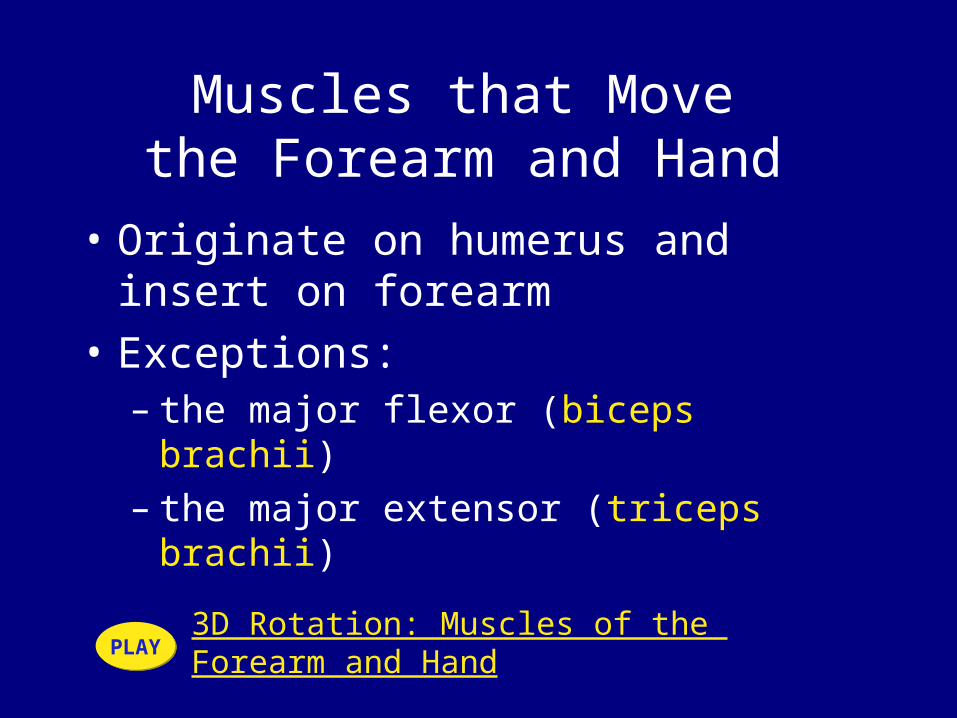
Muscles that Move the Forearm and Hand
• Originate on humerus and insert on forearm
• Exceptions:– the major flexor (biceps brachii)– the major extensor (triceps brachii)
3D Rotation: Muscles of the Forearm and HandPLAYPLAY

Extensors and Flexors
• Extensors:– mainly on posterior and lateral
surfaces of arm
• Flexors:– mainly on anterior and medial
surfaces

13 Muscles that Move the Forearm and Hand
• Biceps brachii:– flexes elbow– stabilizes shoulder joint
• Triceps brachii:– extends elbow– originates on scapula– inserts on olecranon
• Brachialis and brachioradialis:– flex elbow– originates on scapula– inserts on radial tuberosity

13 Muscles that Move the Forearm and Hand
• Anconeus:– opposes brachialis
• Palmaris longus:– superficial– flexes wrist
• Flexor carpi ulnaris: – superficial– flexes wrist– adducts wrist

13 Muscles that Move the Forearm and Hand
• Flexor carpi radialis: – superficial– flexes wrist– abducts wrist
• Extensor carpi radialis:– superficial– extends wrist– abducts wrist
• Extensor carpi ulnaris:– superficial– extends wrist– adducts wrist

13 Muscles that Move the Forearm and Hand (8 of
8)• Pronator teres and supinator:
– originate on humerus and ulna– rotate radius
• Pronator quadratus:– originates on ulna– assists pronator teres

Summary: Muscles that Move the Forearm and Hand
Table 11–13 (1 of 2)

Summary: Muscles that Move the Forearm and Hand
Table 11–13 (2 of 2)

Muscles that Move the Hand and Fingers
Figure 11–17a, b

Muscles that Move the Hand and Fingers
Figure 11–17c, d

Muscles that Move the Hand and Fingers
• Also called extrinsic muscles of the hand
• Lie entirely within forearm• Only tendons cross wrist (in
synovial tendon sheaths)

Tendon Sheaths
• Extensor retinaculum: – wide band of connective tissue– posterior surface of wrist– stabilizes tendons of extensor
muscles
• Flexor retinaculum:– anterior surface of wrist– stabilizes tendons of flexor muscles

Summary: Muscles that Move the Hand and Fingers
Table 11–14

The Intrinsic Muscles of the Hand
Figure 11–18a
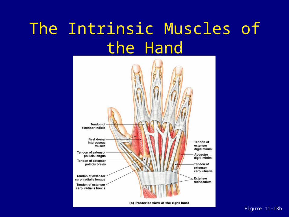
The Intrinsic Muscles of the Hand
Figure 11–18b
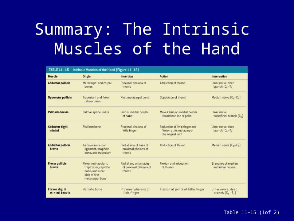
Summary: The Intrinsic Muscles of the Hand
Table 11–15 (1of 2)
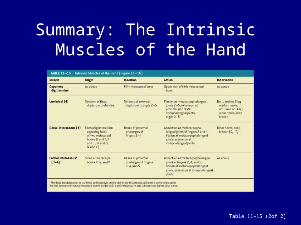
Summary: The Intrinsic Muscles of the Hand
Table 11–15 (2of 2)

Muscles of the Pelvis and Lower Limbs
• Pelvic girdle is tightly bound to axial skeleton:
– permits little movement– has few muscles
Muscles that Position the Lower Limbs
1. Muscles that move the thigh2. Muscles that move the leg3. Muscles that move the foot and toes

Muscles that Move the Thigh
Figure 11–19a, b
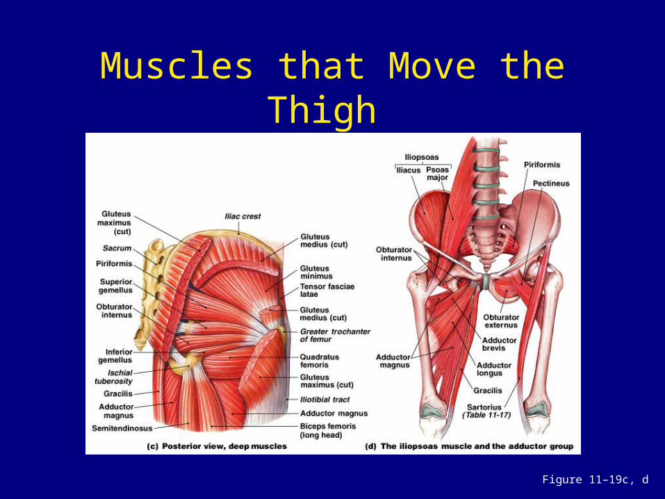
Muscles that Move the Thigh
Figure 11–19c, d

Muscles that Move the Thigh
• Gluteal muscles • Lateral rotators • Adductors • Iliopsoas
3D Peel-Away of Muscles of the ThighPLAYPLAY
3D Peel-Away of Rectus MusclePLAYPLAY

Gluteal Muscles (1 of 2)
• Cover lateral surfaces of ilia• Gluteus maximus:
– largest, most posterior gluteal muscle– produces extension and lateral
rotation at hip

Gluteal Muscles (2 of 2)
• Tensor fasciae latae:– works with gluteus maximus– stabilizes iliotibial tract
• Gluteus medius and gluteus minimus: – originate anterior to gluteus maximus– insert on trochanter

Lateral Rotators
• Group of 6 muscles, including:– piriformis– obturator

Adductors • Adductor magnus:
– produces adduction, extension, and flexion
• Adductor brevis:– hip flexion and adduction
• Adductor longus:– hip flexion and adduction
• Pectineus:– hip flexion and adduction
• Gracilis:– hip flexion and adduction

Iliopsoas
• 2 hip flexors insert on the same tendon:
• psoas major • iliacus

Summary: Muscles that Move the Thigh
Table 11–16 (1 of 2)

Summary: Muscles that Move the Thigh
Table 11–16 (2 of 2)

Muscles that Move the Leg
Figure 11–20a

Muscles that Move the Leg
Figure 11–20b, c

Muscles that Move the Leg
• Flexors of the knee:– originate on the pelvic girdle
• Extensors of the knee:– originate on the femoral surface– insert on the patella

Flexors of the Knee
• Biceps femoris- Hamstrings• Semimembranosus- “• Semitendinosus- “• Sartorius:
– originates superior to the acetabulum
• Popliteus:– rotates the tibia to unlock the knee

Extensors of the Knee
• 4 muscles of the quadriceps femoris: – 3 vastus muscles– rectus femoris muscle

Summary: Muscles that Move the Leg
Table 11–17 (1 of 2)

Summary: Muscles that Move the Leg
Table 11–17 (2 of 2)

Muscles that Move the Foot and Toes
Figure 11–21a, b

Muscles that Move the Foot and Toes
Figure 11–21c, d

Muscles that Move the Foot and Toes
• Extrinsic muscles that move the foot and toes include:– muscles that produce extension at the
ankle– muscles that produce flexion at the ankle– muscles that produce extension at the
toes– muscles that produce flexion at the toes

4 Muscles that Produce Extension at the Ankle
• Gastrocnemius• Soleus• Fibularis• Tibialis posterior+ The Achilles Tendon-
The calcaneal tendon (Achilles tendon):– shared by the gastrocnemius and soleus

Muscles that Produce Flexion at the Ankle
• Tibialis anterior:– opposes the gastrocnemius
Muscles that Produce Flexion at the Toes
• Flexor digitorum longum• Flexor hallucis longus:
– oppose the extensors

Muscles that Produce Extension at the Toes
• Extensor digitorum longum• Extensor hallucis longus• Extensor retinacula:
– fibrous sheaths hold tendons of toes as they cross the ankle

Summary: Muscles that Move the Foot and Toes
Table 11–18
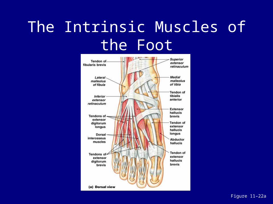
The Intrinsic Muscles of the Foot
Figure 11–22a

The Intrinsic Muscles of the Foot
Figure 11–22b, c

Summary: The Intrinsic Muscles of the Foot
Table 11–19

Effects of Aging on the Muscular System
1. Skeletal muscle fibers become smaller in diameter
2. Skeletal muscles become less elastic:
– develop increasing amounts of fibrous tissue (fibrosis)
3. Decreased tolerance for exercise4. Decreased ability to recover from
muscular injuries

Integration with Other Systems
Figure 11–24

Integration with Other Systems
• Cardiovascular system:– delivers oxygen and fuel– removes carbon dioxide and wastes
• Respiratory system:– responds to oxygen demand of
muscles
• Integumentary system:– disperses heat from muscle activity
• Nervous and endocrine systems:– direct responses of all systems

SUMMARY• Effects of muscle structure on function• Organization of skeletal muscle fibers:
– parallel, convergent, pennate, circular• Relationships between levers and
movement• Actions of first, second, and third class
levers• Origins and insertions of skeletal
muscles• Actions of skeletal muscles:
– agonist, antagonist, synergist• How skeletal muscles are named

SUMMARY• Structures and functions of axial
muscles:– muscles of head and neck– muscle of vertebral column– oblique and rectus muscles– muscles of pelvic floor
• Structures and functions of the appendicular muscles:– muscles of shoulders and upper limbs– muscles of pelvis and lower limbs
• Effects of aging on the muscular system