Chapter 1 · It is therefore not surprising that a decreased platelet count in the peripheral blood...
24
1
Transcript of Chapter 1 · It is therefore not surprising that a decreased platelet count in the peripheral blood...

1

Chapter 1
General introduction1
Chapter 1
12
Thrombocytopenia
Blood is composed of a fluid (plasma) and a cellular fraction. This cellular fraction contains red blood cells (erythrocytes), white blood cells (leukocytes) and anuclear cells that are known as platelets. Platelets play an important role in haemostasis, since they can adhere at sites of injury and pile up to form a plug that stops the bleeding. It is therefore not surprising that a decreased platelet count in the peripheral blood may lead to serious bleeding problems. Normal platelet counts in Caucasians vary between 150 and 350 x 109/L. Africans have lower platelet counts, varying between 115 and 300 x 109/L and in all ethnic groups platelet numbers in women are higher than in men.1 Although thrombocytopenia is defined as a platelet count below the lower limit of 150 x 109 /L, most patients whose platelet count is above 50 x 109/L do not exhibit significant signs of haemostatic impairment. Mild thrombocytopenia (50-100 x 109 platelets/L) is usually associated with an increased bleeding risk after trauma. Serious spontaneous hemorrhage only occurs when platelet counts drop below 10 x 109/L.
Thrombocytopenia can be caused by four general mechanisms: (1) platelet underproduction; (2) platelet sequestration in the spleen; (3) haemodilution; and (4) increased platelet consumption or destruction. The latter develops when the rate of platelet consumption or destruction surpasses the ability of the bone marrow to produce platelets. This can be induced by immune mediated mechanisms, in which antibodies cause a rapid clearance of platelets or by non-immune mechanisms that lead to excessive consumption of platelets. Increased platelet consumption or destruction is thought to play a role in the pathology of clinically very distinct diseases (Table I). Four diseases, hallmarked by thrombocytopenia, were studied in more detail in this thesis. Non-immune consumptive mechanisms have been implicated in three of the diseases, Von Willebrand Disease (VWD) type 2B, Thrombotic Thrombocytopenic Purpura (TTP) and HELLP syndrome (Haemolysis, Elevated Liver enzymes and Low Platelets). The fourth, the antiphospholipid syndrome, has been suggested to be associated with immune-mediated platelet destruction.
General introduction
13
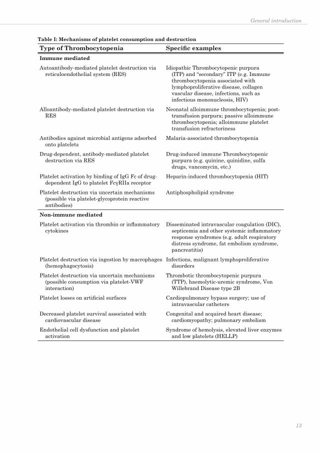
Table I: Mechanisms of platelet consumption and destruction
Type of Thrombocytopenia Specific examplesImmune mediatedAutoantibody-mediated platelet destruction via
reticuloendothelial system (RES)Idiopathic Thrombocytopenic purpura
(ITP) and “secondary” ITP (e.g. Immune thrombocytopenia associated with lymphoproliferative disease, collagen vascular disease, infections, such as infectious mononucleosis, HIV)
Alloantibody-mediated platelet destruction via RES
Neonatal alloimmune thrombocytopenia; post-transfusion purpura; passive alloimmune thrombocytopenia; alloimmune platelet transfusion refractoriness
Antibodies against microbial antigens adsorbed onto platelets
Malaria-associated thrombocytopenia
Drug-dependent, antibody-mediated platelet destruction via RES
Drug-induced immune Thrombocytopenic purpura (e.g. quinine, quinidine, sulfa drugs, vancomycin, etc.)
Platelet activation by binding of IgG Fc of drug-dependent IgG to platelet FcγRIIa receptor
Heparin-induced thrombocytopenia (HIT)
Platelet destruction via uncertain mechanisms (possible via platelet-glycoprotein reactive antibodies)
Antiphospholipid syndrome
Non-immune mediatedPlatelet activation via thrombin or inflammatory
cytokinesDisseminated intravascular coagulation (DIC),
septicemia and other systemic inflammatory response syndromes (e.g. adult respiratory distress syndrome, fat embolism syndrome, pancreatitis)
Platelet destruction via ingestion by macrophages (hemophagocytosis)
Infections, malignant lymphoproliferative disorders
Platelet destruction via uncertain mechanisms (possible consumption via platelet-VWF interaction)
Thrombotic thrombocytopenic purpura (TTP), haemolytic-uremic syndrome, Von Willebrand Disease type 2B
Platelet losses on artificial surfaces Cardiopulmonary bypass surgery; use of intravascular catheters
Decreased platelet survival associated with cardiovascular disease
Congenital and acquired heart disease; cardiomyopathy; pulmonary embolism
Endothelial cell dysfunction and platelet activation
Syndrome of hemolysis, elevated liver enzymes and low platelets (HELLP)
Chapter 1
14
Von Willebrand Factor
A connection with von Willebrand factor (VWF) has been implicated in all 4 diseases.2-
6 VWF is a multimeric plasma protein that mediates adhesion and aggregation of platelets7 and modulates the survival and function of coagulation factor VIII. Factor VIII forms a non-covalent complex with VWF, by binding the amino-terminal (D’D3)-domain of VWF. In this way factor VIII is stabilized8 and protected from clearance via the low-density lipoprotein receptor-related protein (LRP)9 and proteolysis by phospholipid binding proteases, such as activated protein C and factor Xa10,11. Platelet adhesion and platelet-platelet interactions are also mediated by VWF. VWF binds via its A3-domain to exposed collagen at a site of injury, after which VWF captures platelets via its A1-domain (Figure 1A). In this way VWF functions as a molecular bridge between the subendothelial collagen and the GpIb-IX-V receptor complex on platelets.
Synthesis and secretion
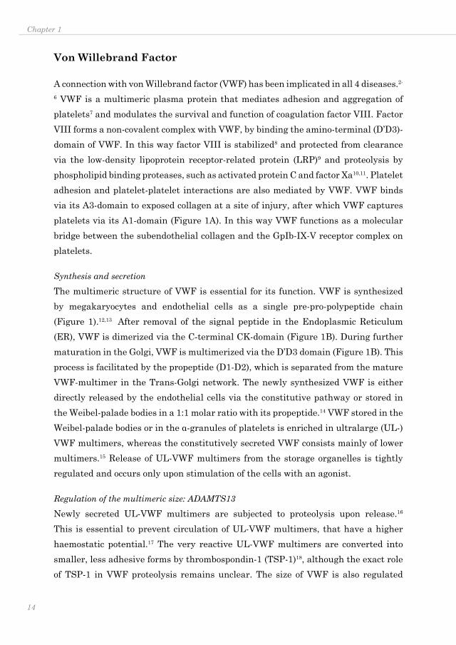
The multimeric structure of VWF is essential for its function. VWF is synthesized by megakaryocytes and endothelial cells as a single pre-pro-polypeptide chain
(Figure 1).12,13 After removal of the signal peptide in the Endoplasmic Reticulum (ER), VWF is dimerized via the C-terminal CK-domain (Figure 1B). During further maturation in the Golgi, VWF is multimerized via the D’D3 domain (Figure 1B). This process is facilitated by the propeptide (D1-D2), which is separated from the mature VWF-multimer in the Trans-Golgi network. The newly synthesized VWF is either directly released by the endothelial cells via the constitutive pathway or stored in the Weibel-palade bodies in a 1:1 molar ratio with its propeptide.14 VWF stored in the Weibel-palade bodies or in the α-granules of platelets is enriched in ultralarge (UL-) VWF multimers, whereas the constitutively secreted VWF consists mainly of lower multimers.15 Release of UL-VWF multimers from the storage organelles is tightly regulated and occurs only upon stimulation of the cells with an agonist.
Regulation of the multimeric size: ADAMTS13
Newly secreted UL-VWF multimers are subjected to proteolysis upon release.16 This is essential to prevent circulation of UL-VWF multimers, that have a higher haemostatic potential.17 The very reactive UL-VWF multimers are converted into smaller, less adhesive forms by thrombospondin-1 (TSP-1)18, although the exact role of TSP-1 in VWF proteolysis remains unclear. The size of VWF is also regulated
General introduction
15
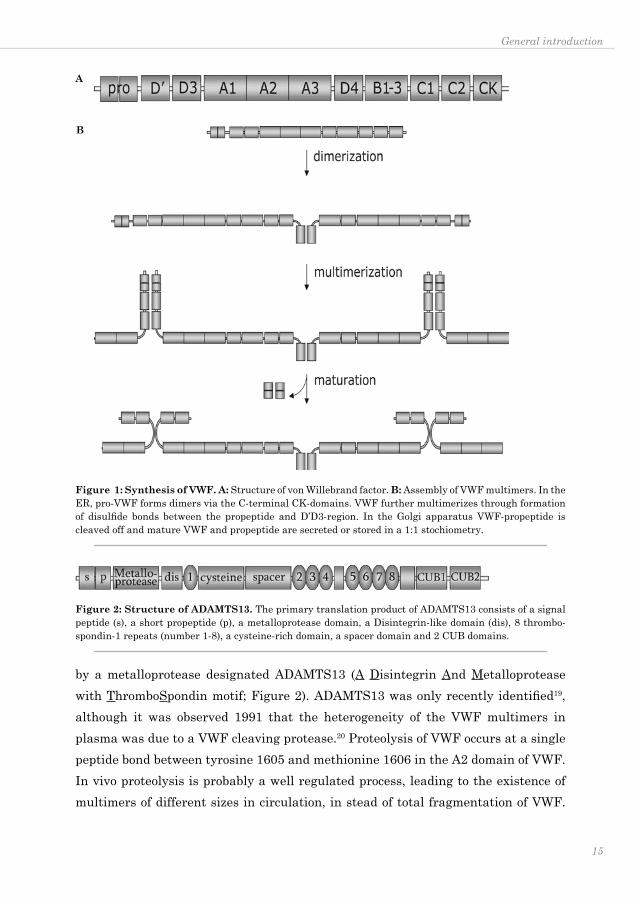
by a metalloprotease designated ADAMTS13 (A Disintegrin And Metalloprotease with ThromboSpondin motif; Figure 2). ADAMTS13 was only recently identified19, although it was observed 1991 that the heterogeneity of the VWF multimers in plasma was due to a VWF cleaving protease.20 Proteolysis of VWF occurs at a single peptide bond between tyrosine 1605 and methionine 1606 in the A2 domain of VWF. In vivo proteolysis is probably a well regulated process, leading to the existence of multimers of different sizes in circulation, in stead of total fragmentation of VWF.
Figure 2: Structure of ADAMTS13. The primary translation product of ADAMTS13 consists of a signal peptide (s), a short propeptide (p), a metalloprotease domain, a Disintegrin-like domain (dis), 8 thrombo-spondin-1 repeats (number 1-8), a cysteine-rich domain, a spacer domain and 2 CUB domains.
Figure 1: Synthesis of VWF. A: Structure of von Willebrand factor. B: Assembly of VWF multimers. In the ER, pro-VWF forms dimers via the C-terminal CK-domains. VWF further multimerizes through formation of disulfide bonds between the propeptide and D’D3-region. In the Golgi apparatus VWF-propeptide is cleaved off and mature VWF and propeptide are secreted or stored in a 1:1 stochiometry.
A
B
Chapter 1
16
Tight regulation is required, since excessive cleavage into minimal fragments would cripple haemostasis, resulting in a bleeding disorder. This is illustrated by VWD type 2A, a bleeding diathesis caused by a lack of larger VWF-multimers, often due to excessive VWF proteolysis.21
Regulation of VWF proteolysis
To make VWF susceptible for cleavage by ADAMTS13, unfolding of the globular VWF molecule seems to be required. This is apparent from the fact that denaturing conditions are necessary for VWF cleavage to occur in vitro.22 Fluid shear stress, therefore, can be considered as one of the determinants of VWF proteolysis by ADAMTS13.23 Furthermore, proteolysis is modulated by chloride ions that bind to VWF and alter its conformation upon secretion into the circulation.24
Cleavage of the A2-domain of VWF is also influenced by the A1-domain. The presence of this domain was shown to decrease proteolysis of A2 when compared to the isolated A2-domain. Binding of GpIbα to the A1-domain abrogated the inhibitory effect of this domain on cleavage.25 Ristocetin has also been suggested to enhance VWF proteolysis by ADAMTS13.26 Botrocetin did not influence proteolysis, possibly because it does not induce a conformational change like ristocetin.27 The overall structure and conformation of VWF also influences the rate of proteolysis as illustrated by VWD type 2A, in which VWF is more susceptible for proteolysis due to mutations.21
On the other hand, the efficacy of proteolysis is determined by the activity and binding of ADAMTS13. Serine proteases like plasmin or thrombin might regulate the activity of ADAMTS13 under normal conditions. In vitro, it has been shown that ADAMTS13 is proteolyzed by thrombin and plasmin and is thereby inactivated.28 There is also evidence that TSP-1 interferes with VWF proteolysis. Although UL-VWF multimers can be processed by TSP-1 itself18, the size of the VWF multimers in TSP-1 knock-out mice was found to be reduced.29 This indicates that TSP-1 might compete with ADAMTS13 for binding to VWF and that ADAMTS13 is a more effective protease in the absence of TSP-1.
Inflammatory cytokine IL-6 has been suggested to inhibit ADAMTS13 activity under flow conditions, but not under static conditions.30 The mechanism behind this inhibition remains to be investigated. A potential mechanism could be that IL-6 impairs docking of ADAMTS13 to VWF under flow. A possible effect of IL-6 on VWF
General introduction
17
cleavage has previously been suggested based on clinical observations. IL-6 levels are increased at the onset of TTP episodes, for instance, and high levels of IL-6 are associated with a poorer prognosis in TTP.31
VWF-platelet interaction
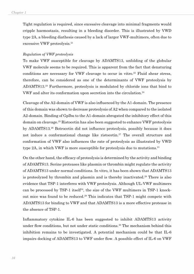
The multimeric pattern of VWF in plasma is very heterogeneic after proteolysis. Under physiological circumstances not only dimers circulate, but also multimers of more than 20.000 kDa are present. Although these multimers circulate together with platelets, VWF and its receptor on platelets do not interact under normal conditions. This indicates that multimers released after cleavage by ADAMTS13, have a latent biological activity. To induce an interaction with the GpIb-IX-V complex, a conformational change in the A1-domain of VWF is required (Figure 3).32 This conformational change is thought to be induced by binding of the A3-domain to exposed collagen at a site of injury33 and can be mimicked in vitro by coating VWF onto a artificial surface or by incubation of VWF with modulators, such as botrocetin or ristocetin.34,35
Figure 3: A1-domain of VWF. A: Solid-ribbon representation of “unliganded” wild type (wt) VWF-A1 with a semi-transparent molecular surface. B: Comparison of unliganded wt-A1 and GpIbα-bound A1, focused on the changes in the α1β2-loop region. Unliganded A1 is depicted in dark grey, liganded A1 in light gray. N = N-terminal C = C-terminal (Figure B was adapted from Dumas et al. J Biol Chem. 2004)
A B
Chapter 1
18
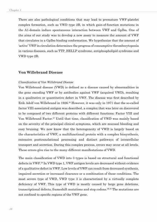
There are also pathological conditions that may lead to premature VWF-platelet complex formation, such as VWD type 2B, in which gain-of-function mutations in the A1-domain induce spontaneous interaction between VWF and GpIbα. One of the aims of our study was to develop a new assay to measure the amount of VWF that circulates in a GpIbα-binding conformation. We hypothesize that the amount of ‘active’ VWF in circulation determines the progress of consumptive thrombocytopenia in various diseases, such as TTP, HELLP syndrome, antiphospholipid syndrome and VWD type 2B.
Von Willebrand Disease
Classification of Von Willebrand Disease
Von Willebrand disease (VWD) is defined as a disease caused by abnormalities in the gene encoding VWF or by antibodies against VWF (acquired VWD), resulting in a qualitative or quantitative defect in VWF. The disease was first described by Erik Adolf von Willebrand in 1926.36 However, it was only in 1971 that the so-called factor VIII-associated antigen was described, a complex that was later on discovered to be composed of two different proteins with different functions: Factor VIII and Von Willebrand Factor.37 Until that time, classification of VWD was mainly based on the severity of the principal clinical symptoms, which are mucosal bleeding and easy bruising. We now know that the heterogeneity of VWD is largely based on the characteristics of VWF, a multifunctional protein with a complex biosynthesis, extensive posttranslational processing and distinct pathways of intracellular transport and secretion. During this complex process, errors may occur at all levels. These errors give rise to the many different manifestations of VWD.
The main classification of VWD into 3 types is based on structural and functional defects in VWF.38 In VWD type 1, VWF antigen levels are decreased without evidence of a qualitative defect in VWF. Low levels of VWF can result from decreased synthesis, impaired secretion or increased clearance or a combination of these conditions. The most severe type of VWD, VWD type 3 is characterized by a virtually complete deficiency of VWF. This type of VWD is mostly caused by large gene deletions, transcriptional defects, frameshift mutations and stop codons.39,40 The mutations are not confined to specific regions of the VWF gene.
General introduction
19
VWD type 2 is hallmarked by qualitative defects, independent of the VWF antigen levels. This type can be subdivided into type 2A, 2B, 2M and 2N. VWD type 2N (Normandie) is caused by a decreased affinity of VWF for factor VIII.41,42 Since VWF stabilizes the heterodimeric structure of factor VIII and protects it from clearance and proteolysis, the clinical manivestations in VWD type 2N mimic haemophilia A.43
VWD type 2M includes patients with decreased platelet-dependent VWF function, although the multimeric size of VWF is normal. Mutations causing this type of VWD are mostly clustered in the A1-domain and results in a reduced GpIb binding capacity.44
VWD type 2A includes all patients with a lack or decrease of high molecular weight multimers, which is accompanied by a decreased VWF function. The reduction of high molecular weight multimers in type 2A can be caused either by an impaired biosynthesis of large VWF multimers (group 1 mutations)45 or an increased proteolysis of VWF multimers in plasma by ADAMTS13 (group 2 mutations).46 Group 2 mutations are mainly located in the A2-domain, the domain that is susceptible for proteolysis. In some cases, such mutations are also found in the A1-domain.47 Type 2A can also be caused by a defective post-translational processing that includes defects of dimerization at the VWF C-terminus or defects of further polymerization of VWF dimers to multimers at the N-terminus. These defects are caused by mutations in the D1 and D2 domains of the propeptide or in the D3-domain.48,49
Von Willebrand disease type 2B
The lack of higher multimers in VWD type 2B is due to an enhanced affinity of VWF for the GpIb-IX-V receptor complex on platelets.5 This enhanced affinity is caused by the presence of gain-of-function mutations in the A1-domain of VWF. At least 20 mutations have been characterized in patients with VWD type 2B. One is a single amino acid insertion50, but the majority are single amino acid substitutions.51 Paradoxically, the gain-of-function of VWF does not induce thrombosis. Instead, it results in a specific loss of the biologically more active higher multimers that react spontaneously with circulating platelets. The combination of loss of high multimers and consumption of platelets results in a bleeding diathesis.5 Thrombocytopenia in VWD type 2B is often moderate to mild, can be intermittent and is exacerbated by surgery, physical effort or the administration of desmopressin (1-deamino-8-D-
Chapter 1
20
arginine vasopressin, DDAVP).52-54 Pregnancy also triggers the release of VWF from the Weibel-palade bodies, resulting in higher VWF antigen levels and lower platelet number. In VWD type 2B the increased amounts of mutant VWF induces more severe thrombocytopenia. The decrease in platelet count is dependent on the length of the pregnancy and is immediately reversed upon delivery. The sudden increase after delivery suggests that thrombocytopenia in VWD type 2B might also be due to other phenomena besides higher production of VWF during pregnancy.52
There is a high degree of heterogeneity in thrombocytopenia in VWD type 2B, even between patients with the same molecular defects. Despite many case reports on thrombocytopenia associated with VWD type 2B, studies in larger cohorts addressing the question of heterogeneity in thrombocytopenia have not been performed yet.
Thrombotic Thrombocytopenic Purpura
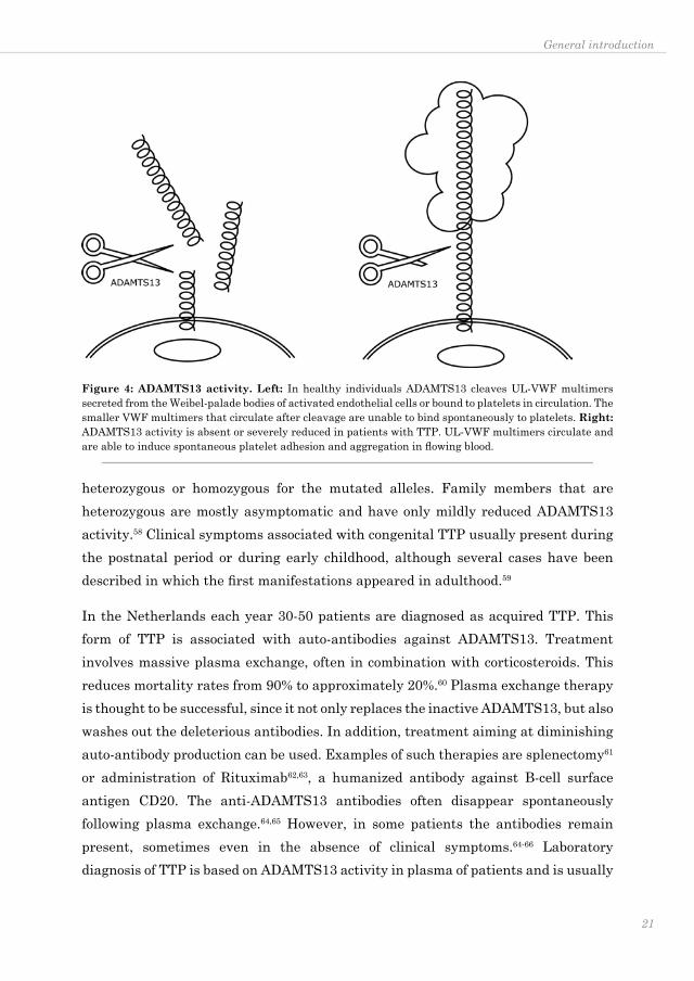
Apart from VWD type 2B, VWF multimers with a high biological activity also circulate in TTP. Under physiological conditions UL-VWF multimers are cleaved by ADAMTS13 into smaller, less reactive VWF multimers (Figure 4).16 ADAMTS13 activity is deficient in TTP, resulting in the presence of UL-VWF multimers in the circulation of patients with TTP (Figure 4). UL-VWF multimers form high strength bonds with the GpIb-IX-V receptor complex on platelets17, which leads to formation of VWF-rich thrombi in the microvasculature of many organs, consumptive thrombocytopenia and microangiopathic hemolytic anemia.4 TTP can also be associated with fever, renal dysfunction and neurological deficits.4 However, the latter three symptoms vary in severity amongst patients or can even be absent, which makes it difficult to distinguish TTP form other microangiopathies.
Reduced ADAMTS13 activity in TTP can be caused by mutations in the ADAMTS13 gene (congenital TTP)55 or by the presence of inhibitory auto-antibodies (acquired TTP; Figure 4).56 Hereditary TTP is an extremely rare disorder, which is treated with regular plasma infusions. Until now, many different mutations have been described, mostly clustered in the N-terminal region of ADAMTS13. Approximately 75% of the described missense mutations are located in the metalloprotease, disintegrin and the cysteine-rich/spacer domains (Figure 2).57 This emphasizes the importance of these domains for the activity of ADAMTS13. Patients with congenital TTP are compound
General introduction
21
heterozygous or homozygous for the mutated alleles. Family members that are heterozygous are mostly asymptomatic and have only mildly reduced ADAMTS13 activity.58 Clinical symptoms associated with congenital TTP usually present during the postnatal period or during early childhood, although several cases have been described in which the first manifestations appeared in adulthood.59
In the Netherlands each year 30-50 patients are diagnosed as acquired TTP. This form of TTP is associated with auto-antibodies against ADAMTS13. Treatment involves massive plasma exchange, often in combination with corticosteroids. This reduces mortality rates from 90% to approximately 20%.60 Plasma exchange therapy is thought to be successful, since it not only replaces the inactive ADAMTS13, but also washes out the deleterious antibodies. In addition, treatment aiming at diminishing auto-antibody production can be used. Examples of such therapies are splenectomy61 or administration of Rituximab62,63, a humanized antibody against B-cell surface antigen CD20. The anti-ADAMTS13 antibodies often disappear spontaneously following plasma exchange.64,65 However, in some patients the antibodies remain present, sometimes even in the absence of clinical symptoms.64-66 Laboratory diagnosis of TTP is based on ADAMTS13 activity in plasma of patients and is usually
Figure 4: ADAMTS13 activity. Left: In healthy individuals ADAMTS13 cleaves UL-VWF multimers secreted from the Weibel-palade bodies of activated endothelial cells or bound to platelets in circulation. The smaller VWF multimers that circulate after cleavage are unable to bind spontaneously to platelets. Right: ADAMTS13 activity is absent or severely reduced in patients with TTP. UL-VWF multimers circulate and are able to induce spontaneous platelet adhesion and aggregation in flowing blood.
Chapter 1
22
measured under static conditions67, although it is generally thought that cleavage in vivo occurs under conditions of shear stress.23 Certain inhibitory antibodies might be missed in static assays, for instance antibodies that impair docking of ADAMTS13 to VWF. Recently, new assays have become available to determine the amount of antibody and ADAMTS13 antigen levels. These assays might provide more insight in the onset of TTP and the role of the antibodies.
Besides primary TTP, TTP can also occur as a secondary manifestation in patients with other autoimmune diseases68,69, cancer70, infection71, bone marrow transplantation72, during use of certain anti-platelet drugs73 or during pregnancy74,75.
TTP-like diseases
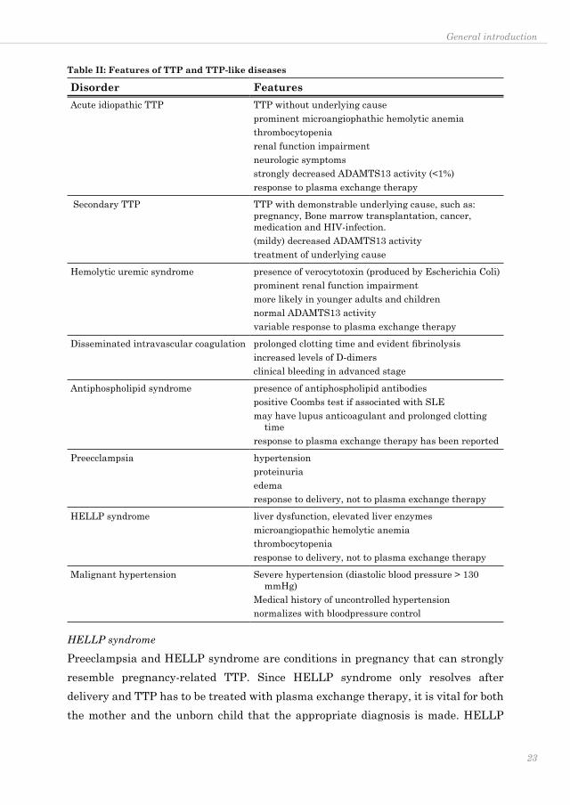
Many conditions have a differential diagnosis that can mimic or overlap with TTP. Since TTP is a fatal disease when left untreated, an accurate diagnosis is critical for a good outcome. Likewise, the decision to omit plasma exchange with its potential side effects76 when it is not TTP, is just as important. The presence of thrombocytopenia and Coombs negative hemolytic anemia without other causes is sufficient to make the diagnosis TTP.60,77,78 However, based on these criteria it is difficult to distinguish TTP from other diseases like disseminated intravascular coagulation (DIC), hemolytic uremic syndrome (HUS), auto-immune diseases such as the antiphospholipid syndrome and during pregnancy the syndrome of hemolysis, elevated liver enzymes and low platelets (HELLP). ADAMTS13 activity has proven to be an important marker for the diagnosis of TTP. Various studies have shown that absence of ADAMTS13 activity or an activity of less than 1% is indicative for TTP and mildly reduced ADAMTS13 activity has been found in patients with other diseases.79-
81 On the other hand, normal ADAMTS13 activity has been found in otherwise typical cases of TTP.82 Moreover, the assay to determine ADAMTS13 activity that is used in many laboratories is laborious and results are often not available soon enough to influence the acute decision-making regarding the treatment. Table II summarizes the features of the different TTP look-alikes, showing the differences and similarities between TTP and the other disorders.
General introduction
23
HELLP syndrome
Preeclampsia and HELLP syndrome are conditions in pregnancy that can strongly resemble pregnancy-related TTP. Since HELLP syndrome only resolves after delivery and TTP has to be treated with plasma exchange therapy, it is vital for both the mother and the unborn child that the appropriate diagnosis is made. HELLP
Table II: Features of TTP and TTP-like diseases
Disorder Features Acute idiopathic TTP TTP without underlying cause
prominent microangiophathic hemolytic anemia thrombocytopeniarenal function impairment neurologic symptomsstrongly decreased ADAMTS13 activity (<1%)response to plasma exchange therapy
Secondary TTP TTP with demonstrable underlying cause, such as: pregnancy, Bone marrow transplantation, cancer, medication and HIV-infection. (mildy) decreased ADAMTS13 activitytreatment of underlying cause
Hemolytic uremic syndrome presence of verocytotoxin (produced by Escherichia Coli)prominent renal function impairmentmore likely in younger adults and childrennormal ADAMTS13 activityvariable response to plasma exchange therapy
Disseminated intravascular coagulation prolonged clotting time and evident fibrinolysisincreased levels of D-dimersclinical bleeding in advanced stage
Antiphospholipid syndrome presence of antiphospholipid antibodies positive Coombs test if associated with SLEmay have lupus anticoagulant and prolonged clotting
time response to plasma exchange therapy has been reported
Preecclampsia hypertensionproteinuriaedemaresponse to delivery, not to plasma exchange therapy
HELLP syndrome liver dysfunction, elevated liver enzymesmicroangiopathic hemolytic anemiathrombocytopeniaresponse to delivery, not to plasma exchange therapy
Malignant hypertension Severe hypertension (diastolic blood pressure > 130 mmHg)
Medical history of uncontrolled hypertensionnormalizes with bloodpressure control
Chapter 1
24
syndrome is thought to be a severe complication of preecclampsia. Both preecclampsia and HELLP syndrome are associated with endothelial injury, fibrin deposition in the vessel lumen, increased platelet activation and platelet consumption.83 Especially in HELLP syndrome platelet counts below 100 x 109 /L are found. Endothelial cell activation in preecclampsia is considered to be one component of a more generalized inflammatory response to pregnancy. This response is common in all pregnancies, but is exaggerated in preecclampsia and is even further aggravated in patients with HELLP syndrome.84 Many inflammatory markers have been reported to be increased in preecclampsia85,86 and in HELLP syndrome87-89. It remains unclear how increased endothelial cell damage and the inflammatory state relate to platelet aggregation and thrombocytopenia observed in patients with HELLP syndrome. It has been suggested that depositions of thrombi in the microvasculature and in the sinusoids of the liver induce hemolytic anemia and increased liver enzymes.90 Variable degrees of hepatic damage, microangiopathic hemolytic anemia and thrombocytopenia characterize HELLP syndrome91. It is still unknown why some preeclamptic women develop HELLP syndrome while others do not and what determines the severity of the symptoms.
Antiphospholipid syndrome
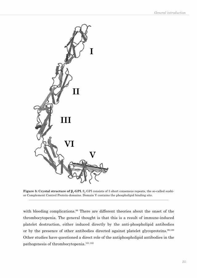
The antiphospholipid syndrome can also present with some of the features of TTP, including thrombocytopenia and hemolysis. However, the main symptoms are arterial and venous thrombosis and recurrent pregnancy-loss.92,93 The syndrome is diagnosed when one of these clinical features is present in combination with the presence of antiphospholipid antibodies.94 Actually, these antibodies recognize proteins that bind anionic phospholipids in stead of the phospholipids themselves.95 Probably the most important antigen in the antiphospholipid syndrome is β2-glycoprotein (GP) I (Figure 5).96 Antibodies that recognize this protein and cause a prolongation of phospholipid-dependent coagulation tests, have been described to correlate strongly with thrombosis.97
Despite all research that has been performed in this field, our understanding of the pathology of the syndrome is still limited. Data concerning the onset and clinical association of thrombocytopenia with the various antiphospholipid syndrome-related manifestations, for instance, are still scarce. Thrombocytopenia in the antiphospholipid syndrome is usually mild (70-120 x 109 /L) and rarely associated
General introduction
25
Figure 5: Crystal structure of β2-GPI. β2-GPI consists of 5 short consensus repeats, the so-called sushi- or Complement Control Protein-domains. Domain V contains the phospholipid binding site.
I
with bleeding complications.98 There are different theories about the onset of the thrombocytopenia. The general thought is that this is a result of immune-induced platelet destruction, either induced directly by the anti-phospholipid antibodies or by the presence of other antibodies directed against platelet glycoproteins.99,100 Other studies have questioned a direct role of the antiphospholipid antibodies in the pathogenesis of thrombocytopenia.101,102
II
III
VIV
Chapter 1
26
Outline of this thesis
Although VWD type 2B, TTP, HELLP syndrome and the antiphospholipid syndrome are four well defined diseases with a distinct clinical entity, several reports documented the development of two of these diseases in one patient. HELLP may reveal the presence of the antiphospholipid syndrome103 and TTP is observed in patients diagnosed with primary antiphospholipid syndrome104. Moreover, clinical features of pregnancy-related TTP and HELLP syndrome are very similar, which makes it difficult to distinguish between the diseases.
As described in the previous paragraphs, all four diseases are associated with a mild to severe thrombocytopenia, which is accompanied by thrombosis in TTP, HELLP syndrome and the antiphospholipid syndrome. Although VWD type 2B is also characterized by a consumptive thrombocytopenia, thrombosis has not been reported in these patients.
A connection with VWF has been implicated in all 4 diseases.2-6 TTP is characterized by UL-VWF multimers, which have an increased biological activity.4,17 The activity of VWF is also increased in VWD type 2B. In this disease a gain-of-function mutation induces spontaneous VWF-platelet interactions.5 The role of VWF in HELLP syndrome and the antiphospholipid syndrome is less clear. VWF antigen levels are dramatically increased in HELLP syndrome, with a normal multimeric pattern.2,3 The multimeric pattern of VWF has been reported to be abnormal in 37% of the patients suffering from the antiphospholipid syndrome. Interestingly, the presence of highly active multimers was found to correlate with the degree of thrombosis in these patients.6
General introduction
27
In order to gain more insight in the role of VWF in the onset of thrombocytopenia in these four diseases, we addressed the following questions:
1) Can we develop an assay to distinguish between VWF in the resting state and VWF in a GpIbα-binding conformation? (Chapter 2)
2) Does VWF circulate in a GpIbα-binding conformation in VWD type 2B, TTP, HELLP syndrome and the antiphospholipid syndrome? (Chapter 2, 3 and 4)
3) What is the cause of the transition of VWF to a GpIb-binding conformation in the different diseases? (Chapter 2, 3 and 4)
4) Can we distinguish TTP from other, clinically related, diseases? (Chapter 5)
Chapter 1
28
Reference List
(1) Bain BJ. Ethnic and sex differences in the total and differential white cell count and platelet count. J Clin Pathol. 1996;49:664-666.
(2) Friedman SA, Schiff E, Emeis JJ, Dekker GA, Sibai BM. Biochemical corroboration of endothelial involvement in severe preeclampsia. Am J Obstet Gynecol. 1995;172:202-203.
(3) Lattuada A, Rossi E, Calzarossa C, Candolfi R, Mannucci PM. Mild to moderate reduction of a von Willebrand factor cleaving protease (ADAMTS-13) in pregnant women with HELLP microangiopathic syndrome. Haematologica. 2003;88:1029-1034.
(4) Moake JL, Rudy CK, Troll JH et al. Unusually large plasma factor VIII:von Willebrand factor multimers in chronic relapsing thrombotic thrombocytopenic purpura. N Engl J Med. 1982;307:1432-1435.
(5) Ruggeri ZM, Pareti FI, Mannucci PM, Ciavarella N, Zimmerman TS. Heightened interaction between platelets and factor VIII/von Willebrand factor in a new subtype of von Willebrand’s disease. N Engl J Med. 1980;302:1047-1051.
(6) Schinco P, Borchiellini A, Tamponi G et al. Lupus anticoagulant and thrombosis: role of von Willebrand factor multimeric forms. Clin Exp Rheumatol. 1997;15:5-10.
(7) Savage B, Saldivar E, Ruggeri ZM. Initiation of platelet adhesion by arrest onto fibrinogen or translocation on von Willebrand factor. Cell. 1996;84:289-297.
(8) Weiss HJ, Sussman II, Hoyer LW. Stabilization of factor VIII in plasma by the von Willebrand factor. Studies on posttransfusion and dissociated factor VIII and in patients with von Willebrand’s disease. J Clin Invest. 1977;60:390-404.
(9) Lenting PJ, Neels JG, van den Berg BM et al. The light chain of factor VIII comprises a binding site for low density lipoprotein receptor-related protein. J Biol Chem. 1999;274:23734-23739.
(10) Walker FJ, Chavin SI, Fay PJ. Inactivation of factor VIII by activated protein C and protein S. Arch Biochem Biophys. 1987;252:322-328.
(11) Koedam JA, Hamer RJ, Beeser-Visser NH, Bouma BN, Sixma JJ. The effect of von Willebrand factor on activation of factor VIII by factor Xa. Eur J Biochem. 1990;189:229-234.
(12) Nachman R, Levine R, Jaffe EA. Synthesis of factor VIII antigen by cultured guinea pig megakaryocytes. J Clin Invest. 1977;60:914-921.
(13) Jaffe EA, Hoyer LW, Nachman RL. Synthesis of antihemophilic factor antigen by cultured human endothelial cells. J Clin Invest. 1973;52:2757-2764.
(14) Wagner DD, Fay PJ, Sporn LA et al. Divergent fates of von Willebrand factor and its propolypeptide (von Willebrand antigen II) after secretion from endothelial cells. Proc Natl Acad Sci U S A. 1987;84:1955-1959.
(15) Sporn LA, Marder VJ, Wagner DD. Inducible secretion of large, biologically potent von Willebrand factor multimers. Cell. 1986;46:185-190.
(16) Dong JF, Moake JL, Nolasco L et al. ADAMTS-13 rapidly cleaves newly secreted ultralarge von Willebrand factor multimers on the endothelial surface under flowing conditions. Blood. 2002;100:4033-4039.
(17) Arya M, Anvari B, Romo GM et al. Ultralarge multimers of von Willebrand factor form spontaneous high-strength bonds with the platelet glycoprotein Ib-IX complex: studies using optical tweezers. Blood. 2002;99:3971-3977.
(18) Xie L, Chesterman CN, Hogg PJ. Control of von Willebrand factor multimer size by thrombospondin-1. J Exp Med. 2001;193:1341-1349.
General introduction
29
(19) Zheng X, Chung D, Takayama TK et al. Structure of von Willebrand factor-cleaving protease (ADAMTS13), a metalloprotease involved in thrombotic thrombocytopenic purpura. J Biol Chem. 2001;276:41059-41063.
(20) Dent JA, Galbusera M, Ruggeri ZM. Heterogeneity of plasma von Willebrand factor multimers resulting from proteolysis of the constituent subunit. J Clin Invest. 1991;88:774-782.
(21) Sadler JE. Biochemistry and genetics of von Willebrand factor. Annu Rev Biochem. 1998;67:395-424.
(22) Furlan M, Robles R, Lamie B. Partial purification and characterization of a protease from human plasma cleaving von Willebrand factor to fragments produced by in vivo proteolysis. Blood. 1996;87:4223-4234.
(23) Dong JF. Cleavage of ultra-large von Willebrand factor by ADAMTS-13 under flow conditions. J Thromb Haemost. 2005;3:1710-1716.
(24) De Cristofaro R, Peyvandi F, Palla R et al. Role of chloride ions in modulation of the interaction between von Willebrand factor and ADAMTS-13. J Biol Chem. 2005;280:23295-23302.
(25) Nishio K, Anderson PJ, Zheng XL, Sadler JE. Binding of platelet glycoprotein Ibalpha to von Willebrand factor domain A1 stimulates the cleavage of the adjacent domain A2 by ADAMTS13. Proc Natl Acad Sci U S A. 2004;101:10578-10583.
(26) Bowen DJ. An influence of ABO blood group on the rate of proteolysis of von Willebrand factor by ADAMTS13. J Thromb Haemost. 2003;1:33-40.
(27) Fukuda K, Doggett TA, Bankston LA et al. Structural basis of von Willebrand factor activation by the snake toxin botrocetin. Structure. 2002;10:943-950.
(28) Crawley JT, Lam JK, Rance JB et al. Proteolytic inactivation of ADAMTS13 by thrombin and plasmin. Blood. 2005;105:1085-1093.
(29) Pimanda JE, Ganderton T, Maekawa A et al. Role of thrombospondin-1 in control of von Willebrand factor multimer size in mice. J Biol Chem. 2004;279:21439-21448.
(30) Bernardo A, Ball C, Nolasco L, Moake JF, Dong JF. Effects of inflammatory cytokines on the release and cleavage of the endothelial cell-derived ultralarge von Willebrand factor multimers under flow. Blood. 2004;104:100-106.
(31) Wada H, Kaneko T, Ohiwa M et al. Plasma cytokine levels in thrombotic thrombocytopenic purpura. Am J Hematol. 1992;40:167-170.
(32) Huizinga EG, Tsuji S, Romijn RA et al. Structures of glycoprotein Ibalpha and its complex with von Willebrand factor A1 domain. Science. 2002;297:1176-1179.
(33) Ruggeri ZM. Von Willebrand factor, platelets and endothelial cell interactions. J Thromb Haemost. 2003;1:1335-1342.
(34) Berndt MC, Du XP, Booth WJ. Ristocetin-dependent reconstitution of binding of von Willebrand factor to purified human platelet membrane glycoprotein Ib-IX complex. Biochemistry. 1988;27:633-640.
(35) Scott JP, Montgomery RR, Retzinger GS. Dimeric ristocetin flocculates proteins, binds to platelets, and mediates von Willebrand factor-dependent agglutination of platelets. J Biol Chem. 1991;266:8149-8155.
(36) von Willebrand EA. Hereditär pseudohemofili. Finska Läkaresällskaptes Handlingar. 672, 7-112. 1926. Finska Läkaresällskaptes Handlingar.
Chapter 1
30
(37) Zimmerman TS, Ratnoff OD, Powell AE. Immunologic differentiation of classic hemophilia (factor 8 deficiency) and von Willebrand’s dissase, with observations on combined deficiencies of antihemophilic factor and proaccelerin (factor V) and on an acquired circulating anticoagulant against antihemophilic factor. J Clin Invest. 1971;50:244-254.
(38) Sadler JE. A revised classification of von Willebrand disease. For the Subcommittee on von Willebrand Factor of the Scientific and Standardization Committee of the International Society on Thrombosis and Haemostasis. Thromb Haemost. 1994;71:520-525.
(39) Ngo KY, Glotz VT, Koziol JA et al. Homozygous and heterozygous deletions of the von Willebrand factor gene in patients and carriers of severe von Willebrand disease. Proc Natl Acad Sci U S A. 1988;85:2753-2757.
(40) Eikenboom JC, Ploos van Amstel HK, Reitsma PH, Briet E. Mutations in severe, type III von Willebrand’s disease in the Dutch population: candidate missense and nonsense mutations associated with reduced levels of von Willebrand factor messenger RNA. Thromb Haemost. 1992;68:448-454.
(41) Nishino M, Girma JP, Rothschild C, Fressinaud E, Meyer D. New variant of von Willebrand disease with defective binding to factor VIII. Blood. 1989;74:1591-1599.
(42) Mazurier C, Dieval J, Jorieux S, Delobel J, Goudemand M. A new von Willebrand factor (vWF) defect in a patient with factor VIII (FVIII) deficiency but with normal levels and multimeric patterns of both plasma and platelet vWF. Characterization of abnormal vWF/FVIII interaction. Blood. 1990;75:20-26.
(43) Schneppenheim R, Budde U, Krey S et al. Results of a screening for von Willebrand disease type 2N in patients with suspected haemophilia A or von Willebrand disease type 1. Thromb Haemost. 1996;76:598-602.
(44) Meyer D, Fressinaud E, Gaucher C et al. Gene defects in 150 unrelated French cases with type 2 von Willebrand disease: from the patient to the gene. INSERM Network on Molecular Abnormalities in von Willebrand Disease. Thromb Haemost. 1997;78:451-456.
(45) Lyons SE, Bruck ME, Bowie EJ, Ginsburg D. Impaired intracellular transport produced by a subset of type IIA von Willebrand disease mutations. J Biol Chem. 1992;267:4424-4430.
(46) Dent JA, Berkowitz SD, Ware J, Kasper CK, Ruggeri ZM. Identification of a cleavage site directing the immunochemical detection of molecular abnormalities in type IIA von Willebrand factor. Proc Natl Acad Sci U S A. 1990;87:6306-6310.
(47) Ginsburg D, Konkle BA, Gill JC et al. Molecular basis of human von Willebrand disease: analysis of platelet von Willebrand factor mRNA. Proc Natl Acad Sci U S A. 1989;86:3723-3727.
(48) Gaucher C, Dieval J, Mazurier C. Characterization of von Willebrand factor gene defects in two unrelated patients with type IIC von Willebrand disease. Blood. 1994;84:1024-1030.
(49) Holmberg L, Karpman D, Isaksson C et al. Ins405AsnPro mutation in the von Willebrand factor propeptide in recessive type 2A (IIC) von Willebrand’s disease. Thromb Haemost. 1998;79:718-722.
(50) Ribba AS, Lavergne JM, Bahnak BR et al. Duplication of a methionine within the glycoprotein Ib binding domain of von Willebrand factor detected by denaturing gradient gel electrophoresis in a patient with type IIB von Willebrand disease. Blood. 1991;78:1738-1743.
(51) Meyer D, Fressinaud E, Hilbert L et al. Type 2 von Willebrand disease causing defective von Willebrand factor-dependent platelet function. Best Pract Res Clin Haematol. 2001;14:349-364.
General introduction
31
(52) Casonato A, Sartori MT, Bertomoro A et al. Pregnancy-induced worsening of thrombocytopenia in a patient with type IIB von Willebrand’s disease. Blood Coagul Fibrinolysis. 1991;2:33-40.
(53) Casonato A, Sartori MT, de Marco L, Girolami A. 1-Desamino-8-D-arginine vasopressin (DDAVP) infusion in type IIB von Willebrand’s disease: shortening of bleeding time and induction of a variable pseudothrombocytopenia. Thromb Haemost. 1990;64:117-120.
(54) Donner M, Kristoffersson AC, Lenk H et al. Type IIB von Willebrand’s disease: gene mutations and clinical presentation in nine families from Denmark, Germany and Sweden. Br J Haematol. 1992;82:58-65.
(55) Levy GG, Nichols WC, Lian EC et al. Mutations in a member of the ADAMTS gene family cause thrombotic thrombocytopenic purpura. Nature. 2001;413:488-494.
(56) Moake JL. Thrombotic microangiopathies. N Engl J Med. 2002;347:589-600.(57) Levy GG, Motto DG, Ginsburg D. ADAMTS13 turns 3. Blood. 2005;106:11-17.(58) Furlan M, Robles R, Solenthaler M et al. Deficient activity of von Willebrand factor-
cleaving protease in chronic relapsing thrombotic thrombocytopenic purpura. Blood. 1997;89:3097-3103.
(59) Furlan M, Lammle B. Aetiology and pathogenesis of thrombotic thrombocytopenic purpura and haemolytic uraemic syndrome: the role of von Willebrand factor-cleaving protease. Best Pract Res Clin Haematol. 2001;14:437-454.
(60) Rock GA, Shumak KH, Buskard NA et al. Comparison of plasma exchange with plasma infusion in the treatment of thrombotic thrombocytopenic purpura. Canadian Apheresis Study Group. N Engl J Med. 1991;325:393-397.
(61) Kappers-Klunne MC, Wijermans P, Fijnheer R et al. Splenectomy for the treatment of thrombotic thrombocytopenic purpura. Br J Haematol. 2005;130:768-776.
(62) Yomtovian R, Niklinski W, Silver B, Sarode R, Tsai HM. Rituximab for chronic recurring thrombotic thrombocytopenic purpura: a case report and review of the literature. Br J Haematol. 2004;124:787-795.
(63) Fakhouri F, Vernant JP, Veyradier A et al. Efficiency of curative and prophylactic treatment with rituximab in ADAMTS13-deficient thrombotic thrombocytopenic purpura: a study of 11 cases. Blood. 2005;106:1932-1937.
(64) Zheng XL, Kaufman RM, Goodnough LT, Sadler JE. Effect of plasma exchange on plasma ADAMTS13 metalloprotease activity, inhibitor level, and clinical outcome in patients with idiopathic and nonidiopathic thrombotic thrombocytopenic purpura. Blood. 2004;103:4043-4049.
(65) Bohm M, Betz C, Miesbach W et al. The course of ADAMTS-13 activity and inhibitor titre in the treatment of thrombotic thrombocytopenic purpura with plasma exchange and vincristine. Br J Haematol. 2005;129:644-652.
(66) Furlan M, Robles R, Solenthaler M, Lammle B. Acquired deficiency of von Willebrand factor-cleaving protease in a patient with thrombotic thrombocytopenic purpura. Blood. 1998;91:2839-2846.
(67) Veyradier A, Girma JP. Assays of ADAMTS-13 activity. Semin Hematol. 2004;41:41-47.(68) Hamasaki K, Mimura T, Kanda H et al. Systemic lupus erythematosus and thrombotic
thrombocytopenic purpura: a case report and literature review. Clin Rheumatol. 2003;22:355-358.
(69) Roberts G, Gordon MM, Porter D, Jardine AG, Gibson IW. Acute renal failure complicating HELLP syndrome, SLE and anti-phospholipid syndrome: successful outcome using plasma exchange therapy. Lupus. 2003;12:251-257.
(70) Gordon LI, Kwaan HC. Thrombotic microangiopathy manifesting as thrombotic thrombocytopenic purpura/hemolytic uremic syndrome in the cancer patient. Semin Thromb Hemost. 1999;25:217-221.
Chapter 1
32
(71) Sutor GC, Schmidt RE, Albrecht H. Thrombotic microangiopathies and HIV infection: report of two typical cases, features of HUS and TTP, and review of the literature. Infection. 1999;27:12-15.
(72) van der Plas RM, Schiphorst ME, Huizinga EG et al. von Willebrand factor proteolysis is deficient in classic, but not in bone marrow transplantation-associated, thrombotic thrombocytopenic purpura. Blood. 1999;93:3798-3802.
(73) Medina PJ, Sipols JM, George JN. Drug-associated thrombotic thrombocytopenic purpura-hemolytic uremic syndrome. Curr Opin Hematol. 2001;8:286-293.
(74) George JN. The association of pregnancy with thrombotic thrombocytopenic purpura-hemolytic uremic syndrome. Curr Opin Hematol. 2003;10:339-344.
(75) Shamseddine A, Chehal A, Usta I et al. Thrombotic thrombocytopenic purpura and pregnancy: report of four cases and literature review. J Clin Apher. 2004;19:5-10.
(76) McMinn JR, Jr., Thomas IA, Terrell DR et al. Complications of plasma exchange in thrombotic thrombocytopenic purpura-hemolytic uremic syndrome: a study of 78 additional patients. Transfusion. 2003;43:415-416.
(77) George JN. How I treat patients with thrombotic thrombocytopenic purpura-hemolytic uremic syndrome. Blood. 2000;96:1223-1229.
(78) Thompson CE, Damon LE, Ries CA, Linker CA. Thrombotic microangiopathies in the 1980s: clinical features, response to treatment, and the impact of the human immunodeficiency virus epidemic. Blood. 1992;80:1890-1895.
(79) Bianchi V, Robles R, Alberio L, Furlan M, Lammle B. Von Willebrand factor-cleaving protease (ADAMTS13) in thrombocytopenic disorders: a severely deficient activity is specific for thrombotic thrombocytopenic purpura. Blood. 2002;100:710-713.
(80) Moore JC, Hayward CP, Warkentin TE, Kelton JG. Decreased von Willebrand factor protease activity associated with thrombocytopenic disorders. Blood. 2001;98:1842-1846.
(81) Hulstein JJ, Rison CN, Kappers-Klunne MC et al. [Activity loss of Von Willebrand factor cleaving protein (ADAMTS-13) is diagnostic for primary and pregnancy-related thrombotic thrombocytopenic purpura]. Ned Tijdschr Geneeskd. 2004;148:1972-1976.
(82) Peyvandi F, Ferrari S, Lavoretano S, Canciani MT, Mannucci PM. von Willebrand factor cleaving protease (ADAMTS-13) and ADAMTS-13 neutralizing autoantibodies in 100 patients with thrombotic thrombocytopenic purpura. Br J Haematol. 2004;127:433-439.
(83) Sibai BM. The HELLP syndrome (hemolysis, elevated liver enzymes, and low platelets): much ado about nothing? Am J Obstet Gynecol. 1990;162:311-316.
(84) Redman CW, Sacks GP, Sargent IL. Preeclampsia: an excessive maternal inflammatory response to pregnancy. Am J Obstet Gynecol. 1999;180:499-506.
(85) Vince GS, Starkey PM, Austgulen R, Kwiatkowski D, Redman CW. Interleukin-6, tumour necrosis factor and soluble tumour necrosis factor receptors in women with pre-eclampsia. Br J Obstet Gynaecol. 1995;102:20-25.
(86) Madazli R, Aydin S, Uludag S, Vildan O, Tolun N. Maternal plasma levels of cytokines in normal and preeclamptic pregnancies and their relationship with diastolic blood pressure and fibronectin levels. Acta Obstet Gynecol Scand. 2003;82:797-802.
(87) Haeger M, Unander M, Norder-Hansson B, Tylman M, Bengtsson A. Complement, neutrophil, and macrophage activation in women with severe preeclampsia and the syndrome of hemolysis, elevated liver enzymes, and low platelet count. Obstet Gynecol. 1992;79:19-26.
(88) Haeger M, Unander M, Andersson B et al. Increased release of tumor necrosis factor-alpha and interleukin-6 in women with the syndrome of hemolysis, elevated liver enzymes, and low platelet count. Acta Obstet Gynecol Scand. 1996;75:695-701.
General introduction
33
(89) Visser W, Beckmann I, Bremer HA, Lim HL, Wallenburg HC. Bioactive tumour necrosis factor alpha in pre-eclamptic patients with and without the HELLP syndrome. Br J Obstet Gynaecol. 1994;101:1081-1082.
(90) Reubinoff BE, Schenker JG. HELLP syndrome--a syndrome of hemolysis, elevated liver enzymes and low platelet count--complicating preeclampsia-eclampsia. Int J Gynaecol Obstet. 1991;36:95-102.
(91) Magann EF, Martin JN, Jr. Critical care of HELLP syndrome with corticosteroids. Am J Perinatol. 2000;17:417-422.
(92) de Groot PG, Derksen RH. Pathophysiology of the antiphospholipid syndrome. J Thromb Haemost. 2005;3:1854-1860.
(93) Miyakis S, Lockshin MD, Atsumi T et al. International consensus statement on an update of the classification criteria for definite antiphospholipid syndrome (APS). J Thromb Haemost. 2006;4:295-306.
(94) Wilson WA, Gharavi AE, Koike T et al. International consensus statement on preliminary classification criteria for definite antiphospholipid syndrome: report of an international workshop. Arthritis Rheum. 1999;42:1309-1311.
(95) Arnout J, Vermylen J. Current status and implications of autoimmune antiphospholipid antibodies in relation to thrombotic disease. J Thromb Haemost. 2003;1:931-942.
(96) McNeil HP, Simpson RJ, Chesterman CN, Krilis SA. Anti-phospholipid antibodies are directed against a complex antigen that includes a lipid-binding inhibitor of coagulation: beta 2-glycoprotein I (apolipoprotein H). Proc Natl Acad Sci U S A. 1990;87:4120-4124.
(97) de Laat HB, Derksen RH, Urbanus RT, Roest M, de Groot PG. beta2-glycoprotein I-dependent lupus anticoagulant highly correlates with thrombosis in the antiphospholipid syndrome. Blood. 2004;104:3598-3602.
(98) Cuadrado MJ, Mujic F, Munoz E, Khamashta MA, Hughes GR. Thrombocytopenia in the antiphospholipid syndrome. Ann Rheum Dis. 1997;56:194-196.
(99) Godeau B, Piette JC, Fromont P et al. Specific antiplatelet glycoprotein autoantibodies are associated with the thrombocytopenia of primary antiphospholipid syndrome. Br J Haematol. 1997;98:873-879.
(100) Macchi L, Rispal P, Clofent-Sanchez G et al. Anti-platelet antibodies in patients with systemic lupus erythematosus and the primary antiphospholipid antibody syndrome: their relationship with the observed thrombocytopenia. Br J Haematol. 1997;98:336-341.
(101) Biasiolo A, Pengo V. Antiphospholipid antibodies are not present in the membrane of gel-filtered platelets of patients with IgG anticardiolipin antibodies, lupus anticoagulant and thrombosis. Blood Coagul Fibrinolysis. 1993;4:425-428.
(102) Martinuzzo ME, Maclouf J, Carreras LO, Levy-Toledano S. Antiphospholipid antibodies enhance thrombin-induced platelet activation and thromboxane formation. Thromb Haemost. 1993;70:667-671.
(103) Le Thi TD, Tieulie N, Costedoat N et al. The HELLP syndrome in the antiphospholipid syndrome: retrospective study of 16 cases in 15 women. Ann Rheum Dis. 2005;64:273-278.
(104) Amoura Z, Costedoat-Chalumeau N, Veyradier A et al. Thrombotic thrombocytopenic purpura with severe ADAMTS-13 deficiency in two patients with primary antiphospholipid syndrome. Arthritis Rheum. 2004;50:3260-3264.