Changes in the Platelet Membrane Glycoprotein … in the Platelet Membrane Glycoprotein IIb*IIIa...
8
THE JOURNAL OF BIOLOGICAL CHEMISTRY 0 1985 by The American Society of Biological Chemists. Inc. Vol. 260, No. Issue of September 15, pp. 11107-11114 1985 Printed in L?.S.A. Changes in the Platelet Membrane Glycoprotein IIb*IIIaComplex during PlateletActivation* (Received for publication, January 21, 1985) Sanford J. Shattil, James A. Hoxie, Michael Cunningham, and Lawrence F. Brass From the Hematology-Oncology Section, Department of Medicine, and the Coagulation and Thrombosis Laboratory, Department of Pathology and Laboratory Medicine, Hospital of the University of Pennsylvania, Philadelphia, Pennsylvania 19104 Platelet activation is accompanied by the appearance on the platelet surface of approximately 45,000 recep- tor sites for fibrinogen. The binding of fibrinogen to these receptors is required for platelet aggregation. Although it is established that the fibrinogen receptor is localized to a heterodimer complex of the membrane glycoproteins, 1% and IIIa, little is known about the changes in this complex during platelet activation that result in the expression of the receptor. In the present studies, we have developed and characterized a murine monoclonal anti-platelet antibody,designated PAC- 1, that binds to activated platelets, but not to unstimu- lated platelets. PAC-1 is a pentameric IgM that binds to agonist-stimulated platelets with an apparent ICd of 5 nM. Binding to platelets is dependent on extracellular Ca2+ (Kc. = 0.4 PM) but is not dependent on platelet secretion. Platelets stimulated with ADP or epineph- rine bind 10,000-15,000 1261-PAC-l molecules/plate- let while platelets stimulated with thrombin bind 20,000-25,000 molecules/platelet. Several lines of evidence indicate that PAC- 1 is specific for theglyco- protein IIb*IIIa complex. First, PAC-l binds specifi- cally to the IIbnIIIa complex on Western blots. Second, PAC-1 does not bind to thrombasthenic platelets or to platelets preincubated with ethylene glycol bis(@-ami- noethyl ether)-N,N,N‘,N‘-tetraacetic acid at 37 “C, both of which lack the intact IIb-IIIa complex. Third, PAC-1 competitively inhibits the binding of 1261-A2Ae, an IgGmonoclonal antibody that is specific for the IIb*IIIacomplex. Fourth, the antibody inhibits fibrin- ogen-mediated platelet aggregation. These data dem- onstrate that PAC-1 recognizes an epitope on the IIb-IIIa complex that is located near the platelet fi- brinogen receptor. Platelet activation appears to cause a Ca2+-dependent change involving the glycoprotein IIb-IIIa complex that exposes the fibrinogen receptor and, at the same time, the epitope for PAC-1. Platelet activation is an ordered sequence of events that begins with the binding of an agonist to itsreceptor and ends with platelet shape change, aggregation, and secretion. Plate- let aggregation requires the expression of receptors for fibrin- ogen on the platelet surface (1-3). While nonactivated plate- lets are unable to bind fibrinogen, activated platelets bind approximately 45,000 fibrinogen molecules/cell. Although the mechanisms that control the expression of fibrinogen recep- tors are unknown, the available evidence suggests that the * This work was supported by Grants HL-26523 and HL-29018 from the National Institutes of Health and funding from the Cytogen Corporation. The costs of publication of this article were defrayed in part by the payment of page charges. This article must therefore be hereby marked “advertisement” in accordance with 18 U.S.C. Section 1734 solely to indicate this fact. receptors are formed entirely or in part by a heterodimer complex of the integral membrane glycoproteins, IIb andIIIa. 1) Thrombasthenic platelets, which lack glycoprotein IIb and IIIa,are unable to bind fibrinogen or aggregate (1, 3). 2) Fibrinogen coupled to photoactivatable cross-linking reagents selectively binds to IIb and IIIa (4,5). 3) The solubilized IIb. IIIa complex binds to immobilized fibrinogen (6). 4) Monoclo- nal antibodies specific for the IIb-IIIa complex inhibit platelet aggregation (7-10). 5) Physical conditions that dissociate IIb from IIIa within the membrane also inhibit fibrinogen binding and platelet aggregation (11, 12). Glycoprotein IIb is composed of two disulfide-linked sub- units of M, 116,000 and 23,000. Glycoprotein IIIa is a M, 95,000 polypeptide containing intramolecular disulfide bonds (13). The stability of the glycoprotein IIb. IIIacomplex, both in detergent solution and within the platelet membrane, is dependent upon the presence of Ca2+ (11, 12, 14-17).One potential explanation for the failure of fibrinogen to bind to resting platelets is that platelet activation results in the formation of the IIb. IIIa complex from its previously separate component parts. However, it appears that the IIb IIIa com- plex is already present on the surface of unstimulated plate- lets. For example, monoclonal antibodies that recognize only the intact complex bind equally well to resting and stimulated platelets (7).Therefore, expression of the fibrinogen receptor during platelet activation must not be due to formation of the complex per se, but to changes in the pre-existing complex. In the present studies, we have developed and characterized a new monoclonal antibody specific for the IIb . IIIa complex that binds preferentially to activated platelets. Our results suggest that changes in the conformation of the1Ib.IIIa complex are necessary for expression of the platelet fibrinogen receptor. EXPERIMENTALPROCEDURES Production and Screening of MonoclonalAntibodies-Platelets ob- tained from a patient with Glanzmann’s thrombasthenia were used to immunize BALB/c mice. Splenic lymphocytes from these mice were fused to mouse myeloma SP-2 cells as described previously (8, 18). Hybridoma culture supernatants were screened for anti-platelet activity in a two-stage process. In the first stage, an enzyme-linked, immunosorbent assay (ELISA’) was employed in which washed, normal platelets were applied to polyvinyl chloride microtiter wells using 0.001% poly(L-lysine) and fixed with 0.25% glutaraldehyde. In preliminary studies, we determined that thisprocess of washing and fixation of the platelets resulted in their becoming activated, at least to the extent that their fibrinogen receptors had become expressed. I The abbreviations used are: ELISA, enzyme-linked, immunosor- bent assay; PBS, phosphate-buffered saline; HEPES, 4-(2-hydroxy- ethyl)-1-piperazineethanesulfonic acid; SDS, sodium dodecyl sulfate; CHAPS, 3- [(3-cholamidopropyl)dimethylammonio] -1-propanesul- fonate; EGTA, ethylene glycol bis(/3-aminoethyl ether)-N,N,N’,N’- tetraacetic acid; PGI?, prostacyclin. 11107
Transcript of Changes in the Platelet Membrane Glycoprotein … in the Platelet Membrane Glycoprotein IIb*IIIa...

THE JOURNAL OF BIOLOGICAL CHEMISTRY 0 1985 by The American Society of Biological Chemists. Inc.
Vol. 260, No. Issue of September 15, pp. 11107-11114 1985 Printed in L?.S.A.
Changes in the Platelet Membrane Glycoprotein IIb*IIIa Complex during Platelet Activation*
(Received for publication, January 21, 1985)
Sanford J. Shattil, James A. Hoxie, Michael Cunningham, and Lawrence F. Brass From the Hematology-Oncology Section, Department of Medicine, and the Coagulation and Thrombosis Laboratory, Department of Pathology and Laboratory Medicine, Hospital of the University of Pennsylvania, Philadelphia, Pennsylvania 19104
Platelet activation is accompanied by the appearance on the platelet surface of approximately 45,000 recep- tor sites for fibrinogen. The binding of fibrinogen to these receptors is required for platelet aggregation. Although it is established that the fibrinogen receptor is localized to a heterodimer complex of the membrane glycoproteins, 1% and IIIa, little is known about the changes in this complex during platelet activation that result in the expression of the receptor. In the present studies, we have developed and characterized a murine monoclonal anti-platelet antibody, designated PAC- 1, that binds to activated platelets, but not to unstimu- lated platelets. PAC-1 is a pentameric IgM that binds to agonist-stimulated platelets with an apparent ICd of 5 nM. Binding to platelets is dependent on extracellular Ca2+ (Kc. = 0.4 PM) but is not dependent on platelet secretion. Platelets stimulated with ADP or epineph- rine bind 10,000-15,000 1261-PAC-l molecules/plate- let while platelets stimulated with thrombin bind 20,000-25,000 molecules/platelet. Several lines of evidence indicate that PAC- 1 is specific for the glyco- protein IIb*IIIa complex. First, PAC-l binds specifi- cally to the IIbnIIIa complex on Western blots. Second, PAC-1 does not bind to thrombasthenic platelets or to platelets preincubated with ethylene glycol bis(@-ami- noethyl ether)-N,N,N‘,N‘-tetraacetic acid at 37 “C, both of which lack the intact IIb-IIIa complex. Third, PAC-1 competitively inhibits the binding of 1261-A2Ae, an IgG monoclonal antibody that is specific for the IIb*IIIa complex. Fourth, the antibody inhibits fibrin- ogen-mediated platelet aggregation. These data dem- onstrate that PAC-1 recognizes an epitope on the IIb-IIIa complex that is located near the platelet fi- brinogen receptor. Platelet activation appears to cause a Ca2+-dependent change involving the glycoprotein IIb-IIIa complex that exposes the fibrinogen receptor and, at the same time, the epitope for PAC-1.
Platelet activation is an ordered sequence of events that begins with the binding of an agonist to its receptor and ends with platelet shape change, aggregation, and secretion. Plate- let aggregation requires the expression of receptors for fibrin- ogen on the platelet surface (1-3). While nonactivated plate- lets are unable to bind fibrinogen, activated platelets bind approximately 45,000 fibrinogen molecules/cell. Although the mechanisms that control the expression of fibrinogen recep- tors are unknown, the available evidence suggests that the
* This work was supported by Grants HL-26523 and HL-29018 from the National Institutes of Health and funding from the Cytogen Corporation. The costs of publication of this article were defrayed in part by the payment of page charges. This article must therefore be hereby marked “advertisement” in accordance with 18 U.S.C. Section 1734 solely to indicate this fact.
receptors are formed entirely or in part by a heterodimer complex of the integral membrane glycoproteins, IIb and IIIa. 1) Thrombasthenic platelets, which lack glycoprotein IIb and IIIa, are unable to bind fibrinogen or aggregate (1, 3). 2) Fibrinogen coupled to photoactivatable cross-linking reagents selectively binds to IIb and IIIa (4,5). 3) The solubilized IIb. IIIa complex binds to immobilized fibrinogen (6). 4) Monoclo- nal antibodies specific for the IIb- IIIa complex inhibit platelet aggregation (7-10). 5) Physical conditions that dissociate IIb from IIIa within the membrane also inhibit fibrinogen binding and platelet aggregation (11, 12).
Glycoprotein IIb is composed of two disulfide-linked sub- units of M, 116,000 and 23,000. Glycoprotein IIIa is a M, 95,000 polypeptide containing intramolecular disulfide bonds (13). The stability of the glycoprotein IIb. IIIa complex, both in detergent solution and within the platelet membrane, is dependent upon the presence of Ca2+ (11, 12, 14-17). One potential explanation for the failure of fibrinogen to bind to resting platelets is that platelet activation results in the formation of the IIb. IIIa complex from its previously separate component parts. However, it appears that the IIb IIIa com- plex is already present on the surface of unstimulated plate- lets. For example, monoclonal antibodies that recognize only the intact complex bind equally well to resting and stimulated platelets (7). Therefore, expression of the fibrinogen receptor during platelet activation must not be due to formation of the complex per se, but to changes in the pre-existing complex. In the present studies, we have developed and characterized a new monoclonal antibody specific for the IIb . IIIa complex that binds preferentially to activated platelets. Our results suggest that changes in the conformation of the 1Ib.IIIa complex are necessary for expression of the platelet fibrinogen receptor.
EXPERIMENTAL PROCEDURES
Production and Screening of Monoclonal Antibodies-Platelets ob- tained from a patient with Glanzmann’s thrombasthenia were used to immunize BALB/c mice. Splenic lymphocytes from these mice were fused to mouse myeloma SP-2 cells as described previously (8, 18). Hybridoma culture supernatants were screened for anti-platelet activity in a two-stage process. In the first stage, an enzyme-linked, immunosorbent assay (ELISA’) was employed in which washed, normal platelets were applied to polyvinyl chloride microtiter wells using 0.001% poly(L-lysine) and fixed with 0.25% glutaraldehyde. In preliminary studies, we determined that this process of washing and fixation of the platelets resulted in their becoming activated, at least to the extent that their fibrinogen receptors had become expressed.
I The abbreviations used are: ELISA, enzyme-linked, immunosor- bent assay; PBS, phosphate-buffered saline; HEPES, 4-(2-hydroxy- ethyl)-1-piperazineethanesulfonic acid; SDS, sodium dodecyl sulfate; CHAPS, 3- [(3-cholamidopropyl)dimethylammonio] -1-propanesul- fonate; EGTA, ethylene glycol bis(/3-aminoethyl ether)-N,N,N’,N’- tetraacetic acid; PGI?, prostacyclin.
11107

11108 Glycoproteins IIb IIIa and Platelet Activation
The final substrate in the assay was orthophenylenediamine, and the reaction product was quantitated at 405 nm with a Multiscan spec- trophotometer (18). Positive supernatants were retested in this ELISA using human lymphoid (HUT 78) and myeloid (K562) cells to exclude positive reactions that were not platelet-specific.
In the second stage of screening, those hybridoma cultures that were positive in the first assay were examined in an ELISA designed to identify antibodies that reacted with activated platelets but not with unstimulated platelets. Whole blood was anticoagulated with 13 mM trisodium citrate, and platelet-rich plasma was obtained by differential centrifugation and then incubated for 20 min at 22 "C with 1 mM aspirin to inhibit platelet cyclooxygenase. Platelets were separated from plasma by gel filtration on Sepharose 2B (Pharmacia Fine Chemicals) using an elution "gel filtration buffer" that contained 137 mM NaCl, 2.7 mM KCl, 1 mM MgCl,, 5.6 mM glucose, 1 mg/ml bovine serum albumin (Sigma), and 20 mM HEPES, pH 7.40 (19). Two-hundred p1 of the platelets (10' platelets/ml) were then placed into plastic microtiter wells and incubated for 5 min with either ADP (10 pM), epinephrine (10 pM), or thrombin (10 milliunits/ml) to stimulate the platelets or with 1 p~ PGI, to prevent platelet activa- tion. After the 5 min, 50 pl of hybridoma culture supernatant were added to each well, and the mixtures were incubated at 22 "C for 30 min. Two hundred pl from each well were transferred to a 0.45-pm nitrocellulose membrane that had been presoaked in 6% bovine serum albumin and then inserted into a "dot blot" apparatus (Schleicher & Schuell). The platelets on the membrane were washed six times with 500 pl of phosphate-buffered saline (PBS), pH 7.4. Peroxidase-con- jugated goat anti-mouse immunoglobulin (100 p1 of a '/lam dilution, Cooper Biomedical, Inc., Malvern, PA) was added for 1 h at 22 "C, and the membrane was washed another six times with PBS. Finally, the membrane was incubated for 15 min with 1-chloro-4-naphthol (Sigma), according to Hawkes et al. (20), then washed with PBS, and air-dried. Anti-platelet antibodies yielded a blue reaction product that was estimated visually from 0 to 4+ or quantitated by reflectance densitometry.
Of the 380 culture supernatants obtained in the fusion, 25 showed platelet-specific reactivity using the first ELISA method. When these 25 were tested further using the second ELISA method, one super- natant, designated PAC-1, consistently reacted 2 to 3+ with agonist- stimulated platelets but only zero-trace with PGIz-treated platelets. This culture was cloned by limiting dilution and produced in quantity in mouse ascitic fluid.
Purification and Characterization of Monoclonal Antibodies-PAC- 1 was shown to be an IgM K using a mouse immunoglobulin subtype identification kit (Boehringer Mannheim). It was purified from 15 to 20 ml of ascites by precipitating the protein in 20 volumes of 2% boric acid for 30 min at room temperature, redissolving the precipitate in 5-10 ml of PBS, and applying the sample to a 1 X 97 cm Sepharose 4B column equilibrated with PBS. The column effluent was collected in 1.6-ml fractions. Pwe immunoglobulin was recovered in the second of three protein peaks (fractions 70-84) and stored at 1 mg of protein/ ml at -70 "C. Antibody purity was assessed by SDS-polyacrylamide slab gel electrophoresis (21). Nonreduced PAC-1 failed to enter a 5% polyacrylamide gel and migrated at the void volume of a Sephadex G-200 column, indicating that the antibody had an apparent M, >600,000. After reduction of disulfides with 0.2 M dithiothreitol, the protein migrated as two bands on a 10% polyacrylamide gel corre- sponding to a M, 82,000 p chain and a M. 26,000 light chain (22). Therefore, PAC-1 was assumed to be a pentameric IgM with a relative molecular mass of 1 X IO6. A2A9, a previously described IgG2a anti- glycoprotein IIb. IIIa monoclonal antibody, was purified by staphy- lococcal protein A-Sepharose chromatography (8).
Identification of the PAC-1-specific Antigen on Platelets-The PAC-1 antigen was identified on Western blots of nondenatured platelet proteins (12). Platelets were solubilized in a buffer containing 0.2% Triton X-100, 10 mM CHAPS, 0.1 mM leupeptin, 0.9 mM phenylmethanesulfonyl fluoride, and 50 mM Tris-HC1, pH 7.4. After sedimenting the detergent-insoluble residue, one hundred pg of solu- bilized protein were applied to a 5-20% polyacrylamide gradient slab gel and electrophoresed under conditions that separate the IIb. IIIa complex from dissociated IIb and IIIa and from other platelet proteins (12). The platelet proteins were transferred from the gel to positively charged Zeta-bind paper (American Cyanamid) (23), and protein bands reacting specifically with PAC-1 were identified using peroxi- dase-conjugated goat anti-mouse immunoglobulin (20).
Direct Binding of Monoclonal Antibodies to Platelets-PAC-1 and AzA9 were radiolabeled with NalZ5I using Enzymobeads (Bio-Rad)
and separated from free Na'"I by gel filtration on Sephadex G-25 followed by overnight dialysis against PBS. The specific activity of the antibodies ranged from 150,000 to 250,000 cpm/pg protein. La- beled antibody co-migrated with the unlabeled species on SDS gels. Furthermore, when varying proportions of PAC-1 and '261-PAC-1 were added to ADP-stimulated platelets in the presence sf a constant amount of '261-PAC-1, a plot of lZI-PAC-1 bound to platelets uersus the per cent of '"I-PAC-1 added was linear. This indicates that the unlabeled and labeled species interacted with platelets with similar affinities.
All steps in the binding assay were performed at room temperature. Unless stated otherwise, platelet-rich plasma was incubated with 1 mM aspirin for 20 min at 22 "C, and the platelets were gel-filtered as described above. If necessary, the platelet concentration was adjusted to 0.5-1.0 X 10'/ml, a range over which '9-PAC-1 binding was linear with platelet count. The binding reaction was started by incubating unstirred platelets for 5 min with either PGIz or with an agonist. Then duplicate or triplicate 500-pl aliquots were transferred to a final reaction tube containing 1-50 p1 of '261-labeled monoclonal antibody. The concentration of the PAC-1 or A2A9 used in each experiment was calculated from its absorbance at 280 nm (PAC-1: e = 1.18 X lo4 cm-' "1. , A2As: e = 1.40 X 10' cm-' M-'). The total amount of antibody in each reaction tube was calculated from the specific radioactivity of the antibody preparation and the total counts/min of '%I in each reaction tube. After incubation of the platelets with the antibody for 15 min, platelets and bound antibody were separated from free antibody by filtration on 0.45-pm nitrocellulose filters that had been presoaked in 6% albumin. The reaction tubes were then rinsed with 4 ml of gel filtration buffer, and the filters were washed with three further 5-ml aliquots of buffer. All wash steps were complete within 15 s. Dried filters were counted for '%I in a y-counter. In preliminary studies, we determined that in the absence of platelets, the filters retained less than 0.7% of applied '261-PAC-1. The amount of platelet-bound antibody determined by this method was identical to that determined by a centrifugation method using silicone oil (19). The amount of free antibody in each reaction tube was determined by subtracting bound antibody from total antibody.
In some experiments, the effect of the antibody and of the agonist on the release of serotonin and platelet factor 4 from platelets was examined. Serotonin release was measured after platelets were loaded with ["Clserotonin, gel-filtered, and then incubated with unlabeled antibody and an agonist (19,241. To measure platelet factor 4 release, gel-filtered platelets were incubated with the antibody and an agonist, then %7 volume of a platelet inhibitor solution (Thrombotect, Bectin- Dickinson, Rutherford, NJ) was added, and the tubes were incubated an additional 30 min in an ice bath (25). The tubes were then centrifuged at 2500 X g for 20 min and the supernatants frozen at -20 "C. Platelet factor 4 in the supernatants was measured within 24 h by radioimmunoassay (25).
The residual free Ca2+ concentration in the gel filtration buffer was approximately 5 p~ (26). In studies of the Caz+ dependence of PAC-1 binding, the free Ca2+ concentration of the incubation mixture was varied from lo-' to M by the addition of CaC12 and EGTA (19, 26). The antibody binding data were analyzed on an Apple Macintosh microcomputer by nonlinear regression analysis using the general equation for ligand binding to one or more noninteracting classes of binding sites (27).
Platelet Function Studies-Since PAC-1 interacted with platelets only after platelet stimulation, these studies were typically carried out by incubating unstirred, gel-filtered platelets in an aggregation cuvette at room temperature simultaneously with an agonist and PAC-1. After 5 min, aggregation was initiated by stirring the platelets in an aggregometer at 37 "C and adding 200 pg/ml fibrinogen and 100 p~ CaC12. The extent of aggregation and ['4C]serotonin release 3 min later was measured (19).
RESULTS
Platelet-specific monoclonal antibodies were generated by fusing the murine myeloma cell line, SP-2, with splenic lym- phocytes from a mouse that had been immunized with plate- lets from a patient with Glanzmann's thrombasthenia. These platelets contained approximately 5% of the normal amount of glycoproteins IIb and IIIa as measured by the binding of IIb- and IIIa-specific monoclonal antibodies. Platelet-specific clones were screened further for preferential reactivity with

Glycoproteins I Ib IIIa and Platelet Activation 11109
“activated” platelets using a solid-phase immunoassay de- scribed under “Experimental Procedures.” One of the positive clones, designated PAC-1, produced antibody that consist- ently reacted with platelets stimulated with ADP, epineph- rine, or thrombin but not with unstimulated platelets or platelets incubated with PGI, (Fig. 1). PAC-1 was purified as described under “Experimental Procedures’’ and identified as a pentameric, IgM K-immunoglobulin.
PAC-1 Binding to Platelets-Antibody binding studies were performed with radioiodinated PAC-1. ’251-PAC-1 bound min- imally to either unstimulated gel-filtered platelets or to plate- lets incubated with 1 p~ PGI, to prevent platelet activation. In contrast, when normal platelets were activated by the addition of ADP, epinephrine, or thrombin, there was a strik- ing increase in PAC-1 binding. Maximum binding was at- tained within 15-20 min (Fig. 2 A ) and was similar at 22 and 37 “C.
The extent of PAC-1 binding to normal platelets was de- pendent on the agonist concentration. Using ADP or epi- nephrine, half-maximal binding was observed at an agonist concentration of 0.1 PM. Maximal binding required 1-2 p~ (Fig. 2B). In the case of thrombin, lZ5I-PAC-1 binding was half-maximal at 1-2 milliunits/ml thrombin and maximal at 5 milliunits/ml. ADP-induced PAC-1 binding was prevented by 25 p~ ATP, an ADP receptor antagonist. Epinephrine- induced binding was prevented by 1 PM yohimbine, an a,- adrenergic receptor antagonist.
PAC-1 binding to platelets at 22 “C was dependent on the extracellular free Ca2+ concentration. In these studies, the M e concentration of the buffer was maintained at 1 mM while the free Ca2+ concentration was set at values that ranged from lo-’ to M by adding CaC1, and EGTA (19, 26). Then, ADP and saturating concentrations of antibody were added to the platelet mixture and lZ5I-PAC-1 binding mea-
sured. Little or no PAC-1 binding was observed at free Ca2+ concentrations <lo nM. Half-maximal binding occurred at 4 pM free Ca2+. Maximal binding required at least 1 pM free Ca2+. At free Ca2+ concentrations >IO0 p ~ , there was a small, but reproducible, decrease in PAC-1 binding (Fig. 3). Addi- tional studies with membrane proteins solubilized in 0.2% Triton X-100 and 10 mM CHAPS indicated that Ca2+ was required for the exposure of the PAC-1 epitope on platelets rather than for the antigen-antibody reaction per se. Using the ELISA shown in Fig. 1, platelets were first solubilized in the presence of 50 p~ free Ca2+, and the proteins were then immobilized on a nitrocellulose membrane. PAC-1 bound to its immobilized antigen, even when the binding reaction was carried out in the presence of 5 mM EGTA.
1251-PAC-1 bound to platelets saturably and with a high apparent affinity (Fig. 4). Unstimulated platelets bound ap- proximately 1000 PAC-1 molecules/platelet (Table I). No further increase in PAC-1 binding was seen after incubation periods as long as 2 h. In contrast, platelets stimulated max- imally with epinephrine, ADP, or thrombin bound an average of 11,393, 13,348, and 21,107 PAC-1 molecules/platelet, re- spectively. Only a single class of PAC-l-binding sites was evident, with an apparent Kd of approximately 5 nM (Fig. 4 and Table I).
The Relationship of PAC-1 Binding to Platelet Secretion- Two other monoclonal antibodies have been reported to bind preferentially to activated platelets (28,29). In each case, the antigen involved proved to be a granule membrane protein that appeared on the platelet surface only after secretion. The following studies were performed to determine whether PAC- 1 binding is also dependent on platelet secretion. First, PAC- 1 binding was compared in platelets prepared with or without aspirin. Inhibition of prostaglandin synthesis by aspirin had no effect on the number of PAC-1 binding sites (Table I).
PGI2 ADP E P I THROMBIN ””
PLA-B SUP
PAC- I ASCITES
( I :loo)
1 BE6 SUP
PAC-I SUP
(UNDI L)
FIG. 1. Screening assay for monoclonal antibodies that bind preferentially to activated platelets. As described under “Experimental Procedures,” aspirin-treated, gel-filtered platelets were incubated for 5 min with 1 p~ PGI, or activated with 10 p~ ADP, 10 PM epinephrine, or 10 milliunits/ml thrombin. The platelets were then incubated for 30 min with hybridoma culture supernatants, transferred to wells of a dot blot apparatus and applied to a nitrocellulose membrane. The membrane was washed with PBS, incubated with a peroxidase-conjugated goat anti-mouse immunoglobulin, washed again, and incubated with Hz02 and the substrate, l-chloro-4-naphthol. Anti- platelet activity was discernible as a blue color that appears on this photograph as dark circles. PLA-B is a monoclonal antibody with known specificity for platelet membrane glycoprotein IIIa. It reacted 4+ with unstimu- lated and stimulated platelets. PAC-1 hybridoma culture supernatant or ascites reacted 2 to 3+ with stimulated platelets but only zero-trace with unstimulated platelets. 1BE6 is an antibody specific for human granulocytes. It did not react with platelets.

11110 Glycoproteins IIb IIIa and Platelet Activation
2 0 0 0 w - 1 ‘0 20 40 60 80 100 120
MINUTES
A S l o g - 2,000-
0 0 1 2 3 4 2 0
[ADP1 PM FIG. 2. Binding of la”I-PAC-l to platelets. A, aspirin-treated,
gel-filtered platelets were incubated for 5 min at room temperature with 1 p~ PGI2 (open circles) or 10 pM ADP (closed circles). The platelets were then incubated with ‘251-PAC-1 (18 pg/ml) at room temperature for various periods of time up to 120 min. lZ5I-PAC-1 binding was measured as described under “Experimental Procedures.” B, platelets were incubated for 5 min with concentrations of ADP ranging from 0.05 to 20 pM. Then ’z51-PAC-1 (10 pg/ml) was added, and the binding of the antibody to the platelets was measured 15 min later. This experiment is representative of three so performed.
01 - 1
IO+ IO-^ IO-^ IO* IO-^ Free [ Ca2+] M
FIG. 3. Effect of extracellular free Cas+ concentration on ‘251-PAC-l binding. EGTA (0.5 mM) and various amounts of CaC12 were added to aspirin-treated, gel-filtered platelets at 22 “C to achieve free Ca2+ concentrations ranging from lo-’ to M (19, 26). Then PG12 (1 p ~ , open squares) or ADP (10 p ~ , closed circles) was added, followed 5 min later by 10 pg/ml of lZ5I-PAC-1. After another 15 min, antibody binding was measured. This experiment is representative of two so performed.
0 0 IO - 20 - 30 40 50
PAC-I BOUND (molecules/platelet x
FIG. 4. The effect of agonists on ““I-PAC-1 binding. Aspirin- treated, gel-filtered platelets were incubated for 5 min with either 1 pM PGIz (closed circles), 10 pM epinephrine (open circles), 10 p~ ADP (closed squares), or 10 milliunits/ml thrombin (open squares). After incubation for another 15 min with 1-45 pg/ml of ‘251-PAC-1, platelet- bound ‘“1-PAC-1 was measured. A, the binding data are plotted as a function of PAC-1 concentration. B, the data are presented as a Scatchard plot. In both cases, the lines through the data points were computer-drawn using a nonlinear regression program (26). The data from three such experiments are summarized in Table I.
TABLE I Binding of ‘251-PAC-1 to platelets
Platelets were incubated f aspirin and gel-filtered as described under “Experimental Procedures.” They were then incubated without stirring with either 1 p~ PGI2, 10 p~ epinephrine, 10 pM ADP, or 10 milliunits/ml thrombin. At 5 min, 0.8-70 pg/ml’Z51-PAC-1 was added. Fifteen min later, the amount of antibody bound to the platelets was determined. The binding data were analyzed as described under “Experimental Procedures.”
Auuarent Kd Maximal PAC-1 binding
No aspirin PGIz Epinephrine ADP Thrombin
+ Aspirin PGIz Epinephrine ADP Thrombin
f lM
2.2 f 1.3” 6.8 f 1.8 5.7 f 0.9 4.4 f 0.5
4.5 -+ 0.6 6.7 f 1.8 5.0 f 1.1 4.2 * 1.0
molecules fplntelet
979 ? 136 11,293 r?s 676 14,041 2 1,423 22,819 r?s 1,162
1,035 +- 272 11,393 k 676 13,348 k 401 21,107 r?s 379
Second, a-granule and dense granule secretion were measured during the antibody binding assays. Over a broad range of ADP, epinephrine, and thrombin concentrations, there was no relationship between the extent of PAC-1 binding and the release of either [‘4C]serotonin (Fig. 5) or platelet factor 4 (not shown). These data suggest that the PAC-1 antigen is neither a protein confined to the granule membrane nor a secreted protein.
Identification of the PAC-1 -specific Platelet Antigen-In order to identify the antigen on the platelet surface that is recognized by PAC-1, binding studies were also performed with platelets from three donors with homozygous Glanz-
a Mean f S.E. of three experiments.
mann’s thrombasthenia (19). Their platelets contained less than 7% the normal amount of glycoproteins IIb and IIIa. Despite the fact that thrombasthenic platelets had been used for immunizing the mouse that produced PAC-1, PAC-1 bind- ing to ADP-stimulated thrombasthenic platelets was reduced by an average of 94% compared to normal platelets (Fig. 6).
This observation suggests that PAC-1 may recognize an epitope on either glycoprotein IIb, IIIa, or the 11b.IIIa com- plex. To investigate this possibility further, we next examined the ability of proteins known to bind to the IIb. IIIa complex

Glycoproteins IIb . IIIa and Platelet Activation 11111
I I I6,OOOt
0 0 I
2 , - t I
O0 OO
I I 0 20 40 60 80 100
'4C-SEROTONIN RELEASE,O/o FIG. 5. Lack of relationship between 12sI-PAC-1 binding
and platelet [14C]serotonin release. Platelet-rich plasma was in- cubated with aspirin and ["C]serotonin as described under "Experi- mental Procedures." After gel filtration, the platelets were incubated for 5 min with either PGI, or various concentrations of agonists. '9- PAC-1 (10 pg/ml) was then added for 15 min and the extent of PAC- 1 binding and ["Clserotonin release measured. Closed circles repre- sent platelets incubated with 0.1-1.0 p~ PGIz; open circles, 0.05-20 /IM epinephrine; closed squares, 0.05-20 p~ ADP; and open squares, 0.5-20 milliunits/ml thrombin. This figure contains data pooled from two experiments.
I I
[ PAC- I ] pg /ml FIG. 6. Binding of 12sI-PAC-1 to platelets from an individ-
ual with Glanzmann's thrombasthenia. Binding studies were performed as described for Fig. 4. Open squares represent thrombas- thenic platelets incubated with 1 p~ PGIz and closed squares the same platelets incubated with 10 ~ L M ADP. Open and closed circles represent normal platelets incubated with PGL and ADP, respec- tively.
to inhibit PAC-1 binding. A2A9 is an IgG anti-platelet mono- clonal antibody that is specific for the IIb. IIIa complex (8). Unlabeled AzA9 inhibited lZ5I-PAC-1 binding competitively with an apparent Kj of 7 nM (Fig. 7A). This value is similar to the apparent Kd for 1251-A2A9 binding to platelets (8). Furthermore, despite the fact that A2A9 bound to approxi- mately twice as many sites/platelet as PAC-1, unlabeled PAC- 1 was an effective competitive inhibitor of '251-AzA9 binding. In this case, the apparent Ki was 9 nM (Fig. 7B). Finally, purified fibrinogen inhibited the binding of lZ5I-PAC-1 to ADP-stimulatedplatelets with an apparent K j of 130 nM. This value is similar to reported values for the Kd of lZ5I-fibrinogen binding to ADP-stimulated platelets (1, 2).
Since PAC-1 is an IgM, it was possible that its relatively large size, rather than its specificity, limited its interaction with unstimulated platelets. To test this possibility, we ex- amined the ability of another pentameric IgM monoclonal
[PAC-I] pqlml FIG. 7. Competition between PAC-1 and AzAe for binding
sites on platelets. A, effect of A2Ae on '251-PAC-1 binding. Aspirin- treated, gel-filtered platelets were stimulated for 5 min with 10 ~ L M ADP. A subsaturating amount of Iz5I-PAC-1 and varying amounts of AzAg were then added for 15 min, and the binding of the "'I-PAC-1 to the platelets was measured. Open squares represent platelets in- cubated with 1.5 pg/ml '2'I-PAC-1; open circles, 5.4 pg/ml PAC-1; open triangles, 10 pg/ml PAC-1; and closed circles, 13 pg/ml PAC-1. The lines were computed by linear regression. The data are presented as a Dixon plot, where the intersection of the lines indicates the -Ki for A2Ag. R, effect of PAC-1 on lZI-AzAg binding. Open squares are platelets incubated with 0.7 pg/ml lmI-A2Ag; open circles, 1.4 pg/ml; open triangles, 5.4 pg/ml; and closed circles, 11 @g/ml l2'I-A2A9.
antibody, A5Gs, to bind to platelets. In preliminary studies, AsG8 failed to bind to thrombasthenic platelets and bound exclusively to the IIb.IIIa complex on Western blots. Using the ELISA shown in Fig. 1, we found that unlike PAC-1, A6G8 bound equally well to both unstimulated and stimulatedplate- lets. Thus, it is unlikely that the large molecular size of PAC- 1, per se, can explain its preferential binding to stimulated proteins.
As further evidence that the IIb. IIIa complex is the antigen recognized by PAC-1, we preincubated platelet-rich plasma with 5 mM EGTA for 1 h at 37 "C. These conditions were shown previously to dissociate the IIb. IIIa complex irrevers- ibly (12). Afterwards, 5 mM CaClz was added, the platelets were gel-filtered, and ADP-stimulated PAC-1 binding was measured. In two experiments, lZ5I-PAC-1 binding to control platelets that had been preincubated at 37 "C without EGTA or at 22 "C with EGTA was normal. Under both of these conditions, the IIb. IIIa complex remains intact (12). In con- trast, the binding of PAC-1 to platelets preincubated with EGTA at 37 "C was decreased by 84%.
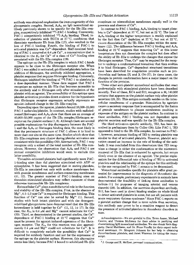
Finally, PAC-1 was shown to bind directly to the 1Ib.IIIa complex on Western blots. Platelets were solubilized with a mixture of Triton X-100 and CHAPS, either in the presence of 1 mM Ca2+ to maintain the IIb. IIIa complex or with 5 mM EDTA to dissociate it. The extracts were electrophoresed in a nondenaturing polyacrylamide gradient gel system that is capable of separating the IIb- IIIa complex from the disso-

11112 Glycoproteins IIb. IIIa and Platelet Activation
ciated glycoproteins (12). Western blots of these gels were then incubated with PAC-1 in the presence of 10 PM Ca2+. Bound antibody was detected using peroxidated-conjugated goat anti-mouse immunoglobulin. PAC-1 bound to the intact IIb. IIIa complex (Fig. 8). However, the antibody did not bind to the dissociated forms of IIb or IIIa.
Effect of PAC-1 on Platelet Function-In the absence of an agonist, PAC-1 a t concentrations as high as 50 pg/ml did not cause platelet aggregation, [14C]serotonin release, or the re- lease of platelet factor 4. PAC-1 also had no effect on ADP- induced platelet shape change, but it did inhibit ADP- and epinephrine-induced platelet aggregation and serotonin re- lease. This effect was most pronounced if the agonist and the PAC-1 were incubated with the platelets for 5 min before initiating the aggregation reaction by adding fibrinogen and stirring. Under these conditions, platelet aggregation was
A B C D E FIG. 8. Binding of PAC-1 to the glycoprotein IIb-IIIa com-
plex. Platelets were solubilized in 0.2% Triton X-100 and 10 mM CHAPS and electrophoresed in the presence of 10 p~ Ca2+ under nondenaturing conditions as described under “Experimental Proce- dures.” A, the gel lane has been stained for protein with Coomassie Brilliant Blue. Arrow indicates the protein band representing the intact IIb. IIIa complex (12). B-E are Western blots of gel lanes similar to that of A . B was incubated with a %oo dilution of AzA9, a monoclonal antibody known to be specific for the IIb. IIIa complex, and then examined in an ELISA to detect bound antibody. Similarly, C was incubated with a Y m dilution of PAC-1, while E was incubated with CA2, an antibody specific for granulocytes. D represents an autoradiograph of a blot incubated with a human polyclonal anti- PIA1 antibody and then Iz5I-labeled staphylococcal protein A. This antibody is specific for glycoprotein IIIa. Note in C that PAC-1 bound only to the area of the blot containing the 1Ib.IIIa complex. Not shown is the fact that when the solubilized IIb-IIIa complex was dissociated with EDTA and then loaded on the gel, the dissociated glycoproteins migrated further into the gel (12), and no PAC-1 binding to the blots was observed.
’ ‘0 10 20 30 40 [PAC- I] pg/ml
FIG. 9. Effect of PAC-1 on platelet aggregation. Platelets were gel-filtered and incubated at room temperature without stirring with the indicated amounts of PAC-1 and with either 10 p~ ADP (closed circles) or epinephrine (open circles). After 5 min, aggregation was initiated by stirring the platelets in an aggregometer at 37 “C and adding 100 pg/ml fibrinogen and 100 p~ CaC12. The extent of aggre- gation was quantitated 3 min later. The data are normalized to a 100% value for control platelets incubated without PAC-1.
inhibited half-maximally by 5-10 nM PAC-1, a value similar to the apparent Kd for lZ5I-PAC-1 binding to stimulated plate- lets (Fig. 9). Similar results were obtained for inhibition of platelet [14C]serotonin release. Thus, PAC-1 inhibited fibrin- ogen-mediated platelet aggregation, and conversely, fibrino- gen inhibited lZ5I-PAC-1 binding. This suggests that PAC-1 inhibited platelet aggregation by preventing the interaction of fibrinogen with its receptor.
DISCUSSION
One of the required steps in platelet aggregation is the expression of receptors for fibrinogen on the platelet surface. Although this receptor is known to be located on the glyco- protein IIb IIIa complex, the changes involving the complex that result in exposure of the binding site for fibrinogen are unknown. Receptor expression does not appear to be the outcome of simple formation of the complex from previously separate components because IIb . IIIa heterodimers are pres- ent in the membrane of unstimulated platelets (7-12). In the present studies, we have described a pentameric IgM mono- clonal antibody, PAC-1, that binds to the surface of platelets activated by agonists such as ADP, epinephrine, and throm- bin, but does not bind to resting platelets. Unlike two previ- ously reported monoclonal antibodies that bind preferentially to activated platelets (28,29), PAC-1 binding is not dependent upon platelet secretion.
Western blots of nondenatured platelet proteins were used to identify the glycoprotein 1Ib.IIIa cmplex as the PAC-1- specific antigen. In addition, PAC-1 has been shown to bind to the complexed but not the dissociated form of IIb and IIIa on crossed immunoelectrophoresis? Several other lines of evidence also indicated that PAC-1 binds to the 1Ib.IIIa complex. First, PAC-1 bound to activated normal platelets, but failed to bind to platelets from three patients with Glanz- mann’s thrombasthenia. It should be noted that thrombas- thenic platelets, containing about 5% the normal amount of IIb and IIIa, had been used to immunize the mouse that produced PAC-1. The fact that an anti-IIb-IIIa monoclonal
T. Kunicki, personal communication.
Glycoproteins IIb. IIIa and Platelet Activation 11113
antibody was obtained emphasizes the immunogenicity of this glycoprotein complex. Second, &As, an IgG monoclonal an- tibody previously shown to be specific for the 1Ib.IIIa com- plex, competitively inhibited lZ5I-PAC-1 binding. Conversely, PAC-1 competitively inhibited lZ5I-A2Ag binding. Third, in- cubation of platelets with EGTA at 37 "C, conditions that dissociate membrane IIb from IIIa (12), also resulted in the loss of PAC-1 binding. Fourth, the binding of PAC-1 to activated platelets was Ca2+-dependent. Half-maximal bind- ing of PAC-1 occurred at 0.4 PM Ca2+, a concentration that is identical to the Kd for Ca2+ of a class of Ca2+-binding sites associated with the 1Ib.IIIa complex (19).
The epitope on the IIb. IIIa complex to which PAC-1 binds appears to be close to the fibrinogen receptor site. When PAC-1 was added to activated, gel-filtered platelets before the addition of fibrinogen, the antibody inhibited aggregation, a platelet response that requires fibrinogen binding. Conversely, fibrinogen inhibited the binding of '251-PAC-1 to platelets in a dose-dependent manner. These data suggest that PAC-1 recognizes an epitope on IIb.IIIa that becomes accessible to the antibody and to fibrinogen only after stimulation of the platelet with an agonist. The accessibility of this epitope upon platelet activation provides direct evidence for the hypothesis that fibrinogen receptor expression actually represents an agonist-induced change in the IIb. IIIa complex.
Depending upon the agonist, platelets bound 10,000-25,000 PAC-1 molecules/platelet. In contrast, studies with radioiod- inated fibrinogen and A2Ag show that there are an average of 40,000-50,000 copies of the IIb. IIIa complex/fibrinogen re- ceptor on the platelet surface (1,8). Although there are several possible explanations for this discrepancy between the PAC- 1 and the A2A9 or fibrinogen binding data, the most likely is that the pentameric structure of PAC-1 allows it to bind to more than one site at the same time. Studies which show that IIb. IIIa complexes may cluster during platelet activation are consistent with this possibility (30). Alternatively, PAC-1 may recognize only a subset of the total number of IIb. IIIa com- plexes. However, the observation that and PAC-1 are mutual competitive inhibitors would seem to exclude this latter possibility.
Thrombin-activated platelets had significantly more PAC- I-binding sites than did platelets stimulated with ADP or epinephrine. It has been suggested that in resting platelets, IIb-IIIa is associated not only with surface membranes but with granule membranes and surface-connecting membranes (31, 32). The greater number of PAC-1-binding sites on thrombin-stimulated platelets may reflect exposure of these otherwise inaccessible IIb. IIla complexes.
Extracellular Ca2+ plays a multifactorial role in the function and stability of the IIb. IIIa complex. First, in the absence of M2', 0.1-1.0 mM Ca2+ is required for fibrinogen binding. This function of Ca2+ can also be served by Mg2+ (1). Second, studies with both intact platelets and with the detergent- solublized glycoproteins have demonstrated that the IIb. IIIa heterodimer is held together by Ca2+ (11, 12, 14-17). In this case, the Kc, is 0.4 PM and Mg2+ cannot substitute for Ca2+ (33). Third, as demonstrated in the present studies, the Ca2+ dependence of PAC-1 binding at 22 "C suggests that Ca2+ must be present for agonist-induced expression of the fibrin- ogen receptor. The KC, for this reaction was also approxi- mately 0.4 p M and M$+ could not substitute for Ca2+. It is difficult to completely exclude the possibility that Ca2+ is required for antibody function and not for the expression of the epitope on the platelet surface. However, this alternative seems less likely because PAC-1 bound to solubilized IIb. IIIa
complexes on nitrocellulose membranes equally well in the presence and absence of Ca2+.
In contrast to PAC-1 binding, A2A, binding to intact plate- lets is Caz+-dependent at 37 "C, but not at 22 "C. The loss of AzAg binding at the higher temperature is readily explained by the fact that Ca2+ depletion at 37 "C, but not at 22 "C, causes dissociation of the IIb. IIIa complex within the mem- brane (12). The difference between PAC-1 binding and A2A9
binding at 22 "C suggests that removing Ca2+ at this lower temperature does not dissociate the complex but does affect the ability of IIb. IIIa to undergo the changes that expose the fibrinogen receptor. Thus, Ca2+ may be required for the recep- tor to undergo a conformational transition that then enables it to bind fibrinogen. Ca2+-dependent structural transitions have been described for coagulation proteins such as pro- thrombin and factors IX and X (34-37). In these cases, the changes in protein conformation have a major impact on the function of the proteins.
Three other anti-platelet monoclonal antibodies that react preferentially with stimulated platelets have been described recently. Two of these, KC4 and S12, recognize a M, 140,000 antigen that appears on the platelet surface only after platelet secretion (28,29). This antigen normally resides on the intra- cellular membranes of a-granules. Stimulation by agonists causes a secretory response that is accompanied by the fusion of granule membranes to plasma membranes resulting in exposure of the antigen on the platelet surface. In contrast to these antibodies, PAC-1 binding was not dependent upon platelet secretion and was specific for the IIb. IIIa complex.
The third activation-specific antibody, 7E3, has been de- scribed in preliminary form by Coller (38). Like PAC-l, 7E3 appeared to bind to the IIb. IIIa complex. In contrast to PAC- 1, however, maximum binding of 7E3 to resting platelets was similar to that of activated platelets. However, platelet acti- vation caused an increase in the rate of binding of the anti- body. It was concluded from this observation that 7E3 recog- nizes a change in either the conformation or the microenvi- ronment of IIb-IIIa that is enhanced by agonists, but occurs slowly even in the absence of an agonist. The precise expla- nation for the differential rate of binding of 7E3 to activated platelets and the relationship of the epitope for this antibody to the one recognized by PAC-1 remain to be determined.
Monoclonal antibodies specific for platelets offer great po- tential for improvements in the diagnosis of thrombotic dis- orders. For example, preliminary experiments in animals have demonstrated the feasibility of linking these antibodies to indium-111 for purposes of imaging arterial and venous thrombi (39). In addition, the secretion-dependent antibody, S12, has been used in direct binding studies on whole blood to detect activated platelets in some, but not all, patients with acute respiratory distress syndrome? Since PAC-1 reports on a platelet surface change that is more subtle than secretion, this antibody may prove to be a sensitive reagent to detect the presence of activated platelets in thrombotic disorders in man.
Acknowledgments-We are grateful to Drs. Terro Asano, Michael Cancro, and Thomas McKearn for their advice in purifying and characterizing IgM monoclonal antibodies. We also thank Beth Hag- garty, David Matthews, and Dr. Prem Pandhi for their expert tech- nical assistance, Dr. Margaret Johnson for her help in obtaining thrombasthenic platelets, and Janet Waxman for typing the manu- script.
J. George and R. McEver, personal communication.

11114 Glycoproteins IIb . IIIa REFERENCES
1. Bennett, J. S., and Vilaire, G. (1979) J. Clin. Inuest. 64, 1393-
2. Marguerie, G. A., Plow, E. F., and Edgington, T. S. (1979) J. Biol.
3. Peerschke, E. I., Zucker, M. B., Grant, R. A., Egan, J. J., and
4. Bennett, J. S., Vilaire, G., and Cines, D. B. (1982) J. Biol. Chem.
5. Marguerie, G. A., Thomas-Maison, N., Ginsberg, M. H., and
6. Nachman, R. L., and Leung, L. L. K. (1982) J. Clin. Znuest. 6 9 ,
7. McEver, R. P., Bennett, E. M., and Martin, M. N. (1983) J. Biol. Chem. 258,5269-5275
8. Bennett, J. S., Hoxie, J. A., Leitman, S. F., Vilaire, G., and Cines, D. B. (1983) Proc. Natl. Acad. Sci. U. S. A. 80, 2417-2421
9. Montgomery, R. R., Kunicki, T. J., Taves, C., Corcoran, M., and Pidard, D. (1982) J. Clin. Inuest. 71 , 385-389
10. Coller, B. S., Peerschke, E. I., Scudder, L. E., and Sullivan, C. A. (1983) J. Clin. Inuest. 7 2 , 325-338
11. Fitzgerald, L. A., and Phillips, D. R. (1984) Circulation 70 , II- 193
12. Shattil, S. J., Brass, L. F., Bennett, J. S., and Pandhi, P. (1985)
13. Phillips, D. R., and Agin, P. P. (1977) J. Biol. Chem. 252,2121- Blood, in press
2126 14. Jennings, L. K., and Phillips, D. R. (1982) J. Biol. Chem. 257 ,
15. Kunicki, T. J., Pidard, D., Rosa, J.-P., and Nurden, A. T. (1981)
16. Hagen, I., Bjerrum, D. J., Gogstad, G., Korsmo, R., and Solum, N. 0. (1982) Biochim. Biophys. Acta 701 , 1-6
17. Howard, L., Shulman, S., Sadanaden, S., andKarpatkin, S. (1982) J. Biol. Chem. 257,8331-8336
18. Kennett, R. H. (1980) in Monoclonal Antibodies, Hybridomas: A New Dimension in Biological Analysis (Kennett, R. H., MC- Kearn, T. J., and Bechtol, K. B., eds) pp. 365-377, Plenum Press, New York
1401
Chem. 254,5357-5363
Johnson, M. M. (1980) Blood 55,841-847
257,8049-8054
Plow, E. F. (1984) Eur. J. Biochem. 139, 5-11
263-269
10458-10466
Blood 58,268-278
and 19. 20.
21. 22.
23.
24.
25.
26.
27.
28.
29.
30.
31.
32.
33.
34.
35.
36.
37.
38. 39.
Platelet Activation Brass, L. F., and Shattil, S. J. (1984) J. Clin. Znuest. 73,626-632 Hawkes, R., Niday, E., and Gordon, J. (1982) Anal. Biochem.
Laemmli, U. K. (1970) Nature 227,680-685 Matthew, W. D., and Reichardt, L. F. (1982) J. Immunol. Methods
Towbin, H., Staehelin, T., and Gordon, J . (1979) Proc. Natl. Acad.
Jerushalmv, Z.. and Zucker. M. B. (1966) Thromb. Diath. Hae-
119,142-147
50,239-253
Sci. U. S. A. 76,4350-4354
morrh. i5,413-419
Br. J . Haematol. 39.129-146
. .
Kaplan, K. L., Nossel, H. L., Drillings, M., and Lesznik, G. (1978)
Brass, L. F., and Shattil, S. J . (1982) J. Biol. Chem. 257 , 14000-
Munson, P. J., and Rodbard, D. (1980) Anal. Biochem. 107,220-
Hsu-Lin, S.-C., Berman, C. L., Furie, B. C., August, D., and Furie,
McEver, R. P., and Martin, M. N. (1984) J. Biol. Chem. 259 ,
Polley, M. J., Leung, L. K., Clark, F. Y., and Nachman, R. L.
Gogstad, G. D., Hagen, I., Korsmo, R., and Solum, N. 0. (1981)
Keller, D. M., Ginsberg, M. H., Plow, E. F., and Woods, V. L.
Brass, L. F., Shattil, S. J., and Bennett, J. S. (1984) Clin. Res.
Madar, D. A., Hall, T. J., Reisner, H. M., Hiskey, R. G., and
Tai, M. M., Furie, B. C., and Furie, B. (1984) J. Biol. Chem. 259,
Lewis, R. M., Reisner, H. M., Chung, K.-S., and Roberts, H. R.
Keyt, B., Furie, B. C., and Furie, B. (1982) J. Biol. Chem. 257 ,
Coller, B. S. (1984) Blood 6 4 , 245a Coller, B. S., Oster, Z. H., Meinken, A. E., Som, P., Brill, A. B.,
Atkins, H. L., Scudder, L. E., and Srivastava, S. C. (1984) Circulation 7 0 , I1 195
14005
239
B. (1984) J. Biol. Chem. 259,9121-9126
9799-9804
(1981) J. Exp. Med. 154, 1058-1068
Biochirn. Biophys. Acta 670, 150-162
(1984) Blood 64,248a
32,494A
Koehler, K. A. (1980) J. Biol. Chem. 255 , 8599-8605
4162-4168
(1980) Blood 56, 608-614
8687-8695