Changes in Leaf Anatomical Traits Enhanced … · The PGPM-induced changes in leaf structure and...
13
ORIGINAL RESEARCH published: 05 May 2017 doi: 10.3389/fpls.2017.00674 Edited by: Barbara De Lucia, Università degli Studi di Bari Aldo Moro, Italy Reviewed by: Antonio Ferrante, Università degli Studi di Milano, Italy Nikos Tzortzakis, Cyprus University of Technology, Cyprus *Correspondence: Roberta Paradiso [email protected] Specialty section: This article was submitted to Crop Science and Horticulture, a section of the journal Frontiers in Plant Science Received: 13 February 2017 Accepted: 12 April 2017 Published: 05 May 2017 Citation: Paradiso R, Arena C, De Micco V, Giordano M, Aronne G and De Pascale S (2017) Changes in Leaf Anatomical Traits Enhanced Photosynthetic Activity of Soybean Grown in Hydroponics with Plant Growth-Promoting Microorganisms. Front. Plant Sci. 8:674. doi: 10.3389/fpls.2017.00674 Changes in Leaf Anatomical Traits Enhanced Photosynthetic Activity of Soybean Grown in Hydroponics with Plant Growth-Promoting Microorganisms Roberta Paradiso 1 *, Carmen Arena 2 , Veronica De Micco 1 , Maria Giordano 1 , Giovanna Aronne 1 and Stefania De Pascale 1 1 Agricultural and Food Sciences, University of Naples Federico II, Naples, Italy, 2 Department of Biology, University of Naples Federico II, Naples, Italy The use of hydroponic systems for cultivation in controlled climatic conditions and the selection of suitable genotypes for the specific environment help improving crop growth and yield. We hypothesized that plant performance in hydroponics could be further maximized by exploiting the action of plant growth-promoting organisms (PGPMs). However, the effects of PGPMs on plant physiology have been scarcely investigated in hydroponics. Within a series of experiments aimed to identify the best protocol for hydroponic cultivation of soybean [Glycine max (L.) Merr.], we evaluated the effects of a PGPMs mix, containing bacteria, yeasts, mycorrhiza and trichoderma beneficial species on leaf anatomy, photosynthetic activity and plant growth of soybean cv. ‘Pr91m10’ in closed nutrient film technique (NFT). Plants were grown in a growth chamber under semi-aseptic conditions and inoculated at seed, seedling and plant stages, and compared to non-inoculated (control) plants. Light and epi-fluorescence microscopy analyses showed that leaves of inoculated plants had higher density of smaller stomata (297 vs. 247 n/mm 2 ), thicker palisade parenchyma (95.0 vs. 85.8 μm), and larger intercellular spaces in the mesophyll (57.5% vs. 52.2%), compared to non-inoculated plants. The modifications in leaf functional anatomical traits affected gas exchanges; in fact starting from the reproductive phase, the rate of leaf net photosynthesis (NP) was higher in inoculated compared to control plants (8.69 vs. 6.13 μmol CO 2 m -2 s -1 at the beginning of flowering). These data are consistent with the better maximal PSII photochemical efficiency observed in inoculated plants (0.807 vs. 0.784 in control); conversely no difference in leaf chlorophyll content was found. The PGPM-induced changes in leaf structure and photosynthesis lead to an improvement of plant growth (+29.9% in plant leaf area) and seed yield (+36.9%) compared to control. Our results confirm that PGPMs may confer benefits in photosynthetic traits of soybean plants even in hydroponics (i.e., NFT), with positive effects on growth and seed production, prefiguring potential application of beneficial microorganisms in plant cultivation in hydroponics. Keywords: beneficial bacteria, chlorophyll fluorescence, controlled ecological life-support system (CELSS), Glycine max (L.) Merr., Mycorrhizae, nutrient film technique (NFT), stomata density, Trichoderma spp. Frontiers in Plant Science | www.frontiersin.org 1 May 2017 | Volume 8 | Article 674
Transcript of Changes in Leaf Anatomical Traits Enhanced … · The PGPM-induced changes in leaf structure and...

fpls-08-00674 May 3, 2017 Time: 15:31 # 1
ORIGINAL RESEARCHpublished: 05 May 2017
doi: 10.3389/fpls.2017.00674
Edited by:Barbara De Lucia,
Università degli Studi di Bari AldoMoro, Italy
Reviewed by:Antonio Ferrante,
Università degli Studi di Milano, ItalyNikos Tzortzakis,
Cyprus University of Technology,Cyprus
*Correspondence:Roberta [email protected]
Specialty section:This article was submitted to
Crop Science and Horticulture,a section of the journal
Frontiers in Plant Science
Received: 13 February 2017Accepted: 12 April 2017Published: 05 May 2017
Citation:Paradiso R, Arena C, De Micco V,
Giordano M, Aronne G andDe Pascale S (2017) Changes in Leaf
Anatomical Traits EnhancedPhotosynthetic Activity of Soybean
Grown in Hydroponics with PlantGrowth-Promoting Microorganisms.
Front. Plant Sci. 8:674.doi: 10.3389/fpls.2017.00674
Changes in Leaf Anatomical TraitsEnhanced Photosynthetic Activity ofSoybean Grown in Hydroponics withPlant Growth-PromotingMicroorganismsRoberta Paradiso1*, Carmen Arena2, Veronica De Micco1, Maria Giordano1,Giovanna Aronne1 and Stefania De Pascale1
1 Agricultural and Food Sciences, University of Naples Federico II, Naples, Italy, 2 Department of Biology, University of NaplesFederico II, Naples, Italy
The use of hydroponic systems for cultivation in controlled climatic conditions and theselection of suitable genotypes for the specific environment help improving crop growthand yield. We hypothesized that plant performance in hydroponics could be furthermaximized by exploiting the action of plant growth-promoting organisms (PGPMs).However, the effects of PGPMs on plant physiology have been scarcely investigatedin hydroponics. Within a series of experiments aimed to identify the best protocol forhydroponic cultivation of soybean [Glycine max (L.) Merr.], we evaluated the effects of aPGPMs mix, containing bacteria, yeasts, mycorrhiza and trichoderma beneficial specieson leaf anatomy, photosynthetic activity and plant growth of soybean cv. ‘Pr91m10’in closed nutrient film technique (NFT). Plants were grown in a growth chamberunder semi-aseptic conditions and inoculated at seed, seedling and plant stages, andcompared to non-inoculated (control) plants. Light and epi-fluorescence microscopyanalyses showed that leaves of inoculated plants had higher density of smaller stomata(297 vs. 247 n/mm2), thicker palisade parenchyma (95.0 vs. 85.8 µm), and largerintercellular spaces in the mesophyll (57.5% vs. 52.2%), compared to non-inoculatedplants. The modifications in leaf functional anatomical traits affected gas exchanges;in fact starting from the reproductive phase, the rate of leaf net photosynthesis (NP)was higher in inoculated compared to control plants (8.69 vs. 6.13 µmol CO2 m−2
s−1 at the beginning of flowering). These data are consistent with the better maximalPSII photochemical efficiency observed in inoculated plants (0.807 vs. 0.784 in control);conversely no difference in leaf chlorophyll content was found. The PGPM-inducedchanges in leaf structure and photosynthesis lead to an improvement of plant growth(+29.9% in plant leaf area) and seed yield (+36.9%) compared to control. Our resultsconfirm that PGPMs may confer benefits in photosynthetic traits of soybean plantseven in hydroponics (i.e., NFT), with positive effects on growth and seed production,prefiguring potential application of beneficial microorganisms in plant cultivation inhydroponics.
Keywords: beneficial bacteria, chlorophyll fluorescence, controlled ecological life-support system (CELSS),Glycine max (L.) Merr., Mycorrhizae, nutrient film technique (NFT), stomata density, Trichoderma spp.
Frontiers in Plant Science | www.frontiersin.org 1 May 2017 | Volume 8 | Article 674

fpls-08-00674 May 3, 2017 Time: 15:31 # 2
Paradiso et al. Plant Growth-Promoting Organisms in Soybean in Hydroponics
INTRODUCTION
When properly managed, the hydroponic systems permit theoptimal water and nutrient supply to the roots, helping toimprove plant growth and yield and resource use efficiencycompared to soil (Savvas et al., 2013; Paradiso et al., 2014b).
Recirculating hydroponic systems are used in most of thestudies aiming to characterize plant production under controlledenvironment, in the sight of their use in CELSS (controlledecological life-support system) in Space (Wheeler and Sager,2003). Together with durum wheat, bread wheat and potato,soybean has been selected as a candidate crop for CELSS dueto the high nutritional value of seeds (Palermo et al., 2012). Tomaximize crop performance, once crops have been chosen, themost suitable cultivar for CELSS has to be selected by consideringplant adaptability to the hydroponic environment and otherrelevant agronomical requirements, such as small size, shortgrowing cycle, high harvest index (as ratio of edible part to totalbiomass per plant), and good tolerance to biotic and abioticstresses (De Micco et al., 2012).
It is conceivable that further improvements in cropproductivity in hydroponics could be achieved by exploitingthe action of beneficial organisms, known as plant growth-promoting organisms (PGPMs) (Lee and Lee, 2015). Whengrown in soil, plants normally establish specific interactionswith PGPMs, which grow in, on, or around plant root tissues.These relationships are well characterized in the most importantcrop/microbe combinations in field (Hayat et al., 2010). PGPMscan promote plant growth and yield, directly or indirectly,through several mechanisms (Vessey, 2003; Mitter et al.,2013). Biological fixation of atmospheric N2, performed byspecific strains of symbiotic Rhizobia bacteria in leguminousplants (Mylona et al., 1995), and non-symbiotic bacteria (e.g.,Azotobacter spp., Pseudomonas spp.) in other crops (Kennedyet al., 2004), provides additional amount of N. Numerousbacteria [e.g., Pseudomonas spp., Bacillus spp., Rhizobium spp.(Rodríguez and Fraga, 1999)] and fungi [e.g., Aspergillus spp.,Penicillium spp. (Whitelaw, 1999)] produce chelators able toconvert insoluble minerals (e.g., phosphorus) to bioavailableforms, or to isolate heavy metals and toxic compounds, includingpathogens metabolites. Some bacteria [e.g., Pseudomonas spp.(Luján et al., 2015)] and fungi [e.g., Streptomyces, Actinomycetes(Haas et al., 2008)] can solubilise ferric iron (Fe3+), by means ofsiderophores. Finally, many bacterial and fungal microorganismsproduce phytohormones, such as indole-3-acetic acid (IAA),cytokinins, gibberellins, promoting plant growth (Weyenset al., 2009). Indirect positive effects of PGPMs on plant canbe related to biocontrol mechanisms (Maksimov et al., 2011).For instance, competition for plant root exudates and mucilage,as source of nutrients, is a mechanism for pathogens exclusion(Compant et al., 2005). Siderophores produced by beneficialbacteria (e.g., Pseudomonas) have higher affinity to Fe3+ thanthose of pathogenic fungi (Beneduzi et al., 2012). SeveralPGPMs (e.g., Pseudomonas spp., Bacillus spp., Trichodermaspp.) produce antibiotic compounds and lytic enzymes whichdegrade pathogens cell walls and toxins (Maksimov et al.,2011); some others induce systemic resistance against pathogens
through the mechanism of priming (Conrath et al., 2006),or help plants cope with abiotic stresses such as droughtand salinity by means of various mechanisms (Yang et al.,2009).
In accordance with the positive effects exerted on plantgrowth, at the present the listed microorganisms are consideredas plant biostimulants (Calvo et al., 2014), similarly to organicmolecules from plant extracts, containing bioactive compoundsable to activate plant metabolism improving plant performance(Bulgari et al., 2015).
Moreover, it is known that PGPMs can influence both gasexchanges and the whole photosynthetic machinery in severalcrops. In sugar beet (Beta vulgaris L.), some endophytic bacteria(e.g., Bacillus pumilus) enhanced the rate of photosynthesisand the maximal photochemical efficiency even at increasingPhotosynthetic Photon Flux Density (PPFD), by promoting thechlorophyll synthesis and the electron transport in thylakoidmembranes, and production of phytohormones, with positiveeffects on growth of both roots and aerial part (Shi et al., 2010).In runner bean (Phaseolus coccineus L.), the co-inoculation ofseeds with two rhizobacteria strains, active for phosphoroussolubilisation and production of siderophores (Bacillus mycoides)and indoleacetic acid (Bacillus pumilus), produced a synergisticaction resulting, in increase in photosynthesis and chlorophyllcontent, particularly during vegetative and early flowering stages,and in the grain yield (Stefan et al., 2013). In plants ofstrawberry (Fragaria × ananassa), inoculation with arbuscularendomycorrhizal fungi (AMF) increased the photosynthetic andphotochemical activity even under drought stress (Borkowska,2002).
Most the listed effects of PGPMs have been well studied in soil,while little is known on these associations in hydroponics, wherethe benefits also depend on the ability of microbes to surviveand proliferate over time, and to colonize plant roots in thespecific environment (i.e., acid recirculating nutrient solution).Some PGPMs (Pseudomonas spp., Bacillus spp., endomycorrhizalfungi) have been successfully tested in hydroponically grownvegetables (e.g., tomato, cucumber, and lettuce), with positiveeffects on plant growth, yield and quality (Lee and Lee, 2015).However, most reports focus on the relief from biotic stress,demonstrating that growth promotion depended on diseasesuppression in recirculating systems, where pathogens can easilydevelop and spread, also considering that the absence of non-pathogenic competitor microorganisms increases their degree ofdanger (Peer and Schippers, 1989; Lee et al., 2010; Stewart-Wade,2011).
Regarding soybean, positive effects of natural association orroot inoculation with PGPMs have been well investigated insoil (Salvagiotti et al., 2008; Tewari et al., 2015). Conversely, alimited number of studies has addressed this topic for soybeanin hydroponics, and they mainly focused on biological N-fixation(Vigue et al., 1977; Paradiso et al., 2014a, 2015).
Only a few works concern the impact of PGPMs on the hostphotosynthetic metabolism, which is crucial in determining plantgrowth and productivity, and they usually refer to single ordouble microbial species, while at the present mixed cultures areoften preferred, since they match multiple scopes (e.g., increasing
Frontiers in Plant Science | www.frontiersin.org 2 May 2017 | Volume 8 | Article 674

fpls-08-00674 May 3, 2017 Time: 15:31 # 3
Paradiso et al. Plant Growth-Promoting Organisms in Soybean in Hydroponics
or restoring microbial diversity of soil, acting effectively in severalplant species) (Lugtenberg et al., 2013).
In this study we aimed at analyzing the effects of inoculationwith PGPMs on soybean leaf structure, which is fundamental indetermining the photosynthetic performance and ultimately theplant growth and yield. Therefore, we investigated the effect of acommercial PGPMs mix on the photosynthetic activity and leafanatomical functional traits of soybean cultivated in hydroponicsunder controlled environment.
MATERIALS AND METHODS
Cultivation Design, Growth ChamberEnvironmental Control, and HydroponicSystem ManagementThe study was conducted on plants of the soybean [Glycinemax (L.) Merr.] cultivar (cv.) ‘PR91M10’ (Pioneer Intl.). Thiscultivar was previously selected according to the European SpaceAgency (ESA) criteria (De Micco et al., 2012) and evaluated inhydroponics in controlled environment (Paradiso et al., 2012; DeMicco et al., 2013).
Plants were grown in a 28 m2 open-gas-exchange growthchamber (7.0 m × 2.1 m × 4.0 m, W × H × D), equippedwith a computer for integrated climate control. The nutrientfilm technique (NFT) system consisted in polypropylene doublegullies (each with two single gullies flanked). Each gully was60 cm high, 100 cm long and 10 cm wide, had an inclinationof about 1%, and was equipped with four sprinklers. Sixteenplants were grown per double gully and three double gullies (48plants in total) were used per treatment. Gullies were sealed withwhite-on-black polyethylene film to prevent evaporation, to avoidthe entrance of light in the root zone, and to enhance the lightdistribution in the basal part of the plants through reflection.
Before starting the experiment, the selected cv. ‘PR91M10’was characterized for photosynthetic response to light intensity,performing a light fast kinetics curve (Figure 1), to identify themost suitable value of PPFD for photosynthesis to be appliedin closed controlled environment. Based on the curve, the lightintensity in the growth chamber was set at 420 µmol m−2 s−1
at the top of the canopy avoiding an excess of light andphotoinhibitory damage risks to photosynthetic apparatus (Evanset al., 1993). Light was provided by High Pressure Sodium(HPS) lamps. Lamps were mounted on a mobile panel, whichwas moved upward following the stem elongation, in orderto keep the PPFD constant at the canopy level, according toa day/night regime of 12/12 h. The other climatic parameterswere chosen according to the Space-related literature on soybean(Dougher and Bugbee, 1997; Wheeler et al., 2008), and keptconstant during the entire growth cycle. Temperature was26/20◦C (light/dark) and relative humidity (RH) was 70–80%.The mean values of temperature and RH recorded at the endof the experiment (98 days) were 26.3 ± 0.1/19.7 ± 0.1◦C and82.4 ± 2.9/68.7 ± 0.5%, respectively (Mean Value ± StandardDeviation). Cultivation was carried out at ambient CO2concentration (370–400 ppm).
FIGURE 1 | Response curve of net photosynthesis (NP) to increasinglight intensity in soybean cv. ‘PR91M10’ grown in closed-loophydroponics. Average values of three non-inoculated plants at vegetativephase; measurement conditions: 26◦C, RH 70%, CO2 400 ppm.
Each double gully was equipped with a polypropylenereservoir (21 liters) for the recirculating nutrient solution, andits own submerged pump (New A Jet 3000) in order to workindependently. Nutrient solution was pumped from the tankinto the gullies at a flow rate of 2.0 L/min and returned to thereservoir by gravity dependent flow. Fertigation started 14 daysafter sowing (DAS), and was performed continuously.
The nutrient solution was based on the standard Hoaglandrecipe 1/2 strength (Hoagland and Arnon, 1950), modified byWheeler et al. (2008), according to the specific requirements ofsoybean. The starting nutrient solution had the following elementconcentration: 7.5 mM N, 3.0 mM K, 0.5 mM P, 2.5 mM Ca,1.0 mM Mg, 1.0 mM S, 60 µM Fe, 7.4 µM Mn, 0.96 µM Zn,1.04 µM Cu, 7.13 µM B, and 0.01 µM Mo. P content was reducedto 0.25 mM during the first 3 weeks of cultivation to avoidnegative effects on mycorrhiza in the inoculated treatment (Collaet al., 2008). The same reduction was applied in control plants.EC and pH of the recirculation nutrient solution were controlledmanually (Multimeter Basic 30, Crison Instruments, Barcelona,Spain) and adjusted every 2 days to the target values by addingdeionised water and/or fresh solution (for EC control) and 1Mnitric acid (for pH control) in the storage tank (Figure 2). The pHwas kept at 5.8 in both the treatments. Since the addition of theinoculum increased the EC value in the fresh solution comparedto control, EC target was raised from 1200 µS cm−1 in controlto 1400 µS cm−1 in inoculated treatment. The starting and thereplenish solution and deionised water were filtered at 0.45 mm.To prevent large fluctuation in the anions/cations concentrations,electrical conductivity and pH, the nutrient solution in both thetreatments was renewed in all tanks 32 DAS.
Disinfection Procedures and SeedInoculation ProtocolBefore starting cultivation, a disinfection of the growthchamber (floor, walls, gullies, conditioning devices, etc.) wasperformed with sodium hypochlorite water solution (NaClO
Frontiers in Plant Science | www.frontiersin.org 3 May 2017 | Volume 8 | Article 674

fpls-08-00674 May 3, 2017 Time: 15:31 # 4
Paradiso et al. Plant Growth-Promoting Organisms in Soybean in Hydroponics
FIGURE 2 | Evolution of pH and EC of recirculating nutrient solution insoybean cv. ‘PR91M10’ grown in closed-loop hydroponics, before theadjustment to the target values (pH 5.8; EC 1200 µS cm−1 in controland 1400 µS cm−1 in inoculated treatment).
5 g l−1). Safety procedures were adopted during the experimentto minimize contamination from operators. In addition,periodic cleaning was performed with NaClO water solutions(5 g l−1 and 1 g l−1, for chamber and measurement tools,respectively).
Prior to start the experiment, the germination performanceof cv. ‘PR91M10’ were evaluated at 8 DAS, according to theInternational Rules for Seed Testing (International Seed TestingAssociation, 1999). The Mean Germination Time (MGT) wascalculated according to the following formula: MGT=6 Dn/6n,where n = number of seeds germinated per day, D = number ofdays from the beginning of the test. The germination percentageand MGT were 98.0± 2.4% and 4.5± 0.2 days, respectively, andwere not affected by seed inoculation.
Seed sterilization was performed according to the protocolof Somasegaran and Hoben (1994). Seeds were rinsed in 95%ethanol for 20 s to remove waxy materials, then they werecompletely submerged in a NaClO water solution (2.5%) andgently swirled for 5 min. Sodium hypochlorite was drained offand seeds were rinsed six times in sterile H2O.
After sterilization, seeds were inoculated by submersion in asolution of Myco Madness mix (Humboldt Nutrients, Eureka,CA, USA), containing a mixture of 14 bacteria, yeasts and 12beneficial fungi species (Mycorrhizae and Trichoderma spp.). The
Myco Madness mix was chosen because of the high diversity ofpotential beneficial microbes.
The inoculum was prepared by adding 0.5 g of Myco Madnesspowder to 1 L of sterile quarter-strength Ringer solution (Oxoid,Milan, Italy). The cell count to verify the final concentration ofthe inoculum was performed by using a Thoma cell countingchamber (depth 0.02 mm, area 1/400 mm2; Thoma, Hawksley,United Kingdom) and the microscope Eclipse E200 Nikon, andresulted 5× 10⁵ cells per ml. Incubation was performed for 12 h,at room temperature in the darkness (Bashan, 1986). Seeds ofcontrol treatment were dipped in sterile quarter-strength Ringersolution only, and submitted to similar conditions.
For germination, seeds were placed on three sterilized layersof filter paper (Watman n. 1) moistened with the inoculum(treated seeds) or with the quarter-strength Ringer solution only(control seeds), in Petri dishes (20 seeds per dish, 10 dishes pertreatment). Dishes were sealed with parafilm and incubated at22◦C, in the darkness (Fernandez-Orozco et al., 2008). After8 DAS, seedlings were transferred into sterilized rockwool plugsand moved in the hydroponic gullies, in the growth chamber.Inoculation was repeated on seedlings at transplanting (12 DAS),by submerging the roots in the Myco Madness solution for24 h. During cultivation, inoculation was repeated by addingthe inoculum to the recirculating nutrient solution, three timesat 10-day interval starting from 38 DAS. Details on inoculum,inoculation protocol and rhizosphere microbiology are reportedby Sheridan et al. (2016).
Sampling and MeasurementsThe effects of the treatments were evaluated on plants in terms ofchanges in leaf structure, photosynthesis, and plant growth andproductivity, also unraveling whether changes in photosynthesisare linked with modifications in leaf functional anatomical traitsand photochemistry.
Functional Anatomical Traits of LeavesSampling for anatomical analyses was done at 57 DAS on the3th fully expanded trifoliate leaf from the top of the plant. Morespecifically, 3 middle leaflets from 3 plants per treatment werecut and immediately submerged in the chemical fixative FAA(40% formaldehyde – glacial acetic acid – 50% ethanol, 5:5:90by volume). After 2 weeks of fixation, leaflets were halved undera dissection microscope (SZX16, Olympus, Germany) to obtaintwo twin groups of subsamples: one devoted to the quantificationof stomata traits, the other to the analysis of lamina cross sections.
For the analysis of stomata, three strips of lamina adaxialepidermis were peeled off from each subsample and mountedon microscope slides with distilled water. Epidermal peels wereanalyzed under a transmitted light microscope (BX60, Olympus)and digital images were collected by means of a digital camera(CAMEDIA C4040; Olympus), avoiding main veins. Digitalimages were analyzed through the software program Analysis3.2 software (Olympus) to quantify stomata frequency and size.More specifically, stomata frequency was expressed as number ofstomata per surface unit (mm2), counted in two regions per peel.Stomata size was quantified by measuring the length of at least 15
Frontiers in Plant Science | www.frontiersin.org 4 May 2017 | Volume 8 | Article 674

fpls-08-00674 May 3, 2017 Time: 15:31 # 5
Paradiso et al. Plant Growth-Promoting Organisms in Soybean in Hydroponics
guard-cells (pole to pole) and the width of the same cells in themedian position.
To obtain cross sections of the leaf lamina, the subsampleswere cut under the SZX16 dissection microscope to obtainsubsamples of 5 mm × 5 mm from the median part ofthe leaflet, avoiding the main vein. These subsamples weredehydrated in an ethanol series (up to 90%) and embeddedin the acrylic resin JB4 (Polysciences, Warrington, PA, USA).Thin cross sections (5 µm) were cut by means of a rotarymicrotome, stained with 0.025% Toluidine blue in 0.1 Mcitrate buffer at pH 4 (Reale et al., 2012) and mounted withCanadian Balsam. The sections were analyzed under a BX60light microscope and digital images were collected at differentmagnifications. By means of the Olympus Analysis 3.2 software,the mesophyll was characterized by measuring the thicknessof palisade and spongy parenchyma tissues and the quantityof intercellular spaces in the spongy tissues. The thickness ofpalisade and spongy tissues were measured in five positionsalong the lamina, avoiding veins. The incidence of intercellularspaces was measured as the percentage of tissue occupied byintercellular spaces over a given surface, in six regions along theleaf lamina.
Photosynthesis, Fluorescence Measurements, andChlorophyll ContentThe light fast kinetics curve was performed at a single leaflevel on non-inoculated plants, using a portable Infra RedGas Analyzer WALZ HCM 1000 (Walz, Effeltrich, Germany)(Supplementary Figure S1). The curve was determined onthe middle leaflet of the 2nd and 3th fully expanded trifoliateleaves from the top of the plant (two leaves per plant, threeplants). Increasing PPFDs (0, 50, 100, 250, 500, 1000, 1500,and 2000 µmol m−2 s−1) were obtained by using a built-in halogen lamp, and the conditions inside the leaf chamberwere kept constant: temperature 25◦C, RH 70%, ambient CO2concentration.
During plant cultivation, NP was measured on the same leaftypes chosen for the light response measurements (two leavesper plant, three plants per treatment), at ambient light intensity(420 µmol m−2 s−1) and the same leaf chamber conditions.Measurements were carried out in the different phenologicalphases: vegetative growth (30 DAS), flowering (44 DAS), podsetting (57 DAS). NP was not detectable during the pod filling,because of the difficulty to position the leaves in the leaf chamberin presence of symptoms of senescence (wilting and curling).
On the same leaves, chlorophyll a fluorescence weredetermined using a portable FluorPen FP100 max fluorometer(Figure 2), equipped with a light sensor (Photon SystemInstruments, Brno, Czech Republic), at room temperature(26◦C). The ground fluorescence Fo was induced on 30′dark adapted leaves, by a blue LED internal light of about1–2 µmol photons m−2 s−1. The maximal fluorescence levelin the dark Fm was induced by 1 s saturating light pulseof 5.000 µmol photons m−2 s−1. The maximum quantumefficiency of PSII photochemistry was calculated as (Fm-Fo)/Fm.The measurements in the light were conducted at PPFD of420 µmol (photons) m−2 s−1 at the canopy level. The PSII
quantum yield (QY) was determined by means of an open leaf-clip suitable for measurements under ambient light, accordingto Genty et al. (1989). QY was used to calculate the linearelectron transport rate (ETR), according to Krall and Edwards(1992). Non-photochemical quenching (NPQ) was calculatedas described by Bilger and Björkman (1990), according to thefollowing formula: NPQ = (Fm/F
′
m)-1. Measurements startedat flowering (44 DAS), as significant differences in NP betweenthe treatments were detected, and repeated during pod setting(57 DAS) and pod filling (84 DAS).
After fluorescence determinations, the leaf greenness wasestimated using a colorimeter (Chlorophyll Meter Konica-Minolta SPAD 502), and expressed as SPAD units, in six plantsper treatment (two leaves per plant, five measurements per leaf),at flowering (44 DAS). Measurements were made at the centralpoint of the leaflet between the midrib and the leaf margin. Inthe same samples, chlorophyll a and b content was determinedby extraction in acetone and spectrophotometer lecture (Jeffreyand Humphrey, 1975), using a Hach DR 4000 Spectrophotometer(Hach Company, Loveland, CO) on one 2-cm2 leaf sample perleaf.
Plant Growth and Productivity, and ChemicalAnalysesGrowth analysis during the growing cycle was based on non-destructive measurements of plant height and leaf number,determined every 7 to 10 days, on six plants per treatment. Plantleaf area (LA) was estimated by the values of leaf length andwidth, using the formula of Wiersma and Bailey (1957), based onthe specific soybean leaf types and shapes.
Soybean seeds were harvested when pods had turned brown(average water content 14%). At each harvest, yield data [freshweight (FW) of pods and seeds] were determined per single plant.Plant productivity was measured as grams of seeds per plant−1
(edible biomass).At the end of the experiment, plants were collected to
determine FW and dry matter (DM), and their partitioningin roots, stems and leaves (non-edible biomass). Measurementswere carried out on six plants per treatment (two plants× doublegully). DM was measured after oven-drying at 60◦C untilconstant weight.
The concentration of the main cations (K+, Ca2+, Mg2+)and anions (NO3
−) in the recirculating nutrient solution andin the leaf tissues was determined using an ion exchangechromatographer (ICS-3000, Dionex, Sunnyvale, CA, USA).Nutrient solution samples were collected at 7-day intervals,starting from the first inoculation of the recirculating solution.Leaf analysis was performed on water extract of DM of 5 healthy,fully expanded leaves per treatment randomly sampled, duringthe flowering phase.
Statistical AnalysisAll data were processed with one-way ANOVA (p < 0.05), usingthe SPSS R© statistical package (SPSS Inc., Chicago, IL, USA).Shapiro–Wilk and Kolmogorov–Smirnov Tests were performedto check for normality. Percent data were transformed througharcsine function before statistical analysis.
Frontiers in Plant Science | www.frontiersin.org 5 May 2017 | Volume 8 | Article 674

fpls-08-00674 May 3, 2017 Time: 15:31 # 6
Paradiso et al. Plant Growth-Promoting Organisms in Soybean in Hydroponics
FIGURE 3 | Effect of root inoculation on mesophyll traits in leaves of soybean cv. ‘PR91M10’ grown in closed-loop hydroponics. Light microscopy viewsof cross sections of leaves from control (A) and inoculated (B) plants. Bar = 50 mm (images are at the same magnification). Thickness of leaf lamina (LL), palisadeparenchyma (PP), and spongy parenchyma (SP) (C), and percent of intercellular spaces (D) measured in control and inoculated plants (Mean ± standard error,n = 45). Different letters indicate significant differences at P < 0.05. In (C), small letters refer to LL, italic small letters refer to PP, capital letters refer to SP.
RESULTS
Leaf Functional Anatomical TraitsAnalyzed leaves showed a typical dorsiventral structure(Figures 3A,B), with mesophyll made of two layers of palisadecells and a spongy parenchyma rich in intercellular spaces.Stomata were frequent on adaxial epidermis, while rare onabaxial surface.
Root inoculation was responsible for a significant increase inleaf lamina thickness, due to the palisade parenchyma that wasthicker in inoculated than in control plants (95.0 vs. 85.8 µm)(Figure 3C). Leaves of inoculated plants also showed a moreloosen spongy parenchyma because of a higher percentage ofintercellular spaces compared with leaves from non-inoculatedplants (57.5% vs. 52.2%) (Figure 3D).
Leaves of inoculated plants showed significantly higherstomata frequency (297 vs. 247 n/mm2) (Figures 4A–C) thannon-inoculated controls. Stomata were significantly smaller in
leaves of inoculated plants compared to the control ones, due toreduced length and width of guard cells (Figure 4D).
Photosynthesis, FluorescenceMeasurements, and Chlorophyll ContentNet photosynthesis of fully developed leaves of soybean controlplants was maximum in the vegetative phase and decreasedprogressively during the reproductive phase, ranging from8.3 µmol CO2 m−2 s−1 (30 days after sowing, DAS) to5.7 µmol CO2 m−2 s−1 (57 DAS) (Figure 5). Measurementsof NP throughout the growing cycle did not show significantdifferences between the treatments during the vegetative phase(until 30 DAS), while they revealed higher CO2 assimilation ininoculated plants from the flowering (Figure 4). Specifically, NPin inoculated plants was 42 and 31% higher compared to thecontrol, at 44 and 57 DAS, respectively.
Root inoculation did not determine any significant effects onplant photochemistry at 44 and 57 DAS, as demonstrated by
Frontiers in Plant Science | www.frontiersin.org 6 May 2017 | Volume 8 | Article 674

fpls-08-00674 May 3, 2017 Time: 15:31 # 7
Paradiso et al. Plant Growth-Promoting Organisms in Soybean in Hydroponics
FIGURE 4 | Effect of root inoculation on stomata traits in leaves of soybean cv. ‘PR91M10’ grown in closed-loop hydroponics. Light microscopy views ofepidermal peels showing stomata in leaves from control (A) and inoculated (B) plants. Bar = 20 mm (images are at the same magnification). Stomata frequency (C)and size (D) quantified in control and inoculated plants (Mean ± standard error, n = 54 for stomata frequency, n = 135 for stomata size). Different letters indicatesignificant differences at P < 0.05. In (D), small letters refer to guard cell width, capital letters refer to guard cell length.
FIGURE 5 | Net photosynthesis of soybean cv. ‘PR91M10’ in controland inoculated plants grown in closed-loop hydroponics, throughoutthe developmental cycle: vegetative growth (30 DAS), flowering(44 DAS), and fruit setting (57 DAS). (Means ± standard error; n = 3).Within each date, different letters indicate significant differences at P < 0.05.
the values of maximal PSII photochemical efficiency (Fv/Fm),PSII quantum yield (QY), linear electron transport rate (ETR),and non-photochemical quenching (NPQ), which were notsignificantly different between control and inoculated plants
(Figure 6). Conversely, at 84 DAS, significant differences weredetected in the efficiency of light conversion to reaction centers.In particular, compared to control, treated plants showed asignificant (P < 0.05) increase in Fv/Fm (0.807 vs. 0.784), QY(0.702 vs. 0.640) and ETR (183 vs. 166), as well as a significant(P < 0.01) reduction in NPQ (0.486 vs. 0.773).
The higher photochemical efficiency in inoculated plants didnot match the chlorophyll content. In fact, the comparisonbetween inoculated and non-inoculated plants showed nodifference between treatments in the leaf chlorophyll content,neither when determined as chl a and chl b concentration, norwhen measured as greenness (Table 1).
Hydroponic System Management, PlantGrowth and Yield, and Leaf ChemicalAnalysesThe evolution of pH and EC in the recirculating nutrient solutionis shown in Figure 2, for both control and inoculated gullies. ThepH value after 2 days of recycling was always higher than in thefresh or adjusted solution (target value 5.8). The fluctuations weresmaller during the 1st weeks of cultivation, when the plant sizewas still small, while they became wider as plant developed, andwater and nutrient uptake increased. Accordingly, EC variationswere very narrow in the first 30 days, increased during the
Frontiers in Plant Science | www.frontiersin.org 7 May 2017 | Volume 8 | Article 674

fpls-08-00674 May 3, 2017 Time: 15:31 # 8
Paradiso et al. Plant Growth-Promoting Organisms in Soybean in Hydroponics
FIGURE 6 | Maximal PSII photochemical efficiency (Fv/Fm), PSII quantum yield (QY), linear electron transport rate (ETR), and non-photochemicalquenching (NPQ) in control and inoculated plants of soybean cv. ‘PR91M10’ grown in closed-loop hydroponics, throughout the developmental cycle:flowering (44 DAS), fruit setting (57 DAS) and pod filling (84 DAS). (Mean ± standard error; n = 18). Within each date, different letters indicate significantdifferences at P < 0.05.
vegetative growth and decreased progressively after the beginningof leaf falling (around day 65). On the average of the entireexperiment, the value was similar between the treatments for pH(6.97 on average), while it was higher in inoculated comparedto control gullies for EC (1465 ± 21 vs. 1381 ± 10 µS cm−1;Means± Standard error of 32 measurements).
The concentration of the main nutrients (in mmol l−1) inthe recirculating nutrient solution was not significantly differentbetween control and inoculated treatment for N (7.79 ± 0.59 vs.7.58 ± 0.74), K (2.92 ± 0.14 vs. 3.38 ± 0.16), Ca (3.04 ± 0.09in C vs. 2.90 ± 0.09), Mg (0.61 ± 0.09 vs. 0.58 ± 0.11)(Mean± Standard error; n= 6).
The growing cycle of soybean ‘PR91M10’ in closed-loop NFTunder controlled environment lasted 98 days, from the sowing tothe end of the harvests, in both inoculated and non-inoculatedtreatments.
In control plants, flowering started around 34 DAS, and itwas followed by the pod setting (until 60 DAS) and the fillingand drying of pods and seeds, until the harvest, which started at88 DAS and lasted 10 days (Figure 7A). Root inoculation did notaffect significantly the earliness and the duration of the differentphenological stages, as well as the harvesting time.
Inoculation promoted an increase, although not significant,in root growth (+10.9% on a DM basis – data not shown),
while it significantly improved the growth of the aerial partand the seed production (Figures 7A,B). Specifically, during thewhole experiment, inoculated plants showed a tendency to highervalues of plant height, and were significantly taller at the harvestcompared to control (48.5 vs. 43.3 cm) (Figure 7A). Accordingly,the total plant leaf area (maximum value before leaf shedding)and the seed yield were higher in inoculated compared to controlplants (+29.9 and+36.9%, respectively) (Figure 7B).
Chemical analysis revealed changes in composition of leaftissues at the stage of flowering. Specifically, inoculated plantsshowed a tendency to higher values compared to control in thecontent (g per 100 g D.M.) of NO3 (0.15 ± 0.04 vs. 0.11 ± 0.01),K (2.40 ± 0.16 vs. 2.18 ± 0.12), Ca (1.28 ± 0.18 vs. 1.03 ± 0.13),even if the difference between the treatments was found to besignificant only for Mg (0.20± 0.04 vs. 0.13± 0.01).
DISCUSSION
The strategy of nutrient solution control adopted in theexperiment, with measurements and adjustments at 2-dayintervals, was efficient in containing the EC and pH fluctuationswithin acceptable values, and to guarantee comparable nutrientssupply in the control and inoculated treatments.
Frontiers in Plant Science | www.frontiersin.org 8 May 2017 | Volume 8 | Article 674
fpls-08-00674 May 3, 2017 Time: 15:31 # 9
Paradiso et al. Plant Growth-Promoting Organisms in Soybean in Hydroponics
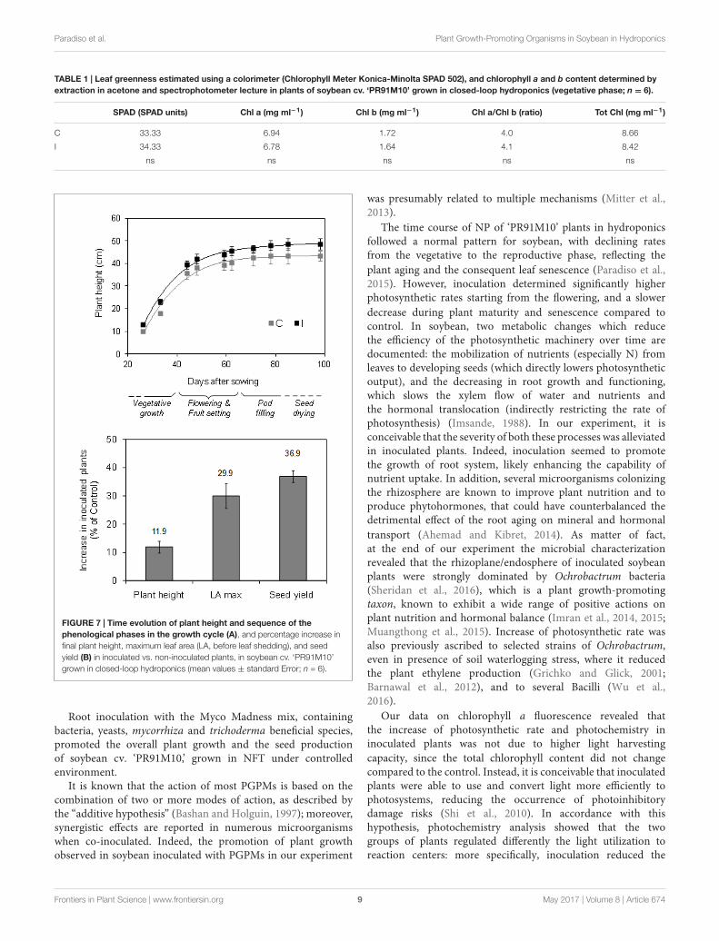
TABLE 1 | Leaf greenness estimated using a colorimeter (Chlorophyll Meter Konica-Minolta SPAD 502), and chlorophyll a and b content determined byextraction in acetone and spectrophotometer lecture in plants of soybean cv. ‘PR91M10’ grown in closed-loop hydroponics (vegetative phase; n = 6).
SPAD (SPAD units) Chl a (mg ml−1) Chl b (mg ml−1) Chl a/Chl b (ratio) Tot Chl (mg ml−1)
C 33.33 6.94 1.72 4.0 8.66
I 34.33 6.78 1.64 4.1 8.42
ns ns ns ns ns
FIGURE 7 | Time evolution of plant height and sequence of thephenological phases in the growth cycle (A), and percentage increase infinal plant height, maximum leaf area (LA, before leaf shedding), and seedyield (B) in inoculated vs. non-inoculated plants, in soybean cv. ‘PR91M10’grown in closed-loop hydroponics (mean values ± standard Error; n = 6).
Root inoculation with the Myco Madness mix, containingbacteria, yeasts, mycorrhiza and trichoderma beneficial species,promoted the overall plant growth and the seed productionof soybean cv. ‘PR91M10,’ grown in NFT under controlledenvironment.
It is known that the action of most PGPMs is based on thecombination of two or more modes of action, as described bythe “additive hypothesis” (Bashan and Holguin, 1997); moreover,synergistic effects are reported in numerous microorganismswhen co-inoculated. Indeed, the promotion of plant growthobserved in soybean inoculated with PGPMs in our experiment
was presumably related to multiple mechanisms (Mitter et al.,2013).
The time course of NP of ‘PR91M10’ plants in hydroponicsfollowed a normal pattern for soybean, with declining ratesfrom the vegetative to the reproductive phase, reflecting theplant aging and the consequent leaf senescence (Paradiso et al.,2015). However, inoculation determined significantly higherphotosynthetic rates starting from the flowering, and a slowerdecrease during plant maturity and senescence compared tocontrol. In soybean, two metabolic changes which reducethe efficiency of the photosynthetic machinery over time aredocumented: the mobilization of nutrients (especially N) fromleaves to developing seeds (which directly lowers photosyntheticoutput), and the decreasing in root growth and functioning,which slows the xylem flow of water and nutrients andthe hormonal translocation (indirectly restricting the rate ofphotosynthesis) (Imsande, 1988). In our experiment, it isconceivable that the severity of both these processes was alleviatedin inoculated plants. Indeed, inoculation seemed to promotethe growth of root system, likely enhancing the capability ofnutrient uptake. In addition, several microorganisms colonizingthe rhizosphere are known to improve plant nutrition and toproduce phytohormones, that could have counterbalanced thedetrimental effect of the root aging on mineral and hormonaltransport (Ahemad and Kibret, 2014). As matter of fact,at the end of our experiment the microbial characterizationrevealed that the rhizoplane/endosphere of inoculated soybeanplants were strongly dominated by Ochrobactrum bacteria(Sheridan et al., 2016), which is a plant growth-promotingtaxon, known to exhibit a wide range of positive actions onplant nutrition and hormonal balance (Imran et al., 2014, 2015;Muangthong et al., 2015). Increase of photosynthetic rate wasalso previously ascribed to selected strains of Ochrobactrum,even in presence of soil waterlogging stress, where it reducedthe plant ethylene production (Grichko and Glick, 2001;Barnawal et al., 2012), and to several Bacilli (Wu et al.,2016).
Our data on chlorophyll a fluorescence revealed thatthe increase of photosynthetic rate and photochemistry ininoculated plants was not due to higher light harvestingcapacity, since the total chlorophyll content did not changecompared to the control. Instead, it is conceivable that inoculatedplants were able to use and convert light more efficiently tophotosystems, reducing the occurrence of photoinhibitorydamage risks (Shi et al., 2010). In accordance with thishypothesis, photochemistry analysis showed that the twogroups of plants regulated differently the light utilization toreaction centers: more specifically, inoculation reduced the
Frontiers in Plant Science | www.frontiersin.org 9 May 2017 | Volume 8 | Article 674

fpls-08-00674 May 3, 2017 Time: 15:31 # 10
Paradiso et al. Plant Growth-Promoting Organisms in Soybean in Hydroponics
dissipation of light energy as heat, promoting the electrontransport rate to C fixation; this allowed to allocate themost part of reductive power in carbon assimilation process,enhancing plant biomass accumulation. Conversely, the controlplants, that exhibit low photochemical efficiency, neededto dissipate thermally the excess of absorbed light withinphotosystems, in order to avoid photoinhibition (Maxwell andJohnson, 2000). This strategy is effective in guarantying theintegrity of photosystems but it reduces the plant efficiencyto assimilate CO2 and to accumulate biomass. The success ofa such regulation is demonstrated by the values of maximumphotochemical efficiency (Fv/Fm) that are comparable inboth the plant groups, indicating the absence of injuriesto photosynthetic apparatus regardless of the treatment.These evidences are consistent with the findings of Shi et al.(2010) who found in sugar beet an increase of gas exchangesand photochemistry triggered by some endophytic bacteria(e.g., Bacillus pumilus). Accordingly, in our experiment,some Bacilli (Staphylococcus spp.) were found in the rootexosphere of inoculated plants, together with other beneficialbacterial taxa such as Actinobacteria, Betaproteobacteria, andAlphaproteobacteria (Sheridan et al., 2016), known to exertseveral useful effects on plant metabolism (Ventorino et al.,2014; Chauhan et al., 2015; Castellano-Hinojosa et al., 2016).Moreover, the higher photosynthetic rate could be likely ascribednot only to a more efficient photochemistry, but also to animprovement of Rubisco carboxylation capacity, hypothesizedon the basis of the enhancement of gas exchanges in inoculatedplants.
In addition to the effect on photochemistry, inoculationenhanced photosynthesis even by inducing changes in leafanatomical traits. The higher stomata frequency found intreated plants could be interpreted as a plant strategy tosatisfy the increasing demand of CO2 needed to match thehigher growth rate in inoculated plants. Beside, higher stomatadensity can greatly amplify the potential for control overwater loss rate and CO2 uptake. Moreover, the occurrenceof smaller stomata in inoculated plants compared to non-inoculated ones would allow better control of stomataopening/closure since small stomata are responsible forfaster dynamic characteristics (Drake et al., 2013). Indeed, in ourexperiment, inoculation induced the formation of leaves whosestructural traits can support more dynamic regulation of stomataopening/closure.
The presence of high frequency of small stomata is knownto have a direct positive influence on the operating stomataconductance which in turn scaled with leaf gas-exchange(Meinzer, 2003; Barbieri et al., 2012): leaves with small andnumerous stomata are considered capable of attain high orlow stomata conductance when environmental conditions are,respectively, favorable or unfavorable (Drake et al., 2013).Moreover, the number and size of stomata are also related to planttranspiration balance (Meinzer, 2003). A strong stomata controlmay allow using the same amount of water more efficiently byroot apparatus of inoculated plants. Generally a high water useefficiency is obtained limiting gas exchanges through the increaseof stomata closure. In the case of inoculated plants, the elevated
number of stomata and their reduced size allow to maintainmore stomata opened on the leaf lamina in order to favor CO2entrance in substomatal spaces and at same time to minimizethe water vapor losses. From this point of view the PGPMsstimulating the evolution of some specific anatomical traits (i.e.,increase on intercellular spaces, elevated number of stomata) mayhave also indirectly affected plant-substrate water relationships,and consequently the nutrient and water uptake (Balliu et al.,2015).
The relations between operating stomata conductanceand the physical attributes of stomata has been shownboth on a large evolutionary scale and on a smaller scale inresponse to specific environmental conditions (Hetheringtonand Woodward, 2003; Franks and Beerling, 2009). Plantphotosynthetic productivity and water-use efficiency are linkednot only to stomatal conductance but also to mesophyllresistance, thus to leaf anatomy (Brodribb et al., 2007;Woodruff et al., 2009). More specifically, traits such asthickness of palisade and spongy parenchyma and theirporosity, affect the lateral and vertical gas diffusion withinthe leaf lamina (Pieruschka et al., 2005). In inoculatedplants, the thicker leaf lamina would not increase mesophyllresistance compared to non-inoculated plants, because itis accompanied by increased intercellular spaces whichwould improve the accessibility to the carboxylation sitesof the chloroplasts inside the cells. Moreover, the improvedphotosynthesis in inoculated plants is in line with the increasedthickness of palisade parenchyma which contain most of thechloroplasts.
CONCLUSION
Root inoculation with the Myco Madness microbial mix,containing bacteria, yeasts, mycorrhiza and trichodermabeneficial species, enhanced the photosynthetic activity ofsoybean ‘Pr91M10’ grown in closed-loop NFT. Starting fromflowering, the rate of leaf NP was higher in inoculated plantscompared to controls. This result was found to be related tochanges in leaf functional anatomical traits promoting leafgas exchanges: leaves of inoculated plants showed higherdensity of smaller stomata, a thicker palisade parenchyma,and larger intercellular spaces in the mesophyll, compared tonon-inoculated plants. In addition, inoculation determinedhigher photochemical efficiency in adult plants, during the stageof seed maturation, thanks to the higher efficiency to use andconvert light to photosystems.
Overall, the positive influence of PGPMs root inoculation onleaf photosynthetic performances enhanced plant growth andseed production of soybean grown hydroponically.
In conclusion, in our experimental conditions, inoculationwith PGPMs conferred benefits in photosynthesis and leaffunctional anatomical traits, which in turn enhanced plantgrowth and productivity of soybean grown in closed-loophydroponics under controlled environment. These resultsprefigure potential application of beneficial microorganisms inhydroponic cultivation of plants.
Frontiers in Plant Science | www.frontiersin.org 10 May 2017 | Volume 8 | Article 674
fpls-08-00674 May 3, 2017 Time: 15:31 # 11
Paradiso et al. Plant Growth-Promoting Organisms in Soybean in Hydroponics
AUTHOR CONTRIBUTIONS
RP and MG performed plant cultivation. RP, CA, and VDMperformed measurements, data collection and statistical analysis,and wrote the manuscript. GA and SDP provided scientificoversight in experimental design and interpretations andcontributed in writing the manuscript. SDP obtained funding forthe study.
ACKNOWLEDGMENTS
Funding was provided by the European Space Agency throughthe MELiSSA project. This publication is dedicated to thememory of Claude Chipaux (1935-2010), father of the MELiSSA
project. The authors thank Prof. Olimpia Pepe and Dr. ValeriaVentorino for their constructive comments to the paper. Theauthors contributed to this study in equal measure.
SUPPLEMENTARY MATERIAL
The Supplementary Material for this article can be found onlineat: http://journal.frontiersin.org/article/10.3389/fpls.2017.00674/full#supplementary-material
FIGURE S1 | Plants of soybean cv. ‘PR91M10’ grown in closed-loop NFT(A). Measurements of photosynthesis with the Infra Red Gas Analyzer WALZ HCM1000: particular of the halogen lamp for light response curves (B), andmeasurement at the ambient light intensity (C). Measurements of chlorophyll afluorescence with the FluorPen FP100 max fluorometer (D).
REFERENCESAhemad, M., and Kibret, M. (2014). Mechanisms and applications of plant growth
promoting rhizobacteria: current perspective. J. King Saud Univ. Sci. 26, 1–20.doi: 10.1016/j.jksus.2013.05.001
Balliu, A., Sallaku, G., and Rewald, B. (2015). AMF inoculation enhances growthand improves the nutrient uptake rates of transplanted, salt-stressed tomatoseedlings. Sustainability 7, 15967–15981. doi: 10.3390/su71215799
Barbieri, G., Vallone, S., Orsini, F., Paradiso, R., De Pascale, S., Negre-Zakharov, F.,et al. (2012). Stomatal density and metabolic determinants mediate salt stressadaptation and water use efficiency in basil (Ocimum basilicum L.). J. Plant Phys.169, 1737–1746. doi: 10.1016/j.jplph.2012.07.001
Barnawal, D., Bharti, N., Maji, D., Chanotiya, C. S., and Kalra, A. (2012).1-Aminocyclopropane-1-carboxylic acid (ACC) deaminase-containingrhizobacteria protect Ocimum sanctum plants during waterlogging stressvia reduced ethylene generation. Plant Physiol. Biochem. 58, 227–235.doi: 10.1016/j.plaphy.2012.07.008
Bashan, Y. (1986). Significance of timing and level of inoculation with rhizospherebacteria on wheat plants. Soil Biol. Biochem. 18, 297–301. doi: 10.1016/0038-0717(86)90064-7
Bashan, Y., and Holguin, G. (1997). Azospirillum-plant relationships:environmental and physiological advances (1990-1996). Can. J. Microbiol.43, 103–121. doi: 10.1139/m97-015
Beneduzi, A., Ambrosini, A., and Passaglia, L. M. P. (2012). Plant growth-promoting rhizobacteria (PGPR): their potential as antagonists andbiocontrol agents. Genet. Mol. Biol. 35, 1044–1051. doi: 10.1590/S1415-47572012000600020
Bilger, W., and Björkman, O. (1990). Role of the xanthophyll cycle inphotoprotection elucidated by measurements of light-induced absorbancechanges, fluorescence and photosynthesis in leaves of Hedera canariensis.Photosynth. Res. 25, 173–185. doi: 10.1007/BF00033159
Borkowska, B. (2002). Growth and photosynthetic activity of micropropagatedstrawberry plants inoculated with endomycorrhizal fungi (AMF) and growingunder drought stress. Acta Physiol. Plant. 24, 365–370. doi: 10.1007/s11738-002-0031-7
Brodribb, T. J., Field, T. S., and Jordan, G. J. (2007). Leaf maximum photosyntheticrate and venation are linked by hydraulics. Plant Physiol. 144, 1890–1898.doi: 10.1104/pp.107.101352
Bulgari, R., Cocetta, G., Trivellini, A., Vernieri, P., and Ferrante, A. (2015).Biostimulants and crop responses: a review. Biol. Agric. Hortic. 31, 1–17.doi: 10.1080/01448765.2014.964649
Calvo, P., Nelson, L., and Kloepper, J. W. (2014). Agricultural uses of plantbiostimulants. Plant Soil 383, 3–41. doi: 10.1007/s11104-014-2131-8
Castellano-Hinojosa, A., Correa-Galeote, D., Palau, J., and Bedmar, E. J. (2016).Isolation of N2-fixing rhizobacteria from Lolium perenne and evaluating theirplant growth promoting traits. J. Basic Microbiol. 56, 85–91. doi: 10.1002/jobm.201500247
Chauhan, H., Bagyaraj, D. J., Selvakumar, G., and Sundaram, S. P. (2015). Novelplant growth promoting rhizobacteria–prospects and potential. Appl. Soil Ecol.95, 38–53. doi: 10.1016/j.apsoil.2015.05.011
Colla, G., Rouphael, Y., Cardarelli, M., Tullio, M., Rivera, C. M., and Rea, E.(2008). Alleviation of salt stress by arbuscular mycorrhizal in zucchini plantsgrown at low and high phosphorus concentration. Biol. Fertil. Soils 44, 501–509.doi: 10.1007/s00374-007-0232-8
Compant, S., Duffy, B., Nowak, J., Clément, C., and Barka, E. A. (2005). Use ofplant growth-promoting bacteria for biocontrol of plant diseases: principles,mechanisms of action, and future prospects. Appl. Environ. Microbiol. 71,4951–4959. doi: 10.1128/AEM.71.9.4951-4959.2005
Conrath, U., Beckers, G. J. M., Flors, V., García-Agustín, P., Jakab, G., Mauch, F.,et al. (2006). Priming: getting ready for battle. Mol. Plant Microbe Interact. 19,1062–1071. doi: 10.1094/MPMI-19-1062
De Micco, V., Buonomo, R., Paradiso, R., De Pascale, S., and Aronne, G. (2012).Soybean cultivar selection for Bioregenerative Life Support Systems (BLSSs):theoretical selection. Adv. Space Res. 49, 1415–1421. doi: 10.1016/j.asr.2012.02.022
De Micco, V., Paradiso, R., Aronne, G., Fogliano, V., and De Pascale, S.(2013). “Agronomical and nutritional characterization of soybean for BLSS:lessons learned from the MELiSSA project – food characterization phase I,”in Proceedings 63rd International Astronautical Congress (IAC), InternationalAstronautical Federation, 1-5 October 2012, Naples, Vol. 2, 1354–1360.
Dougher, T. A. O., and Bugbee, B. (1997). Effect of lamp type and temperatureon development, carbon partitioning and yield of soybean. Adv. Space Res. 20,1895–1899. doi: 10.1016/S0273-1177(97)00857-0
Drake, P. L., Froend, R. H., and Franks, P. J. (2013). Smaller, faster stomata: scalingof stomatal size, rate of response, and stomatal conductance. J. Exp. Bot. 64,495–505. doi: 10.1093/jxb/ers347
Evans, J. R., Jakobsen, I., and Ögren, E. (1993). Photosynthetic light-responsecurves - 2. Gradients of light absorption and photosynthetic capacity. Planta189, 191–200. doi: 10.1007/BF00195076
Fernandez-Orozco, R., Frias, J., Zielinski, H., Piskula, M. K., Kozlowska, H., andVidal-Valverde, C. (2008). Kinetic study of the antioxidant compounds andantioxidant capacity during germination of Vigna radiata cv. emmerald, Glycinemax cv. Jutro and Glycine max cv. Merit. Food Chem. 111, 622–630. doi: 10.1016/j.foodchem.2008.04.028
Franks, P. J., and Beerling, D. J. (2009). CO2-forced evolution of plant gas exchangecapacity and water-use efficiency over the Phanerozoic. Geobiology 7, 227–236.doi: 10.1111/j.1472-4669.2009.00193.x
Genty, B., Briantais, J. M., and Baker, N. R. (1989). The relationship betweenthe quantum yield of photosynthetic electron transport and quenching ofchlorophyll fluorescence. Biochim. Biophys. Acta 990, 87–92. doi: 10.1016/S0304-4165(89)80016-9
Grichko, V. P., and Glick, B. R. (2001). Amelioration of flooding stress byACC deaminase-containing plant growth-promoting bacteria. Plant Physiol.Biochem. 39, 11–17. doi: 10.1016/S0981-9428(00)01212-2
Frontiers in Plant Science | www.frontiersin.org 11 May 2017 | Volume 8 | Article 674
fpls-08-00674 May 3, 2017 Time: 15:31 # 12
Paradiso et al. Plant Growth-Promoting Organisms in Soybean in Hydroponics
Haas, H., Eisendle, M., and Turgeon, B. G. (2008). Siderophores in fungalphysiology and virulence. Annu. Rev. Phytopathol. 46, 149–187. doi: 10.1146/annurev.phyto.45.062806.094338
Hayat, R., Ali, S., Amara, U., Khalid, R., and Ahmed, I. (2010). Soil beneficialbacteria and their role in plant growth promotion: a review. Ann. Microbiol.60, 579–598. doi: 10.1007/s13213-010-0117-1
Hetherington, A. M., and Woodward, F. (2003). The role of stomata in sensing anddriving environmental change. Nature 424, 901–908. doi: 10.1038/nature01843
Hoagland, D. R., and Arnon, D. I. (1950). The water-culture method for growingplants without soil. Circular 347, 1–32.
Imran, A., Mirza, M. S., Shah, T. M., Malik, K. A., and Hafeez, F. Y. (2015).Differential response of kabuli and desi chickpea genotypes toward inoculationwith PGPR in different soils. Front. Microbiol. 6:859. doi: 10.3389/fmicb.2015.00859
Imran, A., Saadalla, M. J. A., Khan, S. U., Mirza, M. S., Malik, K. A.,and Hafeez, F. Y. (2014). Ochrobactrum sp. Pv2Z2 exhibits multiple traitsof plant growth promotion, biodegradation and N-acyl-homoserine-lactonequorum sensing. Ann. Microbiol. 64, 1797–1806. doi: 10.1007/s13213-014-0824-0
Imsande, J. (1988). Enhanced nitrogen fixation increases net photosynthetic outputand seed yield of hydroponically grown soybean. J. Exp. Bot. 39, 1313–1321.doi: 10.1093/jxb/39.9.1313
International Seed Testing Association (1999). International rules for seed testing.Seed Sci. Technol. 27(Suppl.), 333.
Jeffrey, S. W., and Humphrey, G. F. (1975). New spectrophotometric equationsfor determining chlorophylls a, b, c1 and c2 in higher plants, algae and naturalphytoplankton. Biochem. Physiol. Pflanz. 167, 191–194. doi: 10.1016/S0015-3796(17)30778-3
Kennedy, I. R., Choudhury, A. T. M. A., and Kecskés, M. L. (2004). Non-symbioticbacterial diazotrophs in crop-farming systems: can their potential for plantgrowth promotion be better exploited? Soil Biol. Biochem. 36, 1229–1244.doi: 10.1016/j.soilbio.2004.04.006
Krall, J. P., and Edwards, G. E. (1992). Relationship between photosystem II activityand CO2 fixation in leaves. Physiol. Plant. 86, 180–187. doi: 10.1111/j.1399-3054.1992.tb01328.x
Lee, S., Ahn, I., Sim, S., Lee, S., Seo, M., Kim, S., et al. (2010). Pseudomonas sp.LSW25R, antagonistic to plant pathogens, promoted plant growth, and reducedblossom-end rot of tomato fruits in a hydroponic system. Eur. J. Plant Pathol.126, 1–11. doi: 10.1007/s10658-009-9514-3
Lee, S., and Lee, J. (2015). Beneficial bacteria and fungi in hydroponic systems:types and characteristics of hydroponic food production methods. Sci. Hortic.195, 206–215. doi: 10.1016/j.scienta.2015.09.011
Lugtenberg, B. J., Malfanova, N., Kamilova, F., and Berg, G. (2013). “Chapter 53–Plant growth promotion by microbes,” in Molecular Microbial Ecology of theRhizosphere, Vol. 2, ed. F. J. de Bruijn (Hoboken, NJ: Wiley), 561–573.
Luján, A. M., Gómez, P., and Buckling, A. (2015). Siderophore cooperationof the bacterium Pseudomonas fluorescens in soil. Biol. Lett. 11:20140934.doi: 10.1098/rsbl.2014.0934
Maksimov, I. V., Abizgil’dina, R. R., and Pusenkova, L. I. (2011). Plant growthpromoting rhizobacteria as alternative to chemical crop protectors frompathogens (review). Appl. Biochem. Microbiol. 47, 333–345. doi: 10.1134/S0003683811040090
Maxwell, K., and Johnson, G. N. (2000). Chlorophyll fluorescence–a practicalguide. J. Exp. Bot. 51, 659–668.
Meinzer, F. C. (2003). Functional convergence in plant responses to theenvironment. Oecologia 134, 1–11. doi: 10.1007/s00442-002-1088-0
Mitter, B., Brader, G., Afzal, M., Compant, S., Naveed, M., Trognitz, F., et al.(2013). Advances in elucidating beneficial interactions between plants, soiland bacteria. Adv. Agron. 121, 381–445. doi: 10.1016/B978-0-12-407685-3.00007-4
Muangthong, A., Youpensuk, S., and Rerkasem, B. (2015). Isolation andcharacterisation of endophytic nitrogen fixing bacteria in sugarcane. Trop. LifeSci. Res. 26, 41–51.
Mylona, P., Pawlowski, K., and Bisseling, T. (1995). Symbiotic nitrogen fixation.Plant Cell 7, 869–885. doi: 10.1105/tpc.7.7.869
Palermo, M., Paradiso, R., De Pascale, S., and Fogliano, V. (2012). Hydroponiccultivation improves the nutritional quality of soybean and its products. J. Agric.Food Chem. 60, 250–255. doi: 10.1021/jf203275m
Paradiso, R., Buonomo, R., De Micco, V., Aronne, G., Palermo, M., Barbieri, G.,et al. (2012). Soybean cultivar selection for Bioregenerative Life SupportSystems (BLSSs). Hydroponic cultivation. Adv. Space Res. 50, 1501–1511.doi: 10.1016/j.asr.2012.07.025
Paradiso, R., Buonomo, R., Dixon, M. A., Barbieri, G., and De Pascale, S. (2014a).Soybean cultivation for Bioregenerative Life Support Systems (BLSSs): theeffect of hydroponic system and nitrogen source. Adv. Space Res. 53, 574–584.doi: 10.1016/j.asr.2013.11.024
Paradiso, R., Buonomo, R., Dixon, M. A., Barbieri, G., and De Pascale, S. (2015).Effect of bacterial root symbiosis and urea as source of nitrogen on performanceof soybean plants grown hydroponically for bioregenerative life support systems(BLSSs). Front. Plant Sci. 6:888. doi: 10.3389/fpls.2015.00888
Paradiso, R., De Micco, V., Buonomo, R., Aronne, G., Barbieri, G., and DePascale, S. (2014b). Soilless cultivation of soybean for Bioregenerative LifeSupport Systems (BLSSs): a literature review and the experience of the MELiSSAProject - Food characterization Phase I. Plant Biol. 16, 69–78. doi: 10.1111/plb.12056
Peer, R. V., and Schippers, B. (1989). Plant growth responses to bacterization withselected Pseudomonas spp. strains and rhizosphere microbial development inhydroponic cultures. Can. J. Microbiol. 35, 456–463. doi: 10.1139/m89-070
Pieruschka, R., Schurr, U., and Jahnke, S. (2005). Lateral gas diffusion inside leaves.J. Exp. Bot. 56, 857–864. doi: 10.1093/jxb/eri072
Reale, L., Gigante, D., Landucci, F., Ferranti, F., and Venanzoni, R. (2012).Morphological and histo-anatomical traits reflect die-back in Phragmitesaustralis (Cav.) Steud. Aquat. Bot. 103, 122–128. doi: 10.1016/j.aquabot.2012.07.005
Rodríguez, H., and Fraga, R. (1999). Phosphate solubilizing bacteria and their rolein plant growth promotion. Biotechnol. Adv. 17, 319–339. doi: 10.1016/S0734-9750(99)00014-2
Salvagiotti, F., Cassman, K. G., Specht, J. E., Walters, D. T., Weiss, A., andDobermann, A. (2008). Nitrogen uptake, fixation and response to fertilizer Nin soybeans: a review. Field Crops Res. 108, 1–13. doi: 10.1016/j.fcr.2008.03.001
Savvas, D., Gianquinto, G., Tuzel, Y., and Gruda, N. (2013). “Soilless culture,”in Good Agricultural Practices for Greenhouse Vegetable Crops. Principles forMediterranean Climate Areas, ed. Food and Agriculture Organization of theUnited Nations (FAO) (Rome: FAO), 303–354.
Sheridan, C., Depuydt, P., De Ro, M., Petit, C., Van Gysegem, E., Delaere, P.,et al. (2016). Microbial community dynamics and response to plant growth-promoting microorganisms in the rhizosphere of four common food cropscultivated in hydroponics. Microb. Ecol. 73, 378–393. doi: 10.1007/s00248-016-0855-0
Shi, Y., Lou, K., and Li, C. (2010). Growth and photosynthetic efficiency promotionof sugar beet (Beta vulgaris L.) by endophytic bacteria. Photosynth. Res. 105,5–13. doi: 10.1007/s11120-010-9547-7
Somasegaran, P., and Hoben, H. J. (1994). Handbook for Rhizobia: Methodsin Legume-Rhizobium Technology. New York, NY: Springer-Verlag, 450.doi: 10.1007/978-1-4613-8375-8
Stefan, M., Munteanu, N., Stoleru, V., Mihasan, M., and Hritcu, L. (2013).Seed inoculation with plant growth promoting rhizobacteria enhancesphotosynthesis and yield of runner bean (Phaseolus coccineus L.). Sci. Hortic.151, 22–29. doi: 10.1016/j.scienta.2012.12.006
Stewart-Wade, S. (2011). Plant pathogens in recycled irrigation water incommercial plant nurseries and greenhouses: their detection and management.Irrigation Sci. 29, 267–297. doi: 10.1007/s00271-011-0285-1
Tewari, S., Arora, N. K., and Miransari, M. (2015). “Plant growth promotingrhizobacteria to alleviate soybean growth under abiotic and biotic stresses,”in Abiotic and Biotic Stresses in Soybean Production, (Amsterdam: Elsevier),131–153.
Ventorino, V., Sannino, F., Piccolo, A., Cafaro, V., Carotenuto, R., andPepe, O. (2014). Methylobacterium populi VP2: plant growth-promotingbacterium isolated from a highly polluted environment for polycyclic aromatichydrocarbon (PAH) biodegradation. ScientificWorldJournal 2014:931793.doi: 10.1155/2014/931793
Vessey, J. K. (2003). Plant growth promoting rhizobacteria as biofertilizers. PlantSoil 255, 571–586. doi: 10.1023/A:1026037216893
Vigue, J. T., Harper, J. E., Hageman, R. H., and Peters, D. B. (1977). Nodulation ofsoybeans grown hydroponically on urea. Crop Sci. 17, 169–172. doi: 10.2135/cropsci1977.0011183X001700010044x
Frontiers in Plant Science | www.frontiersin.org 12 May 2017 | Volume 8 | Article 674

fpls-08-00674 May 3, 2017 Time: 15:31 # 13
Paradiso et al. Plant Growth-Promoting Organisms in Soybean in Hydroponics
Weyens, N., van der Lelie, D., Taghavi, S., Newman, L., and Vangronsveld, J. (2009).Exploiting plant–microbe partnerships to improve biomass production andremediation. Trends Biotechnol. 27, 591–598. doi: 10.1016/j.tibtech.2009.07.006
Wheeler, R. M., Mackowiak, C. L., Stutte, G. S., Yorio, N. C., Ruffe, L. M.,Sager, J. C., et al. (2008). Crop productivities and radiation use efficiencies forbioregenerative life support. Adv. Space Res. 41, 706–713. doi: 10.1016/j.asr.2007.06.059
Wheeler, R. M., and Sager, J. C. (2003). Crop production for advanced life supportsystems. Observations from the Kennedy Space Center breadboard project.NASA Tech. Mem. 58:211184.
Whitelaw, M. A. (1999). Growth promotion of plants inoculated with phosphate-solubilizing fungi. Adv. Agron. 69, 99–151. doi: 10.1016/S0065-2113(08)60948-7
Wiersma, J. V., and Bailey, T. B. (1957). Estimation of leaflet, trifoliate andtotal leaf area of soybean. Agron. J. 67, 26–30. doi: 10.2134/agronj1975.00021962006700010007x
Woodruff, D. R., Meinzer, F. C., Lachenbruch, B., and Johnson, D. M. (2009).Coordination of leaf structure and gas exchange along a height gradient in atall conifer. Tree Physiol. 29, 261–272. doi: 10.1093/treephys/tpn024
Wu, Y. N., Feng, Y. L., Pare, P. W., Chen, Y. L., Xu, R., Wu, S., et al.(2016). Beneficial soil microbe promotes seed germination, plant growth andphotosynthesis in herbal crop Codonopsis pilosula. Crop Pasture Sci. 67, 91–98.doi: 10.1071/CP15110
Yang, J., Kloepper, J. W., and Ryu, C. M. (2009). Rhizosphere bacteria help plantstolerate abiotic stress. Trends Plant Sci. 14, 1–4. doi: 10.1016/j.tplants.2008.10.004
Conflict of Interest Statement: The authors declare that the research wasconducted in the absence of any commercial or financial relationships that couldbe construed as a potential conflict of interest.
Copyright © 2017 Paradiso, Arena, De Micco, Giordano, Aronne and De Pascale.This is an open-access article distributed under the terms of the Creative CommonsAttribution License (CC BY). The use, distribution or reproduction in other forumsis permitted, provided the original author(s) or licensor are credited and that theoriginal publication in this journal is cited, in accordance with accepted academicpractice. No use, distribution or reproduction is permitted which does not complywith these terms.
Frontiers in Plant Science | www.frontiersin.org 13 May 2017 | Volume 8 | Article 674