Challenges in burns, surgery and specific infections
166
Martin GA Baartmans The paediatric skin at risk Challenges in burns, surgery and specific infections
Transcript of Challenges in burns, surgery and specific infections

Martin GA Baartmans
The paediatricskinat risk
Challenges in burns,
surgery and specific
infections

Stellingen behorend bij het proefschrift:
Challenges in burns, surgery and specific infections
Het totaal verbrand lichaamsoppervlak van een kind wordt door verwijzers systematisch
overschat.(dit proefschrift)
De COMFORT-B score is superieur aan de POCIS en VASobs score om achtergrond- en
procedurele pijn bij kinderen met brandwonden te beoordelen.(dit proefschrift)
Stomen voor een verkoudheid is obsoleet en dient gezien de complicaties ontmoedigd
te worden.(dit proefschrift)
Bij het jonge kind met brandwonden of na curettage van Giant Congenital Melanocytic
Nevi dient men attent te zijn op het zogenaamde “fluid creep”.(dit proefschrift)
Voor het verminderen van pijn en vochtverlies is het gebruik van huidbedekkers bij
Staphylococcal Scalded Skin Syndrome de aangewezen behandeling.(dit proefschrift)
Bij de verdenking op kindermishandeling zijn (hetero)anamnese en lichamelijk
onderzoek van het kind essentieel en dienen altijd verricht te worden.(dit proefschrift)
Om de privacy van de patiënt te waarborgen dienen opnames van patiënt – arts
contact voor de publieke media vooraf getoetst te worden door de medisch ethische
commissie.
Ter voorkoming van intra-uteriene “kindermishandeling” dient de autonomie van de
moeder en foetus onafhankelijk bewaakt te worden.
In de gezondheidzorg, kunnen opeenvolgende kleine incidenten leiden tot ernstige
gevolgen.
Onderwijzen van kinderen in leerjaren leidt tot onnatuurlijke selectie.
(M.Gladwell; Uitblinkers)
Nooit en altijd zijn termen die in de geneeskunde en de liefde met een korreltje zout
genomen moeten worden.
Martin Baartmans
20 juni 2012

The paediatricskinat risk
Challenges in burns, surgery and specific infections
Martin GA Baartmans

ISBN: 978-94-6169-257-3
©MGA Baartmans, 2012-04-22 All rights reserved.
The print, E book and reproduction of this thesis was kindly supported by:Abbott, Bap Medical, Consument en Veiligheid, MSD, Nederlandse Brandwonden Stichting (NBS), Nutricia Nederland bv, Medimast, RPS Nederland, Stichting Spoed-eisende Hulp bij Kinderen (SSHK), Taureon, Vygon Nederland bv.
Model: Mai Lin Stas Photography: MGA BaartmansEdited: Optima Grafische CommunicatieLay-out, printing and cover design by Optima Grafische Communicatie, Rotterdam, The Netherlands.

The Paediatric skin at riskChallenges in burns, surgery and specific infections
Proefschrift
Ter verkrijging van de graad van doctor aan de Erasmus Universiteit Rotterdam
op gezag van derector magnificus
Prof. dr. H.G. Schmidt
en volgens besluit van het College voor Promoties
De openbare verdediging zal plaatsvinden op Woensdag 20 juni 2012 om 17.30 uur
door
Martinus Gerardus Antonius Baartmans
Geboren te Schiedam-Kethel

Promotiecommissie
Promotor: Prof.dr. D. Tibboel
Copromotor : Dr. M.K. Nieuwenhuis
Overige leden: Dr. H. Boxma Dr. M. de Hoog Prof. Dr. J. Klein Prof. Dr. A.P. Oranje Prof. Dr. H. Rode Prof. Dr. P.P.M.van Zuijlen

Voor Eva en Mees,
Het is de tegenwind die de vlieger doet stijgen


Table of contentsChapter 1 Introduction. 9
Part I General aspects
Chapter 2 Early management in children with burns prior to arrival in Dutch burn centres: cooling, wound covering and pain management, a nationwide evaluation.
25
Chapter 3 Accuracy of burn size assessment prior to arrival in Dutch Burn centres and its consequences in children: a nationwide evaluation.
43
Chapter 4 Reliability, validity and clinical utility of three types of pain behavioural observation scales for young children with burns aged 0 - 5 years.
59
Part II Specific conditions
Chapter 5 Steam inhalation therapy: severe scalds as adverse side effect. 79
Chapter 6 Fluid and pain management following curettage of Giant Melanocytic Nevus, a comparative study with severe burns in infants.
89
Chapter 7 Use of skin substitute dressings in the treatment of Staphylococcal scalded skin syndrome in neonates and young infants.
103
Chapter 8 Pain insensitivity syndrome misinterpreted as inflicted burns. 115
Chapter 9 General Discussion. 127
Chapter 10 Summary/Samenvatting. 143


Chapter 1Introduction


Introduction 11
INTRODUCTION
The Skin
The skin, the largest human organ, provides the body shape and is the main organ that protects our body against intruders such as heat, cold, trauma, or infections. A number of important functions are listed here:
� Regulation of body temperature� Sensory function: touch, feel and pain stimuli� Regulation of water loss� Production of vitamin D, needed for bone formation� Food and oxygen supply to the body� Communication: for example, red with embarrassment, pale with fear� Protection from mechanical, chemical and radiation damage� Innate immunity
Severe damage or disorders such as burns scars, giant melanocytic naevi have a major impact on a person’s appearance and will influence not only the skin function but also the interpersonal communication and behaviour.
The skin is built up of three main components: epidermis, dermis, and skin ap-pendages including the pilosebaceous follicle (hair follicle and sebaceous gland), the eccrine sweat glands, and the apocrine glands.
Development of the skin
Skin development starts in utero with two morphologically different skin layers de-rived from two different germ layers; the ectoderm and the mesenchyme.
The epidermis is derived from the surface ectoderm. The surface ectoderm forms a superficial protective layer of simple squamous epithelium, the peridederm. This upper layer is replaced by cells arising from the basal layer by a process of keratinisa-tion and desquamation. The exfoliated cells, together with the sebum from seba-ceous glands, foetal hair and desquamated cells from the amnion, form part of the vernix caseousa, a protective substance that covers the foetal skin. The basal layer (stratum germinativum) produces new cells and by eleven weeks their layers form an intermediate layer. Melanoblasts migrate from the neural crest to the dermoepider-mal junction to form melanocytes, which cells are located in the basal layer of the epidermis. At birth all layers of the adult epidermis are present.
The deeper layer, the dermis, is composed of vascular dense connective tissue derived from mesenchyme underlying the surface ectoderm. By eleven weeks the mesenchymal cells produce collagenous and elastic connective tissue fibres. As the

12 Chapter 1
epidermal ridges form, the dermis projects upward into the epidermis and forms dermal papillae. Capillary loops and sensory nerve endings are formed in these papillae.
Skin appendages such as hairs begin to develop early in the foetal period and are visible by approximately twenty weeks on eyebrows, upper lip and chin. Glands (sebaceous and sweat glands) derive from the epidermal layer and develop together with hairs as a solid downgrowth that extends into the underlying dermis.1,2,3,4
The epidermis
The mature stratified epithelial tissue is constantly renewed by the cells of the basal layer. The cells of the basal layer move upward to the stratum corneum. The transit time of the epidermal cell is relatively fixed; the total life span is approximately 28 days. In hyperproliferative disease the movement of the cells is more rapid, the newly arrived epidermal cells in the stratum corneum, being immature, form a defective barrier and therefore alter permeability.
In addition to the keratinocytes (squamous) the epidermis contains melanocytes and Langerhans cells. Epidermal melanocytes are derived from the neural crest and migrate to the skin during embryonic life. They are responsible for skin and hair colour. Melanosomes containing melanin are congested by the keratinocytes and the melanin is shed with the stratum corneum cells.
The basal membrane lies between the epidermis and dermis and forms the base-ment membrane, which plays the major role in adhesion of epidermis to dermis. It is a complex layered structure comprising a basal lamina of ectodermal origin, a sub-basal lamina, anchor fibrils and micro fibrils reaching into the upper layers of the dermis. It is abnormal in a variety of conditions e.g. epidermolysis bullosa. The membrane is not a rigid impervious barrier between the epidermis and dermis; certain cell types, such as Langerhans cells and possibly lymphocytes can traffic eas-ily through the membrane.
The dermis
The dermis forms a fibrous supporting structure between the epidermis and the sub-cutaneous fat. Collagen and elastic reticular fibres are embedded in an amorphous ground substance; it contains blood vessels, lymphatic, neural structures, eccrine and apocrine sweat glands, hair follicles, sebaceous glands and smooth muscle. Morphologically, the dermis consists of two layers: the superficial papillary layer that interdigitates with the ridges of the epidermis, and the deeper layer that lies beneath the papillary dermis. Since the 70 ‘s tangential excision, originally described by Janzekovic became the technique to remove necrotic tissue while preserving as much viable tissue as possible. She extended her concept to dermal burns by

Introduction 13
excising thin layers of burn until living tissue was reached. Preserving as much of the dermis as possible is important as it is from the dermis that regeneration can take place. In children the skin is thinner and the papillary structures more homogenous and dense.5
The predominantly cell is a spindle-shaped fibroblast that is responsible for the synthesis of collagen, elastic fibres and mucopolysacharides. Nutrients are supplied to both epidermis and dermis via dermal blood vessels.
The appendageal structures
The main skin appendages traverse the dermis and epidermis: the pilosebaceous follicle (hair follicle), the sebaceous glands, apocrine sweat glands and the eccrine sweat glands.
The hair follicle is a complex structure comprising the hair follicle, one or more se-baceous glands, and the erector pili muscle. There are three main varieties of human hair; the terminal hair of the scalp and eyebrow, androgen-dependent terminal hair of the beard, axilla and pubic area, and fine vellus hair present on other body sites.
Sebaceous glands occur in all areas except the palms, soles and dorsa of the feet, but are most numerous on the face, upper chest and back. Sebum is formed by disintegration of glandular cells. In infancy and childhood sebaceous glands are small but at puberty they enlarge and become functionally active due to endocrine stimulation. Foetal sebaceous glands are stimulated by maternal androgens, and their lipid secretion together with desquamated stratum corneum cells comprise the vernix caseosa. The apocrine glands are found mainly in the axillary, areola, perianal genital areas and the periumbilical region. These glands produce a milky odourless fluid that is discharged in response to adrenergic stimuli (during stress). Bacterial de-composition of apocrine sweat accounts for the unpleasant odour associated with perspiration. Apocrine glands do not function in thermoregulation.
The eccrine glands are important in thermoregulation and are distributed over the entire body surface including palms and soles. Those on the hairy skin respond to thermal stimuli and serve to regulate the body temperature by delivering water to the skin surface for evaporation. In contrast, sweat glands on palms and soles respond to psychophysiologic stimuli. The composition of sweat varies with the rate of sweating but it is always hypotonic.3,4
The skin further develops during the first few years of life.6,7 The neonatal skin has other properties than juvenile or adult skin. The skin (especially stratum corneum) is more hydrated and permeable. The microcirculation differs and is fully adapted by 3 to 4 months. Skin-to-skin contact after birth is essential for mother child interaction and has also analgetic effects during painful procedures suck as heel pricks.8,9

14 Chapter 1
Infant skin appears to have thinner epidermis and stratum corneum as well as smaller corneocytes at least until the second year of life. The water-handling prop-erties are not fully developed before the end of the first year and infant stratum corneum contains more water and less amounts of natural moisturizing factors.7 The differences in skin structure, composition and functions in infants versus adults are outlined in table 1.7
Child skin is more sensitive than adult skin because natural defence mechanisms are not fully developed yet. A short exposure to midday sun will result in sunburns.
Table I * Parameters of infant skin physiology compared to adult skin, as evaluated with non-invasive in vivo methods
Parameter Infant compared to adult skin
Skin structure
Surface Denser micro relief network Glyphics more raised, smaller, less defined
Cell size Corneocytes smallerGranular keratinocytes smaller, more densely packed
Thickness Stratum corneum 30% thinner Epidermis 20% thinner
Dermal structure Dermal papillae more homogenous (size, density, distribution), matched one-to-one with surface glyphics
Collagen fibres No marked distinction between papillary and reticular dermis
Skin composition
Water content Skin drier at birth more hydrated in older infants Higher inter-personal variabilityHigher water concentration within the upper 26 μm
Natural moisturizing factor (NMF) Lower concentration
Surface lipids Lower concentration
Melanin Lower concentration
Skin function
Barrier function Weaker, as indicated by the findings below
Trans-epidermal water loss Lower at birth, similar or higher in older infants depending on the anatomical locationHigher inter-personal variability
Water handling Lower water-holding capacity Absorption of greater volumes
pH More alkaline
Cell proliferation Higher turnover rate
* With permission John Wiley & Sons Ltd.

Introduction 15
The deleterious effects of solar ultraviolet radiation (UVR), including immunosup-pression and cutaneous tumorigenesis, are widely acknowledged.10
The paediatric skin at risk
Study aims
The studies described in this thesis focus on the paediatric skin “at risk”. The specific aims are to assess diagnosis, treatment and outcome of the paediatric skin “at risk” in case of burns, major surgery, and specific infections.
PART I
General aspects
Chapters 2 and 3
Guidelines and educational courses help healthcare workers provide optimal care in the first hours after an accident (Advanced Trauma Life Support, Advanced Paediatric Life Support). In the Netherlands, annually almost 240 children with a burn injury of more than 5% of total body surface area (TBSA) or burns in specific body sites (e.g. face, joints, hands and genitals) are treated in one of the three Dutch burn centres. Before admission, medical teams check the child’s vital signs, cool the burns, calcu-late the TBSA burned, and provide intravenous rehydration, pain management and wound dressing. This is a stressful situation for most health care workers in hospitals and ambulances because they individually see and treat very few burn patients. In the Netherlands, Emergency Management Severe Burns (EMSB) courses have been available, however, since 1998. Despite these and other efforts to improve emer-gency burn care, specialists at the burn centres realize that improvements can still be made. To help identify areas that specifically require improvement, we evaluated the pre-hospital diagnosis and treatment of all children transferred and admitted to one of the three Dutch burn centres. Chapter 2 focuses on cooling and covering the burn wound before transfer and initial pain treatment. In the study described in chapter 3 we assessed the pre-hospital diagnosis and treatment with a focus on the accuracy of calculating burn size and intravenous rehydration therapy.
Chapter 4
Burn injury can be very painful and requires appropriate pain management – also in view of the extensive repetitive daily wound care procedures. In this regard, we distinguish between background pain and procedural pain.11 Back ground pain,

16 Chapter 1
experienced while resting, is present immediately post burn when an inflammatory response is initiated. Procedural pain is caused by every manipulation involving the burn (e.g. wound dressing). Procedural pain is usually of higher intensity, but of shorter duration than background pain. Individualized pain management is made easier when patients can self-report their pain, e.g. with the use of a visual analogue scale (VAS). However, approximately one quarter of patients admitted to the three Dutch burn centres are under 4 years of age and cannot reliably self-report pain. These children can show behavioural manifestations of pain (such as crying and fighting off health professionals) but we cannot tell to what extent these manifesta-tions signify pain and what type pain exist, background pain or procedural pain.
At the start of our investigations each Dutch burn centre has a different pain man-agement protocol in place, in which pain is measured, however, with instruments not validated in children with burns. Therefore, we investigated the reliability, valid-ity and clinical utility of three types of pain behavioural observation scales applied to measure procedural and background pain in 0 to 5-year-old children with burns.
The observational scales were: Pain observation scale for young children (POCIS)12, the COMFORT behaviour scale (COMFORT-B)13,14 and the Nurse observational visual analogue scale (VAS obs)15. We administered a questionnaire to nurses to assess the clinical utility of these instruments.
PART II
Specific conditions
Chapter 5
Prevention of burns is one of the cornerstones of the mission of the Dutch Burn foundation. In our burn centre, we admitted two patients with severe scalds after steam inhalation therapy. The one, a 10-year-old boy had inadvertently overturned the bowl of hot water he used for “steam therapy”, with the hot water spilling on his lap. After admission, a bladder catheter was inserted because urinating was painful. He was discharged after 3 days. The other patient was a girl whose sister during steaming overturned a bowl of hot water in her lap. In a Cochrane review (first version 2001, updated in 2006, 2009, 2011)16, it was concluded that steam inhalation had not shown any benefits in the treatment of the common cold and therefore it was not recommended in the routine treatment of common cold symptoms.
In this chapter we studied nationwide admissions to burns centres and emergency departments visits of patients with scalds caused by steam inhalation therapy for

Introduction 17
common cold. Together with the Consumer & Safety organisation we performed an analysis and costs calculation.
Chapter 6
Giant Congenital Melanocytic Naevi (GCMN) are rare (1 : 20.000 newborns) and represent a special group of melanocytic lesions.17 GCMN are pigmented naevi, commonly defined as more than 20 cm in largest diameter, which convey a 14-fold increased risk of melanoma. For cosmetic and functional reasons and risk of ma-lignancy, most dermatologists and plastic surgeons remove these naevi whenever possible and as early as possible.18-21Total removal is not always possible, however, and results are not always satisfactory. Partial thickness removal techniques such as curettage, dermabrasion and laser therapy have been advocated. Curettage is an easy technique to remove the GCMN from the papillary zone of the dermis. This is well feasible within the first two weeks of life when the cleavage plane between the upper and the lower dermis is easily found.21
Given the low incidence of GCMN this therapy is rare and post operative care has not been described earlier. We therefore aimed to describe fluid therapy and pain management and define recommendations for this special group of surgical infants.
We collected data over a period of 10 years and analysed fluid therapy, pain man-agement and length of stay at the intensive care unit. Because the surgical wounds are comparable we also compared the post surgical period of these patients with that of infants (under 6 months old) with burns (TBSA > 10%) treated in the three Dutch burn centres.
Chapter 7
Each year, approximately 45 children in the Netherlands suffer from Staphylococcal Scalded Skin Syndrome (SSSS). (Unpublished data 2010 and 2011, NSCK). SSSS is a generalised superficially exfoliative skin disease caused by an exfoliative toxin, pro-duced by Staphylococcus aureus, interacting with desmosomal protein desmoglein 1 in the stratum granulosum of the epidermis.22
Affected children are younger than five years. Clinically, there are superficial blis-ters without mucosal lesions. SSSS usually presents with prodromas of sore throat and purulent conjunctivitis. In neonates, the umbilical cord is often the source of infection. The patient develops fever, malaise and extremely tender erythematous areas on the face, the neck, the axilla, and the perineum. Within 48 hours flaccid bullae develop within the erythematous areas and the so-called Nikolsky’s sign is positive. The bullae generally affect the flexures and occasionally also large areas of the skin. Bullae enlarge and rupture easily to reveal a moist erythematous base, which gives rise to the scalded appearance. Healing occurs without scarring. SSSS

18 Chapter 1
usually resolves within 7 days.23 Management may be complicated in patients with extensive blistering. With extensive denudation of skin, patients may have decreased thermoregulatory ability, extensive fluid losses and electrolyte imbalance, and are at risk for secondary infection and sepsis. Treatment consists of antibiotics and sup-portive fluid therapy, electrolyte correction, adequate enteral feeding, and pain management23. Severely affected patients should be treated in a paediatric intensive care unit or burn centre.
To prevent excessive fluid loss and reduce pain we treated our patients with skin substitutes (Omiderm® and Suprathel®). To our knowledge this technique has not yet been used in children with SSSS. We analysed the outcome and formulated guidelines for SSSS treatment.
Chapter 8
Child abuse and neglect is one of the causes of burns in children. Studies in the United States of America indicated that this held true for 10% of the children admitted to a burn centre.24 In the Netherlands there are no data on child abuse, neglect and burns. Obviously, much effort is put into identifying abuse and neglect in children; in rare cases, however, suspicion of child abuse is not justified. In this chapter we report a boy with extensive deep contact burns on his buttocks. Initially it was thought these burns were inflicted intentionally. History, physical examination, DNA studies and Quantitative Sensory Testing revealed a Hereditary Sensory and Autonomic Neuropathy (HSAN) type IV25, a rare but understandable cause of burns.
Chapters 9 and 10
Chapter 9 places the findings of the studies in a wider perspective and provides recommendations on optimalisation of therapy, research and education. Chapter 10 provides summaries in the English and Dutch languages.

Introduction 19
REFERENCES
1. Holbroke K. Embryogenesis of the Skinpag. In: Textbook of pediatric dermatology, Volume 1
(Harper J OA, Prose NS, ed), Vol. 1: Blackwell Publishing, 2000: 3-43.2. Hoeger P. Physiol-
ogy of Neonatal Skin. In: Textbook of pediatric dermatology, Volume 1 (Harper J OA, Prose NS,
ed), Vol. 1: Blackwell Publishing, 2000: 42-7.
3. Moore K, Persaud K. The Developing Human: Clinically Oriented Embryology. In: The Integ-
umetary System. The Skin, the Cutaneous appendages, and Teeth Philadelphia, PA: WB Saunders
Company, 1988: 432-45.
4. Esterley N. The newborn infant. In: Nelson Textbook of Pediatrics (Behrman R, Vaughan III V,
eds), 13th edn. Philadelphia, PA : W.B. Saunders Company, 1987: 1385-446.
5. M. Muller. Operative wound management. In: Total burn care (Herndon DH, ed), 3rd edn.:
Saunders Elsevier, 2006: 177-195.
6. Giusti F, Martella A, Bertoni L et al. Skin barrier, hydration, and pH of the skin of infants under
2 years of age. Pediatr Dermatol. 2001; 18: 93-6.
7. Stamatas GN, Nikolovski J, Mack MC et al. Infant skin physiology and development during the
first years of life: a review of recent findings based on in vivo studies. Int J Cosmet Sci. 2011; 33:
17-24.
8. Fluhr JW, Darlenski R, Taieb A et al. Functional skin adaptation in infancy - almost complete
but not fully competent. Exp Dermatol. 19: 483-92.
9. Freire NB, Garcia JB, Lamy ZC. Evaluation of analgesic effect of skin-to-skin contact compared
to oral glucose in preterm neonates. Pain. 2008; 139: 28-33.
10. Paller AS, Hawk JL, Honig P et al. New insights about infant and toddler skin: implications for
sun protection. Pediatrics. 2011; 128: 92-102.
11. Latarjet J, Choinere M. Pain in burn patients. Burns. 1995; 21: 344-8.
12. Boelen-van der Loo WJ, Scheffer E, de Haan RJ et al. Clinimetric evaluation of the pain obser-
vation scale for young children in children aged between 1 and 4 years after ear, nose, and
throat surgery. J Dev Behav Pediatr. 1999; 20: 222-7.
13. Blount RL, Loiselle KA. Behavioural assessment of pediatric pain. Pain Res Manag. 2009; 14:
47-52.
14. Van Dijk M, de Boer JB, Koot HM et al. The reliability and validity of the COMFORT scale as a
postoperative pain instrument in 0 to 3-year-old infants. Pain. 2000; 84: 367-77.
15. Von Baeyer CL, Spagrud LJ. Systematic review of observational (behavioral) measures of pain
for children and adolescents aged 3-18 years. Pain. 2007: 140-59.
16. Singh M, Singh M. Heated, humidified air for the common cold. Cochrane Database Syst Rev.
2011: CD001728.
17. Williams ML, Pennella R. Melanoma, melanocytic nevi, and other melanoma risk factors in
children. J Pediatr. 1994; 124: 833-45.
18. Kopf AW, Bart RS, Hennessey P. Congenital nevocytic nevi and malignant melanomas. J Am
Acad Dermatol. 1979; 1: 123-30.
19. Quaba AA, Wallace AF. The incidence of malignant melanoma (0 to 15 years of age) arising in
“large” congenital nevocellular nevi. Plast Reconstr Surg. 1986; 78: 174-81.
20. Newton Bishop J. Melanocytic naevi and melanoma. In: Textbook of pediatric dermatology,
Volume 1 (Harper J OA, Prose NS, ed), Vol. 1: Blackwell Publishing, 2000: 1105-9.

20 Chapter 1
21. De Raeve LE, Claes A, Ruiter DJ et al. Distinct phenotypic changes between the superficial
and deep component of giant congenital melanocytic naevi: a rationale for curettage. Br J
Dermatol. 2006; 154: 485-92.
22. Ladhani S. Understanding the mechanism of action of the exfoliative toxins of Staphylococ-
cus aureus. FEMS Immunol Med Microbiol. 2003; 39: 181-9.
23. Stanley JR, Amagai M. Pemphigus, bullous impetigo, and the staphylococcal scalded-skin
syndrome. N Engl J Med. 2006; 355: 1800-10.
24. Blyth M, Estela C, Young AE. Severe staphylococcal scalded skin syndrome in children. Burns.
2008; 34: 98-103.
25. Burn Injuries in Child Abuse. Portable Guides to Investigating Child Abuse. In: U.S. Depart-
ment of Justice, Office of Justice Programs, Office of Juvenile Justice and Delinquency
Prevention, 2001.
26. Axelrod FB, Gold-von Simson G, Oddoux C. Hereditary Sensory and Autonomic Neuropathy
IV. In: GeneReviews [Internet] (Pagon R, Bird T, Dolan C et al., eds), 2008 Aug 05 [updated
2009 Nov 24] edn. Seattle (WA): University of Washington, Seattle, 1993.


Part I General Aspects


Chapter 2Early management in children with burns prior to arrival in Dutch burn centres: cooling, wound covering and pain management, a nationwide evaluation.
MGA Baartmans, AEE de Jong, ME van Baar, GIJM Beerthuizen, NEE van Loey, D Tibboel, MK Nieuwenhuis

26 Chapter 2
ABSTRACT
Introduction
Early management in burns, i.e. prior to admission in a burn centre, is essential for an optimal process and outcome of burn care. Several publications have reported suboptimal early management, including low levels of pain medication after trauma, especially in children.
The aim of this study was to evaluate the current practice in the Netherlands and factors related to early management in paediatric burns, i.e. cooling, wound cover-ing and pain management. To study possible change and improvement over time, two study periods were compared.
Methods
This study involved two periods; January 2002–March 2004 (period 1) and January 2007–August 2008 (period 2). All children (0 – 15 years of age) with acute burns admitted within 24 hours post burn to one of the three Dutch Burn centres during these two periods with a formal referral were eligible. Data were obtained from patient records, both retrospectively and prospectively.
Results
A total of 323 and 299 children were included in period 1 and 2, respectively. The vast majority of children in both study periods had been cooled before admission (>90%). Over time, wound covering increased significantly (from 64 to 89%) as well as pain treatment (from 68% to 89%). Predominantly paracetamol and morphine were used. Referral from ambulance services (OR=41.4, 95%CI 16.6-103.0) or general practitioners (OR=59.7, 95%CI 25.1-141.8) were strong independent predictors for not receiving pain medication before admission to the burn centres. On the other hand, flame burns (OR=0.2, 95%CI 0.1-0.5) and more extensive burns (TBSA 5-10%: OR=0.4, 95%CI –0.2-0.8; TBSA≥10%: OR=0.2, 95%CI 0.1-0.4) were independent predictors of receiving pain medication before burn centre admission.
Conclusion
Early management in paediatric burns improved over time. The rate of cooling and wound covering was high. However, still one out of five children did not receive any pharmacological pain management before admission in a burn centre.
Emergency management; cooling, coverage and pain 27
INTRODUCTION
Each year approximately 240 children with burns are admitted to the three burn centres in the Netherlands (Beverwijk, Groningen and Rotterdam), which is 40% of all admissions.1 Even in a relatively small country like the Netherlands, the first critical hours are often spent at the referring hospital, before transport to a specialized burn centre for definitive care. Adequate emergency management is crucial in all (burn) injuries. In a previous paper we investigated the accuracy of burn size assessments and rehydration therapy in paediatric burn patients before they were admitted to a specialised burn centre.2
Cooling is one of the best known first aid measures in burn injury.3-5 It is thought to eliminate the heat, to prevent edema and further tissue damage, and to decrease pain.5, 6 After adequate cooling the wound must be covered to protect the wound and prevent hypothermia, especially in children with severe burns. Ointments are advised against to ensure that experts can easily assess the wound at a later stage. Furthermore, paint treatment is important because burns can be very painful. Several types of nociceptors directly stimulate pain during burning, whereas pain following the injury is due to sensitization of the nociceptive pathways in the peripheral and central nervous systems.7 As lack of adequate early pain management may influence pain perception later on in life,8, 9 pain management should be started as soon as possible.
The Dutch Burns Foundation has a continuous program in place to educate the general public on the prevention of burn injuries and first aid. Furthermore, health care professionals in the Netherlands are being educated in emergency manage-ment of patients with burns. Advanced Trauma Life Support (ATLS) courses started in 1995; Advanced Paediatric Life Support (APLS) and Emergency Management Severe Burns (EMSB) courses in 1998. Despite these efforts, we feel that emergency burn care is still open to improvement. To help identify areas that specifically require im-provement, we evaluated the current practice in the Netherlands with an emphasis on the early management of paediatric burns, i.e. cooling, wound covering and pain management. To study possible changes and improvements over time, two study periods were compared.
METHODS
This study involved the periods from January 2002 until March 2004 (27 months) and from January 2007 until August 2008 (20 months). All children (0-15 years of age) with acute burns admitted within 24 hours of injury, with formal referral, to one of

28 Chapter 2
three Dutch burn centres were eligible. Data on the first period were obtained from patient records retrospectively. In the second period, data on children aged 0-4 years were collected prospectively; those of children aged 5-15 years were obtained retrospectively. Data included socio-demographic and burn-related characteristics, i.e. age, sex, body mass, height, burn etiology, burn size, and referrer. Burn size was expressed as percentage of total body surface area (TBSA).
In addition, data about cooling of the wounds at the accident site and cooling agent were collected. Wound care, i.e. the presence of wound covering before transfer to the burn centres, was recorded. Furthermore, the administration of any topical or systemic pharmacological analgesic treatment was documented, including route of administration, dosage, and type of drug (opioids, non steroid anti-inflammatory drugs (NSAIDS), paracetamol or anaesthetics).
Based on the Paediatric formulary10 the following was considered adequate pain management: for paracetamol, an initial rectal dose of 40 mg/kg; for morphine, an initial intravenous dose of 0.05-0.1 mg/kg or an initial rectal dose of 0.2 mg/kg.
STATISTICS
Differences between children from both study periods were tested using the Chi-square test (age, sex, etiology, referrer) and the Mann Whitney U test (body mass, TBSA). In addition, differences between cooling agents, wound covering, and pain management between periods were tested using the Chi-square test. Stepwise for-ward logistic analysis served to analyse factors related to suboptimal management regarding cooling, wound covering and pain management. In the first step study period was entered; in the second step possible factors were included: sex, age (0 - 11 months, 12-23 months, 24 months and older), body mass (<10 kg, 10-15 kg, ≥15 kg), TBSA (<5%, 5% - 10%, ≥10%), etiology (scald, flame, other). Because age and body mass were strongly related, only the factor most strongly related to the outcome was selected for inclusion in logistic analysis: age (cooling) or body mass (wound cover-ing, pain management). Analyses were conducted with the SPSS Predictive Analytics SoftWare (PASW) version 18.0 program.
RESULTS
A total of 355 and 326 children up to 15 years of age were admitted to the Dutch burn centres in study period 1 (2002-2004) and 2 (2007-2008), respectively. Thirty-two children (8.7%) in period 1 and 27 (8.3%) in period 2 were (re)admitted without

Emergency management; cooling, coverage and pain 29
formal referral, and were therefore excluded from analyses. Socio-demographic and burn-related characteristics of referred children are presented in Table 1. Children from both periods were comparable except for etiology (fewer fat burns in period 2; p<0.01) and burn size (smaller in period 2; p=0.01). In both periods, around two thirds of children were referred by general hospitals.
Cooling
The vast majority of patients had been cooled prior to arrival in the burn centre (Table 2). There were no significant differences in cooling prevalence between refer-rers (ranging from 91.3% in patients from general hospitals to 96.2% in patients re-ferred by ambulance services, p=0.30). In addition, there were no differences in the prevalence of cooling over time, either for the total group, or for specific referrers.
Table 1 Characteristics of children with burns, by study period
2002-2004
n=3232007-2008
n=299p-value
Age, in months (median,IQR) 21 (14-48) 23 (15-64) 0.07
Body mass (median,IQR) 12.5 (10.5-18.0) 13.0 (10.9-21.8) 0.09
Sex (n,%) 0.31
Girl 124 (38.4) 103 (34.4)
Boy 199 (61.6) 196 (65.6)
Etiology (n,%) <0.01
Scald 248 (76.8) 224 (75.9)
Fat 18 (5.6) 2 (0.7)
Flame 50 (15.5) 51 (17.3)
Contact 3 (0.9) 7 (2.4)
Other 4 (1.2) 11 (3.7)
Burn size, total body surface area burned (in %)(median, IQR) 5.5 (4-8) 5 (3-8) 0.01
Referral (n,%) 0.64
General hospital 212 (65.6) 202 (67.6)
General practitioner 30 (9.3) 25 (8.4)
Ambulance service 46 (14.2) 34 (11.4)
University hospital 35 (10.8) 38 (12.7)
Missing values age: body mass period 2 n=30; etiology period 2 n=4; TBSA burn centre period 2 n=1. IQR: interquartile range

30 Chapter 2
Water was the most frequently applied cooling agent. In a minority of cases (max. 15%) other agents were used (wet towels or gauzes, cooling sprays, ice cubes or other alternative methods and cooling blankets), sometimes in combination with water. The combination of water and another agent was more frequently used in period 2 and was specifically significant in patients referred by a general practitioner (period 2: 15.0%) or transferred directly by ambulance (period 2; 33.3%).
Factors related to suboptimal cooling and hypothermia
Patients that had not received cooling at admission differed from patients that had received cooling in only one of the six evaluated demographic and burn-related characteristics, i.e. ‘other burns’ (which included fat or contact burns). Children suffering from ‘other burns’ less frequently (82.5%) received cooling compared to children with scalds (92.7%) and flame burns (96.7%) (p=0.017). In a multivariable analysis a burn not being a scald or flame burn was a significant predictor for no cooling (OR=2.8; 95 CI=1.1-6.9).
Hypothermia (core temperature ≤35˚C) was diagnosed in 4 children in period 1; however, data were missing for the majority of cases (266/323=82.4%). In pe-riod 2, hypothermia was diagnosed in 8 children (3.1% of the patients; missing data n=33/299). The median age of these 8 patients was 26 months, compared to 22 months in patients with normal body temperature.
Hypothermia was more prevalent in children with extensive burns (11.9% when TBSA≥10%) compared to smaller burns (2.6% when TBSA <5%; 1.6% when TBSA 5-10%, p=0.003) and in children referred by ambulance services (12.9%) compared to children referred by university hospitals (5.9%), general hospitals (2.7%), and general practitioners (0%) (p=0.032). No differences were found regarding age, sex, body
Table 2 Prevalence of cooling in paediatric burns over time.
2002-2004 2007-2008 p-value
n=323%
n=299%
Cooling:
Yes 93.1 91.8 0.56
Cooling agent N=283 N=258
Water 83.0 82.2 <0.01
Other cooling agent 6.0 7.4
Water & other agent 2.5 7.4
Cooling agent unknown 8.5 3.1
*Missing values cooling period 1 n=19; period 2 n=18.

Emergency management; cooling, coverage and pain 31
mass or etiology. In a multivariable analysis extensive burns (TBSA ≥10%) was the only independent significant predictor for hypothermia (OR=4.3; 95%CI=1.0-17.9).
Wound covering and trends
Early wound covering had been applied in 64% of the patients in period 1 and in 89% in period 2 (p<0.01) (Table 3). In period 2, almost all children received wound covering before admission to a burn centre. Wound covering increased significantly in all studied sub-groups, regardless of age, body mass, sex, burn size and etiology. However, this trend was not found in children referred by university hospitals and ambulance services; most of these referrers already applied wound covering in period 1 (>70%) and increases were not statistically significant. In children referred by general hospitals and general practitioners increases were significant (from 67.9% to 90.0% and from 20.0% to 80.0%, respectively).
Type of wound covering had changed as well over time: cooling blankets (mela-leuca alternifolia gel) were applied less frequently, all other agents were applied more frequently in period 2 (table 3). There were substantial differences in applied agents between referrers. Ambulance services frequently used cooling blankets in both periods (67.4 and 67.6 %). Physicians from general and university hospitals ap-plied cooling blankets less often in period 2. An identical non significant trend was found in general practitioners. The use of non-medical wound coverage (towels and gauzes) had increased for all referring physicians; significantly for general hospitals and general practitioners.
Table 3 Prevalence of early wound covering in paediatric burns over time
2002-2004 2007-2008 p-value
n=323%
n=299%
Wound covering*
Yes 64.4 88.5 <0.01
No 35.6 11.5
Agent
Cooling blankets (melaleuca alternifolia gel) 45.8 24.4 <0.01
Ointment 4.0 11.4 <0.01
Non Medical covering 14.2 29.8 <0.01
Special Wound covering 0.3 6.4 <0.01
*Missing value wound care period 2 n=38.

32 Chapter 2
Use of ointments by general hospitals and general practitioners had significantly increased as well in period 2. In university hospitals use increased but not signifi-cantly. Ambulance services hardly ever used ointments.
Special wound dressings were used only in general hospitals, and significantly more in period 2 (0.01 vs. 10.6%; p <0.001).
Factors related to suboptimal wound covering
In period 2, one out of every ten children did not receive pre-admission wound covering, especially not from general practitioners. Pre-admission suboptimal wound covering, as defined by absent wound care or wound covering impeding TBSA assessment on admission (i.e. ointments and specialized wound dressings), was only related to type of referral. Patients referred by general practitioners had a higher risk (OR3.9; 95% CI 2.0-8.0), patients transported by ambulance had a lower risk of suboptimal wound covering (OR 0.5; 95% CI 0.3-0.9) compared to patients from general hospitals.
Pain treatment
Early pain treatment and trends
In both periods most patients had received early pain treatment, in any form or dos-age; i.e. 68% in period 1 vs. 79% in period 2 (p<0.01) (table 4). Early pain management had significantly increased over time in specific subgroups: older children, boys, children with a higher body mass, scalds, less extensive burns, and referrals from general hospitals and general practitioners.
Type of analgesia had changed as well: paracetamol had become the most fre-quently used analgesic in the pre-burn centre management, replacing morphine in this respect (table 4). This shift can be attributed to a significant increase in use by general hospitals and general practitioners. Paracetamol was given rectally in the vast majority of children (100% period 1, 92.2% period 2). The median dosage per kilogram was 24mg/kgin both periods. The minority of children, however, received an adequate dose of more than 40 mg/kg (period 1: 9.6%, period 2: 18.4%) (table 5).
Morphine remained an often applied analgesic. From period 1 to 2, there was a trend towards change in morphine route (p=0.07): more IV (39.1 to 50.6%) and no change in rectal use (42.4% to 40.7%). The median bolus dosage IV was 0.10 mg/kg in both periods. The median rectal dosage seemed to increase (Mann-Whitney p=0.07). Overall, only few patients received a too low dose of 0.05 mg/kg or less (9.4% by IV, 5.7% rectally) and half or more of the patients received morphine dos-ages exceeding 0.1 mg/kg (45.4% by IV and 73.0% rectally) (table 5). Dosages over >0.2 mg/kg increased over time from 12.5% to 37.2% in patients with IV morphine

Emergency management; cooling, coverage and pain 33
and from 22.9% to 40.7% in patients with morphine applied rectally. The increase in opioid analgesics was mainly due to a significant increase of its use by university hospitals; the increase in NSAIDs to a significantly increased use in general hospitals.
Factors related to suboptimal pain treatment
One out of five children did not receive any form of pain treatment before admission to the burn centre. Early pain treatment was especially absent in children with a low body mass, small burns, in referrals from ambulance services and general practitio-ners. Age, sex, and burn etiology did not significantly influence early pain treatment.
A multivariable analysis showed that, after correction for trend over time, refer-ral from ambulance services (OR=41.4, 95%CI 16.6-103.0) or general practitioners (OR=59.7, 95%CI 25.1-141.8) were strong independent predictors for not receiving pain medication before admission to the burn centres. On the other hand, flame
Table 4 Prevalence of early pain medication in paediatric burns over time
2002-2004 2007-2008 p-value
n=323%
n=299%
Yes 67.4 78.8 <0.01
Paracetamol 30.7 42.1 <0.01
Morphine 33.7 30.4 0.38
Opioid anaesthetics 8.0 13.0 0.04
NSAID 11.5 17.4 0.04
General anaesthetics / ketamine 0.6 2.3 0.07
*Missing values pain management period 1 n=25, period 2 n=16
Table 5 Prevalence of patients receiving adequate dosage pain medication in paediatric burns over time
2002-2004 2007-2008 p-value
Paracetamol Rectal, >40mg.kg 8/83 (9.6%) 16/87(18.4 %) 0.10
Morphine
Intravenous, >0.05 mg/kg 29/32 (90.6%) 33/35 (94.3%) 0.66
Intravenous. >0,10 mg/kg 10/32 (31.3%) 16/35 (45.7%) 0.23
Rectal, >0.2 mg/kg 8/35 (22.9%) 11/27 (40.7%) 0.13
*Paracetamol rectal: initial dose 40 mg/kg, morphine intravenous: initial dose 0.05-0.1 mg/kg, morphine rectal: initial dose 0.2 mg/kg. Missing value dosage: paracetamol period 1 n=6, period 2 n=18; morphine intravenous period 1 n=4, period 2 n=6; morphine rectal period 1 n=4, period 2 n=6.

34 Chapter 2
burns (OR=0.2, 95%CI 0.1-0.5) and more extensive burns (TBSA 5-10%: OR=0.4, 95%CI –0.2-0.8; TBSA≥10%: OR=0.2, 95%CI 0.1-0.4) were independent predictors of receiving pain medication before burn centre admission.
Children without early pain treatment were also at a higher risk of arriving at the burn centre without wound covering. 32.9% of these children had no wound covering versus 19.7% of children who had received pain medication (p<0.01). In the children without pain treatment, there was a trend towards more frequent application of cooling blankets (42.0 versus 35.1%, p=0.13).
DISCUSSION
We studied the implementation of three international accepted cornerstones in emergency management in children with burns; cooling, wound covering and pain management. The vast majority of children in both study periods had been cooled before admission (>90%). Over time, wound covering increased significantly (from 64 to 89%), and so did pain treatment (from 68% to 89%).
Cooling
The fact that most children received cooling before admission to a burn centre testifies to great awareness of the requirement to cool burn wounds in the Nether-lands. Other high income countries report similar results. Studies from the United Kingdom showed that 68% of patients (n=265) had wounds cooled immediately.11 In one study in 276 patients, prevalence of pre-burn centre cooling with cold water varied between children (65%) and adults (27%).12 Two Australian paediatric studies (n=459 and n=109) reported the use of water in 80%13 to 92%14 of cases. However, adequate cooling, defined in these studies as application of water for more than 20 minutes, was performed in only 12.1% and 22% of the children, respectively. In a third Australian study, in 227 adults, Rea and Woods15 reported similar results: 64% of patients applied water but only 39% did this for longer than 20 minutes. Other studies report very limited use of cooling treatment for burns, especially in Asian, non-English speaking communities.13, 16
We did not record the duration of cooling. First of all because these data were not systematically reported and secondly, there is no consensus on how long a burn needs to be cooled for cooling to be adequate. There is no hard evidence, and dif-ferent guidelines recommend different durations; Dutch guidelines recommend 10 minutes3 with lukewarm tap water whereas New Zealand guidelines advise running tap water for at least 20 minutes.4

Emergency management; cooling, coverage and pain 35
Burns being contact or fat burn are a significant predictor for no cooling in our study. This was not reported earlier; Cuttle et al. 13 reported that toddlers younger than 3.5 years were at risk of not receiving adequate cooling. From experience we know it is very hard to cool a crying, uncooperative distressed child in the shower or with tap water. Alternative methods, such as cooling blankets, should be used with special care however.
In our study hypothermia (<35° Celsius) was documented for no more than 12 patients (; these were notably children with extensive burns (TBSA ≥10%). Singer et al. found that older patients with extensive burns were at risk for hypothermia.17 In their study, however, this was not associated with pre-hospital cooling. Lonnecker et al. reported hypothermia in 212 burn patients who were anaesthetized or ventilated before admission to a burn centre, but also without any association to cooling.18
There is evidence that cooling reduces the severity of tissue damage.13, 16 Jandera et al. compared different cooling methods in a porcine burn model. They found beneficial effects when cooling the partial thickness burn wound for at least 1 h. The wounds were cooled with cold tap water compresses ( 14 – 16 ºC) or cooling blankets with melaleuca hydrogel even if there was a delay with cooling. Not cooling the wound demonstrated less wound recovery.19
Venter et al. also did research in a porcine burn model and compared cooling with ice water and tap water of different temperatures ( 12 -15 º , and 16 – 18 ºC). Wounds cooled with ice water were most damaged (even more than no cooling) and cool-ing with different temperatures of tap water were comparable in wound results. Delayed cooling of up to 30 minutes was still effective in limiting tissue damage to the burn wounds. The wounds that were cooled with tap water for 3 hours showed the least tissue damage.20 Thus, the optimum duration and temperature for cooling are still topics for discussion. In view of the potential importance of this form of early management of burn injury, further studies should attempt to establish these criteria more precisely. Until then, we think that the accepted recommendations should be followed; in the Netherlands that means cooling for 10 minutes with lukewarm tap water.3
Wound covering
Current guidelines are clear about emergency burn wound covering after cooling. Covering the wound with clean bandages or sheets is preferred 3, 4, 21, 22 because this allows burn experts to subsequently judge the wound, estimate the %TBSA burned and start treatment of preference.
In our study most wounds were covered, even the more so in period 2 (increase from 64% to 89%). Taira et al., reporting on 211 patients with burns from the USA, found that dressings were applied in 22% of cases, irrespective of whether they came

36 Chapter 2
in by themselves or were transferred by ambulance.23 Cuttle et al. described that 70.2% (n=459) of Australian burn victims treated by local hospitals received wound covering, and 12.9 % (n=56) were covered by Burnaid® (a tea-tree oiled based hydro-gel dressing).13 Rea et al. reported, in a retrospective study in 227 patients in Australia, that the burn wound was covered with bandages in 33% of cases, with ointments or paraffin gauzes in about 25%; 5 % had no covering of the burn wound and in 33% it was unknown.24 In contrast, a study in the UK, reported that only 5% of the 208 children admitted with burns had covering at arrival.25 Four percent of cases were treated with toothpaste and this was done exclusively in the Asian ethnic minor-ity patients. Nguyen et al. reported, in a study from Vietnam, that none of the 695 children received wound covering; house remedies were applied in 22% of cases (fish oil, tooth paste, fish sauce and plant products).16
In our study we found that while the application of ointments had increased, the use of cooling blankets had decreased. We wonder why this should be so. After all, the receiving centres prefer to judge the burn without application of creams or ointments and there have been no changes in education and guidelines concerning this topic.
Ambulance personnel still used cooling blankets in two thirds of cases, in both periods. Referring hospitals used them less. The intended use of cooling blankets is to cool the wound for a short period when there is no water available. An additional advantage is that it decreases pain. To prevent hypothermia however, especially in children, they should be used only long enough to cool the wound. An interesting alternative, recommended by the UK and New Zealand guidelines4, 5, is using a cel-lophane type wrap (cling foil). If cooling is still indicated, a wet towel can be placed on top of the cling foil.
Early pain management
Pain management had increased over time, but still one of every five children received no pain medication before admission to a burn centre. These findings correspond with those of the few available earlier studies. Nguyen et al.16 reported that 82% (n=695) of children with burns received pain relief at the emergency department. In 62 paediatric burn cases from UK, Palmer et al. 12 reported that 11 children in pain had not received pain medication before arrival at the burn centre, 16 children in pain at arrival received inadequate pain medication and a further 16 children had not received pain medication because pain assessment indicated “no pain”. However, Friedland et al.26 found that pain medication was given in 26% of children with burns (n=52) and Rawlings 25 found that only 13% of children with burns n=208) arrived at the emergency department with pain medication given by their parents or general practitioner. Studies concerning emergency care in general also found inadequate

Emergency management; cooling, coverage and pain 37
pain medication even if the patient was in pain.27, 28 Dutch nationwide guidelines developed in 2008 on pain measurement and management in children29 do not deal with management of acute pain just after burns; although they recommend pain medication for procedural pain (wound dressings). Pain assessment scales are now available to monitor these procedures.30 New guidelines in the area of burns or pain management in children should contain this issue. Cooling and wound cover-ing provide pain relief and should be part of the overall pain management in burn victims.
Uncertainty as to drug dosage and route of administration, as well as unfamiliar-ity of professionals with burn injuries could be barriers to the routine early onset of administration of analgesics in these children. In our study the young and small children were less likely to receive pain medication. Other studies also reported that the very young, apart from the elderly and racial and ethnic minorities, were at a disadvantage to receive pain medication.31
Paracetamol became the most frequently used analgesic in the pre-burn centre management. And although the delivery of adequate dosage increased, this was still only the case in a minority of children (18.4%). At the time of our study intravenous paracetamol was not yet available in the Netherlands; this treatment modality in combination with an iv access may result in more adequate dosing and better pain management. Opioids, the most potent pain medication, were used in 30% of the children who received pain medication, in both periods. The initial intravenous morphine dose in our group was adequate in 45% of cases. Rectal use of pain medi-cation is still popular in the Netherlands and 37% of children who received rectal morphine received an adequate initial dose of 0.2 mg/kg, while 45.8% received an initial dose between 0.1 and 0.2 mg/kg. Rectal administration is not the preferred route in emergency care, because it takes considerable time before it has effect. Physicians, who are aware that an overdose must be avoided because of the risk of respiratory insufficiency, are likely to prescribe the correct morphine dose.
Early adequate pain management is essential. Not only to relieve pain at that time, but possibly also to prevent of post traumatic stress syndrome.32-34 It is not clear why the less potent medication, like paracetamol, became to be used more than the more potent opioids. Adequate wound covering will contribute to pain relief and reduce the need for pain medication.
Appropriate education of medical staff at all levels of training is necessary to improve pain management. The use of serial age-appropriate pain scales can help gauge the initial pain severity as well as the response to therapy and need for ad-ditional analgesics.27 It is not a ‘panacea’ however, as routine pain scoring does not necessarily improve analgesics provison.35 Good pain management should include:
38 Chapter 2
Pain assessment with validated scores, availability of pain algorithms, and evidence based dosing.
In conclusion, in the Netherlands referring physicians of children with burns are well educated: they cool the wound after burns and cover it before transport to prevent hypothermia and reduce the pain. Studies should clarify the duration and temperature for cooling to be effective, before unambiguous recommendations can be made. Pain management must be improved by assessment with validated pain scales to optimise decisions on choice of medication. Education should make health care workers aware of the appropriate initial dose.
Acknowledgements
We thank M Bremer, RN, MSc; R van Komen, RN; S Sie, MD; J Vloemans, MD and J Dokter, MD from the participating burn centres from Rotterdam, Beverwijk and Groningen for their contributions and support. This study was in part supported financially by the Dutch Burn Foundation, the Netherlands.

Emergency management; cooling, coverage and pain 39
REFERENCES
1. Vloemans AF, Dokter J, van Baar ME et al. Epidemiology of children admitted to the
Dutch burn centres. Changes in referral influence admittance rates in burn centres. Burns.
2011;37:1161-1167.
2. Baartmans MG, van Baar ME, Boxma H, Dokter J, Tibboel D, Nieuwenhuis MK. Accuracy of
burn size assessment prior to arrival in Dutch Burn centres and its consequences in children:
A nationwide evaluation. Injury. 2011.
3. Education Committee of the Australian and New Zealand Burn Association. Emergency man-
agement of severe burns (EMSB) course manual. Dutch version, Dutch Burn Foundation. 2009.
4. New Zealand Guidelines Group. Management of burns and scalds in primary care. Wellington
(NZ): Accident Compensation Corporation (ACC); 2007:1-116.
5. Allison K, Porter K. Consensus on the prehospital approach to burns patient management.
Emerg Med J. 2004;21:112-114.
6. Crawford ME, Rask H. Prehospital care of the burned patient. Eur J Emerg Med. 1996;3:247-
251.
7. Werner MU, Lassen B, Pedersen JL, Kehlet H. Local cooling does not prevent hyperalgesia
following burn injury in humans. Pain. 2002;98:297-303.
8. Hohmeister J, Kroll A, Wollgarten-Hadamek I et al. Cerebral processing of pain in school-aged
children with neonatal nociceptive input: an exploratory fMRI study. Pain. 2010;150:257-267.
9. Wollgarten-Hadamek I, Hohmeister J, Demirakca S, Zohsel K, Flor H, Hermann C. Do
burn injuries during infancy affect pain and sensory sensitivity in later childhood? Pain.
2009;141:165-172.
10. Dutch Knowledge centre for Paediatric Phamacotherapy. Paediatric Formulary. 2012.
11. Matthews RN, Rauf KG, Warren J. The Coventry thermal injury study. Burns. 1991;17:33-36.
12. Palmer JH, Sutherland AB. Problems associated with transfer of patients to a regional burns
unit. Injury. 1987;18:250-257.
13. Cuttle L, Kravchuk O, Wallis B, Kimble RM. An Audit of First-Aid Treatment of Pediatric Burns
Patients and Their Clinical Outcome. J Burn Care Res. 2009.
14. McCormack RA, La Hei ER, Martin HC. First-aid management of minor burns in children: a
prospective study of children presenting to the Children’s Hospital at Westmead, Sydney.
Med J Aust. 2003;178:31-33.
15. Rea S, Wood F. Minor burn injuries in adults presenting to the regional burns unit in Western
Australia: a prospective descriptive study. Burns. 2005;31:1035-1040.
16. Nguyen NL, Gun RT, Sparnon AL, Ryan P. The importance of initial management: a case series
of childhood burns in Vietnam. Burns. 2002;28:167-172.
17. Singer AJ, Taira BR, Thode HC, Jr. et al. The association between hypothermia, prehospital
cooling, and mortality in burn victims. Acad Emerg Med. 2010;17:456-459.
18. Lonnecker S, Schoder V. [Hypothermia in patients with burn injuries: influence of prehospital
treatment]. Chirurg. 2001;72:164-167.
19. Jandera V, Hudson DA, de Wet PM. Cooling the burn wound: evaluation of different moda-
lites. Burns. 2000 May;26(3):265-70.
20. Venter TH, Karpelowsky JS, Rode H. Cooling of the burn wound: the ideal temperature of the
coolant. Burns. 2007 Nov;33(7):917-22.
21. Allison K. The UK pre-hospital management of burn patients: current practice and the need
for a standard approach. Burns. 2002;28:135-142.

40 Chapter 2
22. Hudspith J, Rayatt S. First aid and treatment of minor burns. BMJ. 2004;328:1487-1489.
23. Taira BR, Singer AJ, Cassara G, Salama MN, Sandoval S. Rates of compliance with first aid
recommendations in burn patients. J Burn Care Res. 2010;31:121-124.
24. Rea S, Kuthubutheen J, Fowler B, Wood F. Burn first aid in Western Australia--do healthcare
workers have the knowledge? Burns. 2005;31:1029-1034.
25. Rawlins JM, Khan AA, Shenton AF, Sharpe DT. Epidemiology and outcome analysis of 208
children with burns attending an emergency department. Pediatr Emerg Care. 2007;23:289-
293.
26. Friedland LR, Pancioli AM, Duncan KM. Pediatric emergency department analgesic practice.
Pediatr Emerg Care. 1997;13:103-106.
27. Hennes H, Kim MK, Pirrallo RG. Prehospital pain management: a comparison of providers’
perceptions and practices. Prehosp Emerg Care. 2005;9:32-39.
28. Rogovik AL, Goldman RD. Prehospital use of analgesics at home or en route to the hospital in
children with extremity injuries. Am J Emerg Med. 2007;25:400-405.
29. Dutch Association of Pediatrics. Pain assessment and treatment in children: Dutch guidelines.
2008.
30. De Jong A, Baartmans M, Bremer M et al. Reliability, validity and clinical utility of three types
of pain behavioural observation scales for young children with burns aged 0-5 years. Pain
2010; 150: 561-7.
31. Watkins N. Paediatric prehospital analgesia in Auckland. Emerg Med Australas. 2006;18:51-
56.
32. McGhee LL, Slater TM, Garza TH, Fowler M, DeSocio PA, Maani CV. The relationship of early
pain scores and posttraumatic stress disorder in burned soldiers. J Burn Care Res. 2011;32:46-
51.
33. Saxe G, Stoddard F, Courtney D et al. Relationship between acute morphine and the course
of PTSD in children with burns. J Am Acad Child Adolesc Psychiatry. 2001;40:915-921.
34. Tengvall O, Wickman M, Wengstrom Y. Memories of pain after burn injury--the patient’s
experience. J Burn Care Res. 2010;31:319-327.
35. Jadav MA, Lloyd G, McLauchlan C, Hayes C. Routine pain scoring does not improve analgesia
provision for children in the emergency department. Emerg Med J. 2009;26:695-697.


Chapter 3Accuracy of burn size assessment prior to arrival in Dutch Burn centres and its consequences in children: a nationwide evaluation.
Injury. 2011 Jul 6.
MGA Baartmans, ME van Baar, H Boxma, J Dokter, D Tibboel, MK Nieuwenhuis

44 Chapter 3
ABSTRACT
Background
Total body surface area (TBSA) burned, expressed as percentage is one of the most important aspects of the initial care of a burn victim. It determines whether transfer to a burn centre is necessary as well as the need for, and amount of, intravenous fluid resuscitation. Numerous studies, however, have highlighted inaccuracies in TBSA assessment. Therefore, the differences in burn size estimates between referrers and burn centres in children and its consequences in terms of transfer and intravenous fluid resuscitation were investigated.
Methods
This study involved two time periods from January 2002 until March 2004 and Janu-ary 2007 until August 2008. All referred children admitted to a Dutch Burn centre within 24 hours post burn were eligible. Data were obtained from patient records retrospectively and in part prospectively.
Results
A total of 323 and 299 children were included in period 1 and 2, respectively. Refer-ring physicians overestimated burn size with a factor two (mean difference: 6% TBSA ± 5.5). About one in five children was referred to a burn centre without fulfilling the criteria for referral with regard to burn size (assessed by burn specialists), special localization or inhalation trauma. Proportions of children receiving intravenous fluid resuscitation regardless of indication increased from 33% to 49% (p<0.01). The received volumes tended to be higher than necessary.
Conclusions
Referring physicians overestimate burn size in children admitted to Dutch burn centers. This has little negative consequences, however, in terms of unindicated transfers to a burn centre or unnecessary fluid resuscitation .

Emergency management; TBSA and rehydration 45
INTRODUCTION
Each year a total of 500 - 600 patients are admitted to the three burn centres in the Netherlands together (Beverwijk, Groningen and Rotterdam). Among burn victims, children (0-15 years of age) account for approximately 30% of all admitted patients. Even in a relatively small country like the Netherlands, the first critical hours of thermally injured patients often pass at the referring hospital, before transport to a specialized burn centre for definitive care.
Accurate calculation of burn size, expressed as percentage of total body surface area (TBSA) burned, is one of the most important aspects of the initial care of a burn victim. It determines whether transfer to a burn centre is necessary as well as the need for, and amount of, initial intravenous fluid resuscitation. Consequently, ac-curate assessment of the burn size is essential. Numerous studies, however, have highlighted inaccuracies in this assessment.1-6
Various methods have been developed to help improve burn size assessment.7-12 In addition, more attention has been paid to education of health care providers in emer-gency management of patients with burns. In the Netherlands; Advanced Trauma Life Support (ATLS) courses started in 1995, and Advanced Paediatric Life Support (APLS) and Emergency Management Severe Burns (EMSB) courses in 1998. As a result, burn size assessment and intravenous fluid resuscitation should improve over time.
Despite these efforts to improve emergency burn care, we still have the impression that burn size estimation may be flawed and that consequently errors in referral and fluid therapy occur. The primary aim of the study reported here is to investigate differences in burn size estimates in children between referring hospitals and Dutch burn centres. The second aim is to analyze the consequences of inaccurate burn size assessment by referring hospitals in terms of the need for transfer and intravenous fluid resuscitation. To study possible improvement over time, two study periods were compared.
MATERIALS AND METHODS
This study involved the two time periods from January 2002 until March 2004 (27 months) and January 2007 until August 2008 (20 months). All children (0-15 years of age) with acute burns admitted within 24 hours of injury to one of three Dutch burn centres during these two periods were eligible. In the first period, data were obtained from patient records retrospectively. In the second period, data from chil-dren aged 0-4 years were obtained prospectively, those of children aged 5-15 years were obtained retrospectively. The study was approved by the local Medical Ethics Committees.
46 Chapter 3
Socio-demographic and burn-related characteristics (age, gender, body weight, burn etiology) were collected. To assess indication for referral, data based on burn size estimated by the referring physician and by the burn specialist, localization of burns and inhalation injury were included. Data on pre-burn centre intravenous line insertion (yes/no), pre-burn centre fluid resuscitation (yes/no), volume, type and rate of intravenous fluids (period 2 only) and time between burn injury and arrival at the burn centre were obtained to assess intravenous fluid resuscitation. Length of stay and number of surgical procedures were registered as well.
In the burn centres experienced burn specialists assessed burn size using the Lund and Browder charts11 and / or hand rule (the entire palmar side of a patient’s hand represents approximately 1% TBSA). 10,12
In the Netherlands, children are transferred and resuscitated following the criteria of EMSB (table 1).13 This means that for children with TBSA burned ≥ 10%, intravenous fluid requirements are calculated from the time of injury according to the Parkland formula (4ml x TBSA burned (%) x Weight (kg) = fluids in 24 hours. Half the volume (excluding maintenance fluids) must be given in the first 8 hours post burn, the second half over the next period of 16 hours. In addition maintenance fluids are calculated.13 Fluid administration is being titrated to maintain a minimal urine output of 1 ml/kg bodyweight.
Accuracy of burn size assessment and the indication for transfer were assessed in children with a formal referral by general practitioners (GP), ambulance services or hospitals. Intravenous fluid resuscitation was assessed in a selection of children with a formal referral by ambulance services or hospitals; children referred by GPs were excluded because Dutch GPs seldom start intravenous fluid resuscitation.
Statistical analysis
Differences between children from both study periods were tested with the Chi-square test (age, sex, etiology, referrer, accuracy of referral), the Mann Whitney U test (body mass, TBSA), and the t-test (difference TBSA referrer-burn centre, differ-ence TBSA referrer-burn centre, by period). Analyses were performed using SPSS 16.0. p-values <0.05 were considered to reflect a significant difference.
Table 1 Criteria for paediatric referral to Dutch burn centres (13)
·� Burns of partial/full thickness >5% TBSA·� �Burns of special areas: face, hands, feet, perineum, genitalia and major joints·� Electrical or chemical burns·� Circumferential burns of the limbs or chest.·� Burns with concomitant trauma or pre-existing medical condition.·� Burns with associated inhalation injury.·� Suspicion of non-accidental burn injury.·� Burns at the extremes of age – children

Emergency management; TBSA and rehydration 47
RESULTS
A total of 355 and 326 children up to 15 years of age were admitted to the burn centres in study period 1 (2002-2004) and 2 (2007-2008), respectively. Thirty-two children (8.7%) in period 1 and 27 (8.3%) in period 2 were (re)admitted without formal referral, and were therefore excluded from analyses (figure 1).
Socio-demographic and burn-related characteristics of referred children are pre-sented in table 2. Children from both periods were comparable except for etiology (less fat burns) (p<0.01) and a smaller burn size as assessed by burn specialists in period 2 (p=0.01).
TBSA assessment
Referrers overestimated burn size with a factor two; burn size at referral was on average about 6% TBSA higher than burn size assessed by burn specialists (table 3). The magnitude of overestimation did not differ between both study periods (mean differences period 1 and period 2 - 0.1% TBSA, 95%Cl – 1.1; 0.9). Burn size was most
N=355
Exclusion: no formal referral
n=32
N=326
Exclusion:no formal referral
n=27
N=323
Exclusion: referral by general practitioner, n=30
N=299
Exclusion:referral by general practitioner, n=25
N=293 N=274
EligibleBurn centre admissions of children
aged 0-15 years
Analyses burn size assessment, transfer
Children referred by general practitioner, general hospital, university hospital,
ambulance services (table 2, 3 and 4)
Analyses intravenous fluid resuscitation
Children referred by general hospital, university hospital, ambulance serv. (table
5)
Period 1 Period 2
Fig. 1 Flow chart of study population by period
48 Chapter 3
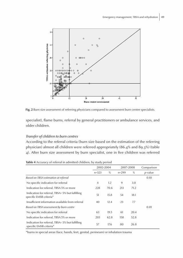
often overestimated by referrers, up to a maximum of 30% TBSA burn. Underestima-tion, up to 13% TBSA, occurred only in a minority of children (figure 2).
In both study periods about 20% of the children were referred without a burn size estimate. This predominantly concerned smaller burns (according to burn centre
Table 2 Characteristics of referred children, by study period
2002-2004
n=3232007-2008
n=299p-value
Age, in months (median, IQR) 21 14-48 23 15-64 0.07
Body mass (median, IQR) 12.5 10.5-18.0 13.0 10.9-21.8 0.09
Sex (n,%) 0.31
Girl 124 38.4 103 34.4
Boy 199 61.6 196 65.6
Etiology (n,%) <0.01
Scald 248 76.8 224 75.9
Fat 18 5.6 2 0.7
Flame 50 15.5 51 17.3
Contact 3 0.9 7 2.4
Other 4 1.2 11 3.7
Referral (n,%) 0.64
General hospital 212 65.6 202 67.6
General practitioner 30 9.3 25 8.4
Ambulance service 46 14.2 34 11.4
University hospital 35 10.8 38 12.7
Burn size assessment
No estimation at referral (n,%) 84 26.0 66 21.9 0.25
TBSA at referral, % (median, IQR) 10 8-15 10 7-16.5 0.21
TBSA burn centre, % (median,IQR) 5.5 4-8 5 3-8 0.01
Length of stay (median, IQR) 7 2-18 6 3-16 0.99
Missing values age: body mass period 2 n=30; etiology period 2 n=4; TBSA burn centre period 2 n=1.IQR: interquartile range.
Table 3 Burn size assessment of admitted children by referrer compared to assessment by burn specialists, by study period
2002-2004
n=239*2007-2008
n=233*p-value
TBSA assessment referrer, median (IQR) 10 (8-15) 10 (7-16.5) 0.21
TBSA assessment burn centre, median (IQR) 6 (4-9) 5 (4-8) <0.01
Difference in TBSA assessments, mean (SD) 5.8 (5.5) 5.9 (5.6) 0.78
*Excluding children without burn size estimate at referral (period 1, n=84; period 2, n=66).IQR: interquartile range
Emergency management; TBSA and rehydration 49
specialist), flame burns, referral by general practitioners or ambulance services, and older children.
Transfer of children to burn centres
According to the referral criteria (burn size based on the estimation of the referring physician) almost all children were referred appropriately (86.4% and 89.3%) (table 4). After burn size assessment by burn specialist, one in five children was referred
Fig. 2 Burn size assessment of referring physicians compared to assessment burn centre specialists.
Table 4 Accuracy of referral in admitted children, by study period
2002-2004 2007-2008 Comparison
n=323 % n=299 % p-value
Based on TBSA estimation at referral 0.10
No specific indication for referral 4 1.2 9 3.0
Indication for referral, TBSA 5% or more 228 70.6 213 71.2
Indication for referral, TBSA< 5% but fulfilling specific EMSB criteria*
51 15.8 54 18.1
Insufficient information available from referral 40 12.4 23 7.7
Based on TBSA assessment by burn centre 0.01
No specific indication for referral 63 19.5 61 20.4
Indication for referral, TBSA 5% or more 203 62.8 158 52.8
Indication for referral, TBSA< 5% but fulfilling specific EMSB criteria*
57 17.6 80 26.8
*burns in special areas (face, hands, feet, genital, perineum) or inhalation trauma

50 Chapter 3
without fulfilling the criteria for referral related to burn size, special localizations or inhalation trauma, in both periods. The majority of these children (81.5%) were un-der the age of 5 years (period 1: n=51, period 2: n=50). They were usually discharged much earlier: median length of stay 3 days (IQR 2-6.5) compared to 7.5 days (IQR 3-17) in children referred in accordance with the EMSB criteria. Nonetheless, a minority (14.5%) of these children without clear indication for referral underwent surgery during admission (period 1: n=11, period 2: n= 7).
In period 2, fewer children with a TBSA of 5% or more (based on TBSA burn centre) were admitted, and more children with burns in special areas and/or inhalation trauma (table 4).
Intravenous fluid resuscitation:
More than half of children referred by hospitals or ambulance services had an in-travenous line insertion on admission (63.1% period 1, 62.3% period 2, p=0.27). The prevalence of documented intravenous fluid resuscitation increased from 33.1% of the referred children in period 1 to 48.5% in period 2 (p<0.01), although median burn size was equal in both study periods. In other children with an intravenous line insertion, referrers did not report on resuscitation volumes. The intravenous line was used for IV pain medication, or patients received small amounts of fluid to keep the intravenous line open.
The higher prevalence of documented intravenous fluid resuscitation in period 2 is mainly due to a significantly higher proportion of intravenous line insertions in children referred by general hospitals (period 1: 39.4% and period 2:60.4%, p<0.01). No significant changes in frequency of intravenous resuscitation were found in chil-dren referred by ambulance services (period 1: 7.5% (3/40), period 2: 21.2% (7/33), p=0.09) or university hospitals (period 1: 54.3% (18/33), period 2: 69.2% (18/26), p=0.25). Information on resuscitation was not available in about 10% of the referrals in both periods (period 1: 27/293; period 2: 31/274).
Table 5 Prevalence of children actually receiving prehospital intravenous fluid resuscitation in admitted children, by burn size and study period
2002-2004 2007-2008
n resuscitation/total n (%)
n resuscitation/total n (%)
TBSA<10% by both referrer and burn centre 10/57 (17.5) 32/81 (39.5)
TBSA at referral ≥10%, TBSA burn centre <10% 53/117 (45.3) 63/84 (75.0)
TBSA at referral <10%, TBSA burn centre ≥10% 1/2 (50.0)
TBSA ≥10% by both referrer and burn centre 31/42 (73.8) 32/37 (86.5)
*Excluding children without burn size estimate at referral or information on intravenous fluid resuscitation (period 1, n=75; period 2, n=72).

Emergency management; TBSA and rehydration 51
Sufficient information to assess appropriateness of resuscitation was available in approximately 63% of the children referred by ambulance services or hospitals. The proportion of children actually receiving intravenous fluid resuscitation without appropriate indication, i.e. a TBSA burn <10% according to the burn specialist, had increased in period 2 (table 5). The proportion of children actually receiving fluids resuscitation with appropriate indication had increased as well, from 73.8% to 86.5%.
Actually given fluids by referrers and ‘necessary’ fluids, as calculated according to protocol were compared for 81 of the 274 children referred by ambulance services or hospitals (29.6%) in period 2. The received volumes tended to be higher than necessary, ranging from 1500 up to a surplus of 2000mL (figure 3). Data on TBSA at referral was available for 76 children; those with more extensive burns (TBSA burn ≥ 10%) received volumes that were almost equal to calculated volumes (mean dif-ference 18 ml)(table 6). Surplus volumes in children with small body weight (<10 kg) were relatively small (mean 126 ml, 95%CI -49; 301) compared to children with a body weight > 20 kg (mean 558 ml, 95%CI 361; 755ml).
Fig. 3 Difference between actual and calculated fluids, by burn size assessment, in period 2007-2008 (n = 81)

52 Chapter 3
DISCUSSION
The aim of this study was to investigate possible differences in burns size estimate between referring hospitals and Dutch burn centres in children with burns and its consequences related to transfer and intravenous fluid resuscitation. Burn size at referral was on average a factor 2 higher than burn size assessed in the burn centres. About one in five children was referred to a burn centre without fulfilling the criteria for referral related to burn size based on the assessment by burn specialists, burns in special localizations or inhalation trauma, in both periods. Proportions of children receiving intravenous fluid resuscitation increased from 33% to 49% overall; from 18% to 40% in the subgroup of children without appropriate indication for resuscita-tion; and from 74% to 87% in children with appropriate indication. Most of the in-travenously rehydrated children received a higher volume than deemed necessary.
TBSA assessment
Various methods to assess burn size have been developed and applied in different settings ((pre-) hospital, burn centre), including the “rule of 9”8, “the hand rule” 10,12, serial halving7 and the Lund Browder charts. 11 Efforts are directed at optimizing burn size assessment 9,14, but errors made by incorrect use and /or calculations of all these methods were reported in several studies6. Notably body proportions, which are age-related, are a source of error in TBSA assessment. For example, in one-year-old children the head accounts for 18% TBSA, which percentage diminishes by one per-cent per year to 9% in adults. Furthermore, the focus on methods to calculate burn size may divert our attention from an underlying and maybe even bigger problem: determining which areas to include in the estimation of the extent of burns, i.e. burns vs. erythema. Our data on overestimation of TBSA burn by referrer compared to burn specialists are in line with previous studies on TBSA burn assessment. 1-5 In 60% to 85% of children, TBSA burn was overestimated at referral, underestimation was
Table 6 Comparison of calculated fluid volumes versus given fluid volumes in children referred by hospitals or ambulance services in 2007-8, by burn size (n=76)
Calculatedfluids (mL)
Given fluids (mL)
Difference given versus calculated fluids (mL)
N Mean SD Mean SD Mean SD (95% CI) Min Max
TBSA <10% 16 0 0 327 259 327 259 (189; 465)* 100 1120
TBSA referrer 10% or more, TBSA burn centre <10% 40 0 0 340 318 340 318 (238; 441)* 30 1500
TBSA 10% or more 21 695 646 677 879 -18 567 (-276; 240) -1544 951
CI : Confidence Interval* p<0.05

Emergency management; TBSA and rehydration 53
less frequent (26%-34%).1,2 These data are predominantly from studies with mixed populations but similar results were also reported in a paediatric study. 15
Overestimation has been reported to be related to burn size and age. Burn size as-sessment in children with burns over 5% TBSA was less accurate than that in children with smaller burns.15 In mixed populations, overestimation of burn size is relatively high for smaller burns (TBSA burn < 20%).3,5 Freiburg et al.4 reported a mean overesti-mation of 4.3% for burns < 20% TBSA and a mean underestimation of 4.9% for burns ≥20%. A higher percentage of overestimation has been reported in children under 36 months of age.3
Next to burn size and the child’s age, other factors may also influence accurate assessment of burn size, such as the child’s condition and circumstances and quality of emergency care provided by pre-hospital workers and referring hospitals. First of all, it is important to know the cause of burns; for example type and amount of liquid involved in the burn. One cup of coffee normally does not cause burns of 20% in a dressed 2-year-old child. Secondly, burn size estimation, regardless of the method used, is difficult especially when a child is crying, when there is confusion about skin redness or when erythema is suspected instead of second degree burn. Furthermore, the smell of burns, a crying, painful child and anxious, scared parents can distract health care workers and force them to make quick decisions and calcu-lations that may lead to errors.
Transfer of children to burn centres
Eighty percent of the children was referred following the EMSB criteria; burn size, burns in special localizations or inhalation trauma. In 20% of the children transfer to a burn centre was not in line with the specific referring criteria for burn size, burns in special localization or inhalation trauma. Most of these children were younger than 5 years old. The referrers may well have lacked experience on diagnosis and management of burns in young children and therefore have decided to transfer regardless of the criteria. Carter et al.16 reported a slightly higher percentage of 88% of adult patients admitted to burn centres meeting the ABA criteria for admission in an USA burn centre. This finding, too, may point at higher professional uncertainty in the case of young children with burns, compared to adults.
In case of uncertainty, (primary care) physicians are advised to consult a burn specialist or refer patients to a burn centre in the Netherlands. However, a transfer puts added stress and anxiety on parents. This should be taken into account when deciding on transferring children to a burn centre.
Our aim is to emphasize these issues in our communication with referring phy-sicians and to address them in our education of health care providers. Another possibility is to use some form of telemedicine to support triage to burn centres.

54 Chapter 3
Recently, Saffle et al.17 evaluated a telemedicine network of one burn centre and three remote hospitals: more patients were treated locally, with lower costs re-lated to emergency air transport. In the Netherlands, we could facilitate a secure website for health care providers, for instance through the Dutch Burn Foundation, where physicians can upload patient information, including TBSA assessment and photographs of the wound. The website should support burn size assessment and management (intravenous fluid resuscitation, pain medication, wound manage-ment) by providing calculation models and other relevant information. The primary care physician should contact the nearest burn care specialist (who also has secure access) to discuss website information, management and transfer to a burn centre, and coordinate admission if indicated.
Our data indicate a shift of admissions in children towards smaller burns and towards burns in special localizations. A similar trend in adults has been described earlier.18
Intravenous fluid resuscitation
Accurate intravenous fluid resuscitation is essential to prevent shock in case of severe burns and to maintain adequate circulation to tissues and perfusion of vital organs; it has been shown to result in an important increase in survival.19, 20 Dutch burn centres, in accordance with the EMSB guideline, recommend to start intravenous fluid resus-citation in children with TBSA burn ≥ 10%, using the Parkland formula and additional maintenance fluid.
In our study we expected a mean over-resuscitation of fluids due to the overall over-estimation of burn size. Surprisingly, there was no over-resuscitation in children with a TBSA burn over 10%. It would seem, therefore, that the inaccuracy of TBSA burn calculation is compensated by inaccuracies in fluid resuscitation calculation. Earlier studies, too, have pointed at the difficulty of calculating correct resuscitation volumes. 21,22 Only one quarter of physicians could accurately recall a burn resusci-tation formula and major mathematical errors were made in adult and paediatric case studies.22 Simple tables have therefore been proposed.23 Alternatively, a digital calculation model of resuscitation fluids could be made available on the internet.24
Comparison to earlier studies is hampered by different criteria for accuracy of fluids administrated during resuscitation and incomplete data due to incomplete transfer records. Over- and underestimation have been reported.3,4 Freiburg et al.4 found a trend towards more complications in case of inappropriate fluid resuscita-tion, but without significant proof.
Klein et al.25 and Dulhunty et al.26 concluded that adults with severe burns were at risk of morbidity if fluid resuscitation was inadequate, such as pneumonia, compart-ment syndrome and organ failure. Mortality of these patients was not higher than

Emergency management; TBSA and rehydration 55
that in patients who had normal fluid administration within 48 hours post burn. In a study on children with extensive burns over 50%, survival was better for those children who received fluid within 2 hours post burn.19
This study does have limitations. First, amount of fluids could be assessed in only a small subgroup of children (n=76). Secondly, the focus was on consequences of overestimating TBSA in terms of transfer and fluids. Because data were limited, we could not analyze the clinical consequences of faulty burn size and fluid resuscita-tion assessments in terms of number of children with worsened vital functions due to over or under fluid resuscitation and complications. Also unnecessary medical interventions were not addressed. For example, Mackie et al. 27 warned against un-necessary intubation and mechanical ventilation before transport in adults, which would hold true for children as well.
Since 1998 health care providers are being educated in the management of se-verely ill and trauma patients, both adults and children. However, the present study failed to find obvious improvements in the pre-hospital management and referral of children with burns over time. We would do well to intensify education and outreach programs based on gaps in the knowledge of the emergency health care providers.
CONCLUSION
Referrers overestimate burn size in children admitted to Dutch burn centres. This has little negative consequences, however, in terms of unindicated transfers to a burn centre or unnecessary intravenous fluid resuscitation. Further improvement of burn size assessment and fluid calculation is desirable.
Traditional education must be improved and new learning methods must be explored (e.g. E-learning). Hospitals and ambulances should have local guidelines and trained health care workers involved in the emergency care of burn patients. National guidelines “Emergency treatment and transfer of children with burns” are a tool to manage these problems and form the first step in improvement of care.

56 Chapter 3
REFERENCES
1. Berkebile BL, Goldfarb IW, Slater H. Comparison of burn size estimates between prehospital
reports and burn center evaluations. J Burn Care Rehabil. 1986;7:411-412.
2. Berry CC, Wachtel T, Frank HA. Differences in burn size estimates between community hos-
pitals and a burn centre. J Burn Care Rehabil. 1982;3:176-177.
3. Collis N, Smith G, Fenton OM. Accuracy of burn size estimation and subsequent fluid resus-
citation prior to arrival at the Yorkshire Regional Burns Unit. A three year retrospective study.
Burns. 1999;25:345-351.
4. Freiburg C, Igneri P, Sartorelli K, et al. Effects of differences in percent total body surface area
estimation on fluid resuscitation of transferred burn patients. J Burn Care Res. 2007;28:42-48.
5. Hammond JS, Ward CG. Transfers from emergency room to burn center: errors in burn size
estimate. J Trauma. 1987;27:1161-1165.
6. Jose RM, Roy DK, Vidyadharan R, et al. Burns area estimation-an error perpetuated. Burns.
2004;30:481-482.
7. Allison K, Porter K. Consensus on the prehospital approach to burns patient management.
Emerg Med J. 2004;21:112-114.
8. Knaysi GA, Crikelair GF, Cosman B. The rule of nines: its history and accuracy. Plast Reconstr
Surg. 1968;41:560-563.
9. Malic CC, Karoo RO, Austin O, et al. Resuscitation burn card--a useful tool for burn injury
assessment. Burns. 2007;33:195-199.
10. Nagel TR, Schunk JE. Using the hand to estimate the surface area of a burn in children. Pediatr
Emerg Care. 1997;13:254-255.
11. Lund CC, Browder NC. The estimation of areas of burns. Surgery, gynecology & obstetrics.
1944;79:352-358.
12. Berkow S. A method of estimating the extensiveness of lesions (burns and scalds) based on
surface area proportions. Arch Surg. 1924;8:138-148.
13. The Education Committee of the Australian and New Zealand Burn Association. Emergency
management of severe burns (EMSB) course manual. Dutch version, Dutch Burn Foundation,
2009.
14. Sheridan RL, Petras L, Basha G, et al. Planimetry study of the percent of body surface repre-
sented by the hand and palm: sizing irregular burns is more accurately done with the palm. J
Burn Care Rehabil. 1995;16:605-606.
15. Irwin LR, Reid CA, McLean NR. Burns in children: do casualty officers get it right? Injury.
1993;24:187-188.
16. Carter JE, Neff LP, Holmes JH. Adherence to burn center referral criteria: are patients appro-
priately being referred? J Burn Care Res. 2010;31:26-30.
17. Saffle JR, Edelman L, Theurer L, et al. Telemedicine evaluation of acute burns is accurate and
cost-effective. J Trauma. 2009;67:358-65.
18. Anwar U, Majumder S, Austin O, et al. Changing pattern of adult burn referrals to a regional
burns centre. J Burn Care Res. 2007;28:299-305.
19. Barrow RE, Jeschke MG, Herndon DN. Early fluid resuscitation improves outcomes in severely
burned children. Resuscitation. 2000;45:91-96.
20. Wolf SE, Rose JK, Desai MH, et al. Mortality determinants in massive pediatric burns. An
analysis of 103 children with > or = 80% TBSA burns (> or = 70% full-thickness). Ann Surg.
1997;225:554-565.

Emergency management; TBSA and rehydration 57
21. Breederveld RS, Nieuwenhuis MK, Tuinebreijer WE, et al. Effect of training in the Emergency
Management of Severe Burns on the knowledge and performance of emergency care work-
ers as measured by an online simulated burn incident. Burns. (accepted)
22. Bhat S, Humphries YM, Gulati S, et al. The problems of burn resuscitation formulae: a need
for a simplified guideline. 2004. Available at: http://www.journalofburnsandwounds.com.
Accessed 2009.
23. Lindford AJ, Lim P, Klass B, et al. Resuscitation tables: a useful tool in calculating pre-burns
unit fluid requirements. Emerg Med J. 2009;26:245-249.
24. Nichter LS, Bryant CA, Edlich RF. Efficacy of burned surface area estimates calculated from
charts--the need for a computer-based model. J Trauma. 1985;25:477-481.
25. Klein MB, Nathens AB, Heimbach DM, et al. An outcome analysis of patients transferred to a
regional burn center: transfer status does not impact survival. Burns. 2006;32:940-945.
26. Dulhunty JM, Boots RJ, Rudd MJ, et al. Increased fluid resuscitation can lead to adverse out-
comes in major-burn injured patients, but low mortality is achievable. Burns. 2008;34:1090-
1097.
27. Mackie DP, Spoelder EJ, Paauw RJ, et al. Mechanical ventilation and fluid retention in burn
patients. J Trauma. 2009;67:1233-1238.


Chapter 4Reliability, validity and clinical utility of three types of pain behavioural observation scales for young children with burns aged 0 - 5 years.
Pain. 2010 Sep;150(3):561-7.
AEE de Jong, MGA Baartmans, M Bremer, R van Komen, E Middelkoop,WE Tuinebreijer, NEE van Loey
60 Chapter 4
ABSTRACT
Pain measurement is a prerequisite for individualized pain management and re-search into pain interventions. There is a need for reliable and valid pain measures for young children with burns. The aim of this study was to investigate whether the Pain Observation Scale for Young Children (POCIS), the COMFORT Behaviour Scale (COMFORT-B) and the Nurse Observational Visual Analogue Scale (VAS obs) are reli-able, valid and clinically useful instruments to measure pain in children with burns aged 0-5 years. Participating trained nurses (N=102) rated pain of 154 children during hospitalization. Two trained nurses simultaneously assessed pain at fixed intervals by using the previous mentioned measures. Cronbach’s alpha for POCIS was .87 for background and .89 for procedural pain. Intraclass Correlation Coefficients (ICC) were .75 for background and .81 for procedural pain. COMFORT-B observations yielded Cronbach’s alpha of .77 for background and .86 for procedural pain and ICC of .83 for background and .82 for procedural pain. The VAS obs resulted in ICC of .55 for background and .60 for procedural pain. Correlation coefficient between POCIS and COMFORT-B was .79 (p < .01), Standardized Response Mean was 1.04 for both POCIS and COMFORT-B. Background pain measured with POCIS and COMFORT-B was lower than procedural pain (p < .001). Nurses found POCIS easier and quicker to use, but COMFORT-B was found to indicate pain more accurately. Both POCIS and COMFORT-B are reliable, valid and practical scales for pain measurement in young children with burns and can be used in practice and research. The VAS obs was found to be unreliable.

Pain assessment 61
INTRODUCTION
Burn pain can be long lasting, has a fluctuating course and is related to extensive repetitive daily wound care procedures. A distinction is made between background pain and procedural pain.1 Background pain, experienced while resting, is caused immediately postburn when an inflammatory response is initiated, which sensitises nociceptors in and around the burn. Procedural pain is caused by every manipula-tion involving the burn, which leads to additional stimulation of the nociceptors. Although procedures like skin transplantations and removal of staples are performed under general anaesthesia, wound care procedures, lasting minimal 30 minutes and including removal of dressings, washing, debridement and application of new dress-ings (usually ceriumsilversulfadiazine cream or hydrofiber for primary burns), are carried out by using pharmacological and non-pharmacological interventions. Pro-cedural pain is usually of higher intensity, but of shorter duration than background pain.
Adequate management of burn pain is important for many reasons. It is essential to the relationship between the patient and the multidisciplinary team, it increases comfort and makes recovery more tolerable. In addition, adequate pain manage-ment affects morbidity by preventing elevated metabolism, thereby reducing the chance of deterioration of the immune system.1 Furthermore, adequate pain man-agement might reduce acute stress symptoms.2,3
To evaluate the adequacy of pain management, pain measurement is essential. Pain measurement is the nurses’ responsibility, because, of all health care professionals, nurses are, as inflictors of pain and providers of pain relief, mostly confronted with pain of patients admitted at the burn centre. Approximately 30% of the admitted patients are children up to four years old who got, due to their development stage of motor and cognitive skills, hold of cups filled with coffee or tea or pull down hot liquid containers2,4,5, causing severe dermal and deep dermal burns. Although some 3-year-olds and many 4-year-olds may be capable of providing self-reports, which is the commonly used method of pain assessment, most of these children are too young to express background and procedural pain by self-reports. Their pain should therefore be assessed by behavioural observation.6
Since pain measurement is a prerequisite for individualized pain management, there is a need for pain behavioural observation measurement instruments with suf-ficient psychometric properties for young children with burns. Therefore, the aim of this study was to investigate the reliability, validity and clinical utility of three types of behavioural observation scales in order to measure procedural and background pain in children with burns aged zero to five years.

62 Chapter 4
METHODS
Participants
Participating nurses were employed at the three Dutch burn centres: the Red Cross Hospital in Beverwijk, the Maasstad Hospital in Rotterdam and the Martini Hospital in Groningen.
Measures
Three pain behavioural observation scales were investigated and a questionnaire was used to assess the clinical utility of these instruments.
Pain behavioural observation scales
Pain Observation Scale for Young Children (POCIS)
The POCIS provides a list of behaviours that are marked as either present or ab-sent. The POCIS was initially developed to measure postoperative pain intensity in children after adenotonsillectomy, adenotomy or insertion of ventilation tubes. The scale comprises seven behavioural items (Table 1) with dichotomous answer catego-ries, which enables easy and quick use of the scale. The presence or absence of each item is scored 0 or 1. The POCIS has proven to be reliable, based on inter-rater agreement and internal consistency, and valid, based on a principal components analysis that supports construct validity.7 The POCIS showed moderate to good reli-ability when children with burns were observed from video fragments.8
COMFORT Behaviour Scale (COMFORT-B)
The COMFORT scale incorporates ratings of intensity and frequency of each be-haviour and is appropriate for longer periods of observation.9 The COMFORT scale that was used in this study, the COMFORT Behaviour Scale, is assumed to measure pain intensity and distress associated with pain and is an adapted version of the one developed by Ambuel et al.10 The adapted version has shown good reliability and congruent validity in children with postoperative pain after abdominal and thoracic surgery.11 The scale comprises six behavioural items with five response categories for each item (Table 2). One of the six items is divided into the options respiratory response and crying. Depending on mechanical ventilation or spontaneous breath-ing, either respiratory response or crying has to be assessed. As it is very rare for children with burns caused by hot liquids to be mechanically ventilated, the option respiratory response was not considered in this study.Nurse Observational Visual Analogue Scale (VAS obs)
A Visual Analogue Scale (VAS) provides a rating of the observer’s global impression of a patient’s pain.6 The VAS is a frequently used instrument by nurses to assess pain in children. 12-18 It may provide information on individual variations in pain sensitivity,

Pain assessment 63
idiosyncratic behaviours and situational influences19. The VAS is a quick and easy to use instrument with ratio scale properties. The scale was considered reliable on the basis of inter-rater reliability for procedural pain in neonates and in children with chronic pain20,21 and demonstrated a high correlation with postoperative behavioural observation pain measurement instruments.16,22 Although the VAS showed poor to moderate inter-rater reliability from video assessments in children with burns8, the scale has not yet been investigated for use with wound care procedures in real life. The VAS in this study is a straight horizontal continuous 10 cm line with clearly
Table 1: Pain observation scale for young children
Score
Facial
At rest, neutral 0
Grimace, nose wrinkled, eyebrows frown 1
Cry
No cry 0
Moan, scream 1
Breath
Relaxed, regular 0
Irregular, hold in, gasping 1
Torso
At rest, neutral, relaxed 0
Tense, restless, contorted, writhed, trembling 1
Arms/fingers
At rest, neutral, relaxed 0
Tense, restless, clenched fist, wild 1
Legs/toes
At rest, neutral, relaxed 0
Tense, restless, pulled up, kicking 1
Arousal
Calm sleepy, calm alert, playing 0
Restless, touchy, fussy 1
Total

64 Chapter 4
marked terminal ends, with the anchor words “no pain” at the left side of the line and “unbearable pain” at the right side. A mark has to be placed on this line and a ruler is needed to read the obtained score. In order to avoid confusion with other applications of the VAS, which is mostly used as a patient self-report tool, in this
Table 2: COMFORT Behaviour Scale
Score
Alertness Deeply asleep (eyes closed, no response to changes in environment)
1
Lightly asleep (eyes mostly closed, occasional responses) 2
Drowsy (child closes eyes frequently, less responsive to environment)
3
Awake and alert (responsive to environment) 4
Awake and hyper-alert (exaggerated responses to environmental stimuli)
5
Calmness/agitation Calm (child appears serene and tranquil) 1
Slightly anxious (child shows slight anxiety) 2
Anxious (child appears agitated but remains in control) 3
Very anxious (child appears very agitated, just able to control) 4
Panicky (severe distress with loss of control) 5
Crying No crying sounds 1
Occasional sobbing or moaning 2
Whining (monotonous sound) 3
Crying 4
Screaming or shrieking 5
Physical movement No movement 1
Occasional (three or fewer), slight movements 2
Frequent (more than three), slight movements 3
Vigorous movements limited to extremities 4
Vigorous movements including torso and head 5
Muscle tone Muscles totally relaxed; no muscle tone 1
Reduced muscle tone; less resistance than normal 2
Normal muscle tone 3
Increased muscle tone and flexion of fingers and toes 4
Extreme muscle rigidity and flexion of fingers and toes 5
Facial tension Facial muscles totally relaxed 1
Normal facial tone 2
Tension evident in some facial muscles (not sustained) 3
Tension evident throughout facial muscles (sustained) 4
Facial muscles contorted and grimacing 5
Total

Pain assessment 65
study, a more specific name for this tool is used, namely the Nurse Observational Visual Analogue Scale (VAS obs).
Clinical utility questionnaire
To survey clinical utility of the scales from the nurses’ point of view, structured closed-ended self-reports by means of a 5-point Likert scale questionnaire were used. The questionnaire is based on clinical utility criteria as assessed by Harris and Warren.23 It includes items about the extent of the scales in providing clinically use-ful patient information and readily understandable scores. In addition, items about ease of use, time required and clarity of the scales were included. The degree of the severity of pain, the ability to differentiate between no pain and unbearable pain and the relevance of the scale items were questioned as well.
Data collection procedure
Approval of the medical ethics committees of the participating hospitals was ob-tained. Parents received written and verbal information about the study and were asked to give verbal consent. They were assured that standard medical and pain treatment remained unchanged and that the study would not cause any burden to their children.
Nurses were trained to use the POCIS and COMFORT-B before taking part in the study. Two nurses from each burn centre followed training at the two hospitals where these scales were developed. Subsequently, these nurses trained their colleagues in the burn centres using a standardized one-hour educational programme about pain and pain assessment. The training also included video and in vivo observations with both scales. The in vivo observations focussed on procedural pain as it was assumed that assessing this type of pain required most training. Each trainee completed ten assessments per scale with one of the trainers, of which five were video observations and another five were in vivo observations. When inter-rater reliability was accept-able, with Intraclass Correlation Coefficients (ICC) of 0.75 or more, nurses were allowed to rate children for the study and train other nurses.
Children that met the inclusion criteria, i.e. children aged zero to five years with burns and without developmental delays, were observed by means of the POCIS, COMFORT-B and VAS obs three times a day at fixed intervals by two nurses who kept independent records. Background pain was recorded in the morning, at least one hour before wound care, and in the afternoon, at least one hour after wound care. Children were observed during two minutes. Procedural pain was assessed directly after wound care. Since procedural pain can be categorised into peak and overall

66 Chapter 4
pain, nurses were asked to rate overall pain of the whole wound care procedure only. Peak pain is usually caused by bandages that stick to one or more areas in the wound, is of short duration but of high intensity and does, if it occurs, not represent pain intensity of the whole procedure. Furthermore, in practice, pain interventions are adapted to accommodate overall pain, not peak pain. Research has also shown that peak pain is included in overall pain ratings: a sizable correlation between peak and overall pain is reported.24,25
Two data collection forms comprising the three measures were developed. On each form, the POCIS and COMFORT-B were ordered differently, to vary the order of completion of scales, which might avoid giving answers that are satisfactory (i.e. a box on the form is filled in), but not optimal.26 The VAS obs was in all cases com-pleted after the POCIS and COMFORT-B. The following instruction for its use was given to nurses: Please estimate the level of the childs’ pain by making a mark on the line. Nurses were requested not to discuss and compare their individual ratings.
The following characteristics of the participating nurses were recorded: age, gender, parenthood, education and number of years working in burn care. As for the in-cluded children, age, gender, extent and cause of the burns and length of stay were recorded. Nurses and children were encoded.
Data analysis
Data was analyzed with the statistical program SPSS 16.0 (SPSS Inc. Chicago USA). Descriptive statistics were used to assess characteristics of nurses and children and clinical utility. Reliability, which is the degree to which an instrument measures a concept in a reproducible fashion, was judged by internal consistency (the degree in which the items of the scale belong to the same concept) and inter-rater reliability (the degree in which observers assign the same ratings).27 Internal consistency was assessed by Cronbach’s alpha, inter-rater reliability by calculating Intraclass Correla-tion Coefficients (ICC). Acceptable reliability coefficients are ≥ .75.27 Validity, which is the degree to which an instrument measures what it is intended to measure, was determined by convergent validity and responsiveness. Convergent validity was as-sessed in order to evaluate how a scale correlates with another measure of the same construct.27 Responsiveness is the ability of an instrument to detect clinically impor-tant change.28,29 Spearman’s rho was used to determine convergent validity, since patient characteristics were not normally distributed. Independent-Samples T-tests and a Standardized Response Mean (SRM) were calculated to assess responsiveness. The value of an SRM can be considered as an effect size index. An acceptable effect size should be d ≥.5 29,30, where .5 is a medium effect and .8 a large effect.

Pain assessment 67
RESULTS
Data was collected from June 2007 until June 2008. All parents gave verbal consent. The number of children included in the study was 154, 101 (66%) of which were boys and 53 (34%) girls. The mean age was 20 months (SD 11). Causes of the burns were scalds in 147 children (95.5%), contact burns in six children and electricity in one child. The mean total body surface area was 6.5% (SD 4.5, min 5-max 28) and the mean length of stay 10 days (SD 7.7, min 1-max 39).
Participants characteristics
A total of 102 nurses working in the three Dutch burn centres, which is 65% of all nurses working in this field, participated in the study. The characteristics of the nurses are described in Table 3.
Reliability
Internal consistency
Internal consistency results of the POCIS and COMFORT-B are presented in Table 4. It shows that both instruments are reliable since Cronbach’s alpha should range between .70 and .90.27 An item contributes to a scale if alpha, when calculated after this item is deleted, has a lower value than alpha of the entire scale. All alpha values were lower when items were deleted. The POCIS showed higher alphas than the COMFORT-B.
Table 3: Nurses’ characteristics (N=102)
Mean age (years ± SD) 40.8 (8.5)
Gender (% female) 86.3
Nurse is parent (%) 67.6
Education (%) BSc 13.7
IC 38.2
PC 33.3
BC 65.7
Years of experience in burn care (%) < 1 year 14.7
>= 1 year < 5 years 27.5
>= 5 years < 10 years 25.5
>= 10 years 32.3
BS: Bachelor of Science, IC: Intensive Care, PC: Paediatric Care, BC: Burn Care

68 Chapter 4
Inter-rater reliability
As presented in Table 4, ICC for the POCIS and COMFORT-B total scores met the criterion of ≥ .75 27 and showed small Confidence Intervals (CI) for background and procedural pain, indicating good reliability. The COMFORT-B showed higher ICC than POCIS. ICC for the VAS obs were not acceptable for both background and procedural pain.
Validity
Convergent validity
In order to assess validity of the POCIS and COMFORT-B, the correlation between these two measures should be rho ≥ .3.27 Spearman’s rho was .45 for background pain and .88 for procedural pain, which was statistically significant (p < .01). As the POCIS and COMFORT-B correlate for both types of pain, they probably measure the same construct. The correlation for background pain, however, is lower than for procedural pain. Since it is first necessary that an instrument measures a concept in a reproducible fashion27, the validity of the VAS obs was not assessed because it did not meet the reliability criterion.
Responsiveness
A t-test demonstrated that the POCIS total scores for background pain were statisti-cally significantly lower than for procedural pain (mean background pain = 0.33 (SD 1.10, median 0), mean procedural pain = 3.41 (SD 2.60, median 4), t = -51.60, df = 3872, p < .001, 95% CI = -3.3 to -3.0). Also, the mean COMFORT-B total scores for background pain were statistically significantly lower than for procedural pain (mean background pain = 12.61 (SD 2.95, median 9), mean procedural pain = 18.54 (SD 4.12, median 18), t = -51.69, df = 3886, p < .001, 95% CI = -6.3 to -5.6).
Table 4. Results reliability for POCIS, COMFORT Behaviour Scale and VAS obs
Type of pain Internal consistency Inter-rater reliability
Cronbach’s � N ICC (CI) N
POCIS Background .872 2552 .75 (.72-.77) 1277
Procedural .883 1322 .81 (.78-.84) 659
COMFORT Background .769 2564 .83 (.82-.85) 1277
Procedural .861 1323 .82 (.80-.85) 659
VAS obs Background .55 (.51-.59) 1277
Procedural .60 (.55-.65) 659
N Cronbach’s �: Number of observationsN ICC: Number of paired observationsICC: Intraclass Correlation Coefficient, CI: Confidence Interval

Pain assessment 69
The POCIS and COMFORT-B turned out to have a similar SRM of 1.04, which is con-sidered a large effect. As background pain differed significantly from procedural pain and the SRM was large, it was assumed that both scales are able to measure change.
Clinical utility
To assess the clinical utility of the POCIS and COMFORT-B, 86 of 102 questionnaires (84% response) were analysed. The results are presented in Table 5. In general, nurses found the POCIS easier and quicker to use than the COMFORT-B, but the COMFORT-B was perceived to address procedural and background pain more ac-curately and to have better properties to connect to a pain management protocol. Since the VAS obs was not reliable and therefore not tested on validity, clinical utility of the VAS obs was not considered.
DISCUSSION
The aim of this study was to assess if the POCIS, COMFORT-B and VAS obs are reli-able, valid and practical instruments to measure procedural and background pain in children with burns aged zero to five years.
Both the POCIS and COMFORT-B seem to be reliable measures to assess two types of pain in children with burns. Both scales showed high and equal internal consistency. Cronbach’s alpha was higher for the POCIS than for COMFORT-B, suggesting that
Table 5. Results clinical utility POCIS and COMFORT Behaviour Scale
POCIS(% agree)
COMFORT(% agree)
Provides information that is clinically useful 60.0 90.1
Is short to administer 81.2 56.1
Is easy to administer 77.6 65.9
Is clear and easy to understand 63.1 71.2
Reflects the extent of background pain 44.0 81.7
Reflects the extent of procedural pain 56.5 85.4
Discriminates children with pain from children without pain 43.5 82.9
Score is readily understandable and allows to adapt pain management to childs’ need
39.3 82.9
Reflects procedural pain specific features 77.4 87.7
Reflects background pain specific features 70.4 87.8
N=86 (number of responding nurses)

70 Chapter 4
the POCIS items show more coherence. This is in line with the assumption that the POCIS is a unidimensional scale, measuring pain intensity, while the COMFORT-B is supposed to be a multidimensional scale, including measurement of distress.6 How-ever, internal consistency of the COMFORT-B does not suggest a multidimensional structure.
Good inter-rater reliability was seen for both the POCIS and COMFORT. This cor-responds with findings of Boelen-van der Loo7 and De Jong et al.8 for the POCIS, and Van Dijk et al.11, Bear and Ward-Smith31 and Caljouw et al.32 for respectively the COMFORT-B, the COMFORT scale and the adapted COMFORT scale. The higher ICC for COMFORT-B background pain than for POCIS may be explained by a restricted range of variance in total POCIS scores, ranging from 0 to 7 when compared to the variance in COMFORT-B total scores ranging from 6 to 30.
The POCIS and COMFORT-B seem to measure the same concept and are able to distinguish between two types of pain with differences in intensity, suggesting valid-ity of both instruments. The lower correlation between POCIS and COMFORT-B for background pain when compared to procedural pain could also be due to a restrict-ed range of variance in total POCIS scores. SRM for both the POCIS and COMFORT-B were large. This can be explained by the substantial difference between procedural and background pain, which was already demonstrated with the t-test.
In contrast to the good psychometric properties of the POCIS and COMFORT-B, the VAS, when used by nurses as a global rating scale to report the patients’ pain, turned out to be unreliable, not only in this study, but also in earlier research in children with burns8, in adults with burns24,25,33,34, and in children without burns but with postoperative or procedural pain.17,20 In children with burns, De Jong et al.8 found ICC between 0.46 and 0.66. In adult patients with burns, Pearson correlation coef-ficients between 0.33 and 0.47 were found.24,33 Geisser et al.24 considered a rating to be correct when the nurse rated pain within 1 cm of the patients’ rating. Using this criterion, it was found that nurses correctly assessed patients’ pain in only 25% of the time. Iafrati34 found correct assessments in only 31% of the time. In non-burn settings a range of correlation coefficients from 0.42 to 0.91 was found.17,20 It should be noted that correlation coefficients may be of limited value to assesses interrater reliability because they reflect only relative positions of scores19 and are usually higher than the true reliability.27 It is possible that nurses are unable to express the patients’ pain on a global rating scale as a 10 cm line, because they have their pain assessment affected by other than behavioural factors. Cognitive, emotional, situational and/or rela-tional factors may play conscious or subconscious roles in their observations. These

Pain assessment 71
findings for the VAS support the statement of Von Baeyer and Spagrud6, namely that global observational scales are not recommended as outcome measures for pain.
An important issue in the pain literature is the distinction between pain intensity and fear-laden items like distress. Although this distinction was not subject of investiga-tion, this study demonstrates that the POCIS and COMFORT-B appear to measure the same construct and it is assumed that this construct is pain. Interestingly, although the POCIS is assumed to measure, according to the developers, pain intensity, while the COMFORT-B measures pain intensity and distress, this distinction could not be confirmed by this study. Distress has been defined as behaviours of negative affect associated with pain, anxiety and fear.10 Although distress is inextricably bound up with pain intensity, especially in children with burns undergoing repetitive wound care procedures, the concept differs from pain intensity. The ability the scales of making distinction between these concepts was not detected in this study.
The question arises whether or not it is possible to distinguish pain intensity from pain-related concepts. Von Baeyer and Spagrud6 have stated that few researchers have presented data showing that their observational instruments can differenti-ate pain intensity from its affective components. This study seems to support this difficulty. Since the POCIS and COMFORT-B seem to measure the same concept, it is assumed that the affective pain component distress is embedded in the POCIS, suggesting that presence of vocalizations, facial expressions and physical move-ments are not only indicators for pain intensity but also for distress. Or, with regard to COMFORT-B, distress is a component of pain intensity in children with burns and cannot be seen separately from pain intensity. This is in accordance with Blount and Loiselle9, who have postulated that both emotional and sensory components of pain seem to be assessed by pain behavioural assessment scales and that many behaviours do not appear to have specificity as an indicator of pain or distress. They consider this however not necessarily problematic, since pain and distress are both included in the most commonly accepted definition of pain. (‘Pain is an unpleasant sensory and emotional experience associated with actual or potential tissue dam-age or described in terms of such damage’35). Although the fact that pain behaviour observation scales assess both emotional and sensory components of pain, which may not be problematic from psychometric and theoretical perspective, we do not think that this solves the problem of differentiating pain from distress and fear from daily practice perspective. Although inextricably linked, pain and fear require dif-ferent treatments. Pain treatment should therefore include pain intensity as well as anxiety reducing interventions and should be started as soon as possible after the burn incident.

72 Chapter 4
A last issue in this study concerns the clinical utility of the scales.
Nurses found the POCIS easier and quicker to use, which can be attributed to the dichotomous answer categories of a behaviour checklist. The COMFORT-B how-ever was perceived addressing procedural and background pain more accurately. According to nurses, this was due to the COMFORT-B’s ability to allow reporting degrees of severity within the answer categories. The multiple answer categories per COMFORT-B item gave nurses the impression that a middle course was also optional and that the POCIS presence or absence options were found to be too limited to accurately assess both types of pain.A limitation of this study is that, although it was assumed that the POCIS and COMFORT-B are able to measure change, we did not assess the minimum clinical significant difference that can be measured. Assessing the minimum clinical sig-nificant difference is essential to be able to evaluate declines in pain intensity and the percentage of clinical significant pain decrease can be achieved by comparing pre- and post-treatment pain measurements.36 These data however were not col-lected during the present study. Another limitation may relate to the use of repeated measurements. Since observations were not independent of each other, this may bias the results. Although it is assumed that repeated measurements have a minor impact on research in which the measurement instrument itself is subject of in-vestigation, analyses were replicated on two subsets of the sample, i.e. one subset comprising the three paired observations attained on one randomly selected day for each child, and a second subset comprising only one paired observation per child. The obtained results remained unchanged, thereby rejecting a possible impact of dependency of observations in this study.
CONCLUSION
Three behavioural observation instruments are investigated for the use in a particular patient group with specific types of pain. These types of pain can be assessed with currently available measurement instruments: the POCIS and COMFORT-B showed good reliability and validity in this study and are considered clinically useful. The VAS obs, when completed by nurses, showed poor reliability to estimate children’s’ pain.
Recommendations for practice
The POCIS and COMFORT-B can be used to measure background and procedural pain in daily burn nursing practice. Development of pain management protocols is recommended in order to connect them to the total scores the scales. A global

Pain assessment 73
observational rating scale like the VAS obs when completed by nurses is not recom-mended as pain measurement instrument in children with burns.
Recommendations for further research
With the aim of connecting the total scores of the scales to a pain management protocol, cut off scores should be assessed to differentiate pain intensity. Also, the minimum clinical significant difference is an important issue to investigate. Further-more, when pain is measured and treated, the adequacy of pain management can be evaluated. Finally, global observational rating scales completed by nurses are not recommended for the use of validity assessment of pain behavioural observation scales in children with burns.
Acknowledgements
The authors would like to thank all nurses from the Dutch burn centres, Mrs. O. Duijndam, Mrs. M. Fuchs, Mrs. C. Hulsmann, Mrs. N. Kruger, Mrs. J. Leeman, Mrs. A. Nijman, Mrs. I. Oen, Mrs. H. Renkema, Mrs. C. Roerhorst and Mr. R. Scheffer for their participation, support and advices. The authors would also like to thank Prof. dr. R. de Vet for her statistical advice. This study was supported by a grant (06.301) from the Dutch Burn Foundation, Beverwijk, the Netherlands. The authors have no financial or other relationships that can lead to conflicts of interest.
74 Chapter 4
REFERENCES
1. Latarjet J, Choinere M. Pain in burn patients. Burns. 1995; 21: 344-8.
2. Stoddard FJ, Ronfeldt H, Kagan J et al. Young burned children: the course of acute stress and
physiological and behavioral responses. Am J Psychiatry. 2006; 163: 1084-90.
3. Taal LA, Faber AW. Posttraumatic stress and maladjustment among adult burn survivors 1-2
years postburn. Burns. 1998; 24: 285-92.
4. van Rijn OJ, Bouter LM, Meertens RM. The aetiology of burns in developed countries: review
of the literature. Burns. 1989; 15: 217-21.
5. Vloemans AF, Soesman AM, Suijker M et al. A randomised clinical trial comparing a hydro-
colloid-derived dressing and glycerol preserved allograft skin in the management of partial
thickness burns. Burns. 2003; 29: 702-10.
6. Von Baeyer CL, Spagrud LJ. Systematic review of observational (behavioral) measures of pain
for children and adolescents aged 3-18 years. Pain. 2007: 140-59.
7. Boelen-van der Loo WJ, Scheffer E, de Haan RJ et al. Clinimetric evaluation of the pain obser-
vation scale for young children in children aged between 1 and 4 years after ear, nose, and
throat surgery. J Dev Behav Pediatr. 1999; 20: 222-7.
8. De Jong AE, Bremer M, Schouten M et al. Reliability and validity of the pain observation scale
for young children and the visual analogue scale in children with burns. Burns. 2005; 31: 198-
204.
9. Blount RL, Loiselle KA. Behavioural assessment of pediatric pain. Pain Res Manag. 2009; 14:
47-52.
10. Ambuel B, Hamlett KW, Marx CM et al. Assessing distress in pediatric intensive care environ-
ments: the COMFORT scale. J Pediatr Psychol. 1992; 17: 95-109.
11. Van Dijk M, de Boer JB, Koot HM et al. The reliability and validity of the COMFORT scale as a
postoperative pain instrument in 0 to 3-year-old infants. Pain. 2000; 84: 367-77.
12. Buchholz M, Karl HW, Pomietto M et al. Pain scores in infants: a modified infant pain scale
versus visual analogue. J Pain Symptom Manage. 1998; 15: 117-24.
13. Hamers JP, Abu-Saad HH, van den Hout MA et al. The influence of children’s vocal expres-
sions, age, medical diagnosis and information obtained from parents on nurses’ pain assess-
ments and decisions regarding interventions. Pain. 1996; 65: 53-61.
14. Hamers JP, van den Hout MA, Halfens RJ et al. Differences in pain assessment and decisions
regarding the administration of analgesics between novices, intermediates and experts in
pediatric nursing. Int J Nurs Stud. 1997; 34: 325-34.
15. Martin-Herz SP, Thurber CA, Patterson DR. Psychological principles of burn wound pain in
children. II: Treatment applications. J Burn Care Rehabil. 2000; 21: 458-72; discussion 7.
16. McGrath P, Johnson G, Goodman J et al. CHEOPS: A behavioral scale for rating postoperative
pain in children. Adv Pain Res Ther. 1985; 9: 395-402.
17. Romsing J, Moller-Sonnergaard J, Hertel S et al. Postoperative pain in children: comparison
between ratings of children and nurses. J Pain Symptom Manage. 1996; 11: 42-6.
18. Van Dijk M, de Boer JB, Koot HM et al. The association between physiological and behavioral
pain measures in 0- to 3-year-old infants after major surgery. J Pain Symptom Manage. 2001;
22: 600-9.
19. Van Dijk M, Koot HM, Huijer Abu-Saad H et al. Observational visual analogue scale in pedi-
atric pain assessment: useful tool or good riddance? Clin J Pain. 2002; 18: 310-6.

Pain assessment 75
20. Lawrence J, Alcock D, McGrath P et al. The development of a tool to assess neonatal pain.
Neonatal Netw. 1993; 12: 59-66.
21. Varni JW, Thompson KL, Hanson V. The Varni/Thompson Pediatric Pain Questionnaire. I.
Chronic musculoskeletal pain in juvenile rheumatoid arthritis. Pain. 1987; 28: 27-38.
22. Tarbell SE, Cohen IT, Marsh JL. The Toddler-Preschooler Postoperative Pain Scale: an obser-
vational scale for measuring postoperative pain in children aged 1-5. Preliminary report. Pain.
1992; 50: 273-80.
23. Harris MR, Warren JJ. Patient outcomes: assessment issues for the CNS. Clin Nurse Spec. 1995;
9: 82-6.
24. Geisser ME, Bingham HG, Robinson ME. Pain and anxiety during burn dressing changes: con-
cordance between patients’ and nurses’ ratings and relation to medication administration
and patient variables. J Burn Care Rehabil. 1995; 16: 165-71; discussion 4.
25. Van der Does AJ. Patients’ and nurses’ ratings of pain and anxiety during burn wound care.
Pain. 1989; 39: 95-101.
26. Krosnick J. Response strategies for coping with the cognitive demands of attitude measures in
surveys. Appl Cogn Psychol. 1991; 5: 213-36.
27. Streiner D, Norman G. Health measurement scales. A practical guide to their development and
use. 3rd edn. Oxford: Oxford University Press, 2006.
28. Burns N, Grove SK. The practice of nursing research. Conduct, critique and utilization. 5th edn,
p316. Philadelphia: W.B. Saunders Company, 2005.
29. Guyatt GH, Deyo RA, Charlson M et al. Responsiveness and validity in health status measure-
ment: a clarification. J Clin Epidemiol. 1989; 42: 403-8.
30. Liang M. Longitudinal construct validity: establishment of clinical meaning in patient evalua-
tive instruments. Med Care. 2000; 38: Supplement II 84-90.
31. Bear LA, Ward-Smith P. Interrater reliability of the COMFORT Scale. Pediatr Nurs. 2006; 32:
427-34.
32. Caljouw M, Kloos M, Olivier M et al. Measurement of pain in premature infants with a ges-
tational age between 28 to 37 weeks: validation of the adapted COMFORT scale. J Neonatal
Nurs. 2007; 13: 13-8.
33. Choiniere M, Melzack R, Girard N et al. Comparisons between patients’ and nurses’ assess-
ment of pain and medication efficacy in severe burn injuries. Pain. 1990; 40: 143-52.
34. Iafrati NS. Pain on the burn unit: patient vs nurse perceptions. J Burn Care Rehabil. 1986; 7:
413-6.
35. Bonica JJ. The management of pain. Philadelphia: Lea & Febiger 1990.
36. Cepeda MR, Carr DB. Clinical meaning of a decline in pain intensity in children and its impli-
cations in care and research. Pediatric Pain Letter. 2006; 8: 23-7.


Part II Specific Conditions

Chapter 5Steam inhalation therapy: severe scalds as adverse side effect.
Provisionally accepted British Journal General Practice
MGA Baartmans, E Kerkhof, AFPM Vloemans, J Dokter, S Nijman, D Tibboel, MK Nieuwenhuis.


Steaminhalation therapy and scalds 81
INTRODUCTION
Steam inhalation is a common home therapy for upper respiratory tract infections. General practitioners (GP) recommend it, and it is included as a recommendation in guidelines and patient brochures issued by societies of general practitioners a.o. in the Netherlands, United States and United Kingdom.1-3
A Cochrane review (first version 2001, updated in 2006, 2009, 2011)4 however, concluded that steam inhalation had not shown any consistent benefits in the treat-ment of the common cold and therefore it was not recommended in the routine treatment of common cold symptoms. This conclusion was based on six random-ized controlled trials (394 trial participants) using heated water vapour in partici-pants with the common cold. Not only however, is there no proven benefit, steam inhalation therapy can have severe adverse side effects i.e. cause burn injuries in a number of ways, the usual scenario being overturning the bowl of steaming water, with the water ending up in the person’s lap, causing severe scalds in sensitive body areas, like lower abdomen and genitals. Case reports5-10 and a number of patients’ series11-14 have already tried to draw attention to the hazards of steam inhalation therapy. The practice unfortunately however, persists. We will argue that the human and economic costs of the complications of this therapy in terms of burn injury are significant, and, as there is no proven benefit, steam inhalation therapy should not be recommended for the common cold.
METHODS
To clarify the human and economic costs of steam inhalation therapy, we investi-gated the frequency and severity of scalds as a complication of steam inhalation therapy and the ensuing health care costs in the Netherlands. We analyzed data from the prospective database of all patients admitted to the three burn centres in the Netherlands (Beverwijk, Groningen and Rotterdam) from 1998 to 2007. Data regis-tered include: age, sex, percentage total body surface area burned (%TBSA), location of burn wounds, and cause of accident. From this database we retrieved the records of all patients admitted with burns due to steam inhalation therapy and selected data of surgery (skin graft), use of bladder catheters and length of stay. Medical ethics committee approved this study.
Secondly, the number of patients with burns related to steam inhalation therapy treated at emergency departments (EDs) was estimated based on the Injury Sur-veillance System (LIS) of the Consumer Safety Institute. LIS records the statistics of people treated at the EDs of selected hospitals in the Netherlands, injured due to

82 Chapter 5
an accident, an act of violence, or self-harm. These hospitals form a representative sample of the general and university hospitals in the Netherlands providing a 24 hours accident and emergency service. The number of ED cases can be estimated from multiplying the number of ED cases registered in LIS by the national number of admissions for injuries divided by the number of admissions for injuries in the LIS hospitals. This is possible for subsets of cases too, provided the numbers are large enough.
All patients with thermal injury due to hot liquid or hot vapour were identified from the 1998-2007 LIS records including one of the following terms in injury scenario; “steam”, “vapour” or “steam inhalation”. Excluded were patients with burns caused by industrial or other home accidents (e.g. steamers used to remove wallpaper).
Thirdly, an approximation was made of the direct costs for medical treatment (hospital, ED) incurred by burn injuries due to steam inhalation therapy. Following established methods15 we calculated real economic costs using the “top down“ ap-proach, which allocates total hospital costs down to the level of a unit (e.g. nursing ward or operating room), resulting in average costs per patient. The financial offices of the burn centres calculated costs of stay and surgery (index year 2008). The aver-age direct medical costs per patient treated at an ED and admitted to a hospital after ED treatment were based on data from the Dutch Burden of Injury Model (version 2007).16 Examples of direct medical costs are: emergency transport by ambulance, emergency care, other outpatients’ care, hospital treatment (initial as well as re-admission) and aftercare by a GP.
RESULTS
At the burn centres in the Netherlands, 31 patients were admitted with burns caused by steam inhalation therapy in the 1998 – 2007 period (Table 1). The burns were due to hot water in 29 cases; to the steam itself in two cases. Nineteen (61%) patients were younger than 16 years of age. The average total body surface area burned was 5.8% (SD: 3.6). In most cases thigh, lower abdomen and genital area were involved. Fourteen patients, including nine children, needed a bladder catheter. Six patients needed a skin graft; five of them were younger than 16 years. The sixth patient was an 82-year-old woman. The mean length of stay was 9.8 days (SD: 7.4).
From the 1998 - 2007 records of the EDs we identified 292 patients with thermal injury due to hot liquid or hot vapour. In 49 patients, the injury was indeed associ-ated with steam inhalation therapy. Seventeen (35%) patients were younger than 16 years. Seven patients had been hospitalized, including two children (aged eight and eleven). The average length of stay of six patients admitted to a LIS hospital was 6.3

Steaminhalation therapy and scalds 83
days (range: 2-20 days), one patient was admitted to a burn centre. All these patients recovered without the need for skin grafting. These 49 patients however, were not uniformly distributed across the various hospitals participating in LIS. Therefore, a wide margin was used concerning the national extrapolation of the data (further details can be obtained from the authors). This resulted in an estimate of 40 patients (95% CI: 30-50) per year treated at the EDs for burns due to steam inhalation therapy.
The direct costs of stay in a burn centre were set at €1,800 ($ 2,340) per day. The average costs for surgical intervention were €900 ($1,230). Therefore, the cost of burns due to steam inhalation therapy requiring admission to a burn centre was es-timated at €55,500 ($72,000) per year, based on the average number of admissions (3.1), length of stay (9.8 days) and surgeries (0.6 interventions) per year. The direct medical costs (treatment at the ED and hospitalization), based on the Dutch Burden of Injury Model 2007, varied widely. The mean direct medical costs per accident were €1,500 (range €560 - €7,800). Therefore, the mean direct costs per year were €60,000 ($78,000) based on 40 patients each year. The total direct medical costs for Burn centre and ED treatment were €115,500 ($150,000).
DISCUSSION
Annually, on average three persons are admitted to a burn centre in the Netherlands for burns resulting from steam inhalation therapy. Our inventory showed that scalds occurred predominantly in sensitive body areas like lower abdomen and genitals,
Table 1 Admissions of patients with scalds due to steam inhalation therapy between 1998-2007.
Burn centre A Burn centre B Burn centre C Total
N 9 13 9 31
Age < 16 5 9 5 19
TBSA (%)* 6.3 (4.0) 6.1 (3.1) 5.1 (3.7) 5.8 (3.6)
Location
- head/face 1 0 3 4
- upper extremity 4 2 1 7
- trunk (abdomen) 8 (7) 4 (3) 3 (1) 15 (11)
- genital 6 7 3 16
- lower extremity 8 13 6 27
Bladder catheter 6 7 1 14
Surgery 1 3 2 6
Length of stay* 9.8 (7.3) 7.7 (5.5) 12.0 (9.7) 9.8 (7.4)
* mean (SD)

84 Chapter 5
often necessitating bladder catheterization. Most victims were children, and chil-dren needed skin grafting more often than did adults. A possible explanation for deeper burns in children is their thinner skin. A thin skin would also explain why the elderly patient underwent a skin graft. The total costs of care were substantial, certainly in view of the fact that the accidents were unnecessary.
Besides case reports5-10 only four consecutive series have been described so far. Two series only covered several months11, 13, the other two series covered a more extensive period (years).12, 14 All these series involved only children and specialized burn centres. Barich et al. (1972) identified two children with burns due to steam inhalation therapy out of 23 children (9%) during a 5-month period.11 Murphy et al. (2004) described seven children (also representing 9% of all children admitted) with burns due to steam inhalation therapy during six months.13 Although only one child required surgery, four children had permanent scarring. Ebrahim et al. (1990) reported on 11 infants (0-2 years) from a total of 193 seen from 1984 – 1987.12 Mean length of stay was 14.7 days (range 1-39 days) and four infants underwent surgery. In their series spanning from 2001 – 2006, Wallis et al. (2008) found 27 children with burns associated with steam inhalation therapy, 17 of which were scalds from hot water spills; and 10 were contact burns from contact with the steamer.14 Two children underwent skin grafting and four were hospitalized for a long time. From these published studies it is clear that the hazards of steam inhalation therapy are not unrecognized; however, as the reports span four decades, it seems they remain underestimated.
Regarding our inventory the following has to be kept in mind. The burn victims from steam inhalation therapy we identified are in all probability just part of the total amount of steam inhalation therapy burn victims. In the Netherlands, most people who seek medical help, attend their GP. Lack of central registration of victims of ac-cidents treated by their GP made it impossible to study this group. Our cost analysis therefore underestimated the true costs because primary care (by GPs) was not included in the calculations. A second limitation of this study is that we did not as-sess how many people with common cold use steam inhalation therapy. Therefore, nothing can be said about the actual risk of burns injuries as a result of steam inhala-tion therapy. Last but by no means least, costs were included to put a price on steam inhalation therapy; however, the emotional costs, pain and anxiety of patients and parents, spouses are not reflected this way, and neither are the costs of absenteeism, loss of productivity by patients, and by family and parents caring for the patient.
The hazards of steam inhalation therapy in terms of burn injury are in our opinion underestimated. The previously cited Cochrane review4 did describe undesirable effects of steam inhalation therapy such as irritation and swelling of the nasal mu-cosa, but did not mention scalds as a complication. With rhinothermy, the method
Steaminhalation therapy and scalds 85
studied to apply heated, humidified air, scalds may be less of a problem, explaining why it was not noticed, however, when using the everyday method, it is a significant problem. In various patient brochures recommending steam inhalation therapy there is a warning regarding the risk of scalding,1, 3 but apparently this is not enough,
and whereas the patient information from the BMJ Group does refer to the lack of evidence for steam inhalation therapy, it is not rejected as a treatment option.17
CONCLUSION
The proposition “there is no harm in trying” does not apply to steam inhalation therapy. As steam inhalation therapy has no proven benefit and the number and ex-tent of complications of this therapy in terms of burn injury are significant, especially in children, steam inhalation therapy should be considered a dangerous procedure and not recommended anymore in professional guidelines and patient brochures.

86 Chapter 5
REFERENCES
1. Patient letter concerning upper respiratory infections by the Dutch College of General Prac-
titioners (NHG) “Colds in children” version May 2011, “Nose and sinusitis in adults” version
October 2005 and “Common Colds” version May 2011
2. Family doctor.org: health information for the whole family. By the American Academy of Fam-
ily Physicians: Bronchiolitis and your child, OTC Cough and Cold Medicines and My Child:
What Do I Need to Know?, Sinusitis. Accessed November 3, 2011 at http://familydoctor.org/
familydoctor/en/diseases-conditions.html
3. NHS Clinical Knowledge Summaries: Common Cold. Accessed November 3, 2011 at http://
www.cks.nhs.uk/common_cold/management/scenario_common_cold#-261033
4. Singh M. Heated, humidified air for the common cold. Cochrane Database Syst Rev.
2006;3:CD001728.
5. Aggarwal A, Edlich RF, Himel HN. Steam vaporizer burn injuries. J Emerg Med. 1995;13:55-58.
6. Akhavani MA, Baker RH. Steam inhalation treatment for children. Br J Gen Pract. 2005;55:557.
7. Bhootra BL, Kitinya J. Deaths from accidental steam inhalation during traditional therapy. J
Clin Forensic Med. 2005;12:214-217.
8. Colombo JL, Hopkins RL, Waring WW. Steam vaporizer injuries. Pediatrics. 1981;67:661-663.
9. Greally P, Cheng K, Tanner MS, Field DJ. Children with croup presenting with scalds. BMJ.
1990;301:113.
10. Meuwly J. [Burns in children caused by steam air humidifier]. Helv Chir Acta. 1969;36:152-154.
11. Barich DP. Steam vaporizers--therapy or tragedy? Pediatrics. 1972;49:131-133.
12. Ebrahim MK, Bang RL, Lari AR. Scald accidents during water aerosol inhalation in infants.
Burns. 1990;16:291-293.
13. Murphy SM, Murray D, Smith S, Orr DJ. Burns caused by steam inhalation for respiratory tract
infections in children. BMJ. 2004;328:757.
14. Wallis BA, Turner J, Pearn J, Kimble RM. Scalds as a result of vapour inhalation therapy in
children. Burns. 2008;34:560-564.
15. Olieman JF, Poley MJ, Gischler SJ, Penning C, Escher JC, van den Hoonaard TL, et al. Interdis-
ciplinary management of infantile short bowel syndrome: resource consumption, growth,
and nutrition. J Pediatr Surg. 2010;45:490-8.
16. Meerding WJ, Birnie E, Mulder S, den Hertog PC, Toet H, van Beeck EF. Costs of injuries in the
Netherlands. Amsterdam: Consumer Safety Institute; 2000.
17. Patient information from the BMJ Group “Common cold” Jan 7, 2011. Accessed Novem-
ber 3, 2011 at http://clinicalevidence.bmj.com.proxy-ub.rug.nl/ceweb/conditions/
rda/1510/532030-ce_patient_leaflet.pdf


Chapter 6 Fluid and pain management following curettage of Giant Melanocytic Nevus, a comparative study with severe burns in infants.
SAP Bergmans, MGA Baartmans, LNA Adrichem, RJ Houmes, ME van Baar, D Tibboel

90 Chapter 6
ABSTRACT
Background
Giant Congenital Melanocytic Nevi (GCMN) is a rare disease. Most lesions cannot be removed by classical surgical excision. Curettage is an alternative procedure. Fluid and pain management are potentially similar to the treatment of infants with burns.The aim of this study is to describe fluid and pain management after curettage of GCMN and to compare this with treatment of infants after burns.
Methods:
Children with GCMN admitted for curettage to the Erasmus MC-Sophia Children’s Hospital, the Netherlands from January 2000 up to December 2010 were eligible for inclusion in this study. In addition, young infants up to 6 months of age with burns equal or over 10% total body surface area (TBSA) and admitted to one of the three Dutch burn centres in this period were selected.
Results
Five infants with GCMN and four infants with burns were included. Compared to GCMN infants, infants with burns were older (p<0.01), had a higher body mass (p<0.01), differed in ICU days and ventilation (p<0.01). TBSA was similar (p=0.30). Mean total intake was 172,5 ml/kg/day for GCMN patients, and 131,9 ml/kg/day in burn patients. All GCMN patients were ventilated and received midazolam and mor-phine (dosage 20 mcg/kg/h, after day to 15 microgram/kg/h). Burn patients were not ventilated and received mainly paracetamol (50 mg/kg/day).
Conclusion
Infants after GCMN curettage received more fluids than infants with burns. Urinary output in both groups is equal, but higher than target volume (1-2 ml/kg/hr). The Parkland formula was appropriate in GCMN patients but our results suggest fluid overload. In young infants, resuscitation formulas based on body surface area are probably more suitable. Pain medication in both groups was different and not com-parable, also because of the need for mechanical ventilation in GCMN patients.

Management of infants with GCMN vs burns 91
INTRODUCTION
Giant Congenital Melanocytic Nevi (GCMN) is a rare disease reported in about one in 20,000 newborns.1 The nevi are larger than 20 cm in diameter and several studies reported a 100- to 1000 fold increased risk for the occurrence of melanoma in pa-tients with GCMN.2-4 Five percent of nevi transform into melanoma before puberty. Unfortunately, most lesions are too large to be removed by classical surgical exci-sion and other techniques are required (e.g. tissue expanders) whose functional and cosmetic results are not always satisfactory.
An alternative treatment modality is curettage, which involves curetting through a natural cleavage plane that separates the highly nevus populated dermis from the relatively less nevus populated deeper dermis. This cleavage plane is only present during the first weeks of life.5-8
This method can often be performed as a single procedure and thus is less hazard-ous (in terms of anesthesia9 and surgery) than serial excisions. Acceptable cosmesis is achieved when performed in early life by experienced surgeons.8 Nevertheless it is a painful, major procedure that requires postoperative intensive care. The chal-lenges in postoperative care consist of adequate fluid and pain management and are potentially similar to the treatment of thermal injuries.10
The aim of this study is to describe the fluid and pain management after curettage of GCMN and to compare this with treatment of infants who sustained a thermal injury.
METHODS
Study population; All children with GCMN admitted for curettage to the Erasmus MC-Sophia Children’s Hospital from January 2000 up to and including December 2010 were eligible for inclusion. Children for whom no data on pain management and fluids were available were excluded. In addition, all young infants up to 6 months of age with burns equal to or over 10% total body surface area (TBSA) admitted to one of the three designated Dutch burn centres (Beverwijk, Groningen or Rotterdam) in this period were selected.
We analysed prospectively collected data on patients (age in weeks, sex, body mass); (burn) injury (aetiology, TBSA, body region, data of injury); treatment (date of admission and discharge, surgery, ICU stay, ventilation days, fluids (type and volume (ml) itravenous fluids, nutrition, blood transfusion, other, urine output); and pain management (type, dosage per hour (GCMN) or per day (burns). Data on young infants with GCMN were derived from the Patient Data Management System

92 Chapter 6
(PDMS), containing all physiological parameters, including prescribed medication, fluid intake and output.
The local ethical committees approved this study. Informed consent from parents was waived as only data analysis took place, without interference with therapy or patient contact.
Differences between infants after burns and after GCMN were analysed using the independent T-test (age, body mass, TBSA, ICU, ventilation, fluids). The mean daily fluids and medication doses were calculated by assessing the mean per patient over all admissions days. Regarding fluids, data of day 0 (admission or surgery) were excluded; these data were incomplete (see also figure 1) and their inclusion would have resulted in an underestimation of fluids.
GCMN versus Burns: urine output
0
1
2
3
4
5
6
0 1 2 3 4 5 6 7 8 9 10
day of admisison
ml/kg/hGCMN
Burns
Figure 1A en B: Daily fluids in infants after surgery for GCMN and after burn

Management of infants with GCMN vs burns 93
RESULTS
A total of nine infants with GCMN were admitted for surgery in the study period. Data on fluids and pain management were available for five of them, four girls and one boy, with a mean age of 4.2 weeks (range 2-6 weeks). They underwent curettage for a mean of 18.0% TBSA (range 8-35%). In all cases the back was affected, mostly in combination with adjacent body regions. They received intensive care for a mean of 10.2 days (SD 1.6) with a mean 7.6 days (SD 1.2) mechanical ventilation.
Four infants in the age up to 6 months with TBSA burn ≥10% were admitted to Dutch burn centres in the study period. The two boys and two girls had a mean age of 22.3 weeks (range 20-25 weeks). They were suffering from scald burns with a mean size of 11.9% (range 10-13.5%TBSA). None of these four received intensive care or ventilation. (Table 1) These infants were older than those with GCMN (p<0.01), had a higher body mass (p<0.01) and also differed significantly in days at the ICU and ventilation (p<0.01). The size of the affected skin was not significantly different (p=0.30).
Fluid management
GCMN patients received 160 ml/kg/day intravenous fluids one day after the curet-tage; combined with enteric feeding they received more than 200ml/kg (table 2 and fig 1). Total fluid intake gradually decreased and stabilized after day 6 at about 150ml/kg. Mean total intake was 172.5 ml/kg/day.
Burn patients’ fluid intake was 115 ml/kg/day at the day of admission, which vol-ume increased after day 4 to nearly 140ml/kg/day. Intravenous fluid was given from admission and discontinued at day 3 (table 2 and figure 1a).
Table 1 Characteristics of children after surgery for GCMN and after burns
NrAge
(wks) sexeBody mass etiology TBSA surgery body region
ICU days
Ventilationdays
Year admission
1 4 F 4.0 GCMN 20.0 Y back, trunk, arm 11 9 2005
2 6 F 4.6 GCMN 8.0 Y back, shoulder 11 6 2005
3 4 F 4.5 GCMN 20.0 Y back, bottom 9 8 2005
4 6 M 5.7 GCMN 8.0 Y Back 8 7 2007
5 6 F 3.3 GCMN 35.0 y back, trunk f, leg 12 8 2009
1 24 M 8.5 scald 12.0 Y trunk f, arm, leg 0 0 2001
2 20 F 5.2 scald 12.0 N Back 0 0 2003
3 20 F 7.5scald
13.5 Nback, bottom,
leg 0 0 2007
4 25 M 9.3 scald 10.0 N trunk f arm, leg 0 0 2008

94 Chapter 6
Mean total intake was 131.9 ml/kg/day. Calculated by ml/kg TBSA%, the fluid was up to 15ml/kg/TBSA % in GCMN patients versus 10 to 12ml in burn patients (table 2).
Fig 1b shows the mean urine output; this resulted in a median of 4 ml/kg/hour for both groups.
Pain Management
All GCMN patients required postoperative mechanical ventilation and received intravenous midazolam and morphine for sedation and pain relief. Midazolam requirements started at a mean of 0.1 mg/kg/h and increased after day 3 to 0.12 mg/kg/h; the dose decreased after mean day 7, i.e. after extubation. Morphine require-ments started at 20 mcg/kg/h and decreased after day 2 to 15 mcg/kg/h.
Burn patients received mainly rectal paracetamol for pain relief, in a dose of 50 mg/kg/day. Three patients received diclophenac for some days, in a low dose of 0.25 mg/kg/day or less to reduce fever. One patient received procedural midazolam and morphine in a dose of 0,04 mg/kg. Two of the four patients received additional diazepam for 3 and 10 days, respectively, in a once-daily dose of 0.1 mg/kg.
Table 2 Mean daily fluids in children after surgery for GCMN and after burns.
GCMN Burnsp-value
difference
N Mean SD Min-max N Mean SD Min-max
total input* 5 172.5 17.1
148.0- 194.6 4 131.9 13.9
119.8-151.2 0.01
total input** 5 13.1 7.8 4.2-21.8 4 11.3 2.0 9.2-13.3 0.63
urine output*** 5 4.7 0.9 3.3-5.8 4 4.3 1.2 3.5-6.1 0.37
* ml per kg
** ml per kg per %TBSA
*** ml per kg per hour
Table 3 Mean daily pain management*** in children after surgery for GCMN and after burns.
GCMN Burns
N Mean SD N Mean SD
Acetaminophen** 5 0 0 4 48.10 9.20
Morphine* 5 15.8 8 4 0 0
Midazolam* 5 102.5 40.8 4 0 0
* mcg per kg per hour ** mg per kg per day***Pain management per population per day

Management of infants with GCMN vs burns 95
DISCUSSION
While studies on fluid and pain management in young infants after burns are rare11, such data are not at all available following surgery for GCMN. Not surprisingly, GCMN and burn patients in the present study differed on age and body weight: GCMN patients were younger and had a lower body weight than the burn patients. In spite of these differences we compare fluid and pain management data where possible.
Fluid management
Normal fluid intake of 5-week-old infants is 150ml/kg. In the GCMN patients the mean intake at postoperative day 1 was 200ml/kg. Extra 50ml/kg was administered in these patients.
On the basis of the mean weight of 4.4 kg and the mean 18% affected TBSA the Parkland formula (Table 4) would calculate a fluid requirement of 10 ml/hour after 8 hours (4 x 4.4(weight) x 18 (TBSA%); the first 8 hours half of the calculated volume, the next 16 hours the other half ). According to the Parkland formula the mean extra
Figure 2A en B: Daily pain medication in children after surgery for GCMN (2A) and after burns (2B)

96 Chapter 6
fluid necessary to compensate for edema and fluid loss 8 hours post burn or curet-tage is 55 ml/kg/day. In all four GCMN patients this volume was reached. According to the Galveston formula (5000ml/m2 TBSA burn per 24 hours + 2000ml/m2TBSA per 24 hours) an extra 38ml/kg should be added to the daily fluid maintenance.
Normal fluid intake in 22-week-old infants (the mean age of the burn patients) is 130-150ml/kg/day. The burn patients’ mean intake on post burn day 1 was 115 ml/kg/day. According to the Parkland formula 24 ml/kg/day extra fluid would be required. As the total calculated requirement is at least 154 ml/kg/day, the burn patients had a deficit of approximately 40 ml/kg/day.
According to the Galveston formula (on the basis of mean 0.37 m2 burned TBSA ) 21ml/kg/day extra fluid would be required. Nevertheless, in spite of the differences in fluid intake, the urine output was similarly high in both groups (4 ml vs.1-2 ml/kg/hour recommended in children admitted at ICU’s).
Several studies recommended higher fluid resuscitation volumes in children compared with adults with similar thermal injury. Merrel et al. demonstrated that children required more fluids and recommended 5.8 ml/kg/% TBSA burn.12 Graves et al. substantiated that children received 6.3 ± 2 ml/kg% TBSA burn.13 Our study demonstrates an even higher fluid input, i.e. from 10 - 15ml/kg/TBSA % burn. It is generally accepted that adequate fluid resuscitation is a crucial factor in survival of children with burns. Erickson et al. emphasized that death in 55% of burn patients could be ascribed to fluid resuscitation failure.14
Cox et al. studied thermal injury within the first 4 months of life and concluded that fluid resuscitation failed in newborns admitted with burns on the first day of life11. In three neonates with TBSA burn of 15, 12 and 8% the Parkland formula predicted a volume 30 - 45% below the fluid volume needed to achieve 1–2 ml/kg/day urine output. It should be noted however that neonates in normal physiological condi-tions have an impaired renal function and typically void a total of 30-60 ml within the first 24-48 hours.15,16
Attempts to achieve a high urine output are questioned when vital functions like blood pressure, heart frequency and refill are normal and should not be considered as a therapy goal. Graves et al. emphasized this and recommended to achieve a
Table 4 Formulas for estimating paediatric resuscitation needs.
Formula Fluid volume (mL) in 24 hours post burn
Parkland 4 mL x Weight( Kg) x % TBSA burn + 1500mL x m2 TBSA
Half of this volume in first 8 hours
4 mL x Weight( Kg) x % TBSA burn + 1500mL x m2 TBSA (maintenance fluid)
Half of this volume the next 16 hours
Galveston 5000mL/TBSA burn + 2000mL/TBSA (maintenance fluid)
Volume first 24 hours post burn

Management of infants with GCMN vs burns 97
minimal output of 0.5 – 1.5 ml/kg /hr and not to strive for urine output in excess of this output.13 It offers no potential benefit and may be deleterious to the patient. In ten older infants, in the study of Cox et al.11, the Parkland formula was considered to be satisfactory. Six infants received too little fluid and had a mean fluid deficit of 31% (by the Parkland formula). Nevertheless, their urine output was sufficient and fluid corrections were easily estimated. Cox and colleagues recommended to individualize fluid requirements especially in neonates. While the Parkland formula underestimates requirements, the Galveston formula, a calculation of fluid resusci-tation needs based on body surface area, allows for more precise calculation of the actual needs.17
In our study total fluid intake was not excessively high in view of the Parkland and Galveston formulas. Fluid management was adequate, as demonstrated by a urine output that was twice as aimed for. The GCMN patients might have been overhydrated, potentially resulting in prolonged need for mechanical ventilation and increased peep levels. Excessive fluid resuscitation in burn patients is known as “fluid creep”, which means that more fluid is actually given, for unclear reasons, than calculated with the Parkland formula. Complication like generalised oedema, ab-dominal compartiment syndrome pulmonary oedema and the need for fasciotomy in uninjured limbs are described Therefore fluid administration should be kept between narrow limits to prevent prolonged mechanical ventilation and ventilation associated pneumonia.18,19,20,21
Comparative studies in these patients are not available.
Pain management
Pain after curettage of GCMN should theoretically be comparable with pain in burn wounds. Studies how to manage pain after curettage of GCMN are not available. In pain from burn wounds a distinction is made between background pain and pro-cedural pain.22 Background pain, experienced while resting, is present immediately post burn when an inflammatory response is initiated that sensitizes nociceptors in and around the burn. Procedural pain is caused by every manipulation involving the burn that leads to additional stimulation of the nociceptors.23 Both background pain and procedural pain can be treated by pharmacological and non-pharmacological interventions.
All GCMN patients were mechanically ventilated and received intravenous mid-azolam for sedation in a dose of 0.1-0.13 mg/kg/h, which was only lowered when patients were ready for extubation. Intravenous morphine was administered as pain medication in a mean dose of 10-20 mcg/kg/h. In one individual case morphine administration was as high as 40 mcg/kg/h for the first days post-operative. In some patients morphine could be discontinued after weaning from the ventilator. Other

98 Chapter 6
patients received intravenous morphine at a rate of 10 mcg/kg/h at least until inten-sive care discharge at eleven days. All pain medication and sedatives were dosed and evaluated on the guidance of the Comfort-B scale, which has been found valid for use postoperatively in major non cardiac surgery in newborns.24,25
All burn patients were treated with paracetamol for background pain at a mean dose of 50 mg/kg/day. Three out of the four patients received low-dose diclophenac to reduce fever, not to reduce pain.
To reduce stress two out of four patients received diazepam (1mg/day in one dose). Only one patient received a two-time bolus dose of midazolam and morphine for treatment of procedural pain. Treatment of background pain in this patient was reached with paracetamol. Pain management was not monitored with pain scores at that time.
The main difference between the groups was the fact that only the GCMN patients were mechanically ventilated. Mechanical ventilation alone is a significant stressor that can lead to stress responses and ventilator asynchrony in neonates; both of which can be reduced by opioid infusion. In term newborns, infants and older chil-dren, sedation, and often, analgesia is necessary to achieve acceptance of mechani-cal ventilation and associated medical procedures (like suctioning).22,26
Striking are the differences in pain medication between the two groups. Different factors influenced pain management. The burn centers did not use pain assessment scales and this could result in underestimation of the need for pain medication. On the other hand, pain in GCMN patients could have been overestimated, influenced by mechanical ventilation. Secondly, GCMN wounds are caused by curettage, con-tain no dead tissue and vital sensory nerves are therefore stimulated. Burn wounds consist of both dead and vital tissue and depending on the burn depth, dead or functional (alive) nerve endings. This could be an explanation for the difference in pain experienced in burn wounds compared to post-curettage wounds. Further pro-spective studies on wound covering, fluid management and pain assessment may reveal the cause of the differences in fluid and or pain management.
Limitation
The small numbers of patients and the difference in age and weight between the groups are the main limiting factors. We also assumed that the wounds are equivalent, although the etiology and circumstances are different. Yet we made this comparison to improve the management of these newborns and young infants with severe skin injuries that are rarely seen around the world.

Management of infants with GCMN vs burns 99
CONCLUSION
The aim of our study was to describe the fluid and pain management of GCMN patients after curettage in early life and compare this with the management of infants who sustained severe burns. This study demonstrated that infants after GCMN curet-tage received more fluids than infants with burns. Urinary output in both groups was equal but much higher than the target volume of 1-2 ml/kg hr. In GCMN patients the Parkland formula for burn resuscitation was appropriate but overestimated the fluid needs in burn patients. In young children, resuscitation formulas based on body surface area are more suitable. Pain medication in burn patients was quite different from that in GCMN patients, on account of the fact that only the GCMN patients received mechanical ventilation and that the wounds had different etiology.
When we compare our results to the available literature it seems reasonable to state that an individualized, goal directed (urinary output) therapy seems the best way to resuscitate GCMN and burn patients.
Acknowledgements
We thank H Eshuis RN, MJ Hop MD and MBA van der Wal MD, from the participat-ing burn centres in Rotterdam, Beverwijk and Groningen for their help in the data collection.

100 Chapter 6
REFERENCES
1. Williams ML, Pennella R. Melanoma, melanocytic nevi, and other melanoma risk factors in
children. J Pediatr. 1994; 124: 833-45.
2. Kopf AW, Bart RS, Hennessey P. Congenital nevocytic nevi and malignant melanomas. J Am
Acad Dermatol. 1979; 1: 123-30.
3. Quaba AA, Wallace AF. The incidence of malignant melanoma (0 to 15 years of age) arising in
“large” congenital nevocellular nevi. Plast Reconstr Surg. 1986; 78: 174-81.
4. Newton Bishop J. Melanocytic naevi and melanoma. In: Textbook of pediatric dermatology,
Volume 1 (Harper J OA, Prose NS, ed), Vol. 1: Blackwell Publishing, 2000: 1105-9.
5. De Raeve LE, Claes A, Ruiter DJ et al. Distinct phenotypic changes between the superficial
and deep component of giant congenital melanocytic naevi: a rationale for curettage. Br J
Dermatol. 2006; 154: 485-92.
6. De Raeve LE, Roseeuw DI. Curettage of giant congenital melanocytic nevi in neonates: a
decade later. Arch Dermatol. 2002; 138: 943-7.
7. Casanova D, Bardot J, Andrac-Meyer L et al. Early curettage of giant congenital naevi in chil-
dren. Br J Dermatol. 1998; 138: 341-5.
8. Moss AL. Congenital “giant” naevus: a preliminary report of a new surgical approach. Br J Plast
Surg. 1987; 40: 410-9.
9. Backman ME, Kopf AW. Iatrogenic effects of general anesthesia in children: considerations in
treating large congenital nevocytic nevi. J Dermatol Surg Oncol. 1986; 12: 363-7.
10. Rehal B, Golshanara N, Maguina P. Burn unit experience treating giant melanocytic nevi: 10-
year review. J Burn Care Res. 2011; 32: 475-80.
11. Cox SG, Rode H, Darani AN et al. Thermal injury within the first 4 months of life. Burns. 2011;
37: 828-34.
12. Merrell SW, Saffle JR, Sullivan JJ et al. Fluid resuscitation in thermally injured children. Am J
Surg. 1986; 152: 664-9.
13. Graves TA, Cioffi WG, McManus WF et al. Fluid resuscitation of infants and children with
massive thermal injury. J Trauma. 1988; 28: 1656-9.
14. Erickson EJ, Merrell SW, Saffle JR et al. Differences in mortality from thermal injury between
pediatric and adult patients. J Pediatr Surg. 1991; 26: 821-5.
15. Grupe W. The Kidney. In: Care of the High-risk Neonate (Klaus MH FA, ed): Saunders (W.B.) Co
Ltd, 1986: 314-35.
16. Esterley N. The newborn infant. In: Nelson Textbook of Pediatrics (Behrman R, Vaughan III V,
eds), 13th edn. Philadelphia, PA, : W.B. Saunders Company, 1987: 1385-446.
17. Herndon DN, Thompson PB, Desai MH et al. Treatment of burns in children. Pediatr Clin
North Am. 1985; 32: 1311-32.
18. Young A. Fluid resuscitation in children’s burn care; are current guidelines harming children?
In: 14th European Burns Association Congress. World Forum Congress Center, The Hague, The
Netherlands, 2011.
19. Rogers AD, Karpelowsky J, Millar AJ et al. Fluid creep in major pediatric burns. Eur J Pediatr
Surg. 2010; 20: 133-8.
20. Pruitt BA Jr. Protection from excessive resuscitation: “pushing the pendulumback”. J Trauma.
2000 Sep;49(3):567-8.
21. Saffle JI. The phenomenon of “fluid creep” in acute burn resuscitation. J Burn Care Res. 2007
May-Jun;28(3):382-95. Review.

Management of infants with GCMN vs burns 101
22. Latarjet J, Choinere M. Pain in burn patients. Burns. 1995; 21: 344-8.
23. De Jong A, Baartmans M, Bremer M et al. Reliability, validity and clinical utility of three types
of pain behavioural observation scales for young children with burns aged 0-5 years. Pain.
2010; 150: 561-7.
24. Van Dijk M, de Boer JB, Koot HM et al. The association between physiological and behavioral
pain measures in 0- to 3-year-old infants after major surgery. J Pain Symptom Manage. 2001;
22: 600-9.
25. Bouwmeester NJ, Anand KJ, van Dijk M et al. Hormonal and metabolic stress responses after
major surgery in children aged 0-3 years: a double-blind, randomized trial comparing the
effects of continuous versus intermittent morphine. Br J Anaesth. 2001; 87: 390-9.
26. Hunseler C, Merkt V, Gerloff M et al. Assessing pain in ventilated newborns and infants:
validation of the Hartwig score. Eur J Pediatr. 2011; 170: 837-43.


Chapter 7Use of skin substitute dressings in the treatment of Staphylococcal scalded skin syndrome in neonates and young infants.
Neonatology. 2011;100(1):9-13.
MGA Baartmans, J Dokter, JC den Hollander, AA Kroon, AP Oranje

104 Chapter 7
ABSTRACT
Background
Staphylococcal scalded skin syndrome (SSSS) is a rare toxin-mediated skin disease caused by Staphylococcus aureus and seen in infants and children younger than 5 years. Objectives: The supportive role of skin substitutes in SSSS is stressed as a new and relatively unknown method.
Methods
Retrospective observational case-series study, inneonates and young infants diagnosed with SSSS.
Results
Seven infants with SSSS, treatment with antibiotics, skin substitutes, strict pain relief strategy and prognosis were described. One of them was severely affected and deceased.
Conclusion
This study describes 7 infants with SSSS and stresses the important role of skin substi-tutes as Omiderm® and Suprathel® as valuable adjuvant treatment modality.

Skin treatment of SSSS 105
INTRODUCTION
Staphylococcal scalded skin syndrome (SSSS) is a toxin-mediated skin disease caused by Staphylococcus aureus and usually seen in neonates and children younger than 5 years. It is rarely seen in adults. The Staphylococcus aureus produce exfoliative toxins which destroy the desmosomes in the stratum granulosum of the skin. Clinically, there are superficial blisters without mucosal lesions.1,2
SSSS usually presents with sore throat and purulent conjunctivitis as the source of infection. In neonates, the umbilical cord is often the source of infection. Within 48 hours the patient develops fever, malaise and extremely tender erythematous areas on the face, neck, axilla and perineum (fig 1, left panel). Flaccid bullae develop within the erythematous areas and the Nikolsky’s sign is positive.2 The bullae generally af-fect the bending sides of the extremities and large areas of the skin may be affected. Bullae enlarge and rupture easily to reveal a moist erythematous base, which gives rise to the scalded appearance. The diagnosis of SSSS is clinically and by histopatho-logical examination of a sample from the roof of a blister (fig 1, right panel).
Healing occurs without scarring. SSSS usually resolves within 7 days but manage-ment may be very challenging and complicated including repeated and daily use of sedatives and analgesics for skin treatment.3-6 Adhesive wound dressings were not recommended for SSSS, but are practical solutions for skin substitution in burns.7,8 In this communication, we introduce the treatment of 7 infants with SSSS treated with adhesive skin substitutes such as Omiderm®9 or Suprathel®.
METHOD
We collected retrospective data of children younger than one year old admitted to the hospital with SSSS over a period of 4 years (2004- 2008). Data were collected in two medical centres, ErasmusMC-Sophia Children’s Hospital and Burn Centre Maas-stad Ziekenhuis, Rotterdam, the Netherlands.
Age, gender, total affected area, culture results, histopathology, time of intensive care treatment, amount of analgesics and sedatives, skin treatment, and length of stay were evaluated.
RESULTS
Table1 summarizes patients data. Patient number five was published earlier.10

106 Chapter 7
Four neonates and three infants were analysed. All patients were infected with Staphyloccocus aureus and needed intensive care support. All patients required opioid infusion (morphine) and all received paracetamol (acetaminophen). Three patients were sedated with midazolam. Six patients were treated with Omiderm® and/or Suprathel®, five patients were discharged within one week. One patient died (no 4) after severe pseudomonas aeroginosa sepsis, another patient (no 1) needed special care for his prematurity after recovering SSSS.
DISCUSSION
Staphylococcal scalded skin syndrome (SSSS) is a potential life-threatening disease especially in the first month of life. Differential diagnosis can be difficult in prodro-mal phase with erythroderma.11 When blistering is the presenting symptom, SSSS should be differentiated from bullous impetigo, epidermolysis bullosa, mastocyto-sis, pemphigus and other congenital bullous disorders such as bullous ichthyosis and toxic epidermal necrolysis (TEN). TEN is extremely rare in infancy and caused by necrosis of the epidermis, whereas SSSS is caused by acantholysis.12-14 In TEN the oral mucosa is always involved, whereas it is spared in SSSS. A skin biopsy for histological examination is not necessary, the roof of the blisters shows acantholytic cells in SSSS. The Tzanck smear is also an easy method to illustrate acantholysis.15 Determining the exfolative toxine produced by the Staphyloccocus aureus can be helpful in epide-miological studies during outbreaks on neonatal wards but is not necessary for the diagnosis SSSS.16 To prevent outbreaks of SSSS on neonatal wards it is necessary to isolate the patient and during outbreaks it is also necessary to treat health care work-ers or parents with Mupirocin to eradicate nasal carriage of Staphylococcus aureus and prevent contamination of other patients.16
Fig 1 left panel: Scald appearance with blisters and erythema of the trunk.Fig 1 right panel: Acantholytic cells in the subcorneal region.

Skin treatment of SSSS 107
Recently, Blyth et al. described the diagnose and treatment in four older children with SSSS.17 Skin treatment consist of silver sulphadiazine creams and daily dress-ings change under anaesthesia on the unit with sedation. Antibiotic or antiseptic ointments should also not be applied on large areas of the skin because of systemic toxicity by absorption of antibiotics special in neonates or infants.3,6
Therefore, treatment is required with skin substitutes that are applied only. We introduced a new skin treatment for SSSS with Omiderm® in a case report in 2006.10 After that period we treated 6 patients with SSSS and used Omiderm; in one case added with Suprathel® (case 3). Others reported the use of Omiderm® protecting the skin of low birth weight infants or in aplasia cutis congenita.18,19
A case report emphasis the use of Suprathel® in the treatment of an infant with TEN.20 Recently, Mueller reported an infant with SSSS who was success-
fully treated with Suprathel®.21 Greenwood described the use of Mepitel® in SSSS patients. Major disadvantages of the use of Mepitel® are that it is not transparant which renders it impossible to evaluate the skin and daily changing of the covering bandages are required.22
Omiderm® and Suprathel® must be left on the skin because both skins substitutes are synthetic and there is no need to change the dressing (fig 2, left panel). The dress-ings will gradually peel off on their own (fig 2, right panel). Suprathel® is also biode-gradable (Appendix). In older children we also prefer treatment with skin substitutes to improve pain relief and fluid control.
Table 1 characteristics of neonates and young infants
1 2 3 4* 5 6 7
Age (days/moths
3d 7m 6d 18d 6d 5m 11m
Gender m m m m m f m
Total affected area(%)
70 20 70 70 90 30 40
Source of infection
Umb Airway Umb Umb Umb Nose No source
Histopathology roof of blister
+ + + + - - -
Skin therapy Omiderm®
+ +† + - + + +
Highest dose morphine in μg/kg/h
15 20 20 20 40 10 20
Intensive care (days)
3 3 4 4 6 4 3
7 cases with SSSS, *deceased, † also treated with Suprathel® , umb.=umbilical,case 5 published before (11)

108 Chapter 7
The proposed treatment requires hospitalisation, as intravenous antibiotics are generally necessary to eradicate the source of staphylococcal infection or coloni-zation. However, antibiotics do not influence the progression of the ‘skin’ mani-festations of SSSS. Affected skin and blisters were in most cases sterile in classical SSSS by definition, but exception are reported.23 The toxins will be neutralized by antibodies and are eliminated in the urine.1,7 Generally, sepsis therapy will be chosen for neonates which also covers infections with Pseudomonas aeroginosa. Case 4 was not treated with skin substitutes and deceased, unfortunately due to pseudomonas aeroginosae sepsis. This case stresses the indication of early use of broad spectrum antibiotics to prevent pseudomonas aeroginosae sepsis.
After determining the Staphylococcus aureus, a penicillinase-resistant, anti-staphy-lococcal antibiotics such as flucloxacillin is preferred. Depending on the response to treatment, oral antibiotics can be substituted within several days. The use of pooled human immunoglobulin or fresh frozen plasma (FFP) in SSSS has been reported17, but has not been investigated in randomised controlled trials.24 The use of corticoste-roids is contraindicated.4 In general, patients with SSSS have extreme pain as did our patients. Not only touch but even a draught can cause pain. Neonates may become irritated but may also present with hypotonia and lethargic states due to extreme pain. Therefore, it is important to cover the wounds with an adhesive skin substitute.
We acknowledge the limitation of our study as no control group was used. Our first experience with Omiderm® was positive and has shown that the use of the Omiderm® and Suprathel® have an analgesic effect. These dressings should be applied as soon as possible on the exfoliated skin. In addition, pain relief is nec-essary. Paracetamol in combination with opiates are required. In our patients we administering paracetamol and intravenous morphine (one case needed dosages up to 40 μg/kg/hour) and in three cases we added also low dose midazolam (0.1mg/kg/hour). NSAID’s are contraindicated. Premature neonates with diminished renal
Fig 1 left panel: Abdomen and leg covered with Omiderm®Fig 1 right panel: Omiderm® treratment after 4 days.
Skin treatment of SSSS 109
function due to hypovoleamia, immaturity or use of NSAID’s are prone for SSSS due to limited renal elimination clearance of exfoliative toxins.1,7
In most cases, we initially started with a fluid bolus when the circulation is threat-ened by hypovolemia. Many referring specialists calculate the fluid needed on the basis of burns resuscitation formulas (Parkland formula). However, patients with SSSS are not similar to burn patients who have extensive dermal necrosis and capillary leakage. The amount of fluid administered using the Parkland formula is too much and there is a risk of fluid overload and hyponatremia.17 In our patients, we used another formula that is used in burn care, after the initial resuscitation phase.25 For children, we advise the maintenance requirements plus (35 + TBSA (%)) x (BSA/ m2) = the amount of fluid in ml per hour. There is a risk of hyponatremia and isotonic fluids such as saline ( 0,9% NaCl) should be used when substituting for fluid losses. In some cases, the serum albumin or colloid osmotic pressure is low and human albumin or plasma expanders are necessary. To control body temperature neonates are nursed in an incubator with high humidity. Children are sometimes nursed in burn units were air, humidity and ambient temperature is controlled.
Enteral feeding is encouraged and should be started as soon as possible. Our therapeutic approach is summarized in table 2.
In conclusion, although the external signs of SSSS appear worse, infants generally recover well and healing is usually complete within 5-7 days after starting treatment. Skin substitutes and special care including appropriate fluids and analgesics are es-sential to achieve the best results.
Acknowledgement
The authors would like to thank Prof. D Tibboel, of the paediatric intensive care unit, at the ErasmusMC Sophia’s children Hospital of Rotterdam, for critical reading of the manuscript.

110 Chapter 7
Table 2 Diagnostics and Treatment of SSSS
Diagnosis
Clinical picture Age younger than five yearsSource of infection: umbilical, conjunctivitis, perioral infectionErythema with blisteringProgression within hours/day
Histopatholog: Roof of blister
Culture Skin, umbilical and nose (Staphyloccocus aureus)
Treatment
Environment Sterile under-sheets (non-sticky)Control of humidity and temperature
Antibiotics Neonates: sepsis therapy with aminoglycosides (staphylococcus, pseudomonas)Flucloxacillin
Control fluid balance Maintenance fluidExtra fluid based on formula (35+ % damage skin) x BSA( m²)= ml/hoursCovering skin
Analgesia and sedation ParacetamolOpiatesMidazolam
Skin care Covering with skin substitutes (Omiderm®, Suprathel®)Minimal handlingNo application of antibiotic ointments
Skin treatment of SSSS 111
REFERENCES
1. Ladhani S. Understanding the mechanism of action of the exfoliative toxins of Staphylococ-
cus aureus. FEMS Immunol Med Microbiol. 2003; 39: 181-9.
2. Stanley JR, Amagai M. Pemphigus, bullous impetigo, and the staphylococcal scalded-skin
syndrome. N Engl J Med. 2006; 355: 1800-10.
3. Patel GK. Treatment of staphylococcal scalded skin syndrome. Expert Rev Anti Infect Ther.
2004; 2: 575-87.
4. Patel GK, Finlay AY. Staphylococcal scalded skin syndrome: diagnosis and management. Am
J Clin Dermatol. 2003; 4: 165-75.
5. Gait CR, Johnston GA. Acute skin conditions in children. Br J Hosp Med (Lond). 2008; 69:
M56-8.
6. Johnston GA. Treatment of bullous impetigo and the staphylococcal scalded skin syndrome
in infants. Expert Rev Anti Infect Ther. 2004; 2: 439-46.
7. Ladhani S, Joannou CL, Lochrie DP et al. Clinical, microbial, and biochemical aspects of the
exfoliative toxins causing staphylococcal scalded-skin syndrome. Clin Microbiol Rev. 1999; 12:
224-42.
8. Uhlig C, Rapp M, Hartmann B et al. Suprathel-an innovative, resorbable skin substitute for the
treatment of burn victims. Burns. 2007; 33: 221-9.
9. Benmier P. Omiderm as an initial burn dressing. J Burn Care Rehabil. 1994; 15: 461.
10. Baartmans MG, Maas MH, Dokter J. Neonate with staphylococcal scalded skin syndrome.
Arch Dis Child Fetal Neonatal Ed. 2006; 91: F25.
11. Hoeger PH, Harper JI. Neonatal erythroderma: differential diagnosis and management of the
“red baby”. Arch Dis Child. 1998; 79: 186-91.
12. de Groot R, Oranje AP, Vuzevski VD. [Staphylococcal scalded skin syndrome]. Ned Tijdschr
Geneeskd. 1984; 128: 1459-63.
13. Scully MC, Frieden IJ. Toxic epidermal necrolysis in early infancy. J Am Acad Dermatol. 1992;
27: 340-4.
14. Amagai M. Pemphigus as a paradigm of autoimmunity and cell adhesion. Keio J Med. 2002;
51: 133-9.
15. Oranje AP, Folkers E. The Tzanck smear: old, but still of inestimable value. Pediatr Dermatol.
1988; 5: 127-9.
16. Jursa-Kulesza J, Kordek A, Kopron K et al. Molecular studies of an impetigo bullosa epidemic
in full-term infants. Neonatology. 2009; 96: 61-8.
17. Blyth M, Estela C, Young AE. Severe staphylococcal scalded skin syndrome in children. Burns.
2008; 34: 98-103.
18. Canter HI, Vargel I, Nasir S et al. Use of a water-vapour permeable polyurethane film (omi-
derm) in the non-surgical treatment of aplasia cutis congenita. Scand J Plast Reconstr Surg
Hand Surg. 2004; 38: 232-5.
19. Barak M, Hershkowitz S, Rod R et al. The use of a synthetic skin covering as a protective layer
in the daily care of low birth weight infants. Eur J Pediatr. 1989; 148: 665-6.
20. Pfurtscheller K, Zobel G, Roedl S et al. Use of Suprathel dressing in a young infant with TEN.
Pediatr Dermatol. 2008; 25: 541-3.
21. Mueller E, Haim M, Petnehazy T et al. An innovative local treatment for staphylococcal
scalded skin syndrome. Eur J Clin Microbiol Infect Dis. 2010; 29: 893-7.

112 Chapter 7
22. Greenwood JE, Dunn KW, Davenport PJ. Experience with severe extensive blistering skin
disease in a paediatric burns unit. Burns. 2000; 26: 82-7.
23. Makhoul IR, Kassis I, Hashman N et al. Staphylococcal scalded-skin syndrome in a very low
birth weight premature infant. Pediatrics. 2001; 108: E16.
24. Orange JS, Hossny EM, Weiler CR et al. Use of intravenous immunoglobulin in human dis-
ease: a review of evidence by members of the Primary Immunodeficiency Committee of the
American Academy of Allergy, Asthma and Immunology. J Allergy Clin Immunol. 2006; 117:
S525-53.
25. Warden GD. Fluid resuscitation and early management. In: Total burn care (Herndon DH, ed),
3rd edn.: Saunders Elsevier, 2006: 107-18.
Appendix Characteristics of Omiderm® versus Suprathel®
Brand name Omiderm Suprathel
Generic name water-vapourpermeable polyurethanefilm
synthetic copolymerbased on dl-lacticacid
Application time once once
Non-toxic + +
Flexibility + +
Elasticity ± +
Sticky - +
Pain relief + +
Transparent + After attachment
Expensive - +


Chapter 8Pain insensitivity syndrome misinterpreted as inflicted burns.
MGA Baartmans, P Vos, J Dokter, GE van den Bosch, D Tibboel.


Pain insensitivity syndrome and burns 117
INTRODUCTION
Hereditary sensory and autonomic neuropathy (HSAN type IV) or congenital insen-sitivity to pain with anhidrosis (CIPA) (phenotype OMIM number 191315 and 256800 respectively) is a rare disease characterised by absences of pain and temperature sensation combined with oral (self ) mutilation, mental retardation, fractures and im-paired bone healing. HSAN type IV belongs to the family of HSANs and five HSANs are recognized.1-3 We present a case of severe burns and general features in a boy which were misinterpreted as inflicted burns
CASE
A 10-year-old boy was admitted to our burn centre with contact burns on his but-tocks.
During a family visit he had played computer games with his nephew while sitting on top of a central heater system. After a few hours he noticed severe blisters on his buttocks without pain. The parents sought medical help in the nearby general hos-pital and were referred to our burn centre. The referring hospital suspected inflicted burns because the blisters had not been cooled and both parents and patient did not have an explanation for the burns. Not until after extensive questioning on what he had done before his buttocks started to feel painful, the hot central heater system was identified as the possible cause of the burns. Physical examination revealed a cooperative healthy boy with a total body surface area (TBSA) burn of 4%. (Figure 1 upper panel) The burns were deep dermal and surgery was needed to close the wound. (Figure 1 lower panel). The tongue (Figure 2 upper panel) and lips (Figure 2 lower panel) showed several scars from earlier lacerations as well as burns caused by tongue biting and drinking very hot liquids.
Neurological examination pointed at normal cranial nerve function, normal vibra-tion sense, normal stature and movement sense. Deep tendon reflexes were low. The boy was able to differentiate between cold and heat stimuli. In conclusion, a disorder in pain sense was suspected.
History
Youngest child of non consanguineous parents of Turkish ethnicity. During infancy he had no feeding or respiratory problems. After the first tooth eruptions he had lingual lacerations. Developmental milestones in the early years and learning were normal, but he showed remarkable hyperactivity. After start walking he had frequent bruising, skin laceration and bone fractures of legs and ankles; most injuries had no

118 Chapter 8
clear cause. The parents noted that he did not sweat just like his older brother and mother, whose pain sensation is normal. The father also had no abnormalities with regard to pain sensation and sweating. Remarkable, as the boy grew older, he learned to handle painless injuries carefully. E.g. when hit in the face by a ball during soccer he would spit on the ground to ex-amine his saliva. Blood stained saliva would point at mucosal damage in his mouth. Due to the frequent injuries, which had remained unexplained so far, the parents were already suspected of child abuse. The boy had been under supervision of the Child Care Board for two years.Differential diagnosis: Hereditary Sensory and Autonomic Neuropathy (HSAN) type IV or congenital insen-sitivity to pain with anhidrosis (CIPA)
Additional Testing
Histamine flare test: Intra dermal injection of histamine produced a wheal. DNA diagnosis revealed no gene mutation in HSN2 (HSAN type 2) and IKBKAP (HSAN type 3 or Riley Day syndrome). Testing gene mutation of NTRK1 (TKRA) was not available in Dutch neurogenetic laboratories.
Figure 1Upper panel: the boy’s burnsLower panel: scars after surgical closure
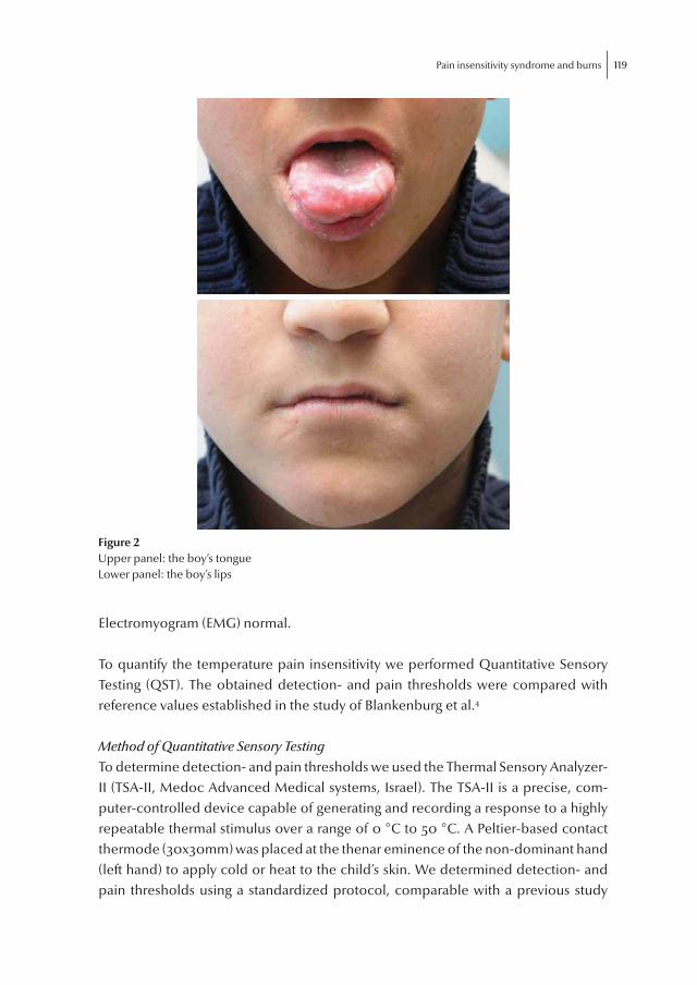
Pain insensitivity syndrome and burns 119
Electromyogram (EMG) normal.
To quantify the temperature pain insensitivity we performed Quantitative Sensory Testing (QST). The obtained detection- and pain thresholds were compared with reference values established in the study of Blankenburg et al.4
Method of Quantitative Sensory Testing
To determine detection- and pain thresholds we used the Thermal Sensory Analyzer-II (TSA-II, Medoc Advanced Medical systems, Israel). The TSA-II is a precise, com-puter-controlled device capable of generating and recording a response to a highly repeatable thermal stimulus over a range of 0 °C to 50 °C. A Peltier-based contact thermode (30x30mm) was placed at the thenar eminence of the non-dominant hand (left hand) to apply cold or heat to the child’s skin. We determined detection- and pain thresholds using a standardized protocol, comparable with a previous study
Figure 2Upper panel: the boy’s tongueLower panel: the boy’s lips

120 Chapter 8
from our research group.5 After explaining the test we first determined the child’s re-action time by means of a reaction time task on the computer. Subsequently we de-termined the detection- and pain thresholds for cold and warmth using the reaction time dependent Method of Limits (MLI). The test started at a baseline temperature of 32 °C, which was then steadily linearly decreased at a rate of 1 °C/sec. The child was asked to press the button as soon as the cold stimulus was felt. After pressing the button, the stimulus reversed to the baseline temperature of 32 °C with a rate of 1 °C/sec. We repeated this five times. The first two stimuli served as rehearsal stimuli. The detection threshold was calculated as the mean value of the last four stimuli. Next, the temperature was steadily linearly increased at a rate of 1 °C/sec to determine the detection threshold for warmth using the same method. Secondly, the MLI was applied to determine the pain thresholds for cold and warmth. Starting again from a baseline temperature of 32 °C, the temperature was steadily linearly decreased at a rate of 1.5 °C/sec. The child was asked to press the button when the cold sen-sation started to feel painful. Now also, the temperature reversed to the baseline temperature with a rate of 10.0 °C/sec. This was repeated four times. The last four obtained temperatures were used to calculate the mean pain threshold. Next, the pain threshold for warmth was determined in the same manner. When the child did not press the button before 0 °C or 50 °C, the test automatically terminated.
Furthermore we determined the detection thresholds for cold and warmth again, but now using the reaction time independent Method of Levels (MLE).
The researcher told the child that the thermode either could become cold, or would not change in temperature. The first thermal stimulus was 3.0 °C below the baseline temperature of 32.0 °C. Following each thermal stimulus the researcher asked the child ‘did the thermode become cold or not’? Dependent on the child’s response, the next stimulus was increased or decreased in temperature. The test terminated when the step size of the stimulus had decreased to a level of 0.1 °C. The warm detection threshold was determined in the same manner starting with a stimulus temperature of 3.0 °C above the baseline temperature.
Results Quantitative Sensory Testing
The test took place at home, in a quit room with a constant temperature of 21 °C. The temperature of the boy’s hand was 32.8 °C. The mean detection thresholds obtained with the MLI and the MLE, as well as the pain thresholds obtained with the MLI are presented in
Table 1. The third column lists the reference values of 9 to 12-year-old boys4. These values are also presented in figure 3.

Pain insensitivity syndrome and burns 121
Table 1 – Mean detection- and pain thresholds
Mean thresholdsin °C (SD)
Reference values4 for boys 9-12 years old
Detection thresholdcold
28.53 (1.15) (MLI)26.15 (MLE)
31.0 (1.6)
Detection thresholdwarmth
38.86 (0.18) (MLI)34.25 (MLE)
33.4 (1.6)
Pain thresholdcold
4.45 (3.94) 16.27 (8.3)
Pain thresholdwarmth
47.19 (1.15) 41.24 (3.84)
26.15
28.53
31
34.25
38.86
33.4
4.45
16.27
47.19
41.24
�
�
�
�
�
��
��
��
��
��
��
��
��
��
��
��
��
��
��
��
��
��
��
��
��
�
Detection cold(MLE)
Detection cold(MLI)
Reference Detection warmth(MLE)
Detection warmth(MLI)
Reference Pain cold (MLI) Reference Pain warmth(MLI)
Reference
Fig 3 Mean detection- and pain threshold with the references values

122 Chapter 8
Only once, during the determination of the cold pain threshold, the child did not press the button and reached the minimum temperature of 0 °C. On this occasion the value of 0 °C was used in the calculation of the mean cold pain threshold.
DISCUSSION
The diagnosis of HSAN type IV or CIPA requires three clinical criteria, i.e. anhidrosis, decreased pain and temperature perception, and mental retardation.6 However; there is a wide variety of intellectual performance. Common in all five types is the absence of axon flare following intradermal histamine. In our case the patient pro-duced a wheal, which is in contradiction with a HSAN but has been described before.9
HSAN type IV is caused by mutations in the NTRK1 (TRKA) gene. This gene is lo-cated on chromosome 1 (1q21-q22) and encodes for neurotrophic tyrosine kinase re-ceptor type 1 that is autophosphorylated in response to NGF (nerve growth factor).6 As a result of loss of function mutations, signal transduction at the NGF receptor is impeded and NGF dependent neurons, the small sensory and sympathetic neurons, fail to survive. Individuals who are compound heterozygotes (i.e. have two different abnormal NTKR1 alleles) may in some cases have unusually mild presentation. In contrast to type III there is no ethnic distribution but one half of reported cases have occurred in consanguineous marriages.2,6,8 Numerous mutations do not allow simple DNA diagnosis. Penetrance is complete, but expression varies widely and may be related to the site of the mutation on the NGF receptor or whether there is genetic homo or heterozygocity.1, 9 The quantitative sensory test showed that the detection- and pain thresholds of our patient considerably deviated from reference values of healthy children4. The boy was less sensitive in detecting a warm or a cold stimulus. Besides, he was less sensitive for both cold and warm pain stimuli. These findings support our suspicion of pain insensitivity. A limitation of the comparison with the reference values is that the healthy children in the study of Blankenburg et al. were tested on the dorsal side of the hand, whereas we tested at the thenar eminence of the hand. However, we do not expect substantial differences in results between these two methods.
In our patient, history, clinical signs of anhidrosis, pain insensibility, abnormal QST, histamine test combined with clinical findings, sufficed to confirm the diagno-sis: HSAN type IV or CIPA. With this diagnosis we repudiate the suspicion of child abuse confirm the caring attitude of the parents. The parents and the boy have been informed about the disease and treatment by a rehabilitation physician was begun.
Pain insensitivity syndrome and burns 123
The only similar case was presented by Makari in 1994, who reported a 5-year-old girl with severe burns and her 15-year-old brother, both probably with HSAN type V.10 Although both children had shown severe laceration, fractures and injuries, child abuse was not suggested until after the girl showed severe burns. The girl was placed in care because of suspected child abuse, but was allowed to return home after an examination of her brother confirmed that both children had HSAN. Other reports of children with HSAN or CIPA misinterpreted as child abuse are not known.
CONCLUSION:
We describe a case of suspicion of inflicted burns in a 10-year-old boy. History, physical examination and QST all pointed at the diagnosis of HSAN type IV or con-genital insensitivity of pain with anhidrosis. Health care workers should be aware of the potential presence of this disease. Thorough history taking and physical exami-nation are necessary to reveal the diagnosis. In case of oral mucosal laceration and scars, multiple fractures of lower extremities, anhidrosis and, infrequently, mental retardation, a diagnosis of HSAN type 4 or CIPA should be suspected and thoroughly evaluated.

124 Chapter 8
REFERENCES
1. Axelrod FB, Gold-von Simson G. Hereditary sensory and autonomic neuropathies: types II,
III, and IV. Orphanet J Rare Dis. 2007; 2: 39.
2. Axelrod FB, Gold-von Simson G, Oddoux C. Hereditary Sensory and Autonomic Neuropathy
IV. In: GeneReviews [Internet] (Pagon R, Bird T, Dolan C et al., eds), 2008 Aug 05 [updated
2009 Nov 24] edn. Seattle (WA): University of Washington, Seattle, 2008
3. Tanaka T, Satoh T, Tanaka A et al. Congenital insensitivity to pain with anhidrosis: a case with
preserved itch sensation to histamine and partial pain sensation. Br J Dermatol. 2011.
4. Blankenburg M, Boekens H, Hechler T et al. Reference values for quantitative sensory test-
ing in children and adolescents: developmental and gender differences of somatosensory
perception. Pain. 2010; 149: 76-88.
5. de Graaf J, Valkenburg AJ, Tibboel D et al. Thermal detection thresholds in 5-year-old pre-
term born children; IQ does matter. Early human development. 2012.
6. Indo Y, Tsuruta M, Hayashida Y et al. Mutations in the TRKA/NGF receptor gene in patients
with congenital insensitivity to pain with anhidrosis. Nat Genet. 1996; 13: 485-8.
7. Berkovitch M, Copeliovitch L, Tauber T, Vaknin Z, Lahat E. Hereditary insensitivity to pain with
anhidrosis. Pediatr Neurol. 1998; 19: 227-9.
8. Toscano E, Andria G. Congenital insensitivity to pain with anhidrosis: an NGF/TrkA-related
disorder. Am J Med Genet. 2001; 99: 164-5.
9. Oddoux C, Wang J, Clayton C et al. Genetic heterogeneity in hereditary and autonomic
sensory neuropathy (HSAN4). In: Society of Human Genetics, 1999.
10. Makari GS, Carroll JE, Burton EM. Hereditary sensory neuropathy manifesting as possible
child abuse. Pediatrics. 1994; 93: 842-4.


Chapter 9General Discussion


General Discussion 129
GENERAL DISCUSSION
Prelude
It is a challenge being a paediatric consultant in a burn centre – one of the three designated burn centres in the Netherlands – and one feels privileged indeed to have the opportunity to do research. This thesis is the fruit of 12 years of clinical experience with burns of various severity and causes (accident, neglect or inflicted) in children as well as severe skin diseases such the staphylococcal scalded skin syndrome (SSSS), Stevens-Johnson Syndrome, and toxic epidermal necrolysis (TEN). In 2004 we started to collect data of the pre-hospital management and transfer of children to a burn centre, first only regarding the Rotterdam burn centre but later also the other two. The first nation wide data were presented at the European Burn Association (EBA) congress in Budapest (2007). Chapters 2 & 3 present these data, supplemented with additional data from a second, later period. The research fo-cused on cooling and covering of the wound, as well as pain management, calcula-tion of burn size and fluid management. As we wished to study changes over time, the two study periods were compared: January 2002 until March 2004 (27 months) and January 2007 until August 2008 (20 months).
Furthermore, with the help of the Association of Dutch Burn Centres (ADBC) we evaluated the validity of pain scales in children with burns (Chapter 4). And con-fronted with cases of severe scalds after steam inhalation therapy in children we collected data on these incidents from the national Dutch Injury Surveillance System (LIS) and the three Dutch burn centres, assisted by the Consumer Safety Institute (Amsterdam, the Netherlands). The findings are presented in Chapter 5.
Finally, Chapters 6 - 8 deal with the intensive care support of children with severe skin disorders in the Erasmus MC-Sophia Children’s Hospital (Rotterdam, the Neth-erlands). Chapter 6 describes the fluid and pain management of young infants after curettage of giant congenital melanocytic nevi in comparison with severe burns. After having published a case report on the first use of skin substitutes in the manage-ment of SSSS1, we collected data of an additional seven infants with SSSS which we treated with Omiderm® and Suprathel® in the department of Paediatric Dermatology of the Erasmus MC-Sophia Children’s Hospital (Chapter 7). A very rare disease of pain insensitivity presenting as intentionally inflicted burns is reviewed in Chapter 8.
Part I. General Aspects
Cooling and covering, pain management, and determining burn size and need of fluid resuscitation are the cornerstones of emergency care in burns.

130 Chapter 9
Cooling
Cooling is one of the best known first aid measures following burn injury, and as such recommended in many guidelines.2-4 It is thought to eliminate the heat, prevent oedema and ongoing tissue damage, and finally to decrease pain.4,5 Though cool-ing is felt to be essential, care must be taken to prevent hypothermia. In our study we found that more than 90% of the children received cooling before burn centre admission. This indicates that in the Netherlands both the general public and health professionals are highly aware of the requirement to cool burn wounds. This aware-ness is long-standing as there was no change over time and evidently most people are acquainted with the slogan “eerst water de rest komt later” (first water the rest can wait), disseminated by the Dutch Burns Foundation.
The best cooling agent is water but there is still discussion about the optimal water temperature, duration of cooling, and time elapsed after the burn for cool-ing to still be effective.4,6 Dutch guidelines advise using lukewarm water within one hour. Evidently, these factors can only be studied experimentally, not in actual burn patients. Numerical models can help us to measure the effectiveness of cooling but can not replace studies in vivo. Besides, apart from heat transfer of the skin, the burn wound condition is also influenced by other factors such as oedema and biochemi-cal substances.7 Further studies should determine the effectiveness of water cooling (how long was effectively cooled, did the water cool the whole burn, what was the temperature of the cooling water, etc) and compare this with the effectiveness of other cooling methods such as the use of blankets or other applications.8
Cooling blankets containing melaleuca alternifolia gel (e.g. burnshields®) are alternative agents to cool the wound when water is not available. They demonstrate cooling and pain reducing effects and are frequently used by ambulance services. In a practical sense, it is sometimes difficult to cool children with water. When they are distressed and uncooperative the cooling water may spill all over the child and thus cause hypothermia, especially in the very young. Cooling blankets give a quick cool-ing effect and pain relief without disturbing the child. However, permanent wound cooling is not advocated by the provider of these blankets as this might result in hypothermia.
Wound covering
After adequate cooling, the wound must be covered to protect it, to provide pain relief, and to prevent hypothermia, especially in children with severe burns. Oint-ments should not be applied, however, so that experts can assess the depth of burn wounds without delay. We found that wound covering was applied in 65% of cases in the first period versus nearly 90% in the second period studied. (Chapter 2). Cooling blankets, discussed above, carry the risk of decreased blood supply and destruction

General Discussion 131
of vital tissue and may also result in hypothermia. Young children are at higher risk because their body surface area is relatively larger and the skin thinner. Rehydration with cold fluids may also contribute to the risk for hypothermia. Therefore, as a first aid the wound should be covered with non sticky material and the patient should be kept warm during transport to the burn centre. An interesting alternative, recom-mended by the UK and New Zealand guidelines3,4, is a cellophane type wrap (cling film). If cooling is still indicated, a wet towel can be placed on top of the cling film.
Pain management
Burns can be very painful due to direct stimulation of several types of nociceptors or to later sensitization of the nociceptive pathways in the peripheral and central nervous systems.9 Inadequate pain management may influence children’s pain perception later on in life.10,11 Over the two time periods the percentages of children receiving pain medication increased but still one out of every five children received no pain medication before they were transferred to a burn centre. Furthermore, although there was an increase in cases of adequate dosing, still it was adequate only in 41% of children in the second study period. Further education should make health care workers aware of the appropriate initial dose, and pain management may also be improved by with the use of validated pain scales to assess the actual effect of treatment.
Burn size
Accurate calculation of burn size, expressed as percentage of total body surface area (TBSA), is one of the most important aspects of the initial care of a burn victim. The outcome determines whether transfer to a burn centre is necessary as well as the need for, and amount of, initial intravenous fluid resuscitation. Accurate calculation is difficult in children and a mean twofold overestimation was demonstrated in our study (Chapter 2.) Fortunately, it had no consequences in terms of referral, as the cri-teria for referral to a Dutch burn centre also include vital locations like the face and the hands, and not only extent of burn. Many methods are advocated to perform and improve TBSA burn calculations, such as the Lund and Browder charts, the rule of nine (modified for children), the percent/hand rule, and the halving method.12-15 An interesting development in this regard also is telemedicine, which allows the burn specialist to assess the extent of the burn and to give additional advice.16
Accurate intravenous fluid resuscitation is essential to prevent shock in case of severe burns and to maintain adequate circulation to tissues and perfusion of vital organs. It was found to considerably increase chances of survival.17,18 Dutch burn centres, in accordance with the EMSB guideline, recommend intravenous fluid resuscitation in children with TBSA burn ≥ 10%, using the Parkland formula and ad-

132 Chapter 9
ditional maintenance fluid. In our study (Chapter 3) no over-resuscitation in children with a TBSA burn over 10% occurred. We concluded that the inaccuracy of TBSA burn calculation is ‘accidentally’ compensated for by inaccuracies in fluid resuscita-tion calculation.
To optimize pre-hospital burn care it is essential to develop recommendations and to educate first aid healthcare workers how to estimate burn size and compute an appropriate fluid amount. A working group consisting of burn care professionals, general practitioners, ambulance personnel, emergency medicine physicians, trau-ma surgeons, and paediatricians is currently preparing Dutch guidelines. Advanced training programs will facilitate the implementation of these guidelines in daily practice. Organizations such as the Dutch Foundation for the Emergency Medical Care for Children (SSHK), the Dutch Burn Foundation (NBS), Foundation Advanced Trauma Life Support (Stichting ATLS), Edupack and SOSA provide training programs like APLS, EMSB, ATLS and courses for ambulance services (Edupack ,SOSA) and should participate in training programs based on the new guidelines. E-learning modules could be used to spread the guidelines to the larger population of health care workers. Future solutions to help manage the emergency care of burn patients are telemedicine with video communication and secured websites that provide for the exchange of patient information, pictures and movies.
Pain management
Burn pain can be long lasting, has a fluctuating course, and is related to extensive repetitive daily wound care procedures. Adequate management of burn pain is im-portant for many reasons. It is essential for the relationship between the patient and the multidisciplinary team; it increases comfort, and makes recovery more tolerable. In addition, adequate pain management affects morbidity by preventing elevated metabolism, thereby reducing the chance of deterioration of the immune system.19 Furthermore, adequate pain management might reduce acute stress symptoms.
The cornerstone of pain management is pain assessment coupled with treatment decision trees and evidenced base dosing. Chapter 5 analyzes three different meth-ods to evaluate background pain and procedural pain in children younger than five years;
1) The pain observation scale for young children (POCIS) provides seven behav-ioural items with dichotomous answer categories (present or absent), which enables easy and quick use of the scale. It was initially developed to measure postopera-tive pain intensity in children after adenotonsillectomy, adenotomy or insertion of ventilation tubes.20
2) The COMFORT behaviour (COMFORT-B) scale comprises six behavioural items with five response categories for each item. One item has two options, i.e. respiratory

General Discussion 133
response and crying, to be used in ventilated and non-ventilated patients, respec-tively. The original COMFORT scale was developed and validated for all patients of all ages admitted to ICUs.21
3) Nurse observational visual analogue scale (VASobs) Is a visual analogue scale (VAS) providing a rating of the observer’s global impression of a patient’s pain. Nurses often use the VAS to assess children’s pain.22
Both the POCIS and the COMFORT-B scale can be used to measure background and procedural pain in daily burn nursing practice. The VASobs is not recommended. The POCIS was preferred by nurses for its ease of use, the COMFORT-B was per-ceived to address procedural and background pain more accurately and to have better properties to connect to a pain management protocol. In 2010 all Dutch burn centres embedded COMFORT-B assessments in daily practice. A treatment decision tree based on the pain scores was developed and the next step is to evaluate its accuracy and reliability. This should be considered as a major step forwards enabling benchmarking.
Lack of pain assessment tools also hampers adequate pain management in emer-gency care. Cooling, wound covering and medication are essential in reducing the pain, but adequate pain management must start with pain assessment. Chapter 4 stresses the importance of pain assessment training for nurses in pain scores and deals with the introduction of the COMFORT-B scale in burn centres. It is pointed out (chapter 4) that none of the available pain scales can differentiate between pain intensity and the emotional component of pain. However, each element would require a different treatment in terms of multimodal pharmacological and non-pharmacological interventions. With regard to the latter, pain distraction methods such as virtual-reality video gaming during wound dressing are very intriguing meth-ods to relieve pain and to decrease the need for pharmacological approaches.23 A recent innovation is multi modal distraction with a handheld interactive device that uses customized programs to inform the child about the procedure he/she is about to experience and to distract the child during wound dressings.24 Introduction of such non-pharmacological methods can further improve the pain management and clinical efficiency with reduction of treatment lengths. Another important non phar-macological approach consists of optimizing wound dressing. Reducing the need for dressing changes will decrease the number of potentially painful procedures and thus increase the patient’s comfort. Also dressings that fit well and comfortably on the wound is important and may contribute to the reduction in background pain. Research for optimal wound dressings should also include pain reducing and com-fort increasing effects.

134 Chapter 9
PART II. SPECIFIC CONDITIONS
Steam
Steam inhalation is a common home therapy for upper respiratory tract infections. General practitioners recommend it, and it is included as a recommendation in guidelines and patient brochures issued by societies of general practitioners, among others in the Netherlands, United States and United Kingdom.25-28 A Cochrane re-view (first version 2001, updated in 2006, 2009, 2011)29 however, concluded that steam inhalation had not shown to have any consistent benefit in the treatment of the common cold – and therefore it was not recommended in the routine treatment of common cold symptoms.
Annually, on average three persons were found to be admitted to a burn centre in the Netherlands for scalds resulting from steam inhalation therapy. (Chapter 5) With wide margins we estimated that at least a further 40 people were treated at emergency departments and an unknown number in general practices. In this study we highlighted that steam inhalation therapy has no benefits in common cold and that steam therapy with the use of a bowl of hot water carries a risk of severe scalds.
A limitation of this study was that we were unable to investigate the risk of scald-ing in relation to the number of times the procedure is used. As obtaining financial support to fund such a study of sufficient scale to provide the necessary data is highly unlikely, we concluded our data were the only and best available data at present. This study was performed in collaboration with an important Dutch organisation: Consumer Safety Institute. From their data system we could retrieve the amount of burn victims treated at Dutch emergency departments. By paying attention to the risk of scalds caused by steam inhalation we hope to provide general practitioners and nurse specialists with good arguments to discourage their patients to use steam inhalation. This study can also provide arguments for organizations such as the Dutch Burns Foundation and Consumer Safety Institute to incorporate awareness of the risk of scalds caused by steaming inhalation therapy in their campaigns aimed at the general public.
Giant congenital melanocytic nevi
Surgical treatment (curettage) of neonates and young infants with burns or giant con-genital melanocytic nevi (GCMN) is rare – and so is the literature on fluid and pain management for these conditions.30 In the study reported in Chapter 6, children with GCMN and, to a lesser extent, burn patients, received fluid predicted by the Parkland formula, which is based on body mass. This resulted in a urinary output that was far more than considered necessary. Too excessive fluid resuscitation in burn patients is known as “fluid creep”, which means that more fluid is actually given than

General Discussion 135
calculated with the Parkland formula.31,32 Besides this phenomenon, there is a risk of fluid overload in children even when the Parkland formula is calculated correctly and appropriate volumes are administered.33,34 Neonates and young infants have a relatively large body surface area. For this reason fluid resuscitation formula based on body surface area are more appropriate.35 Fluid overload is a risk in young infants and adequate fluid administration or permissive hypovolemia in the postoperative management of GCMN patients could prevent prolonged mechanical ventilation and ventilator associated pneumonia.36 In order to prevent fluid overload, we recom-mend to adapt fluid management to, for example, urinary output, in which a urinary output of 0.5 - 1 .0 ml/kg/hr is considered a solid parameter. Beside the amount of fluid, type of fluid solution is important to prevent electrolyte disturbances or hypoalbuminemia and is an issue for further studies.
Regarding pain management of infants with severe skin disorders such as GCMN and burns, the nationwide Dutch guideline “pain measurement and pain man-agement in children” contains advice on procedural pain only (changing wound dressings). PICUs are familiar with pain scales, but the Dutch burn centres only re-cently introduced these scales for children (Comfort-B scale).37 Further steps in pain management are integrated therapy algorithms and evidence based dosing. Beside these cornerstones of pain management, local skin care is important in reducing pain. Severe skin disorders in children are a challenge to manage, and pain and fluid management must be individualized. Exchange of experience with these children between centres (nationally and internationally) and or centralization can improve the quality of post-operative care of surgical treatment of GCMN and post burn care of infants.
Staphylococcal scalded skin syndrome
Staphylococcal scalded skin syndrome (SSSS) is a rare toxin-mediated skin disease caused by Staphylococcus aureus and is usually seen in infants and children younger than 5 years. It is rarely seen in adults. The bacteria produce exfoliative toxins that destroy the desmosomes in the stratum granulosum of the skin. Clinically, there are superficial blisters without mucosal lesions.38,39 In chapter 7 we describe seven infants with severe SSSS admitted to a PICU or burn centre and 6 of them success-fully treated with skin substitutes (Omiderm® and Suprathel®). This treatment was not described earlier. It decreases fluid loss from denuded skin and has pain reducing capacities. We also describe how to manage these children and gave recommenda-tions regarding antibiotics, fluid management, pain and sedation and the optimal way to use skin substitutes.
SSSS is rare (personal communication MB), 40 patients annually in the Nether-lands) and severe SSSS are best managed in a PICU or burn centre. Burn centres are

136 Chapter 9
particularly suitable for patients with skin diseases such a SSSS, the Stevens-Johnson syndrome, and toxic epidermal necrolysis. In these centres expert multidisciplinary care is provided, and patient’s rooms have special air processing to prevent wound infections and hypothermia. In the Netherlands, the burn centres are not located in university medical centres with a top level PICU, but all burn centres have partner-ships with nearby PICUs in university medical centres, thus guaranteeing high-quality and safe intensive care if needed.
Intentionally inflicted burns or rare disease?
Hereditary sensory and autonomic neuropathy (HSAN type IV) or congenital insen-sitivity to pain with anhidrosis (CIPA) is a rare disease characterised by absences of pain and temperature sensation combined with oral (self ) mutilation, mental retar-dation, fractures and impaired bone healing.40 In chapter 8 we present a boy with severe burns and general features that were misinterpreted as intentionally inflicted burns. The burns were suspected as inflicted burns for several reasons. The patient was not aware of his burns, the wound had not been cooled, and parents sought help not until after a few hours. Besides, the patient was known to and his family was supervised by the council of child protection. History taking, however, revealed that the boy was insensitive for pain, had anhidrosis and several unexplained fractures, lacerations of the skin, his lips and tongue.
With additional testing the diagnosis of HSAN type 4 was made. This rare disease is hereditary and the diagnosis can be made by three symptoms: pain insensitivity, anhidrosis and mental retardation. Physical examination demonstrated scars of the lips and scars of the tongue. For the diagnosis DNA analysis is not mandatory but absence of the flare response to intra dermal histamine phosphate supports the di-agnosis.41 With Quantitative Sensory Testing we qualified the disease. By publishing this case we emphasize that this disease is easily mistaken for child abuse but can be detected by a thorough history and careful and detailed physical examination. Secondly, we are not aware how many children with burns, admitted or treated on emergency departments have intentionally inflicted burns or burns caused by neglect. Furthermore, in these cases we need to apply validated methods to exclude child abuse or neglect. This may shed a light on this important issue in relation to burns.
CONCLUSION
Both the general public and professionals are already highly aware of the necessity to cool burn wounds, however improvements may still be made by establishing op-

General Discussion 137
timal temperature and duration of cooling, and the time after which cooling would no longer be effective. Concerning emergency care in paediatric burn patients, areas that require specific attention are pain management, assessment of extent of the burns and subsequent fluid resuscitation. A start has been made with the develop-ment of Dutch guidelines and subsequent educational efforts and innovations in emergency care such as telemedicine may contribute to the improvement of burn size assessment and the management of these children in years to come.
To assess pain in children during their stay at a burn centre, the COMFORT-B was found to be the best option. With the embedding of this scale in daily practice and the development of a treatment decision tree based on the pain scores, a major step forwards will be taken regarding pain management in paediatric patients with burns in the Netherlands.
As steam inhalation therapy has no proven benefit and the number and extent of complications of this therapy in terms of scalds are significant, especially in chil-dren, the proposition “there is no harm in trying” does not apply to steam inhalation therapy. Therefore steam inhalation therapy should be discouraged and not recom-mended anymore in professional guidelines and patient brochures.
This thesis is the result of collaboration between Erasmus MC-Sophia Children’s hospital, extramural organisations (Consumer Safety Institute, Association of Dutch Burn Centres) and the burn centres of Groningen, Rotterdam and Beverwijk. Both paediatric patients with burns and children with other serious and/or rare skin conditions (SSSS, GCMN and HSAN typeIV) benefit from the collaboration and exchange of knowledge and experience by the different professionals involved in their treatment. This exchange, now and in the future, helps improve the quality of care and outcome, of children with skins “at risk”.
138 Chapter 9
REFERENCES
1. Baartmans MG, Maas MH, Dokter J. Neonate with staphylococcal scalded skin syndrome.
Arch Dis Child Fetal Neonatal Ed. 2006; 91: F25.
2. Baartmans MG, van Baar ME, Boxma H et al. Accuracy of burn size assessment prior to arrival
in Dutch Burn centres and its consequences in children: A nationwide evaluation. Injury.
2011.
3. Education Committee of the Australian and New Zealand Burn A. Emergency management of
severe burns (EMSB) course manual. Dutch version, Dutch Burn Foundation, 2009.
4. New Zealand Guidelines G. Management of burns and scalds in primary care. Wellington (NZ):
Accident Compensation Corporation (ACC), 2007.
5. Allison K. The UK pre-hospital management of burn patients: current practice and the need
for a standard approach. Burns. 2002; 28: 135-42.
6. Bartlett N, Yuan J, Holland AJ et al. Optimal duration of cooling for an acute scald contact
burn injury in a porcine model. J Burn Care Res. 2008; 29: 828-34.
7. Baldwin A, Xu J, Attinger D. How to Cool a Burn: A Heat Transfer Point of View. J Burn Care Res.
2012.
8. Schnell HM, Zaspel JG. Cooling extensive burns: sprayed coolants can improve initial cooling
management - a thermography-based study. Burns. 2008; 34: 505-8.
9. Werner MU, Lassen B, Pedersen JL et al. Local cooling does not prevent hyperalgesia follow-
ing burn injury in humans. Pain. 2002; 98: 297-303.
10. Wollgarten-Hadamek I, Hohmeister J, Demirakca S et al. Do burn injuries during infancy
affect pain and sensory sensitivity in later childhood? Pain. 2009; 141: 165-72.
11. Hohmeister J, Kroll A, Wollgarten-Hadamek I et al. Cerebral processing of pain in school-
aged children with neonatal nociceptive input: an exploratory fMRI study. Pain. 2010; 150:
257-67.
12. Knaysi GA, Crikelair GF, Cosman B. The rule of nines: its history and accuracy. Plast Reconstr
Surg. 1968; 41: 560-3.
13. Nagel TR, Schunk JE. Using the hand to estimate the surface area of a burn in children. Pediatr
Emerg Care. 1997; 13: 254-5.
14. Lund CC, Browder NC. The estimation of areas of burns. Surgery, gynecology & obstetrics 1944;
79: 352-8.
15. Berkow S. A method of estimating the extensiveness of lesions (burns and scalds) based on
surface area proportions. Arch Surg. 1924; 8: 138-48.
16. Saffle JR, Edelman L, Theurer L et al. Telemedicine evaluation of acute burns is accurate and
cost-effective. J Trauma. 2009; 67: 358-65.
17. Barrow RE, Jeschke MG, Herndon DN. Early fluid resuscitation improves outcomes in severely
burned children. Resuscitation. 2000; 45: 91-6.
18. Wolf SE, Rose JK, Desai MH et al. Mortality determinants in massive pediatric burns. An
analysis of 103 children with > or = 80% TBSA burns (> or = 70% full-thickness). Ann Surg. 1997;
225: 554-65.
19. Latarjet J, Choinere M. Pain in burn patients. Burns. 1995; 21: 344-8.
20. Boelen-van der Loo WJ, Scheffer E, de Haan RJ et al. Clinimetric evaluation of the pain obser-
vation scale for young children in children aged between 1 and 4 years after ear, nose, and
throat surgery. J Dev Behav Pediatr. 1999; 20: 222-7.

General Discussion 139
21. van Dijk M, de Boer JB, Koot HM et al. The reliability and validity of the COMFORT scale as a
postoperative pain instrument in 0 to 3-year-old infants. Pain. 2000; 84: 367-77.
22. Von Baeyer CL, Spagrud LJ. Systematic review of observational (behavioral) measures of pain
for children and adolescents aged 3-18 years. Pain. 2007: 140-59.
23. Malloy KM, Milling LS. The effectiveness of virtual reality distraction for pain reduction: a
systematic review. Clin Psychol Rev. 2010; 30: 1011-8.
24. Miller K, Rodger S, Bucolo S et al. Multi-modal distraction. Using technology to combat pain
in young children with burn injuries. Burns. 2011; 36: 647-58.
25. Dutch College of General Practitioners. Colds in children - Patient letter. Utrecht, 2011.
26. American Academy of Family Physicians. Health information for the whole family. Bronchiol-
itis and your child. Accessed November 3, 2011 at http://familydoctor.org/familydoctor/en/
diseases-conditions.html
27. American Academy of Family Physicians. Health information for the whole family. OTC
Cough and Cold Medicines and My Child: What Do I Need to Know? Accessed November 3,
2011 at http://familydoctor.org/familydoctor/en/diseases-conditions.html
28. NHS. NHS Clinical Knowledge Summaries: Common Cold. Accessed November 3, 2011 at
http://www.cks.nhs.uk/common_cold/management/scenario_common_cold#-261033.
29. Singh M, Singh M. Heated, humidified air for the common cold. Cochrane Database Syst Rev.
2011: CD001728.
30. De Raeve LE, Claes A, Ruiter DJ et al. Distinct phenotypic changes between the superficial
and deep component of giant congenital melanocytic naevi: a rationale for curettage. Br J
Dermatol. 2006; 154: 485-92.
31. Saffle JI. The phenomenon of “fluid creep” in acute burn resuscitation. J Burn Care Res. 2007;
28: 382-95.
32. Rogers AD, Karpelowsky J, Millar AJ et al. Fluid creep in major pediatric burns. Eur J Pediatr
Surg. 2010; 20: 133-8.
33. Graves TA, Cioffi WG, McManus WF et al. Fluid resuscitation of infants and children with
massive thermal injury. J Trauma. 1988; 28: 1656-9.
34. Warden GD. Fluid resuscitation and early management. In: Total burn care (Herndon DH, ed),
3rd edn.: Saunders Elsevier, 2006: 107-18.
35. Herndon DN, Thompson PB, Desai MH et al. Treatment of burns in children. Pediatr Clin
North Am. 1985; 32: 1311-32.
36. Young A. Fluid resuscitation in children’s burn care; are current guidelines harming children?
In: 14th European Burns Association Congress. World Forum Congress Center, The Hague, The
Netherlands, 2011.
37. de Jong A, Baartmans M, Bremer M et al. Reliability, validity and clinical utility of three types
of pain behavioural observation scales for young children with burns aged 0-5 years. Pain
2010; 150: 561-7.
38. Ladhani S. Understanding the mechanism of action of the exfoliative toxins of Staphylococ-
cus aureus. FEMS Immunol Med Microbiol. 2003; 39: 181-9.
39. Stanley JR, Amagai M. Pemphigus, bullous impetigo, and the staphylococcal scalded-skin
syndrome. N Engl J Med. 2006; 355: 1800-10.
40. Axelrod FB, Gold-von Simson G. Hereditary sensory and autonomic neuropathies: types II,
III, and IV. Orphanet J Rare Dis. 2007; 2: 39.

140 Chapter 9
41. Axelrod FB, Gold-von Simson G, Oddoux C. Hereditary Sensory and Autonomic Neuropathy
IV. In: GeneReviews [Internet] (Pagon R, Bird T, Dolan C et al., eds), 2008 Aug 05 [updated
2009 Nov 24] edn. Seattle (WA): University of Washington, Seattle, 2008.


Chapter 10Summary/Samenvatting


Summary/Samenvatting 145
SUMMARY
This thesis focuses on the paediatric skin “at risk”. The included studies investigate diagnosis, treatment and outcome of children’s skin lesions in case of burns, major surgery, and specific infections.
CHAPTER 1
The skin, the largest human organ, provides the body shape and protects our body against intruders such as heat, cold, trauma, or infections. Severe skin lesions such as burn scars and giant melanocytic naevi have a major impact on a person’s ap-pearance and will influence not only the skin function but also the interpersonal communication and behaviour.
The skin is built up of three main components: epidermis, dermis, and skin ap-pendages. Skin development starts in utero with two morphologically different skin layers derived from two different germ layers; the ectoderm and the mesenchyme. At birth all layers of the adult epidermis are present. The skin further develops during the first few years of life. The skin (especially stratum corneum) will become more hydrated and permeable. The microcirculation undergoes changes and is fully adapted by 3 to 4 months. A child’s skin is more sensitive than an adult’s skin because natural defence mechanisms are not fully developed yet.
PART I. GENERAL ASPECTS
CHAPTERS 2 AND 3
In the Netherlands, annually approximately 250 children with a burn injury of more than 5% of total body surface area (TBSA) or burns in specific body sites are treated in one of the three Dutch burn centres. Before admission, medical teams check the child’s vital signs, cool the burns, calculate the TBSA burned, and then provide intra-venous rehydration, pain management and wound dressing. This puts great stress on most health care workers in hospitals and ambulances. Courses on how to handle these situations have been available in the Netherlands since 1998. Despite these and other efforts to improve emergency burn care, specialists at the burn centres realize that improvements can still be made. To identify areas for improvement, we evaluated the pre-hospital management of all children transferred and admitted to

146 Chapter 10
three Dutch burn centres. As we wished to study changes over time, the two study periods were compared: January 2002 until March 2004 (27 months) and January 2007 until August 2008 (20 months).
Chapter 2 focuses on cooling and covering of the burn wound and the initial pain treatment. Both the general public and professionals are highly aware of the neces-sity to cool burn wounds. The best cooling agent is water but there is still discussion about its optimal temperature, the duration of cooling, and the time after which cooling would no longer be effective. New studies should demonstrate the actual effectiveness of cooling with water (how long was effectively cooled, did the water cool the whole burn, what was the temperature of the cooling water, et cetera) and compare it with the effectiveness of other methods, such as the use of cooling blan-kets. The use of cooling blankets decreased over time; and in rare cases hypothermia as a result of cooling occurred. After cooling the wound must be covered to protect it from further damage, to lessen the pain and to prevent hypothermia, especially in children. We found that indeed 90% of the children were covered before admission. An interesting alternative in this respect, recommended by the UK and New Zealand guidelines, is using a cellophane type wrap (cling film). If cooling is still indicated, a wet towel can be placed on top of the cling film.
Burns can be very painful due to direct stimulation of several types of nociceptors or to later sensitization of the nociceptive pathways in the peripheral and central nervous systems.6 Inadequate pain management may influence children’s pain perception later on in life .7,8 Over the two time periods the percentages of children receiving pain medication increased but still one out of every five children received no pain medication before they were transferred to a burn centre. Furthermore, al-though there was an increase in cases of adequate dosing, it was still only adequate in 41% of children in the second study period. Further education should make health care workers aware of the appropriate initial dose, and pain management may also be improved by with the use of validated pain scales to assess the actual effect of treatment.
In chapter 3 we assessed the pre-hospital diagnosis and treatment of paediatric burn patients with a focus on the accuracy of calculating burn size and intravenous rehydration therapy. Accurate calculation of burn size is difficult in children and a mean twofold overestimation was demonstrated in our study. Fortunately it had no consequences in terms of indication of referral to a Dutch burn centre. Methods and alternatives are described to improve the calculation of burn size. In our study no over-resuscitation in children with a TBSA burn over 10% occurred. We concluded that the inaccuracy of TBSA burn calculation was ‘accidentally’ compensated for by inaccuracies in fluid resuscitation calculation. To optimize pre-hospital burn care it

Summary/Samenvatting 147
is essential to develop recommendations and to educate healthcare workers how to estimate burn size and to compute an appropriate fluid amount.
A working group consisting of burn and health care professionals is currently preparing Dutch guidelines. Advanced training programs will facilitate the imple-mentation of these guidelines in daily practice. Organizations such as the Dutch Foundation for the Emergency Medical Care for Children (SSHK), the Dutch Burn Foundation (NBS), Foundation Advanced Trauma Life Support (Stichting ATLS), Edu-pack, and SOSA provide training programs like APLS, EMSB, ATLS and courses for ambulance services and should participate in training programs based on the new guidelines. E-learning modules could be used to spread the guidelines to the larger population of health care workers. Future solutions to help manage the emergency care of burn patients are telemedicine with video communication and secured websites that provide for the exchange of patient information, pictures and movies.
CHAPTER 4
Burn injury requires appropriate pain management – also in view of the extensive repetitive daily wound care procedures. In this regard, we distinguish between background pain and procedural pain. Individualized pain management is made easier when patients can self-report their pain. However, approximately one quarter of patients admitted to the three Dutch burn centres are under 4 years of age and cannot reliably self-report pain. Therefore, we investigated the reliability, validity and clinical utility of three pain behavioural observation scales applied to measure procedural and background pain in 0 to 5-year-old children with burns: The pain observation scale for young children (POCIS), the COMFORT behaviour scale (COMFORT-B scale), and the Nurse observational visual analogue scale (VASobs). We demonstrated that both the POCIS and the COMFORT-B scale are suitable to mea-sure background and procedural pain in daily burn nursing practice. The COMFORT-B scale was perceived to address procedural and background pain more accurately and to have better properties to connect to a pain management protocol. All Dutch burn centres have chosen to embed the COMFORT-B scale in daily practice. Non-pharmacological approaches can probably further improve the pain management of children with burns.

148 Chapter 10
PART II. SPECIFIC CONDITIONS
CHAPTER 5
In this chapter we studied cases of scalds caused by steam inhalation therapy for common cold that necessitated emergency care or admission to a burn centre. To-gether with the Consumer and Safety Institute we performed the analyses and costs calculation. Each year, three patients are admitted to one of the three burn centres due to scalds after steam inhalation for common cold and children are especially at risk for severe burns. At least 40 people visit an emergency department for this reason. In a Cochrane review it was concluded that steam inhalation had not shown any benefits in the treatment of the common cold. So there is every reason to advice against it. By paying attention to the risk of scalds caused by steam inhalation we hope and expect that general practitioners and nurse specialists have good argu-ments to discourage the use of steam inhalation therapy. This study can also provide arguments for organizations such as the Dutch Burns Foundation and Consumer Safety Institute to incorporate awareness of the risk of scalds caused by steaming inhalation therapy in their campaigns aimed at the general public.
CHAPTER 6
Giant congenital melanocytic naevi (GCMN) are rare and represent a special group of melanocytic lesions. Curettage is an easy technique to remove the GCMN from the papillary zone of the dermis. This is well feasible within the first two weeks of life when the cleavage plane between the upper and the lower dermis is easily found. Given the low incidence of GCMN this therapy is rare and postoperative care has not been described earlier. We therefore aimed to describe fluid therapy and pain management and define recommendations for this special group of surgical infants. We compared the fluid and pain management in theses infants to that of young infants with burns. There was evidence that both groups received too much fluid. Pain management differed between paediatric intensive care units and burn centres. We recommend exchange of experience in the management of postoperative care between centres to improve expertise and quality of care.

Summary/Samenvatting 149
CHAPTER 7
Each year, approximately 40 children in the Netherlands suffer from staphylococcal scalded skin syndrome (SSSS). This is a generalised superficially exfoliative skin dis-ease caused by an exfoliative toxin produced by Staphylococcus aureus. To prevent excessive fluid loss and reduce pain we treated our patients with skin substitutes (Omiderm® and Suprathel®). To our knowledge this technique had not yet been used in children with SSSS. Management may be complicated in patients with extensive blistering. With extensive denudation of skin, patients may have decreased thermo-regulatory ability, extensive fluid losses and electrolyte imbalance, and are at risk for secondary infection and sepsis. Treatment consists of antibiotics and supportive fluid therapy, electrolyte correction, adequate enteral feeding, and pain manage-ment. Severely affected patients should be treated in a paediatric intensive care unit or burn centre. We analysed the outcomes of seven young infants (6 treated with skin substitutes) and formulated guidelines for treatment. The use of skin substitutes is an essential contribution in the management of SSSS. Burn centres are particularly suitable for patients with skin diseases such as Stevens-Johnson syndrome, toxic epi-dermal necrolysis (TEN) or SSSS. In these centres multidisciplinary care is provided, there is expertise in wound care with skin substitutes and patient’s rooms have special air processing to prevent wound infections and hypothermia.
CHAPTER 8
Child abuse and neglect is one of the causes of burns in children. In the Netherlands there are no data on child abuse, neglect and burns. Obviously, much effort is put into identifying abuse and neglect in children; in rare cases, however, suspicion of child abuse is not justified. In this chapter we report a boy with severe burns and HSAN type IV misinterpreted as intentionally inflicted burns. We emphasize that HSAN type IV is easily mistaken for child abuse. Furthermore, we need to assess patients with validated methods for child abuse and neglect to reveal data regarding this important issue in burns.
Conclusion
This thesis is the result of a collaboration between a university children’s hospital, extramural organisations and burn centres and. The exchange of knowledge and experience will help improve the quality of care and outcome of children with skins “at risk”.

150 Chapter 10
SAMENVATTING
Dit proefschrift richt zich op kinderen met bedreigende huidaandoeningen. De inhoud bestaat uit hoofdstukken die handelen over de diagnose, behandeling en uitkomst van huidaandoeningen bij kinderen zoals brandwonden, na grote huid-operaties en speciale infecties.
HOOFDSTUK 1
De huid is het grootste orgaan van de mens, het geeft vorm aan het lichaam en beschermt ons lichaam tegen warmte, kou, verwondingen of infecties. Ernstige huidaandoeningen zoals brandwonden, littekens en congenitale reuzen naevi heb-ben een grote invloed op het uiterlijk en zullen niet alleen de huidfunctie, maar ook de intermenselijke communicatie en het gedrag beïnvloeden.
De huid is opgebouwd uit drie hoofdcomponenten: epidermis, dermis, en de huidaanhangsels. De huidontwikkeling begint in de baarmoeder met twee mor-fologisch verschillende huidlagen afkomstig uit twee verschillende kiembladen, het ectoderm en het mesenchym. Bij de geboorte zijn alle lagen van de volwassen epidermis aanwezig. De huid ontwikkelt zich verder tijdens de eerste jaren van het leven en is dan (vooral stratum corneum) meer gehydrateerd en doorlaatbaar. De microcirculatie van de huid verandert eveneens en is volledig aangepast op de leeftijd van 3 tot 4 maanden. Een kinderhuid is gevoeliger dan de huid van een volwassene. Natuurlijke afweermechanismen zijn nog niet volledig ontwikkeld.
DEEL I. ALGEMENE ASPECTEN
HOOFDSTUKKEN 2 EN 3
Jaarlijks worden er ongeveer 250 kinderen met brandwonden opgenomen in een van de drie Nederlandse brandwondencentra. Indicatie voor opname zijn met name het totaal verbrand lichaamsoppervlakte (TVLO) waarbij 5% of meer is verbrand of brandwonden in functionele gebieden. Voor overplaatsing beoordelen medische teams van verwijzende ziekenhuizen het kind, stabiliseren de vitale functies, koelen de brandwonden, berekenen het TVLO en geven daarna, wanneer noodzakelijk, intraveneuze rehydratie, pijnbestrijding en wondverzorging. Deze opvang van pa-tiënten met ernstige brandwonden komt relatief weinig voor en legt grote druk op
Summary/Samenvatting 151
de artsen en verpleegkundigen in ziekenhuizen en ambulances. Sinds 1998 zijn er cursussen hoe men in dergelijke situaties moet handelen. Ondanks deze cursussen kan de eerstehulpzorg vóór verwijzing naar brandwondencentra beter. Om te bepa-len waar verbetering nodig is hebben we geëvalueerd hoe de brandwondenzorg is voor kinderen voordat zij overgeplaatst worden naar een van de drie Nederlandse brandwondencentra. Om veranderingen in de tijd te kunnen analyseren, werden twee periodes met elkaar vergeleken; van januari 2002 tot maart 2004 (27 maanden) en van januari 2007 tot augustus 2008 (20 maanden).
Hoofdstuk 2 richt zich op het koelen, het bedekken van de brandwonden en de pijnbestrijding. Zowel de leek als de professionals zijn zich zeer bewust van de noodzaak om brandwonden te koelen. Het beste koelmiddel is water. Er is echter nog discussie over de optimale temperatuur, de duur van de koeling, en de tijd waarna koeling niet meer effectief is. Dit zou verder onderzocht moeten worden en vergeleken met de effectiviteit van andere methoden, zoals koeldekens of andere middelen. Ook wat er werkelijk gebeurd is aan koelen, is iets dat wij niet onderzocht hebben. Over de twee tijdsperioden van de studie wordt gezien dat het gebruik van koeldekens is afgenomen. In zeldzame gevallen wordt onderkoeling gezien. Na het koelen moet de wond worden afgedekt ter bescherming, ter vermindering van pijn en om onderkoeling, vooral bij kinderen, te voorkomen. We vonden dat bij 90% van de kinderen de wonden bedekt waren. Een interessant alternatief in dit opzicht is het gebruik van plastic huishoudfolie dat op de wond gelegd wordt en daarna bedekt met een doek. Dergelijke alternatieven staan vermeld in richtlijnen van het Verenigd Koninkrijk en Nieuw-Zeeland. Wanneer er nog koeling nodig is, kan er ook een natte doek op de folie gelegd worden.
Brandwonden zijn erg pijnlijk als gevolg van directe stimulatie van de verschillende soorten nociceptoren of in een latere fase de sensibilisering van de nociceptieve paden in het perifere en het centrale zenuwstelsel. Bij onvoldoende pijnbestrijding kan de pijnperceptie beïnvloed worden voor het latere leven. Wanneer de twee tijdsperioden worden vergeleken dan is het percentage kinderen dat pijnbestrijding krijgt toegenomen. In de tweede periode kreeg echter één op de vijf kinderen geen pijnbestrijding voor overplaatsing naar een brandwondencentrum. Daarnaast bleek dat maar in 41% van de gevallen de dosering adequaat was. Onderwijs en trainingen moeten de professionals in de gezondheidzorg bewust maken dat pijnbestrijding nodig is en dat juiste doseringen worden gegeven. Pijnbestrijding kan worden ver-beterd door gebruik te maken van gevalideerde pijnscores om de effectiviteit van de behandeling te beoordelen.
In hoofdstuk 3 onderzochten we de diagnostiek en behandeling van kinderen met brandwonden gericht op het berekenen van het TVLO en de berekening van de

152 Chapter 10
hoeveelheid intraveneuze vochttherapie. Nauwkeurig berekenen van het TVLO is moeilijk bij kinderen en wij concludeerden dat er een gemiddelde overschatting was met een factor 2. Gelukkig bleek ondanks verkeerde inschatting, overplaatsing naar een brandwondencentrum geïndiceerd.
In veel gevallen waren er ook andere indicaties, waaronder de lokalisatie van de verbranding, die een overplaatsing naar een brandwondencentrum rechtvaar-digden. Er worden een aantal manieren beschreven hoe het verbrand lichaamsop-pervlakte kan worden berekend. In onze studie werd niet gezien dat kinderen met een TVLO van 10% of meer te veel vocht toegediend kregen. We concludeerden dat de onnauwkeurigheid van de TVLO berekening ‘per ongeluk’ gecompenseerd werd door onnauwkeurigheden in de berekening voor vochttoediening. Voor het optimaliseren van de eerste opvang van patiënten met brandwonden voorafgaand aan overplaatsing naar een brandwondencentrum is het essentieel om richtlijnen hebben. Momenteel is een werkgroep, bestaande uit professionals in de gezond-heidzorg die te maken hebben met de opvang van patiënten met brandwonden, bezig om een Nederlandse richtlijn te ontwikkelen. Via bijscholingsprogramma’s dienen deze richtlijnen geïmplementeerd te worden in de dagelijkse praktijk. Or-ganisaties zoals de Nederlandse Stichting spoedeisende hulp bij kinderen (SSHK), de Nederlandse Brandwonden Stichting (NBS), Stichting ATLS, Edupack en SOSA die opleidingen verzorgen zoals APLS, EMSB, ATLS en cursussen voor de ambulance-diensten zullen deze richtlijnen moeten implementeren in hun trainingsprogramma en uitdragen. E-learning modules kunnen worden gebruikt om de richtlijnen onder de aandacht te brengen bij een grote groep professionals in de gezondheidzorg. Toekomstige oplossingen om vanuit de brandwondencentra de spoedeisende zorg van voor patiënten met brandwonden te ondersteunen zijn “telemedicine” en be-veiligde websites die zorgen voor de snelle uitwisseling van patiëntgegevens zoals foto’s en films.
HOOFDSTUK 4
Brandwonden vereisen goede pijnbestrijding - mede gelet op de uitgebreide dage-lijkse wondverzorgingsprocedures. We maken onderscheid tussen achtergrondpijn (pijn die gehele dag aanwezig is) en procedurele pijn (pijn tijdens wondverzorging). Individuele pijnbestrijding kan worden geoptimaliseerd wanneer patiënten hun pijn zelf melden. Echter, een kwart van de patiënten opgenomen in de drie Nederlandse brandwondencentra is 4 jaar of jonger en kunnen niet zelf hun pijn betrouwbaar rapporteren. Daarom hebben we onderzoek gedaan naar de betrouwbaarheid, validiteit en klinische bruikbaarheid van drie pijn gedragsobservatie schalen

Summary/Samenvatting 153
toegepast op de procedurele en achtergrondpijn bij kinderen met brandwonden onder de vijf jaar; de pijn observatieschaal voor jonge kinderen (POCIS), de COM-FORT gedragschaal (COMFORT-B schaal), en de observationele verpleegkundige visueel analoge schaal (VASobs). We hebben aangetoond dat zowel de POCIS en de COMFORT-B schaal geschikt zijn voor achtergrond en procedurele pijn in de dagelijkse brandwondenzorg. De COMFORT-B schaal werd gezien als de beste schaal om achtergrond en procedurele pijn te meten en daarnaast heeft deze de beste eigenschappen om te gebruiken in combinatie met pijn protocollen en voor onderzoek. Alle Nederlandse brandwondencentra hebben ervoor gekozen om de COMFORT-B schaal te gebruiken in de dagelijkse praktijk. Niet-farmacologische benadering van pijn kan een verdere verbetering van de pijnbestrijding bij kinderen met brandwonden opleveren.
DEEL II. SPECIALE CONDITIES
HOOFDSTUK 5
In dit hoofdstuk bestudeerden we het aantal opnames in brandwondencentra en spoedeisende hulp behandelingen van mensen met brandwonden die werden veroorzaakt door heet water dat zij over zich heen kregen tijdens het stomen voor een verkoudheid. In de drie brandwondencentra worden ieder jaar gemiddeld drie patiënten opgenomen met dergelijke brandwonden. Kinderen hadden daarbij een verhoogd risico op ernstige brandwonden. Samen met Consument en Veiligheid hebben we een analyse uitgevoerd hoeveel slachtoffers er behandeld worden op een spoedeisende hulp voor deze brandwonden. Daarnaast hebben wij de kosten berekend voor deze ongevallen. Op de spoedeisende hulp werden ieder jaar gemiddeld ten minste 40 mensen behandeld voor deze brandwonden. In een Cochrane-review werd geconcludeerd dat stoominhalatie geen positief effect heeft op de behandeling van een verkoudheid. Gezien de ernstige brandwonden en kosten voor behandeling hiervan is er alle reden om stoom inhalatietherapie af te raden. Door aandacht te besteden aan het risico van brandwonden veroorzaakt door stoom inhalatietherapie hopen en verwachten we dat huisartsen en gespecia-liseerde verpleegkundigen goede argumenten hebben om stoom inhalatietherapie te ontmoedigen. Deze studie kan ook als basis dienen voor organisaties zoals de Nederlandse Brandwonden Stichting en de Stichting Consument en Veiligheid om stoom inhalatietherapie af te raden en het grote publiek bewust te maken dat er een risico bestaat op brandwonden.

154 Chapter 10
HOOFDSTUK 6
Congenitale Reuzen naevi( GCMN) zijn zeldzaam en vertegenwoordigen een spe-ciale groep van melanocytaire laesies. Curettage is een techniek om het GCMN uit de papillaire zone van de dermis te verwijderen. Dit is mogelijk in de eerste twee levensweken als de splitsing tussen de bovenste en onderste dermis gemakkelijk te onderscheiden is. Gezien de lage incidentie van GCMN is deze therapie zeldzaam en postoperatieve zorg voor deze kinderen nog niet eerder beschreven. Ons doel was het postoperatieve vocht- en pijnbeleid te beschrijven en aanbevelingen te definiëren voor deze speciale groep van chirurgische baby’s. We vergeleken het vocht-en pijnbeleid van deze groep met jonge zuigelingen met brandwonden met een TVLO van 10% of meer. Wij concludeerden dat beide groepen te veel vocht kregen en dat het pijnbeleid tussen de pediatrische intensive care units (PICU’s) en brandwondencentra enorm verschilde. Omdat deze patiëntengroepen zeldzaam zijn, zou centralisering en uitwisseling van expertise omtrent het post-operatieve beleid de expertise vergroten en de kwaliteit van zorg verbeteren.
HOOFDSTUK 7
Per jaar zijn er ongeveer 40 kinderen in Nederland met de diagnose staphylococ-cen scalded skin syndrome (SSSS). Dit is een blaarvormende huidziekte veroorzaakt door het exfoliatieve toxine geproduceerd door de Staphylococcus aureus. De be-handeling en het verloop kunnen gecompliceerd zijn wanneer de blaarvorming zeer uitgebreid is. De temperatuursregulatie kan ontregeld zijn, overmatig vochtverlies uit de wonden, verstoringen van de elektrolytenbalans, en secundaire infectie met sepsis. De behandeling bestaat uit antibiotica en ondersteunende vochttherapie, elektrolytcorrecties, adequate voeding en pijnbestrijding. Ernstig aangedane patiën-ten moeten worden behandeld in op een PICU of brandwondencentrum.
Om overmatig vochtverlies te voorkomen en daarnaast de pijn te verminderen hebben we onze patiënten behandeld met speciale wondbedekkers (Omiderm ® en Suprathel ®). Deze methode was nog niet eerder toegepast bij kinderen met SSSS. We beschreven de uitkomsten van 7 jonge zuigelingen met ernstige SSSS (waarvan zes werden behandeld met wondbedekkers). Daarnaast formuleerden we richtlij-nen voor de behandeling van SSSS en adviseerden de speciale wondbedekkers als essentieel in de behandeling.
Brandwondencentra zijn met name geschikt voor patiënten met huidaandoenin-gen zoals Stevens-Johnson syndroom, toxische epidermale necrolyse (TEN) of SSSS. In deze centra wordt multidisciplinaire zorg verleend, is er deskundigheid in de

Summary/Samenvatting 155
wondverzorging met wondbedekkers en de patiëntenkamers zijn voorzien van een speciale luchtverwerking om wondinfecties en onderkoeling te voorkomen.
HOOFDSTUK 8
Kindermishandeling en verwaarlozing zijn oorzaken van brandwonden bij kinderen. Er wordt veel energie gestoken in het identificeren van kindermishandeling. In zeld-zame gevallen is het vermoeden van kindermishandeling niet gerechtvaardigd. In dit hoofdstuk beschrijven we een jongen met ernstige brandwonden die aanvankelijk werden geïnterpreteerd als gevolg van kindermishandeling. Bij verder onderzoek bleek hij een overerfbare aandoening te hebben waarbij hij ongevoelig is voor pijn en minder transpireert (HSAN type IV). Met deze casus willen we artsen en verpleegkundigen bewustmaken dat dit ziektebeeld makkelijk verward kan worden met kindermishandeling en goed te identificeren is door anamnese en lichamelijk onderzoek. Brandwonden bij kinderen als gevolg van kindermishandeling of verwaarlozing komt voor. Aantallen zijn in Nederland echter onbekend. Om deze belangrijke oorzaak van brandwonden te identificeren en kwantificeren zouden we onderzoek moeten doen met gevalideerde methoden.
CONCLUSIE
Dit proefschrift is het resultaat van samenwerking tussen een universitair kinder-ziekenhuis, extramurale organisaties en de Nederlandse brandwondencentra. De uitwisseling van kennis en ervaring draagt nu en in de toekomst bij aan de kwaliteit van de zorg voor kinderen die lijden aan bedreigende huidaandoeningen.


Curriculum Vitae
Dankwoord


Curriculum Vitae 159
CURRICULUM VITAE
Martin Baartmans (Schiedam 1958) volgde na het voortgezet onderwijs (HAVO) de opleiding tot operatie-assistent. Na het behalen van het VWO diploma startte hij in 1983 met zijn opleiding tot arts aan de Erasmus Universiteit te Rotterdam. In het Sophia kinderziekenhuis en Zuiderziekenhuis volgde hij de opleiding tot kinderarts. Zijn opleiders waren Prof. H.K.A. Visser en Dr. R.N. Sukhai. In 1998 rondde hij zijn opleiding tot neonatoloog (opleider Prof. P.J.J. Sauer) af. Vanaf 1998 is hij werkzaam in het Maasstad ziekenhuis en heeft naast zijn neonatologie specialisatie de brand-wondenzorg voor kinderen als aandachtsgebied. Hij woont met zijn twee kinderen, Eva en Mees in Schiedam-Kethel.


Dankwoord 161
DANKWOORD
Promoveren is nooit een doel op zich geweest maar nu het zover is, ben ik blij dat het ervan is gekomen. Ik ben trots op de samenwerking waarin deze prestatie tot stand is gekomen. Dit proefschrift is niet alleen mijn inspanning geweest maar een gezamenlijke inspanning. Daarom wil ik iedereen die, op wat voor wijze dan ook, betrokken was, bedanken voor hun geleverde werk. Ik wil graag een aantal mensen speciaal bedanken:
Sintha Sie, omdat jij als enthousiaste arts-assistent bent begonnen met het verzame-len van de data uit het voormalig Zuiderziekenhuis terwijl je dienst had op locatie Clara.
Marianne Nieuwenhuis voor de gastvrijheid om in Grun de patiëntendossiers door te nemen. Ik ben blij dat je mijn co-promotor wilde zijn.
Margriet van Baar, omdat je altijd klaar stond om data te verzamelen en te bewerken. Samen met Marianne was jij mijn klankbord tijdens het schrijven van de artikelen en het bespreken van mijn plannen.
Han Boxma en Jan Dokter, voor het aanleveren van data, de gezamenlijke congres-sen die we bezocht hebben en de adviezen die ik kreeg als niet-chirurg.
Nancy van Loey en Alette de Jong, bedankt dat jullie mij betrokken bij jullie project waardoor ik de kans kreeg nog meer data te verzamelen.
Professor Dick Tibboel, ik wil je bedanken voor het delen van je visie, en de hulp en raad tijdens het schrijven van de artikelen. Ook was je op de meest onmogelijke tijden bereid commentaar te geven op de geschreven stukken.
Daarnaast wil ik je feliciteren omdat ik inmiddels je 75 ste promovendus ben. Ik hoop dat we de samenwerking kunnen behouden op wetenschappelijk en klinisch gebied.
Han Boxma, Arnold Oranje, Paul van Zuijlen dank ik voor hun bereidwilligheid zit-ting te nemen in de promotiecommissie.
Matthijs de Hoog en emeritus professor Heinz Rode als leden van de kleine com-missie voor het doornemen van het manuscript.

162 Dankwoord
Lid van de kleine commissie, Jan Klein, bedank ik niet alleen voor zijn zittingname in deze commissie maar ook voor zijn vriendschap die wij als zonen van bevriende vaders al jaren hebben. Nelis en Henk zouden trots zijn.
De vele medeauteurs waar het prima mee samenwerken was; Andre, Bas, Esther, Evelien, Gerard, Gerbrich, Jan, Jos, Marco, Leon, Paul, Rob, Robert-Jan, Susanne en Wim.
Mijn collega kinderartsen, Annelies, Annemie, Carsten, Desiree, Frank, Jan, Jennifer, Jurjen, Marieke, Michael, Riwka en Rob, wil ik bedanken omdat zij mij de ruimte gaven dit werk te doen. Helene in het bijzonder omdat zij mij, als mijn collega van de neonatologie en brandwondenzorg, aanmoedigde maar ook mijn gemopper aanhoorde wanneer het tegen zat.
De stille krachten: Jacco van Meel, Annemarie Illsley- de Jonge, Ko Hagoort, ik ben blij met jullie onmisbare hulp.
Mijn paranimfen Bram Keizer en Kees Harmsen dank ik voor hun vriendschap en hulp tijdens de voorbereidingen en hun steun bij de promotie.
De Nederlandse Brandwonden Stichting, de stichting Consument en Veiligheid, de Nederlandse vereniging van samenwerkende brandwondencentra en het Maasstad ziekenhuis die mij op allerlei wijzen faciliteerden om deze promotie mogelijk te maken.
Lieve Eva en Mees, fijn dat jullie er waren het afgelopen jaar. Deze promotie draait om een betere zorg voor kinderen met een bedreigde huid maar ik hoop dat dit voor jullie ook een inspiratie is om het beste uit je zelf te halen.
Martin


The paediatricskinat risk
The skin, the largest human organ, provides
the body shape and protects our body against
intruders such as heat, cold, trauma, or infec-
tions. Severe skin lesions such as burn scars and
giant melanocytic naevi have a major impact on
a person’s appearance and will influence not
only the skin function but also the interpersonal
communication and behaviour.
This thesis focuses on the paediatric skin
“at risk”. The included studies investigate
diagnosis, treatment and outcome of
children’s skin lesions in case of
burns, major surgery, and spe-
cific infections.