Ch6. Bones & Skeletal Tissues I. Skeletal Cartilages...
4
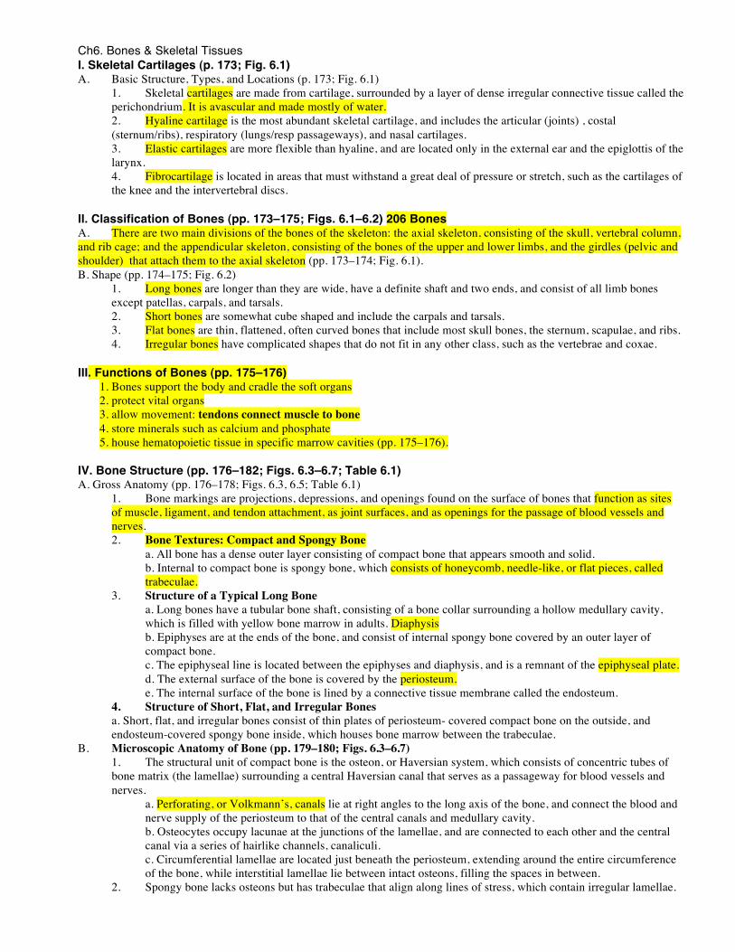
Ch6. Bones & Skeletal Tissues I. Skeletal Cartilages (p. 173; Fig. 6.1) A. Basic Structure, Types, and Locations (p. 173; Fig. 6.1) 1. Skeletal cartilages are made from cartilage, surrounded by a layer of dense irregular connective tissue called the perichondrium. It is avascular and made mostly of water. 2. Hyaline cartilage is the most abundant skeletal cartilage, and includes the articular (joints) , costal (sternum/ribs), respiratory (lungs/resp passageways), and nasal cartilages. 3. Elastic cartilages are more flexible than hyaline, and are located only in the external ear and the epiglottis of the larynx. 4. Fibrocartilage is located in areas that must withstand a great deal of pressure or stretch, such as the cartilages of the knee and the intervertebral discs. II. Classification of Bones (pp. 173–175; Figs. 6.1–6.2) 206 Bones A. There are two main divisions of the bones of the skeleton: the axial skeleton, consisting of the skull, vertebral column, and rib cage; and the appendicular skeleton, consisting of the bones of the upper and lower limbs, and the girdles (pelvic and shoulder) that attach them to the axial skeleton (pp. 173–174; Fig. 6.1). B. Shape (pp. 174–175; Fig. 6.2) 1. Long bones are longer than they are wide, have a definite shaft and two ends, and consist of all limb bones except patellas, carpals, and tarsals. 2. Short bones are somewhat cube shaped and include the carpals and tarsals. 3. Flat bones are thin, flattened, often curved bones that include most skull bones, the sternum, scapulae, and ribs. 4. Irregular bones have complicated shapes that do not fit in any other class, such as the vertebrae and coxae. III. Functions of Bones (pp. 175–176) 1. Bones support the body and cradle the soft organs 2. protect vital organs 3. allow movement: tendons connect muscle to bone 4. store minerals such as calcium and phosphate 5. house hematopoietic tissue in specific marrow cavities (pp. 175–176). IV. Bone Structure (pp. 176–182; Figs. 6.3–6.7; Table 6.1) A. Gross Anatomy (pp. 176–178; Figs. 6.3, 6.5; Table 6.1) 1. Bone markings are projections, depressions, and openings found on the surface of bones that function as sites of muscle, ligament, and tendon attachment, as joint surfaces, and as openings for the passage of blood vessels and nerves. 2. Bone Textures: Compact and Spongy Bone a. All bone has a dense outer layer consisting of compact bone that appears smooth and solid. b. Internal to compact bone is spongy bone, which consists of honeycomb, needle-like, or flat pieces, called trabeculae. 3. Structure of a Typical Long Bone a. Long bones have a tubular bone shaft, consisting of a bone collar surrounding a hollow medullary cavity, which is filled with yellow bone marrow in adults. Diaphysis b. Epiphyses are at the ends of the bone, and consist of internal spongy bone covered by an outer layer of compact bone. c. The epiphyseal line is located between the epiphyses and diaphysis, and is a remnant of the epiphyseal plate. d. The external surface of the bone is covered by the periosteum. e. The internal surface of the bone is lined by a connective tissue membrane called the endosteum. 4. Structure of Short, Flat, and Irregular Bones a. Short, flat, and irregular bones consist of thin plates of periosteum- covered compact bone on the outside, and endosteum-covered spongy bone inside, which houses bone marrow between the trabeculae. B. Microscopic Anatomy of Bone (pp. 179–180; Figs. 6.3–6.7) 1. The structural unit of compact bone is the osteon, or Haversian system, which consists of concentric tubes of bone matrix (the lamellae) surrounding a central Haversian canal that serves as a passageway for blood vessels and nerves. a. Perforating, or Volkmann’s, canals lie at right angles to the long axis of the bone, and connect the blood and nerve supply of the periosteum to that of the central canals and medullary cavity. b. Osteocytes occupy lacunae at the junctions of the lamellae, and are connected to each other and the central canal via a series of hairlike channels, canaliculi. c. Circumferential lamellae are located just beneath the periosteum, extending around the entire circumference of the bone, while interstitial lamellae lie between intact osteons, filling the spaces in between. 2. Spongy bone lacks osteons but has trabeculae that align along lines of stress, which contain irregular lamellae.
Transcript of Ch6. Bones & Skeletal Tissues I. Skeletal Cartilages...
Ch6. Bones & Skeletal Tissues I. Skeletal Cartilages (p. 173; Fig. 6.1) A. Basic Structure, Types, and Locations (p. 173; Fig. 6.1)
1. Skeletal cartilages are made from cartilage, surrounded by a layer of dense irregular connective tissue called the perichondrium. It is avascular and made mostly of water. 2. Hyaline cartilage is the most abundant skeletal cartilage, and includes the articular (joints) , costal (sternum/ribs), respiratory (lungs/resp passageways), and nasal cartilages. 3. Elastic cartilages are more flexible than hyaline, and are located only in the external ear and the epiglottis of the larynx. 4. Fibrocartilage is located in areas that must withstand a great deal of pressure or stretch, such as the cartilages of the knee and the intervertebral discs.
II. Classification of Bones (pp. 173–175; Figs. 6.1–6.2) 206 Bones A. There are two main divisions of the bones of the skeleton: the axial skeleton, consisting of the skull, vertebral column, and rib cage; and the appendicular skeleton, consisting of the bones of the upper and lower limbs, and the girdles (pelvic and shoulder) that attach them to the axial skeleton (pp. 173–174; Fig. 6.1). B. Shape (pp. 174–175; Fig. 6.2)
1. Long bones are longer than they are wide, have a definite shaft and two ends, and consist of all limb bones except patellas, carpals, and tarsals. 2. Short bones are somewhat cube shaped and include the carpals and tarsals. 3. Flat bones are thin, flattened, often curved bones that include most skull bones, the sternum, scapulae, and ribs. 4. Irregular bones have complicated shapes that do not fit in any other class, such as the vertebrae and coxae.
III. Functions of Bones (pp. 175–176)
1. Bones support the body and cradle the soft organs 2. protect vital organs 3. allow movement: tendons connect muscle to bone 4. store minerals such as calcium and phosphate 5. house hematopoietic tissue in specific marrow cavities (pp. 175–176).
IV. Bone Structure (pp. 176–182; Figs. 6.3–6.7; Table 6.1) A. Gross Anatomy (pp. 176–178; Figs. 6.3, 6.5; Table 6.1)
1. Bone markings are projections, depressions, and openings found on the surface of bones that function as sites of muscle, ligament, and tendon attachment, as joint surfaces, and as openings for the passage of blood vessels and nerves. 2. Bone Textures: Compact and Spongy Bone
a. All bone has a dense outer layer consisting of compact bone that appears smooth and solid. b. Internal to compact bone is spongy bone, which consists of honeycomb, needle-like, or flat pieces, called trabeculae.
3. Structure of a Typical Long Bone a. Long bones have a tubular bone shaft, consisting of a bone collar surrounding a hollow medullary cavity, which is filled with yellow bone marrow in adults. Diaphysis b. Epiphyses are at the ends of the bone, and consist of internal spongy bone covered by an outer layer of compact bone. c. The epiphyseal line is located between the epiphyses and diaphysis, and is a remnant of the epiphyseal plate. d. The external surface of the bone is covered by the periosteum. e. The internal surface of the bone is lined by a connective tissue membrane called the endosteum.
4. Structure of Short, Flat, and Irregular Bones a. Short, flat, and irregular bones consist of thin plates of periosteum- covered compact bone on the outside, and endosteum-covered spongy bone inside, which houses bone marrow between the trabeculae.
B. Microscopic Anatomy of Bone (pp. 179–180; Figs. 6.3–6.7) 1. The structural unit of compact bone is the osteon, or Haversian system, which consists of concentric tubes of bone matrix (the lamellae) surrounding a central Haversian canal that serves as a passageway for blood vessels and nerves.
a. Perforating, or Volkmann’s, canals lie at right angles to the long axis of the bone, and connect the blood and nerve supply of the periosteum to that of the central canals and medullary cavity. b. Osteocytes occupy lacunae at the junctions of the lamellae, and are connected to each other and the central canal via a series of hairlike channels, canaliculi. c. Circumferential lamellae are located just beneath the periosteum, extending around the entire circumference of the bone, while interstitial lamellae lie between intact osteons, filling the spaces in between.
2. Spongy bone lacks osteons but has trabeculae that align along lines of stress, which contain irregular lamellae.
C. Chemical Composition of Bone (p. 180) 1. Organic components of bone include cells (osteoblasts bone destroying, osteocytes mature bone cells, and osteoclasts bone forming) and osteoid (ground substance and collagen fibers), which contribute to the flexibility and tensile strength of bone. 2. Inorganic components make up 65% of bone by mass, and consist of hydroxyapatite, a mineral salt that is largely calcium phosphate, which accounts for the hardness and compression resistance of bone.
VI. Bone Homeostasis: Repair (pp. 185–190; Figs. 6.11–6.15; Table 6.2) B. Bone Repair (pp. 188–190; Fig. 6.15; Table 6.2)
1. Fractures are breaks in bones, and are classified by: the position of the bone ends after fracture, completeness of break, orientation of the break relative to the long axis of the bone, Closed (simple) fracture – break that does not penetrate the skin and Open (compound) fracture whether the bone ends penetrate the skin. Comminuted, Compression, Greenstick, Spiral, Epiphyseal and Depressed 2. Repair of fractures involves four major stages: hematoma formation, fibro- cartilaginous callus formation, bony callus formation, and remodeling of the bony callus.
A. Rickets (p. 189) is inadequate mineralization of bones in children caused by insufficient calcium or vitamin D deficiency. B. Osteoporosis refers to a group of disorders in which the rate of bone resorption exceeds the rate of formation (pp. 189–191, Fig. 6.16).
1. Bones have normal bone matrix, but bone mass is reduced and the bones become more porous and lighter, increasing the likelihood of fractures. 2. Older women are especially vulnerable to osteoporosis, due to the decline in estrogen after menopause. 3. Other factors that contribute to osteoporosis include a petite body form, insufficient exercise or immobility, a diet poor in calcium and vitamin D, abnormal vitamin D receptors, smoking, and certain hormone-related conditions.
Ch.7: The Skeleton PART 1: THE AXIAL SKELETON I. The Skull (pp. 199–216; Figs. 7.1–7.15; Table 7.1) A. The skull consists of 22 cranial and facial bones that form the framework of the face, contain cavities for special sense organs, provide openings for air and food passage, secure the teeth, and anchor muscles of facial expression (p. 200). B. Except for the mandible, which is joined to the skull by a movable joint, most skull bones are flat bones joined by interlocking joints called sutures (p. 200). C. Overview of Skull Geography (p. 200)
2. The cavities of the skull include the cranial cavity (houses the brain), ear cavities, nasal cavity, and orbits (house the eyeballs). 3. The skull has about 85 named openings that provide passageways for the spinal cord, major blood vessels serving the brain, and the cranial nerves.
D. The cranium consists of eight strong, superiorly curved bones (pp. 199–209; Figs. 7.2–7.10). 1. The frontal bone articulates posteriorly with the parietal bones via the coronal suture 2. The parietal bones are two large, rectangular bones on the superior and lateral aspects of the skull 3. The occipital bone articulates with the parietal, temporal, and sphenoid bones, forming most of the posterior wall and base of the skull. a. The foramen magnum, a large opening through which the brain connects to the spinal cord, is located in the base of the occipital bone. 4. The temporal bones articulate with the parietal bones
E. Facial Bones (pp. 209–211; Fig. 7.11) 1. The mandible, or lower jawbone 2. The maxillary bones form the upper jaw and central portion of the face
F. The hyoid bone lies inferior to the mandible in the anterior neck. It is the only bone that does not articulate directly with any other bone (p. 211; Fig. 7.12). H. Paranasal sinuses are air-filled sinuses clustered around the nasal cavity that lighten the skull and enhance resonance of the voice (p. 216; Figs. 7.14–7.15). II. The Vertebral Column (pp. 216–221; Figs. 7.16–7.21; Table 7.2) A. General Characteristics (pp. 216–218; Figs. 7.16–7.17)
1. The vertebral column consists of 26 irregular bones, forming a flexible, curved structure extending from the skull to the pelvis that surrounds and protects the spinal cord. It provides attachment for ribs and muscles of the neck and back. 2. Divisions and Curvatures a. The vertebrae of the spine fall in five major divisions: seven cervical, twelve thoracic, five lumbar, five fused

vertebrae of the sacrum, and four fused vertebrae of the coccyx. 3. The major supporting ligaments of the spine are the anterior and posterior longitudinal ligaments, which run as continuous bands down the front and back surfaces of the spine. They support the spine and prevent hyperflexion and hyperextension. 4. Intervertebral discs are cushion-like pads that act as shock absorbers and allow the spine to flex & extend
C. Regional Vertebral Characteristics (pp. 219–223; Figs. 7.19–7.21; Table 7.2) 1. Cervical vertebrae are the smallest vertebrae a. The atlas has no body or spinous process. It articulates with the skull superiorly, and the second cervical vertebra, the axis, inferiorly. 2. Thoracic vertebrae have a roughly heart-shaped body, which bear two facets on each side for rib articulation 3. Lumbar vertebrae are large vertebrae that have kidney-shaped bodies 4. The sacrum is formed by five, fused vertebrae in adults, and articulates with the fifth lumbar vertebra superiorly, the coccyx inferiorly 5. The coccyx (tailbone) is a small bone consisting of four, fused vertebrae that articulate superiorly with the sacrum.
III. The Thoracic Cage (pp. 223–225; Figs. 7.22–7.23) A. The thoracic cage consists of the thoracic vertebrae dorsally, the ribs laterally, and the sternum and costal cartilages anteriorly. It forms a protective cage around the organs of the thoracic cavity, and provides support for the shoulder girdles and upper limbs (pp. 223–224; Fig. 7.22). B. Sternum (pp. 223–224; Fig. 7.22)
1. The sternum (breastbone) is a flat bone resulting from the fusion of three bones: the manubrium, body, and xiphoid process. (superior to inferior) 2. The manubrium articulates with the clavicles and the first two pairs of ribs. The body articulates with the cartilages of ribs two through seven. The xiphoid process forms the inferior end, articulating only with the body.
C. Ribs (pp. 224–225; Figs. 7.22–7.23) 1. The sides of the thoracic cage are formed by twelve pairs of ribs that attach posteriorly to the thoracic vertebrae and curve inferiorly toward the anterior body surface. 2. The superior seven pairs of ribs are called true ribs. They attach directly to the sternum via individual costal cartilages. 3. The lower five pairs of ribs are called false ribs. They either attach indirectly to the sternum or lack a sternal attachment entirely. Floating ribs-attach posteriorly
F. Effects of old age on the skeleton (p. 244). 1. The intervertebral discs become thinner, less hydrated, and less elastic. 2. The thorax becomes more rigid, due to calcification of the costal cartilages. 3. All bones lose bone mass.
PART 2: THE APPENDICULAR SKELETON IV. The Pectoral (Shoulder) Girdle (pp. 225–228; Figs. 7.24–7.25; Table 7.3) A. The pectoral (shoulder) girdle consists of the clavicle, which joins the sternum anteriorly, and the scapula, which is attached to the posterior thorax and vertebrae via muscular attachments (pp. 225–226; Fig. 7.24). B. The clavicles (collarbones) extend horizontally across the thorax, articulating medially with the sternum(p. 226; Fig. 7.24). C. The scapulae (shoulder blades) are thin, flat bones that lie on the dorsal surface of the rib cage (pp. 226–228; Fig. 7.25). V. The Upper Limb (pp. 228–233; Figs. 7.26–7.28; Table 7.3) A. Arm (p. 228; Fig. 7.26)
1. The arm is the region extending from shoulder to elbow, and has one bone, the humerus. 2. The humerus is the largest, longest bone of the upper limb. It articulates with the scapula at the shoulder, and with the radius and ulna at the elbow.
B. Forearm (pp. 228–231; Fig. 7.27) 1. The forearm is the region between the elbow and wrist. It consists of two bones, the ulna and the radius. 2. The ulna forms the elbow joint with the humerus 3. The radius articulates with the humerus and the ulna
C. Hand (pp. 231–233; Fig. 7.28) 1. The carpus (wrist) consists of eight short bones 2. The metacarpus (palm) consists of five small, long bones numbering one through five from thumb to little finger 3. There are 14 phalanges of the fingers: the thumb (pollex) is digit 1, and has two phalanges. The other fingers, numbered 2–5, have three phalanges each.
VI. The Pelvic (Hip) Girdle (pp. 233–237; Figs. 7.29–7.30; Tables 7.4–7.5) A. The pelvic girdle attaches the lower limbs to the axial skeleton. It is formed by a pair of coxal bones, each consisting of three separate bones: the ischium, ilium, and pubis, that are fused in adults (pp. 233–235; Fig. 7.29). E. Pelvic Structure and Childbearing (pp. 235–237; Table 7.4)
1. The female pelvis is modified for childbearing. It tends to be wider, shallower, lighter, and rounder than the male pelvis.
VII. The Lower Limb (pp. 237–241; Figs. 7.31–7.34; Table 7.5) A. Thigh (p. 237; Fig. 7.31)
1. The thigh is the region between the hip and knee. It has one bone, the femur. 2. The femur is the largest, longest, and strongest bone in the body. Since it is a long bone, the epiphyseal plates assure growth during childhood until 25 years. 3. The patella is a triangular sesamoid bone that articulates with the femur at the patellar surface.
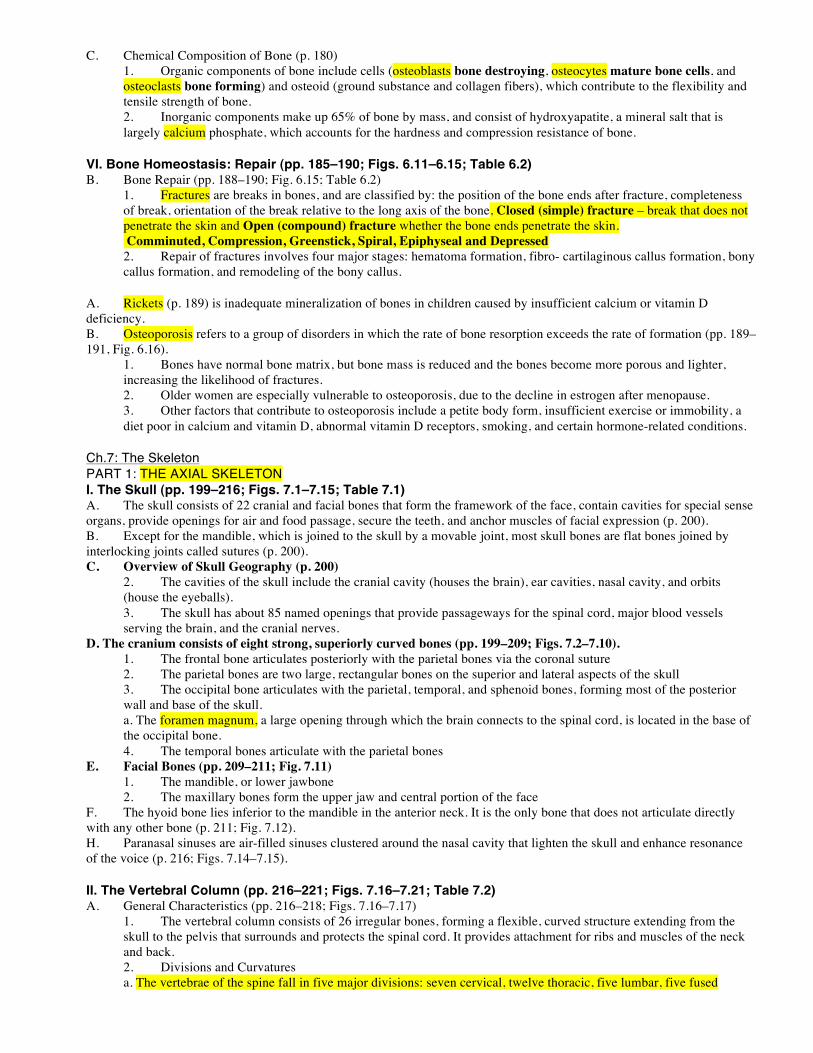
B. Leg(pp. 237–240; Fig. 7.32) 1. The leg is the region between the knee and ankle. It has two bones, the tibia and fibula. 2. The tibia is the weight-bearing bone of the leg. 3. The fibula is a sticklike, non-weight-bearing bone. Homeostatic Imbalance Know- Rickets, Osteroporosis, and Rheumatoid Arthritis
!
!!"!#$%&!'(!')*$'!$!+)$,'-)*.!!"#!$%&'()!*(+,(-./%0!-()12,3&.4,5(!)&2,&-!6/.371%/,('!,5(!8&.(!3.,&!7&'3,3&.#!!9#!:7(.!*3.,(-./%0!-()12,3&.!3'!'(21-()!8;!/!'1-<(-;!7-&2()1-(!3.=&%=3.<!73.'!&-!>3-('!
!!
/0120345467!618595:723!0;!80:2!5:<!=06:43!!
?3'(/'(! $/1'(! @5&!3,!/AA(2,'! B;67,&6'!CD$EFGB! 2/1'()!8;!%/2H!
&A!2/%2316!&-!I3,/!?#!
!)3'(/'(!3.!253%)-(.J!
8&>()!%(<'K!)(A&-63,3('!&A!,5(!7(%=3'K!'H1%%!/.)!-38!2/<(!/-(!2&66&.#!F7375;'(/%!7%/,('!/-(!.&,!2/%23A3()!/.)!>3)(.K!(.)'!&A!%&.<!8&.('!8(2&6(!(.%/-<()#!!
:BGF:L:C:BDB! 8&.(!-('&-7,3&.!*&',(&2%/',0!3'!A/',(-!,5/.!8&.(!)(7&'3,!*&',(&8%/',0!
!)3'(/'(!3.!,5(!(%)(-%;J!
M&.('!8(2&6(!A-/<3%(!8;!8(3.<!7&-&1'!/.)!%3<5,#!B7&.<;!8&.(!3.!,5(!'73.(!6&',!=1%.(-/8%(K!81,!/%'&!/AA(2,'!,5(!(.,3-(!'H(%(,&.#!
:BGF:!NCGOCDGDB!
D.A%/6()!&-!)/6/<(!,&!,5(!P&3.,'!
)3'(/'(!3.!,5(!(%)(-%;J!6&',!2&66&.K!&221--3.<!3.!6&-(!>&6(.!,5/.!6(.!
Q>(/-!/.)!,(/-R!/-,5#J!/-,321%/-!2/-,3%/<(!8-(/H!)&>.!/.)!3'!)(',-&;()!A/',(-!,5/.!-(7%/2()!
COFST#!NCGOCDGDB!
! N<('!UVWXVJ!/1,&3661.(!! Y&3.,!,(.)(-.(''!/.)!',3AA.(''!3.!,5(!';.&=3/%!P&3.,'!
Z:SG[!NCGOCDGDB!
M%&&)!%(=(%'!5/=(!1-32!/23)!*1-3.(0!>5325!A&-6!3.,&!2-;',/%'!3.!,5(!P&3.,'!
$&66&.!3.!6(.!&=(-!>&6(.!8(2/1'(!,5(;!5/=(!53<5(-!8%&&)!%(=(%'!&A!1-32!/23)J!3.5(-3,()!)3'(/'(!
S.,-(/,()!<&1,!2/.!8(!)(',-12,3=(J!/-,321%/,3.<!8&.(!(.)'!A1'(!/.)!366&83%3\(!,5(!P&3.,#!G-(/,6(.,!4!6()'!7-(=(.,!<&1,!/,,/2H'#!T1',!)-3.H!>/,(-!/.)!/=&3)!/%2&5&%!(+2(''!
!!
Chapter 6 Bones and Skeletal Tissues 189
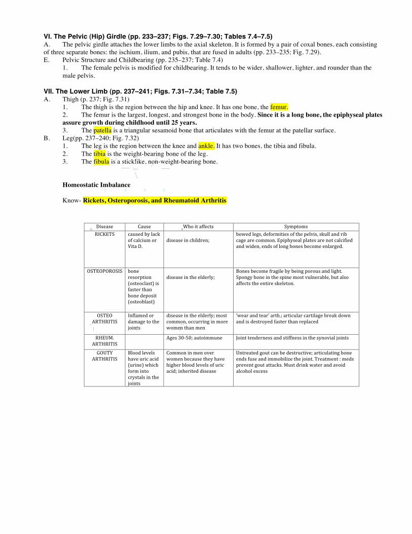
6begin cleaning up the debris. Meanwhile, fibroblasts andosteoblasts invade the fracture site from the nearby perios-teum and endosteum and begin reconstructing the bone.The fibroblasts produce collagen fibers that span the breakand connect the broken bone ends, and some differentiateinto chondroblasts that secrete cartilage matrix. Within thismass of repair tissue, osteoblasts begin forming spongybone, but those farthest from the capillary supply secretean externally bulging cartilaginous matrix that later calci-fies. This entire mass of repair tissue, now called thefibrocartilaginous callus, splints the broken bone.
Bony callus forms. Within a week, new bone trabeculaebegin to appear in the fibrocartilaginous callus and graduallyconvert it to a bony (hard) callus of spongy bone. Bonycallus formation continues until a firm union is formedabout two months later.
Bone remodeling occurs. Beginning during bony callus for-mation and continuing for several months after, the bonycallus is remodeled. The excess material on the diaphysisexterior and within the medullary cavity is removed, andcompact bone is laid down to reconstruct the shaft walls.The final structure of the remodeled area resembles that ofthe original unbroken bony region because it responds tothe same set of mechanical stressors.
C H E C K Y O U R U N D E R S TA N D I N G
19. If osteoclasts in a long bone are more active thanosteoblasts, what change in bone mass is likely?
20. Which stimulus—PTH (a hormone) or mechanical forces act-ing on the skeleton—is more important in maintaining ho-meostatic blood calcium levels?
21. How does an open fracture differ from a closed fracture?22. How do bone growth and bone remodeling differ?
For answers, see Appendix G.
4
3
Homeostatic Imbalances of Bone! Contrast the disorders of bone remodeling seen in osteo-
porosis, osteomalacia, and Paget’s disease.
Imbalances between bone deposit and bone resorption underlienearly every disease that affects the adult skeleton.
Osteomalacia and RicketsOsteomalacia (os!te-o-mah-la!she-ah; “soft bones”) includes anumber of disorders in which the bones are inadequately min-eralized. Osteoid is produced, but calcium salts are not de-posited, so bones soften and weaken. The main symptom is painwhen weight is put on the affected bones.
Rickets is the analogous disease in children. Because youngbones are still growing rapidly, rickets is much more severe thanadult osteomalacia. Bowed legs and deformities of the pelvis,skull, and rib cage are common. Because the epiphyseal platescannot be calcified, they continue to widen, and the ends of longbones become visibly enlarged and abnormally long.
Osteomalacia and rickets are caused by insufficient calciumin the diet or by a vitamin D deficiency. For this reason, drink-ing vitamin D–fortified milk and exposing the skin to sunlight(which spurs the body to form vitamin D) usually cure thesedisorders. Although the seeming elimination of rickets in theUnited States has been heralded as a public health success, rick-ets still rears its head in isolated situations. For example, if amother who breast-feeds her infant becomes vitamin D defi-cient because of dreary winter weather, the infant too will bevitamin D deficient and will develop rickets.
OsteoporosisFor most of us, the phrase “bone problems of the elderly”brings to mind the stereotype of a victim of osteoporosis—ahunched-over old woman shuffling behind her walker.
1 A hematoma forms. 2 Fibrocartilaginous callus forms.
3 Bony callus forms. 4 Bone remodeling occurs.
Figure 6.15 Stages in the healing of a bone fracture.