Ch04 hemorrhage and shock
149
Paramedic Care: Death by PowerPoint Part 2 Shock !
-
Upload
djorgenmorris -
Category
Healthcare
-
view
490 -
download
1
Transcript of Ch04 hemorrhage and shock
Paramedic Care:Death by PowerPoint Part 2
Shock !

EMT classshock lecture:
“Air goes in and out… blood goes around and around…. Anything that gets in the way is a big problem….”

Hemorrhage
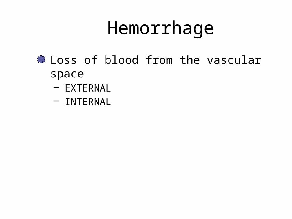
Hemorrhage
Loss of blood from the vascular space – EXTERNAL– INTERNAL
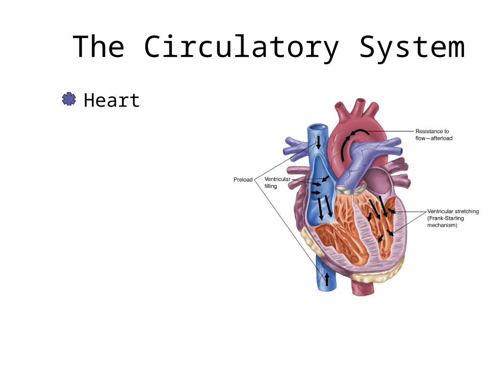
The Circulatory System
Heart
CARDIAC OUTPUT
STROKE VOLUME X HEART RATE

WHAT IS STROKE VOLUME?

HeartStroke volume is the amount of blood pumped from the ventricle with each contraction.

WHAT IS STROKE VOLUME DEPENDENT ON?

PRELOADAFTERLOADCONTRACTILITYThe normal heart, at rest, beats about 70 times per minute and moves about 70 mL of blood with each beat.

The Circulatory System
The Vascular System– Consists of arteries, capillaries, and veins

ArteriesConsist of three distinct tissue layers – Tunica adventicia– Tunica media– Tunica intima

CapillariesCapillary flow provides essential nutrients and oxygen and removes waste products. – Only one-cell thick
Hydrostatic pressure pushes the plasma into the interstitial space.– Filtration

VeinsCollect blood and return it to the heart Contains the vast majority of the total blood volume Able to constrict in early stages of hemorhage
The Circulatory System
Progressive reduction in pressure as blood is moved through the circulatory system

BloodBlood is the tissue that circulates within the cardiovascular system – A mixture of cells, proteins, water, and other
suspended elements Blood Volume– Average adult male has a blood volume of 7% of
total body weight– Average adult female has a blood volume of 6.5%
of body weight– Normal adult blood volume is 4.5–5 L
Remains fairly constant in the healthy body

Blood Components
Erythrocytes: 45%– Hemoglobin– Hematocrit
Miscellaneous blood products: <1%– Platelets– Leukocytes
Plasma: 54%

Blood Components
Erythrocytes (RBC’s)– The major blood
Contains hemoglobinA molecule to which oxygen attaches
– Efficient transporter of oxygen from the lungs to body cells.

Blood Components
Plasma– Approximately 92% water– Circulates salts, minerals, sugars, fats, and
proteins throughout the body
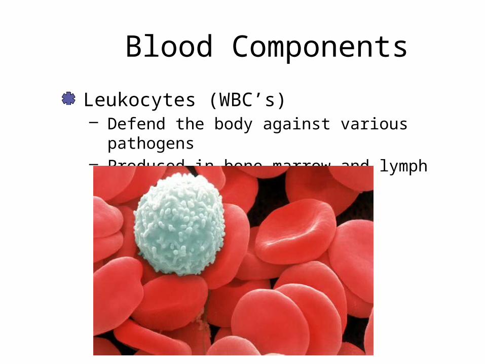
Blood Components
Leukocytes (WBC’s)– Defend the body against various pathogens– Produced in bone marrow and lymph glands

Blood Components
Platelets– Part of the body’s defense mechanism– Formed in red bone marrow– Work by swelling and adhering together to form
sticky plugs (initiating the clotting phenomenon)

Hemmorhage Classification

ClottingThree-Step Process– Vascular phase
Vasoconstriction– Platelet phase
– CoagulationRelease of enzymesNormal coagulation in 7–10 minutes

Clotting
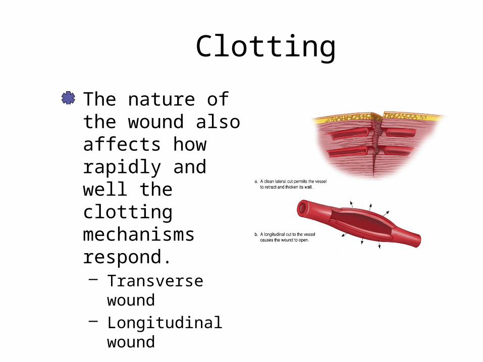
Clotting
The nature of the wound also affects how rapidly and well the clotting mechanisms respond. – Transverse wound– Longitudinal wound

What affects clotting?
Blood thinnersBody temperatureMovementAggressive fluid therapy

Hemorrhage ControlExternal Hemorrhage – External hemorrhage is relatively easy to
recognize and control. Bleeding from small vessels can often be controlled by firmly bandaging a dressing in place. Fingertip pressure
– With careful application of direct pressure you can halt virtually all hemorrhage.

Hemorrhage Control

Hemorrhage Control
External Hemorrhage (cont.)– If you consider using a tourniquet, be extremely
cautious. The need for a tourniquet is rare.
– In the absence of perfusion, lactic acid, potassium, and other anaerobic metabolites accumulate
Will be released into the circulation when released– Use a wide-band if considering use
Internal Hemorrhage
Can result from:– Blunt or penetrating trauma– Acute or chronic medical illnesses
Internal bleeding that can cause hemodynamic instability usually occurs in one of four body cavities:– Chest– Abdomen– Pelvis– Retroperitoneum

Internal HemorrhageSigns and symptoms that suggest significant internal hemorrhage include:– Bright red blood from mouth, rectum, or other
orifice– Coffee-ground appearance of vomitus– Melena (black, tarry stools)– Orthostatic hypotension
Chronic hemorrhage may result in anemia

Internal Hemorrhage ControlGeneral Management– Immobilization,
stabilization, elevation
– Epistaxis: Nose Bleed
Causes: trauma, hypertensionTreatment: lean forward, pinch nostrils

How Much Blood Loss?
Humerus 500-750 mLFemur up to 1500 mLPelvis up to 2000 mL

Know These Numbers !
152535

Stages of Hemorrhage
Stage 1 – 15% loss of CBV (circulating blood volume)
70 kg pt = 500–750 mL– Compensation
VasoconstrictionNormal BP, pulse pressure, respirationsSlight elevation of pulseRelease of catecholamines
EpinephrineNorepinephrine
Anxiety, slightly pale and clammy skin

Stages of HemorrhageStage 2 – 15–30% loss of CBV
750–1500 mL– Early decompensation
Unable to maintain BPTachycardia and tachypnea
– Decreased pulse strength– Narrowing pulse pressure– Significant catecholamine release
Increase PVRCool, clammy skin and thirstIncreased anxiety and agitationNormal renal output

Stages of HemorrhageStage 3 – 30–40% loss of CBV
1500–2000 mL– Late decompensation (early irreversible)– Classic Shock
Weak, thready, rapid pulseNarrowing pulse pressure
TachypneaAnxiety, restlessnessDecreased LOC and AMSPale, cool, and clammy skin

Stages of Hemorrhage
Stage 4 – >40% CBV loss
>1750 mL– Irreversible
Pulse: Barely palpableRespiration: Rapid, shallow, and ineffectiveLOC: Lethargic, confused, unresponsiveGU: CeasesSkin: Cool, clammy, and very paleUnlikely survival

Different People, Different Blood Volumes
PregnantAthleticObeseElderlyChildren

Geriatric Patients
Discussed in another chapterOld people take beta blockers and other medications that slow heart rate. May not show typical signs of shock.Break bones easilyHave curvy spines Can’t hear very well

Pediatric Trauma
Discussed in another presentation
PALS CONCEPT:70 + (2 X Patient’s Age) is cutoff for hypotension in a pediatric patientCompensate well, then decline rapidly – (don’t circle the drain like adults)

Stages of Hemorrhage
Concomitant Factors – Pre-existing condition– Rate of blood loss– Patient Types
Pregnant>50% greater blood volume than normalFetal circulation impaired when mother compensating
AthletesGreater fluid and cardiac capacity
ObeseCBV is based on IDEAL weight (less CBV)

Stages of Hemorrhage
Concomitant Factors – Children
CBV 8–9% of body weightPoor compensatory mechanisms
– ElderlyDecreased CBVMedications
BPAnticoagulants

Hemorrhage Assessment
Assessment of the hemorrhage patient is directed at identifying the source of the hemorrhage. – Halt any serious and controllable loss.
Examine the nature of the injury.
Hemorrhage Assessment
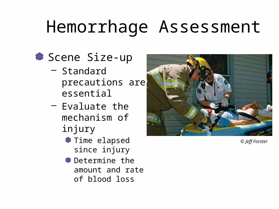
Scene Size-up– Standard
precautions are essential
– Evaluate the mechanism of injury
Time elapsed since injuryDetermine the amount and rate of blood loss
© Jeff Forster

Hemorrhage Assessment
Primary Assessment– General Impression
Obvious Bleeding– Mental Status– CABC– Interventions
Manage as you goO2
Bleeding controlShockBLS before ALS!

Hemorrhage Assessment
Secondary Assessment– Rapid Trauma Assessment
Full head to toeConsider air medical if stage 2+ blood loss
– Focused Physical ExamGuided by c/c
– Vitals, SAMPLE, and OPQRST– Additional Assessment
Search for signs of internal bleedingBleeding from body orifice, melena, hematochezia
Orthostatic hypotension

Hemorrhage AssessmentOngoing Assessment– Reassess vitals and mental status:
Q 5 min: UNSTABLE patientsQ 15 min: STABLE patients
– Reassess interventions:OxygenETIVMedication actions
– Trending: improvement vs. deteriorationPulse oximetryEnd-tidal CO2 levels

Hemorrhage Management
Assure that the airway is patent and breathing is adequate. – Maintain the airway and provide the necessary
ventilatory support.– Administer high-flow oxygen.
Assure that the patient has a palpable carotid pulse.Care for serious (arterial and heavy venous) hemorrhage, immediately after you correct airway and breathing problems.
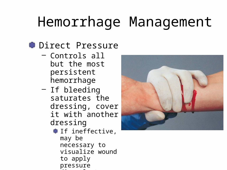
Hemorrhage ManagementDirect Pressure– Controls all but the
most persistent hemorrhage
– If bleeding saturates the dressing, cover it with another dressing
If ineffective, may be necessary to visualize wound to apply pressure directly to site
Hemorrhage Management
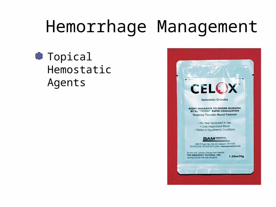
Topical Hemostatic Agents
Hemorrhage Management
ElevationPressure Point

Hemorrhage Management
Tourniquet

Bleeding Assessment
Click here to view an animation on bleeding assessment.

Specific Wound Considerations
Head Wounds– Presentation
Severe bleedingSkull fracture
– ManagementGentle direct pressureFluid drainage from ears and nose
DO NOT packCover and bandage loosely
Neck Wounds– Presentation
Large vessel can entrap air
– ManagementConsider direct digital pressureOcclusive dressing

Specific Wound Considerations
Gaping Wounds– Presentation
Multiple sitesGaping prevents uniform pressure
– ManagementBulky dressing
Trauma dressingSterile, non-adherent surface to woundCompression dressing
Crush Injury– Presentation
Difficult to locate source of bleedingNormal hemorrhage control mechanism non-functional
– ManagementConsider an air-splint and pressure dressingConsider tourniquet

Transport Considerations
Consider rapid transport if:– Suspected serious blood loss– Suspected serious internal bleeding– Decompensating shock– If in doubt, rapid transport indicated
Other Considerations– Sympathetic response– Anxiety

Shock!

ShockInadequate tissue perfusion– Transitional stage between normal life, called
homeostasis, and death – Can result from a variety of disease states and
injuries– Can affect the entire organism, or it can occur at a
tissue or cellular level
Biology 101
Cellular Metabolism– Glycolysis– Kreb’s Cycle– Electron Transport Chain

Biology for Paramedics
Glycolysis– Anaerobic (no Oxygen needed)– Produces pyruvic acid and 2 ATPs

Don’t Degrade My Pyruvate

Cellular MetabolismATP is a product of the cellular breakdown of glucose – Breakdown occurs in three steps
Glycolysis Does not require oxygenProduces pyruvic acid and 2 ATP’s
Kreb’s CycleRequires oxygenConverts pyruvic acid into water, carbon dioxide and 2 ATP’s
Electron transport chain Occurs in mitochondriaResults in the production of 32 ATPS
Biology 101.b
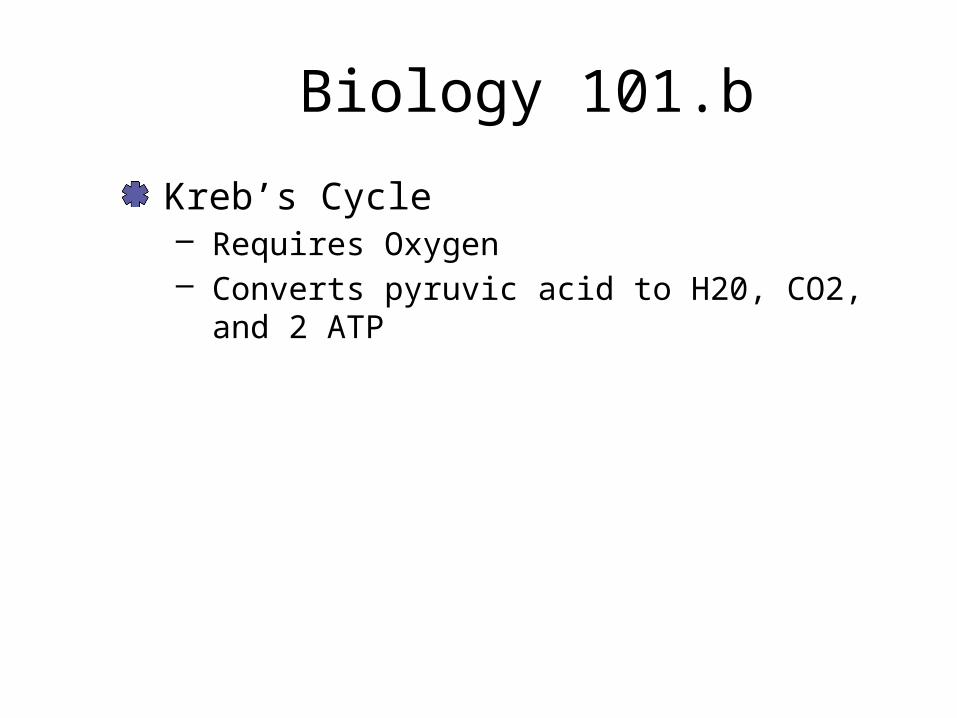
Kreb’s Cycle– Requires Oxygen– Converts pyruvic acid to H20, CO2, and 2 ATP


Electron Transport Chain(ITS COMPLICATED)

Electron Transport Chain
32 ATP

WHY IS AEROBIC RESPIRATION SO GOOD?

Answer:
More ATPNo lactic acid

Oxygen Transport
Oxygen must uptake on the hemoglobin molecule. – Efficiently carries 97% of the oxygen– Remaining 3% dissolves in plasma
The cardiovascular system then moves the red blood cells from the pulmonary system, through the heart, through the arterial system and into the tissues.

Oxygen Transport
In the capillaries oxygen diffuses across the capillary wall, into the interstitial fluid and then to the cell.– Internal respiration

Cellular Metabolism
The cardiovascular system is also responsible to help maintain other elements of the homeostatic environment – Removal of CO2 and water– Heat regulation– Provides the glucose necessary for the cellular
metabolism

Digestion, Filtration, Hormone Production, Excretion
The digestive system absorbs carbohydrates and lipids (fats), moving them through the portal system to the liver for processing.The pancreas regulates blood glucose.– Glucagon increases blood glucose– Insulin decreases blood glucose
Digestion, Filtration, Hormone Production, Excretion
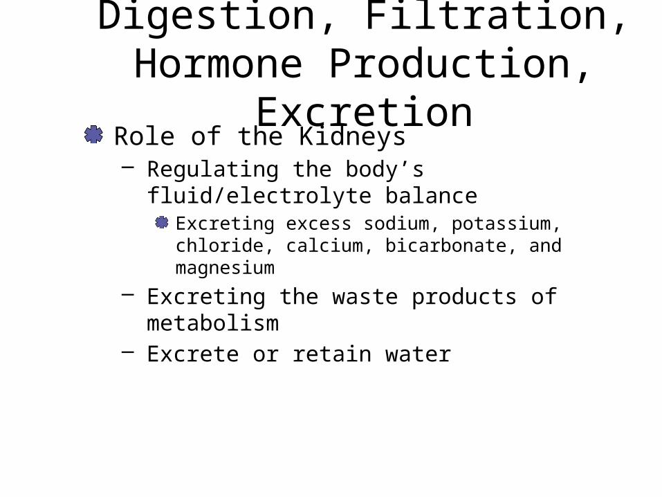
Role of the Kidneys– Regulating the body’s fluid/electrolyte balance
Excreting excess sodium, potassium, chloride, calcium, bicarbonate, and magnesium
– Excreting the waste products of metabolism – Excrete or retain water

Circulation
The cardiovascular system – Responsible for assuring that the necessary
materials travel to and from the body’s cells – Cardiac output
Preload, cardiac contractility, and afterload Systolic blood pressure is most indicative of the strength and volume of cardiac output Lowest pressure in the arteries is the diastolic blood pressure

Circulation

CirculationMicrocirculation– Blood flow in the
arterioles, capillaries, and venules
– Sphincter functioning

Microcirculation
Venules and veins serve as collecting channels and storage vessels (capacitance)Normally contain 70% of the blood volumeMuscular movement aids in blood return to the heart

Circulation
Respiration also facilitates blood return to the heart – Changes in pressure draws blood towards the
heartThoracoabdominal pump
In states of hypovolemia, blood return to the heart is diminished– Reduces cardiac output, arterial blood pressure,
and the body’s ability to direct blood flow to critical organs

Cardiovascular System Regulation
The human body is controlled by the autonomic branch of the nervous system. – Parasympathetic branch– Sympathetic branch
These two systems act in balance Many sympathetic nervous system activities are aimed at defending the organism. – These mechanisms may be detrimental in shock
states

Cardiovascular System Regulation
Parasympathetic Nervous SystemFEED AND BREATHE
Decrease – Heart rate– Strength of contractions– Blood pressure
Increase– Digestive system– Kidneys
Cardiovascular System Regulation
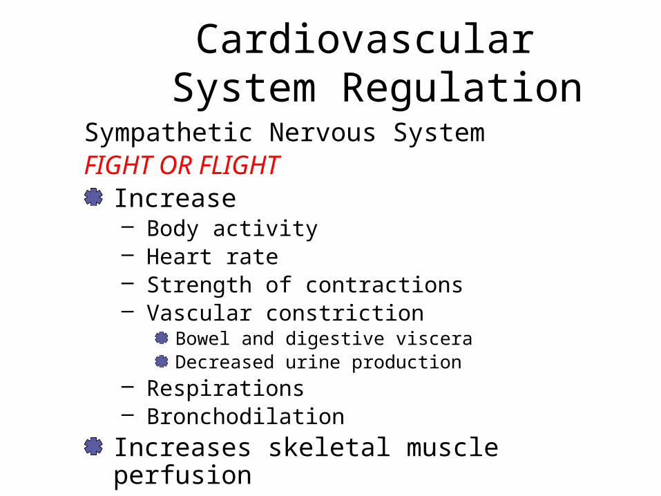
Sympathetic Nervous SystemFIGHT OR FLIGHT
Increase– Body activity– Heart rate– Strength of contractions– Vascular constriction
Bowel and digestive visceraDecreased urine production
– Respirations– Bronchodilation
Increases skeletal muscle perfusion

Cardiovascular System Regulation
A system of receptors, autonomic centers, and nervous and hormonal interventions maintains control over the cardiovascular system

Baroreceptors
Aortic ArchAtriaCarotid SinusMonitors pressure

Chemoreceptors
Aortic ArchCarotid SinusBrain (monitoring CSF)Monitors CO2 (and Oxygen) levels
Cardiovascular System Regulation
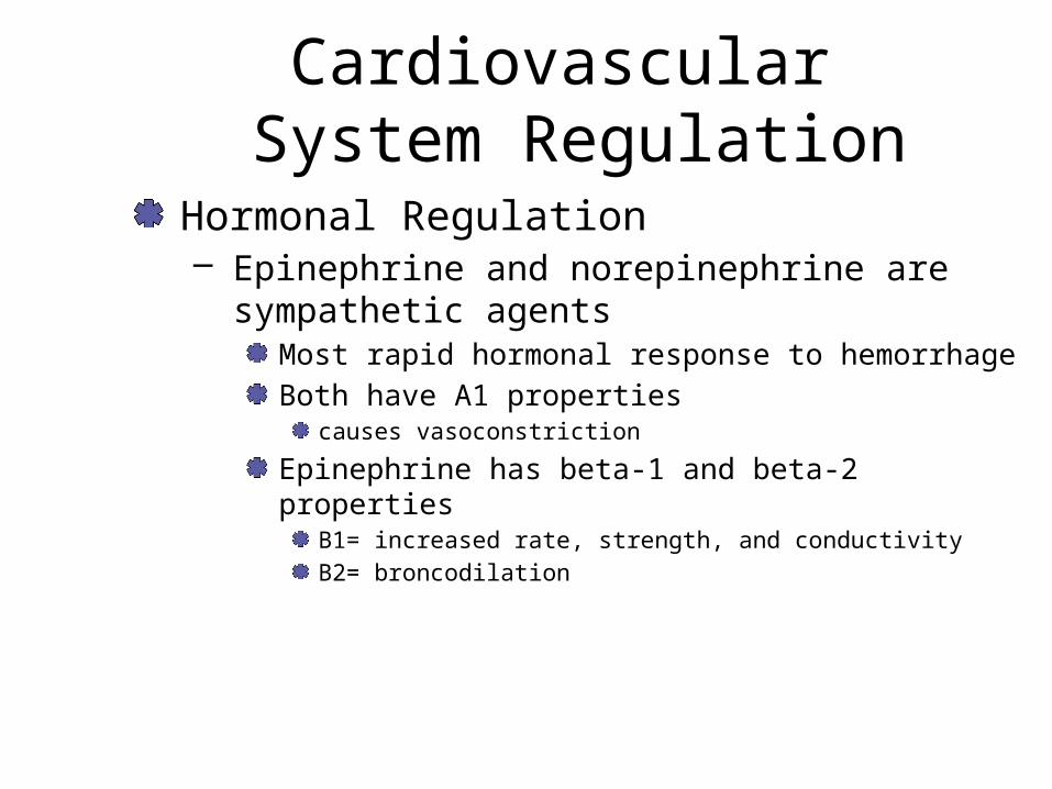
Hormonal Regulation– Epinephrine and norepinephrine are sympathetic
agents Most rapid hormonal response to hemorrhage Both have A1 properties
causes vasoconstriction
Epinephrine has beta-1 and beta-2 propertiesB1= increased rate, strength, and conductivityB2= broncodilation
Hormonal Regulation
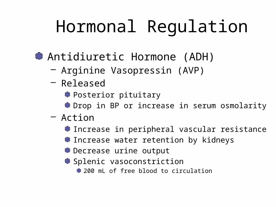
Antidiuretic Hormone (ADH)– Arginine Vasopressin (AVP)– Released
Posterior pituitaryDrop in BP or increase in serum osmolarity
– ActionIncrease in peripheral vascular resistanceIncrease water retention by kidneysDecrease urine outputSplenic vasoconstriction
200 mL of free blood to circulation

Hormonal Regulation
Angiotensin– Released
Primary chemical from kidneysStimulus is lowered BP and decreased perfusion
– ActionConverted from renin into angiotensin I
Modified in lungs to angiotensin IIPotent systemic vasoconstrictorCauses release of ADH, aldosterone, and epinephrine


Hormonal Regulation
Aldosterone– Release
Adrenal cortexStimulated by angiotensin II
– ActionMaintain kidney ion balanceRetention of sodium and waterReduce insensible fluid loss
Hormonal Regulation
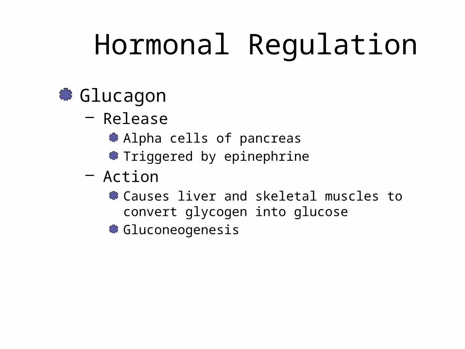
Glucagon– Release
Alpha cells of pancreasTriggered by epinephrine
– ActionCauses liver and skeletal muscles to convert glycogen into glucoseGluconeogenesis

Hormonal Regulation
Insulin– Release
Beta cells of pancreas
– ActionFacilitates transport of glucose across cell membrane
Erythropoietin– Release
KidneysHypoperfusion or hypoxia
– ActionIncreases production and maturation of RBCs in the bone marrow

Hormonal Regulation
Adrenocorticotropic hormone – Stimulates the release of glucocorticoids from the
adrenal cortex Increases glucose productionReduces the body’s inflammation responseProlongs clotting time, wound healing, and infection fighting processes
Growth hormone – Promotes the uptake of glucose and amino acids
in the muscle cells

The Body’s Responseto Blood Loss
As stroke volume decreases, cardiac output decreases resulting in decreased systolic BP– Carotid and aortic baroreceptors recognize this
decrease in blood pressure Stimulate the cardiovascular center of the medulla oblongata
Mechanisms compensate for small blood losses
The Body’s Responseto Blood Loss
Cellular Ischemia– Constriction of arterioles means that less and less
blood is directed to the noncritical organs Results in hypoxia
– Anaerobic metabolism resultsFollowed by ischemia

Cellular Ischemia
If blood loss continues, waste products accumulate and blood becomes acidic.– Increase in depth and rate of respirations– Decreased LOC– Increased circulating catecholamines causes
anxiousness, restlessness, and possibly a combative patient
– Decreased myocardial oxygen supply

Cellular Ischemia
If the blood loss stops, the blood draws fluid from within the interstitial space – Up to 1 L per hour
Kidneys reduce urine output

The Body’s Responseto Blood Loss
Capillary Microcirculation– Sympathetic stimulation and reduced perfusion to
the kidneys, pancreas, and liver cause the release of hormones
Angiotensin II causes reduced blood flow– Perfusion is further limited to only those organs
most critical to life More cells begin to use anaerobic metabolism for energy = Increased acids
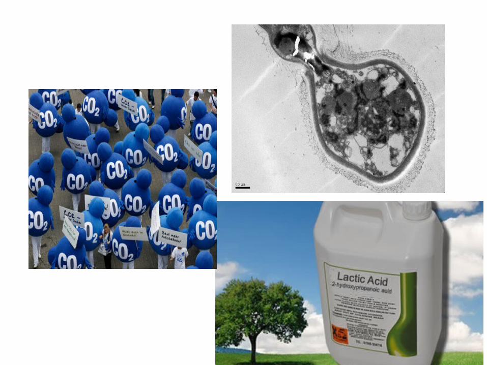
Capillary MicrocirculationThe build-up of lactic acid and carbon dioxide relaxes the precapillary sphincters Postcapillary sphincters remain closed Capillary and cell membranes begin to break down Red blood cells begin to clump together – Rouleaux

Rouleaux
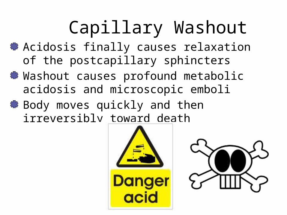
Capillary WashoutAcidosis finally causes relaxation of the postcapillary sphinctersWashout causes profound metabolic acidosis and microscopic emboli Body moves quickly and then irreversibly toward death

Stages of Shock
Three stages:– Compensated– Decompensated– Irreversible
Stages are progressively more serious

Stages of Shock
Compensated ShockSize of container is reduced– The body is capable of meeting its critical
metabolic needs through a series of progressive compensating actions.

Compensated
High pulse rateNarrowing pulse pressureVasoconstrictionTachypneaAir hungerThirstPale, ashen skinRestlessness

Stages of Shock
Decompensated Shock (Progressive)– Mechanisms that compensate for blood loss fail – Systolic BP drops significantly– Vital organs are no longer perfused– Patient displays a rapidly dropping level of
responsiveness
Stages of Shock
Irreversible Shock– The body’s cells die.– Cell membrane lyses.– Toxic chemicals released.– Aggressive resuscitation will be ineffective.– The longer a patient is in decompensated shock,
the more likely he has moved to irreversible shock.
Etiology of Shock
Shock can have many causesClassifications according to origin:– Hypovolemic, – distributive – obstructive – cardiogenic– respiratory

Hypovolemic Shock
Big containerLess volumeCauses– Bleeding– Dehydration– Third space issues

Distributive Shock
Distributive Shock– Mechanisms that interfere with the ability of the
vascular system to distribute the cardiac output – Causes
NeurogenicAnaphylacticSepsis

We will talk about this after spring break

Obstructive
Obstructive– Results from interference with the blood flowing
through the cardiovascular system – Causes
Tension pneumothoraxCardiac tamponadePulmonary emboli

Obstructive Shock
Tension pneumothoraxCardiac tamponadePulmonary emboli

Etiology of Shock
Cardiogenic Shock– Results from a problem with the cardiovascular pump – Causes
InfarctionDisturbances in the cardiac electrical systemFailure of the valves Cardiac rupture Reduced cardiac pumping action
– May present with the signs and symptoms of myocardial infarction or pulmonary edema

Etiology of Shock
Respiratory Shock– Occurs when the respiratory system is not able to
bring oxygen into the alveoli and remove carbon dioxide
– CausesFlail chestRespiratory muscle paralysisPneumothoraxPulmonary edemaTension pneumothorax

Respiratory Shock
Flail chestRespiratory muscle paralysisPneumothoraxPulmonary edemaTension pneumothorax

Etiology of ShockNeurogenic Shock– Results from an interruption in the communication
pathway between the central nervous system and the rest of the body
– CausesSpinal injury
Skin remains warm and dry above injury siteHead injury
Temporary or permanent– Body’s compensatory mechanisms are often
affected Tachycardia and increased diastolic are not present

Let’s Look at the #sType of Shock Heart Rate BPHemorrhagic/hypovolemicCardiogenic
Neurogenic
Anaphylactic
Warm septic shock
Late septic shock

Had Enough Yet?
Cardiac Output: Heart rate X stroke volumeMAP = 1/3 (2 X diastolic + systolic)SVR= MAP ÷ Cardiac Output
Type of Shock Cardiac Output SVRHemorrhagic/hypovolemic
Cardiogenic
Neurogenic
Anaphylactic
Warm septic shock
Late septic shock

Shock Assessment
You must be able to recognize shock as early as possible in your patient assessment. Search out the signs and symptoms of shock in each phase of the assessment process. Carefully monitor for the development or progression of shock.

The Lethal Triad
Acidosis Hypothermia
Coagulopathy
Death
Brohi, K, et al. J Trauma, 2003.

Shock Assessment
Scene Size-up– Analyze the forces that caused the trauma.
Possibility of both external and internal injury.– Look for mechanisms that might result in internal
chest, abdominal, or pelvic injuries. – Observe for external hemorrhage.
Shock Assessment
Initial Assessment– Determine the patient’s level of consciousness,
responsiveness, and orientation. – Assess the airway for patency and breathing for
adequacy.Administer high-concentration oxygen.
– Note the heart rate and pulse strength.Skin color and temperature.

Initial Assessment
Pulse oximetry– If you note erratic or intermittent readings with the
device, suspect increasing cardiovascular compensation.
Capnography– Decreased ETCO2 levels
Reflect cardiac arrest, shock, pulmonary embolism, or incomplete airway obstruction
– Increased ETCO2 levelsReflect hypoventilation, respiratory depression, or hyperthermia
Focused History and Physical Exam
Vary with the patient’s priority as determined by the initial assessment Patients who have no significant mechanism of injury : focused trauma assessmentTrauma patients who have signs or symptoms of serious injury : rapid trauma assessment

Assessment Techniques
Orthostatic HypotensionTilt Test- increase by 20
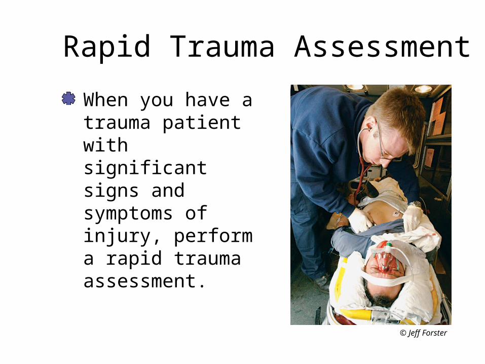
Rapid Trauma Assessment
When you have a trauma patient with significant signs and symptoms of injury, perform a rapid trauma assessment.
© Jeff Forster

Rapid Trauma AssessmentInspect and palpate the patient head to toe. Pay special attention to the areas most likely to produce serious, life-threatening injury. Rule out the possibility of obstructive shock. Set the patient’s priority for transport and for injury care.
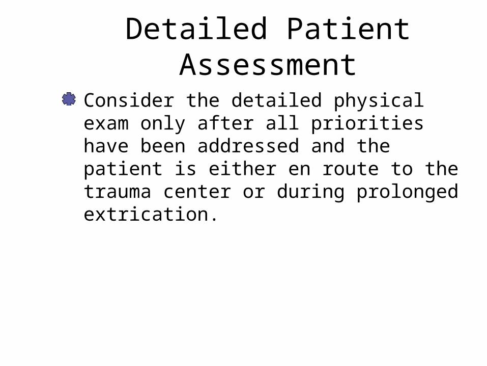
Detailed Patient Assessment
Consider the detailed physical exam only after all priorities have been addressed and the patient is either en route to the trauma center or during prolonged extrication.

Ongoing Assessment
Perform serial ongoing assessments– Mental status, airway, breathing, and circulation – Perform the ongoing assessment every 5 minutes
in the serious trauma patientPay particular attention to the pulse rate and pulse pressure Check the adequacy and effectiveness of any interventions you have performed
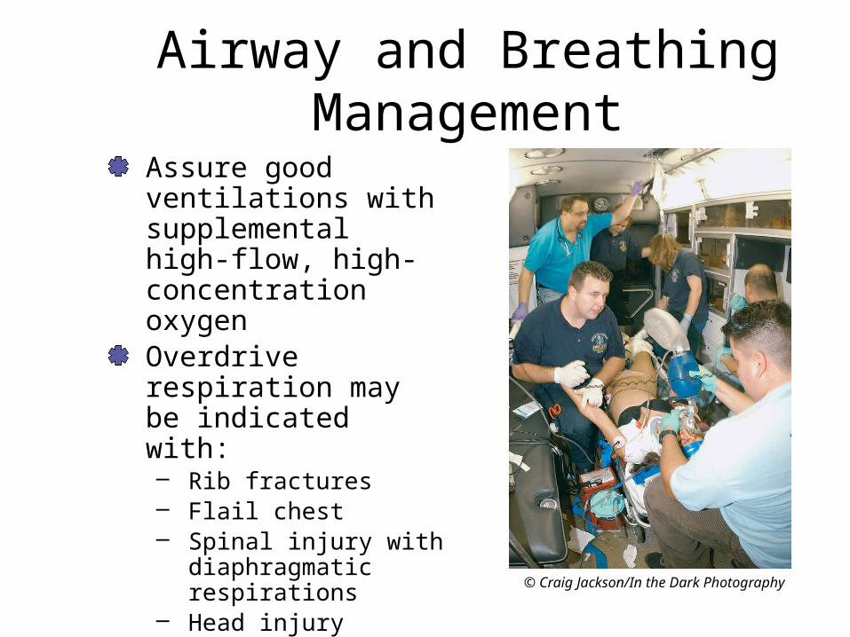
Airway and Breathing Management
Assure good ventilations with supplemental high-flow, high-concentration oxygen Overdrive respiration may be indicated with:– Rib fractures – Flail chest – Spinal injury with
diaphragmatic respirations
– Head injury
© Craig Jackson/In the Dark Photography

Airway and Breathing Management
Positive end-expiratory pressure (PEEP) and continuous positive airway pressure (CPAP) Protect the airway with an oral airway, nasal airway or possibly, endotracheal intubation Provide pleural decompression as necessary

Hemorrhage Control
Provide ongoing hemorrhage control as previously described.

Fluid Resuscitation Basics
Warm fluid if possibleAfter 250-500 mL, check BP and lung soundsPermissive hypotension -80 mmHgChildren- 20 mL/kgBP cuff on IV bag or pressure infuserLarge bore IV

Fluid Replacement
The field treatment of choice for significant blood loss in trauma is whole blood. – Generally not practical in the field setting– Most practical fluid for prehospital administration is
an isotonic crystaloid Polyhemoglobins– Contain either animal or human hemoglobin – Prolonged shelf life– Relatively inexpensive– Efficacy not well established
Fluid Replacement
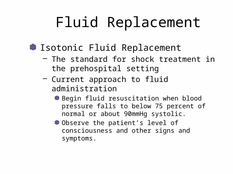
Isotonic Fluid Replacement– The standard for shock treatment in the
prehospital setting – Current approach to fluid administration
Begin fluid resuscitation when blood pressure falls to below 75 percent of normal or about 90mmHg systolic.Observe the patient’s level of consciousness and other signs and symptoms.

Isotonic Fluid Replacement
Employ aggressive fluid resuscitation– Use lactated Ringer’s solution or normal saline via
two lines – Administer until blood pressure returns to 100
mmHg and the level of consciousness increases In children, infuse 20 mL/kg of body weight
Permissive Hypotension

Isotonic Fluid ReplacementConsider the internal lumen size of both the catheter and the administration set – Utilize largest bore possible– Catheter length and fluid
pressure Ideal catheter for the shock patient is relatively short, 1 1/2" or shorter
Cautiously control fluid – Maintain V/S – don’t increase them

Treat the Patient… Don’t Fix the Patient
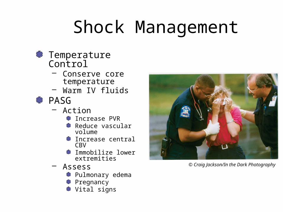
Shock ManagementTemperature Control– Conserve core
temperature– Warm IV fluids
PASG– Action
Increase PVRReduce vascular volumeIncrease central CBVImmobilize lower extremities
– AssessPulmonary edemaPregnancyVital signs
© Craig Jackson/In the Dark Photography
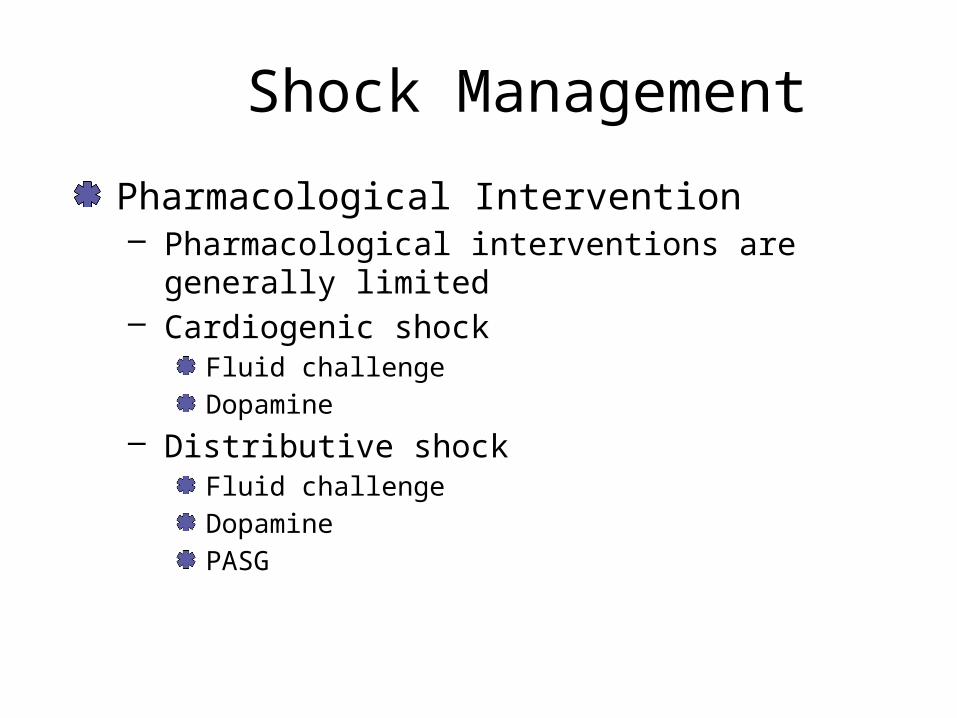
Shock Management
Pharmacological Intervention– Pharmacological interventions are generally limited – Cardiogenic shock
Fluid challengeDopamine
– Distributive shockFluid challengeDopaminePASG

Trauma Score
See page 505Based on BP, respirations, GCSMaximum of 12 Points

Revised Trauma Score

Helicopter Transport
Follow local protocolHave helicopter enroute while you are enrouteCancel if neededUsed designated LZ