Ch. 12: Respiratory Physiology - Francis Marion...
26
6/17/2019 1 Objectives: 1. Review respiratory anatomy. 2. Understand mechanics of breathing, gas pressure vocabulary, and the principles of surface tension, compliance, and recoil. 3. Respiratory disorders and spirometry 4. How gas exchange occurs between the alveoli & pulmonary vessels, and between capillaries & tissue. 5. Regulation of breathing (voluntary vs involuntary) 6. Hemoglobin & hemoglobin disorders 1 Ch. 12: Respiratory Physiology 2 2 Zones of Respiratory System: 1) Conduction zone, 2) Respiratory zone 1) Conduction Zone = from oral/nasal cavities to, _____________________ _____________________ _____________________ _____________________ _____________________ _____________________ terminal bronchioles 1. Respiratory Anatomy - REVIEW
Transcript of Ch. 12: Respiratory Physiology - Francis Marion...

6/17/2019
1
Objectives:
1. Review respiratory anatomy. 2. Understand mechanics of breathing, gas pressure
vocabulary, and the principles of surface tension, compliance, and recoil.
3. Respiratory disorders and spirometry 4. How gas exchange occurs between the alveoli &
pulmonary vessels, and between capillaries & tissue. 5. Regulation of breathing (voluntary vs involuntary) 6. Hemoglobin & hemoglobin disorders
1
Ch. 12: Respiratory Physiology
2
2 Zones of Respiratory System: 1) Conduction zone, 2) Respiratory zone 1) Conduction Zone = from oral/nasal cavities to, _____________________ _____________________ _____________________ _____________________ _____________________ _____________________ terminal bronchioles
1. Respiratory Anatomy - REVIEW

6/17/2019
2
Functions of Conducting Zone
• Transports air to the lung..
• Warms, humidifies, filters, and cleans the air.
– Mucus traps small particles, and cilia move it away from the lungs. Expectoration = coughing up the mucus & debris.
• Voice production in the larynx as air passes over the vocal folds
Structures in the larynx
glottis – opening between vocal cords
epiglottis – closes upon swallowing to
prevent food from entering airway
false vocal cords – muscles fibers that
assist the epiglottis
true vocal cords – muscles folds that
vibrate when air passes by them

6/17/2019
3
5
2) Respiratory zone
Respiratory bronchioles = smallest bronchioles, branch from tertiary bronchioles. Alveolar sacs = honey-comb shaped, 1-cell thick sacs for gas exchange. [~300 mill in lungs! ~760 sq ft area!]
6
How gases are exchanged w/blood
Surrounded by arterial & venous capillaries (“capillary plexus”) for gas exchange between alveoli & blood.
QUES: Is this a pulmonary artery or vein?
QUES: Is this a pulmonary artery or vein?

6/17/2019
4
7
Gas exchange occurs at the Alveoli
- thin cellular walls covered with capillary networks
- 300 million sacs! - very large surface area
- surfactant keeps the alveoli inflated
8
2 Types Alveolar Cells:
Type 1 Alveolar Cells = Make up wall of alveolus. 97% of total lung surface area
where most gas exchange occurs.
Type 2 Alveolar Cells - secrete surfactant
Surfactant =

6/17/2019
5
9
Why is surfactant important?
Your text here Your text here
Non-obstructive Atelectasis = ___________________________ ____________________________ ____________________________ _____________________________ Not to be confused with
Obstructive Atelectasis =
___________________________ ____________________________ ____________________________ _____________________________
Surfactants ↓ intra-alveolar pressure & prevent collapse Infant Respiratory Distress Syndrome (IRDS)
Acute Respiratory Distress Syndrome (ARDS)
• Surfactant is produced > 28 weeks (7-8 months)
• Babies are born < 28 wks - not enough surfactant. High surface tension inside alveoli, results in collapsed alveoli, which collapses lung (non-obstructive atelectasis)
• Tx = synthetic surfactant delivered into baby’s lungs & mechanical ventilator until Type 2 alveolar cells can make surfactant.
• Due to inflammation from infection (septic shock) • Results in protein (serum) secretion in lungs. • Fluid dilutes surfactant, surface tension, alveoli collapse, • could cause lung collapse (non-obstructive atelectasis)

6/17/2019
6
Thoracic cavity: REVIEW!
Membranes of the lungs: Visceral pleura = _____________________________________________ Parietal pleura = ____________________________________________________ - Parietal pleura held tight against thoracic wall by surface tension of water layer. - As thoracic cage changes volume (w/ breathing) so do the lungs.
Intrapleural space = ______________________________________________ - The 2 pleura pressed together w/serous fluid between them.
1) Air moves from high to low pressure
- Atmospheric air pressure = constant (760 mmHg)
- Lung air pressure depends on volume of thoracic cavity
2) Air pressure in lungs (closed chamber) changes with volume of chamber
“Boyle’s Law” = as volume of closed chamber ↑, air pressure within _______
as volume of closed chamber ↓, air pressure within ________
2. Mechanics of Respiration
Translates to lung volume & air pressure within lungs (“intrapulmonary pressure”): As thoracic volume ↑, lung volume ↑, & intrapulmonary pressure _______ As thoracic volume ↓, lung volume ↓, & intrapulmonary pressure _______

6/17/2019
7
Boyle’s Law
13
Chamber volume larger BUT air pressure lower
Chamber volume smaller BUT air pressure higher
14
Inspiration Expiration

6/17/2019
8
Mechanism of Breathing
Inspiration/inhalation = When the diaphragm and intercostal muscles
contract, the thoracic cage volume , & the lung volume .
Rate of breathing is controlled
by the medulla oblongata,
which sends signals to the
diaphragm and intercostal
muscles.
Mechanism of Breathing
Expiration/exhalation = When the diaphragm and intercostal muscles
relax, the thoracic cage volume, , & the lung volume .
6/17/2019
9
Muscles involved in inspiration
Diaphragm – contracts (moves downward) to expand thorax.
external intercostal muscles – contraction lifts ribs & sternum
scalenes, pectoralis minor, & sternocleidomastoid – contract to pull thoracic
cage up and outward.
Muscles involved in expiration Diaphragm – relaxes (diaphragm moves upward) to compress thorax.
internal intercostal muscles – contraction pulls ribs and sternum in
abdominal wall muscles – contract to push diaphragm higher
6/17/2019
10
• Intrapulmonary pressure = pressure inside lungs – During inhalation – is lower than atmospheric pressure (-3 mmHg)
– During exhalation – is above atmospheric pressure (+3 mmHg)
• Intrapleural pressure = pressure between the pleural membranes due to elastic recoil (parietal pleura sticks to wall) – During inhalation – is lower than atmospheric (-6 mmHg)
– During exhalation – is still lower atmospheric (-3 mmHg)
*** intrapleural pressure should ALWAYS be negative. If air enters this space, the lung can detach from thoracic wall, trapped air puts pressure on lung, & lung can collapse. ***
• Transpulmonary pressure = difference between intrapulmonary & intrapleural pressure (is ALWAYS above atmospheric pressure).
19
Gas Pressure Vocabulary:
20
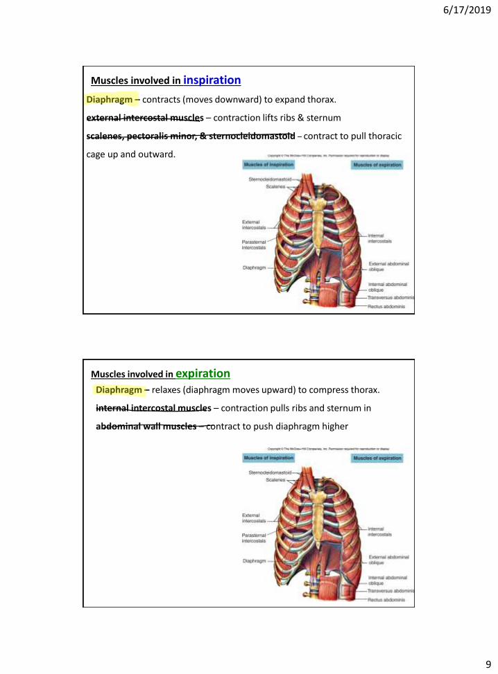
Pneumothorax in lung
“Pneumothorax” = _______________ ________________________________ _______________________________. (Can be traumatic or tension) - Air trapped between the two pleural membranes removes the pressure gradient.
Result = can’t expand lungs to get air to enter! Lung collapses.
2nd pneumothorax video (tension pneumothorax)
Causes of a collapsed lung”

6/17/2019
11
21
Important properties of the lungs:
A) Surface tension = pressure resulting from thin film of water lining alveoli that resists their expansion. Makes alveoli want to collapse with exhalation.
B) Compliance = lungs expand when stretched (when thoracic volume ).
- more lung compliance = greater capacity for “stretchiness”
- less lung compliance = less capacity for “stretchiness”
C) Elasticity/Recoil = tendency of lungs to return to normal shape after stretching. (When thoracic volume , lungs volume also parietal pleura keeps lungs “stuck” to thoracic wall).
22
6/17/2019
12
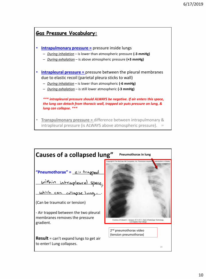
A) Surface Tension & Law of La Place
23 Fig 16.10
Lower: - Air pressure - Surface tension
Greater: - Air pressure - Surface tension
Law of LaPlace & surface tension in alveoli: Small alveoli have surface tension within, & air pressure within (intra-alveolar pressure) Large alveoli have surface tension, & air pressure within
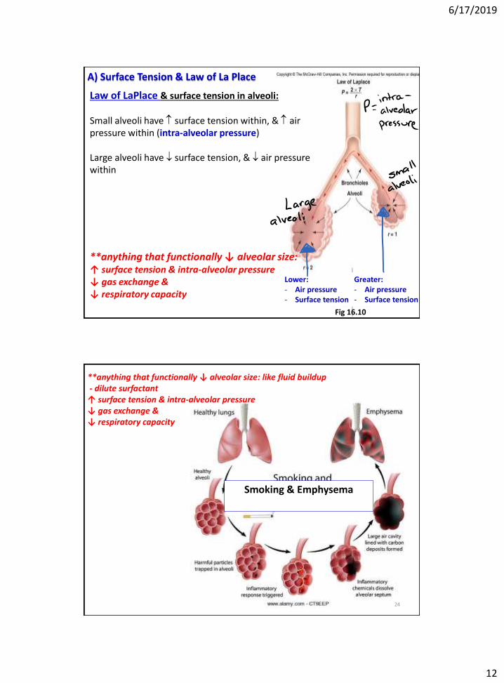
**anything that functionally ↓ alveolar size: ↑ surface tension & intra-alveolar pressure ↓ gas exchange & ↓ respiratory capacity
**anything that functionally ↓ alveolar size: like fluid buildup - dilute surfactant ↑ surface tension & intra-alveolar pressure ↓ gas exchange & ↓ respiratory capacity
24
Smoking & Emphysema

6/17/2019
13
Factors that increase compliance: - pulmonary “surfactants” Factors that decrease compliance: - many, many things! - Anything that causes chronic inflammation can lead to compliance
- Chronic inflammation of the airways (bronchitis) can lead to - scar tissue in lungs (pulmonary fibrosis), - and narrowing of airways (bronchoconstriction)
B) Lung compliance
25
For Ex. - Lung damage from smoking causes chronic bronchitis and formation of scar tissue (fibrosis)
26

6/17/2019
14
Review • The respiratory system
– The conduction & respiration zones
• Airway, lung, and thoracic cavity anatomy
• Alveoli (gas exchange, surfactant, factors that affect intra-alveolar surface tension and pressure)
• Mechanics of breathing (Boyle’s law and respiratory muscles), muscles of respiration.
• Gas pressure vocabulary, and pneumothorax,
• Important properties of the lungs
– Surface tension, compliance, & elasticity.
– Factors that affect these properties
27
Respiratory Disorders
A Restrictive Disorder = Lung tissue is damaged. Lungs are stiff, or respiratory muscles are weak.
Examples: Pulmonary fibrosis
Obstructive Disorder = Lung tissue is normal, but resistance is increased (airways are narrowed)
Examples: Asthma
COPD (which includes emphysema & chronic bronchitis)
Cystic fibrosis
3. Respiratory Disorders, & Diagnosing Them

6/17/2019
15
29
- Pulmonary fibrosis = buildup of fibrous tissue in lungs stiffens them (restrictive disorder) MANY CAUSES > Breathing in small particles that accumulate in & irritate the lungs: Ex: Silicosis = (inhalation of fine glass, rock, or sand particles) Ex: Anthracosis (black lung disease) = inhalation of coal dust. Ex. Mesothelioma – breathing in asbestos. - Breathing in chemicals that irritate the lungs: Ex. smoking
Silicosis
Respiratory Disorders – Pulmonary Fibrosis
Respiratory Illnesses - Asthma
Muscles around the bronchioles are hyper-excitable
- Obstructive disorder due to inflammation, mucous secretion, & narrowing
of airways (bronchoconstriction).
Treatment? __________________________________________

6/17/2019
16
Respiratory Illnesses – Chronic Obstructive Pulmonary Disease (COPD)
Chronic inflammation of airways alveolar tissue - narrows airways & destroys alveolar walls - proliferation of mucus-secreting goblet cells - development of scar (fibrous) tissue = pulmonary fibrosis - Obstructive disorder – due to mucus buildup and narrowed airways.
32

6/17/2019
17
Respiratory Illnesses - Emphysema
http://www.nlm.nih.gov/medlineplus/ency/images/ency/fullsize/17055.jpg
Chronic destruction of alveolar tissue (walls between alveoli lost) - reduces area for gas exchange - alveoli expand easily, but can’t empty easily (air-trapping disorder) - obstructive disorder
34
Respiratory Illnesses - Cystic Fibrosis - Genetic disorder affecting Cl- channels on alveoli membrane. Obstructive disorder Results in buildup of mucus within alveoli causing: - Dilutes surfactant - decreased functional alveolar size - surface tension & intra-alveolar pressure (harder for alveoli to expand) - with gas exchange - Warmth & moisture (mucus) aids bacterial growth.
(Vulnerable to pneumonia)

6/17/2019
18
Pulmonary Function Tests
• Spirometry: air movement during respiration recorded on a spirogram.
– Measures lung volumes and capacities
– Can diagnose restrictive and obstructive lung disorders
36
Spirometry = clinical evaluation of pulmonary (respiratory) function, which allows diagnosis of lung disorders.
Fig 16.14
3. Lung Volumes & Respiratory Vocabulary

6/17/2019
19
37
Additional Respiratory Vocabulary:
38
Apnea = absence of breathing
Dyspnea = labored or difficult breathing
Eupnea = normal breathing at rest
Hyperventilation = excessively rapid ventilation (will decrease alveolar CO2)
Hypoventilation = low ventilation (will increase alveolar CO2)
Pneumothorax = presence of gas in intrapleural space causing lung collapse

6/17/2019
20
Review • Respiratory disorders
– Restrictive vs Obstructive
– Pulmonary fibrosis (and its causes)
– Asthma
– COPD
– Emphysema
– Cystic fibrosis
• Testing for respiratory disorders
– Spirometry
– Spirometry vocabulary (IRV, ERV, TV, VC, RV, and TLC, minute ventilation)
– Additional respiratory vocabulary (eupnea, dyspnea, apnea, hyperventilation, hypoventilation)
39
40
Gas exchange between 2 structures is dependent on pressure gradient of dissolved O2 & CO2 * Gas moves from side with higher pressure (from dissolved gases) to side with lower pressure & visa versa
*Gas wants to move “downhill” from high to low pressure!
4. Basics of Gas Exchange at Lungs and at Body Tissues

6/17/2019
21
41
Gas exchange between lung alveoli & pulmonary vessels: > Alveolar PO2 = 105 mmHg, higher than that in pulmonary arteries (40 mmHg) > Alveolar PCO2 = 40 mmHg, lower than that in pulmonary arteries (46 mmHg)
PO2 = 105 mmHg PCO2 = 40 mmHg
PO2 = 40 mmHg PCO2 = 46 mmHg
PO2 = 100 mmHg PCO2 = 40 mmHg
42
Gas exchange between systemic capillaries & tissues: > Tissue PO2 (<100 mmHg) = lower than O2-rich arterial blood (100 mmHg) > Tissue PCO2 (>40 mmHg) = higher than that in arterial blood (40 mmHg)

6/17/2019
22
Review
• Pulmonary function tests (spirometry)
• Alveolar PO2 lower than atmospheric
• Gas exchange at tissues & at alveoli of lungs Depends on differences in partial pressures of O2 and CO2
43
44
Motor neurons from 3 brain areas control breathing muscles: 1) Voluntary Breathing = primary motor cortex of frontal cerebral lobe. 2) Involuntary Breathing = Medulla – respiratory center regulates respiratory rate. Pons – apneustic center (stimulate inhalation) – pneumotaxic center (inhibit inhalation)
5. Regulation of Respiration – regulation of blood O2 & CO2

6/17/2019
23
What happens to minute ventilation after:
• Hypoventilation?
• Hyperventilation?
45
Autonomic motor control breathing involves:
Chemoreceptors: Aorta & carotid artery chemoreceptors (called
peripheral chemoreceptors) - sense blood O2 and CO2 levels
Medulla chemoreceptors (called central chemoreceptors) - sense CSF O2 and CO2 levels

6/17/2019
24
47
Stimulus = blood pH (acidosis) blood O2 is too low (CO2 is high) Sensors = arterial chemoreceptors detect high CO2 in blood, Medulla senses high CO2 in CSF. Integrating center = Medulla’s respiratory center Effectors = respiratory muscles respiratory depth & rate ( minute ventilation) Get rid of excess CO2 Effect = blood pH to normal Stimulus = blood pH (alkalosis) blood O2 is too high (CO2 is too low) Sensors = arterial chemoreceptors detect low CO2 in blood, Medulla senses low CO2 in CSF. Integrating center = Medulla’s respiratory center Effectors = respiratory muscles respiratory depth & rate ( minute ventilation) Retain a little CO2 Effect = blood pH to normal
Negative Feedback regulation of blood pH (by blood CO2 content)
The role of chemoreceptors in respiration
The most important chemical in
influencing breathing rate is
carbon dioxide.
CO2 + H2O H2CO3 H+ + HCO3-
Oxygen-sensitive chemoreceptors only
alter breathing rate when blood oxygen
levels fall critically low (or CO2 is too
high)

6/17/2019
25
49
Blood pH (Acid/Base balance) based primarily on blood CO2 content and metabolic activities in body: 1) Respiratory component = where CO2 (a volatile acid) in blood eliminated by lungs (exhalation). - Increased respiratory rate ↑blood pH (respiratory alkalosis) - Decreased respiratory rate ↓ blood pH (respiratory acidosis) 2) Metabolic component = non-volatile acids in blood (i.e. lactic acid, fatty acids, ketones) eliminated by liver, kidneys, or other organs.
Normal Blood pH = 7.35 – 7.45 Blood pH maintained by buffering CO2 with HCO3- Blood with high CO2 or H+ content = acidic (acidosis) Blood w/lower CO2 or high HCO3- content = alkaline (alkalosis)
50
Acidosis = increased acids in blood (pH below 7.35) Alkalosis = decreased acids in blood (pH above 7.45) Respiratory acidosis = ↓ blood pH due to ↓ respiratory rate (hypoventilation) – not enough CO2 waste exhaled by lungs. Respiratory alkalosis = ↑blood pH due to ↑ respiratory rate (hyperventilation) – too much CO2 exhaled by lungs. Metabolic acidosis = excess metabolic production of acids (i.e. ketosis) OR loss of bases (i.e. bicarbonate) from chronic diarrhea or kidney problems (excrete too much HCO3-) Metabolic alkalosis = too much bicarbonate (not enough excreted by kidneys) OR loss of metabolic acids such as with chronic vomiting (lose HCL).

6/17/2019
26
Review
• Regulation of breathing (voluntary vs involuntary)
– Primary motor cortex (voluntary)
– Medulla & Pons (involuntary)
• Acid / Base imbalance
– Metabolic Acidosis & Alkalosis versus
– Respiratory Acidosis & alkalosis
51
Review
• Regulation of breathing
– Medulla & pons
• Chemoreceptors
– central, peripheral
• Hemoglobin O2 transport:
– Oxyhemoglobin & deoxyhemoglobin
– Abnormal hemoglobin (carboxyhemoglobin, methemoglobin)
– Neonatal jaundice
– Sickle cell
52