CF patient with hypoxia - cdn.doctorsonly.co.il · reported in 1966 by Berthelot et al. first...
38
CF patient with hypoxia David Shoseyov MD David Shoseyov MD
Transcript of CF patient with hypoxia - cdn.doctorsonly.co.il · reported in 1966 by Berthelot et al. first...

CF patient with hypoxia
David Shoseyov MDDavid Shoseyov MD
D GD.G.
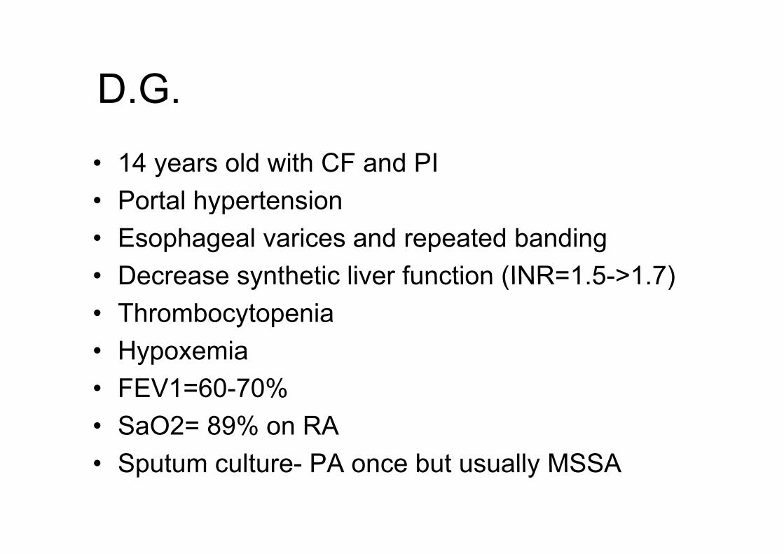
• 14 years old with CF and PI• Portal hypertensionyp• Esophageal varices and repeated banding• Decrease synthetic liver function (INR=1 5->1 7)• Decrease synthetic liver function (INR=1.5->1.7)• Thrombocytopenia• Hypoxemia• FEV1=60-70%• SaO2= 89% on RA• Sputum culture- PA once but usually MSSA• Sputum culture- PA once but usually MSSA
D GD.G.
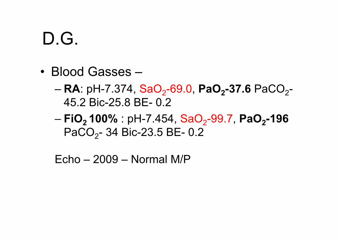
• Blood Gasses –– RA: pH-7.374, SaO2-69.0, PaO2-37.6 PaCO2-p , 2 , 2 2
45.2 Bic-25.8 BE- 0.2– FiO2 100% : pH-7 454 SaO2-99 7 PaO2-196FiO2 100% : pH 7.454, SaO2 99.7, PaO2-196
PaCO2- 34 Bic-23.5 BE- 0.2
Echo – 2009 – Normal M/P
D GD.G.
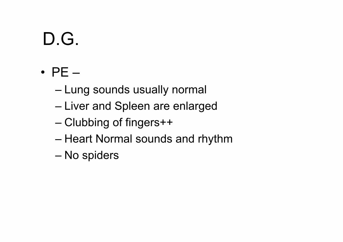
• PE –– Lung sounds usually normalg y– Liver and Spleen are enlarged
Clubbing of fingers++– Clubbing of fingers++ – Heart Normal sounds and rhythm – No spiders

Chest CT (J 2012)Chest CT (Jan 2012)

HRCTHRCT (Jan 2012)

Why is he hypoxic?Why is he hypoxic?Th h i l i l h i th t i d• The physiological mechanisms that induce hypoxemia include:
Hi h ltit d– High altitude– Hypoventilation
Diff i d f t– Diffusion defects– Shunts
V til ti f i d f t– Ventilation–perfusion defects.
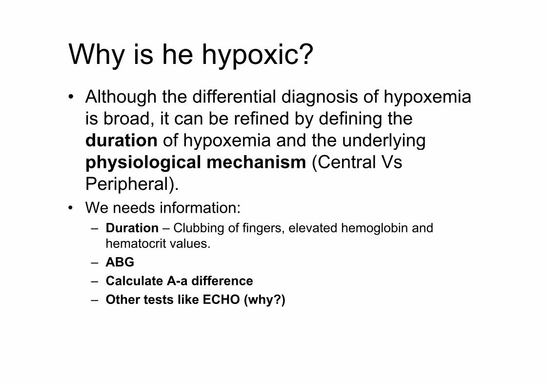
Why is he hypoxic?Why is he hypoxic?• Although the differential diagnosis of hypoxemia g g yp
is broad, it can be refined by defining the duration of hypoxemia and the underlying yp y gphysiological mechanism (Central Vs Peripheral).p )
• We needs information:– Duration – Clubbing of fingers, elevated hemoglobin and
hematocrit values.– ABG
Calculate A a difference– Calculate A-a difference– Other tests like ECHO (why?)

A-a GradientA l A di t f d lt kA normal A-a gradient for a young adult non-smoker breathing air, is between 5-10 mmHg.
At Sea level A-a Gradient =(21%*(746 -40) - 5/4(PCO2)) - PaO2At Sea level A-a Gradient =(21% (746 -40) - 5/4(PCO2)) - PaO2
Approximately
In Jerusalem A-a Gradient =(21%*(700 -40) - 5/4(PCO2)) - PaO2
At Sea level A-a Gradient = 147 - 5/4(PCO2)) - PaO2

Why is he hypoxic?y yp
• The value for the partial pressure of oxygen should improve with oxygen therapy.
• Altitude is not a concern in this case since he is• Altitude is not a concern in this case, since he is hypoxic in Jerusalem which is not too high from sea level.
• It improves with oxygen and CO2 is often low due to hyperventilation

Why is he hypoxic?Why is he hypoxic?• Hypoventilation - failure of the central respiratory center
or abnormalities of the peripheral nerves and muscles.
• Hepatic Encephalopathy - unlikely
• The PaO2 depressed and that of PaCO2 is elevated.The PaO2 depressed and that of PaCO2 is elevated.• There is a normal alveolar–arterial (A-a) gradient and a
robust response to oxygen unlike the findings in this caserobust response to oxygen, unlike the findings in this case.

Why is he hypoxic?Why is he hypoxic?• Diffusion defects - arise when the distance that oxygen must travel
from the alveolus to the hemoglobin in the pulmonary capillary is
altered.
• Such defects result in hypoxemia with exercise (6 min walk test etc) or
at altitude but rarely at rest.
• This type of hypoxemia responds to inhaled oxygen.
• DG response to 100% O2 was partial and insufficient

Where is the Shunt?Where is the Shunt?Shunting of blood through intracardiac defects or• Shunting of blood through intracardiac defects or abnormal intrapulmonary intrapulmonary vessels is a frequent cause of hypoxemia.
• The hallmarks of shunting are a poor response to inhaled oxygen therapy and an increased alveolar– arterial gradient. g
• In some cases, there may be a partial response to oxygen therapy.
• The most frequent cause of hypoxemia is a ventilation• The most frequent cause of hypoxemia is a ventilation–perfusion (V/Q) mismatch, which can lead to clinically significant hypoxemia but is responsive to oxygen therapy (lik i B hi liti )(like in Bronchiolitis).
Intra cardiac ShuntIntra cardiac Shunt
• They occur in approximately 20 % of patients with congenital heart disease. p g
• Conditions that could cause such a shunt in the age group of the patient include:in the age group of the patient include:– Tetralogy of Fallot, – Eisenmenger’s syndrome (with an ASD, VSD
or PDA)– Pulmonary artery hypertension with a PFO

Intrapulmonary ShuntIntrapulmonary Shunt• Chronic intrapulmonary shunts areChronic intrapulmonary shunts are
unusual, and the differential diagnosis includes two conditions:includes two conditions:– Pulmonary arteriovenous malformations
(AVM)(AVM)– Hepatopulmonary syndrome.

Pulmonary arteriovenous malformations (AVM)
• Such malformations are rare, detected in only 2 of 15,000 consecutive autopsies.
• They occur twice as often in females as in males and are rarely identified in infancy but are y yincreasingly identified with advancing age.
• Approximately two thirds of pulmonary AVMApproximately two thirds of pulmonary AVM occur in the context of hereditary hemorrhagic telangiectasia, also known as the:telangiectasia, also known as the:
Rendu–Osler–Weber syndrome.

Hepatopulmonary syndromeHepatopulmonary syndrome• The unique striking pathological feature of• The unique striking pathological feature of
HPS is :– gross dilatation of the pulmonary
precapillary and capillary vessels (15 to 100 μm in diameter when the patient is at rest), coupled with an absolute increase in the number of dilated vessels.

NormalNormal Classic Shunt Hepatopulmonary Syndrome
8-15 m15-100 m
The unique striking pathological feature of hepatopulmonary syndrome is gross dilatation of the pulmonary precapillary and capillary vessels (to 15 to 100 μm in diameter when the patient is at rest).

Hepatopulmonary syndromeHepatopulmonary syndrome• The hepatopulmonary syndrome is suspected inThe hepatopulmonary syndrome is suspected in
any patient with known liver disease who reports dyspnea. y p
• Arterial blood gasses should be measured on room air in patients with clinically significant p y gsymptoms.

Hepatopulmonary syndromeHepatopulmonary syndromeA useful diagnostic test is contrast gechocardiography.
I t i b bbl ( 10Intravenous microbubbles (>10 min diameter) from agitated normal saline that are normally obstructedsaline that are normally obstructed by pulmonary capillaries (normally <8 to 15 m) rapidly transit the lung ) p y gand appear in the left atrium of the heart within 7 heart beats.

Hepatopulmonary syndromeHepatopulmonary syndrome• Similarly, intravenousSimilarly, intravenous
technetium-99m–labeled albumin may transit the lungs and appear in thelungs and appear in the kidney and brain.
• Pulmonary angiographyPulmonary angiography may reveal diffusely fine or blotchy vascular configuration Theconfiguration. The distinction has to be made with an intracardiac right-to-left shunt.

Hepatopulmonary syndromeHepatopulmonary syndrome

DG echo bubbleDG echo bubble

D GD.G.
• Blood Gasses –– RA: pH-7.374, SaO2-69.0, PaO2-37.6 PaCO2-p 2 2 2
45.2 Bic-25.8 BE- 0.2– FiO2 100% : pH-7.454, SaO2-99.7, PaO2-196FiO2 100% : pH 7.454, SaO2 99.7, PaO2 196
PaCO2- 34 Bic-23.5 BE- 0.2In Jerusalem A-a Gradient =(134 - 5/4(PCO2)) - PaO2
A-a Gradient =(134 - 5/4(45.2)) – 40
In Jerusalem A-a Gradient =(134 - 5/4(PCO2)) - PaO2
A-a Gradient – 42.6 High
Severe HPS

Hepatopulmonary Synd Clinical PresentationHepatopulmonary Synd – Clinical Presentation
D ti t t b th i th• Dyspnea on exertion, at rest, or both is the predominant presenting symptom, usually after years of liver disease.
• There are no signs, symptoms, or hallmarks of the HPS on physical examination.
• However, the presence of:– spider nevi
digital clubbing– digital clubbing– Cyanosis– severe hypoxemia (partial pressure of oxygen, <60 mm Hg)
strongly suggests Hepatopulmonary syndrome

Hepatopulmonary Synd Clinical PresentationHepatopulmonary Synd – Clinical Presentation
• Orthodeoxia - arterial blood O2 decreases by 5% or more or by 4 mm Hg (0.5 kPa) or more when the patient moves from a supine to an upright position
• Worsening dyspnea (platypnea) related to further ventilation–perfusion mismatch

Lab findingLab finding
• A decrease in the DLCO is the only routine pulmonary-function test that is p yconsistently abnormal in patients with the hepatopulmonary syndromehepatopulmonary syndrome
• The CXR is frequently nonspecific, with a mild interstitial pattern in the lower lung that may reflect the existence of diffuse ypulmonary vascular dilatation.

Chest angio CTChest angio CT

PREVALENCE AND NATURAL HISTORYPREVALENCE AND NATURAL HISTORY
• The term “hepatopulmonary syndrome,” which was probably coined in 1977, was preceded by compelling descriptions based on autopsy and clinical findings.
• An autopsy study in patients with liver cirrhosis, reported in 1966 by Berthelot et al. first suggested that marked pulmonary vascular dilatation may play a role in this condition.
•Kennedy TC, Knudson RJ. Exercise-aggravated hypoxemia and orthodeoxia in cirrhosis. Chest 1977; 72:305-309G S S f•Berthelot P, Walker JG, Sherlock S, Reid L. Arterial changes in the lungs in cirrhosis of the liver -- lung spider nevi. N Engl J
Med 1966; 274:291-298

PREVALENCE AND NATURAL HISTORYPREVALENCE AND NATURAL HISTORY
S i l i ifi tl ti t ith ti l• Survival was significantly worse among patients with a partial pressure of oxygen of less than 50 mm Hg at the time of diagnosis.
• Patients with HPS (Not candidates for liver Tx)– median survival of 24 months and a 5-year survival rate of 23%.
• Patients without the HPS matched for the cause and severity ofPatients without the HPS matched for the cause and severity of liver disease– median survival of 87 months, with a 5-year survival rate of 63%.
100
Survival
20
40
60
80
100
0
20
5 Yrs Surviaval %Median (months)
HPS + No HPS

ManagementManagementPharmacological treatment• Pharmacological treatment.
• A small number of small uncontrolled trials :– somatostatin analogue,g ,– -blockers, – cyclooxygenase inhibitors, – glucocorticoids and immunosuppressors (cyclophosphamide)glucocorticoids and immunosuppressors (cyclophosphamide)– pulmonary vasoconstrictors (almitrine), – NO inhibitors, inhaled NO,
antimicrobials– antimicrobials – garlic preparation.
• None of the studies demonstrated consistent i t d t i d t i t t t ffiimprovement due to inadequate size to test efficacy.
• In addition, rare spontaneous recovery has been observed in HPS

ManagementManagement• Long-term oxygen therapy. No data are available,
• Transjugular intrahepatic portosystemic shunt (TIPS). – Only a few case reports using TIPS for HPS have been published, and
have shown variable short-term effects on pulmonary gas exchange. – Our single experience was a failure.
• Cavoplasty. Venous decompression by abscess drainage resolved HPS in a single case with Budd- Chiari syndrome.
• Embolisation. Coil embolisation in type II angiographic pattern HPS has been reported to improve arterial oxygenation (as a temporary measure) in a single case reportmeasure) in a single case report.

ManagementManagement• Orthotopic liver transplantation *.p p• Complete resolution of HPS following OLT has been
observed in >80% of reported cases,• Many centers currently view HPS as an indication for OLT,
particularly in the pediatric population.
*O th t i li t l t ti f t d i hi h f il d li iOrthotopic liver transplantation refers to a procedure in which a failed liver is removed from the patient's body and a healthy donor liver is transplanted

Algorithm for screening and therapeutic decisions in hepatopulmonary syndrome (OLT: orthotopic liver transplantation CEE: Contrast p p y y (Enhanced Echocardiography; MAA: macroaggregated albumin)
R. Rodrıguez-Roisin Pulmonary–Hepatic vascular Disorders (PHD) Eur Respir J 2004; 24: 861–880

Add onAdd on• The correlation between the degree of hepatic
d f ti d t l h t i d th ldysfunction and portal hypertension and the prevalence and severity of the hepatopulmonary syndrome remains controversial.
• Rare congenital cardiac disorders without liver injury in which either hepatic venous blood flow does not reach th l t l bl d h th i f ithe lung or portal venous blood reaches the inferior vena cava without passing through the liver (i.e., the type 1 Abernethy malformation) have clinical similarities to y )the hepatopulmonary syndrome
• This provides support for the hypothesis that blood from th t t th li t t lthe gut must cross the liver to prevent pulmonary vascular dilatation.

Abernethy Malformation a rare cause of HPS The firstAbernethy Malformation a rare cause of HPS. The first account of congenital absence of the portal vein (CAPV) was given by John Abernethy in 1793

Which animal suffers frequently from Abernethy malformation ?
Patellar luxation and portosystemic shunt, can often be identified early. Parents can be screened for retinal dysplasia, and their histories should reveal
il ll i t hany epilepsy or collapsing trachea .
Th h titi i t t llThough pancreatitis is not actually a hereditary disease, the fact that the Yorkshire terrier, along with a few other breeds, shows a higher occurrence rate than among dogs ina higher occurrence rate than among dogs in general