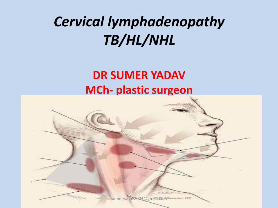


Cervical lymph adenopathy
88
-
Upload
sumer-yadav -
Category
Health & Medicine
-
view
244 -
download
0
Transcript of Cervical lymph adenopathy
Lymph Nodes
• Anatomy
– Collection of lymphoid cells attached to both vascular and lymphatic systems
– Over 600 lymph nodes in the body
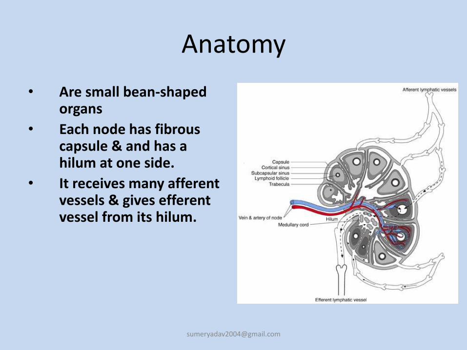
Anatomy
• Are small bean-shaped organs
• Each node has fibrous capsule & and has a hilum at one side.
• It receives many afferent vessels & gives efferent vessel from its hilum.
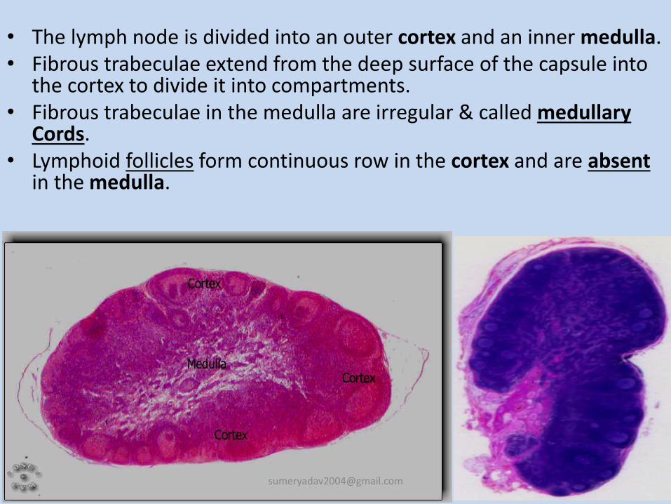
• The lymph node is divided into an outer cortex and an inner medulla. • Fibrous trabeculae extend from the deep surface of the capsule into
the cortex to divide it into compartments.• Fibrous trabeculae in the medulla are irregular & called medullary
Cords.• Lymphoid follicles form continuous row in the cortex and are absent
in the medulla.
Lymph
• Fluid similar in composition to blood plasma.
• Derived from blood plasma by filtration through capillary walls at the arterial end.
• As soon as the interstitial fluid enters the lymph capillaries, it is called lymph.
• Returning the fluid to the blood helps to maintain normal blood volume and pressure.
Function
• To provide optimal sites for the concentration of free or cell-associated antigens and recirculating lymphocytes –“sensitization of the immune response”
• To allow contact between B-cells, T-cells and macrophages
• Lymph nodes and other lymphatic organs filter the lymph to remove & destroy microorganisms and other foreign particles
• It returns excess interstitial fluid to the blood to maintain blood volume and blood pressure .
• Absorption of fat and fat-soluble vitamins from the digestive system by special lymph capillaries, called lacteals The lymph in the lacteals has a milky appearance due to its high fat content and is called chyle.
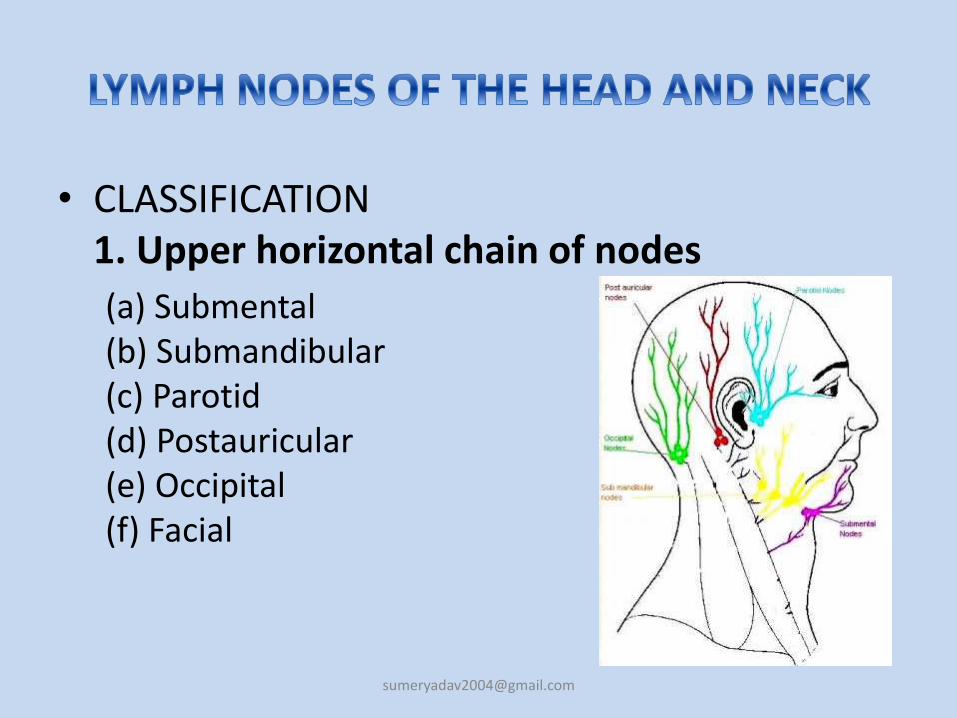
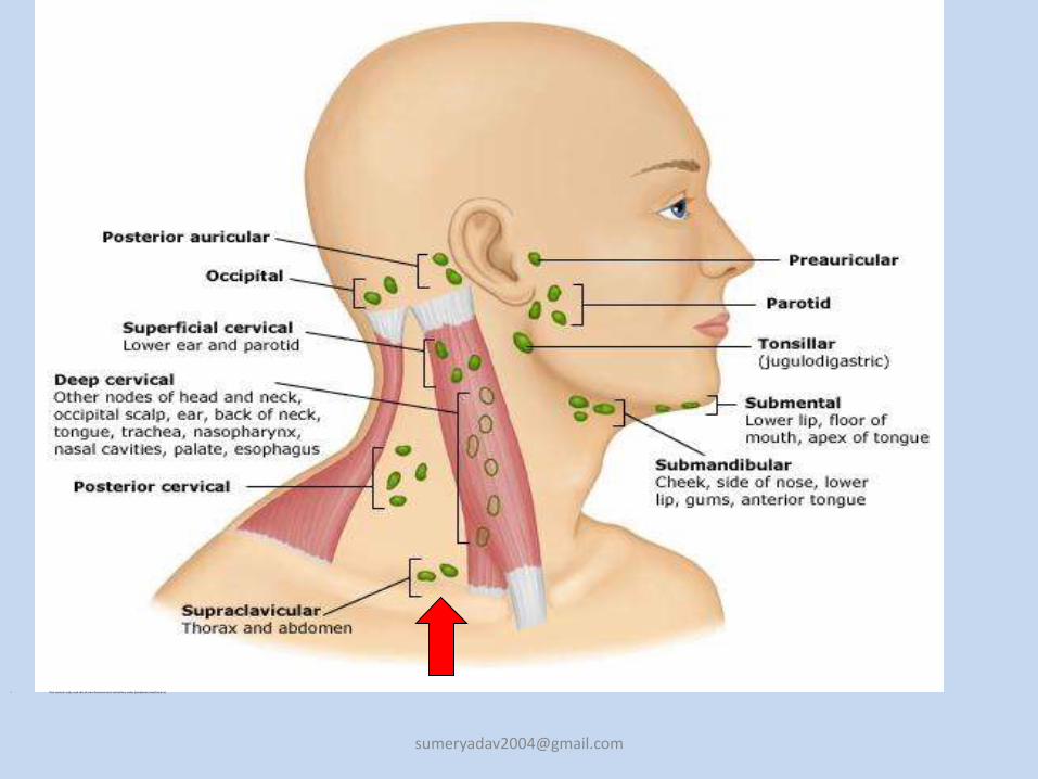
• CLASSIFICATION1. Upper horizontal chain of nodes
(a) Submental(b) Submandibular(c) Parotid(d) Postauricular(e) Occipital(f) Facial
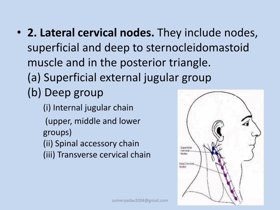
• 2. Lateral cervical nodes. They include nodes, superficial and deep to sternocleidomastoid muscle and in the posterior triangle.(a) Superficial external jugular group(b) Deep group
(i) Internal jugular chain
(upper, middle and lowergroups)(ii) Spinal accessory chain(iii) Transverse cervical chain
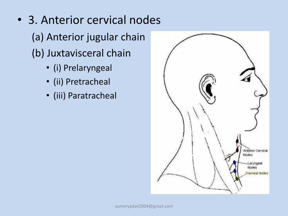
• 3. Anterior cervical nodes
(a) Anterior jugular chain
(b) Juxtavisceral chain
• (i) Prelaryngeal
• (ii) Pretracheal
• (iii) Paratracheal
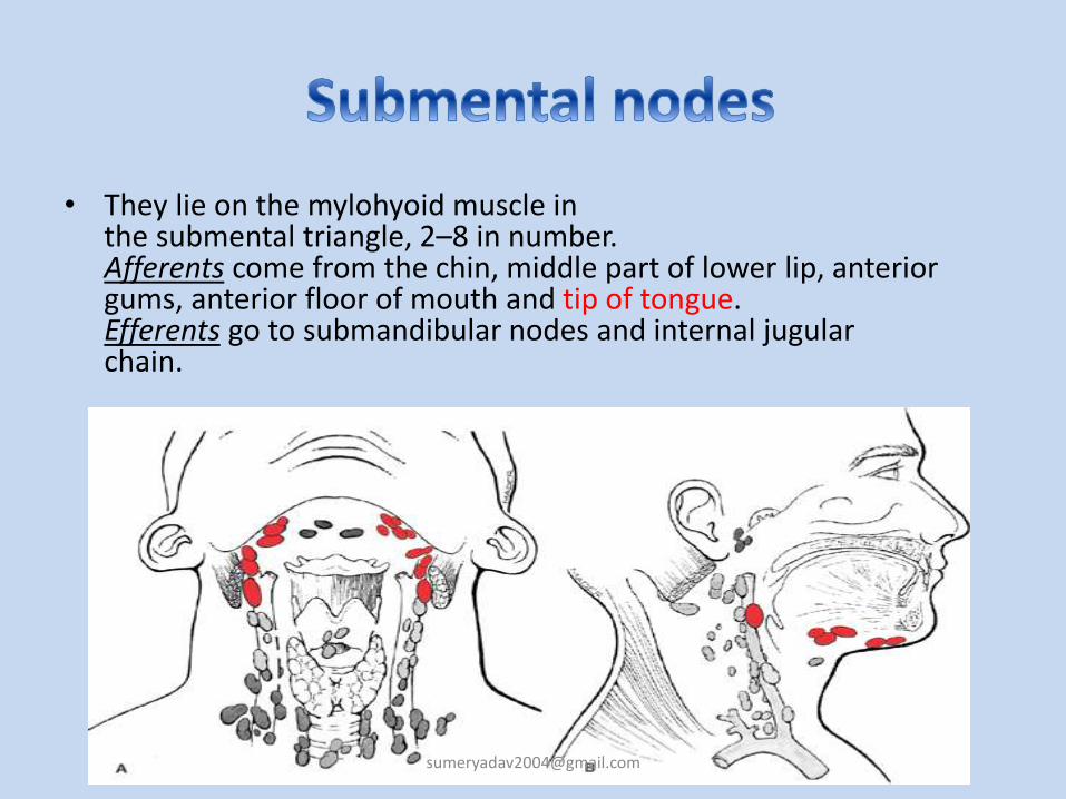
• They lie on the mylohyoid muscle inthe submental triangle, 2–8 in number.Afferents come from the chin, middle part of lower lip, anterior gums, anterior floor of mouth and tip of tongue.Efferents go to submandibular nodes and internal jugularchain.
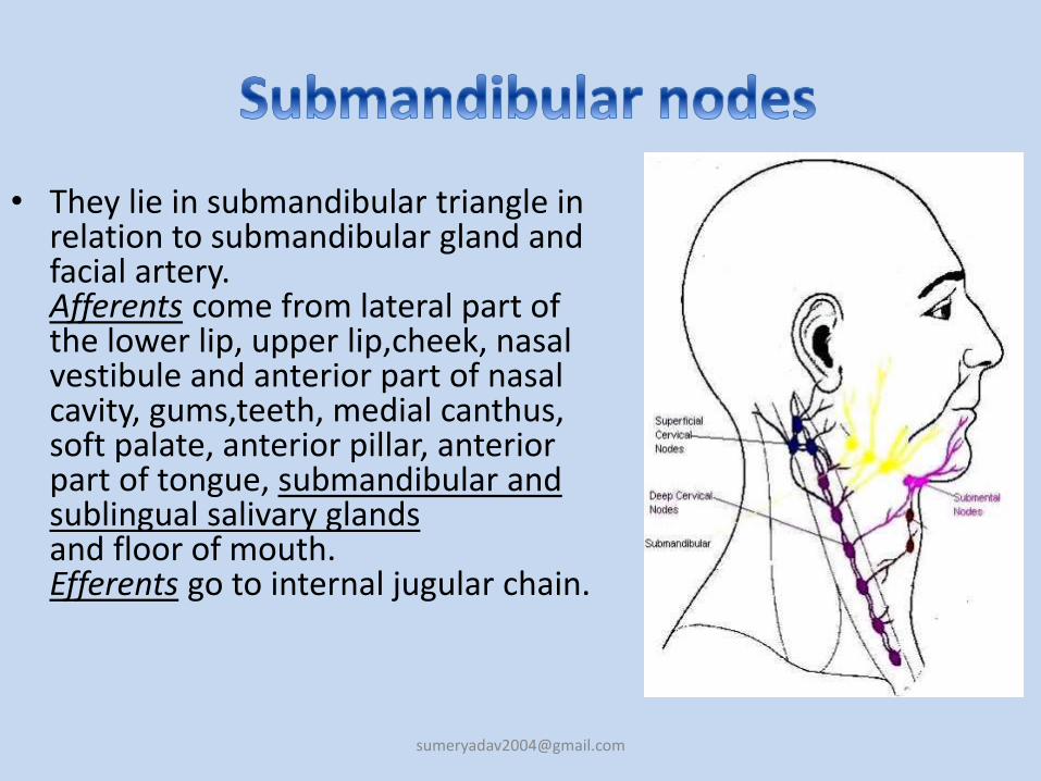
• They lie in submandibular triangle in relation to submandibular gland and facial artery.Afferents come from lateral part of the lower lip, upper lip,cheek, nasal vestibule and anterior part of nasal cavity, gums,teeth, medial canthus, soft palate, anterior pillar, anterior part of tongue, submandibular and sublingual salivary glandsand floor of mouth. Efferents go to internal jugular chain.
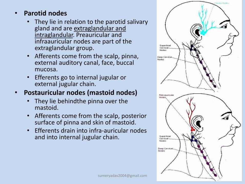
• Parotid nodes• They lie in relation to the parotid salivary
gland and are extraglandular and intraglandular. Preauricular and infraauricular nodes are part of the extraglandular group.
• Afferents come from the scalp, pinna, external auditory canal, face, buccal mucosa.
• Efferents go to internal jugular or external jugular chain.
• Postauricular nodes (mastoid nodes)• They lie behindthe pinna over the
mastoid.• Afferents come from the scalp, posterior
surface of pinna and skin of mastoid.• Efferents drain into infra-auricular nodes
and into internal jugular chain.
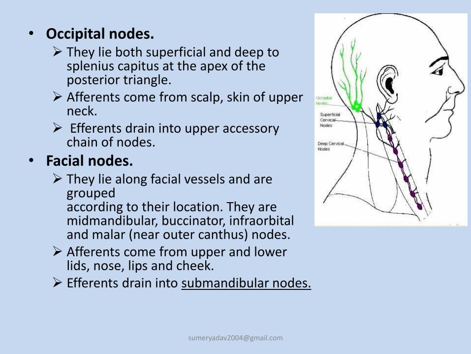
• Occipital nodes. They lie both superficial and deep to
splenius capitus at the apex of the posterior triangle.
Afferents come from scalp, skin of upper neck.
Efferents drain into upper accessory chain of nodes.
• Facial nodes. They lie along facial vessels and are
groupedaccording to their location. They are midmandibular, buccinator, infraorbital and malar (near outer canthus) nodes.
Afferents come from upper and lower lids, nose, lips and cheek.
Efferents drain into submandibular nodes.
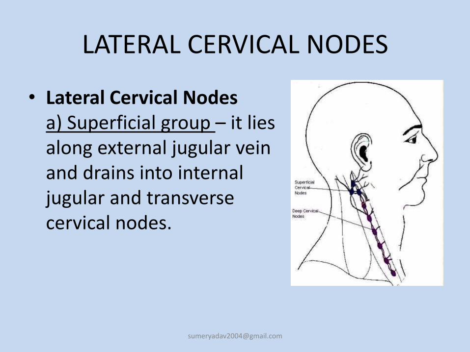
LATERAL CERVICAL NODES
• Lateral Cervical Nodesa) Superficial group – it liesalong external jugular veinand drains into internaljugular and transversecervical nodes.
b.Deep Group
• It consists of three chains,
1. the internal jugular chain2. spinal accessory and3. Transverse cervical
• Internal jugular chainLymph nodes of internal jugular chain lie anterior, lateral and posterior to internal jugular vein.Upper group (jugulodigastric node) – drains oral cavity, orpharynx, nasopharynx, hypopharynx, larynx and parotid.Middle group drains hypopharynx, larynx, throid, oral cavity, oropharynx.Lower jugular group drains larynx, thyroid and cervical oesophagus.
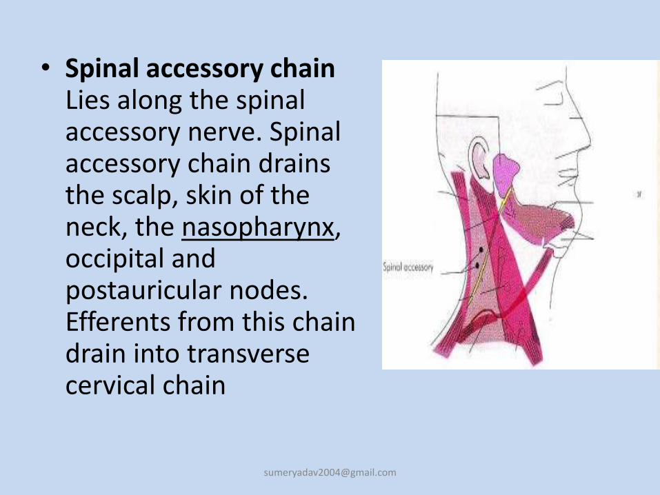
• Spinal accessory chainLies along the spinal accessory nerve. Spinal accessory chain drains the scalp, skin of the neck, the nasopharynx, occipital andpostauricular nodes.Efferents from this chain drain into transverse cervical chain
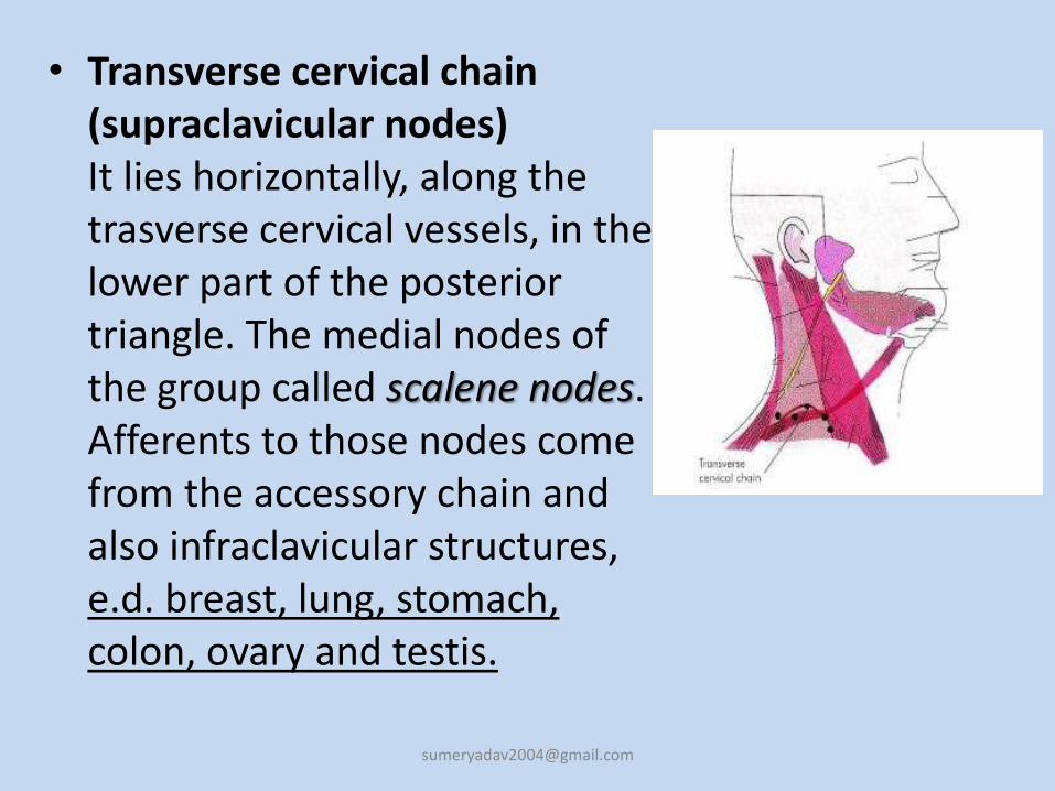
• Transverse cervical chain (supraclavicular nodes)It lies horizontally, along the trasverse cervical vessels, in the lower part of the posterior triangle. The medial nodes of the group called scalene nodes. Afferents to those nodes come from the accessory chain and also infraclavicular structures, e.d. breast, lung, stomach, colon, ovary and testis.
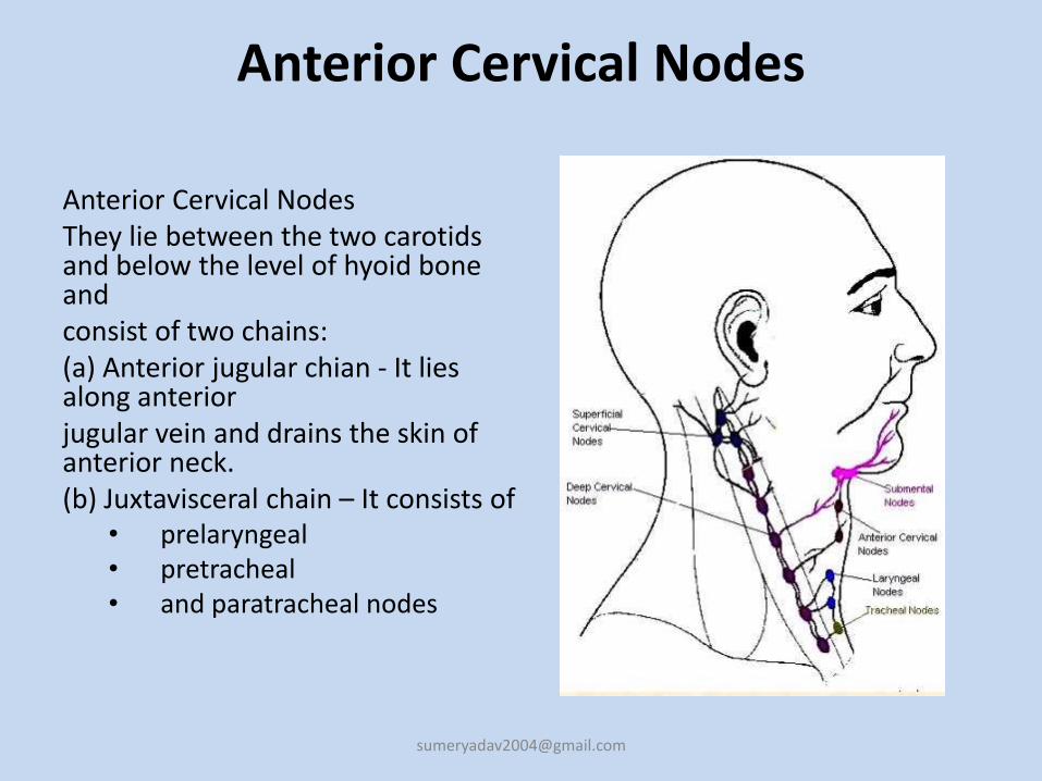
Anterior Cervical Nodes
Anterior Cervical NodesThey lie between the two carotids and below the level of hyoid bone andconsist of two chains:(a) Anterior jugular chian - It lies along anteriorjugular vein and drains the skin of anterior neck.(b) Juxtavisceral chain – It consists of
• prelaryngeal• pretracheal• and paratracheal nodes
(i) Prelaryngeal node (Delphian node)lies on cricothyroid membrane and drains subgottic
region of larynx and pyriform sinuses.
(ii) Pretracheal nodeslie in front of the trachea, and drainthyroid gland and the trachea. Efferents from thesenodes go to paratracheal, lower internal jugular andanterior mediastinal nodes.
(iii) Paratracheal Nodes drain the thyroid lobes, subglotticlarynx, tracha and cervical oesophagus
• Post cervical: scalp, neck skin of arms thorax cervical and axillary nodes (lymphoma, head/neck ca)
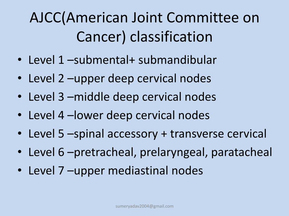
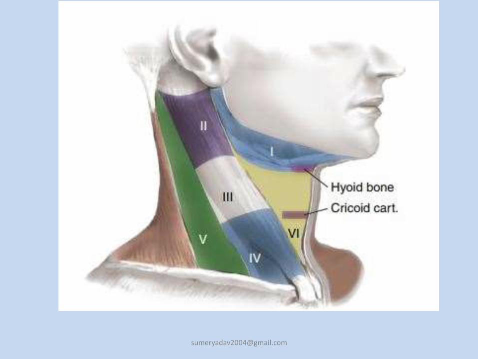
AJCC(American Joint Committee on Cancer) classification
• Level 1 –submental+ submandibular
• Level 2 –upper deep cervical nodes
• Level 3 –middle deep cervical nodes
• Level 4 –lower deep cervical nodes
• Level 5 –spinal accessory + transverse cervical
• Level 6 –pretracheal, prelaryngeal, paratacheal
• Level 7 –upper mediastinal nodes
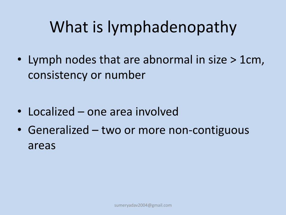
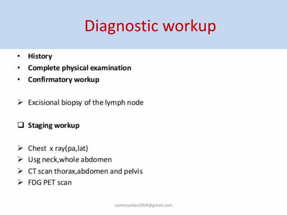
What is lymphadenopathy
• Lymph nodes that are abnormal in size > 1cm, consistency or number
• Localized – one area involved
• Generalized – two or more non-contiguous areas
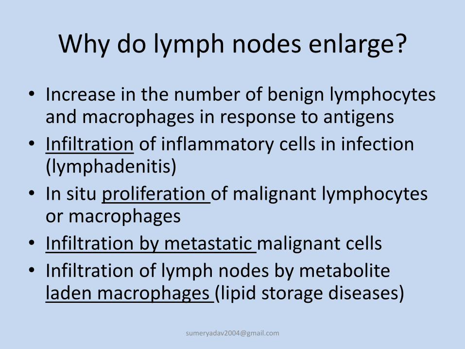
Why do lymph nodes enlarge?
• Increase in the number of benign lymphocytes and macrophages in response to antigens
• Infiltration of inflammatory cells in infection (lymphadenitis)
• In situ proliferation of malignant lymphocytes or macrophages
• Infiltration by metastatic malignant cells
• Infiltration of lymph nodes by metabolite laden macrophages (lipid storage diseases)
Epidemiology
• 0.6% annual incidence of unexplained adenopathy in the general population
• 10% were referred to a subspecialist and 3.2 % required a biopsy and 1.1% had a malignancy
When to worry?
• Age
• Characteristics of the node
• Location of the node
• Clinical setting associated with lymphadenopathy
Age
• Children/young adults – more likely to respond to minor stimuli with lymphoid hyperplasia– Lymph nodes in patients less than the age of 30
are clinically benign in 80% of cases whereas in patients over the age of 50 only 40% are benign
– Biopsies done in patients less than 25 yrs have a incidence of malignancy of <20% vs the over-50 age group has an incidence of malignancy of 55-80%
Clinical examination
• Localized adenopathy should prompt a search for an adjacent precipitating lesion and an examination of other nodal areas to rule out generalized lymphadenopathy. In general, lymph nodes greater than 1 cm in diameter are considered to be abnormal. Supraclavicular nodes are the most worrisome for malignancy. A three- to four-week period of observation is prudent in patients with localized nodes and a benign clinical picture.
• The body has approximately 600 lymph nodes, but only those in the submandibular, axillary or inguinal regions may normally be palpable in healthy people.1 Lymphadenopathy refers to nodes that are abnormal in either size, consistency or number. There are various classifications of lymphadenopathy, but a simple and clinically useful system is to classify lymphadenopathy as “generalized” if lymph nodes are enlarged in two or more noncontiguous areas or “localized” if only one area is involved.
Characteristics of the node
• Nodes lasting less than 2 weeks or greater than one year with no progression of size have a low likelihood of being neoplastic – excludes low grade lymphoma
• Cervical nodes – up to 56% of young adults have adenopathy on clinical exam
• Inguinal adenopathy is common – up to 1-2 cm in size and often benign reactive nodes
Characteristics of the node
i. Consistency – Hard/Firm vs Soft/Shotty; Fluctuant
ii. Mobile vs Fixed/Matted
iii. Tender vs Painless
iv. Clearly demarcated
v. Sizei. When to worry – 1.5-2cm in size
ii. Epitroclear nodes over 0.5cm; Inguinal over 1.5cm
vi. Duration and Rate of Growth
vii. Mobile vs fixed
viii. Symmetrical vs asymmetrical
Consistency
• Stony hard: typical of cancer usually metastatic• Firm rubbery: can suggest lymphoma• Soft: infection or inflammation• Fluctuant : Suppurated nodes.• Matting : . A group of nodes that feels connected
and seems to move as a unit is said to be “matted.” Nodes that are matted can be either benign (e.g., tuberculosis, sarcoidosis or
lymphogranuloma venereum) or malignant (e.g., metastatic carcinoma or
lymphomas).
Pain/Tenderness
• When a lymph node rapidly increases in size, its capsule stretches and causes pain. Pain is usually the result of an inflammatory process or suppuration, but pain may also result from hemorrhage into the necrotic center of a malignant node. The presence or absence of tenderness does not reliably differentiate benign from malignant nodes
size• in one series of 213 adults with unexplained
lymphadenopathy, no patient with a lymph node smaller than 1 cm2 (1
cm × 1 cm) had cancer, while cancer was present in 8 percent of those with
nodes from 1 cm2 to 2.25 cm2 (1 cm × 1 cm to 1.5 cm × 1.5 cm) in size, and
in 38 percent of those with nodes larger than 2.25 cm2 (1.5 cm × 1.5 cm).
• In children, lymph nodes larger than 2 cm in diameter (along with an abnormal chest radiograph and the absence of ear, nose and throat symptoms) were predictive of granulomatous diseases (i.e., tuberculosis, cat-scratch disease or sarcoidosis) or cancer (predominantly lymphomas).
Location of the node
• The anatomic location of localized adenopathy will sometimes be helpful in narrowing the differential diagnosis. For example, cat-scratch disease typically causes cervical or axillary adenopathy, infectious mononucleosis causes cervical adenopathy and a number of sexually transmitted diseases are associated with inguinal adenopathy .
Location of the node
• Supraclavicular lymphadenopathy Highest risk of malignancy – estimated as 90% in
patients older than 40 years vs 25% in those younger than 40 yrs
Right sided node – cancer in mediastinum, lungs, esophagus
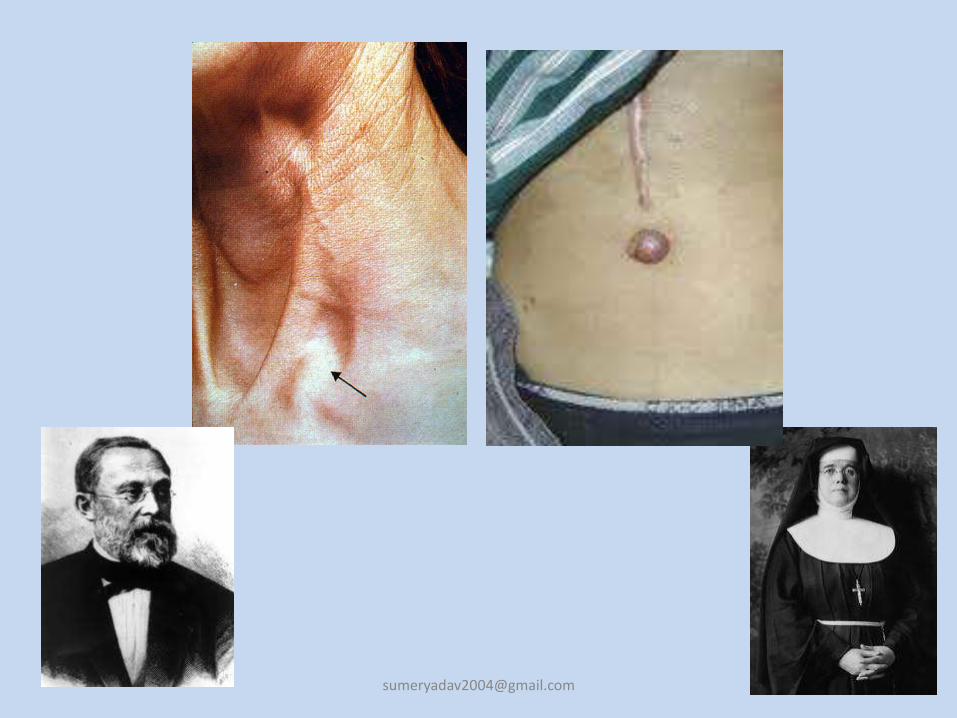
Left sided node (Virchow’s) – testes, ovaries, kidneys, pancreas, stomach, gallbladder or prostate
• Paraumbilical node (Sister mary Joseph’s)– Abdominal or pelvic neoplasm
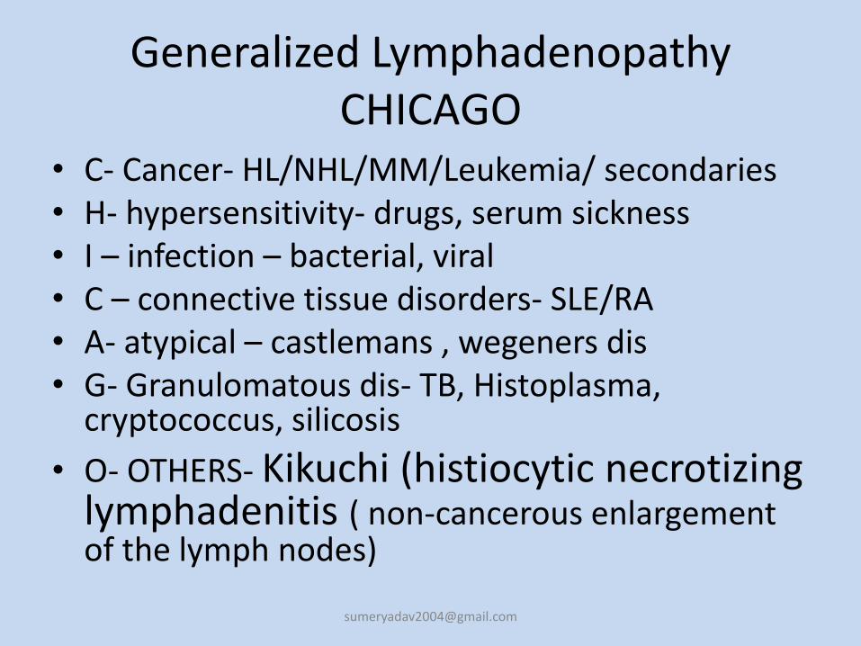
Generalized LymphadenopathyCHICAGO
• C- Cancer- HL/NHL/MM/Leukemia/ secondaries• H- hypersensitivity- drugs, serum sickness• I – infection – bacterial, viral• C – connective tissue disorders- SLE/RA• A- atypical – castlemans , wegeners dis• G- Granulomatous dis- TB, Histoplasma,
cryptococcus, silicosis
• O- OTHERS- Kikuchi (histiocytic necrotizing lymphadenitis ( non-cancerous enlargement of the lymph nodes)
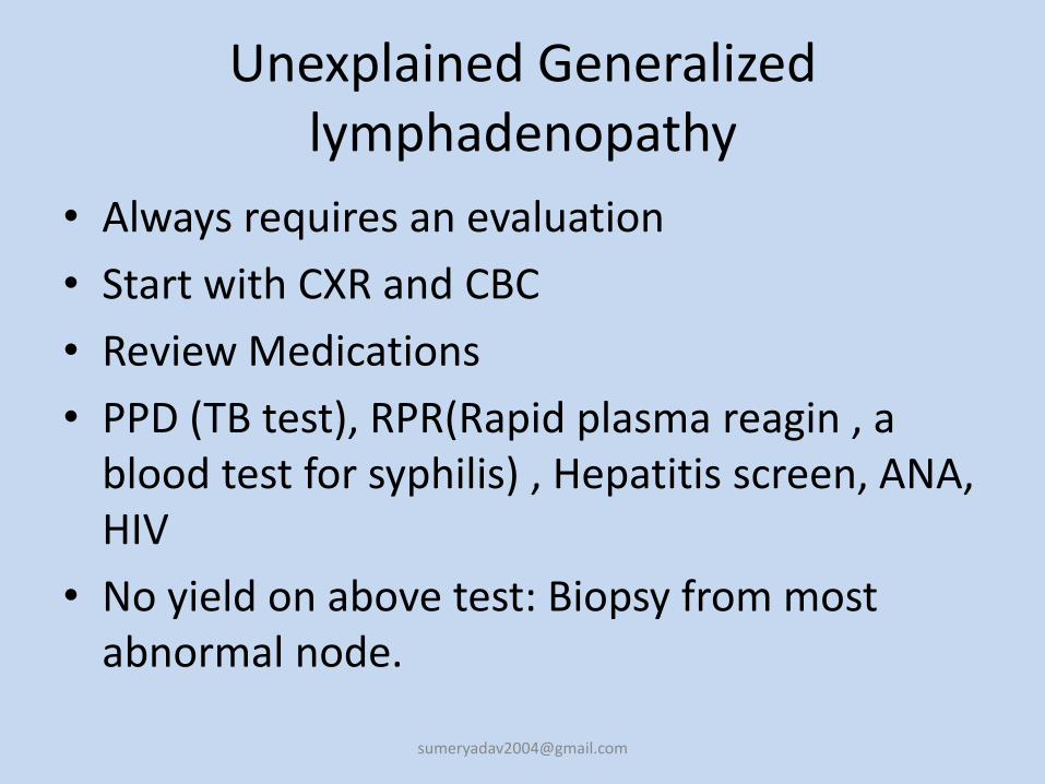
Unexplained Generalized lymphadenopathy
• Always requires an evaluation
• Start with CXR and CBC
• Review Medications
• PPD (TB test), RPR(Rapid plasma reagin , a blood test for syphilis) , Hepatitis screen, ANA, HIV
• No yield on above test: Biopsy from most abnormal node.
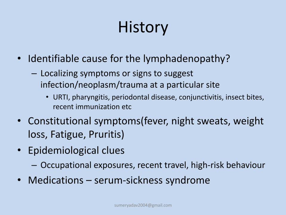
History
• Identifiable cause for the lymphadenopathy?
– Localizing symptoms or signs to suggest infection/neoplasm/trauma at a particular site• URTI, pharyngitis, periodontal disease, conjunctivitis, insect bites,
recent immunization etc
• Constitutional symptoms(fever, night sweats, weight loss, Fatigue, Pruritis)
• Epidemiological clues
– Occupational exposures, recent travel, high-risk behaviour
• Medications – serum-sickness syndrome
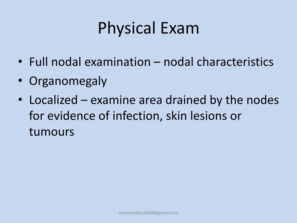
Physical Exam
• Full nodal examination – nodal characteristics
• Organomegaly
• Localized – examine area drained by the nodes for evidence of infection, skin lesions or tumours
Radiographic Investigation of the Head and Neck Masses
• MRI – Magnetic Resonance Imaging can clearly highlight soft tissue pathologies better than the C.T. Scan.– It uses a magnetic field rather than x-rays (radiation).
• CT SCAN – Computed tomography is less accurate than M.R.I for the soft tissue examination, but is very useful to locate bony tumorsand their dimensions and extensions.– C.T with contrast is used to enhance the visibility of abnormal tissue
during examination.
• PET (Positron Emission Tomography) and SPECT (Single Photon Emission Tomography) are useful after diagnosis to help determine the grade of a tumor or to distinguish between cancerous and dead or scar tissue.– They involve injection with a radioactive tracer.
• Gallium scanning
Fine Needle Aspirate
• Safe Convenient, less invasive, quicker turn-around time• especially beneficial for verification of lymphoid
origin of the enlarged growth and in differentiatingbetween metastatic, infectious, reactive andlymphomatous causes of lymphadenopathy. It alsohelps in the determination of the extent of tumor;detection of recurrence; monitoring of the course ofdisease; obtaining of material for special studies suchas microbiological cultures, immunological or genetic studies as well as electron microscopy. Furthermore
• Most patients with a benign diagnosis on FNA biopsy do not undergo a surgical biopsy
• overall sensitivity was 92.7%, specificity 98.5%
• If the LN arenot palpable, endoscopic ultrasound-guided fineneedle aspiration (EUS-FNA) has been shown to accurately diagnose mediastinal lymph nodepathology with diagnostic accuracy of 84%
• endobronchial ultrasound guided transbronchialneedle aspiration (EBUS-TBNA) have been shown tobe highly sensitive and specific in the diagnosis ofmediastinal and hilar lesions
• Limitations of FNA:
– the lack of propertissue sample to run special studies including cytogenetics, flow cytometry, electron microscopy,
– the potential risk of seeding a tract with malignancy as a result of FNA
BIOPSY• Can be done by bedside, open surgery, mediastinoscopy FNA
cannot distinguish between lymphomas (nodal architecture needs to be intact) The preservation of nodal architecture is critical to theproper diagnosis of lymphadenopathy, particularly when differentiating lymphoma from benign reactive hyperplasia
• Biopsy should be avoided in patients with probable viral illnessbecause lymph node pathology in these patients may sometimes simulate lymphoma and lead to a false-positive diagnosis of malignancy.
• The diagnostic yield of the biopsy can be maximized by obtaining an excisional biopsy of the largest and most abnormal node (which is not necessarily the most accessible node). If possible, the physician should not select inguinal and axillary nodes for biopsy, since they frequently show only reactive hyperplasia.
• Patients should be cautioned to remain alert for the reappearance of the nodes because lymphomatous nodes have been known to temporarily regress.
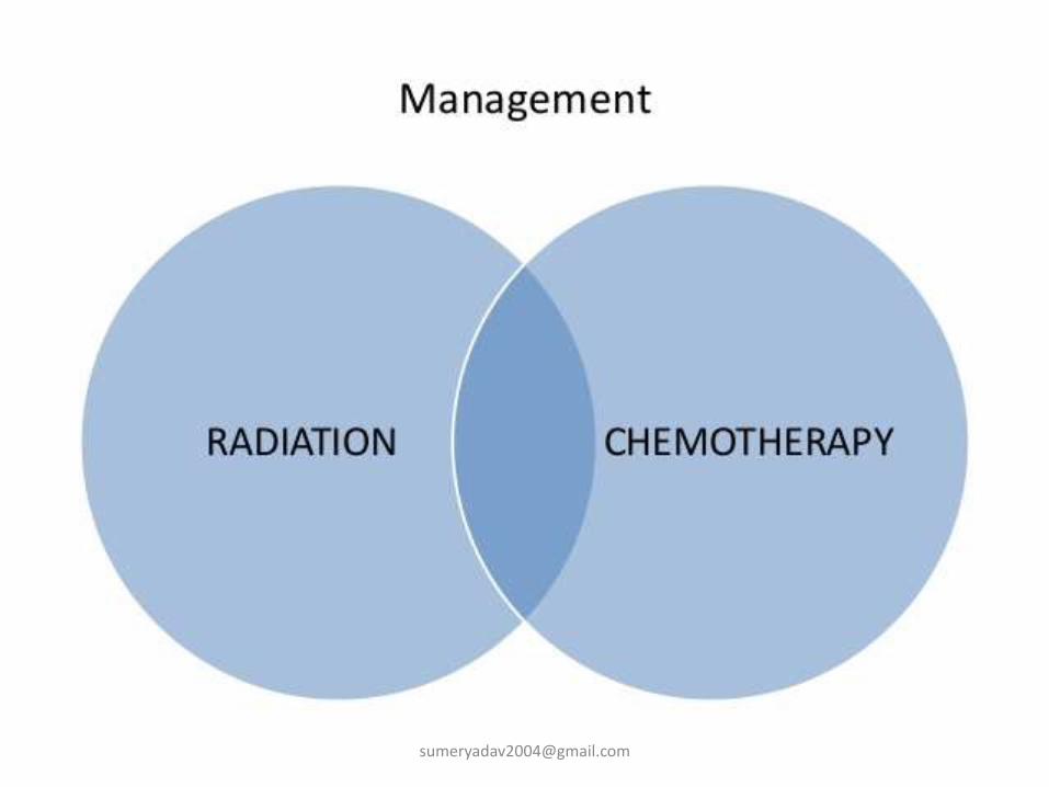
Management
• Identify underlying cause and treat as appropriate – confirmatory tests
• Generalized adenopathy – usually has identifiable cause
• Localized adenopathy
– 3-4 week observation period for resolution if not high clinical suspicion for malignancy
– Biopsy if risk for malignancy - excisional
Microbiology
• M. tuberculosis is the usual cause of tuberculouslymphadenitis.
• Other infectious causes of chronic lymphadenitis include :
– Nontuberculous mycobacteria (including M. scrofulaceum, M. avium, and M. haemophilum)
– Pseudomonas pseudomallei
– Toxoplasma species
– Bartonella species
– Fungi.
Symptomatology
• Patients do not generally report significant pain at presentation.
• Node tenderness during examination is noted in only 10%–35% of cases.
• A draining sinus may be present in 4%–11% of cases.
• Unilateral involvement of 1–3 nodes has been noted in 85% of cases.
• Cervical chain involvement is most common (45%–70%) with 12%–26% in the supraclavicularregion; 20% of cases are bilateral.
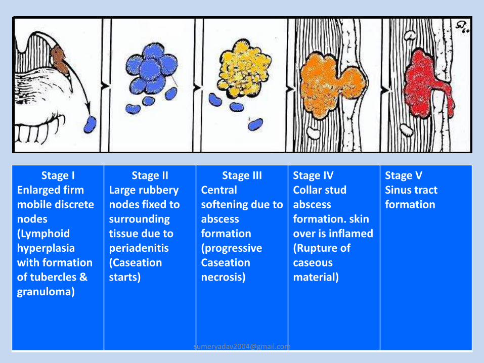
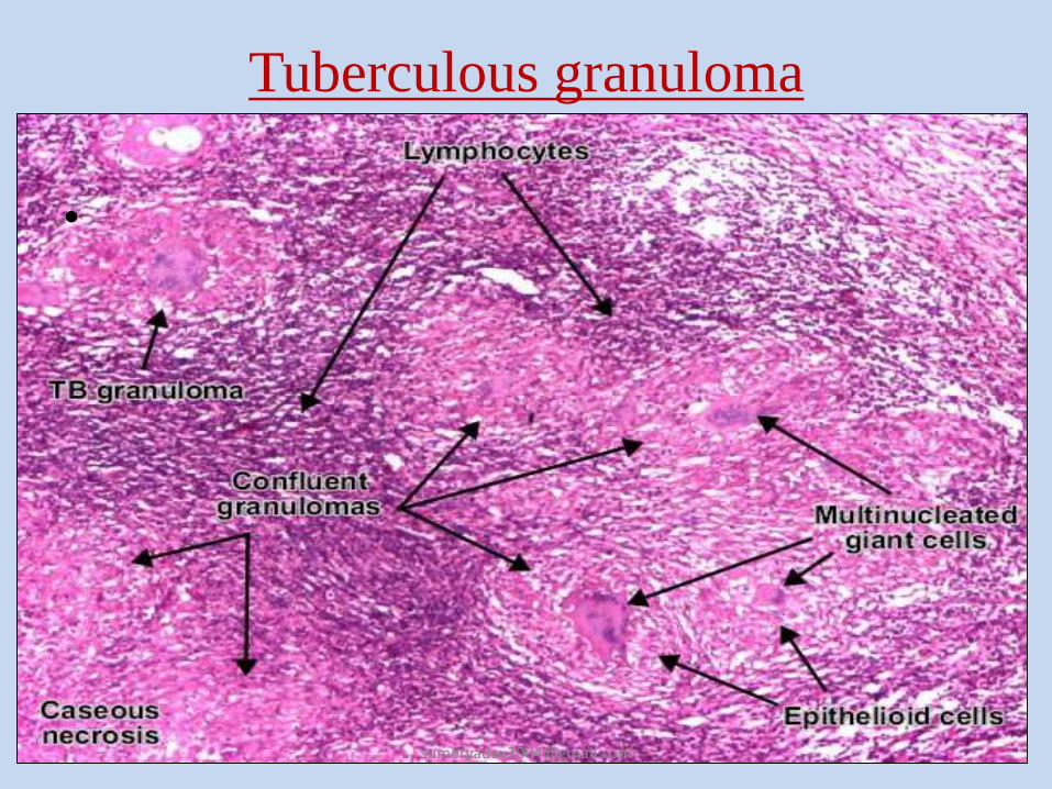
Stage I Enlarged firm mobile discrete nodes(Lymphoid hyperplasia with formation of tubercles & granuloma)
Stage IILarge rubbery nodes fixed to surrounding tissue due to periadenitis(Caseationstarts)
Stage IIICentral softening due to abscess formation(progressive Caseationnecrosis)
Stage IVCollar stud abscess formation. skin over is inflamed(Rupture of caseousmaterial)
Stage VSinus tract formation
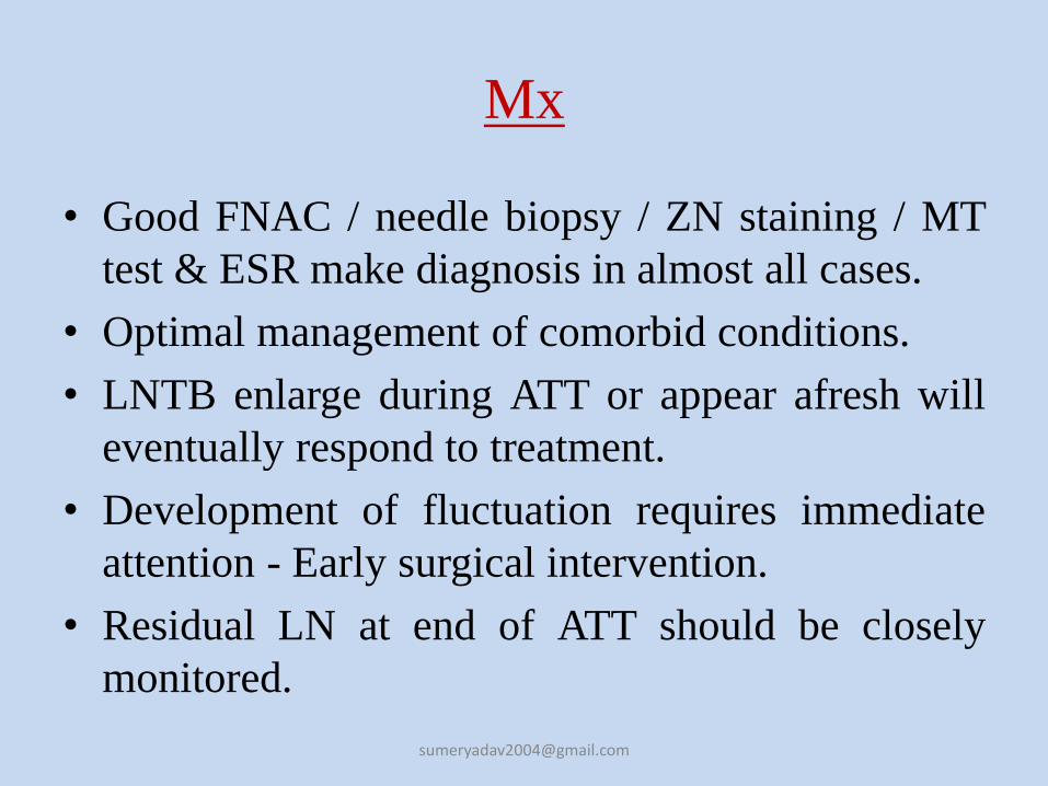
Mx
• Good FNAC / needle biopsy / ZN staining / MT
test & ESR make diagnosis in almost all cases.
• Optimal management of comorbid conditions.
• LNTB enlarge during ATT or appear afresh will
eventually respond to treatment.
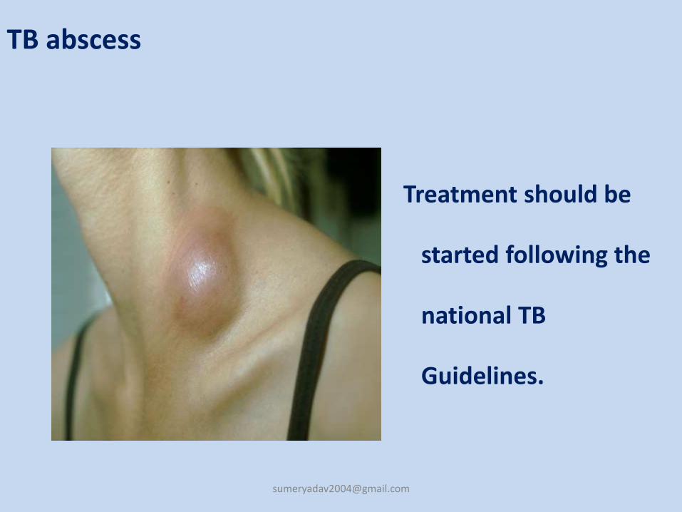
• Development of fluctuation requires immediate
attention - Early surgical intervention.
• Residual LN at end of ATT should be closely
monitored.
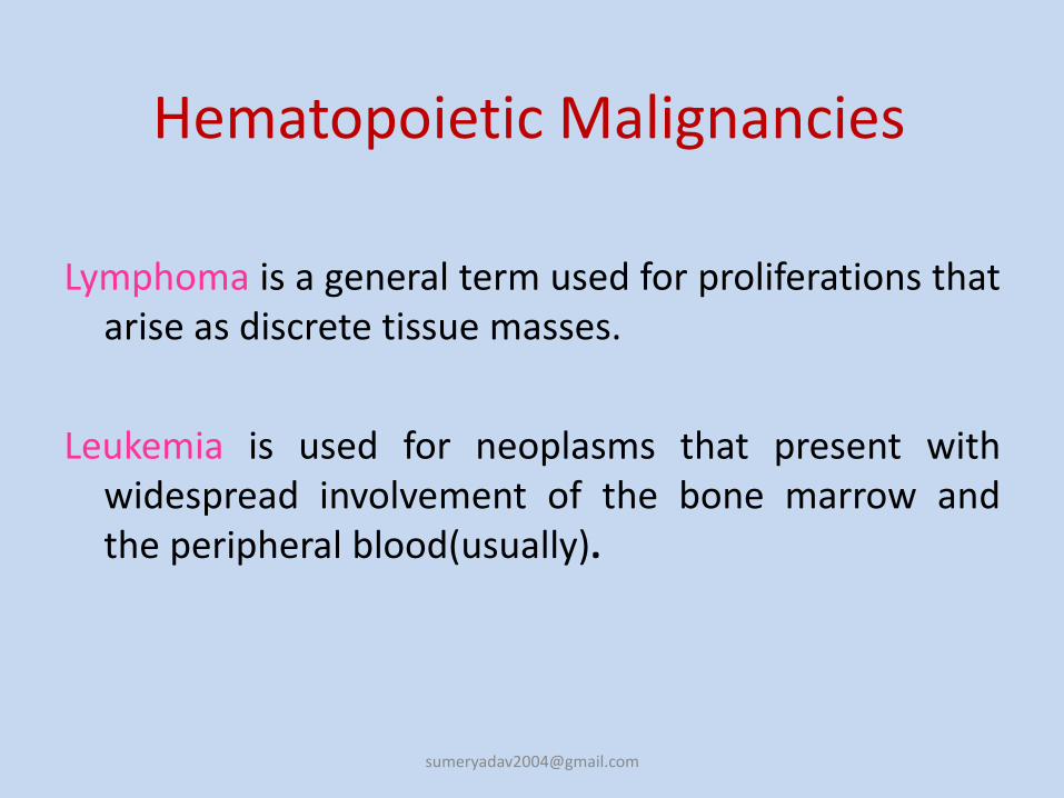
Hematopoietic Malignancies
Lymphoma is a general term used for proliferations thatarise as discrete tissue masses.
Leukemia is used for neoplasms that present withwidespread involvement of the bone marrow andthe peripheral blood(usually).
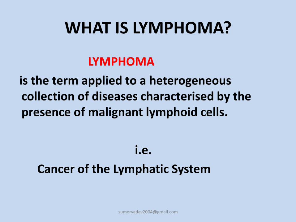
WHAT IS LYMPHOMA?
LYMPHOMA
is the term applied to a heterogeneous collection of diseases characterised by the presence of malignant lymphoid cells.
i.e.
Cancer of the Lymphatic System
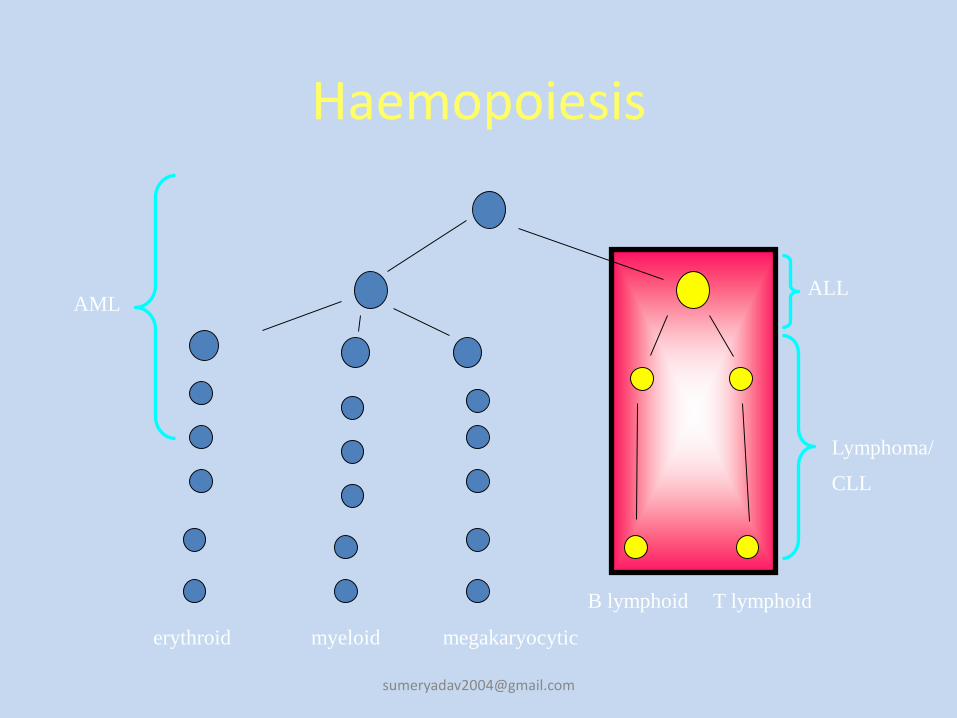
Haemopoiesis
erythroid myeloid megakaryocytic
B lymphoid T lymphoid
AML
Lymphoma/
CLL
ALL
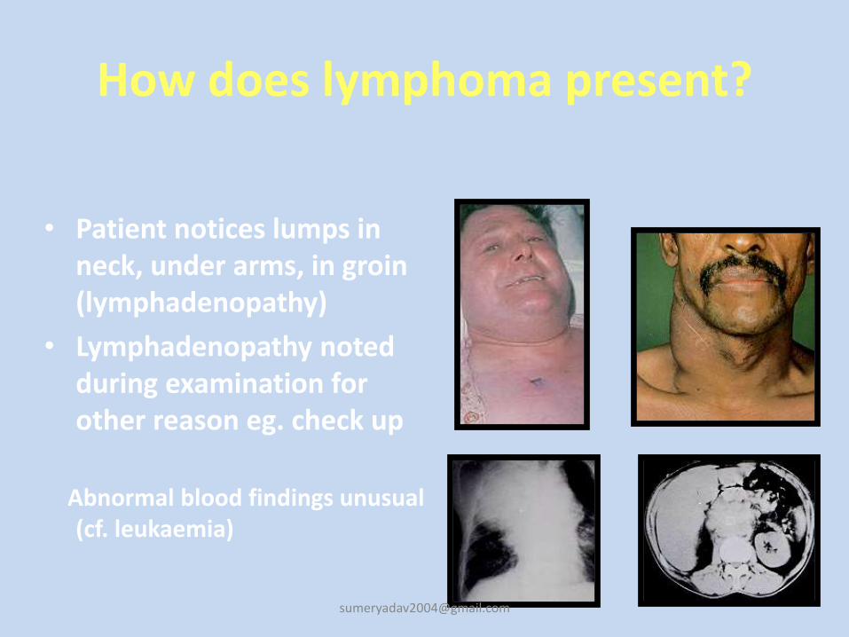
How does lymphoma present?
• Patient notices lumps in neck, under arms, in groin (lymphadenopathy)
• Lymphadenopathy noted during examination for other reason eg. check up
Abnormal blood findings unusual (cf. leukaemia)
Clinical features
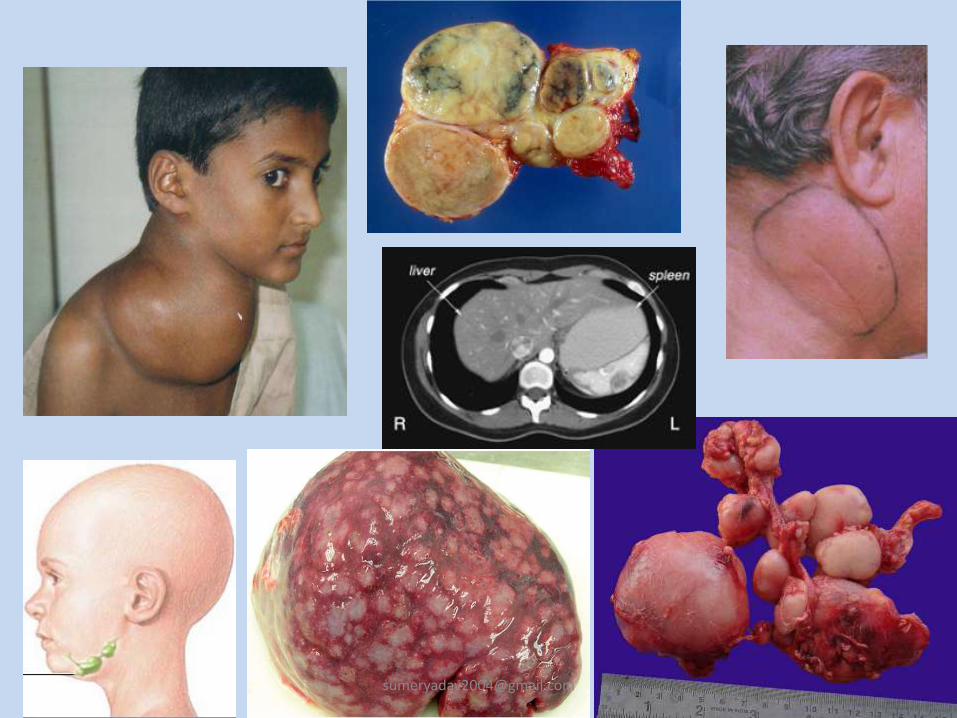
• Most common presentation is asymptomatic lymphnode enlargement, typically in the neck.
• Cervical lymphnodes are involved in 80% cases.
• Mediastinal involvement is seen in about 50% cases. They produce symptoms like chest pain, cough and dyspnoea.
• Infradiaphrgamatic involvement is seen in 5% cases and usually seen with older patients.
• Other less common symptoms are :
Pruritis, alcohol induced pain over involved lymphnodes, nephrotic syndrome, erythema nodosum, cerebellar degeneration, immune hemolytic anaemia, thrombocytopenia, hypercalcemia.
B symptoms
• About 33 % present with B symptoms overall
• Only 15-20% of stage I-III have B symptoms like
1. Fever(>38^C)
• May first present as fever of unknown origin
• Fever persists for days to weeks followed by afebrile intervals and then recurrence.
• This pattern is called Pel Ebstein fever.
2. Drenching night sweats
3. Weight loss (>10% in 6 months)
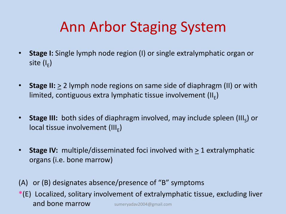
Ann Arbor Staging System
• Stage I: Single lymph node region (I) or single extralymphatic organ or site (IE)
• Stage II: > 2 lymph node regions on same side of diaphragm (II) or with limited, contiguous extra lymphatic tissue involvement (IIE)
• Stage III: both sides of diaphragm involved, may include spleen (IIIS) or local tissue involvement (IIIE)
• Stage IV: multiple/disseminated foci involved with > 1 extralymphaticorgans (i.e. bone marrow)
(A) or (B) designates absence/presence of “B” symptoms
*(E) Localized, solitary involvement of extralymphatic tissue, excluding liver and bone marrow [email protected]
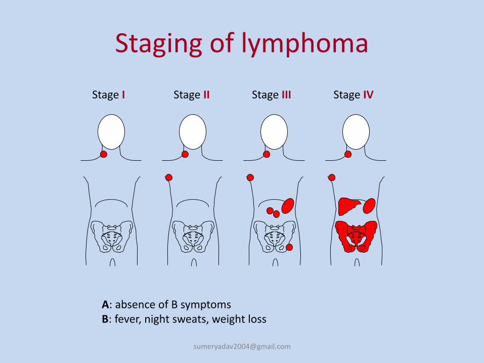
Stage I Stage II Stage III Stage IV
Staging of lymphoma
A: absence of B symptomsB: fever, night sweats, weight loss
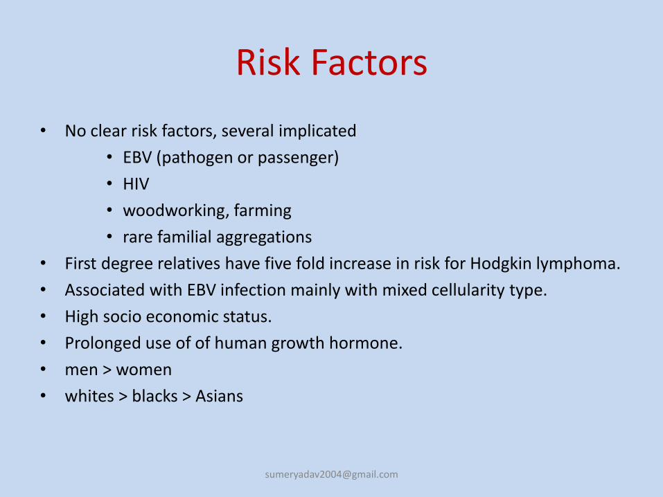
Risk Factors
• No clear risk factors, several implicated
• EBV (pathogen or passenger)
• HIV
• woodworking, farming
• rare familial aggregations
• First degree relatives have five fold increase in risk for Hodgkin lymphoma.
• Associated with EBV infection mainly with mixed cellularity type.
• High socio economic status.
• Prolonged use of of human growth hormone.
• men > women
• whites > blacks > Asians
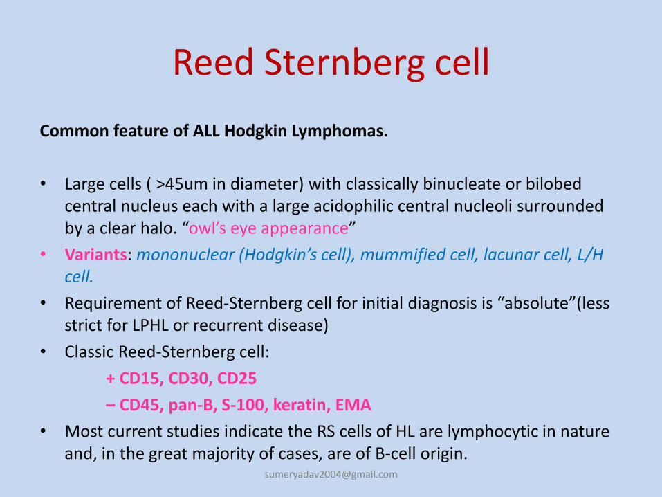
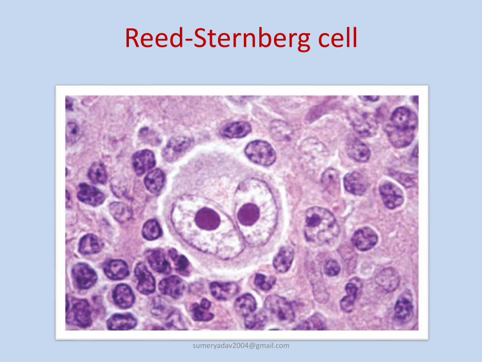
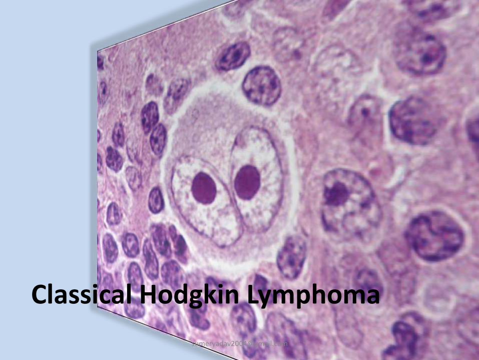
Reed Sternberg cell
Common feature of ALL Hodgkin Lymphomas.
• Large cells ( >45um in diameter) with classically binucleate or bilobedcentral nucleus each with a large acidophilic central nucleoli surrounded by a clear halo. “owl’s eye appearance”
• Variants: mononuclear (Hodgkin’s cell), mummified cell, lacunar cell, L/H cell.
• Requirement of Reed-Sternberg cell for initial diagnosis is “absolute”(less strict for LPHL or recurrent disease)
• Classic Reed-Sternberg cell:
+ CD15, CD30, CD25
– CD45, pan-B, S-100, keratin, EMA
• Most current studies indicate the RS cells of HL are lymphocytic in nature and, in the great majority of cases, are of B-cell origin.
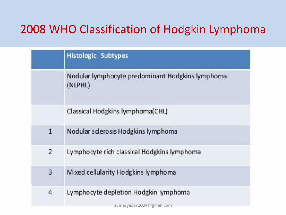
Lymphocyte predominant Hodgkin lymphoma
• <5% of Hodgkin lymphoma
• Mainly involves cervical, axillary or mediastinal
• L&H cells or Popcorn cells are seen
• Positive for CD20, 45, CD79a, Bcl-6, J-chain, and PAX-5. EMA positive in 50% cases.
• Negative for CD15, 30.
• Differential Diagnosis: Well differentiated lymphocytic lymphoma, mononucleosis, malignant melanoma,, progressive transformation of germinal centers
1. Nodular Sclerosis
• Most common type diagnosed
• About 70%
• Lacunar cells seen
• CD15, 30 positive
• EBV negative
• Only subtype without a male predominance
• Seen in younger patients with stage I-II disease.
• Differential diagnosis: Large cell Non Hodgkin lymphoma, carcinoma, germ cell tumour and thymoma.
2. Mixed Cellularity
• Constitutes about 20%
• More than 50% present as stage III or IV disease
• Biphasic incidence, peaking in young adults and again in adults older than 55
• CD15, 30, EBV positive
• Presents in advanced stages
• Tendency to involve spleen, bone marrow.
• Differential diagnosis: Some cases of MCHL display an interfolliculargrowth pattern. Such cases may be difficult to distinguish from peripheral T-cell lymphomas. Lennert’s lymphoma (diffuse mixed T-cell ML with excessive histiocytes). Diffuse follicular lymphoma.
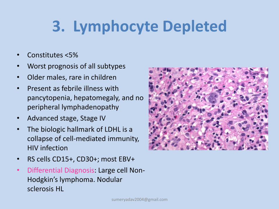
3. Lymphocyte Depleted
• Constitutes <5%
• Worst prognosis of all subtypes
• Older males, rare in children
• Present as febrile illness with pancytopenia, hepatomegaly, and no peripheral lymphadenopathy
• Advanced stage, Stage IV
• The biologic hallmark of LDHL is a collapse of cell-mediated immunity, HIV infection
• RS cells CD15+, CD30+; most EBV+
• Differential Diagnosis: Large cell Non-Hodgkin’s lymphoma. Nodular sclerosis HL
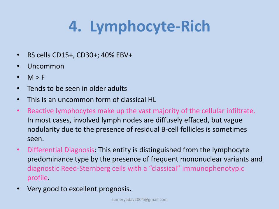
4. Lymphocyte-Rich
• RS cells CD15+, CD30+; 40% EBV+
• Uncommon
• M > F
• Tends to be seen in older adults
• This is an uncommon form of classical HL
• Reactive lymphocytes make up the vast majority of the cellular infiltrate. In most cases, involved lymph nodes are diffusely effaced, but vague nodularity due to the presence of residual B-cell follicles is sometimes seen.
• Differential Diagnosis: This entity is distinguished from the lymphocyte predominance type by the presence of frequent mononuclear variants and diagnostic Reed-Sternberg cells with a “classical” immunophenotypicprofile.
• Very good to excellent [email protected]
Spread
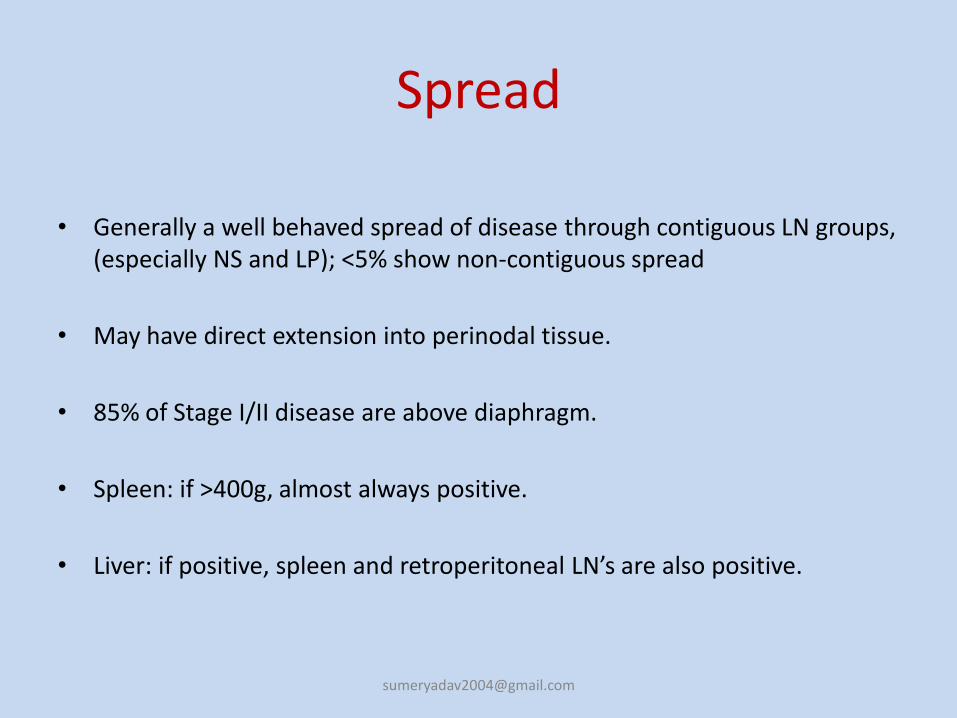
• Generally a well behaved spread of disease through contiguous LN groups, (especially NS and LP); <5% show non-contiguous spread
• May have direct extension into perinodal tissue.
• 85% of Stage I/II disease are above diaphragm.
• Spleen: if >400g, almost always positive.
• Liver: if positive, spleen and retroperitoneal LN’s are also positive.
PROGNOSIS
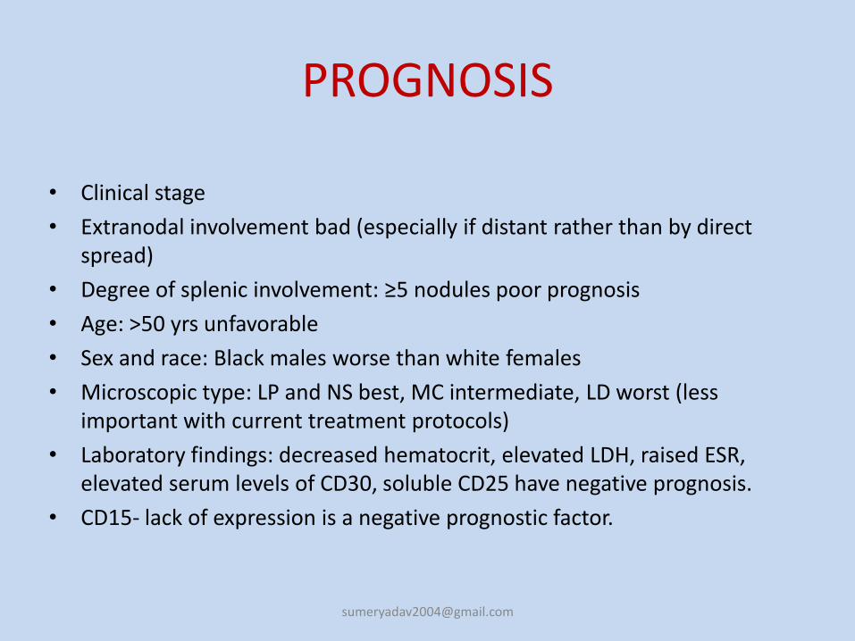
• Clinical stage
• Extranodal involvement bad (especially if distant rather than by direct spread)
• Degree of splenic involvement: ≥5 nodules poor prognosis
• Age: >50 yrs unfavorable
• Sex and race: Black males worse than white females
• Microscopic type: LP and NS best, MC intermediate, LD worst (less important with current treatment protocols)
• Laboratory findings: decreased hematocrit, elevated LDH, raised ESR, elevated serum levels of CD30, soluble CD25 have negative prognosis.
• CD15- lack of expression is a negative prognostic factor.
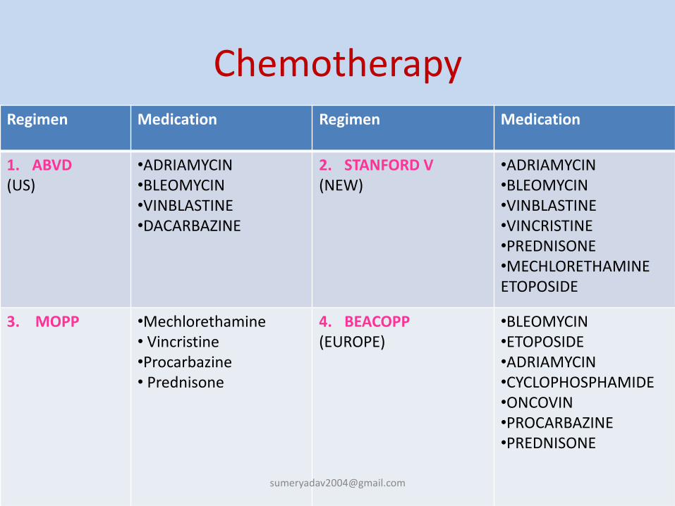
Chemotherapy Regimen Medication Regimen Medication
1. ABVD(US)
•ADRIAMYCIN•BLEOMYCIN•VINBLASTINE•DACARBAZINE
2. STANFORD V(NEW)
•ADRIAMYCIN•BLEOMYCIN•VINBLASTINE•VINCRISTINE•PREDNISONE•MECHLORETHAMINEETOPOSIDE
3. MOPP •Mechlorethamine• Vincristine•Procarbazine• Prednisone
4. BEACOPP(EUROPE)
•BLEOMYCIN•ETOPOSIDE•ADRIAMYCIN•CYCLOPHOSPHAMIDE•ONCOVIN•PROCARBAZINE•PREDNISONE
Radiotherapy
• Radiation therapy is the most effective single thrapeutic agent for treating Hodgkin lymphoma.
• The main objective of radiation in Hodgkin lymphoma is to treat involved and contiguous field.
• Radiotherapy can be given by
1. 2D planning
2. 3D planning
3. IFRT
• Involved field radiotherapy is the most commonly used technique at present. It targets a smaller area rather than a classical extended field.
Complications
• Autologous bone marrow transplantation can cure half of patients who fail effective chemotherapy regimens.
• Because of the very high cure rate in patients with Hodgkin's disease, long-term complications have become a major focus for clinical research. The most serious late side effects include secondary malignancies, cardiac injury, infertility and Lhermitte's syndrome.
Non-Hodgkin’s lymphomas-definition and epidemiology
1. Definition: malignant disease of the lymphoid system, highly heterogeneous, both histologicallyand clinically.
2. Epidemiology:
- annual incidence: 5-10 new cases per 100 000 persons,
- age distribution: middle-age patients and the elderly,
- males are affected more often than females (1.5:1.0).
Non-Hodgkin’s lymphomas-Clinical features
1. Constitutional symptoms (fever, night sweats, weight loss)
2. Lymphadenopathy
(cervical, supraclavicular, axillary, inguinal, mediastinal, retroperitoneal, mesenteric, pelvic).
3. Mediastinal adenopathy (T cell lymphoma)
4. Extralymphatic involvement (gastrointestinal, testicular masses, solitary bone lesions, CNS).
5. Unexplained anemia and thrombocytopenia ( bone marrow infiltration).
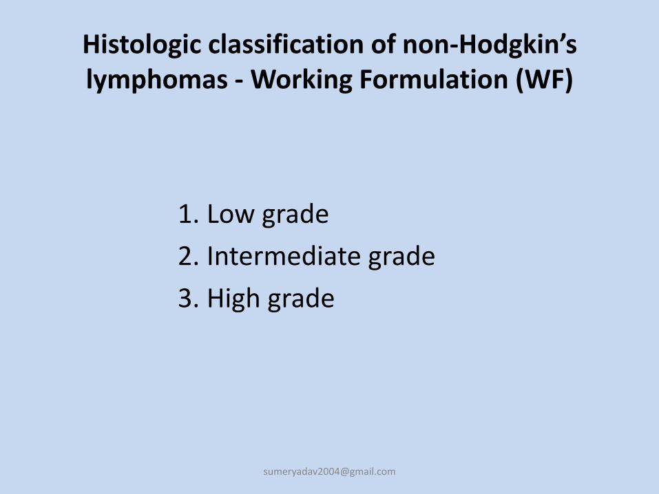
Histologic classification of non-Hodgkin’s lymphomas - Working Formulation (WF)
1. Low grade
2. Intermediate grade
3. High grade
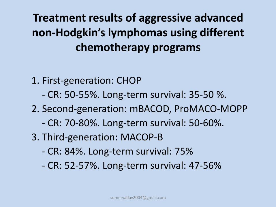
Treatment results of aggressive advanced non-Hodgkin’s lymphomas using different
chemotherapy programs
1. First-generation: CHOP
- CR: 50-55%. Long-term survival: 35-50 %.
2. Second-generation: mBACOD, ProMACO-MOPP
- CR: 70-80%. Long-term survival: 50-60%.
3. Third-generation: MACOP-B
- CR: 84%. Long-term survival: 75%
- CR: 52-57%. Long-term survival: 47-56%
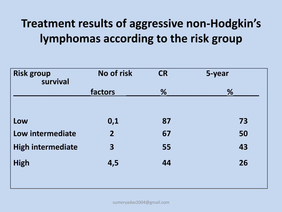
Treatment results of aggressive non-Hodgkin’s lymphomas according to the risk group
Risk group No of risk CR 5-year survival
__________________factors ________%______________%______
Low 0,1 87 73
Low intermediate 2 67 50
High intermediate 3 55 43
High 4,5 44 26


![Annals of Clinical Case Reports Case Report · epithelium are found in axillary lymph nodes [14,15]. Ectopic thyroid tissueis found occasionally in cervical lymph nodes. Among nonepithelial](https://static.fdocuments.net/doc/165x107/5f1cd159d6b56138e82777d7/annals-of-clinical-case-reports-case-epithelium-are-found-in-axillary-lymph-nodes.jpg)