Cerebrovascular anatomy
79
Cerebrovascular Anatomy Naresh Mullaguri MD Neurology Resident Physician PGY4 University of Missouri-Columbia
-
Upload
cleveland-clinic-foundation -
Category
Health & Medicine
-
view
80 -
download
0
Transcript of Cerebrovascular anatomy

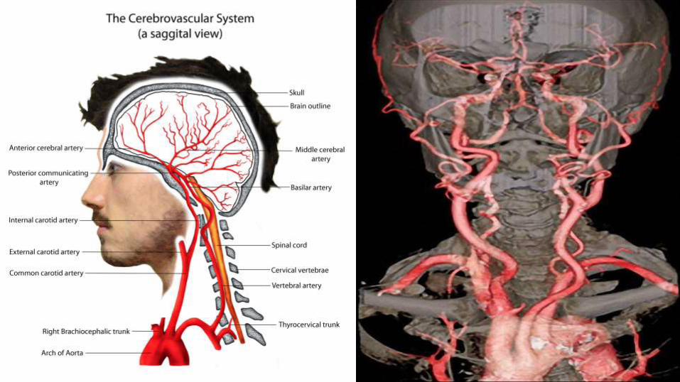
Cerebrovascular Anatomy
Naresh Mullaguri MD
Neurology Resident Physician PGY4
University of Missouri-Columbia

Cerebral Circulation Math
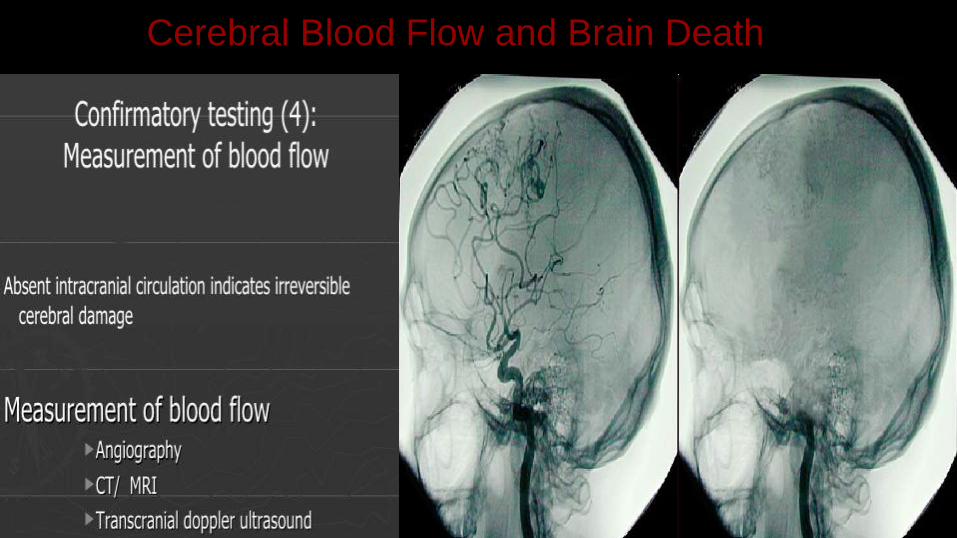
Cerebral Blood Flow and Brain Death
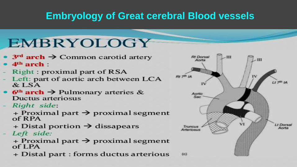
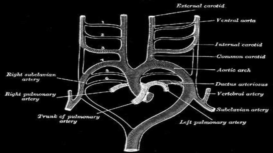
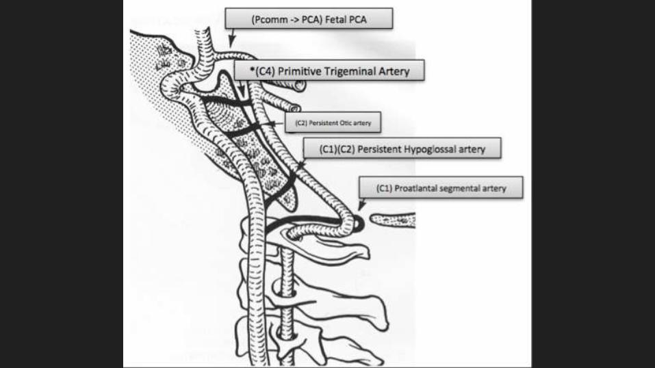
Embryology of Great cerebral Blood vessels
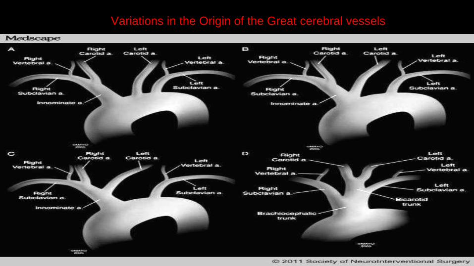
Variations in the Origin of the Great cerebral vessels
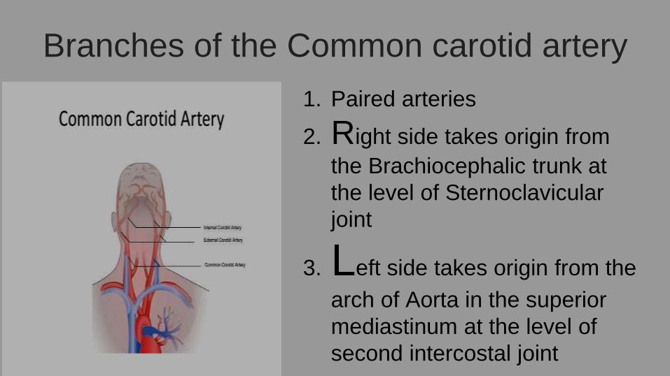
Branches of the Common carotid artery
1. Paired arteries
2. Right side takes origin from
the Brachiocephalic trunk at
the level of Sternoclavicular
joint
3. Left side takes origin from the
arch of Aorta in the superior
mediastinum at the level of
second intercostal joint
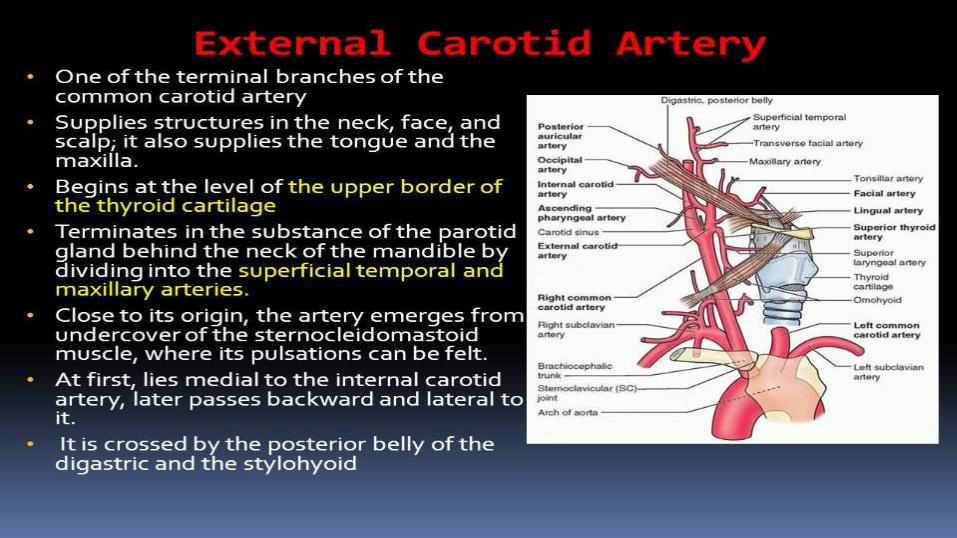
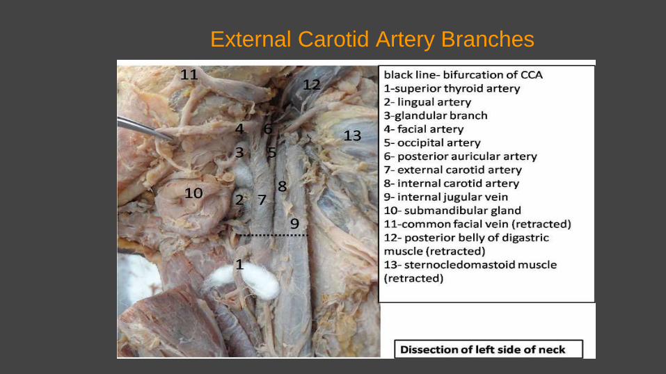
External Carotid Artery Branches
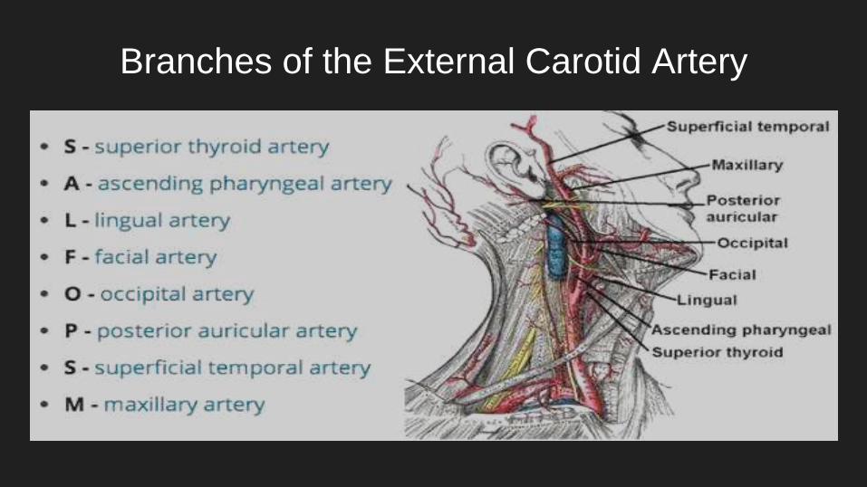
Branches of the External Carotid Artery

Who don’t like Mnemonics
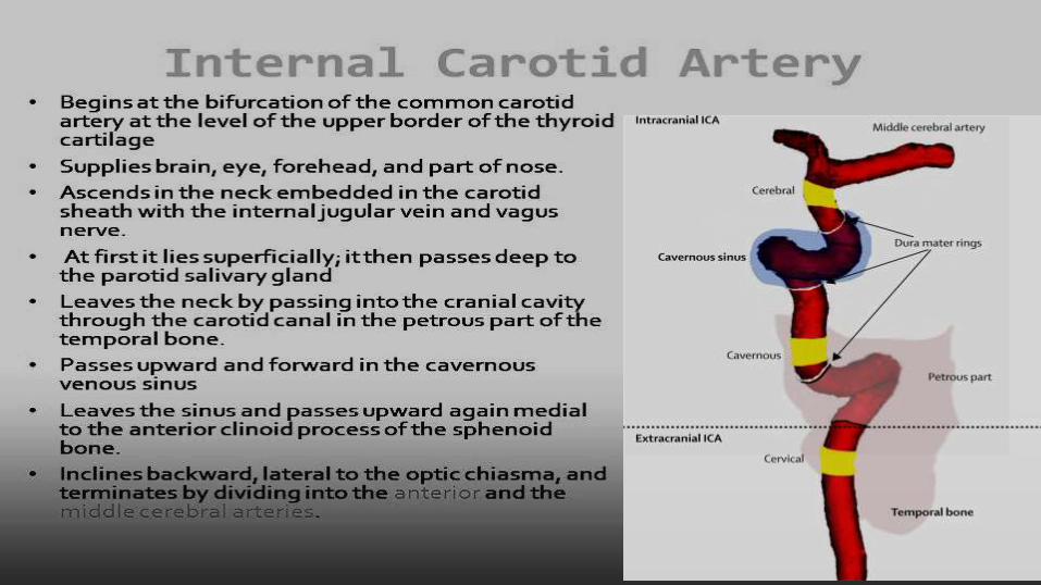
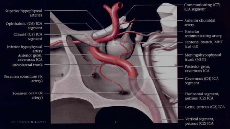
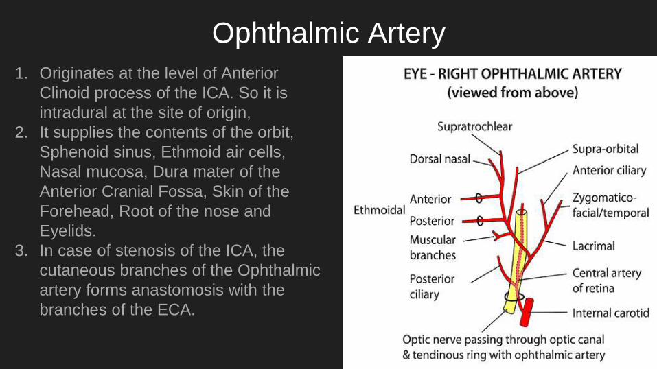
Ophthalmic Artery
1. Originates at the level of Anterior
Clinoid process of the ICA. So it is
intradural at the site of origin,
2. It supplies the contents of the orbit,
Sphenoid sinus, Ethmoid air cells,
Nasal mucosa, Dura mater of the
Anterior Cranial Fossa, Skin of the
Forehead, Root of the nose and
Eyelids.
3. In case of stenosis of the ICA, the
cutaneous branches of the Ophthalmic
artery forms anastomosis with the
branches of the ECA.
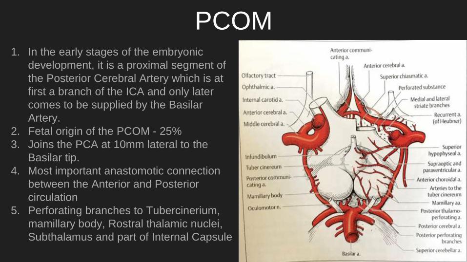
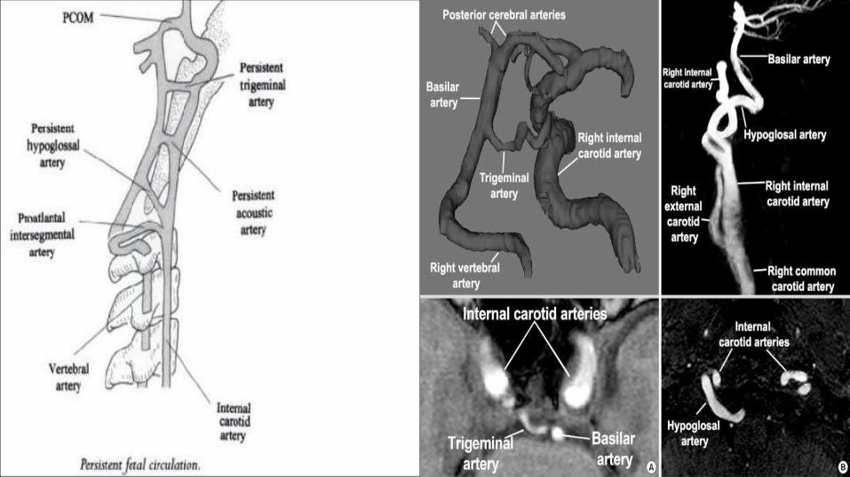
PCOM1. In the early stages of the embryonic
development, it is a proximal segment of
the Posterior Cerebral Artery which is at
first a branch of the ICA and only later
comes to be supplied by the Basilar
Artery.
2. Fetal origin of the PCOM - 25%
3. Joins the PCA at 10mm lateral to the
Basilar tip.
4. Most important anastomotic connection
between the Anterior and Posterior
circulation
5. Perforating branches to Tubercinerium,
mamillary body, Rostral thalamic nuclei,
Subthalamus and part of Internal Capsule
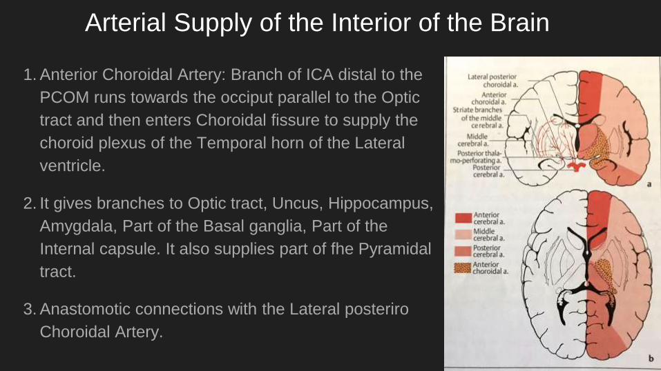
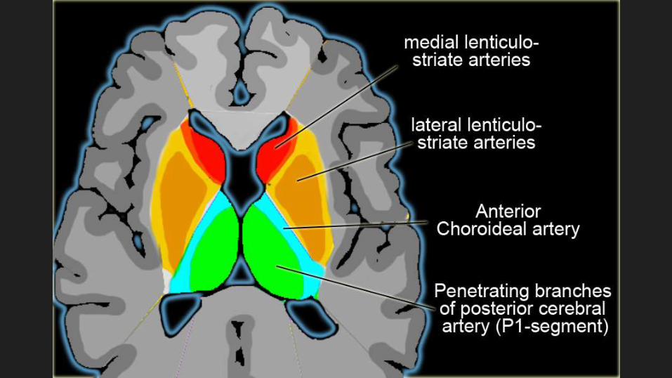
Arterial Supply of the Interior of the Brain
1. Anterior Choroidal Artery: Branch of ICA distal to the
PCOM runs towards the occiput parallel to the Optic
tract and then enters Choroidal fissure to supply the
choroid plexus of the Temporal horn of the Lateral
ventricle.
2. It gives branches to Optic tract, Uncus, Hippocampus,
Amygdala, Part of the Basal ganglia, Part of the
Internal capsule. It also supplies part of fhe Pyramidal
tract.
3. Anastomotic connections with the Lateral posteriro
Choroidal Artery.
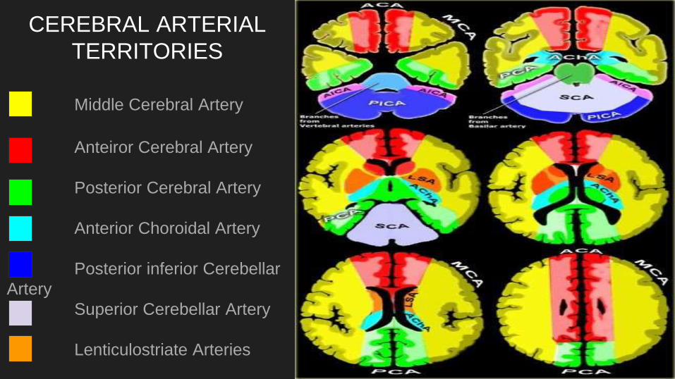
CEREBRAL ARTERIAL
TERRITORIES
Middle Cerebral Artery
Anteiror Cerebral Artery
Posterior Cerebral Artery
Anterior Choroidal Artery
Posterior inferior Cerebellar
Artery
Superior Cerebellar Artery
Lenticulostriate Arteries
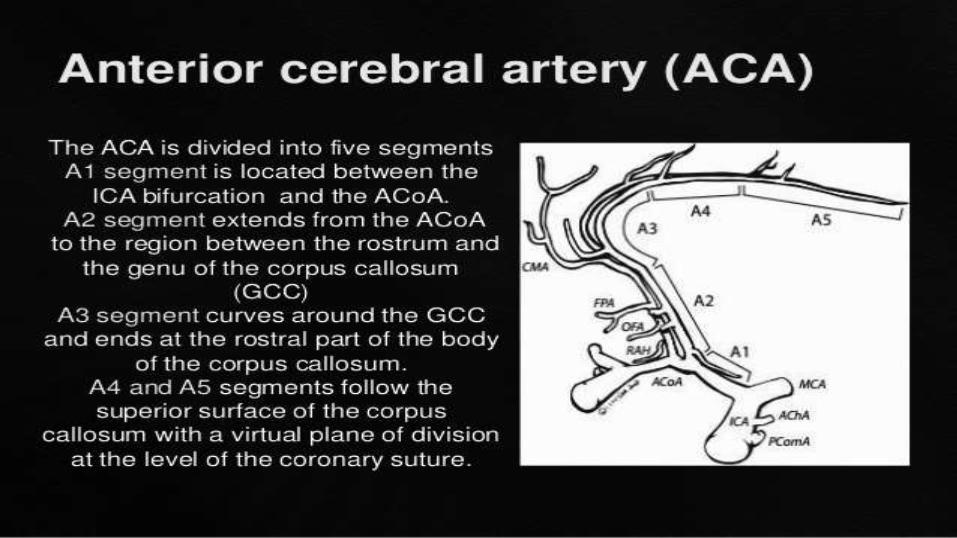
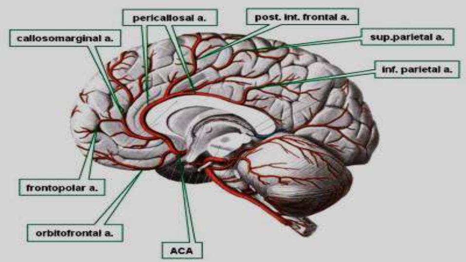
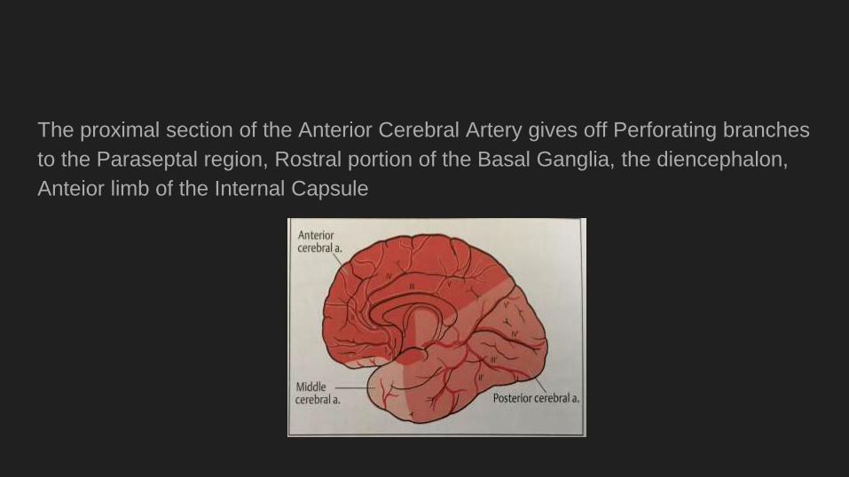
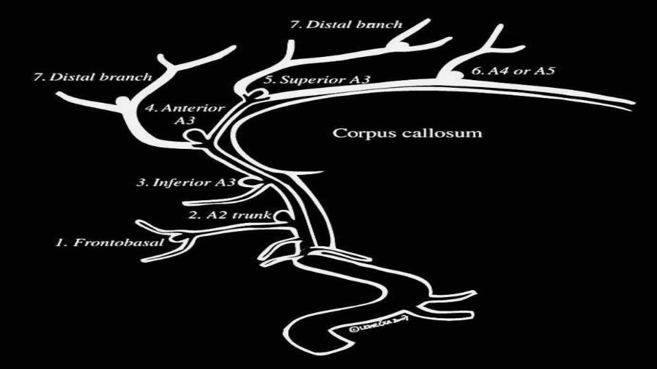
The proximal section of the Anterior Cerebral Artery gives off Perforating branches
to the Paraseptal region, Rostral portion of the Basal Ganglia, the diencephalon,
Anteior limb of the Internal Capsule
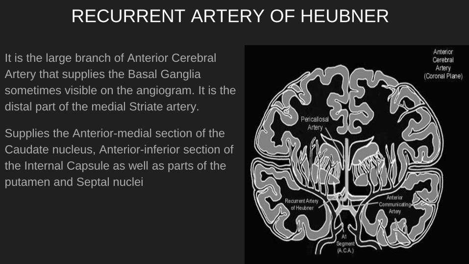
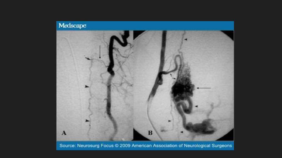
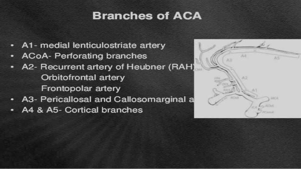
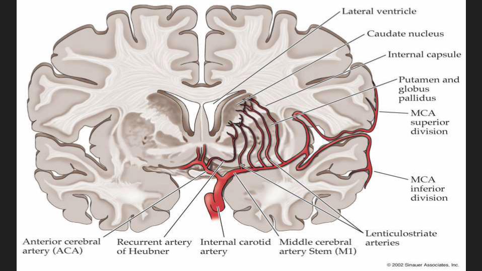
RECURRENT ARTERY OF HEUBNER
It is the large branch of Anterior Cerebral
Artery that supplies the Basal Ganglia
sometimes visible on the angiogram. It is the
distal part of the medial Striate artery.
Supplies the Anterior-medial section of the
Caudate nucleus, Anterior-inferior section of
the Internal Capsule as well as parts of the
putamen and Septal nuclei
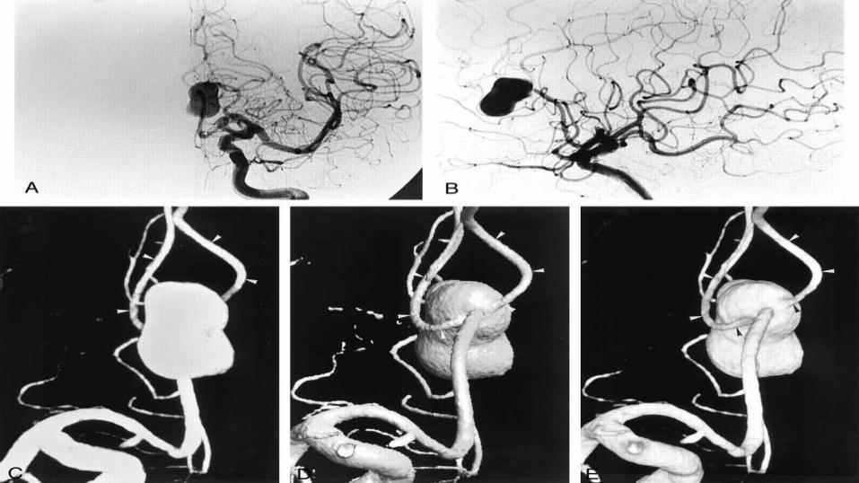
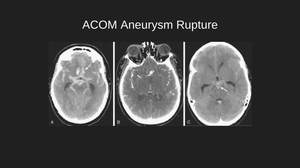
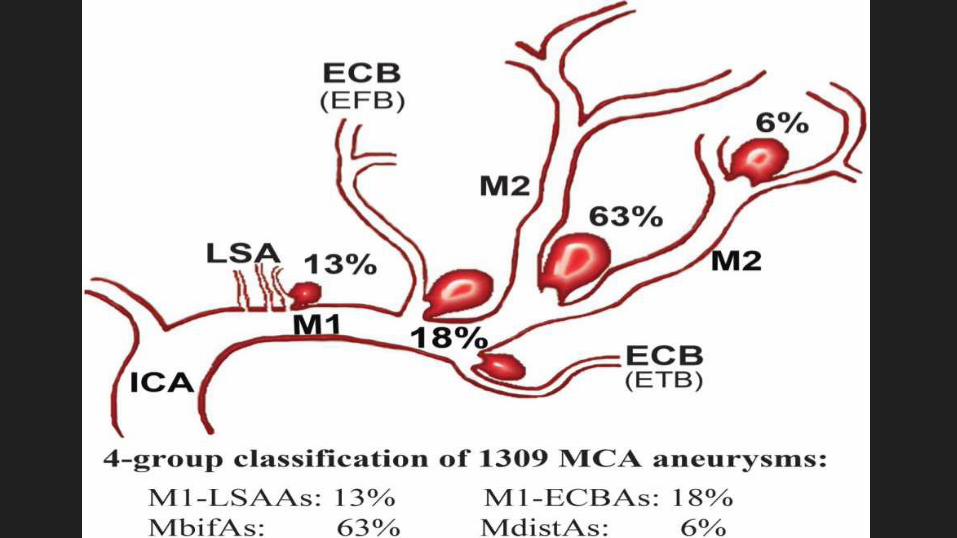
ACOM Aneurysm Rupture

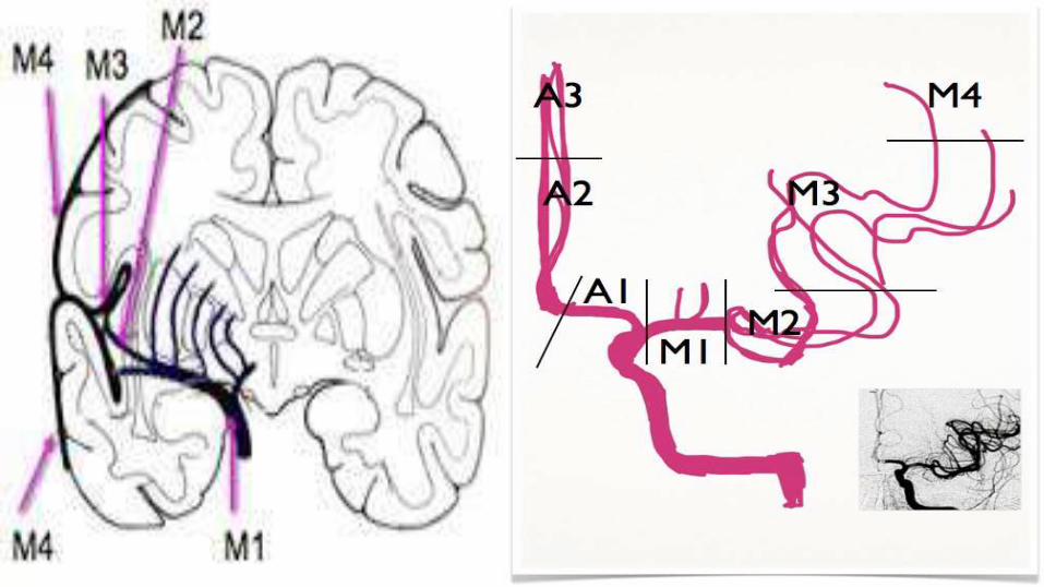
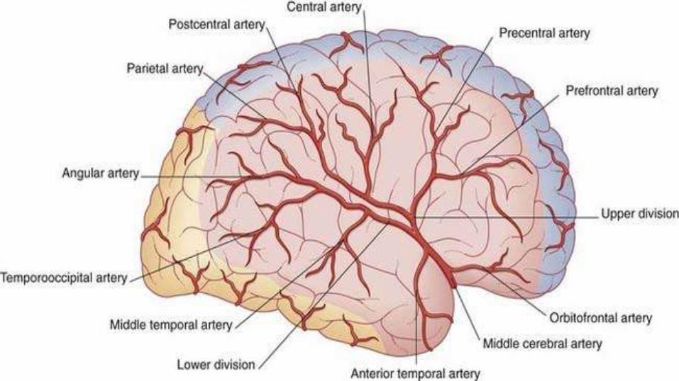
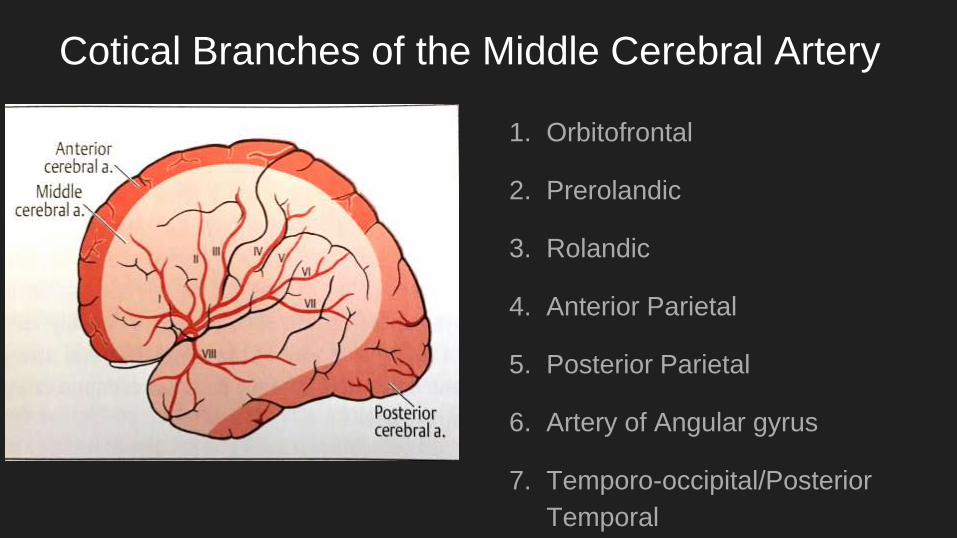
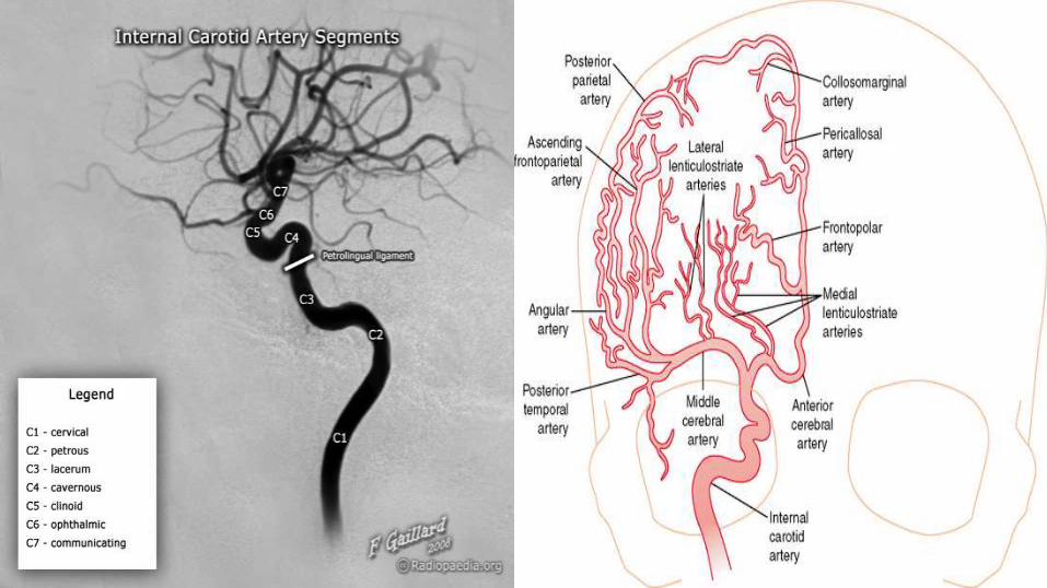
Cotical Branches of the Middle Cerebral Artery
1. Orbitofrontal
2. Prerolandic
3. Rolandic
4. Anterior Parietal
5. Posterior Parietal
6. Artery of Angular gyrus
7. Temporo-occipital/Posterior
Temporal
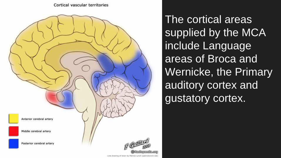
The cortical areas
supplied by the MCA
include Language
areas of Broca and
Wernicke, the Primary
auditory cortex and
gustatory cortex.
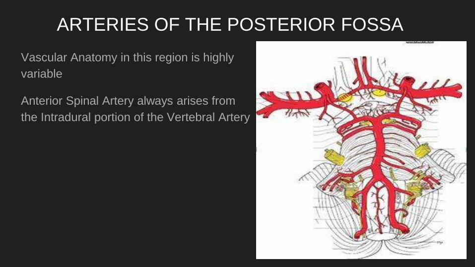
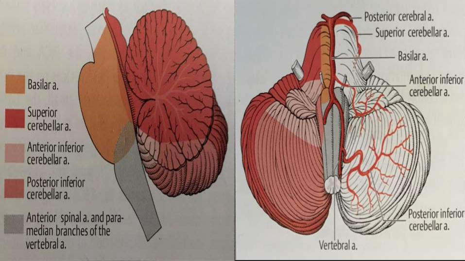
ARTERIES OF THE POSTERIOR FOSSA
Vascular Anatomy in this region is highly
variable
Anterior Spinal Artery always arises from
the Intradural portion of the Vertebral Artery
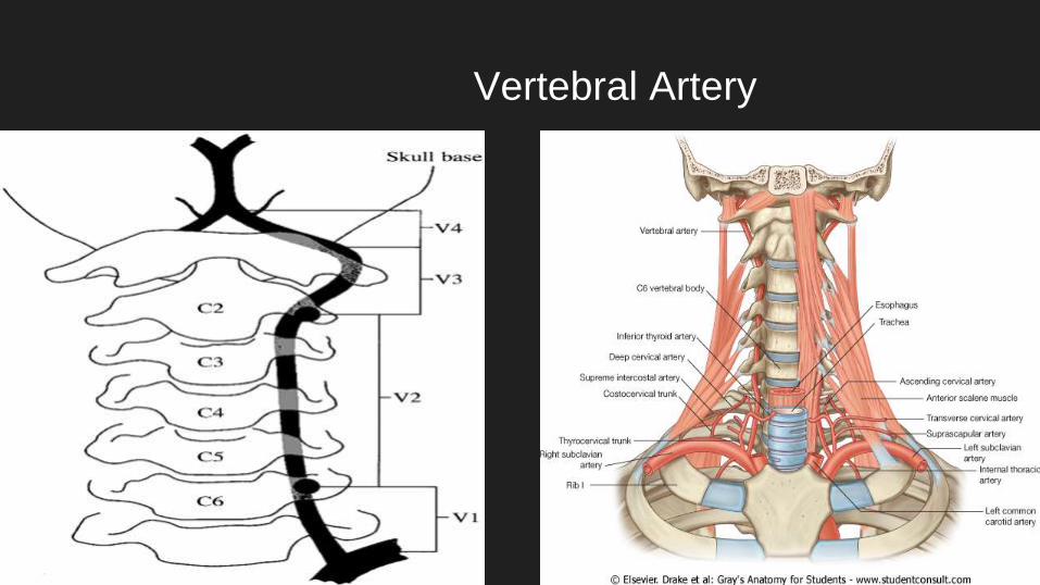
Vertebral Artery
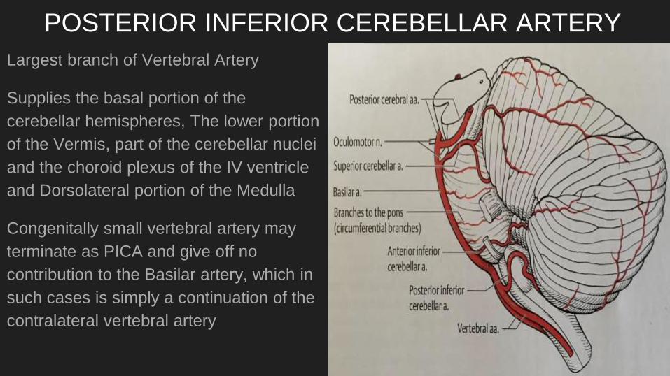
POSTERIOR INFERIOR CEREBELLAR ARTERY
Largest branch of Vertebral Artery
Supplies the basal portion of the
cerebellar hemispheres, The lower portion
of the Vermis, part of the cerebellar nuclei
and the choroid plexus of the IV ventricle
and Dorsolateral portion of the Medulla
Congenitally small vertebral artery may
terminate as PICA and give off no
contribution to the Basilar artery, which in
such cases is simply a continuation of the
contralateral vertebral artery
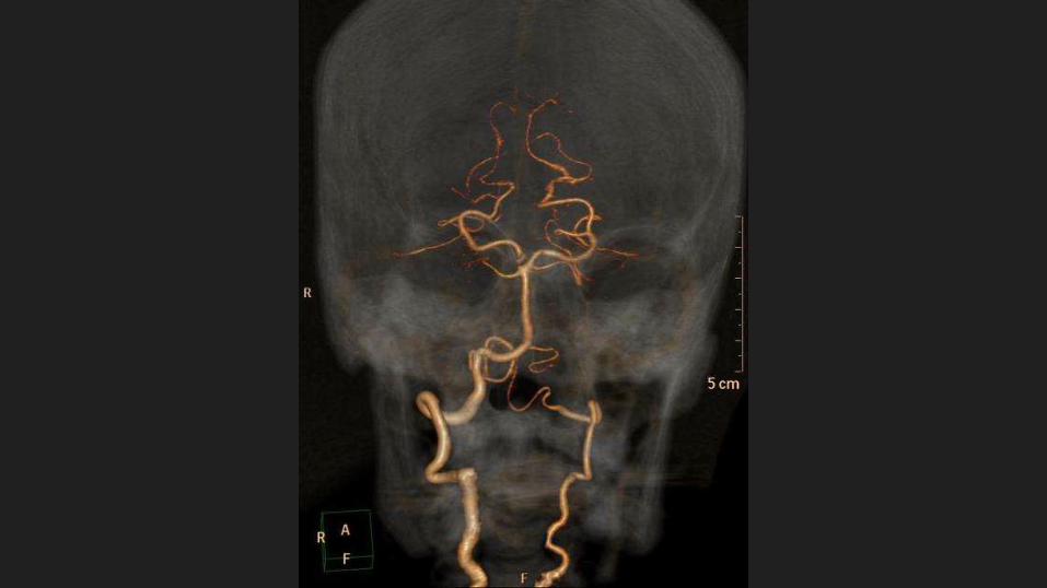
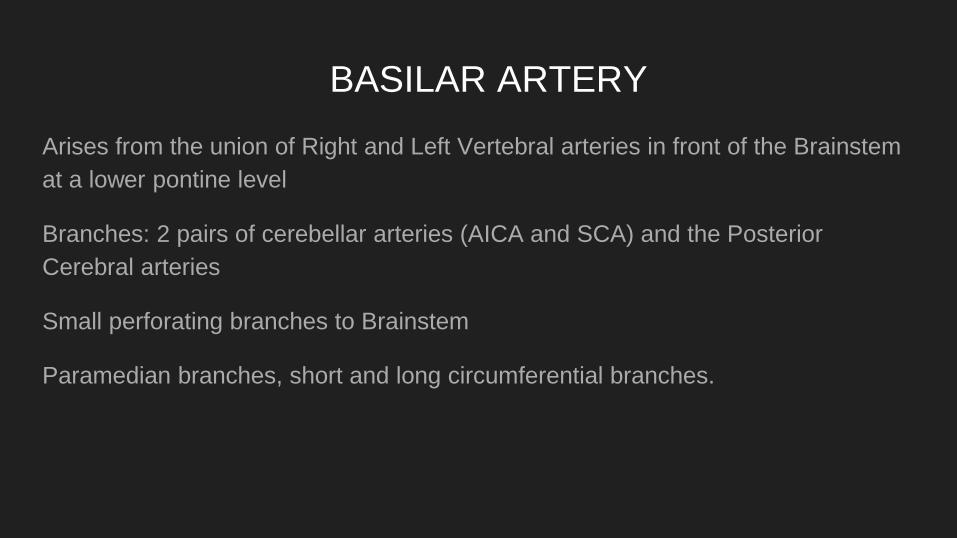
BASILAR ARTERY
Arises from the union of Right and Left Vertebral arteries in front of the Brainstem
at a lower pontine level
Branches: 2 pairs of cerebellar arteries (AICA and SCA) and the Posterior
Cerebral arteries
Small perforating branches to Brainstem
Paramedian branches, short and long circumferential branches.
ANTERIOR INFERIOR CEREBELLAR ARTERY
First major branch of the Basilar artery
Supplies the Flocculus, Anterior portion of the Cerebellar hemisphere
Anastomoses with branches of PICA and its distribution is highly variable.
Gives of Labyrinthine artery to the inner ear
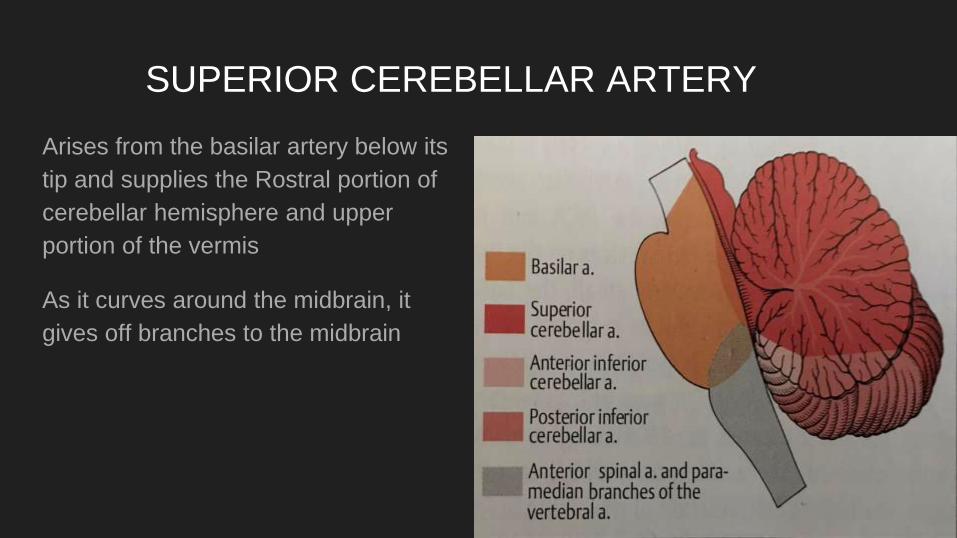
SUPERIOR CEREBELLAR ARTERY
Arises from the basilar artery below its
tip and supplies the Rostral portion of
cerebellar hemisphere and upper
portion of the vermis
As it curves around the midbrain, it
gives off branches to the midbrain
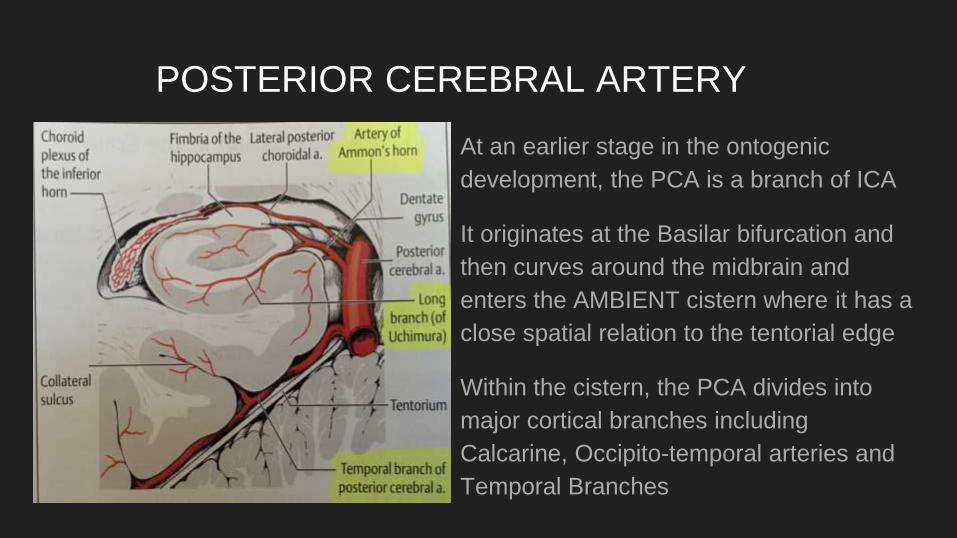
POSTERIOR CEREBRAL ARTERY
At an earlier stage in the ontogenic
development, the PCA is a branch of ICA
It originates at the Basilar bifurcation and
then curves around the midbrain and
enters the AMBIENT cistern where it has a
close spatial relation to the tentorial edge
Within the cistern, the PCA divides into
major cortical branches including
Calcarine, Occipito-temporal arteries and
Temporal Branches
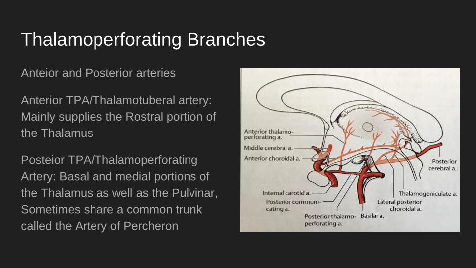
Thalamoperforating Branches
Anteior and Posterior arteries
Anterior TPA/Thalamotuberal artery:
Mainly supplies the Rostral portion of
the Thalamus
Posteior TPA/Thalamoperforating
Artery: Basal and medial portions of
the Thalamus as well as the Pulvinar,
Sometimes share a common trunk
called the Artery of Percheron
Thalamogeniculate Artery
DIstal to the origin of the PCOM
It supplies the lateral portion of the Thalamus
Posterior Choroidal Arteries
Medial Branch : Supplies the midbrain and also the choroid plexus of the III
ventricle
Lateral Branch supplies the choroid plexus of the Lateral ventricle and has an
anastomotic connection with the anterior choroidal artery.
Both arteries supply the Geniculate bodies, Medial and posteromedial thalamic
nuclei and the Pulvinar
CORTICAL BRANCHES OF THE PCA
PCA territory is delimited by the Sylvian fissure. In others the MCA supplies the
entire convexity of the Brain including the Occipital pole.
The visual cortex of the calcarine sulcus is always supplied by the PCA. The optic
radiation is however often supplied by the MCA so that homonymous hemianopsia
doesn’t always incline an infarct in the territory of the PCA
The PCA also has temporal branches to the temporal lobes
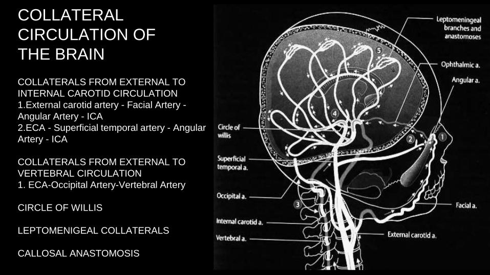
COLLATERAL
CIRCULATION OF
THE BRAIN
COLLATERALS FROM EXTERNAL TO
INTERNAL CAROTID CIRCULATION
1.External carotid artery - Facial Artery -
Angular Artery - ICA
2.ECA - Superficial temporal artery - Angular
Artery - ICA
COLLATERALS FROM EXTERNAL TO
VERTEBRAL CIRCULATION
1. ECA-Occipital Artery-Vertebral Artery
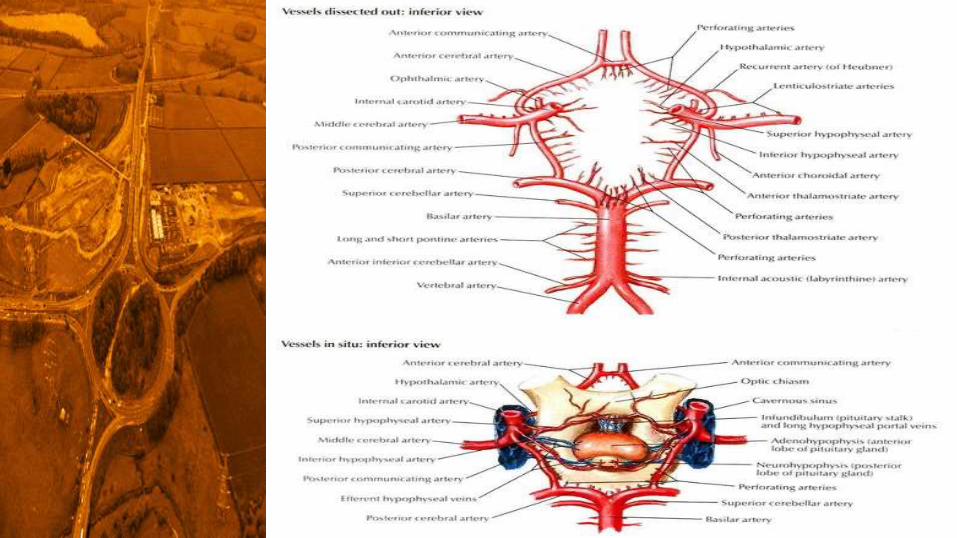
CIRCLE OF WILLIS
LEPTOMENIGEAL COLLATERALS
CALLOSAL ANASTOMOSIS
Unlike the rest of the body Veins don’t run together with its arterial counterparts.
The territories of the Cerebral arteries do not coincide with the cerebral veins
Venous blood from Brain parenchyma crosses the subarachnoid and subdural
spaces in short cortical veins like Superior anastomotic vein of Trolard, Dorsal
superior Cerebral Vein, Superficial middle cerebral vein and Inferior Anastomotic
vein of Labbe.
VENOUS ANATOMY
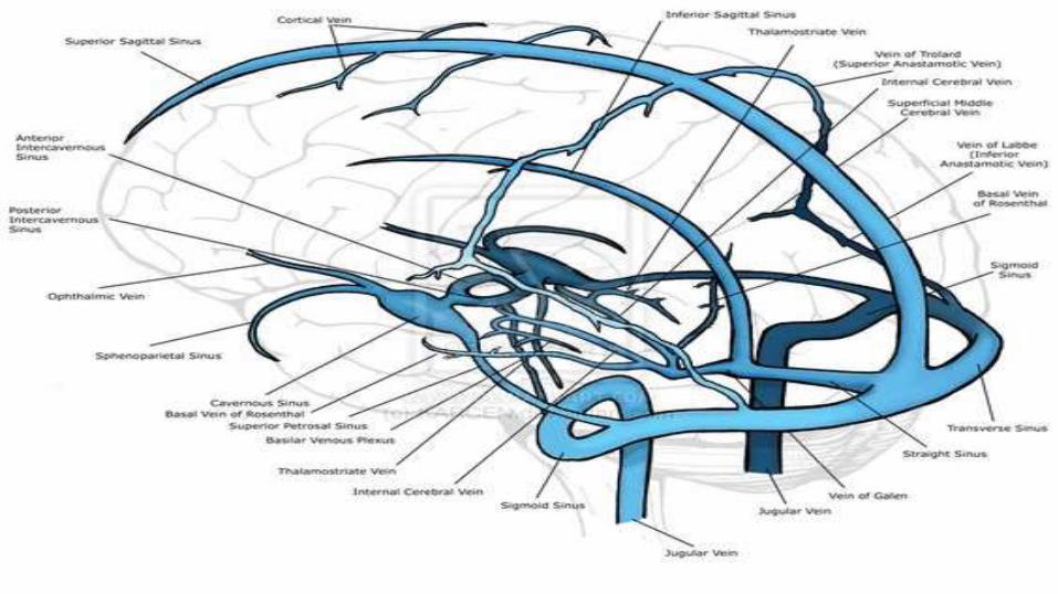
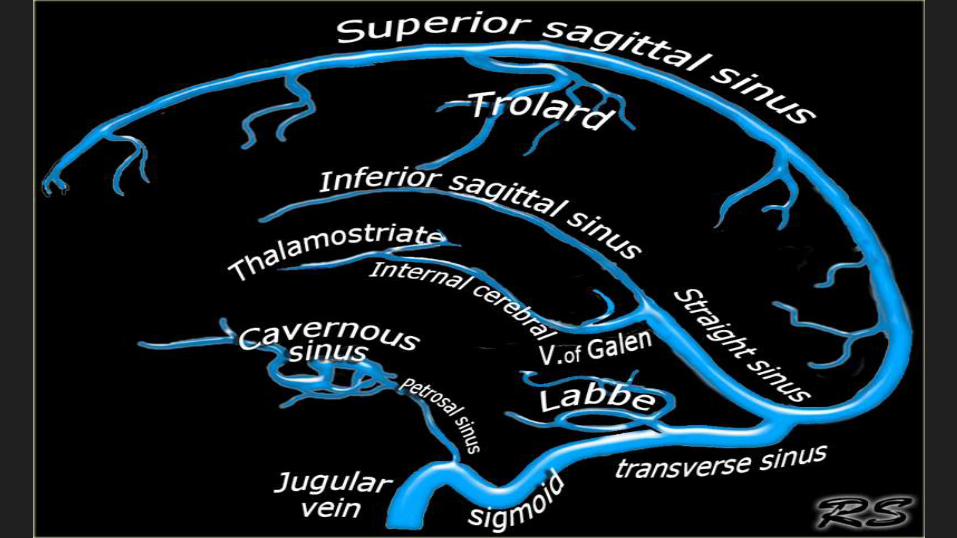
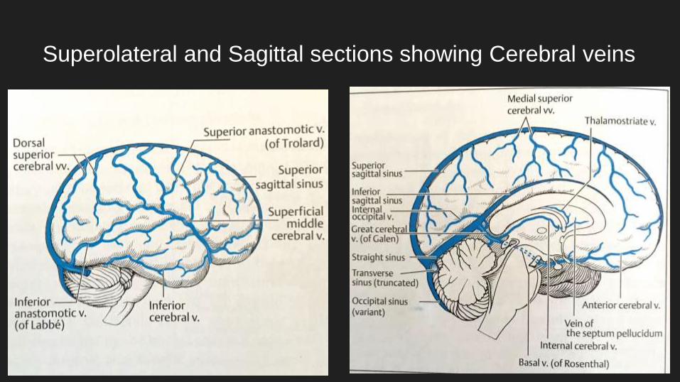
Superolateral and Sagittal sections showing Cerebral veins
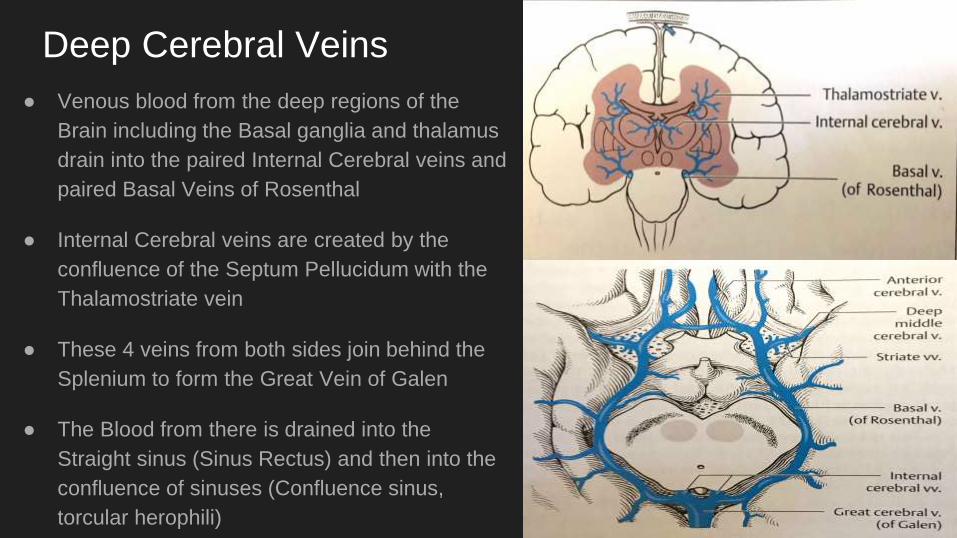
Deep Cerebral Veins
● Venous blood from the deep regions of the
Brain including the Basal ganglia and thalamus
drain into the paired Internal Cerebral veins and
paired Basal Veins of Rosenthal
● Internal Cerebral veins are created by the
confluence of the Septum Pellucidum with the
Thalamostriate vein
● These 4 veins from both sides join behind the
Splenium to form the Great Vein of Galen
● The Blood from there is drained into the
Straight sinus (Sinus Rectus) and then into the
confluence of sinuses (Confluence sinus,
torcular herophili)
Dural Sinuses
● Superficial and deep veins of the Brain drain
into the Dural Venous sinuses.
● Most of the venous drainage in the Superior
Sagittal sinus travel from front to back which
runs in the midline along the attachment of Falx
Cerebri
● At the point in the back of the Head where the
Falx cerebri merges with the Tentorium, the
SSS is joined by the Straight sinus which runs
in the midline along the attachment of tentorium
and carries blood from deep regions of the
Brain.
● The blood from the SSS and SS is then
distributed to two Transverse Sinuses in the
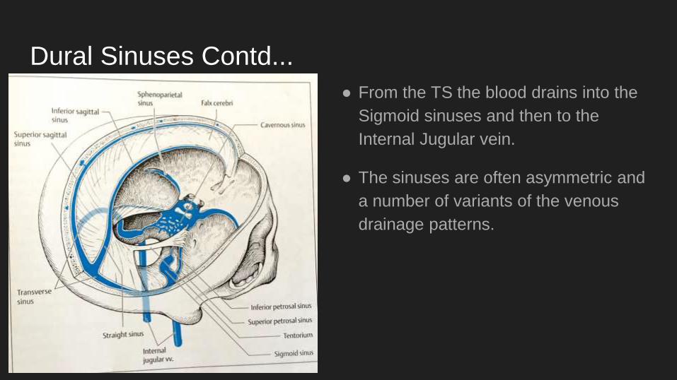
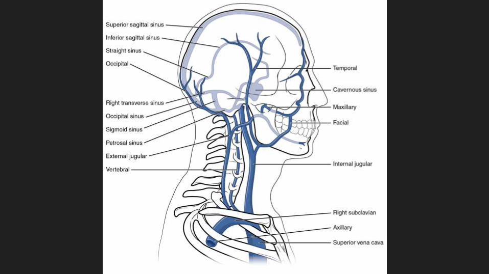
Dural Sinuses Contd...
● From the TS the blood drains into the
Sigmoid sinuses and then to the
Internal Jugular vein.
● The sinuses are often asymmetric and
a number of variants of the venous
drainage patterns.
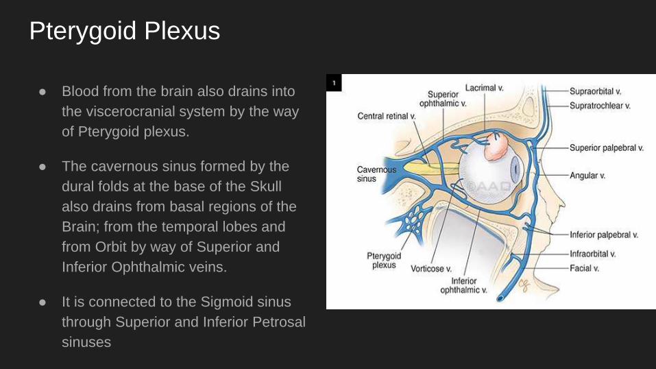
Pterygoid Plexus
● Blood from the brain also drains into
the viscerocranial system by the way
of Pterygoid plexus.
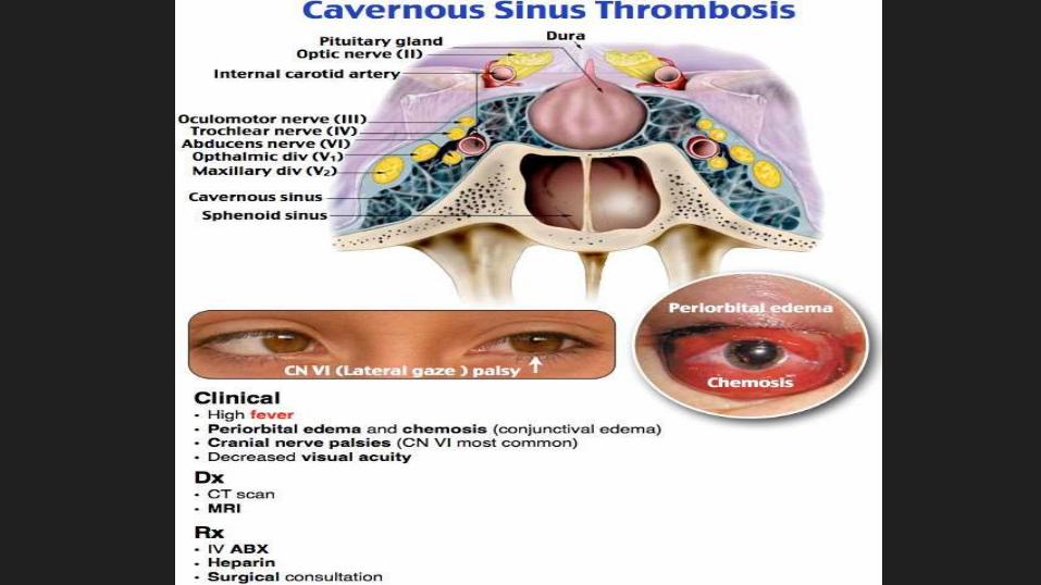
● The cavernous sinus formed by the
dural folds at the base of the Skull
also drains from basal regions of the
Brain; from the temporal lobes and
from Orbit by way of Superior and
Inferior Ophthalmic veins.
● It is connected to the Sigmoid sinus
through Superior and Inferior Petrosal
sinuses
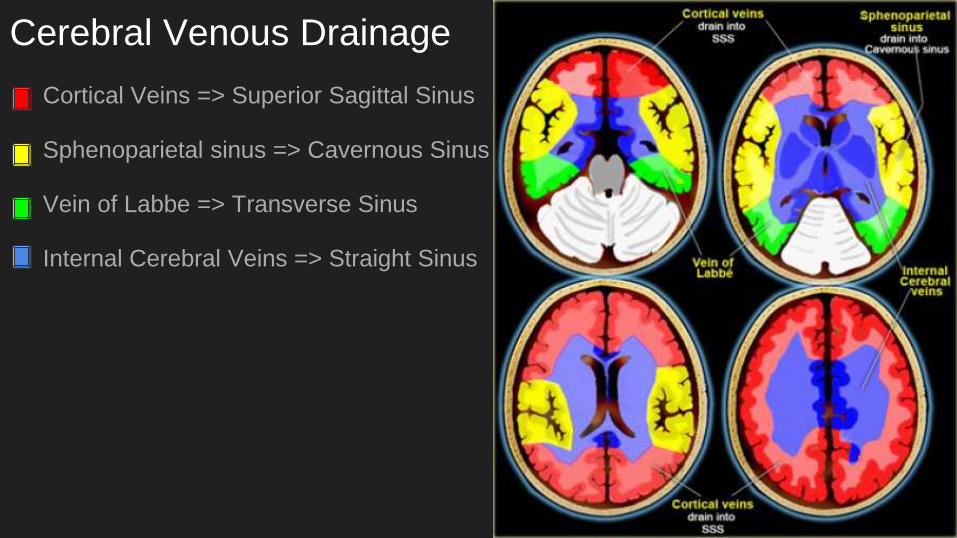
Cerebral Venous Drainage
Cortical Veins => Superior Sagittal Sinus
Sphenoparietal sinus => Cavernous Sinus
Vein of Labbe => Transverse Sinus
Internal Cerebral Veins => Straight Sinus
VASCULAR ANATOMY OF SPINAL CORD
Mostly anastomotic blood supply from Anterior Spinal Artery and paired Posterior
Spinal arteries
Anterior Spinal Artery:
● It runs down the ventral surface of the Spinal cord at the anterior edge of the
anterior median fissure. It receives Segmental contribution from a number of
arteries and supply to the ventral part of the spinal grey matter through
perforating vessels known as SULCO-COMMISSURAL ARTERIES.
● Each artery supplies one half of the spinal cord, important structures supplied
by the ASA include Anterior Horns, Lateral Spinothalamic tract and part of the
Pyramidal tract.
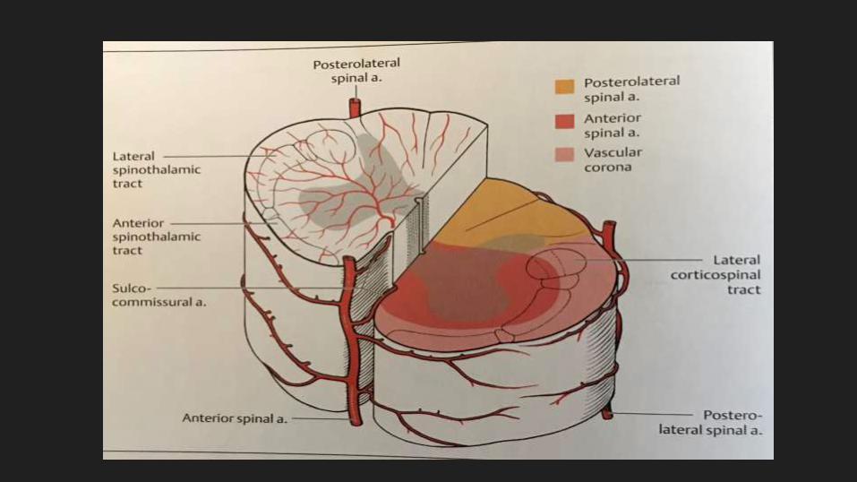
POSTEROLATERAL SPINAL ARTERIES
● Paired
● Runs on the Dorsal side between the Posterior roots and lateral columns on
either side.
● Supplies the posterior columns, roots and Dorsal horns
● The longitudinal axis are connected by radicular anastomosis. These
arteries supply the anterior and Lateral columns through perforating
branches.
● In the periphery however, the arteries of the spinal cord are functional end
arteries. Intramedullary embolic occlusion of a Sulco-commissural artery
therefore causes infarction of the Spinal cord.
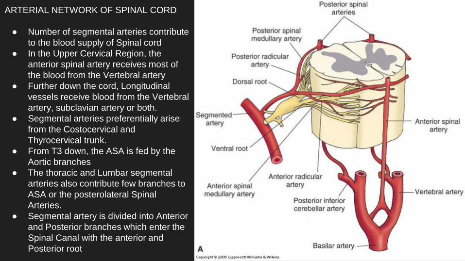
ARTERIAL NETWORK OF SPINAL CORD
● Number of segmental arteries contribute
to the blood supply of Spinal cord
● In the Upper Cervical Region, the
anterior spinal artery receives most of
the blood from the Vertebral artery
● Further down the cord, Longitudinal
vessels receive blood from the Vertebral
artery, subclavian artery or both.
● Segmental arteries preferentially arise
from the Costocervical and
Thyrocervical trunk.
● From T3 down, the ASA is fed by the
Aortic branches
● The thoracic and Lumbar segmental
arteries also contribute few branches to
ASA or the posterolateral Spinal
Arteries.
● Segmental artery is divided into Anterior
and Posterior branches which enter the
Spinal Canal with the anterior and
Posterior root
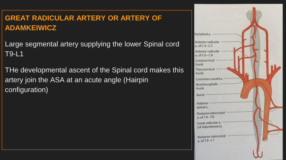
GREAT RADICULAR ARTERY OR ARTERY OF
ADAMKEIWICZ
Large segmental artery supplying the lower Spinal cord
T9-L1
THe developmental ascent of the Spinal cord makes this
artery join the ASA at an acute angle (Hairpin
configuration)
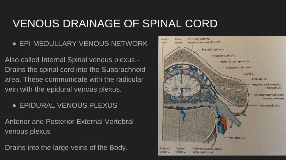
VENOUS DRAINAGE OF SPINAL CORD
● EPI-MEDULLARY VENOUS NETWORK
Also called Internal Spinal venous plexus -
Drains the spinal cord into the Subarachnoid
area. These communicate with the radicular
vein with the epidural venous plexus.
● EPIDURAL VENOUS PLEXUS
Anterior and Posterior External Vertebral
venous plexus
Drains into the large veins of the Body.