Cerebrocerebellar system and Arnold’s bundle · Dement Neuropsychol 2010 December;4(4):293-299...
7
Original Article Dement Neuropsychol 2010 December;4(4):293-299 Engelhardt E, et al. Cerebrocerebellar system: a tractographic study 293 Cerebrocerebellar system and Arnold’s bundle A tractographic study: preliminary results Eliasz Engelhardt 1 , Denise Madeira Moreira 2,4 , Jerson Laks 3 Abstract – The cerebellum, traditionally considered a structure involved in balance and movement control, was more recently recognized as important in cognitive, emotional and behavioral functions. These functions appear to be related to the more recent parts of the cerebellum that belong to the cerebrocerebellar system. One of the key segments of this system is the (pre)fronto-[penduncule]-pontine projection that represents the Arnold’s bundle. Diffusion tensor imaging and tractography (DTI-TR) has permitted in vivo virtual dissection of white matter tracts, including those of the cerebellar. Objective: To study the fronto-[peduncule]-pontine projection (Arnold’s bundle), with DTI-TR. Methods: Ten normal subjects were included (mean age 30 years). Standard acquisitions in three planes were obtained with a 1.5T GE Signa Horizon scanner, complemented with DTI acquisitions. Post-processing and analysis was performed using an ADW 4.3 workstation running Functool 4.5.3 (GE Medical Systems). A single ROI was placed on the medial third of the cerebral peduncle base, considered the site of convergence of the fibers of Arnold’s bundle, bilaterally. Results: Twenty tractograms were obtained. All were constituted by a significant number of fibers in correspondence to the frontal lobe, and part of them anterior to the coronal plane at the anterior commissure, which characterizes them as associated to the prefrontal region. Conclusions: For the first time, frontal lobe related projections were systematically revealed with DTI-TR seeded from cerebral peduncle base ROIs. They showed anatomic coherence with Arnold’s bundle, which includes the prefrontopontine segment of the cortico-ponto-cerebellar path, one of the components of the cerebrocerebellar system, acknowledged as fundamental for non-motor functions such as cognition, emotion and behavior. Key words: tractography, frontopontine bundle, Arnold’s bundle, cerebrocerebellar system. Sistema cerebrocerebelar e feixe de Arnold – um estudo de tratografia: resultados preliminares Resumo – O cerebelo, tradicionalmente considerado como estrutura relacionada ao equilíbrio e controle do movimento, foi mais recentemente reconhecido ser importante em funções cognitivas, emocionais e comportamentais. Tais funções encontram-se relacionadas às partes mais novas do cerebelo que pertencem ao sistema cerebrocerebelar. Um dos segmentos de relevo desse sistema é a projeção (pré)fronto-[pedúnculo]- pontina que representa o feixe de Arnold. A imagem com tensor de difusão e a tratografia (DTI-TR) vem possibilitando a dissecção virtual de feixes da substância branca, inclusive os cerebelares. Objetivo: Estudar a projeção fronto-[pedúnculo]-pontina (feixe de Arnold), com DTI-TR. Métodos: Foram incluídos dez sujeitos normais (média de idade 30 anos). Sequências padrão em tres planos foram obtidas com equipamento Signa Horizon GE de 1,5T e complementadas com aquisições de DTI. Pós-processamento e análise foram realizados em uma estação de trabalho ADW 4.3 com Functool 4.5.3 (GE Medical Systems). Foi utilizado ROI único colocado no terço medial da base do pedúnculo cerebral, considerado o local de convergência das fibras do feixe de Arnold, bilateralmente. Resultados: Foram obtidos vinte tratogramas. Todos eram constituídos por um número significativo de fibras em correspondência ao lobo frontal e parte dessas em posição anterior ao plano coronal à comissura anterior, que as caracteriza como relacionadas à região pré-frontal. Conclusões: Foram mostradas, pela primeira vez de modo sistemático, projeções relacionadas ao lobo frontal com DTI-TR através de ROIs colocados na base do pedúnculo cerebral. Apresentavam coerência anatômica com o feixe de Arnold, que compreende o segmento pré-frontopontino da via córtico-ponto-cerebelar, um dos componentes do sistema cerebrocerebelar, considerado subjacente a funções não motoras, como cognição, emoção e comportamento. Palavras-chave: tratografia, feixe frontopontino, feixe de Arnold, sistema cerebrocerebelar. 1 Cognitive and Behavioral Neurology Unit – INDC/CDA/IPUB-UFRJ. 2 Neuroradiology Unit – INDC – UFRJ. 3 Center for Alzheimer Disease/CDA/IPUB – UFRJ. 4 PróCardíaco Hospital Radiology Service – RJ. Eliasz Engelhardt – Av. N.S. de Copacabana, 749/708 - 22050-002 Rio de Janeiro RJ - Brazil. Disclosure: The authors report no conflicts of interest. Received September 10, 2010. Accepted in final form November 11, 2010.
Transcript of Cerebrocerebellar system and Arnold’s bundle · Dement Neuropsychol 2010 December;4(4):293-299...

Original ArticleDement Neuropsychol 2010 December;4(4):293-299
Engelhardt E, et al. Cerebrocerebellar system: a tractographic study 293
Cerebrocerebellar system and Arnold’s bundleA tractographic study: preliminary results
Eliasz Engelhardt1, Denise Madeira Moreira2,4, Jerson Laks3
Abstract – The cerebellum, traditionally considered a structure involved in balance and movement control, was
more recently recognized as important in cognitive, emotional and behavioral functions. These functions appear
to be related to the more recent parts of the cerebellum that belong to the cerebrocerebellar system. One of the
key segments of this system is the (pre)fronto-[penduncule]-pontine projection that represents the Arnold’s
bundle. Diffusion tensor imaging and tractography (DTI-TR) has permitted in vivo virtual dissection of white
matter tracts, including those of the cerebellar. Objective: To study the fronto-[peduncule]-pontine projection
(Arnold’s bundle), with DTI-TR. Methods: Ten normal subjects were included (mean age 30 years). Standard
acquisitions in three planes were obtained with a 1.5T GE Signa Horizon scanner, complemented with DTI
acquisitions. Post-processing and analysis was performed using an ADW 4.3 workstation running Functool 4.5.3
(GE Medical Systems). A single ROI was placed on the medial third of the cerebral peduncle base, considered the
site of convergence of the fibers of Arnold’s bundle, bilaterally. Results: Twenty tractograms were obtained. All
were constituted by a significant number of fibers in correspondence to the frontal lobe, and part of them anterior
to the coronal plane at the anterior commissure, which characterizes them as associated to the prefrontal region.
Conclusions: For the first time, frontal lobe related projections were systematically revealed with DTI-TR seeded
from cerebral peduncle base ROIs. They showed anatomic coherence with Arnold’s bundle, which includes the
prefrontopontine segment of the cortico-ponto-cerebellar path, one of the components of the cerebrocerebellar
system, acknowledged as fundamental for non-motor functions such as cognition, emotion and behavior.
Key words: tractography, frontopontine bundle, Arnold’s bundle, cerebrocerebellar system.
Sistema cerebrocerebelar e feixe de Arnold – um estudo de tratografia: resultados preliminaresResumo – O cerebelo, tradicionalmente considerado como estrutura relacionada ao equilíbrio e controle
do movimento, foi mais recentemente reconhecido ser importante em funções cognitivas, emocionais e
comportamentais. Tais funções encontram-se relacionadas às partes mais novas do cerebelo que pertencem
ao sistema cerebrocerebelar. Um dos segmentos de relevo desse sistema é a projeção (pré)fronto-[pedúnculo]-
pontina que representa o feixe de Arnold. A imagem com tensor de difusão e a tratografia (DTI-TR) vem
possibilitando a dissecção virtual de feixes da substância branca, inclusive os cerebelares. Objetivo: Estudar a
projeção fronto-[pedúnculo]-pontina (feixe de Arnold), com DTI-TR. Métodos: Foram incluídos dez sujeitos
normais (média de idade 30 anos). Sequências padrão em tres planos foram obtidas com equipamento Signa
Horizon GE de 1,5T e complementadas com aquisições de DTI. Pós-processamento e análise foram realizados em
uma estação de trabalho ADW 4.3 com Functool 4.5.3 (GE Medical Systems). Foi utilizado ROI único colocado
no terço medial da base do pedúnculo cerebral, considerado o local de convergência das fibras do feixe de
Arnold, bilateralmente. Resultados: Foram obtidos vinte tratogramas. Todos eram constituídos por um número
significativo de fibras em correspondência ao lobo frontal e parte dessas em posição anterior ao plano coronal à
comissura anterior, que as caracteriza como relacionadas à região pré-frontal. Conclusões: Foram mostradas, pela
primeira vez de modo sistemático, projeções relacionadas ao lobo frontal com DTI-TR através de ROIs colocados
na base do pedúnculo cerebral. Apresentavam coerência anatômica com o feixe de Arnold, que compreende o
segmento pré-frontopontino da via córtico-ponto-cerebelar, um dos componentes do sistema cerebrocerebelar,
considerado subjacente a funções não motoras, como cognição, emoção e comportamento.
Palavras-chave: tratografia, feixe frontopontino, feixe de Arnold, sistema cerebrocerebelar.
1Cognitive and Behavioral Neurology Unit – INDC/CDA/IPUB-UFRJ. 2Neuroradiology Unit – INDC – UFRJ. 3Center for Alzheimer Disease/CDA/IPUB – UFRJ. 4PróCardíaco Hospital Radiology Service – RJ.
Eliasz Engelhardt – Av. N.S. de Copacabana, 749/708 - 22050-002 Rio de Janeiro RJ - Brazil.
Disclosure: The authors report no conflicts of interest.
Received September 10, 2010. Accepted in final form November 11, 2010.

Dement Neuropsychol 2010 December;4(4):293-299
294 Cerebrocerebellar system: a tractographic study Engelhardt E, et al.
The cerebellum, traditionally considered a structure involved in balance and movement control,1-3 is now also recognized as playing an important role in cognitive, emo-tional and behavioral functions,3-7 and is also implicated in neuropsychiatric disorders.8 These more recently studied functions (and dysfunctions) are ascribed to the integra-tive activity of the cerebrocerebellar system constituted by distinctive brain regions and related fiber pathways. Asso-ciation areas of the cerebral cortex and neocerebellar struc-tures (cortical and subcortical) are interconnected. The cortical efferents constitute the corticopontine and mossy fiber projections to the cerebellum. Reciprocally, the cere-bellum (dentate nucleus) projects to cortical association ar-eas via cerebellothalamic and thalamocortical pathways.9,10
Classically, neuroanatomical studies have been per-formed on post-mortem human specimens, with dissec-tion of normal brains and gross and microscopic tracking of degenerated fiber bundles of brains that had suffered lesions (vascular, traumatic, surgical). Despite the limita-tions of such studies the main tracts of the central nervous system were fairly accurately demonstrated and described, including the corticopontine group of fibers.11-19
The knowledge that the corticopontine-[cerebellar] projections originate from several areas of the cerebral cortex (frontal, parietal, occipital, and temporal) and con-verge to the homolateral cerebral peduncle base(s) (basis pedunculus[i], pes pedunculus[i]) is not new,20,13 although has achieved a higher level of detail only more recently.
The description of these projections into the brain stem (pedunculus, pons, medulla) preceded the elucidation of their cortical origin.21 It is presently acknowledged, albeit not fully settled, that the frontopontine tract stems from the prefrontal cortex (Brodmann areas [BA] 10, 9, 8, 45, and 46), containing also fibers from the precentral region (BA 4 and 6), while the cortico-(bulbo)-spinal tract origi-nates from the central region (mainly from BA 4, somes-thetic [BA 3, 2, 1], and less from SMA, and BA 6, 8, 5 and 7),22-28 and the posterior projections come from the exten-sive retrorolandic region, including parietal, occipital and temporal areas.15
In spite of a long history of investigations, including numerous anatomo-pathological studies in humans and experimental studies in non-human primates,29-30 the topographical organization of the fibers at the base of the cerebral peduncle still has some unresolved details con-cerning its constituent fibers. It is recognized that the fron-topontine projection first depicted and described by Ar-nold (1838)31 extends from the frontal cortex through the anterior limb of the internal capsule to the pons, passing through the medial part of the cerebral peduncle base.12-13 The lateral peduncle base is occupied by the tract described
by Türck (1851), formed by retrorolandic (parieto-occip-ito-temporo-pontine) projections.13,15 The intermediate part is the route of the cortico-(bulbo)-spinal or cortico-(nucleo)-spinal tract, that extends through the internal capsule to caudad levels, and comprises the corticospinal part (pyramidal tract as named by Türck, 1851),21 and the corticobulbar tract. Most fibers of the corticobulbar tract accompany the corticospinal fibers through the internal capsule (genual fibers) and separate as they traverse the brainstem. Part of these fibers terminate in the pontine nuclei which originate projections to the cerebellum.32-33 The proportion corresponding to each segment varies ac-cording to several authors, with the classical trend consid-ering the middle segment to be larger than the medial and lateral ones.13,34-36 However, there are few such studies in the human brain, and it should be noted that these have been carried out in brains with natural13,15 or surgical12 le-sions which render them with imprecision, or resulting from post-mortem dissections.37 On the other hand, stud-ies with magnetic resonance imaging (MRI) and diffusion tensor acquisition have shown that the distribution of the peduncular fibers is somewhat different in comparison to the classical model, with roughly equal segmentation.38-43
The frontopontine bundle (Arnold’s bundle) together with that originating from the posterior cortex (Türck’s bundle) includes numerous fibers of the cortico-ponto-[cerebellar] limb of the cerebrocerebellar system that un-derlies non-motor functions.30 Additionally, clinical and neurophysiologic research in patients with lesions of the cerebellum and/or its pathways have allowed clinicians to identify the presence of significant cognitive and affective disorders,6 and recent functional neuroimaging studies substantiate such findings.44
Studies with MRI and DTI followed by tractography (DTI-TR) have permitted virtual dissection of white mat-ter tracts, including cerebellar ones, in vivo, in the human brain,38, 41,45-48 but systematic studies of the cerebrocerebel-lar system in the human brain are limited, while studies on corticopontine projections are lacking. A preliminary report on this issue was recently presented.49
Given the scarcity of tractographic studies on the hu-man cerebrocerebellar system, the aim of this study was to visualize cortico-[peduncle]-pontine projections from (pre)frontal cortex (Arnold’s bundle) with DTI-TR, with the seeding ROIs placed at the level of the cerebral pedun-cle base.
MethodsTen normal subjects of both genders were included (age
30.67±5.73 years). Standard acquisitions in three planes were obtained with a 1.5T GE Signa Horizon scanner. Dif-
Dement Neuropsychol 2010 December;4(4):293-299
Engelhardt E, et al. Cerebrocerebellar system: a tractographic study 295
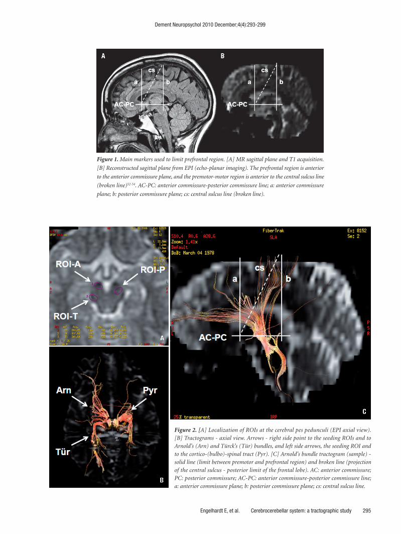
Figure 1. Main markers used to limit prefrontal region. [A] MR sagittal plane and T1 acquisition.
[B] Reconstructed sagittal plane from EPI (echo-planar imaging). The prefrontal region is anterior
to the anterior commissure plane, and the premotor-motor region is anterior to the central sulcus line
(broken line)52-54. AC-PC: anterior commissure-posterior commissure line; a: anterior commissure
plane; b: posterior commissure plane; cs: central sulcus line (broken line).
Figure 2. [A] Localization of ROIs at the cerebral pes pedunculi (EPI axial view).
[B] Tractograms - axial view. Arrows - right side point to the seeding ROIs and to
Arnold’s (Arn) and Türck’s (Tür) bundles, and left side arrows, the seeding ROI and
to the cortico-(bulbo)-spinal tract (Pyr). [C] Arnold’s bundle tractogram (sample) -
solid line (limit between premotor and prefrontal region) and broken line (projection
of the central sulcus - posterior limit of the frontal lobe). AC: anterior commissure;
PC: posterior commissure; AC-PC: anterior commissure-posterior commissure line;
a: anterior commissure plane; b: posterior commissure plane; cs: central sulcus line.

Dement Neuropsychol 2010 December;4(4):293-299
296 Cerebrocerebellar system: a tractographic study Engelhardt E, et al.
fusion tensor acquisitions, similar to those found in other studies on the issue,45,50 were used in the present study with parameters TR/TE=10 000/89.1 msec, matrix=128×128, FOV=30×24 mm, NEX=1, 25 gradient directions, b=1000 sec/mm,2 slice thickness=5 mm, number of slices=30 without gap, in the axial plane, scan time=4 min, 40 sec. Post-processing and analysis were performed on an ADW 4.3 workstation running Functool 4.5.3 (GE Medical Sys-tems). The fiber tracts were reconstructed starting from voxels with an FA>0.18 on the axial plane up to those with an FA<0.18, or up to a maximum step size of 160 μm. A single ROI (of around 40 mm2) was placed occupying the medial third of the cerebral peduncle base,51 considered the site of convergence of the fibers of the frontopontine tract (Arnold’s bundle), on both sides. Smaller ROIs (of around 5 mm2) were placed at more medial and more lateral lo-calizations in the same territory for a better understanding
of the topographical distribution of the fibers (Figure 1 for the reference lines, and Figure 2 for the ROIs localiza-tion and sample tractograms) The fibers selected with the peduncular ROI originate mainly in the frontal cortex as DTI-TR cannot distinguish between efferent and afferent fibers, and the cerebral peduncles are known to contain fibers that project from the cortex to the pontine nuclei through the pes pedunculus.41,51
The present study is part of a larger project on Vascu-lar Cognitive Disorder, approved by the Research Ethics Committee of IPUB-UFRJ. Informed consent was obtained from all participants.
ResultsTwenty tractograms were obtained (Figure 3). The
demonstrated fibers tracked from the medial ROI were localized in correspondence to the frontal lobe, anterior to
Figure 3. Arnold’s bundle tractograms (right and left) of the 10 subjects (A-01 to A-10).

Dement Neuropsychol 2010 December;4(4):293-299
Engelhardt E, et al. Cerebrocerebellar system: a tractographic study 297
a plane running parallel to the central sulcus.54 A signifi-cant proportion of these fibers was localized anterior to the coronal plane to the anterior commissure, considered a conservative landmark for the limit between prefrontal and premotor cortex,53,55 which characterizes them as associated to the prefrontal region (Arnold’s bundle). The fibers re-lated to more posterior regions of the frontal lobe probably concern premotor and motor areas, which are the main origin of the cortico-(bulbo)-spinal tract, as confirmed by the smaller ROIs. Although the core ROIs were placed practically in the same regions, the tractograms showed intra-subject and inter-subjects variability and asymmetry.
It may be possible that fewer prefrontal fibers than ex-pected were visualized in some subjects. An explanation for this would be a truncation effect as a result of the acute angulation of the frontopontine tract at the level of the cerebral peduncle, in its trajectory from the frontal cor-tex.50,56 On the other hand, the apparent presence of more than the expected number of fibers localized in relation to the premotor-motor region in other subjects may be attributed to a better visualization of less angulated fibers and/or some overlap with perirolandic originating fibers. Individual variations, considering both possibilities, can-not be ruled out.
DiscussionProjections from the frontal lobe were revealed in the
present study with DTI-TR, seeded from cerebral pedun-cle base medially-placed ROIs. These showed anatomic coherence with the frontopontine bundle (Arnold’s bun-dle), which includes the prefrontopontine segment of the cortico-ponto-cerebellar path. Several studies containing cerebellar-related tractograms have previously been pub-lished,46,57-59 but few studies have focused specifically on cortico-peduncular projections as has the present inves-tigation. One of these studies,57 for instance, showed the cortical fibers descending to the cerebral peduncle, revealed by placing one ROI around the pes of the cerebral peduncle of one side and the second around the middle cerebellar peduncle on the other side. This approach permitted the whole assembly of fibers that traverse the cerebral pes pe-dunculus to be visualized, including the cortico-(bulbo)-spinal tract. However, it was unable to show the other two specifically cerebellum-related bundles. Another more detailed study assessed the contributions of cortical areas (prefrontal, motor and posterior parietal) projections to the cerebral peduncle in humans (and macaque mon-keys).41,60 The cited study showed seven probable distribu-tion maps of the cerebral peduncle, one for each cortical zone, and a clear topographic organization in the cerebral peduncle. Fibers from the prefrontal, primary motor and
premotor regions, and from posterior cerebral cortex, oc-cupied almost equal areas, as did those medially placed by the prefrontal fibers. The cited study, according to the authors, adds to the understanding of the corticopontine component of the cortico-ponto-cerebellar system by pro-viding anatomical evidence of a major contribution from the prefrontal cortex to the pontine nuclei.41 However, in spite of showing the cortical origin and the peduncular termination of the fibers, the authors did not display the trajectories of the bundles under analysis. Thus, the present study appears to be the first time that the trajectory of the (pre)fronto-[peduncule]-pontine projection is visualized in a systematic study by way of a single ROI placed on its convergence area at the cerebral peduncle base.
The present study showed intra- and inter-subject vari-ability and asymmetry of the obtained tractograms, even though the ROIs were placed in the same location. The variability of the studied tract was previously observed (among subjects, between hemispheres) in a classical study (post-surgical),12 and recently by the tractographic tech-nique showing the case-to-case anatomical variability of the peduncular tracts41 seen for other tracts.61-63 The vari-ability observed in the present study, reflected by different fiber densities comparing the anterior (less than expected?) and more posterior fibers (more than expected?), could also be explained at least in part by a truncation effect due to different degrees of angulation of these fibers.50,56
The prefrontal segment of the fronto-[peduncle]-pontine fibers emerges from prefrontal associative cortical areas, and is anatomically related with the evolutionary newer parts of the cerebellum (lateral hemispheric cere-bellar cortex and dentate nucleus).9,60 Reciprocally, the cer-ebellum (dentate nucleus) sends fibers out to these cortical areas (dentato-thalamo-frontal projection),9 which were also demonstrated tractographically.38 These pathways are responsible for the cerebellar’s participation in the neural circuits that underpin cognitive, emotional, and behavioral control.5,6,10
Some limitations should be acknowledged. It is possible that fewer prefrontal fibers than expected were visualized because of a truncation effect at the level of the cerebral pe-duncle due to the acute angulation of the studied tract. On the other hand, the apparent presence of more fibers than expected localized in the posterior frontal region could be attributed to better visualization of less angulated fibers and/or some overlap with perirolandic-derived fibers. Individual variations, in both scenarios cannot be ruled out. Some of these limitations will probably be resolved by future studies aimed at visualizing the projections from (pre)frontal areas to the cerebral peduncle base employing alternative approaches.

Dement Neuropsychol 2010 December;4(4):293-299
298 Cerebrocerebellar system: a tractographic study Engelhardt E, et al.
ConclusionThe cerebellum, long considered a motor modulat-
ing structure, has more recently been recognized as an important structure underlying cognitive, emotional and behavioral functions. The evolutionary more recent parts of the cerebellum (lateral hemispheric cortex and dentate nucleus), reciprocally connected with associative cortical areas, constitute the cerebrocerebellar system that underpin these functions. One of the key segments of this system is the (pre)fronto-[peduncle]-pontine projection (Arnold’s bundle). The present paper reveals and analyses this bundle with single ROI placement at the medial part of the cere-bral peduncle base, and displays it systematically with this approach for the first time.
Acknowledgements – The authors would like to thank Luzinete Alvarenga for her editorial assistance.
References1. Barlow JS. The cerebellum and adaptive control [Internet].
Cambridge: Cambridge University Press, 2002:3-5. Retrieved
in: 2009. Available from: http://books.google.com.br/books.
2. Glickstein M, Voogd J. Lodewijk Bolk and the comparative
anatomy of the cerebellum. Trends Neurosci 1995;18:206-210.
3. Schmahmann JD. Rediscovery of an early concept. In:
Schmahmann JD (Editor), The cerebellum and cognition.
San Diego: Academic Press; 1997:4-30.
4. Leiner HC, Leiner AL, Dow RS. Does the cerebellum con-
tribute to mental skills? Behav Neurosci 1986;100:443-454.
5. Rapoport M, van Reekum R, Mayberg H. The role of the
cerebellum in cognition and behavior: a selective review. J
Neuropsychiatry Clin Neurosci 2000;12:193-198.
6. Schmahmann JD, Caplan D. Cognition, emotion and the cer-
ebellum. Brain 2006;129:290-292.
7. Watson PJ. Nonmotor functions of the cerebellum. Psychol
Bull 1978;85:944-967.
8. Baldaçara L, Borgio JGF, Lacerda ALT, Jackowski AP. Cerebellum
and psychiatric disorders. Rev Bras Psiquiatr 2008;30: 281-289.
9. Middleton FA, Strick PL. Basal ganglia and cerebellar loops:
motor and cognitive circuits. Brain Res Brain Res Rev 2000;
31:236-250.
10. Schmahmann JD, Pandya DN. The cerebrocerebellar system.
Int Rev Neurobiol 1997a;41:31-60
11. Axer H, Keyserlingk DG. Mapping of fiber orientation in
human internal capsule by means of polarized light and
confocal scanning laser microscopy. J Neurosci Methods
2000;94:165-175.
12. Beck E. The origin, course and termination of the prefronto-
pontine tract in the human brain. Brain 1950;73:368-391.
13. Dejerine J. Anatomie des centres nerveux. Paris: J Rueff Ed.,
1901:1-586.
14. Gebbink TB. Structure and connections of the basal ganglia
in man. Thesis Leiden. Assen: Van Gorcum & Comp., 1967.
15. Marin OSM, Angevine Jr JB, Locke S. Topgrtaphical organiza-
tion of the lateral segmento f the basis pedunculi in man. J
Comp Neurol 1962;118:165-183.
16. Probst M. Zur Kenntniss des Binderarms, der Haubenstrh-
alung und des Regio Subthalamica. Mschr Psychiat Neurol
1901;10:288-309
17. Voogd J. Cerebellum and precerebellar nuclei. In: Paxinos G,
Mai JK (Editors), The human nervous system. 2nd ed. Amas-
terdam: Elsevier; 2004:321-392.
18. Schmahmann JD, Pandya DN. Fiber pathways of the brain.
Oxford: Oxford University Press, 2006:654.
19. Smith MC. Histological findings after hemicerebellectomy in
man: anterograde, retrograde and transneuronal degenera-
tion. Brain Res 1975;95:423-442.
20. Arnold F. Handbuch der anatomie des menschen. v. 2, 5th
Book: Vom Nervensystem. Freiburg im Breisgau: Herder’sche
Verlagshandlung, 1851:636-740.
21. Armand J. The origin, course and terminations of cortico-
spinal fibers in various mammals. In: Kuypers GJM, Martin
GF (Editors). Anatomy of descending pathways to the spinal
cord. v.57, Amsterdam: Elsevier Progress in Brain Research
1982:329-360.
22. Clark DL, Boutros NN, Mendez MF. The Brain and behavior:
an introduction to behavioral neuroanatomy. 3rd ed. Cam-
bridge: Cambridge University Press; 2010:325.
23. Ebeling U, Reulen HJ. Subcortical topography and propor-
tions of the pyramidal tract. Acta Neurochir (Wien) 1992;118:
164-171.
24. Iragui VJ, McCutchen CB. Capsular ataxic hemiparesis. Arch
Neurol 1982;39:528-529.
25. Kumar A, Juhasz C, Asano E, et al. Diffusion tensor imaging
study of the cortical origin and course of the corticospinal tract
in healthy children. Am J Neuroradiol 2009;30:1963-1970.
26. Lassek A. The human pyramidal tract: VII. A critical review
of its origin. J Nerv Ment Dis 1944;99:22-28.
27. Martin JH. Neuroanatomy: text and atlas. 3rd ed. New York:
McGraw-Hill, 2003:532.
28. Penfield W, Boldrey E. Somatic motor and sensory represen-
tation in the cerebral cortex of man as studied by electrical
stimulation. Brain 1937;60:389-443.
29. Sunderland S. The projection of the cerebral cortex on the
pons and cerebellum in the macaque monkey. J Anat 1940;74:
201-226.
30. Schmahmann JD, Pandya DN. Anatomic organization of the
basilar pontine projections from prefrontal cortices in Rhesus
monkey. J Neurosci 1997b;17:438-458.
31. Türe U, Yasargil MG, Friedman AH, Al-Mefty O. Fiber dis-
section technique: lateral aspect of the brain. Neurosurgery
2000;47:417-427.

Dement Neuropsychol 2010 December;4(4):293-299
Engelhardt E, et al. Cerebrocerebellar system: a tractographic study 299
32. Lassek AM. The pyramidal tract: its status in medicine.
Springfiel: Charles C Thomas, 1954:166.
33. Mendoza J, Foundas AL. Clinical neuroanatomy: a neurobe-
havioral approach. New York: Springer Science, 2008:704.
34. Crosby EC, Huphrey T, Lauer EW. Correlative anatomy of
the nervous system. New York: MacMillan Co, 1962:188-220.
35. Gray H. Anatomy of the human body [Internet]. Philadel-
phia: Lea & Febiger, 1918:788-796. Retrieved in: 2009. Avail-
able from: http://www.bartleby.com/107/.
36. Nieuwenhuys R, Voogd J, van Huijzen C. The human central
nervous system. Berlin: Springer Verlag, 1980.
37. Ross ED. Localization of the pyramidal tract in the internal
capsule by whole brain dissection. Neurology 1980;30:59-64.
38. Jissendi P, Baudry S, Balériaux D. Diffusion tensor imaging
(DTI) and tractography of the cerebellar projections to pre-
frontal and posterior parietal cortices: a study at 3T. J Neuro-
radiol 2008;35:42-50.
39. Naidich TP, Duvernoy HM, Delman BN, Sorensen AG, Kollias
SS, Haacke EM. Duvernoy’s atlas of the human brain stem
and cerebellum: high-field MRI, surface anatomy, internal
structure, vascularization and 3D sectional anatomy. Wien:
Springer-Verlag; 2009:876.
40. Park JK, Kim BS, Choi G, Kim SH, Choi JC, Khang H. Evalu-
ation of the somatotopic organization of corticospinal tracts
in the internal capsule and cerebral peduncle: results of diffu-
sion-tensor MR tractography. Korean J Radiol 2008;9:191-195.
41. Ramnani N, Behrens TEJ, Johansen-Berg H, et al. The evolu-
tion of prefrontal inputs to the cortico-pontine system: diffu-
sion imaging evidence from macaque monkeys and humans.
Cerebral Cortex 2006;16:811-818.
42. Salamon N, Sicotte N, Alger J, et al. Analysis of the brain-stem
white-matter tracts with diffusion tensor imaging. Neurora-
diology 2005;47:895-902.
43. Stieltjes B, Kaufmann WE, van Zijl PCM, et al. Diffusion ten-
sor imaging and axonal tracking in the human brainstem.
NeuroImage 2001;14:723-735.
44. Stoodley CJ, Schmahmann JD. Evidence for topographic or-
ganization in the cerebellum of motor control versus cogni-
tive and affective processing. Cortex 2010; 46:831-844.
45. Conturo TE, Lori NF, Cull TS, et al. Tracking neuronal fiber
pathways in the living human brain. PNAS 1999;96:10422-
10427.
46. Engelhardt E, Moreira DM. O cerebelo e suas principais
conexões: estudo com tensor de difusão. Rev Bras Neurol
2009;45:17-32.
47. Salamon N, Sicotte N, Drain A, et al. White matter fiber trac-
tography and color mapping of the normal human cerebellum
with diffusion tensor imaging. J Neuroradiol 2007;34: 115-128.
48. Schmahmann JD, Pandya DN. Fiber pathways of the brain.
Oxford: Oxford University Press; 2006:654.
49. Engelhardt E, Moreira DM. Cerebrocerebellar system -
Arnold’s bundle tractography. Preliminary results, (abstr).
Sixth International Congress on Vascular Dementia. Barce-
lona, 2009.
50. Mukherjee P, Chung SW, Berman JI, Hess CP, Henry RG. Dif-
fusion tensor MR imaging and fiber tractography: technical
considerations. AJNR 2008;29:843-852.
51. Hagmann P, Thiran JP, Jonasson L, et al. DTI mapping of hu-
man brain connectivity: statistical fibre tracking and virtual
dissection. Neuroimage 2003;19:545-554.
52. Brett M. Finding commissures [Internet]. lastly edited on
2006-07-31. Retrieved in: 2010 ago. Available from: http://
ccn.ucla.edu/wiki/index.php/Find_the_AC-PC_line.
53. Nakayama N, Okumura A, Shinoda J, Nakashima T, Iwama
T. Relationship between regional cerebral metabolism and
consciousness disturbance in traumatic diffuse brain injury
without large focal lesions: an FDG-PET study with statistical
parametric mapping analysis. J Neurol Neurosurg Psychiatry
2006;77:856-862.
54. Nii Y, Uematsu S, Lesser RP, Gordon B. Does the central sulcus
divide motor and sensory functions? Cortical mapping of hu-
man hand areas as revealed by electrical stimulation through
subdural grid electrodes. Neurology 1996;46:360-367.
55. Vorobiev V, Govoni P, Rizzolatti G, Matelli M, Luppino G. Par-
cellation of human mesial area 6: cytoarchitectonic evidence
for three separate areas. Eur J Neurosci 1998;10:2199-2203.
56. Yamada K, Kizu O, Mori S, et al. Brain fiber tracking with
clinically feasible diffusion-tensor MR imaging: initial experi-
ence. Radiology 2003;227:295-301.
57. Catani M, Thiebaut de Schotten M. A diffusion tensor imag-
ing tractography atlas for virtual in vivo dissections. Cortex
2008;44:1105-1132.
58. Hagmann P, Thiran JP, Jonasson L, et al. DTI mapping of hu-
man brain connectivity: statistical fibre tracking and virtual
dissection NeuroImage 2003;19:545-554.
59. Wakana S, Jiang H, Nagae-Poetscher LM, van Zijl PCM, Mori
S. Fiber tract-based atlas of human white matter anatomy.
Radiology 2004;230:77-87.
60. Ramnani N. The primate cortico-cerebellar system: anatomy
and function. Nature Rev Neurosci 2006;7:511-522.
61. Bürgel U, Amunts K, Hoemke L, Mohlberg H, Gilsbach JM,
Zilles K. White matter fiber tracts of the human brain: three-
dimensional mapping at microscopic resolution, topography
and intersubject variability. NeuroImage 2006;29:1092-1105.
62. Ciccarelli O, Toosy AT, Parker GJM, et al. Diffusion tractogra-
phy based group mapping of major white-matter pathways in
the human brain. NeuroImage 2003;19:1545-1555.
63. Catani M, Mesulam M. The arcuate fasciculus and the discon-
nection theme in language and aphasia: history and current
state. Cortex 2008; 44:953-961.