
Cerebralresponsesto pain inpatientswith atypical facial ...
7
1Journal of Neurology, Neurosurgery, and Psychiatry 1994;57:1166-1172 PAPERS Cerebral responses to pain in patients with atypical facial pain measured by positron emission tomography S W G Derbyshire, A K P Jones, P Devani, K J Friston, C Feinmann, M Harris, S Pearce, J D G Watson, R S J Frackowiak MRC Cyclotron Unit, Hammersmith Hospital, London, UK S W G Derbyshire A K P Jones K J Friston J D G Watson R S J Frackowiak Eastman Dental Hospital, London, UK P Devani C Feinmann M Harris Psychology Department, University College London, UK S Pearce Correspondence to: Dr Stuart Derbyshire, MRC Cyclotron Unit, Hammersmith Hospital, London W12 OHS, UK. Received 19 July 1993 and in final revised form 16 February 1994. Accepted for publication 28 February 1994 Abstract The localised PET cerebral correlates of the painful experience in the normal human brain have previously been demonstrated. This study examined whether these responses are different in patients with chronic atypical facial pain. The regional cerebral responses to non- painfil and painful thermal stimuli in six female patients with atypical facial pain and six matched female controls were studied by taking serial measurements of regional blood flow by PET. Both groups displayed highly significant differences in responses to painful heat compared with non-painful heat in the thalamus, ante- rior cingulate cortex (area 24), lentiform nucleus, insula, and prefrontal cortex. These structures are closely related to the "medial pain system". The atypical facial pain group had increased blood flow in the anterior cingulate cortex and decreased blood flow in the prefrontal cortex. These findings show the impor- tance of the anterior cingulate cortex and the reciprocal (possibly inhibitory) con- nections with the prefrontal cortex in the processing of pain in patients with this disorder. A hypothesis is proposed to explain the mechanisms of cognitive and pharmacological manipulation of these pain processes. (7 Neurol Neurosurg Psychiatry 1994;57:1166-1172) Changes in regional cerebral blood flow (rCBF) can be directly observed by PET and used as an index of neuronal activity.'-3 This technique has recently been employed to investigate pain processing in normal volun- teers,45 and studies have shown activity in areas of the brain associated with painful experience. These include the thalamus, ante- rior cingulate, and prefrontal cortices. These structures relate to the "media pain sys- tem", 9 which is associated with the process- ing of the emotional components of pain thought to be predominant in chronic rather than acute pain. Acute pain has been associ- ated with the "lateral pain system", which relates to the somatosensory cortex. '0 The involvement of "medial structures" in the pro- cessing of acute pain challenges this classical distinction and indicates the importance of emotional components to the processing of acute pain. This is not surprising in that pain is now accepted as an experience involving sensory, cognitive-evaluative, and affective motiva- tional components.'1 This view is comple- mented by the proposal that the sensory and affective components of pain are processed in parallel,'2-'4 rather than sequentially as had been previously assumed,'5 and is also sup- ported by evidence that the affective compo- nent of pain can be differentially suppressed with morphine,'6 cingulectomy,'7 or cognitive intervention.'8 It is interesting in this context that morphine analgesia is associated with increased blood flow in the prefrontal and anterior cingulate cortex'9 as well as in the insula and temporal cortex. On the basis of these studies we predicted that the relation between anterior cingulate activity and pre- frontal activity would be altered in patients with an idiopathic facial pain presumed to have a major affective component. Around 40% of the general population report frequent facial pain and headache,20 and many patients referred for specialist con- sultation are found to have a pain with a strong affective component and a psychiatric basis. Other specialties have similar patients; in one study, 63% of women presenting with a pelvic disorder were considered to have no demonstrable physical disorder.2' Because it is difficult to identify these patients, they are usually subjected to excessive non-invasive and invasive investigation. We have extended the findings of Jones et a14 to study cerebral responses to acute pain in patients with chronic atypical facial pain. This is a common form of facial pain that is usually described as a continuous dull to severe ache localised to one or both sides of the face. The aetiology of the pain is largely unknown, although it is often associated with an adverse life event and depression.22 It is one of many unexplained pain conditions such as fibromyalgia and irritable bowel syn- drome. We have identified major differences in the functional cortical correlates of acute pain between pain free volunteers and patients with atypical facial pain. 166 on March 21, 2022 by guest. Protected by copyright. http://jnnp.bmj.com/ J Neurol Neurosurg Psychiatry: first published as 10.1136/jnnp.57.10.1166 on 1 October 1994. Downloaded from
Transcript of Cerebralresponsesto pain inpatientswith atypical facial ...
1Journal ofNeurology, Neurosurgery, and Psychiatry 1994;57:1166-1172
PAPERS
Cerebral responses to pain in patients withatypical facial pain measured by positron emissiontomography
S W G Derbyshire, A K P Jones, P Devani, K J Friston, C Feinmann, M Harris, S Pearce,J D G Watson, R S J Frackowiak
MRC Cyclotron Unit,HammersmithHospital, London, UKS W G DerbyshireA K P JonesK J FristonJ D G WatsonR S J FrackowiakEastman DentalHospital, London, UKP DevaniC FeinmannM HarrisPsychologyDepartment,University CollegeLondon, UKS PearceCorrespondence to:Dr Stuart Derbyshire,MRC Cyclotron Unit,Hammersmith Hospital,London W12 OHS, UK.Received 19 July 1993and in final revised form16 February 1994.Accepted for publication28 February 1994
AbstractThe localised PET cerebral correlates ofthe painful experience in the normal
human brain have previously beendemonstrated. This study examinedwhether these responses are different inpatients with chronic atypical facial pain.The regional cerebral responses to non-
painfil and painful thermal stimuli in sixfemale patients with atypical facial painand six matched female controls werestudied by taking serial measurements ofregional blood flow by PET. Both groupsdisplayed highly significant differences in
responses to painful heat compared withnon-painful heat in the thalamus, ante-rior cingulate cortex (area 24), lentiformnucleus, insula, and prefrontal cortex.These structures are closely related to the"medial pain system". The atypical facialpain group had increased blood flow inthe anterior cingulate cortex anddecreased blood flow in the prefrontalcortex. These findings show the impor-tance of the anterior cingulate cortex andthe reciprocal (possibly inhibitory) con-nections with the prefrontal cortex in theprocessing of pain in patients with thisdisorder. A hypothesis is proposed toexplain the mechanisms of cognitive andpharmacological manipulation of thesepain processes.
(7 Neurol Neurosurg Psychiatry 1994;57:1166-1172)
Changes in regional cerebral blood flow(rCBF) can be directly observed by PET andused as an index of neuronal activity.'-3 Thistechnique has recently been employed toinvestigate pain processing in normal volun-teers,45 and studies have shown activity inareas of the brain associated with painfulexperience. These include the thalamus, ante-rior cingulate, and prefrontal cortices. Thesestructures relate to the "media pain sys-tem", 9 which is associated with the process-ing of the emotional components of painthought to be predominant in chronic ratherthan acute pain. Acute pain has been associ-ated with the "lateral pain system", whichrelates to the somatosensory cortex.'0 The
involvement of "medial structures" in the pro-cessing of acute pain challenges this classicaldistinction and indicates the importance ofemotional components to the processing ofacute pain.
This is not surprising in that pain is nowaccepted as an experience involving sensory,cognitive-evaluative, and affective motiva-tional components.'1 This view is comple-mented by the proposal that the sensory andaffective components of pain are processed inparallel,'2-'4 rather than sequentially as hadbeen previously assumed,'5 and is also sup-ported by evidence that the affective compo-nent of pain can be differentially suppressedwith morphine,'6 cingulectomy,'7 or cognitiveintervention.'8 It is interesting in this contextthat morphine analgesia is associated withincreased blood flow in the prefrontal andanterior cingulate cortex'9 as well as in theinsula and temporal cortex. On the basis ofthese studies we predicted that the relationbetween anterior cingulate activity and pre-frontal activity would be altered in patientswith an idiopathic facial pain presumed tohave a major affective component.Around 40% of the general population
report frequent facial pain and headache,20and many patients referred for specialist con-sultation are found to have a pain with astrong affective component and a psychiatricbasis. Other specialties have similar patients;in one study, 63% of women presenting witha pelvic disorder were considered to have nodemonstrable physical disorder.2' Because it isdifficult to identify these patients, they areusually subjected to excessive non-invasiveand invasive investigation.We have extended the findings of Jones
et a14 to study cerebral responses to acute painin patients with chronic atypical facial pain.This is a common form of facial pain that isusually described as a continuous dull tosevere ache localised to one or both sides ofthe face. The aetiology of the pain is largelyunknown, although it is often associated withan adverse life event and depression.22 It isone of many unexplained pain conditionssuch as fibromyalgia and irritable bowel syn-drome. We have identified major differencesin the functional cortical correlates of acutepain between pain free volunteers andpatients with atypical facial pain.
166
on March 21, 2022 by guest. P
rotected by copyright.http://jnnp.bm
j.com/
J Neurol N
eurosurg Psychiatry: first published as 10.1136/jnnp.57.10.1166 on 1 O
ctober 1994. Dow
nloaded from

Cerebral responses to pain by positron emission tomography
MethodSUBJECTSSix female patients with atypical facial pain(age range 42-65 (mean 53) years) and sixhealthy age matched female controls (agerange 47-69 (mean 54-2) years) took part inthe study. All 12 subjects were right handedand postmenopausal.The six patients had had left sided atypical
facial pain from one to 16 (mean seven) years,and all had other associated symptoms suchas headache, neck ache, pelvic pain, irritablebowel, and pruritus. These patients were cho-sen because of the refractory nature of theirpain in response to antidepressant medica-tion. All such medication was stopped threeweeks before the scans. Their neurological,radiological (orthopantomogram and CT),and dental examination findings were normal.The patients were diagnosed as having atypi-cal facial pain on the basis of history, consis-tent absence of neurological and radiologicalsigns, and negative CT.23
Permission to carry out these studies wasobtained from the Administration ofRadioactive Substances Advisory Committee,UK (ARSAC-UK) and the research ethicscommittee of Hammersmith Hospital. Fullyinformed signed consent was obtained frompatients before each procedure.
DESIGNThe patients with atypical facial pain and con-trols were compared in their response to aseries of painful and non-painful intermittentheat stimuli applied to the back of the righthand. Thus two independent variables wereexplored-namely, painful v non-painful heatin patients with chronic pain v non-pain con-trols. A non-painful heat was deliberately cho-sen as a baseline to control for the temporaland somatotopic localisation components ofthe painful stimulus. A range of dependentvariables was also investigated; pain quality asmeasured by the McGill pain questionnaire,pain intensity as measured by a visual ana-logue scale, and the regional corticalresponses as measured by PET. In these stud-ies changes in blood flow were used as a mea-sure of change in synaptic activity.24
APPARATUSThe stimulus for both hot and painful hotconditions was produced by a Marstock ther-mal threshold stimulator (Somedic: ther-motest Type 1),25 which delivers reproducibleintermittent ramps of increasing heat to theskin via a water cooled probe.The visual analogue scales and the McGill
scale were displayed by the Macintosh"Hypercard" system in between rCBF mea-surements.
Scans were obtained with a PET scanner;CTI model 931-08/12 Knoxville, USA (itsphysical characteristics have been describedelsewhere26).
PROCEDUREPatients with atypical facial pain wererecruited from the facial pain clinic of the
Eastman Dental Hospital. Volunteers wererecruited from hospital secretarial staff. Allsubjects were given a thorough explanation ofthe procedure.
Before scanning, anxiety and depressionwere assessed with the Spielberger state/traitself evaluation questionnaire27 and Beckdepression inventory.28 All subjects were thenfamiliarised with the pain visual analoguescale and the McGill pain questionnaire.29They then rated their chronic pain on thevisual analogue scale and the McGill painquestionnaire during the scan.
Temperatures that, when applied to theback of the right hand, were reproduciblyexperienced as non-painful heat or painfulheat were established for each subject usingthe thermal stimulator before the scans.
Each subject was positioned in the scannerso that its axis was roughly parallel to theglabellar-inion line, which in turn is parallelwith the line between the anterior and pos-terior commissures (AC-PC line). A transmis-sion scan was performed with an external ringsource of positrons to provide an image ofregional tissue density for the correction ofemission scans for tissue attenuation effects.
Each subject underwent six sequentialscans over the course of a single two hour ses-sion, each scan providing measurements ofrelative regional cerebral blood flow (rCBF).In each subject rCBF was measured byrecording the distribution of cerebral radioac-tivity after inhalation of the freely diffusiblepositron emitting "50-labelled tracer, carbondioxide (C"02). Any increase in rCBF entailsan increase in the amount of radioactivityrecorded from that region.30 31
Each thermal stimulus was commencedfive seconds before the start of the scan.Subjects were warned before the start of eachstimulation but were not told whether thepainful or non-painful temperature was tobe applied. The two stimuli were alternatedfrom scan to scan. To avoid any possibleorder effects, the series commenced withnon-painful heat in half the subjects andpainful heat in the other half. Each scan lastedtwo minutes, during which time an intermit-tent and precisely reproducible ramp ofincreasing heat was applied to the back of theright hand every 15 seconds. During the timeof stimulation the lights were dimmed andsilence maintained in order not to con-taminate the sensory input. Movement andverbalisation of the subjects during the scanswere monitored by observation. After eachmeasurement verbal confirmation wasobtained that subjects had experienced thestimulus appropriately as non-painful heator painful heat. After each scan painscores were obtained as described. Whereapplicable, McGill responses and visualanalogue scale scores for both the retrospec-tive acute pain and chronic pain wererecorded.
Scans were reconstructed with a Hanningfilter with a cut off frequency of 0'5 cycles pervolume element (pixel), giving a transaxialresolution of 8-5 mm full width at half
1167
on March 21, 2022 by guest. P
rotected by copyright.http://jnnp.bm
j.com/
J Neurol N
eurosurg Psychiatry: first published as 10.1136/jnnp.57.10.1166 on 1 O
ctober 1994. Dow
nloaded from

Derbyshire, J'ones, Devani, Friston, Feinmann, Harris, Pearce, Watson, Frackowiak
maximum. This implies that two structuresmust be at least 8-5 mm apart to appear asdistinct structures. The reconstructed imagescontained 128 x 128 volume elements (pix-els), each 2-05 x 2-05 x 6&75 mm. The 15original scan slices were interpolated to pro-duce 43 planes to make these volume ele-ments roughly cubic.
PET DATA ANALYSISThe object of the analysis of these studies wasto compare changes in blood flow between thedifferent stimulation conditions so that theeffect of increasing heat intensity withoutpain could be contrasted with the effect ofpainful thermal stimulation. Additionally wecompared changes in cerebral blood flowbetween the two groups so that the effect ofpain on patients with chronic pain could becompared with the effect of pain on normalvolunteers. To make these comparisons thefollowing procedures were carried out. Headmovement between scans was corrected for byaligning all scans with the first one, usingautomated image registration softwarespecifically developed for the purpose.'2Each realigned set of scans from every patientwas reorientated into a standardised stereo-tactic anatomical space. A correction wasmade for global changes in blood flowbetween scans. These two procedures allowflow values for each stimulus condition to bepooled across subjects. Finally a statisticalcomparison of blood flow distributionsbetween conditions and groups was per-formed to identify sites of significantlychanged regional flow.33The AC-PC line was identified directly
from the PET image and the data trans-formed into standard stereotactic space of thestereotactic atlas of Talairach andToumoux.'4 To increase the signal to noiseratio and accommodate variability in func-tional anatomy, each image was smoothed inX, Y, and Z dimensions with a Gaussian filterof 20 mm (full width at half maximum).Differences in global activity were removedafter a pixel by pixel analysis of covariance.The differences between one condition and
another were assessed with the appropriatecontrast (weighting of the six conditionmeans) by the t statistic." This analysis wasperformed for each pixel and the resulting setof t values constituted a statistical parametricmap (SPM{t}).
Table I Within group comparison for the control group
Coordinates (mm)
Region Side X Y Z Associated Z value
Control group: rCBF increases:Periaqueductal grey M 2 -44 -16 3-102Lentiform nucleus L -26 -12 8 3-282Anterior cingulate (area 24) L -12 2 40 3-152Frontal pole (area 10) R 26 42 8 4-069Medial frontal (area 32) R 18 38 24 5-493Inferior parietal (area 40) R 54 -42 28 3 939
Control group: rCBF decreases:Prestriate (areas 18 and 19) L -20 -56 4 4-016Premotor (area 6) L -42 6 48 3-282
All Z values shown are significant at p < 0 001. The X, Y, and Z coordinates refer to the atlasof Talairach and Tournoux34; see text for details. M = Midline; L = left; R = right.
The significance of each SPM{t} wasassessed by comparing the observed andexpected pixels above a specific criterion(p < 0.001). The threshold of p < 0001 waschosen because empirical studies with phan-toms have shown that this threshold protectsagainst false positives.'6 Because the signifi-cance relates to the profile of rCBF changesindividual foci are reported for descriptivepurposes only.Two planned statistical comparisons were
performed; (a) to assess the effects of painwithin both groups; and (b) to assess any dif-ferences in neurophysiological correlations ofpain and heat between the two groups.
Effects of inducedpain within each groupThe non-painful heat conditions (increasingheat, anticipation of pain) were comparedwith the painful heat conditions (increasingheat, anticipation of pain, pain). The resultingSPM{t} highlighted brain regions in whichchanges of synaptic activity were associatedwith pain.
Effects of induced pain between groupsThis was assessed by contrasting the changesassociated with pain between the groups usingthe appropriate contrast to define the t statistic.The test for a significant difference in therCBF responses due to painful heat stimula-tion in the two groups (normal subjects andpatients with atypical facial pain) used theaverage error variance for the two groups foreach pixel.One tailed tests of significance were made
looking for (a) increases in rCBF associatedwith induced thermal pain in each group sepa-rately, and (b) increases in the pain inducedrCBF response in patients with atypical facialpain over and above the increases seen in thevolunteers.
ResultsCOMPARISON OF NON-PAINFUL AND PAINFULCORTICAL ACTIVITY DISTRIBUTIONSControl groupTable 1 shows the areas of significant changeof rCBF on comparing heat with pain in nor-mal volunteers. Increased rCBF in the regionof the periaqueductal grey was found in themidline, increases in the lentiform nucleuswere found contralateral to the side of stimula-tion, and increases in the prefrontal cortex(areas 10, 32) and inferior parietal cortex(area 40) were seen ipsilaterally. The lateralityof the increased rCBF in the anterior cingu-late cortex (area 24) cannot be determinedwithin the resolution. Figure 1 shows theincreases in blood flow in response to pain forthis group. These focal rCBF increases are inthe form of SPM{t}.
Significant decreases in rCBF were seen inthe contralateral prestriate (areas 18, 19) andpremotor cortex (area 6). There was no evi-dence of change in rCBF in the primarysomatosensory cortex on either side.
Patients with atypicalfacial painTable 2 shows the areas of significant change
1168
on March 21, 2022 by guest. P
rotected by copyright.http://jnnp.bm
j.com/
J Neurol N
eurosurg Psychiatry: first published as 10.1136/jnnp.57.10.1166 on 1 O
ctober 1994. Dow
nloaded from

Cerebral responses to pain by positron emission tomography
Figure I Data averagedfrom the group ofsix femalecontrols. At the top aretransverse images of thebrain after stereotaxicnormalisation, with thedistances from the AC-PCplane indicated.(A) Averaged bloodflowscans. Anatomicallandmarks are clearlyidentified due to differencesinflow between grey andwhite matter.(B) The arithmeticaldifference between adjustedmean bloodflows forpainful hot and non-painfulhot phasic stimuli. (C)SPM{t} values derivedfrom the formal pixel bypixel comparison of theadjusted mean bloodflowsand variances for each ofthe two conditions. Thecolour scale is arbitrary;threshold significance isindicated by the lower leftpixelfor each plane. (D)Orthogonal projections ofthe statistical comparison atp < 0-001 (Z threshold3 09). The areas showingsignificant increases inbloodflow are within theregions ofperiaqueductalgrey, lentiform nucleus,insula, frontal areas 32 and10, parietal area 40, andanterior cingulate cortex.AC-PC = anteriorcommissure-posteriorcommissure; SPM{t} =statistical parametric map.
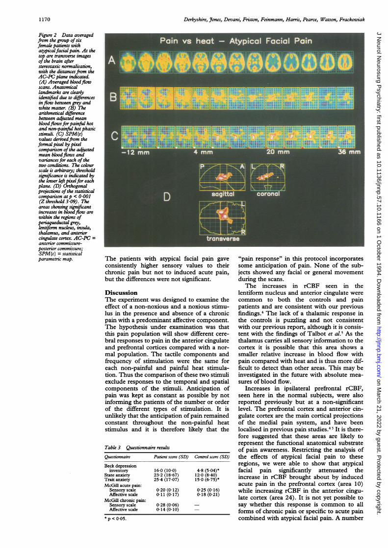
in rCBF on comparing heat with pain inpatients with atypical facial pain. The mainareas of increased rCBF were contralateral inthe lentiform nucleus, insula, and thalamus.The laterality of the increased rCBF in theanterior cingulate cortex (area 24) was againindeterminate. Figure 2 shows the increases inblood flow in response to pain for this group.These focal rCBF increases are in the form ofSPM{t}.
Significant decreases in rCBF were seenbilaterally in the prestriate cortex, contralat-eral premotor (area 6), parietal, and frontalcortices (area 8), and ipsilateral prefrontalcortex (area 10).- There was no evidence ofsignificant change in rCBF in the primarysomatosensory cortex on either side.
Table 2 Within group comparison for the atypicalfacial pain group
Coordinates (mm)
Region Side X Y Z Associated Z value
Patients with atypical facial pain: rCBF increases:Periaqueductal grey M -2 -44 -16 3-980Anterior cingulate (area 24) M 0 -16 36 3-417Lentiform nucleus L -16 -10 0 3-243Insula L -38 2 0 3-386Thalamus L -18 -18 12 3-406
Patients with atypical facial pain: rCBF decreases:Inferior parietal (area 40) L -42 -66 28 4-084Area 31 L -10 -58 24 3-356Prestriate L -34 -78 32 3-275Area 8 L -28 10 44 4-847Premotor (area 6) L -38 6 52 3-273Frontal pole (area 10) R 6 46 -8 3-982Medial frontal (area 32) R 4 46 -4 3-171Prestriate (areas 18 and 19) R 12 -88 24 3-475Area 7 R 10 -74 40 3-222
M = Midline; L = left; R = right.
COMPARISON OF rCBF INCREASES IN THECONTROL GROUP WITH INCREASES IN THEATYPICAL FACIAL PAIN GROUPFigure 3 shows the comparison of the acutepain changes between the two groups in termsof significant focal differences in the form ofSPMs at the appropriate levels in the brain.Activation in the atypical facial pain groupwas greater in the anterior cingulate cortex(area 24) and significantly less in the ipsi-lateral prefrontal cortex (area 10).
MEASURES OF DEPRESSION AND PAINEXPERIENCETable 3 shows that the patients with atypicalfacial pain scored higher on all the tests ofdepression and anxiety. These scores wereonly significantly higher (p = 0 05), however,on measures of depression and trait anxiety;not state anxiety (Student's t test).To determine a measure of sensory inten-
sity, all descriptors selected within the sensorycategories of the McGill pain questionnairewere summated by rank value and thendivided by the highest possible score. Thisscoring method yielded values ranging from 0to 1 with a score of 0 indicating that the subjectdid not select any adjectives from any of thesensory categories and a score of 1 indicatingthat the patient selected the highest rankedword in each category. This same procedurewas used to obtain a quantitative measure ofaffective descriptors. These values were aver-aged for the three retrospective acute painmeasures in both groups and for the sixchronic atypical facial pain measures in thefacial pain group. Table 3 gives the results.
1169
on March 21, 2022 by guest. P
rotected by copyright.http://jnnp.bm
j.com/
J Neurol N
eurosurg Psychiatry: first published as 10.1136/jnnp.57.10.1166 on 1 O
ctober 1994. Dow
nloaded from
Derbyshire, J7ones, Devani, Friston, Feinmann, Harris, Pearce, Watson, Frackowiak
Figure 2 Data averagedfrom the group ofsixfemale patients withatypicalfacialpain. At thetop are transverse imagesof the brain afterstereotaxtc normalisation,with the distancesfrom theAC-PC plane indicated.(A) Averaged bloodflowscans. Anatomicallandmarks are clearlyidentified due to differencesinflow between grey andwhite matter. (B) Thearithmetical differencebetween adjusted meanbloodflows for painful hotand non-painful hot phasicstimuli. (C) SPM{t}values derivedfrom theformal pixel by pixelcompanson of the adjstedmean bloodflows andvariances for each of thetwo conditions. The colourscale is arbitrary; thresholdsignificance is indicated bythe lower left pixelfor eachplane. (D) Orthogonalprojections of the statisticalcomparison atp < 0-001(Z threshold 3 09). Theareas showing significantincreases in bloodflow arewithin the regions ofperiaqueductal grey,lentiforn nucleus, insula,thalamus, and anteriorcingulate cortex. AC-PC =anterior commissure-posterior commissure;SPM{t} = statisticalparametric map. The patients with atypical facial pain gave
consistently higher sensory values to theirchronic pain but not to induced acute pain,but the differences were not significant.
DiscussionThe experiment was designed to examine theeffect of a non-noxious and a noxious stimu-lus in the presence and absence of a chronicpain with a predominant affective component.The hypothesis under examination was thatthis pain population will show different cere-bral responses to pain in the anterior cingulateand prefrontal cortices compared with a nor-
mal population. The tactile components andfrequency of stimulation were the same foreach non-painful and painful heat stimula-tion. Thus the comparison of these two stimuliexclude responses to the temporal and spatialcomponents of the stimuli. Anticipation ofpain was kept as constant as possible by notinforming the patients of the number or orderof the different types of stimulation. It isunlikely that the anticipation of pain remainedconstant throughout the non-painful heatstimulus and it is therefore likely that the
Table 3 Questionnaire results
Questionnaire Patient score (SD) Control score (SD)
Beck depressioninventory 16-0 (10 0) 4-8 (5 04)*
State anxiety 23-2 (18-67) 12-0 (8 40)Trait anxiety 25-4 (17-07) 15-0 (6 75)*McGill acute pain:
Sensory scale 0-20 (0-12) 0-25 (0-16)Affective scale 0 11 (0-17) 0-18 (0-21)
McGill chronic pain:Sensory scale 0-28 (0 06) -
Affective scale 0-14 (0 10)
*p < 005.
"pain response" in this protocol incorporatessome anticipation of pain. None of the sub-jects showed any facial or general movementduring the scans.The increases in rCBF seen in the
lentiform nucleus and anterior cingulate werecommon to both the controls and painpatients and are consistent with our previousfindings.4 The lack of a thalamic response inthe controls is puzzling and not consistentwith our previous report, although it is consis-tent with the findings of Talbot et al.5 As thethalamus carries all sensory information to thecortex it is possible that this area shows asmaller relative increase in blood flow withpain compared with heat and is thus more dif-ficult to detect than other areas. This may beinvestigated in the future with absolute mea-sures of blood flow.
Increases in ipsilateral prefrontal rCBF,seen here in the normal subjects, were alsoreported previously but at a non-significantlevel. The prefrontal cortex and anterior cin-gulate cortex are the main cortical projectionsof the medial pain system, and have beenlocalised in previous pain studies.45 It is there-fore suggested that these areas are likely torepresent the functional anatomical substrateof pain awareness. Restricting the analysis ofthe effects of atypical facial pain to theseregions, we were able to show that atypicalfacial pain significantly attenuated theincrease in rCBF brought about by inducedacute pain in the prefrontal cortex (area 10)while increasing rCBF in the anterior cingu-late cortex (area 24). It is not yet possible tosay whether this response is common to allforms of chronic pain or specific to acute paincombined with atypical facial pain. A number
1170
on March 21, 2022 by guest. P
rotected by copyright.http://jnnp.bm
j.com/
J Neurol N
eurosurg Psychiatry: first published as 10.1136/jnnp.57.10.1166 on 1 O
ctober 1994. Dow
nloaded from

Cerebral responses to pain by positron emission tomography
Figure 3 Two statistical parametric maps (SPM{t}) in the transaxial plane ofcomparisons described in the main text. The left part of thefigure shows the anatomycorresponding to the parametric maps. The top right part of the figure illustrates anSPM{t} showing activation of the anterior cingulate in the comparison of the differencebetween non-painfid heat and painful heat between the two groups, with significantlygreater activity beingfound in the group with atypicalfacial pain. The bottom right part ofthe figure illustrates an SPM{t} showing right sided prefrontal cortex in the comparison ofnon-painful heat and painful heat differences between the two groups, with greater activityfound in the control group. The colour scale is arbitrary and the significance levelcorresponds to p = 0 001. The top section is 28 mm above and parallel to the AC-PC line,the bottom section is 16 mm above the AC-PC line. AC-PC = anterior commissure-posterior commissure; SPM{t} = statistical parametric map.
of explanations for this pattern of rCBF arepossible and studies are ongoing with otherchronic pain disorders, such as arthritic pain,to discriminate between these possibilities.
Blood flow in the anterior cingulate cortexis thought to reflect the attentional,' motiva-tional, and emotional37 aspects of pain pro-cessing and response. Studies with rabbitshave shown that anterior cingulate lesionsinterfere with aversive conditioning38 andstimulation of area 24 of the anterior cingulatein animals produces a shrill vocalisationresponse.39 Atypical facial pain is often associ-ated with an emotional disturbance22 involv-ing some serious life event, such as a
bereavement, with inadequate support fromrelatives or spouse. The anterior cingulatecortex is well placed to integrate variablestressors and to disrupt analgesic mechanismshaving reciprocal connections with the medialthalamic nuclei40 and projecting to the pre-frontal cortex,4' striatum,42 and periaqueduc-tal grey.4' Atypical facial pain, therefore, maybe a "hyperemotional" response to incomingsensory information. The finding that chronicpain loses its emotional component after
frontal leucotomy and cingulotomy44 in com-bination with the findings reported here lendssupport to this hypothesis.The prefrontal cortex has important projec-
tions to the anterior cingulate cortex and basalganglia45 as well as weaker connections withthe insula cortex. Prefrontal cortical rCBF(area 10) was significantly increased in thecontrol group. Shallice46 has proposed that theprocess by which complex behavioural unitsor schemas are brought to conscious attentionis the function of the "supervisory attentionsystem". This is part of the "programming,regulation, and verification of human activ-ity"47 by the frontal lobes. Posner andRothbart48 argue that this alert state is later-alised to the right lateral frontal lobe based onits close involvement with the regulations ofthe heart. The maintenance of vigilance isindexed by a pronounced slowing of the heart.The abnormal pattern of right prefrontal andanterior cingulate responses in these patientsmay therefore reflect an abnormal "super-vision" of attention and emotional schemas.This is consistent with the perception of phys-ical symptoms proposed by Pennebaker.49The common conviction of these patients thatthere is something structurally wrong withtheir face,50 and their high trait anxiety, wouldbe seen as a schema in which the likelihood ofperceiving painful sensory input from the faceis high.5"
It is apparent from the McGill scores thatthe chronic and acute pain were not triggeringany exceptional emotional response in thepatients with atypical facial pain. The affectiveMcGill scores were the same for both groupsin response to the acute pain stimulus. This isnot consistent with larger group studies50 andmay relate to a desire by these patients to provethe reality of their atypical facial pain to themedical staff carrying out the scan by denyingany emotional input to their disorder.52The pharmacological substrates for the
abnormal patterns of pain rCBF seen in thepatients with atypical facial pain are not clear.Although about 80% of cases respond to tri-cyclic or monoamine oxidase inhibitors only45% are found to be depressed.5' Further-more, these patients seem to have a deficit inthe excretion of conjugated tyramine compa-rable to patients with endogenous depression.This biological marker is independent ofdepression in the patients with atypical facialpain suggesting a neuropharmacologicaldeficit common to both conditions.54 It hasbeen suggested that descending cinguloperi-aqueductal efferents modulate activity of thedescending 5-hydroxytryptamine (5HT)mediated inhibitory system via the middleraphe nuclei in the brain stem.55 If uncon-trolled this system may deplete serotoninreserves and so disrupt descending analgesia,56or directly interfere with opiate organisationin the cingulate cortex itself.'9 It is also knownthat patients with depression have low levelsof 5HT breakdown products in the CSF andthat suicide victims have decreased 5HT andnoradrenaline concentrations with increasedconcentrations of 5HT2 and ,B receptors inthe frontal cortex. Increased receptors have
1 171
on March 21, 2022 by guest. P
rotected by copyright.http://jnnp.bm
j.com/
J Neurol N
eurosurg Psychiatry: first published as 10.1136/jnnp.57.10.1166 on 1 O
ctober 1994. Dow
nloaded from
Derbyshire, Jones, Devani, Friston, Feinmann, Harris, Pearce, Watson, Frackowiak
been suggested as a compensatory mechanismfor reduced postsynaptic concentration ofthese amines. The success of tricyclic antide-pressants in patients with atypical facial painmay be explained by the restoration of aminestores. Studies to examine the effects of tri-cyclic antidepressants on the reversal of rCBFpatterns seen in these patients are ongoingand necessary to clarify the possible role ofcentral amine depletion.
In conclusion, important differencesbetween patients with atypical facial pain andnormal volunteers have been discovered in theresponse of the prefrontal cortex and anteriorcingulate cortex to pain. These differences inblood flow may be responsible for the mainte-nance of chronic pain through the failure ofinhibition of other cortical and limbic struc-tures. It is likely that this mechanism is relatedto both overt emotional processing, anxiety,and attentional mechanisms. There is there-fore the possibility that, at least for some ofthese patients, the pain may be brought underconscious control.This research was supported in part by the Economic andSocial Research Council (ESRC grant R00429134074) and inpart by the Medical Research Council (MRC).
1 Lueck CJ, Zeki S, Friston KJ, et al. The colour centre inthe cerebral cortex of man. Nature 1989;340:386-9.
2 Friston KJ, Frith CD, Liddle PF, Lanmmertsma AA, DolanRD, Frackowiak RSJ. The relationship between localand global changes in PET scans. J Cereb Blood FlowMetabol 1990;10:458-66.
3 Pardo JV, Pardo PJ, Janer KW, Raichle ME. The anteriorcingulate cortex mediates processing selection in theStroop attentional conflict paradigm. Neurobiology1990;87:256-9.
4 Jones APK, Brown WD, Friston KJ, Qi LY, FrackowiakRSJ. Cortical and subcortical localization of response topain in man using positron emission tomography. Proc RSoc Lond [Biol] 1991;244:39-44.
5 Talbot JD, Marret S, Evans AC, Meyer E, Bushnell MC,Duncan GH. Multiple representations of pain in humancerebral cortex. Science 1991;251:1355-8.
6 Roland PE. Cortical representation of pain. TrendsNeurosci 1992;15:3-5.
7 Stea RA, Apkarian AV. Pain and somatosensory activa-tion. Trends Neurosci 1992;15:250-1.
8 Evans AC, Meyer E, Marret S. Pain and activation in thethalamus. Trends Neurosci 1992;15:252.
9 Roland PE. Reply. Trends Neurosci 1992;15:252-3.10 Guyton AC. Basic neuroscience: anatomy and physiology.
Philadelphia: WB Saunders, 1991.11 Melzack R, Wall P. The challenge ofpain. London: Penguin
Books, 1991.12 Leventhal H, Everhart D. Emotion, pain, and physical ill-
ness. In: CE Izard, ed. Emotions in personality and psy-chopathology. New York: Plenum Press, 263-99.
13 Leventhal H. A perceptual-motor theory of emotion.Advances in Experimental Social Psychology 1984;17:117-75.
14 Leventhal H, Scherer K. The relationship of emotion tocognition: a functional approach to a semantic contro-versy. Cognition and Emotion 1987;1:3-28.
15 Beecher HK. Quantification of the subjective pain experi-ence. In: PH Hoch, J Zubin, eds. Psychopathology ofper-ception. New York: Grune and Stratton, 1965:111-28.
16 Kupers R, Konings H, Adriaensen H, Gybels J. Morphinedifferentially affects the sensory and affective pain rat-ings in neurogenic and idiopathic forms of pain. Pain1991;47:5-12.
17 Foltz EL, White LEJ. Pain relief by frontal cingulotomy.Neurosurgery 1962;19:89-100.
18 Leventhal H, Brown D, Shacham S, Engquist G. Effects ofpreparatory information about sensations, threat of pain,and attention on cold pressor distress. J Pers Soc Psychol1979;37:688-714.
19 Jones AKP, Friston KJ, Qi LY, et al. The sites of action ofmorphine on the human brain studied with positronemission tomography. Lancet 1991;338:825.
20 Agerberg D, Carison GE. Functional disorders of the mas-ticatory system. Distribution of symptoms according toage and sex as judged by questionnaire. Acta OdontolScand 1972;30:577-613.
21 Pearce S. The concept of psychogenic pain. A psychophys-ical investigation of women with pelvic pain. In: MJohnson, T Marteu, eds. Applications in health psychology.New Jersey: Transaction, 1989.
22 Feinmann C, Harris M. Psychogenic facial pain, part 1:the clinical presentation. Br DentJ 1984;156:165-8.
23 Feinmann C, Harris M. Psychogenic facial pain (part 2):management and prognosis. Br DentJ 1984;156:205-8.
24 Friston KJ, Frackowiak RSJ. Imaging functional anatomy.Alfred Benzon symposium 31. Copenhagen: Munksgaard,1991.
25 Fruhstorfer H, Lindblom U, Schnidt WG. Method forquantitative estimation of thermal thresholds in patients.Journal ofNeurology and Psychiatry 1976;39:1071-5.
26 Spinks TJ, Jones T, Gilardi MC, HeatherJD. Performancecharacteristics of a whole body positron tomograph.IEEE Transactions on Nuclear Science 1988;35:721-5.
27 Spielberger CD, Gorsuch RL, Lushene R. Manual for thestate-trait anxiety inventory. Palo Alto, CA: ConsultingPsychologists' Press, 1970.
28 Beck AJ, Ward CH, Medelson M, Mock J, Erbaugh J. Aninventory for measuring depression. Arch Gen Psychiatry1961;4:561-7.
29 Melzack R. The McGill pain questionnaire: major proper-ties and scoring methods. Pain 1975;1:277-99.
30 Mazziotta JC, Huang SC, Phelps ME, Carson RE,Macdonald NS, Mahoney K. A non-invasive positroncomputed tomography technique using oxygen-15labeled water for the evaluation of neurobehavioural taskbatteries. J Cereb Blood Flow Metab 1985;5:70-8.
31 Fox PT, Mintum MA. Noninvasive functional brain map-ping by change-distribution analysis of averaged PETimages of H,"1O tissue activity. Y Nud Med 1989;30:141-9.
32 Woods RP, Cherry SR, Mazziotta JC. A rapid automatedalgorithm for accurately aligning and reslicing positronemission tomography images. J Comput Assist Tomogr1992;16:620-33.
33 Friston KJ, Frith PF, Liddle PF, Frackowiak RSJ.Comparing functional (PET) images: the assessment ofsignificant change. J Cereb Blood Flow Metab 1991;11:690-9.
34 Talairach J, Tournoux P. Co-planar stereotaxic adas of thehuman brain. Stuttgart: Georg Thieme, 1988.
35 Friston KJ, Frith CD, Liddle PF, Lammertsma AA, DolanRD, Frackowiak RSJ. The relationship between localand global changes in PET scans. J Cereb Blood FlowMetab 1990;10:458-66.
36 Bailey DL, Jones T, Spinks TJ. A method for measuringthe absolute sensitivity of positron emission tomographicscanners. EurJ Nucl Med 1991;18:374-9.
37 Gabriel M, Orona E, Foster K, Lambert RW. Mechanismsand generality of stimulus significance coding in a mam-malian model system. Advances in Behavioural Biology1982;26:535-67.
38 Gabriel M. Functions of anterior and posterior cingulatecortex during avoidance learning in rabbits. In: HBMUylings, CG Van Eden, JPC De Bruin, MA Corner,MGP Feenstra, eds. The prefrontal cortex. Amsterdam:Elsevier, 1990:467-83.
39 Vogt BA, Rosene DL, Pandya DN. Thalamic and corticalafferents differentiate anterior from posterior cingulatecortex in the monkey. Science 1979;204:205-7.
40 Domesick V. Projections from the cingulate cortex in therat. Brain Res 1969;12:296-320.
41 Pandya D, Van Hoesen G, Mesulam MM. Efferent con-nections of the cingulate gyrus in the rhesus monkey.Exp Brain Res 1981;42:319-30.
42 Royce G. Laminar origin of cortical neurons which projectupon the caudate nucleus: a horseradish peroxidaseinvestigation in the cat. J Comp Neurol 1982;205:8-29.
43 Muller-Preuss P, Jurgens U. Projections from the "cingu-lar" vocalization area in the squirrel monkey. Brain Res1976;103:29-43.
44 Bouckoms AJ. Psychosurgery. In: Wall PD, Melzack R,eds. Textbook of pain: 2nd ed. Edinburgh: ChurchillLivingstone, 1989:666-76.
45 Groenewegen HJ, Berendse HW, Wolters JG, LohmanAHM. The anatomical relationship of the prefrontal cor-tex with the striatopallidal system, the thalamus and theamygdala: evidence for a parallel organization. In: HBMUylings, CG Van Eden, JPC De Bruin, MA Corner,MGP Feenstra, eds. The Prefrontal Cortex. Amsterdam:Elsevier, 1990.
46 Shallice T. Specific impairements of planning. Phil Trans RSoc LondB 1982;298:199-209.
47 Luria AR. The working brain. An introduction to neuropsy-chology. Translated by B Haigh. New York: Basic Books,1977:187.
48 Posner MI, Rothbart MK. Attentional mechanisms andconscious experience. In: D Milner, M Rugg, eds. Theneuropsychology of consciousness. London: AcademicPress, 1991.
49 Pennebaker J. The psychology of physical symptoms. NewYork: Springer-Verlag, 1982.
50 Harris M, Feinmann C. Psychosomatic disorders. In: MKMason, JG Jones, eds. Oral manifestations of systemic dis-ease. Philadelphia: WB Saunders, 1990.
51 Eysenck MW, Macleod C, Mathews A. Cognitive func-tioning and anxiety. Psychol Res 1987;49: 189-95.
52 Pilowsky I, Bassett D, Barret R, Petrovic L, Minniti R.The illness behaviour assessment schedule: reliabilityand validity. IntJ Psychiatry Med 1983-4;131:11-28.
53 Feinmann C. Pain relief by antidepressants: possiblemodes of action. Pain 1985,23:1-8._
54 Ashabeigi B, Feinmann C, Glover V, Goodwin B, HannahP, Sandler M, Wasil M. Tyramine conjugation deficit inpatients with chronic idiopathic temporomandibularjoint and orofacial pain. Pain 1993;54:159-63.
55 Sikes RW, Vogt BA. Nociceptive neurons in area 24 ofrabbit cingulate cortex. J Neurophysiol 1992;68:1720-32.
56 Melzack R, Stotler WA, Livingstone WK. Effects of dis-crete brainstem lesions in cats on perception of noxiousstimulation. J Neurophysiol 1958;21:353-67.
1172
on March 21, 2022 by guest. P
rotected by copyright.http://jnnp.bm
j.com/
J Neurol N
eurosurg Psychiatry: first published as 10.1136/jnnp.57.10.1166 on 1 O
ctober 1994. Dow
nloaded from


![r n a l o f Pain o u elief Journal of Pain & Relief · 2018. 3. 6. · Atypical odontalgia is a subtype of persistent idiopathic facial pain (PIFP) [10], which has pain characteristics](https://static.fdocuments.net/doc/165x107/60320a51cb58d26e8967360c/r-n-a-l-o-f-pain-o-u-elief-journal-of-pain-relief-2018-3-6-atypical-odontalgia.jpg)
