CerebralAngiographyforEvaluationofPatientswithCT … INTERVENTIONAL...
8
ORIGINAL RESEARCH INTERVENTIONAL Cerebral Angiography for Evaluation of Patients with CT Angiogram-Negative Subarachnoid Hemorrhage: An 11-Year Experience X J.J. Heit, X G.T. Pastena, X R.G. Nogueira, X A.J. Yoo, X T.M. Leslie-Mazwi, X J.A. Hirsch, and X J.D. Rabinov ABSTRACT BACKGROUND AND PURPOSE: CT angiography is increasingly used to evaluate patients with nontraumatic subarachnoid hemorrhage given its high sensitivity for aneurysms. We investigated the yield of digital subtraction angiography among patients with SAH or intra- ventricular hemorrhage and a negative CTA. MATERIALS AND METHODS: An 11-year, single-center retrospective review of all consecutive patients with CTA-negative SAH was performed. Noncontrast head CT, CTA, DSA, and MR imaging studies were reviewed by 2 experienced interventional neuroradiologists and 1 neuroradiologist. RESULTS: Two hundred thirty patients (mean age, 54 years; 51% male) with CTA-negative SAH were identified. The pattern of SAH was diffuse (40%), perimesencephalic (31%), sulcal (31%), isolated IVH (6%), or identified by xanthochromia (7%). Initial DSA yield was 13%, including vasculitis/vasculopathy (7%), aneurysm (5%), arteriovenous malformation (0.5%), and dural arteriovenous fistula (0.5%). An addi- tional 6 aneurysms/pseudoaneurysms (4%) were identified by follow-up DSA, and a single cavernous malformation (0.4%) was identified by MRI. No cause of hemorrhage was identified in any patient presenting with isolated intraventricular hemorrhage or xanthochromia. Diffuse SAH was due to aneurysm rupture (17%); perimesencephalic SAH was due to aneurysm rupture (3%) or vasculitis/vasculopathy (1.5%); and sulcal SAH was due to vasculitis/vasculopathy (32%), arteriovenous malformation (3%), or dural arteriovenous fistula (3%). CONCLUSIONS: DSA identifies vascular pathology in 13% of patients with CTA-negative SAH. Aneurysms or pseudoaneurysms are identified in an additional 4% of patients by repeat DSA following an initially negative DSA. All patients with CT-negative SAH should be considered for DSA. The pattern of SAH may suggest the cause of hemorrhage, and aneurysms should specifically be sought with diffuse or perimesencephalic SAH. ABBREVIATION: IVH intraventricular hemorrhage N ontraumatic subarachnoid hemorrhage occurs in 30,000 pa- tients per year in the United States, which accounts for 5% of strokes. 1 Patient mortality approximates 45% within a month af- ter SAH, 1 and the identification of a treatable cause of SAH is imperative. Cerebral aneurysm rupture accounts for most SAHs, 2 but a cause of hemorrhage is not identified in 15%–20% of pa- tients. 2-4 Prior studies have demonstrated that the pattern of SAH on a noncontrast head CT may predict the probability of identifying a causative vascular lesion, though CT angiography or digital subtraction angiography is needed to identify these lesions. 5-9 The evaluation of SAH varies, and no consensus exists as to the best algorithm. Increasingly, CTA is performed in the evaluation of SAH, given its noninvasive nature and wide availability. CTA has a reported a sensitivity of 97%–100% for the detection of intracranial aneurysms. 10-13 However, it fails to identify a cause for SAH in 5%– 30% of patients. 14,15 DSA remains the criterion standard in the diag- nosis of vascular lesions resulting in SAH with a reported sensitivity of 99%. 16,17 Given the superior sensitivity of DSA and the impor- tance of identifying a treatable cause of SAH, DSA is frequently per- formed in patients presenting with SAH and negative CTA findings. Prior studies demonstrated that DSA identifies a cause of SAH Received May 12, 2015; accepted after revision June 22. From the Department of Radiology (J.J.H.), Interventional Neuroradiology Division, Stanford University Hospital, Stanford, California; Department of Radiology (G.T.P.), Albany Medical Center, Albany, New York; Departments of Neurology, Neurosurgery, and Radiology (R.G.N.), Emory University School of Medicine, Mar- cus Stroke and Neuroscience Center, Atlanta, Georgia; Department of Neuroradi- ology and Interventional Neuroradiology (T.M.L.-M., J.A.H., J.D.R.), Massachusetts General Hospital, Boston, Massachusetts; and Texas Stroke Institute (A.J.Y.), Plano, Texas. Please address correspondence to James D. Rabinov, MD, Interventional Neurora- diology, Gray 241, Massachusetts General Hospital, 55 Fruit St, Boston, MA 02114; e-mail: [email protected] http://dx.doi.org/10.3174/ajnr.A4503 AJNR Am J Neuroradiol 37:297–304 Feb 2016 www.ajnr.org 297
Transcript of CerebralAngiographyforEvaluationofPatientswithCT … INTERVENTIONAL...

ORIGINAL RESEARCHINTERVENTIONAL
Cerebral Angiography for Evaluation of Patients with CTAngiogram-Negative Subarachnoid Hemorrhage: An 11-Year
ExperienceX J.J. Heit, X G.T. Pastena, X R.G. Nogueira, X A.J. Yoo, X T.M. Leslie-Mazwi, X J.A. Hirsch, and X J.D. Rabinov
ABSTRACT
BACKGROUND AND PURPOSE: CT angiography is increasingly used to evaluate patients with nontraumatic subarachnoid hemorrhagegiven its high sensitivity for aneurysms. We investigated the yield of digital subtraction angiography among patients with SAH or intra-ventricular hemorrhage and a negative CTA.
MATERIALS AND METHODS: An 11-year, single-center retrospective review of all consecutive patients with CTA-negative SAH wasperformed. Noncontrast head CT, CTA, DSA, and MR imaging studies were reviewed by 2 experienced interventional neuroradiologists and1 neuroradiologist.
RESULTS: Two hundred thirty patients (mean age, 54 years; 51% male) with CTA-negative SAH were identified. The pattern of SAH wasdiffuse (40%), perimesencephalic (31%), sulcal (31%), isolated IVH (6%), or identified by xanthochromia (7%). Initial DSA yield was 13%,including vasculitis/vasculopathy (7%), aneurysm (5%), arteriovenous malformation (0.5%), and dural arteriovenous fistula (0.5%). An addi-tional 6 aneurysms/pseudoaneurysms (4%) were identified by follow-up DSA, and a single cavernous malformation (0.4%) was identified byMRI. No cause of hemorrhage was identified in any patient presenting with isolated intraventricular hemorrhage or xanthochromia. DiffuseSAH was due to aneurysm rupture (17%); perimesencephalic SAH was due to aneurysm rupture (3%) or vasculitis/vasculopathy (1.5%); andsulcal SAH was due to vasculitis/vasculopathy (32%), arteriovenous malformation (3%), or dural arteriovenous fistula (3%).
CONCLUSIONS: DSA identifies vascular pathology in 13% of patients with CTA-negative SAH. Aneurysms or pseudoaneurysms areidentified in an additional 4% of patients by repeat DSA following an initially negative DSA. All patients with CT-negative SAH should beconsidered for DSA. The pattern of SAH may suggest the cause of hemorrhage, and aneurysms should specifically be sought with diffuseor perimesencephalic SAH.
ABBREVIATION: IVH � intraventricular hemorrhage
Nontraumatic subarachnoid hemorrhage occurs in 30,000 pa-
tients per year in the United States, which accounts for 5% of
strokes.1 Patient mortality approximates 45% within a month af-
ter SAH,1 and the identification of a treatable cause of SAH is
imperative.
Cerebral aneurysm rupture accounts for most SAHs,2 but a
cause of hemorrhage is not identified in 15%–20% of pa-
tients.2-4 Prior studies have demonstrated that the pattern of
SAH on a noncontrast head CT may predict the probability of
identifying a causative vascular lesion, though CT angiography
or digital subtraction angiography is needed to identify these
lesions.5-9
The evaluation of SAH varies, and no consensus exists as to the
best algorithm. Increasingly, CTA is performed in the evaluation of
SAH, given its noninvasive nature and wide availability. CTA has a
reported a sensitivity of 97%–100% for the detection of intracranial
aneurysms.10-13 However, it fails to identify a cause for SAH in 5%–
30% of patients.14,15 DSA remains the criterion standard in the diag-
nosis of vascular lesions resulting in SAH with a reported sensitivity
of 99%.16,17 Given the superior sensitivity of DSA and the impor-
tance of identifying a treatable cause of SAH, DSA is frequently per-
formed in patients presenting with SAH and negative CTA findings.
Prior studies demonstrated that DSA identifies a cause of SAH
Received May 12, 2015; accepted after revision June 22.
From the Department of Radiology (J.J.H.), Interventional Neuroradiology Division,Stanford University Hospital, Stanford, California; Department of Radiology(G.T.P.), Albany Medical Center, Albany, New York; Departments of Neurology,Neurosurgery, and Radiology (R.G.N.), Emory University School of Medicine, Mar-cus Stroke and Neuroscience Center, Atlanta, Georgia; Department of Neuroradi-ology and Interventional Neuroradiology (T.M.L.-M., J.A.H., J.D.R.), MassachusettsGeneral Hospital, Boston, Massachusetts; and Texas Stroke Institute (A.J.Y.), Plano,Texas.
Please address correspondence to James D. Rabinov, MD, Interventional Neurora-diology, Gray 241, Massachusetts General Hospital, 55 Fruit St, Boston, MA 02114;e-mail: [email protected]
http://dx.doi.org/10.3174/ajnr.A4503
AJNR Am J Neuroradiol 37:297–304 Feb 2016 www.ajnr.org 297

in 4%–14% of patients with negative CTA findings.3,4,14,18-20
Moreover, the diagnostic yield of repeat DSA after initially nega-
tive DSA findings is reported to be between 4% and 16%.14,15,21
We describe our experience with initial and repeat DSA in pa-
tients with CTA negative for SAH during an 11-year period at a
large neurovascular referral center.
MATERIALS AND METHODSPatient SelectionWe retrospectively reviewed the radiology data base and medical
records of all patients who presented to our hospital (Massachu-
setts General Hospital) during an 11-year period (January 1, 2002,
through December 31, 2012) with the following: 1) nontraumatic
subarachnoid hemorrhage or isolated intraventricular hemor-
rhage identified by noncontrast head CT or xanthochromia on
lumbar puncture, 2) initial evaluation with a CTA that failed to
identify a cause of SAH, and 3) at least 1 cerebral DSA. If an initial
cerebral DSA was negative for SAH, patients underwent addi-
tional studies.
Follow-up DSA was planned in all patients at an interval of
7–10 days unless there was a contraindication such as arterial
dissection or death, and some patients underwent DSA in a
shorter time interval due to a clinical deterioration or other evi-
dence of cerebral arterial vasospasm. If a diagnosis was made by a
noninvasive imaging study, including CTA or MR imaging/MRA,
a follow-up DSA was not performed. Noninvasive imaging stud-
ies were performed at the discretion of the physician caring for the
patient.
Incidental aneurysms identified by DSA were not considered
causative of SAH and comprised additional aneurysms distinct
from the ruptured lesions or aneurysms remote from the region of
SAH. The study was approved by the institutional review board of
our hospital and complied with the Health Insurance Portability
and Accountability Act.
Image Acquisition and AnalysisNCCT was performed with standard protocols by using a 16- or
64-section helical CT scanner (LightSpeed; GE Healthcare, Mil-
waukee, Wisconsin). Axial NCCT images were obtained with
120 –140 kV(peak), 170 mA, and 5-mm-section-thickness recon-
struction. CTA was performed by axial acquisition from the base
of the C1 vertebral body to the vertex by using an 0.5 pitch,
1.25-mm collimation, 350 maximal mA,
120 kVP, and 22-cm FOV. We adminis-
tered 65– 85 mL of iodinated contrast by
power injector at 4 –5 mL/s into an an-
tecubital vein, and image acquisition
was initiated with either a fixed 25-sec-
ond delay after injection, SmartPrep
software (GE Healthcare), or semiauto-
matic contrast bolus triggering. Axial,
coronal, and sagittal maximum-intensity-
projection images were available at the
time of study review and curvilinear 3D
reformations were created by an inde-
pendent processing laboratory.MR imaging and MRA were per-
formed on either 1.5T or 3T scanners(Signa 1.5T; GE Healthcare; or Tim Trio
3T; Siemens, Erlangen, Germany) by using standard departmen-tal and vendor protocols.
DSA was performed in a biplane neuroangiography suite (Ax-iom Artis; Siemens) following transfemoral arterial access underintravenous conscious sedation or general anesthesia. BiplanarDSA images were obtained after selective catheterization of thebilateral common carotid arteries and at least 1 vertebral arteryand injection with iodinated contrast. Additional selective injec-tion of the internal, external, or contralateral vertebral artery wasperformed at the discretion of the neurointerventionalist. 3D ro-tational angiography was performed at the discretion of the inter-ventional neuroradiologist. Standard views typically includedfrontal, lateral, and oblique views of each selected vessel.
All NCCT, CTA, and DSA images were independently re-viewed by 2 interventional neuroradiologists and 1 neuroradiolo-gist to determine the presence and pattern of SAH, including thefollowing: 1) absence of SAH, or 2) perimesencephalic, 3) sulcal,and 4) diffuse SAH. Perimesencephalic SAH was consideredlargely within the interpeduncular, prepontine, ambient, quadri-geminal plate or premedullary cistern with minimal extensioninto the medial Sylvian fissures. This imaging review was per-formed near the end of the 11-year study period when the manu-script was being prepared. The presence of intraventricular hem-orrhage (IVH) was also identified. CTA images were reviewed fora vascular cause of SAH. If vessel irregularity was identified, DSAwas performed to determine whether the irregularity representedvasospasm in the region of SAH versus a vasculitis or vasculopa-thy and for the presence of an aneurysm. If an aneurysm wasidentified by DSA after a CTA negative for SAH, the CTA wasreviewed retrospectively. These studies were included as false-negative ones.
Medical Record ReviewPatient demographic information and clinical outcome were re-
viewed in the electronic Longitudinal Medial Record system avail-
able at our hospital.
Statistical AnalysisStatistical analysis was performed by using Excel (Microsoft, Red-
mond, Washington) and XLSTAT (Addinsoft, New York, New
York). A P value of .05 was statistically significant.
Table 1: Demographic data of patients presenting with subarachnoid or isolatedintraventricular hemorrhage without a causative lesion on CTA
Total Men WomenNo. of Patients 230 118 112Mean age (yr) 54 55 54Age range 19–92 21–92 19–87SAH found by CT 216 106 110SAH found by lumbar puncture 14 7 7Mean time between CTA and DSA (days) 1.5 1.0 2.0Time range between CTA and DSA (days) 0–110 0–5 0–110Second follow-up study performed (No. of patients) 169 86 83Mean time between initial and follow-up study (days) 33 24 42Time range between initial and follow-up study (days) 0–1836 0–203 0–1836Second follow-up modality
CTA 54 28 26DSA 98 45 53MRI/MRA 17 10 7
298 Heit Feb 2016 www.ajnr.org
RESULTSFrom January 1, 2002, to December 31, 2012, 1288 patients pre-
sented with nontraumatic SAH. CTA identified an aneurysm as
the cause of SAH in 1058 patients (82%), and the CTAs typically
provided sufficient data for treatment triage by microsurgical
clipping or endovascular coil embolization. The 1058 patients
with aneurysms identified by CTA were excluded from the anal-
ysis. The remaining 230 patients had SAH or isolated IVH and a
CTA that failed to reveal a causative lesion. These 230 patients
(17.9% of patients with SAH) included 118 men (51%) and 112
women (49%), and the average patient age was 54 years (Table 1).
SAH was identified by CT in 216 patients (94%) and by CSF
xanthochromia on lumbar puncture in 14 patients (6%). In
patients with SAH identified by CT,
the SAH distribution was perimesen-
cephalic in 71 patients (31%), sulcal in
37 patients (16%), and diffuse in 93 pa-
tients (40%). Representative imaging
examples of each type of SAH are show
in Fig 1. Sulcal SAH was identified in 22
women and 15 men (18% and 14%, re-
spectively; P � .001), and there was no
significant difference in the prevalence
of other distributions of SAH between
men and women (Table 2). Isolated
IVH was identified in 13 patients (6%)
by CT (Table 2).
All 230 patients underwent an initial
cerebral DSA within a mean of 1.5 days
(median, 1 day; range, 0 –14 days; and a
single outlier performed at 110 days).
These studies identified a causative le-
sion for SAH in 29 patients (13%), in-
cluding 12 aneurysms (5%), 15 cases of
vasculitis or vasculopathy (7%), 1 arte-
riovenous malformation (0.05%), and 1
dural arteriovenous fistula (0.05%).
Vasculitis or vasculopathy (Fig 2) was
identified in 11 female patients and 4
men (73% and 27%, respectively; P �
.03), and there was no significant dif-
ference in the prevalence of other
causative lesions between men and
women (Table 3).
No causative lesion for SAH was
identified by initial DSA, CTA, or MR
imaging in patients with xanthochromia
on lumbar puncture and no CT evidence of SAH (16 patients,
Table 4). Similarly, no causative lesion for SAH was identified by
DSA, CTA, or MR imaging in patients with isolated IVH (13 pa-
tients, Table 4).
In patients with perimesencephalic SAH (71 patients), 2 aneu-
rysms (3%) and 1 case of vasculitis/vasculopathy (1.5%) were
identified (Table 4). However, no source of the hemorrhage was
identified in 68 of these patients (96%). By contrast, in patients
presenting with sulcal SAH (37 patients), 12 cases of vasculitis/
vasculopathy (30%), 1 AVM (3%), and 1 dural AVF (3%) were
identified. No aneurysms were identified that resulted in a sul-
cal pattern of SAH. In patients with diffuse SAH (93 patients),
16 aneurysms (17%) and no other vascular lesions were
identified.
Additional studies were performed in 169 patients who had an
initial CTA negative for SAH and DSA, including repeat DSA (98
patients), repeat CTA (54 patients), MR imaging/MRA (17 patients),
or a combination of these modalities. These follow-up studies were
performed within a mean of 33 days (median, 8 days; range, 0–252
days with an outlier at 1836 days; Table 1). A causative lesion for SAH
was identified through these follow-up studies in an additional 7
patients (4%). Six de novo aneurysms/pseudoaneurysms were iden-
tified by CTA (and subsequently also by DSA, Figs 3 and 4), and all 6
FIG 1. Noncontrast head CT examples of SAH. Perimesencephalic SAH: axial image from anoncontrast head CT demonstrates acute SAH in the prepontine and interpeduncular cistern,consistent with a perimesencephalic pattern of SAH (A). Sulcal SAH: axial image from a noncon-trast head CT demonstrates acute SAH in the left precentral sulcus and in the sulci overlying theleft middle frontal gyrus, consistent with a sulcal pattern of SAH (B). Diffuse SAH: axial image froma noncontrast head CT demonstrates acute SAH in the bilateral Sylvian fissures, overlying the sulciof the bilateral temporal lobes, consistent with a diffuse pattern of SAH. Note also intraventric-ular hemorrhage within the third ventricle (C). Isolated IVH: axial image from a noncontrast headCT demonstrates acute intraventricular hemorrhage casting the right lateral ventricle (D).
Table 2: Pattern of subarachnoid hemorrhage and the presenceof intraventricular hemorrhage
All Men WomenPattern of SAH
None 29 (13%) 15 (14%) 14 (12%)Perimesencephalic 71 (31%) 38 (34%) 33 (28%)Sulcal 37 (16%) 15 (14%)a 22 (18%)Diffuse 93 (40%) 43 (39%) 50 (42%)
Xanthochromia on lumbar puncture 16 (7%) 8 (7%) 8 (7%)Isolated intraventricular hemorrhage 13 (6%) 7 (6%) 6 (5%)
a P � .0001 by Fisher exact test.
AJNR Am J Neuroradiol 37:297–304 Feb 2016 www.ajnr.org 299
patients initially presented with a diffuse pattern of SAH. One cav-
ernous malformation was identified by MR imaging in a patient who
presented with xanthochromia (Table 5).
The neurologic complication rate from diagnostic cerebral
angiography was �0.5% in all patients.
DISCUSSIONThe use of CTA as a primary imaging and triage technique in
patients presenting with SAH results in decreased time to diagno-
sis and reduced medical costs.22 At our large neurovascular refer-
ral center, all patients presenting with SAH undergo a CTA of the
head, and this technique identified an aneurysm as the cause of
SAH in 82% of patients during an 11-year period. For much of
this period, the bulk of patients who underwent subsequent cra-
niotomy for ruptured aneurysms did not undergo preoperative
DSA examination.
We investigated the yield of DSA in the remaining 230 patients
with SAH and a negative initial evaluation by CTA (17.9% of all
patients presenting with nontraumatic SAH). Initial DSA deter-
mined the cause of SAH in 13% of these patients, corresponding
to a 13% false-negative rate of CTA, which is similar to that in
prior studies.7,23,24 By contrast, other studies have found a lower
yield of 4%–7% of DSA in patients with SAH and a negative initial
evaluation by CTA,5,6,25 possibly reflecting differences in patient
populations. 3D rotational angiography was not routinely per-
formed, and the benefit of this technique in the evaluation of SAH
remains controversial.26,27 These data further underscore the im-
portance of pursuing DSA in patients with CTA negative for SAH
to identify a treatable vascular pathology.
Vascular Lesions Resulting in SAHThe most common identified cause of SAH in our series was vas-
culitis or vasculopathy, including Call-Flemming or reversible ce-
rebral vasoconstriction syndrome, which accounted for 7% of the
abnormalities identified on the initial DSA (Fig 2). Vascular nar-
rowing and irregularity on DSA may also be seen in patients with
vasospasm following SAH. However, vasospasm typically occurs
3–5 days after initial intracranial hemorrhage, is most commonly
observed after rupture of an intracranial aneurysm, and is most
strongly correlated with a diffuse pattern of SAH.28 By contrast,
the vascular narrowing and irregularity identified in our series
was identified by DSA performed within the first 2 days of presen-
tation with SAH. Moreover, the pattern of SAH in patients with
vasculitis or vasculopathy was sulcal (11 patients) or perimesen-
cephalic (1 patient), and these patterns are less commonly associ-
ated with vasospasm than diffuse patterns of hemorrhage. These
differences strongly suggest that the vascular narrowing and irreg-
ularity in these patients are most consistent with a primary vascu-
lar pathology rather than vasospasm.
The second most frequent cause of SAH in our series was rup-
ture of an intracranial aneurysm (12 patients; 5%). Repeat DSA
identified an additional 6 patients with de novo aneurysms or
pseudoaneurysms. Thus, aneurysms were identified in 18 pa-
tients, which accounts for an 8% yield of DSA for this pathology.
Types of aneurysms that may potentially be missed by initial CTA
include small bifurcation aneurysms or those on a curve of a ves-
sel, dissecting aneurysms (Fig 3), perforator aneurysms (Fig 4),
and small infectious or myxomatous aneurysms. This yield of
initial DSA is slightly higher than that in prior studies, which
identified aneurysms in approximately 2% of patients with CTA
negative for SAH.5,7 This discrepancy in the yield of DSA for the
detection of aneurysms may reflect differences in the patient pop-
ulations, the size of patient cohorts, or even differences in the
sensitivity of CTA for aneurysms among different centers.
Vascular lesions resulting in abnormal arteriovenous shunt-
ing, including AVMs and dural AVFs, were uncommonly identi-
fied by DSA in our series. Only 1 AVM and 1 dural AVF were
identified, both of which presented with a sulcal pattern of SAH.
The relative rarity of AVMs and AVFs in our series may reflect the
FIG 2. Cerebral arterial vasculitis identified on DSA. A 45-year-old woman who presented with sulcal SAH (arrow) isolated to the left Sylvianfissure (A). A CTA at the time of admission demonstrated multifocal arterial narrowing (arrowheads) within the bilateral anterior and middlecerebral arteries (B). DSA identified more extensive bilateral arterial irregularity with multifocal narrowing and dilation that was consistent withvasculitis.
Table 3: Diagnosis as determined by digital subtractionangiography
All Men WomenDiagnosis after initial DSA
No causative lesion 201 (87%) 110 (55%)a 91 (45%)Aneurysm 12 (5%) 3 (25%) 9 (75%)Arteriovenous
malformation2 (1%) 1 (50%) 1 (50%)
Arteriovenous fistula 1 (0.5%) 1 (100%) 0 (0%)Vasculitis/vasculopathy 15 (7%) 3 (21)b 11 (79%)
a P � .01 by Fisher exact test.b P � .03 by Fisher exact test.
300 Heit Feb 2016 www.ajnr.org

high sensitivity of CTA for the detection
of these lesions, which would lead to
their exclusion in our study. Moreover,
patients with ruptured AVMs frequently
present with intraparenchymal hemor-
rhage and SAH. At our institution, pa-
tients with intraparenchymal hemor-
rhage most often undergo MR imaging
to exclude the presence of an underlying
mass lesion. Our MR imaging protocols
include advanced sequences such as ar-
terial spin-labeling and susceptibility-
weighted imaging, which have been
shown to be very sensitive for arterio-
venous shunting.29,30 Based on this di-
agnostic algorithm, a presumptive diag-
nosis of an AVM or dural AVF is
typically made by CTA and MR imaging
at our institution, which likely accounts
for the relative paucity of these lesions in
this series.
Patterns of SAH and Cause ofHemorrhageIn our series, the distribution of SAH
was diffuse (40%), perimesencephalic
(31%), sulcal (16%), or identified by
lumbar puncture (13%), which is simi-
lar to the distribution identified in prior
studies.5,7,20 Similar to authors in prior
studies,2,13 we found the distribution of
SAH to be highly correlated with specific
vascular lesions, as described in detail
below.
The yield of DSA in patients with dif-
fuse SAH was 17%, which is similar to
the 10%–15% in prior studies.5,7 Aneu-
rysms were the only pathology identified
that resulted in diffuse SAH, and most of
these aneurysms (12 patients; 13%) were
identified on initial DSA. However, 6
additional de novo aneurysms/pseudoa-
neurysms were identified on repeat DSA
in patients with diffuse SAH (see below).
Therefore, we recommend careful in-
spection for the presence of aneurysms
in patients presenting with diffuse SAH.
By contrast, the yield of DSA in pa-
tients presenting with perimesen-
cephalic hemorrhage was 4.5%, which is
similar to the 0%–5% yield in prior
studies.5,7-9 Perimesencephalic SAH
typically correlates with a more benign
clinical course compared with other pat-
terns of SAH and is the least likely pat-
tern to result in a positive finding on
DSA.2,31 Several studies have argued
that no further investigation is necessary
FIG 3. Supraclinoid internal carotid artery aneurysm identified on repeat DSA. A 72-year-old manwho presented with perimesencephalic SAH that is localized near the right clinoid process (A). Aninitial CTA performed on the day of presentation did not identify a lesion responsible for the SAH(B). DSA performed on the day after presentation demonstrates relative narrowing of the rightsupraclinoid internal carotid artery, the right middle cerebral artery, and the right anterior cere-bral artery in the anteroposterior (C) and lateral (D) projections, which was thought to representearly vaspospasm. A follow-up DSA was performed 7 days after presentation, which demon-strates an irregular saccular outpouching (arrows) arising from the supraclinoid internal carotidartery in the anteroposterior (E) and lateral (F) projections, consistent with a dissecting aneurysm.
Table 4: Subarachnoid hemorrhage pattern and final diagnosisa
Pattern of SAH
No SAHb Perimesencephalic Sulcal Diffuse IVHNo source identified 0 68 (96%) 24 (65%) 79 (85%) 13 (100%)Aneurysm/pseudoaneurysm 0 2 (3%) 0 16 (17%) 0AVF 0 0 1 (3%) 0 0AVM 0 0 1 (3%) 0 0Vasculitis 0 1 (1.5%) 12 (32%) 0 0Cavernous malformation 1 (3%) 0 0 0 0
a Percentages reflect patient percentage with a vascular pathology within each SAH pattern.b “No SAH” refers to patients with xanthochromia or isolated IVH.
AJNR Am J Neuroradiol 37:297–304 Feb 2016 www.ajnr.org 301
in patients with CTA negative for SAH based on a negative yield of
DSA in their series.5,9 By contrast, we and others23 have found
that though the yield of DSA was low in these patients, an aneu-
rysm was responsible for perimesencephalic SAH in 3% of pa-
tients. We also found a single case of vasculitis accounting for
perimesencephalic SAH, which resulted in an active change to the
clinical management of this patient. On the basis of these data and
the low risk of DSA, we recommend that all patients presenting
with CTA negative for perimesencephalic SAH continue to un-
dergo DSA.
A sulcal pattern of SAH resulted in the highest yield of DSA,
with a finding rate of 38%. Similar to a prior study,6 nearly one-
third (32%) of patients with sulcal SAH were found to have vas-
culitis or vasculopathy. We found a single AVM and a single dural
AVF that resulted in a sulcal pattern of SAH. No aneurysms re-
sulted in sulcal SAH. All of these vascular pathologies were iden-
tified on the initial DSA, suggesting that follow-up DSA is not
required for diagnostic purposes in patients presenting with sulcal
SAH. Most interesting, sulcal SAH was significantly more likely to
be found in women. Although the reported incidence of central
nervous system vasculitis is equal among men and women,32
other cerebral vasculopathies, including reversible cerebral vaso-
constriction syndrome, are more common in women, which may
account for this difference in our series.33
The yield of DSA in patients with either xanthochromia or
isolated IVH was zero in our series. However, prior studies have
found aneurysms as a cause of xanthochromia in approximately
8% of patients.34 Similarly, prior studies have found a high yield
of DSA in patients with isolated IVH, though the sensitivity of
CTA in the setting of isolated IVH has been poorly described.35
Therefore, at least 1 DSA is prudent in these patients.
Follow-Up Diagnostic StudiesAt our institution, patients with CTA negative for SAH and neg-
ative findings on initial DSA undergo further imaging evaluation.
We found an additional 6 aneurysms/pseudoaneurysms in 169
patients on repeat DSA performed 1 week after the initial DSA
(4% detection rate). Similarly, prior studies have found a repeat
DSA yield of 4%–16% in patients presenting with SAH and an
initial CTA and DSA negative for SAH.7,20,23,24,36 Nondetection
of a ruptured aneurysm on initial DSA may be due to vasospasm
of the parent vessel, compression of the aneurysm by an adjacent
hematoma, development of a de novo aneurysm/pseudoaneu-
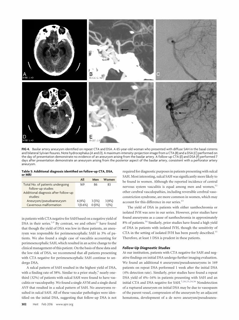
FIG 4. Basilar artery aneurysm identified on repeat CTA and DSA. A 65-year-old woman who presented with diffuse SAH in the basal cisternsand bilateral Sylvian fissures. Note hydrocephalus (A and D). A maximum-intensity-projection image from a CTA (B) and a DSA (C) performed onthe day of presentation demonstrate no evidence of an aneurysm arising from the basilar artery. A follow-up CTA (E) and DSA (F) performed 7days after presentation demonstrate an aneurysm arising from the posterior aspect of the basilar artery, consistent with a perforator arteryaneurysm.
Table 5: Additional diagnosis identified on follow-up CTA, DSA,or MRI
All Men WomenTotal No. of patients undergoing
follow-up studies169 86 83
Additional diagnosis after follow-upstudies:
Aneurysm/pseudoaneurysm 6 (4%) 3 (3%) 3 (4%)Cavernous malformation 1 (0.6%) 0 (0%) 1 (1%)
302 Heit Feb 2016 www.ajnr.org

rysm, or a small size of the aneurysm in combination with these
factors.37 Most interesting, the 6 aneurysms found on repeat DSA
were also prospectively identified by repeat CTA that was per-
formed for vasospasm triage purposes before the repeat DSA. It
would be of interest to determine the sensitivity of CTA in the
delayed detection of a ruptured aneurysm among a greater num-
ber of patients.
An MR imaging performed after a negative initial DSA iden-
tified a cortical cavernous malformation as a cause of SAH in a
single patient. This patient initially presented with an acute head-
ache and NCCT that was negative for intracranial hemorrhage.
Xanthochromia was identified by lumbar puncture, which even-
tually led to the diagnosis. Cavernous malformations have previ-
ously been identified as a cause of perimesencephalic SAH,38
though SAH is an uncommon presentation of these lesions.
LimitationsThe limitations of this study include a selection bias due to the
study occurring at a large tertiary care and neurovascular referral
center and the retrospective nature of the study. This selection
bias may limit the generalizability of our findings.
CONCLUSIONSCareful evaluation of patients presenting with CTA negative for
SAH should include DSA, even in case of perimesencephalic SAH,
given the significant 13% yield of DSA in identifying a vascular
pathology that resulted in the hemorrhage. Furthermore, contin-
ued investigation is prudent by CTA, DSA, and/or MR imaging in
patients with an initial DSA negative for SAH after a CTA negative
for SAH, given a 4% yield of these follow-up studies.
Disclosures: Raul G. Nogueira—UNRELATED: Consultancy: Covidien (Study of Ta-moxifen and Raloxifene Trial core lab); OTHER RELATIONSHIPS: Stryker/ConcentricMedical: Trevo 2 Trial Principal Investigator, DWI/PWI and CTP Assessment in theTriage of Wake-Up and Late Presenting Strokes Undergoing Neurointervention TrialPrincipal Investigator (unpaid); Covidien/ev3: Solitaire Flow Restoration Device ver-sus the Merci Retriever in Patients with Acute Ischaemic Stroke; and Solitaire Withthe Intention For Thrombectomy as PRIMary Endovascular Treatment Trials SteeringCommittee (unpaid); Covidien: 3D Separator Trial Executive Committee (unpaid).Joshua A. Hirsch—UNRELATED: Consultancy: Medtronic, CareFusion, Comments:Medtronic, ongoing consultancy concerning interventional spine; CareFusion, taughta non-Continuing Medical Education course; Stock/Stock Options: Intratech,Brainstorm.
REFERENCES1. Bederson JB, Connolly ES Jr, Batjer HH, et al; American Heart Asso-
ciation. Guidelines for the management of aneurysmal subarach-noid hemorrhage: a statement for healthcare professionals from aspecial writing group of the Stroke Council, American Heart Asso-ciation. Stroke 2009;40:994 –1025 CrossRef Medline
2. Rinkel GJ, Wijdicks EF, Hasan D, et al. Outcome in patients withsubarachnoid haemorrhage and negative angiography according topattern of haemorrhage on computed tomography. Lancet 1991;338:964 – 68 CrossRef Medline
3. Duong HH, Melancon DD, Tampieri DD, et al. The negative angio-gram in subarachnoid haemorrhage. Neuroradiology 1996;38:15–19CrossRef Medline
4. Kaim A, Proske M, Kirsch E, et al. Value of repeat-angiography incases of unexplained subarachnoid hemorrhage (SAH). Acta NeurolScand 1996;93:366 –73 Medline
5. Agid R, Andersson T, Almqvist H, et al. Negative CT angiographyfindings in patients with spontaneous subarachnoid hemorrhage:
when is digital subtraction angiography still needed? AJNR Am JNeuroradiol 2010;31:696 –705 CrossRef Medline
6. Agid R, Lee SK, Willinsky RA, et al. Acute subarachnoidhemorrhage: using 64-slice multidetector CT angiography to “tri-age” patients’ treatment. Neuroradiology 2006;48:787–94 CrossRefMedline
7. Delgado Almandoz JE, Crandall BM, Fease JL, et al. Diagnostic yieldof catheter angiography in patients with subarachnoid hemorrhageand negative initial noninvasive neurovascular examinations.AJNR Am J Neuroradiol 2013;34:833–39 CrossRef Medline
8. Schievink WI, Wijdicks EF. Pretruncal subarachnoid hemorrhage:an anatomically correct description of the perimesencephalic sub-arachnoid hemorrhage. Stroke 1997;28:2572 Medline
9. Cruz JP, Sarma D, Noel de Tilly L. Perimesencephalic subarachnoidhemorrhage: when to stop imaging? Emerg Radiol 2011;18:197–202CrossRef Medline
10. Fox AJ, Symons SP, Aviv RI. CT angiography is state-of-the-art firstvascular imaging for subarachnoid hemorrhage. AJNR Am J Neuro-radiol 2008;29:e41– 42; author reply e46 – 47 CrossRef Medline
11. Prestigiacomo CJ, Sabit A, He W, et al. Three dimensional CT an-giography versus digital subtraction angiography in the detectionof intracranial aneurysms in subarachnoid hemorrhage. J Neuroin-terv Surg 2010;2:385– 89 CrossRef Medline
12. Kershenovich A, Rappaport ZH, Maimon S. Brain computed tomog-raphy angiographic scans as the sole diagnostic examination forexcluding aneurysms in patients with perimesencephalic subarach-noid hemorrhage. Neurosurgery 2006;59:798 – 801; discussion801– 02 CrossRef Medline
13. Kelliny M, Maeder P, Binaghi S, et al. Cerebral aneurysm exclu-sion by CT angiography based on subarachnoid hemorrhagepattern: a retrospective study. BMC Neurol 2011;11:8 CrossRefMedline
14. Bradac GB, Bergui M, Ferrio MF, et al. False-negative angiograms insubarachnoid haemorrhage due to intracranial aneurysms. Neuro-radiology 1997;39:772–76 CrossRef Medline
15. Urbach H, Zentner J, Solymosi L. The need for repeat angiography insubarachnoid haemorrhage. Neuroradiology 1998;40:6 –10 CrossRefMedline
16. Luo Z, Wang D, Sun X, et al. Comparison of the accuracy of subtrac-tion CT angiography performed on 320-detector row volume CTwith conventional CT angiography for diagnosis of intracranial an-eurysms. Eur J Radiol 2012;81:118 –22 CrossRef Medline
17. Yeung R, Ahmad T, Aviv RI, et al. Comparison of CTA to DSA indetermining the etiology of spontaneous ICH. Can J Neurol Sci 2009;36:176 – 80 CrossRef Medline
18. Forster DM, Steiner LL, Hakanson SS, et al. The value of repeat pan-angiography in cases of unexplained subarachnoid hemorrhage.J Neurosurg 1978;48:712–16 CrossRef Medline
19. Huttner HB, Hartmann M, Kohrmann M, et al. Repeated digitalsubstraction angiography after perimesencephalic subarachnoidhemorrhage? J Neuroradiol 2006;33:87– 89 CrossRef Medline
20. Jung JY, Kim YB, Lee JW, et al. Spontaneous subarachnoid haemor-rhage with negative initial angiography: a review of 143 cases. J ClinNeurosci 2006;13:1011–17 CrossRef Medline
21. Nishioka H, Torner JC, Graf CJ, et al. Cooperative study of intracra-nial aneurysms and subarachnoid hemorrhage: a long-term prog-nostic study, III: subarachnoid hemorrhage of undetermined etiol-ogy. Arch Neurol 1984;41:1147–51 CrossRef Medline
22. Hoh BL, Cheung AC, Rabinov JD, et al. Results of a prospectiveprotocol of computed tomographic angiography in place of cathe-ter angiography as the only diagnostic and pretreatment planningstudy for cerebral aneurysms by a combined neurovascular team.Neurosurgery 2004;54:1329 – 40; discussion 1340 – 42 CrossRefMedline
23. Delgado Almandoz JE, Jagadeesan BD, Refai D, et al. Diagnostic yieldof repeat catheter angiography in patients with catheter and com-puted tomography angiography negative subarachnoid hemor-rhage. Neurosurgery 2012;70:1135– 42 CrossRef Medline
AJNR Am J Neuroradiol 37:297–304 Feb 2016 www.ajnr.org 303

24. Topcuoglu MA, Ogilvy CS, Carter BS, et al. Subarachnoid hemor-rhage without evident cause on initial angiography studies: diag-nostic yield of subsequent angiography and other neuroimagingtests. J Neurosurg 2003;98:1235– 40 CrossRef Medline
25. Dalyai R, Chalouhi N, Theofanis T, et al. Subarachnoid hemorrhagewith negative initial catheter angiography: a review of 254 casesevaluating patient clinical outcome and efficacy of short- and long-term repeat angiography. Neurosurgery 2013;72:646 –52; discussion651–52 CrossRef Medline
26. Ringelstein A, Mueller O, Monninghoff C, et al. 3D rotationalangiography after non-traumatic SAH. Rofo 2014;186:675–79CrossRef Medline
27. van Rooij WJ, Peluso JP, Sluzewski M, et al. Additional value of 3Drotational angiography in angiographically negative aneurysmalsubarachnoid hemorrhage: how negative is negative? AJNR Am JNeuroradiol 2008;29:962– 66 CrossRef Medline
28. Fisher CM, Kistler JP, Davis JM. Relation of cerebral vasospasm tosubarachnoid hemorrhage visualized by computerized tomo-graphic scanning. Neurosurgery 1980;6:1–9 CrossRef Medline
29. Jagadeesan BD, Delgado Almandoz JE, Moran CJ, et al. Accuracy ofsusceptibility-weighted imaging for the detection of arteriovenousshunting in vascular malformations of the brain. Stroke 2011;42:87–92 CrossRef Medline
30. Zaharchuk G, Bammer R, Straka M, et al. Arterial spin-label imagingin patients with normal bolus perfusion-weighted MR imaging
findings: pilot identification of the borderzone sign. Radiology2009;252:797– 807 CrossRef Medline
31. van Gijn J, van Dongen KJ, Vermeulen M, et al. Perimesencephalichemorrhage: a nonaneurysmal and benign form of subarachnoidhemorrhage. Neurology 1985;35:493–97 CrossRef Medline
32. Salvarani C, Brown RD Jr, Calamia KT, et al. Primary central ner-vous system vasculitis: analysis of 101 patients. Ann Neurol 2007;62:442–51 CrossRef Medline
33. Singhal AB, Hajj-Ali RA, Topcuoglu MA, et al. Reversible cerebralvasoconstriction syndromes: analysis of 139 cases. Arch Neurol2011;68:1005–12 CrossRef Medline
34. Wallace AN, Dines JN, Zipfel GJ, et al. Yield of catheter angiographyafter computed tomography negative, lumbar puncture positivesubarachnoid hemorrhage [corrected]. Stroke 2013;44:1729 –31CrossRef Medline
35. Flint AC, Roebken A, Singh V. Primary intraventricularhemorrhage: yield of diagnostic angiography and clinical outcome.Neurocrit Care 2008;8:330 –36 CrossRef Medline
36. Maslehaty HH, Barth HH, Petridis AKA, et al. Special features ofsubarachnoid hemorrhage of unknown origin: a review of a seriesof 179 cases. Neurol Res 2012;34:91–97 CrossRef Medline
37. van Gijn J, Rinkel GJ. Subarachnoid haemorrhage: diagnosis, causesand management. Brain 2001;124:249 –78 CrossRef Medline
38. Yaghi S, Oomman S, Keyrouz SG. Non-aneurysmal perimesen-cephalic subarachnoid hemorrhage caused by a cavernous angi-oma. Neurocrit Care 2011;14:84 – 85 CrossRef Medline
304 Heit Feb 2016 www.ajnr.org