Ceratocystis bhutanensis sp. nov., associated with the bark beetle
16
STUDIES IN MYCOLOGY 50: 365–379. 2004. 365 Ceratocystis bhutanensis sp. nov., associated with the bark beetle Ips schmutzenhoferi on Picea spinulosa in Bhutan Marelize van Wyk 1* , Jolanda Roux 1 , Irene Barnes 2 , Brenda D. Wingfield 2 , Dal Bahadur Chhetri 3 , Thomas Kirisits 4 and Michael J. Wingfield 1 1 Department of Microbiology and Plant Pathology, Forestry and Agricultural Biotechnology Institute (FABI), University of Pretoria, Pretoria, South Africa, 0002; 2 Department of Genetics, Forestry and Agricultural Biotechnology Institute (FABI), University of Pretoria, Pretoria, South Africa, 0002; 3 Renewable Natural Resources Research Centre (RNR-RC), Yusipang, Council of Research & Extension, Ministry of Agriculture, P.O. Box No. 212, Thimphu, Bhutan; 4 Institute of Forest Entomol- ogy, Forest Pathology and Forest Protection (IFFF), Department of Forest and Soil Sciences, BOKU – University of Natural Resources and Applied Life Sciences, Vienna, Hasenauerstrasse 38, A-1190, Vienna Austria * Correspondence: Marelize van Wyk, [email protected] Abstract: The Eastern Himalayan spruce bark beetle, Ips schmutzenhoferi, is a serious pest of Picea spinulosa and Pinus wallichiana in Bhutan. A study to identify the ophiostomatoid fungi associated with this bark beetle resulted in the isolation of a Ceratocystis sp. from I. schmutzenhoferi, collected from galleries on P. spinulosa. Morphological characteristics and comparisons of DNA sequence data were used to identify this fungus. Based on morphology, the Ceratocystis sp. from Bhutan resembled C. moniliformis and C. moniliformopsis, but was distinct from these species, both in micro-morphological characteristics, growth at different temperatures, as well as in the odour that it produces in culture. Comparisons of DNA sequences for the ITS regions of the rDNA operon, -tubulin and elongation factor 1- genes, confirmed that this fungus represents a taxon distinct from all other species of Ceratocystis. Based on morphological characteristics, comparisons of DNA sequence data and its unique ecology, we provide the name C. bhutanensis sp. nov. for it. Taxonomic novelty: Ceratocystis bhutanensis M. van Wyk, M.J. Wingf. & T. Kirisits sp. nov. Key words: Bark beetles, blue-stain, Ceratocystis, Ips schmutzenhoferi, ophiostomatoid fungi, Picea spinulosa, Scolytidae. INTRODUCTION The Kingdom of Bhutan is renowned for its intact forest resources, which are of immense socio- economic and ecological importance for this Himala- yan country. Sixty-four percent of Bhutan is covered by forests (FAO 1999, 2001). Conifer forests form the natural vegetation in most parts of the mountainous areas at elevations above 1800 m a.s.l. (FAO 1999). Eastern Himalayan spruce (Picea spinulosa (Griffith) A. Henry) and Himalayan blue pine (Pinus wallichi- ana Jackson) are important tree species in these forests, forming either pure stands or stands of mixed species, together with other conifers and occasionally also hardwoods. Bark beetles (Coleoptera: Scolytidae) are amongst the most damaging agents affecting conifer forests, worldwide. Some of the most aggressive of these insects are species of Ips de Geer (Postner 1974, Wood & Bright 1992). The best known of these is the eight-spined European spruce bark beetle, I. typog- raphus L., which can cause extensive mortality of Norway spruce (Picea abies (L.) Karst.) in Europe (Postner 1974, Christiansen & Bakke 1988). In Bhu- tan, the Eastern Himalayan spruce bark beetle, I. schmutzenhoferi Holzschuh is a serious pest in conifer forests at elevations between 2500 and 3800 m a.s.l. (Holzschuh 1988, Schmutzenhofer 1988). This scoly- tid attacks mainly living trees or infests freshly felled logs of Eastern Himalayan spruce, Himalayan blue pine and occasionally Himalayan larch (Larix grif- fithiana (Lindl. & Gord.) Carrière) (Schmutzenhofer 1988, Tshering & Chhetri 2000). During the 1980s, I. schmutzenhoferi caused a destructive outbreak in Western and Central Bhutan, during which more than 2060 ha of forest were affected (Chhetri 1991) and losses of approximately 2 million m 3 of timber oc- curred (Schmutzenhofer 1988). Conifer-infesting bark beetles are well known vectors of blue-stain fungi belonging to the ascomy- cete genera Ceratocystis Ellis & Halst. and Ophio- stoma H. Syd. & P. Syd. and related anamorph genera (Francke-Grosmann 1967, Upadhyay 1981, Whitney 1982, Wingfield et al. 1993, Jacobs & Wingfield 2001, Kirisits 2004). These fungi cause blue, grey or black discoloration in the sapwood of living trees, logs and lumber, mostly on conifers. This damage results from the presence of pigmented fungal hyphae in the ray parenchyma cells and tracheids of the
Transcript of Ceratocystis bhutanensis sp. nov., associated with the bark beetle
STUDIES IN MYCOLOGY 50: 365–379. 2004.
365
Ceratocystis bhutanensis sp. nov., associated with the bark beetle Ips schmutzenhoferi on Picea spinulosa in Bhutan
Marelize van Wyk1*, Jolanda Roux1, Irene Barnes2, Brenda D. Wingfield2, Dal Bahadur Chhetri3, Thomas Kirisits4 and Michael J. Wingfield1 1Department of Microbiology and Plant Pathology, Forestry and Agricultural Biotechnology Institute (FABI), University of Pretoria, Pretoria, South Africa, 0002; 2Department of Genetics, Forestry and Agricultural Biotechnology Institute (FABI), University of Pretoria, Pretoria, South Africa, 0002; 3Renewable Natural Resources Research Centre (RNR-RC), Yusipang, Council of Research & Extension, Ministry of Agriculture, P.O. Box No. 212, Thimphu, Bhutan; 4Institute of Forest Entomol-ogy, Forest Pathology and Forest Protection (IFFF), Department of Forest and Soil Sciences, BOKU – University of Natural Resources and Applied Life Sciences, Vienna, Hasenauerstrasse 38, A-1190, Vienna Austria *Correspondence: Marelize van Wyk, [email protected] Abstract: The Eastern Himalayan spruce bark beetle, Ips schmutzenhoferi, is a serious pest of Picea spinulosa and Pinus wallichiana in Bhutan. A study to identify the ophiostomatoid fungi associated with this bark beetle resulted in the isolation of a Ceratocystis sp. from I. schmutzenhoferi, collected from galleries on P. spinulosa. Morphological characteristics and comparisons of DNA sequence data were used to identify this fungus. Based on morphology, the Ceratocystis sp. from Bhutan resembled C. moniliformis and C. moniliformopsis, but was distinct from these species, both in micro-morphological characteristics, growth at different temperatures, as well as in the odour that it produces in culture. Comparisons of DNA sequences for the ITS regions of the rDNA operon, �-tubulin and elongation factor 1-� genes, confirmed that this fungus represents a taxon distinct from all other species of Ceratocystis. Based on morphological characteristics, comparisons of DNA sequence data and its unique ecology, we provide the name C. bhutanensis sp. nov. for it.
Taxonomic novelty: Ceratocystis bhutanensis M. van Wyk, M.J. Wingf. & T. Kirisits sp. nov. Key words: Bark beetles, blue-stain, Ceratocystis, Ips schmutzenhoferi, ophiostomatoid fungi, Picea spinulosa, Scolytidae.
INTRODUCTION The Kingdom of Bhutan is renowned for its intact forest resources, which are of immense socio-economic and ecological importance for this Himala-yan country. Sixty-four percent of Bhutan is covered by forests (FAO 1999, 2001). Conifer forests form the natural vegetation in most parts of the mountainous areas at elevations above 1800 m a.s.l. (FAO 1999). Eastern Himalayan spruce (Picea spinulosa (Griffith) A. Henry) and Himalayan blue pine (Pinus wallichi-ana Jackson) are important tree species in these forests, forming either pure stands or stands of mixed species, together with other conifers and occasionally also hardwoods. Bark beetles (Coleoptera: Scolytidae) are amongst the most damaging agents affecting conifer forests, worldwide. Some of the most aggressive of these insects are species of Ips de Geer (Postner 1974, Wood & Bright 1992). The best known of these is the eight-spined European spruce bark beetle, I. typog-raphus L., which can cause extensive mortality of Norway spruce (Picea abies (L.) Karst.) in Europe (Postner 1974, Christiansen & Bakke 1988). In Bhu-tan, the Eastern Himalayan spruce bark beetle, I.
schmutzenhoferi Holzschuh is a serious pest in conifer forests at elevations between 2500 and 3800 m a.s.l. (Holzschuh 1988, Schmutzenhofer 1988). This scoly-tid attacks mainly living trees or infests freshly felled logs of Eastern Himalayan spruce, Himalayan blue pine and occasionally Himalayan larch (Larix grif-fithiana (Lindl. & Gord.) Carrière) (Schmutzenhofer 1988, Tshering & Chhetri 2000). During the 1980s, I. schmutzenhoferi caused a destructive outbreak in Western and Central Bhutan, during which more than 2060 ha of forest were affected (Chhetri 1991) and losses of approximately 2 million m3 of timber oc-curred (Schmutzenhofer 1988). Conifer-infesting bark beetles are well known vectors of blue-stain fungi belonging to the ascomy-cete genera Ceratocystis Ellis & Halst. and Ophio-stoma H. Syd. & P. Syd. and related anamorph genera (Francke-Grosmann 1967, Upadhyay 1981, Whitney 1982, Wingfield et al. 1993, Jacobs & Wingfield 2001, Kirisits 2004). These fungi cause blue, grey or black discoloration in the sapwood of living trees, logs and lumber, mostly on conifers. This damage results from the presence of pigmented fungal hyphae in the ray parenchyma cells and tracheids of the
VAN WYK ET AL.
366
sapwood (Münch 1907, Liese & Schmid 1961, Seifert 1993).
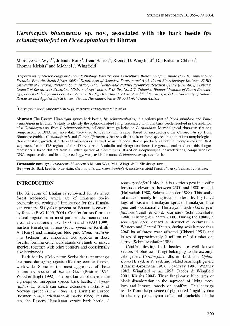
Fig. 1. A. A map of Bhutan showing the administrative districts (dzongkhags) of the country and Bhutan’s capital Thimphu. B. A map of the dzongkhags Thimphu and Wangdi (Wangdue Phodrang) showing the localities where samples for fungal isolation were collected from Picea spinulosa and Pinus wallichiana. Ceratocystis bhutanensis was isolated only from individuals of Ips schmutzenhoferi obtained from Jelekha. Damage due to sapstain is cosmetic rather than struc-tural, and results in substantial financial loss, because markets prefer non-stained wood (Münch 1907, Seifert 1993, Uzunovic et al. 1999). Some bark bee-tle-associated blue-stain fungi also cause vascular stain diseases on living conifer trees and are thought to aid their insect vectors in exhausting the defence mechanisms of their host trees (Whitney 1982, Paine et al. 1997, Kirisits 2004). Most fungal associates of bark beetles belong to the genus Ophiostoma. In contrast, Ceratocystis spp. usually have loose relationships with insects (Kile 1993). However, there are some Ceratocystis spp. that are consistently associated with conifer bark beetles (Harrington & Wingfield 1998). Ceratocystis polonica (Siemaszko) C. Moreau is associated with I. typog-raphus and other species of Ips on Picea abies in Europe (Solheim 1986, 1992, Krokene & Solheim 1996, Kirisits 2004), and with I. typographus japoni-cus Niijima on Picea jezoensis (Sieb. & Zucc.) Carr. in Japan (Yamaoka et al. 1997). Ceratocystis larici-cola Redfern & Minter, is associated with the larch bark beetle I. cembrae Heer on Larix Miller spp. in
Europe and Japan (Redfern et al. 1987, Yamaoka et al. 1998, Stauffer et al. 2001, Kirisits 2004). Cerato-cystis rufipennis M.J. Wingf., T.C. Harr. & H. Sol-heim, is associated with the spruce bark beetle Den-droctonus rufipenniss Kirby on Picea engelmannii Parry and Picea glauca (Moench) Voss in Western North America (Solheim 1995, Wingfield et al. 1997). The small number of Ceratocystis spp. that are associated with bark beetles, display high levels of pathogenicity, when compared to Ophiostoma spp. from the same niches (Solheim 1988, Solheim & Safranyik 1997, Kirisits 1998, Krokene & Solheim 1998). Ceratocystis polonica is highly pathogenic to Norway spruce and contributes to tree death following attack by I. typographus (Christiansen 1985, Solheim 1988, 1992, Kirisits & Offenthaler 2002). Likewise, C. laricicola is considered to play an important role in the death of Larix spp. infested by I. cembrae (Red-fern et al. 1987, Yamaoka et al. 1998, Kirisits 2001, Harrington et al. 2002) and C. rufipennis can kill Sitka spruce (Picea sitchensis (Bongard) Carrière) in mass inoculation experiments (Solheim & Safranyik 1997). During a recent survey of ophiostomatoid fungi associated with I. schmutzenhoferi in Bhutan, a Cera-tocystis sp. resembling C. moniliformis Hedgcock and C. moniliformopsis Z.Q. Yuan & C. Mohammed was isolated. Despite its morphological similarity to these Ceratocystis spp., the association of the Ceratocystis sp. from Bhutan with a conifer bark beetle aroused suspicion that it might represent an undescribed taxon. This study compares the Ceratocystis sp. from Bhutan with C. moniliformis and C. moniliformopsis and assesses their phylogenetic relationships based on gene sequences of the ITS region of the rDNA operon as well as parts of the �-tubulin and EF1-� regions. MATERIALS AND METHODS Fungal cultures and isolations A survey of ophiostomatoid fungi associated with I. schmutzenhoferi in Bhutan was conducted in July 2001. Samples for isolation were collected at several locations in Western and Central Bhutan (administra-tive districts Thimphu and Wangi) (Fig. 1) where I. schmutzenhoferi had infested living trees or freshly felled logs of Picea spinulosa and/or Pinus wallichi-ana (Fig. 2). The collection sites included mixed conifer forests at Jelekha (3300 m a.s.l.), Changaphug (3600 m a.s.l.), Phobjikha valley (3100 m a.s.l.), and near the Renewable Natural Resources Research Centre (RNR-RC) in Yusipang (2700 m a.s.l.) as well as wood depots at Gidakom (2200 m a.s.l.) and Ram-tokto (2100 m a.s.l.) (Fig. 1).

CERATOCYSTIS BHUTANENSIS SP. NOV.
367
Fig. 2. Eastern Himalayan spruce (Picea spinulosa), blue stain and Ips schmutzenhoferi in Bhutan. A. A landscape view of the valleys and mountains in Western Bhutan with Wangdi dzong on the left. B. Eastern Himalayan spruce (P. spinulosa) dying due to infestation by I. schmutzenhoferi. C, D. Blue stain resulting from colonization by ophiostomatoid fungi seen around nuptial chambers and female galleries of I. schmutzenhoferi on P. spinulosa. E. A female I. schmutzenhoferi. F. A male I. schmutzenhoferi. The large elongated third spine at the elytral declivity, which bends downwards at the apex, is a distinct characteristic of this bark beetle species. Logs and standing trees, infested by I. schmutzen-hoferi were examined for suitable material to conduct fungal isolations (Fig. 2) Galleries of the insects occurring in the bark or on the surface of the sapwood on logs and standing pine and spruce trees were inspected on site, with the aid of a 10 × magnification hand lens, for the occurrence of sexual and asexual stages of ophiostomatoid fungi. At the research station in Yusipang, adult beetles of I.
schmutzenhoferi (2nd generation) were collected from a pheromone trap installed specifically for the purpose of insect specimen collection. Adult and juvenile beetles, breeding galleries, stem discs and stem sections from beetle-infested P. spinu-losa and P. wallichiana trees and logs were collected for further investigation (Fig. 2). All samples were stored in plastic bags and transported to the laboratory at RNR-RC in Yusipang.

VAN WYK ET AL.
368
Table 1. Isolates of Ceratocystis used in this study.
Species Isolate no.d Alternative numberse GenBank accession no.
Year of isolation
Host Geographical origin Associated insect Collector(s)
C. moniliformis CMW 8240a − AY528989f
AY529000g
AY529010h
2001 Cassia fistula Punakha, Bhutan − MJW, TK & DBC
CMW 8238c CBS 115771 − 2001 C. fistula Wangdi, Bhutan − MJW, TK & DBC
CMW 9590a,c CBS 116452 AY528985f
AY528996g
AY529006h
2002 Eucalyptus grandis
Mpumalanga, South Africa
− JR
CMW 4114a AY528986f
AY528997g
AY529007h
1997 Schizolobium parahybum
Ecuador, South America − MJW
CMW 10134c − 2002 Eucalyptus grandis
Mpumalanga, South Africa
− MvW
C. moniliformopsis CMW 9986a,c CBS 109441 AY528987f
AY528998g
AY529008h
1999 Eucalyptus obliqua
Tazmania, Australia − ZQY
CMW 10214a,c CBS 115792, ORB 33 AY528988f
AY528999g
AY529009h
1989 Eucalyptus sieberi
Victoria, Australia − MJ D
CMW 10215c 115793, ORB 346 − 1990 E. sieberi Victoria, Australia − MJD
C. bhutanensis CMW 8215a CBS 114290, PREM 57805 AY528953f
AY528958g
AY528963h
2001 Picea spinu-losa
Jelekha, Bhutan Ips schmutzenhoferi TK & DBC
CMW 8242a,b CBS 112907 PREM 57809
AY528951f
AY528956g
AY528961h
2001 P. spinulosa Jelekha, Bhutan I. schmutzenhoferi TK & DBC
CMW 8217a,b,c CBS 114289 PREM 57807
AY528952f
AY528957g
AY528962h
2001 P. spinulosa Jelekha, Bhutan I. schmutzenhoferi TK & DBC
CMW 8241a,b,c CBS 115773, PREM 57808 − 2001 P. spinulosa Jelekha, Bhutan I. schmutzenhoferi TK & DBC
CMW 8396a CBS 114286 BH 8/5, PREM 57812
− 2001 P. spinulosa Jelekha, Bhutan I. schmutzenhoferi TK & DBC
CMW 8399a CBS 115772, BH 8/8 AY528954f
AY528959g
AY528964h
2001 P. spinulosa Jelekha, Bhutan I. schmutzenhoferi TK & DBC
CERATOCYSTIS BHUTANENSIS SP. NOV.
369
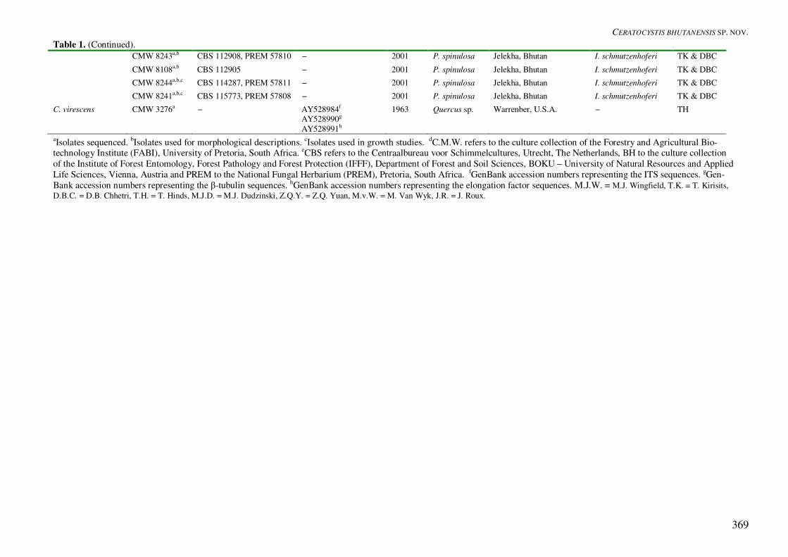
Table 1. (Continued). CMW 8243a,b CBS 112908, PREM 57810 − 2001 P. spinulosa Jelekha, Bhutan I. schmutzenhoferi TK & DBC
CMW 8108a,b CBS 112905 − 2001 P. spinulosa Jelekha, Bhutan I. schmutzenhoferi TK & DBC
CMW 8244a,b,c CBS 114287, PREM 57811 − 2001 P. spinulosa Jelekha, Bhutan I. schmutzenhoferi TK & DBC
CMW 8241a,b,c CBS 115773, PREM 57808 − 2001 P. spinulosa Jelekha, Bhutan I. schmutzenhoferi TK & DBC
C. virescens CMW 3276a − AY528984f
AY528990g
AY528991h
1963 Quercus sp. Warrenber, U.S.A. − TH
aIsolates sequenced. bIsolates used for morphological descriptions. cIsolates used in growth studies. dC.M.W. refers to the culture collection of the Forestry and Agricultural Bio-technology Institute (FABI), University of Pretoria, South Africa. eCBS refers to the Centraalbureau voor Schimmelcultures, Utrecht, The Netherlands, BH to the culture collection of the Institute of Forest Entomology, Forest Pathology and Forest Protection (IFFF), Department of Forest and Soil Sciences, BOKU – University of Natural Resources and Applied Life Sciences, Vienna, Austria and PREM to the National Fungal Herbarium (PREM), Pretoria, South Africa. fGenBank accession numbers representing the ITS sequences. gGen-Bank accession numbers representing the �-tubulin sequences. hGenBank accession numbers representing the elongation factor sequences. M.J.W. = M.J. Wingfield, T.K. = T. Kirisits, D.B.C. = D.B. Chhetri, T.H. = T. Hinds, M.J.D. = M.J. Dudzinski, Z.Q.Y. = Z.Q. Yuan, M.v.W. = M. Van Wyk, J.R. = J. Roux.

VAN WYK ET AL.
370
Dry bark samples were sprayed with distilled water and the bags sealed for a few days to create a moist environment, conducive for sporulation of fungi within the beetle galleries. Reference specimens of I. schmutzenhoferi were stored in ethanol and are main-tained at the Institute of Forest Entomology, Forest Pathology and Forest Protection (IFFF), Department of Forest and Soil Sciences (BOKU) – University of Natural Resources and Applied Life Sciences, Vienna, Austria. In addition to material obtained from pine and spruce infested by I. schmutzenhoferi, wood samples were collected from broken Cassia fistula L. trees near Punakha (ca. 1300 m a.s.l.) and Wangdi (Wang-due Phodrang) (ca. 1100 m a.s.l.). The purpose of these collections was to search for Ceratocystis moniliformis on this subtropical hardwood species. Fungi were isolated on 2 % malt extract agar (MEA) (20 % w/v) (Biolab, Midrand, South Africa) or on 2 % malt agar (MA) (20 % w/v) (DiaMalt, Hefe Schweiz AG, Stettfurt, Switzerland), both supple-mented with 100 mg/L streptomycin sulphate (SIGMA or VWR International). In order to obtain a comprehensive view of the fungi associated with I. schmutzenhoferi, various isolation methods were applied. Fungi were isolated directly from adult bee-tles (2nd generation) collected from two spruce logs at Jelekha, from young beetles (1st generation) obtained from a pine log at Ramtokto, and from swarming beetles (2nd generation) collected from a pheromone trap at Yusipang. To obtain isolates directly from the insects, their body parts were dissected and spread onto 2 % MA. Fungi were also isolated from the sapwood of a spruce tree from Jelekha. Six stem discs (ca. 10–15 cm thick, 18–21 cm diam), were cut from the upper part of this tree. These were split vertically and isola-tions from the sapwood were done along radii beneath I. schmutzenhoferi female galleries, following the procedure described by Solheim (1992). Three radii per disc were sampled, resulting in a total of 18 radii. Small pieces of sapwood were transferred onto 2 % MA plates. From each radius, samples were taken 2, 5 and 10 mm apart from the cambium into the sapwood. Most isolations were made from ascospores and conidia taken directly from sexual and asexual fungal structures occurring in and around female and larval galleries and pupal chambers of the insects. Bark and sapwood samples from spruce and pine collected at Jelekha, Gidakom, Ramtokto, Changaphug (adminis-trative district Thimphu) and Phobjikha valley (admin-istrative district Wangdi) (Fig. 1) were examined with a dissecting microscope at magnifications ranging from 10 × to 40 ×. With a fine needle, ascospores and conidia accumulating at the apices of perithecia and on conidiophores, respectively, were carefully re-moved and transferred to 2 % MA or 2 % MEA plates. Isolation of C. moniliformis from C. fistula collected at Punakha and Wangdi was done in a similar manner,
from ascospores obtained from perithecia occurring on the wood surface. A selective method for the isolation of Ceratocystis spp. was also used (Moller & DeVay 1968). Fresh carrots were washed and lightly sprayed with 70 % ethanol. Carrot discs (5–10 mm thick) were cut and four to eight discs were placed in plastic Petri dishes (90 mm). Beetles, larvae and pupae of I. schmutzen-hoferi were dissected and spread over the surface of the carrot discs. Larval frass, collected from the insect galleries was also put onto the carrots. The discs were examined for the incidence of perithecia after 5–10 d incubation at ca. 20 °C. Pure cultures were obtained by transferring asco-spore or conidial masses as well as small pieces of mycelium from the primary isolation plates onto fresh 2 % MA or 2 % MEA plates. Fungal cultures are maintained in the culture collection (CMW) of the Forestry and Agricultural Biotechnology Institute (FABI), University of Pretoria, South Africa, the Institute of Forest Entomology, Forest Pathology and Forest Protection (IFFF), Department of Forest and Soil Sciences, BOKU – University of Natural Re-sources and Applied Life Sciences, Vienna, Austria and the Centraalbureau voor Schimmelcultures (CBS), Utrecht, The Netherlands. Holotype material of the new Ceratocystis sp. from Bhutan, consisting of a dried culture of isolate CMW 8217 on 2 % MEA has been lodged at the National Fungal Herbarium (PREM), Pretoria, South Africa (Table 1). Culture characteristics and morphology The growth of isolates CMW 8217, CMW 8241 and CMW 8244 representing the Ceratocystis sp. obtained from I. schmutzenhoferi, was determined on 2 % MEA (Table 3). Three isolates of C. moniliformis (CMW 9590, CMW 8238, CMW 10134) and C. monilifor-mopsis (CMW 9986, CMW 10214, CMW 10215) were used for comparisons in the growth studies (Table 3). Prior to the growth assays, the isolates were grown for 2 wk at 25 ºC on 2 % MEA (Fig. 3). Myce-lial plugs were taken from actively growing cultures using a 5 mm cork borer and a single mycelial plug was transferred to the centre of a 90 mm Petri dish containing 2 % MEA. For each isolate, five plates were incubated at 4, 10, 15, 20, 25, 30 and 35 ºC, respectively. Colony diameter for each culture was assessed by taking two daily measurements at right angles to each other, for 4 d or until the plates were almost completely covered by mycelium. Averages were computed separately for each isolate and each specified temperature. The entire experiment was repeated once. Micro-morphological characteristics were de-scribed from 10-d-old cultures, on 2 % MEA supple-mented with streptomycin sulphate (0.001 g/L) (SIGMA, Steinheim, Germany) and Thiamine (0.001 g/L) (SIGMA, Steinheim, Germany). Fungal struc-
CERATOCYSTIS BHUTANENSIS SP. NOV.
371
tures were mounted in lactophenol containing cotton blue. Fifty measurements for each taxonomically relevant structure were made from isolate CMW 8217, and 10 further measurements were made for each of seven other isolates of the Ceratocystis sp. from Bhutan (Tables 1, 2). Ranges, averages, and standard deviations of the corresponding measurements were calculated. Measurements are given in the format: (minimum–) mean minus standard deviation – mean plus standard deviation (–maximum). The microscopic observations were made using a Carl Zeiss micro-scope and the photographic images were made with a Zeiss Axio Vision camera system. Colour descriptions were determined using the colour charts of Rayner (1970). The measurements and morphological charac-teristics of the Ceratocystis sp. from Bhutan were compared with those of the descriptions of C. monili-formis (Hedgcock 1906, Bakshi 1951, Upadhyay 1981) and C. moniliformopsis (Yuan & Mohammed 2002) (Table 2).
PCR, sequencing and analysis Representative isolates of the Ceratocystis sp. from I. schmutzenhoferi in Bhutan as well as isolates of C. moniliformis, C. moniliformopsis and C. virescens (R.W. Davidson) C. Moreau (Table 1) were selected for DNA extraction and sequencing. An ascospore mass was transferred from each actively growing and sporulating culture, to 50 mL 3 % ME broth, in Er-lenmeyer flasks, and incubated at 25 °C. After 2 wk, the thick mycelial mats were filtered from the broth and lyophilised for two d. The freeze-dried mycelium was placed in liquid nitrogen and ground to a powder using a glass rod, and DNA was extracted using the method described by Barnes et al. (2001). The two ITS regions (ITS1 and ITS2) and the 5.8S gene of the rDNA operon were amplified using prim-ers ITS1 and ITS4 (White et al. 1990) at an annealing temperature of 55 °C. The �-tubulin gene was partially amplified using primers �t1a and �t1b at an annealing temperature of 55 °C (Glass & Donaldson 1995) and the EF1-� gene of the rDNA operon was amplified using primers EF1-728F and EF1-986R at an anneal-ing temperature of 56 °C (Carbone & Kohn 1999). Polymerase chain reaction (PCR) mixtures con-sisted of 200 nM of the forward and reverse primers, 200 �M of each dNTP, Expand High Fidelity PCR System enzyme mix (1.75 U) (Roche Diagnostics, Mannheim, Germany), 1 × Expand HF Buffer contain-ing 1.5 mM MgCl2 (supplied with the enzyme) and 2–10 ng DNA. Reaction volumes were adjusted to 25 �L with sterile water. The PCR programme was set at 96 ºC for 2 min, followed by 10 cycles at 94 ºC for 20 s, x ºC (x = the annealing temperature specified for each set of primers) for 40 s and 72 ºC for 45 s. A
further 30 cycles were included with the annealing time altered to 40 s and a 5 s extension after each cycle. A final step of 10 min at 72 ºC completed the programme. Amplification of the respective genes was confirmed on a 2 % agarose (Roche diagnostics, Mannheim, Germany) gel supplemented with ethidium bromide. PCR amplicons were purified using the Magic PCR Preps, Purification System (Promega, Madison, U.S.A.). PCR amplicons were sequenced in both directions using the ABI PRISMTM Big DYE Terminator Cycle Sequencing Ready Reaction Kit (Applied BioSystems, Foster City, California). The same primers as those in the PCR reactions were used for sequencing of the respective gene areas. Sequence reactions were run on an ABI PRISMTM 3100 Autosequencer (Applied BioSystems, Foster City, California) and sequences were analysed using Sequence Navigator version 1.0.1 (Applied BioSystems, Foster City, California). The sequences obtained for the Ceratocystis sp. from I. schmutzenhoferi were compared with those for morphologically similar Ceratocystis spp. (Table 1). Sequences were aligned manually and analysed using PAUP v4.0b10 (Phylogenetic Analysis Using Parsi-mony) (Swofford 2002). Gaps were treated as “new-state” and trees were obtained via stepwise addition of 1000 replicates with the Mulpar option in effect. The heuristic search based on parsimony with tree bisec-tion reconstruction was used to obtain the phylogram. Confidence intervals using 1000 bootstrap replicates were calculated. The out-group taxon, C. virescens, was rooted as a midpoint with respect to the in-group taxa. All sequences derived from this study have been deposited in GenBank (Table 1). A partition homoge-neity test (Swofford 2002) was used to determine whether the sequence data sets for the three different genome regions could be combined. The Markov Chain Monte Carlo (MCMC) method (Larget & Simon 1999), with a Bayesian framework was used to estimate the posterior probability of nodes in the phylogenetic tree. One hundred thousand ran-dom trees were generated using the MCMC proce-dure, sampling every 100th tree and printing every 10th tree. To avoid including trees that might have been sampled before convergence of the Markov chain, the chain was assessed for the number of trees that were formed before the stabilization and these trees (8600) were discarded. For the combined analysis of the three gene sequences, gamma rate heterogeneity was set, and no codon-specific sites were included for the ITS gene. For �-tubulin and EF1-� sequences, codon specific sites were specified with a site-specific substi-tution rate and the site partition was treated as a by-codon.

VAN WYK ET AL.
372
Table 2. Comparison of Ceratocystis bhutanensis with the morphologically similar species, C. moniliformis and C. monilifor-mopsis. Character C. bhutanensis C. moniliformis
(Hedgcock 1906) C. moniliformopsis (Yuan & Mohammed 2002)
ASCOMATA Base Colour Dark brown to black Brown to black Dark brown to black Diameter 138−178 �m 90−180 �m 200−300 �m Ornamentation Conical spines and hyphal hairs Conical spines (sparse) Hyphal hairs & conical spines Form Globose Globose Ovoid Neck Colour Dark brown to black becoming
pale brown towards apex Pale brown becoming transparent at the apexa
Dark brown to black
Disc-form at base Yes Yesa Yes Length 453−519 �m 730−896 �ma 470−780 �m Width (Tip) 12−14 �m 14 �m a 18−22 �m Width (Base) 34−42 �m 39.2−51.8 �ma 40−50 �m Ostiolar hyphae Shape Divergent Divergent Convergent Measurement 18−26 �m 12−18 × 2 �m 25−45 × 1.5−2 �m Ascus Not seen Fugacious Not seen Ascospores Colour Hyaline Hyaline Hyaline Shape (Side view) Hat-shaped Oval, one side flat Hat-shaped Measurements 4−6 × 2−5 �m 4−5 × 3−4 �m 4−5 × 2−2.5 �m Aggregation Mucilaginous Slimy grey mass Gelatinous sheath CONIDIOPHORES Measurements 23−39 × 4−6 �m (1) 3−13.7 × 3.5−8.9 �ma
(2) 7.3−13.7 × 4.5 �ma 5−32.5 × 4−5.3 �m
Shape Phialides Phialides (2 types) Phialides (2 types) Conidia Shape (1) Cylindrical
(2) Barrel-shaped (1) Oval or cylindricala
(2) Cylindricala (1) Cylindrical (2) Oblong or ellipsoidal
Measurements (1) 7−9 × 1−3 �m (2) 3−5 × 2−3 �m
(1) 7.3−13.7 × 3.5−8.9 �ma
(2) 4.3−15.5 × 1−2.5 �m a (1) 13−21 × 2−3 �m (2) 12−17.5 × 5−7.5 �m
CULTURES Growth rate 20 mm per day at 25 ºC
in the dark 60 mm in 10 days at 22 ºC in the darka
6.3−7.5 mm per day at 22 ºC in the dark
Colour Cream-buff to dark olive to black Hyaline to grey to black Colourless to white grey, centre becoming greenish brown
Odour Fermenting odour Pear dropsa None Mycelia Smooth and granulated Coarsely granular Smooth aDescription by Bakshi (1951). RESULTS Fungal cultures and isolations Conspicuous blue-stain was observed on the surface of the sapwood and in the bark around nuptial cham-bers as well as female and larval galleries of I. schmutzenhoferi on spruce and pine (Fig. 2). How-ever, intensive blue-stain, deeply penetrating into the sapwood was not seen on any of the wood samples. On stem discs of the spruce from which isolations were made, a narrow zone of desiccation, extending 5 to 8 mm deep into the sapwood and recognizable by its white to yellowish colour, occurred. The Ceratocystis sp. was isolated directly from second-generation beetles collected from galleries on
P. spinulosa at Jelekha. In a sample of 20 beetles from this site 16 (80 %) yielded the Ceratocystis sp. and this fungus was thus among the dominant species recovered from this niche. Fourteen isolates of the Ceratocystis sp. representing isolations from separate beetles were initially maintained and 10 of these strains were used for characterization of this fungus (Table 1). The Ceratocystis sp. was neither isolated from beetles obtained from Ramtokto and Yusipang nor from desiccated sapwood of the spruce tree collected at Jelekha. Perithecia and conidiophores were never located in galleries of I. schmutzenhoferi on spruce or pine. Attempts to isolate this fungus from adult and juvenile beetles, larvae, and pupae or from larval frass

CERATOCYSTIS BHUTANENSIS SP. NOV.
373
using carrot discs were unsuccessful. Ascomata re-sembling those of C. moniliformis were common on the surface of the wood of broken C. fistula trees near Punakha and Wangdi (Wangdue Phodrang). This fungus was easily isolated by transferring ascospores from the perithecial tips to MA and MEA plates. Two isolates of this fungus from Bhutan were included in the comparisons with the Ceratocystis sp. from I. schmutzenhoferi (Table 1).
PCR, sequencing and analysis Amplification of the ITS regions and the 5.8S gene of the rDNA resulted in amplification products of ~500 bp. Amplification products for �-tubulin were ~500 bp, while those for the EF1-� gene were ~300 bp. In the partition homogeneity tests, all DNA se-quence data sets gave P-values greater than the mini-mum required value of P = 0.05 and they could thus be combined. The combined sequences of the three gene areas resulted in a data set that was 1491 bp long, had a single most parsimous tree, with a consistency index (CI) of 0.95, a homoplasy index (HI) of 0.05, a retention index (RI) of 0.93 and a rescaled consistency index (RC) of 0.88. The posterior probability of the branch nodes of the combined tree, generated with the Bayesian inference programme supported the boot-strap values. The posterior probability for the branch nodes for the three clades representing C. monili-formis, C. moniliformopsis and the Ceratocystis sp. isolated from I. schmutzenhoferi was 100 %. A heuristic search resulted in a single well-resolved tree with species of Ceratocystis residing in three distinct sub-clades (Fig. 5). One of these sub-clades included the Ceratocystis sp. isolated from I. schmutzenhoferi in Bhutan, supported by a bootstrap value of 100 %. The other sub-clades included isolates of C. moniliformis and C. moniliformopsis, respec-tively (Fig. 5). Taxonomy Comparison of DNA sequence data confirmed mor-phological observations that the Ceratocystis sp. from I. schmutzenhoferi in Bhutan is related to C. monili-formis and C. moniliformopsis. The data also provided robust support for the view that this fungus represents a new and previously undescribed species of Cerato-cystis. The fungus is, therefore, described as follows. Ceratocystis bhutanensis M. Van Wyk, M.J. Wingf. & T. Kirisits, sp. nov. MycoBank MB500092. Anamorph: Thielaviopsis sp.
Etymology: bhutanensis refers to Bhutan, the country of origin. Coloniae juvenes cremeo-fulvidae, infra mellinae, seniores griseo-mustelinae, infra umbrinae, dein atro-olivaceae, infra nigrae. Mycelium plerumque in medio immersum; myce-lium album aerium adest. Crescit optime ad 20–25 ºC, nullo incremento supra 35 ºC. Hyphae leves vel granulatae, ad septa non constrictae, 1–3.5 µm latae. Bases ascomatum atrobrunneae vel nigrae, globosae, spinis hyphisque ornatae, spinis atrobrunneis vel nigris, (4.5–)8–19(–27) µm longis, bases (112–)138–178(–206) µm diametro. Colla ascomatum basin versus atrobrunnea vel nigra, apicem versus pallidiora, (450–)453–519 µm longa, ad basim, 34–42(–44) µm lata, basi discoidea dilata. Hyphae ostiolares divergentes, hyalinae, (13–)18–26(–34) µm longae. Asci non visi. Ascosporae e latere visae galeiformes, aseptatae, hyalinae, in vagina investitae, vagina inclusa 4–6 × 2–5 µm, vagina exclusa 2–5 × 2–5 µm. Ascosporae in massis mucilagineis fulvo-luteis in apicibus collorum ascomatum cumulantes. Anamorphe Thielaviopsis: conidiophora in mycelio singula, hyalina, basi tumida, apicem versus angustata, (15–)23–39(–51) µm longa, basi (3–)4–6(–9) µm lata, apice 1–3 µm lata. Conidiogenesis phialidica incremento parietis annulari; conidia in catenis biformibus facta: conidia primaria hyalina, aseptata, cylindrica, (6–)7–9(–10) × 1–3 µm, conidia secundaria hyalina, aseptata, doliiformia, 3–5 × (1.5–)2–3(–3.5) µm. Colonies of young cultures with submerged mycelium honey-coloured (19”b), aerial mycelium cream-buff (19”d). Cultures that were ± 14-d-old, had umber (15 m) submerged mycelium, aerial mycelium ecru-drab (13”””d) (Fig. 3). Cultures that were ± 28-d-old, had black (7”””k) submerged mycelium, aerial mycelium abundant, dark olive (21”m). Mycelium submerged in medium, abundant white aerial mycelium present. Optimal temperature range 20–25 ºC, no growth at 35 ºC. Isolates growing up to 20 mm per day at 20 ºC. Hyphae smooth or granulated, not constricted at septa, 1–3.5 �m wide. Ascomatal bases dark brown to black, globose, ornamented with spines and hyphae, spines dark brown to black, (4.5–)8–19(–27) �m long, bases (112–)138–178(–206) �m diam. Ascomatal necks dark brown to black at base, becoming light brown towards the apex, (450–)453–519 �m long, 34–42(–44) �m wide at the base, (11–)12–14(–17) �m wide at the apex, with a disc-like (disciform) base. Ostiolar hyphae divergent, hyaline, (13–)18–26(–34) �m long. Asci not observed. Ascospores cucullate in side view, aseptate, hyaline, invested in a sheath, 4–6 × 2–5 �m with sheath, 2–5 × 2–5 �m without sheath. Ascospores accumulating in white and later buff-yellow (19d) mucilaginous masses on the apices of ascomatal necks.

VAN WYK ET AL.
374
Fig. 3. Characteristics of the cultures of Ceratocystis bhutanensis and the two Ceratocystis spp. most closely related to it. A. White to pale mouse grey (15””’d) colony of C. moniliformis (CMW 9590). B. Grey to black (7”””k) colony of C. bhutanensis (CMW 8217). C. Buffy brown (17”’i) culture of C. moniliformopsis (CMW 9986). All cultures were grown on 2 % MEA at 20 ºC in the dark for approximately 10 d.
Fig. 4. Morphological characteristics of Ceratocystis bhutanensis (PREM 57807, CMW 8217, CBS 114289). A. Globose ascoma with long neck. B. Neck base with disc and ascoma base with short, conical spines. C. Divergent ostiolar hyphae on the top of the ascomatal neck. D. Hat-shaped ascospore in side view. E. Hyphae with smooth walls. F. Hyphae with rough walls. G. Cylindrical conidia forming a chain. H. Barrel-shaped conidia. I. Phialidic conidiophore with emerging cylindrical conidia. Scale Bars: A, B = 40, C = 10, D–F, I = 5 �m, G = I = 30 �m.
CERATOCYSTIS BHUTANENSIS SP. NOV.
375
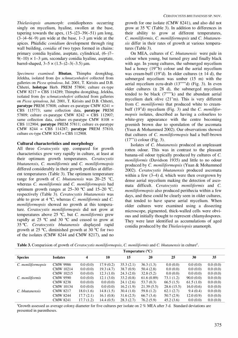
Thielaviopsis anamorph: conidiophores occurring singly on mycelium, hyaline, swollen at the base, tapering towards the apex, (15–)23–39(–51) �m long, (3–)4–6(–9) �m wide at the base, 1–3 �m wide at the apices. Phialidic conidium development through ring wall building, conidia of two types formed in chains: primary conidia hyaline, aseptate, cylindrical, (6–)7–9(–10) × 1–3 �m, secondary conidia hyaline, aseptate, barrel-shaped, 3–5 × (1.5–)2–3(–3.5) �m. Specimens examined: Bhutan, Thimphu dzongkhag, Jelekha, isolated from Ips schmutzenhoferi collected from galleries on Picea spinulosa, Jul. 2001, T. Kirisits and D.B. Chhetri, holotype Herb. PREM 57804; culture ex-type CMW 8217 = CBS 114289; Thimphu dzongkhag, Jelekha, isolated from Ips schmutzenhoferi collected from galleries on Picea spinulosa, Jul. 2001, T. Kirisits and D.B. Chhetri, paratype PREM 57808; culture ex-paratype CMW 8241 = CBS 115773; same collection data, paratype PREM 57809; culture ex-paratype CMW 8242 = CBS 112907; same collection data, culture ex-paratype CMW 8108 = CBS 112904; paratype PREM 57811; culture ex-paratype CMW 8244 = CBS 114287; paratype PREM 57810, culture ex-type CMW 8243 = CBS 112908. Cultural characteristics and morphology All three Ceratocystis spp. compared for growth characteristics grew very rapidly in culture, at least at their optimum growth temperatures. Ceratocystis bhutanensis, C. moniliformis and C. moniliformopsis differed considerably in their growth profiles at differ-ent temperatures (Table 3). The optimum temperature range for growth of C. bhutanensis was 20–25 °C, whereas C. moniliformis and C. moniliformopsis had optimum growth ranges at 25–30 ºC and 15–20 ºC, respectively (Table 3). Ceratocystis bhutanensis was able to grow at 4 ºC, whereas C. moniliformis and C. moniliformopsis showed no growth at this tempera-ture. Ceratocystis moniliformopsis did not grow at temperatures above 25 ºC, but C. moniliformis grew rapidly at 25 ºC and 30 ºC and ceased to grow at 35 ºC. Ceratocystis bhutanensis displayed rapid growth at 25 ºC, diminished growth at 30 ºC for two of the isolates (CMW 8244 and CMW 8217), and no
growth for one isolate (CMW 8241), and also did not grow at 35 ºC (Table 3). In addition to differences in their ability to grow at different temperatures, C. moniliformis, C. moniliformopsis and C. bhutanen-sis differ in their rates of growth at various tempera-tures (Table 3). On MEA, cultures of C. bhutanensis were pale in colour when young, but turned grey and finally black with age. In young cultures, the submerged mycelium had a honey (19'”b) colour and the aerial mycelium was cream-buff (19''d). In older cultures (± 14 d), the submerged mycelium was umber (15 m) with the aerial mycelium ecru-drab (13”””d) (Fig. 3). In even older cultures (± 28 d), the submerged mycelium tended to be black (7”””k) and the abundant aerial mycelium dark olive (21''m). This is very different from C. moniliformis that produced white to cream-buff (19”d) mycelium (Fig. 3) and the C. monilifor-mopsis isolates, described as having a colourless to white-grey appearance with the centre becoming greenish brown due to sporulation of the ascomata (Yuan & Mohammed 2002). Our observations showed that cultures of C. moniliformopsis had a buff-brown (17”’i) colour (Fig. 3). Isolates of C. bhutanensis produced an unpleasant rotten odour. This was in contrast to the pleasant banana-oil odour typically produced by cultures of C. moniliformis (Davidson 1935) and little to no odour produced by C. moniliformopsis (Yuan & Mohammed 2002). Ceratocystis bhutanensis produced ascomata within a few (3–4) d, which were then overgrown by dense aerial mycelium making the detection of asco-mata difficult. Ceratocystis moniliformis and C. moniliformopsis also produced perithecia within a few days, and these could be clearly seen in older cultures, that tended to have sparse aerial mycelium. When older cultures were examined using a dissecting microscope, pigmented, thick-walled cells were obvi-ous and initially thought to represent chlamydospores. They were later identified as accumulations of aged conidia produced by the Thielaviopsis anamorph.
Table 3. Comparision of growth of Ceratocystis moniliformopsis, C. moniliformis and C. bhutanensis in culturea. Temperature (ºC) Species Isolates 4 10 15 20 25 30 35
C. moniliformopsis CMW 9986 0.0 (0.0) 17.9 (0.2) 35.3 (2.1) 36.3 (1.3) 0.0 (0.0) 0.0 (0.0) 0.0 (0.0) CMW 10214 0.0 (0.0) 19.3 (4.7) 38.7 (0.9) 50.4 (2.8) 0.0 (0.0) 0.0 (0.0) 0.0 (0.0) CMW 10215 0.0 (0.0) 12.3 (1.0) 24.3 (2.0) 32.8 (5.2) 0.0 (0.0) 0.0 (0.0) 0.0 (0.0)
C. moniliformis CMW 9590 0.0 (0.0) 12.1 (3.0) 33.2 (0.8) 61.6 (0.89) 73.1 (1.2) 90.0 (0.0) 0.0 (0.0) CMW 8238 0.0 (0.0) 0.0 (0.0) 24.1 (2.6) 53.7 (0.3) 66.5 (1.5) 61.5 (1.0) 0.0 (0.0) CMW 10134 0.0 (0.0) 0.0 (0.0) 16.2 (1.9) 21.39 (5.5) 28.6 (15.5) 16.0 (0.6) 0.0 (0.0)
C. bhutanensis CMW 8217 18.0 (1.6) 14.8 (1.5) 30.4 (1.0) 59.8 (1.2) 62.1 (2.7) 9.4 (0.4) 0.0 (0.0) CMW 8244 17.7 (2.1) 16.1 (0.8) 31.6 (2.5) 66.7 (3.4) 50.7 (2.9) 12.0 (0.9) 0.0 (0.0) CMW 8241 17.7 (1.2) 14.4 (0.5) 28.3 (2.7) 76.2 (5.9) 45.2 (3.6) 0.0 (0.0) 0.0 (0.0)
aGrowth assessed as average colony diameter for five cultures per isolate on 2 % MEA after 3 d. Standard deviations are presented in parentheses.

VAN WYK ET AL.
376
Fig. 5. A phylogenetic tree based on the combined sequence data of three gene regions (ITS, �-tubulin and EF1-�). The phylogram was obtained using the heuristic search option based on parsimony and Ceratocystis virescens was treated as the out-group. Bootstrap values are indicated above of the branches while Bayesian values are indicated below the branches. Cultures of C. bhutanensis tended to degenerate on MEA. This phenomenon has also been observed in C. moniliformis and in C. moniliformopsis. Degenerated cultures became white in colour, displayed reduced growth and ceased to produce ascomata in culture DISCUSSION There is very little known regarding the occurrence of ophiostomatoid fungi in the Himalayas or their role as tree pathogens and agents of blue-stain. Prior to the present study, only one ophiostomatoid fungus, Ophi-ostoma himal-ulmi Brasier & M.D. Mehrotra was known from the Western Himalayas, where it occurs on Ulmus wallichiana Planchon and is associated with elm bark beetles (Brasier & Mehrotra 1995). The discovery of C. bhutanensis and the detection of C. moniliformis in Bhutan represent the first reports of species of Ceratocystis from this country or the Hima-layas in general. Ceratocystis bhutanensis, described in this study is also the first fungus to be recorded as an associate of bark beetles, specifically of I. schmutzenhoferi in Bhutan. Many other ophiostoma-toid fungi, including species of Ophiostoma, Cerato-cystiopsis Upadhyay & W.B. Kendr., Leptographium
Lagerb. & Melin and Pesotum Crane & Schoknecht were found in the survey of these fungi in Bhutan (Kirisits et al. 2002) and these are currently being examined and identified. Ceratocystis bhutanensis is morphologically simi-lar to C. moniliformis and C. moniliformopsis. Its occurrence in Bhutan on a conifer tree, in association with a bark beetle provided the first suspicion that it might represent a new species. The unusual colony morphology and distinct odour of this Ceratocystis sp., also suggested that it might be new. Morphologi-cal comparisons of C. bhutanensis with C. monili-formis and C. moniliformopsis revealed minor differ-ences between these fungi. DNA sequence compari-sons for three nuclear gene regions provided un-equivocal evidence that isolates of this fungus were unique. In addition to C. bhutanensis, there are six Cerato-cystis spp. that have hat-shaped ascospores. These include C. fimbriata Ellis & Halst. (Upadhyay 1981), C. moniliformis (Davidson 1935), C. albifundus M.J. Wingf., De Beer & M.J. Morris (Wingfield et al. 1996), C. moniliformopsis (Yuan & Mohammed 2002), C. pirilliformis Barnes & M.J. Wingf. (Barnes et al. 2003b) and C. acericola H.D. Griffin (Griffin 1968). Of these species, only C. moniliformis and C. moniliformopsis have spines on their ascomatal bases and both have very characteristic disc-shaped bases at their ascomatal necks. Ceratocystis bhutanen-sis can be distinguished from both of these species based on host, biogeography, association with a coni-fer bark beetle, and odour in culture, as well as various morphological characteristics (Table 2). Ceratocystis moniliformopsis has convergent ostiolar hyphae while these structures are divergent in C. bhutanensis (Table 2, Fig. 3). The ascomatal bases in these two species differ in that they are ovoid in C. moniliformopsis and globose in C. bhutanensis (Table 2). Two types of conidiogenous cells have been described for C. moniliformopsis, while C. bhutanensis has only one morphological form for these structures (Table 2). Ceratocystis bhutanensis has hyphae with walls that are smooth and granulated while C. moniliformis has only smooth-walled hyphae (Table 2). Comparison of DNA sequences for three gene regions provided strong support for the recognition of C. bhutanensis as a new species. Sequence data for the ITS regions alone did not provide convincing separa-tion between C. bhutanensis, C. moniliformis and C. moniliformopsis. However, addition of �-tubulin and EF1-� sequences provided clear resolution of the clades, in which these three species reside. Phyloge-netically, C. bhutanensis grouped within the larger C. coerulescens clade (Witthuhn et al. 1998) together with C. moniliformis and C. moniliformopsis as its closest relatives. This clade is separate from the C. fimbriata clade, in which the other Ceratocystis spp. with hat-shaped ascospores reside (Witthuhn et al.

CERATOCYSTIS BHUTANENSIS SP. NOV.
377
1999, Barnes et al. 2003a). This study also provides the first DNA sequence data for C. moniliformopsis and supports the view that this is a distinct species, even though it is morphologically very similar to C. moniliformis (Yuan & Mohammed 2002). Besides C. polonica, C. laricicola and C. rufipen-nis, C. bhutanensis is the fourth Ceratocystis sp. known to be associated with a conifer-infesting bark beetle. The new Ceratocystis sp. from Bhutan is, however, very different to the other three species, morphologically, phenotypically and phylogenetically. Ceratocystis polonica, C. laricicola and C. rufipennis are closely related to each other and form part of the C. coerulescens species complex on conifers (Harring-ton & Wingfield 1998, Witthuhn et al. 1998). In contrast, C. bhutanensis is more distantly related to species in this complex and groups closely with C. moniliformis and C. moniliformopsis that typically occur on hardwoods. The strong aroma produced by C. bhutanensis is of special interest, since this is a general characteristic of Ceratocystis spp. that are not specifically associated with bark beetles. Species of Ceratocystis that produce strong aromas are typically carried by non-specific insects that are attracted to fermenting organic mate-rial and that also visit wounds on trees (Kile 1993, Harrington & Wingfield 1998). In contrast to C. bhutanensis, cultures of C. polonica, C. laricicola and C. rufipennis lack strong aromas, which is considered as a modification to their consistent association with bark beetles (Yamaoka et al. 1997, Harrington & Wingfield 1998). Ips schmutzenhoferi is an insect that is biologically very similar to I. typographus and I. cembrae (Postner 1974, Christiansen & Bakke 1988, Schmutzenhofer 1988). Both of the latter insects carry a wide range of Ophiostoma spp. and their anamorphs, and they are particularly interesting in that they are also consis-tently associated with a pathogenic Ceratocystis spp. (Solheim 1986, Redfern et al. 1987, Solheim 1992, Yamaoka et al. 1997, Yamaoka et al. 1998, Kirisits 2001, 2004). In this respect, we might have expected to encounter a Ceratocystis sp. associated with I. schmutzenhoferi in Bhutan. However, the fact that C. bhutanensis was isolated only from adult insects at one locality and not from beetles obtained at other sites or from galleries or symptomatic sapwood tissue is intriguing. It also raises the question regarding the intimacy of the relationship between this fungus and I. schmutzenhoferi. Ceratocystis bhutanensis may be a rare associate of I. schmutzenhoferi or it may display a restricted geo-graphical distribution. Variation in the assemblages of fungi associated with bark beetles at different study sites has also been well documented. This might explain the isolation results in the present study. For example, C. polonica has been reported as a frequent or even as the dominant associate of I. typographus in
some parts of Europe, while it was not recorded or occurred rarely in studies conducted in other parts of the continent (Solheim 1986, 1992, Kirisits 2001, 2004). It has also been suggested that the population dynamics of I. typographus has a strong influence on the incidence and frequency of C. polonica, the fungus occurring less frequently during endemic periods, but becoming more frequent during outbreaks of the insect (Solheim 1993, Kirisits 2004). Similar phenom-ena may also occur in the I. schmutzenhoferi – C. bhutanensis system. It is possible that the isolation of C. bhutanensis from I. schmutzenhoferi was accidental and that this fungus is casually associated with this conifer bark beetle. Its unusual features for a Ceratocystis sp. associated with conifer bark beetles, especially its intensive aroma, and its close phylogenetic relation-ship to two Ceratocystis spp. from hardwoods might support this view. At present, the ecology of C. bhuta-nensis remains enigmatic. Further investigations, especially isolations from various niches at the local-ity where the fungus was first discovered and from the entire distribution range of I. schmutzenhoferi would be useful. In addition, pathogenicity tests with the fungus on spruce and pine to consider its role as an associate of I. schmutzenhoferi are planned. ACKNOWLEDGEMENTS We thank the Conifer Research and Training Partnership (CORET) http://woek.boku.ac.at/coret/, funded by the Austrian Development Co-operation (Austrian Ministry of Foreign Affairs) and the Royal Government of Bhutan for the opportunity and partial funding to undertake this study. G. Gratzer, G. Glatzel, L. Norbu, K. Wangdi, P. Namgyel and P.B. Chhetri helped to organise the research visit of T. Kirisits and M.J. Wingfield to Bhutan in July 2001. We also acknowledge the National Research Foundation (NRF) of the Republic of South Africa, and the Tree Pathology Co-operative Programme (TPCP) for financial support. Thanks are also due to Sylvia Presslauer (IFFF-BOKU) and staff of the Renewable Natural Resources Research Centre (RNR-RC), Yusipang and the Forestry and Agricultural Biotechnology Institute (FABI), for technical assistance in the field and laboratory and to Dr H. Glen for providing the Latin description. REFERENCES Bakshi BK (1951). Studies on four species of Ceratocystis,
with a discussion of fungi causing sap-stain in Britain. Mycological Papers 35: 1–16.
Barnes I, Roux J, Coetzee MPA, Wingfield MJ (2001). Characterization of Seiridium spp. associated with cy-press canker based on �-tubulin and histone sequences. Plant Disease 85: 317–321.
Barnes I, Roux J, Wingfield BD, O’Neil M, Wingfield MJ (2003a). Ceratocystis fimbriata infecting Eucalyptus

VAN WYK ET AL.
378
grandis in Uruguay. Australasian Plant Pathology 32: 361–366.
Barnes I, Roux J, Wingfield MJ, Old KM, Dudzinski M (2003b). Ceratocystis pirilliformis, a new species from Eucalyptus nitens in Australia. Mycologia 95: 865–871.
Brasier CM, Mehrotra MD (1995). Ophiostoma himal-ulmi sp. nov., a new species of Dutch elm disease fungus en-demic to the Himalayas. Mycological Research 99: 205–215.
Carbone I, Kohn LM (1999). A method for designing primer sets for speciation studies in filamentous asco-mycetes. Mycologia 91: 553–556.
Chhetri DB (1991). History of bark beetle outbreak in Western Bhutan. Tsenden 3: 68–72.
Christiansen E (1985). Ceratocystis polonica inoculated in Norway spruce: blue-staining in relation to inoculum density, resinosis and tree growth. European Journal of Forest Pathology 15: 160–167.
Christiansen E, Bakke A (1988). The spruce bark beetles of Eurasia. In: Dynamics of forest insect populations. Pat-tern, causes, implications (Berryman AA, ed.). Plenum Press, New York and London: 479–503.
Davidson RW (1935). Fungi causing stain in logs and lumber in the Southern states, including five new spe-cies. Journal of Agricultural Research 50: 789–807.
FAO (1999). Forest Resources of Bhutan – Country report. Rome, Italy: Forest Resources Assessment Programme (FRA), Working Paper 14: 71.
FAO (2001). Global Forest Resources Assessment 2000 – Main report. Rome, Italy: FAO Forestry Paper 120: 479.
Francke-Grosmann H (1967). Ectosymbiosis in wood-inhabiting insects. In: Symbiosis (Henry SM, ed.). Aca-demic Press, New York and London: 141–205.
Glass NL, Donaldson GC (1995). Development of primer sets designed for use with the PCR to amplify conserved genes from filamentous Ascomycetes. Applied and En-vironmental Microbiology 61: 1323–1330.
Griffin HD (1968). The genus Ceratocystis in Ontario. Canadian Journal of Botany 46: 689–718.
Harrington TC, Pashenova NV, McNew DL, Steimel J, Konstantinov M (2002). Species delimination and host specialization of Ceratocystis laricicola and C. polonica to larch and spruce. Plant Disease 86: 418–422.
Harrington TC, Wingfield MJ (1998). The Ceratocystis species on conifers. Canadian Journal of Botany 76: 1446–1457.
Hedgcock GG (1906). Studies upon some chromogenic fungi which discolor wood. Missouri Botanical Garden Annual Report 17: 59–111.
Holzschuh C (1988). Eine neue Art der Gattung Ips aus Bhutan (Coleoptera, Scolytidae). Entomologica Brasil-iensia 12: 481–485.
Jacobs K, Wingfield MJ (2001). Leptographium species: tree pathogens, insect associates and agents of blue-stain. APS Press, St. Paul, Minnesota.
Kile GA (1993). Plant diseases caused by species of Cera-tocystis sensu stricto and Chalara. In: Ceratocystis and Ophiostoma: Taxonomy, Ecology, and Pathogenicity (Wingfield MJ, Seifert KA, Webber JF, eds.). APS Press, St. Paul, Minnesota: 173–183.
Kirisits T (1998). Pathogenicity of three blue-stain fungi associated with the bark beetle Ips typographus to Nor-
way spruce in Austria. Österreichische Zeitschrift für Pilzkunde 7: 191–201.
Kirisits T (2001). Studies on the association of ophiostoma-toid fungi with bark beetles in Austria with special em-phasis on Ips typographus and Ips cembrae and their associated fungi Ceratocystis polonica and Ceratocystis laricicola. Rerum Naturalium Technicarum Doctoral Thesis, Universität für Bodenkultur Wien (BOKU), Vienna, Austria.
Kirisits T (2004). Fungal associates of European bark beetles with special emphasis on the ophiostomatoid fungi. In: Bark and Wood Boring Insects in Living Trees in Europe, a Synthesis (Lieutier F, Day KR, Grégoire JC, Evans H, eds). Kluwer, Dordrecht, The Netherlands: 181–235.
Kirisits T, Offenthaler I (2002). Xylem sap flow of Norway spruce after inoculation with the blue-stain fungus Cera-tocystis polonica. Plant Pathology 51: 359–364.
Kirisits T, Wingfield MJ, Chhetri DB (2002). Ophiostoma-toid fungi associated with the Eastern Himalayan spruce bark beetle Ips schmutzenhoferi and other bark beetles in Bhutan. In: The 7th International Mycological Con-gress, 11–17 August 2002, Oslo, Norway, IMC 7 Book of Abstracts (Ryvarden L, Schumacher T, eds.). 94, Ab-stract no. 296.
Krokene P, Solheim H (1996). Fungal associates of five bark beetle species colonizing Norway spruce. Cana-dian Journal of Forest Research 26: 2115–2122.
Krokene P, Solheim H (1998). Pathogenicity of four blue-stain fungi associated with aggressive and non-aggressive bark beetles. Phytopathology 88: 39–44.
Larget B, Simon DL (1999). Markov Chain Monte Carlo alogorithms for the Bayesian analysis of phylogenetic trees. Molecular Biology and Evolution 16: 750–759.
Liese W, Schmid R (1961). Licht- und elektronenmikroskopische Untersuchungen über das Wachstum von Bläuepilzen in Kiefern- und Fichtenholz. Holz als Roh- und Werkstoff 19: 329–337.
Moller W, DeVay J (1968). Insect transmission of Ceratocystis fimbriata in deciduous fruit orchards. Phytopathology 58: 1499–1508.
Münch E (1907). Die Blaufäule des Nadelholzes. I-II. Naturwissenschaftliche Zeitschrift für Land- und Forstwirtschaft 5: 531–573.
Paine TD, Raffa KF, Harrington TC (1997). Interactions among scolytid bark beetles, their associated fungi, and live host conifers. Annual Review of Entomology 42: 179–206.
Postner M (1974). Scolytidae (Ipidae), Borkenkäfer. In: Die Forstschädlinge Europas. Bd. 2. (Schwencke W, ed.). Paul Parey Verlag, Hamburg, Berlin: 334–482.
Rayner RW (1970). A Mycological Colour Chart. Com-monwealth Mycological Institute and British Mycologi-cal Society, Kew, Surrey.
Redfern DB, Stoakley JT, Steele H, Minter DW (1987). Dieback and death of larch caused by Ceratocystis laricicola sp. nov. following attack by Ips cembrae. Plant Pathology 36: 467–480.
Schmutzenhofer H (1988). Mass outbreaks of Ips bark beetles in Bhutan and the revision of the genus Ips de Geer for the Himalayan region. In: Integrated control of Scolytid bark beetles (Payne TL, Saarenmaa H, eds.). Proceedings of the IUFRO working party and XVII. In-ternational Congress of Entomology Symposium, “Inte-

CERATOCYSTIS BHUTANENSIS SP. NOV.
379
grated control of Scolytid bark beetles”, Vancouver, B. C., Canada: 345–355.
Seifert KA (1993). Sapstain of commercial lumber by species of Ophiostoma and Ceratocystis. In: Ceratocys-tis and Ophiostoma: Taxonomy, Ecology, and Patho-genicity (Wingfield MJ, Seifert KA, Webber JF, eds.). APS Press, St. Paul, Minnesota: 141–151.
Solheim H (1986). Species of Ophiostomataceae isolated from Picea abies infested by the bark beetle Ips typog-raphus. Nordic Journal of Botany 6: 199–207.
Solheim H (1988). Pathogenicity of some Ips typographus-associated blue-stain fungi to Norway spruce. Meddelel-ser fra Norsk Institutt for Skogforskning 40: 1–11.
Solheim H (1992). Fungal succession in sapwood of Nor-way spruce infested by the bark beetle Ips typographus. European Journal of Forest Pathology 22: 136–148.
Solheim H (1995). A comparison of blue-stain fungi associ-ated with the North American spruce bark beetle Den-droctonus rufipenniss and the Eurasian spruce bark bee-tle Ips typographus. In: Forest pathology research in the Nordic countries 1994. Proceedings from the SNS-meeting in forest pathology at Skogbrukets Kurssenter, Biri, Norway 9–12 August 1994 (Aamlid D, ed.). Ak-tuelt fra Skogforsk 4/95: 61–67.
Solheim H, Safranyik L (1997). Pathogenicity to Sitka spruce of Ceratocystis rufipennis and Leptographium abietinum, blue-stain fungi associated with the spruce beetle. Canadian Journal of Forest Research 27: 1336–1341.
Stauffer C, Kirisits T, Nussbaumer C, Pavlin R, Wingfield MJ (2001). Phylogenetic relationships between the European and Asian eight spined larch bark beetle popu-lations (Coleoptera: Scolytidae) inferred from DNA se-quences and fungal associates. European Journal of En-tomology 98: 99–105.
Swofford DL (2002). PAUP*. Phylogenetic Analysis Using Parsimony (*and other methods). Version 4.0b10. Sinauer Associates, Sunderland, Massachusetts.
Tshering G, Chhetri DB (2000). Important forest insect pests and diseases of Bhutan with control measures. Renewable Natural Resources Research Centre, Yusi-pang and Natural Resources Training Institute, Lobesa. MoA, Field guide 2000/1.
Upadhyay HP (1981). A monograph of Ceratocystis and Ceratocystiopsis. University of Georgia Press. Athens, GA.
Uzunovic A, Yang DQ, Gagné P, Breuil C, Bernier L, Byrne A, Gignac M, Kim SH (1999). Fungi that cause
sap stain in Canadian softwoods. Canadian Journal of Microbiology 45: 914–922.
White TJ, Bruns T, Lee S, Taylor J (1990). Amplification and direct sequencing of fungal ribosomal RNA genes for phylogenetics. In: PCR Protocols: A sequencing guide to methods and applications (Innis MA, Gelfand DH, Sninsky JJ, White TJ, eds.). Academic Press, San Diego: 315–322.
Whitney HS (1982). Relationships between bark beetles and symbiotic organisms. In: Bark Beetles in North American Conifers (Mitton JB, Stugeon KB, eds.). Uni-versity of Texas Press, U.S.A.: 183–211.
Wingfield MJ, De Beer C, Visser C, Wingfield BD (1996). A new Ceratocystis species defined using morphologi-cal and ribosomal DNA sequence comparisons. System-atic and Applied Microbiology 19: 191–202.
Wingfield MJ, Harrington TC, Solheim H (1997). Two species in the Ceratocystis coerulescens complex from conifers in western North America. Canadian Journal of Botany 75: 827–834.
Wingfield MJ, Seifert KA, Webber JF (eds.) (1993). Ophio-stoma and Ceratocystis: Taxonomy, Biology and Pa-thology. APS Press, St.Paul, Minnesota.
Witthuhn RC, Wingfield BD, Wingfield MJ, Harrington TC (1999). PCR-based identification and phylogeny of spe-cies of Ceratocystis sensu stricto. Mycological Research 103: 743–749.
Witthuhn RC, Wingfield BD, Wingfield MJ, Wolfaardt M, Harrington TC (1998). Monophyly of the conifer spe-cies in the Ceratocystis coerulescens complex based on DNA sequence data. Mycologia 90: 96–101.
Wood SL, Bright DE (1992). A catalogue of Scolytidae and Platypodidae (Coleoptera), Part 2: Taxonomic Index, Volumes A and B. Great Basin Naturalist Memoirs 13: 1–1553.
Yamaoka Y, Wingfield MJ, Ohsawa M, Kuroda Y (1998). Ophiostomatoid fungi associated with Ips cembrae in Japan. Mycoscience 39: 367–378.
Yamaoka Y, Wingfield MJ, Takahashi I, Solheim H (1997). Ophiostomatoid fungi associated with the spruce bark beetle Ips typographus f. japonicus in Japan. Mycologi-cal Research 101: 1215–1227.
Yuan ZQ, Mohammed C (2002). Ceratocystis monilifor-mopsis sp. nov., an early colonizer of Eucalyptus obli-qua logs in Tasmania, Australia. Australian Systematic Botany 15: 125–133

VAN WYK ET AL.
380