CERAMENT™|BONE VOID FILLER |BONE VOID FILLER Bone Healing Technical Monograph 2 I. Overview...
20
CERAMENT™|BONE VOID FILLER Bone Healing Technical Monograph
Transcript of CERAMENT™|BONE VOID FILLER |BONE VOID FILLER Bone Healing Technical Monograph 2 I. Overview...
CERAMENT™|BONE VOID FILLER Bone Healing Technical Monograph

CERAMENT™|BONE VOID FILLER Bone Healing Technical Monograph
1
Table of contents
I. Overview 2
II. Conditions for bone healing 3
III. History of bone graft substitutes 4
i) Calcium sulfate 4
ii) Calcium phosphate 4
IV. Importance of porosity and chemistry 5
V. The Science of CeramenT™ 6
i) Biphasic 6
ii) Injectable 7
iii) Bioactive 8
VI. Preclinical findings 9
i) Biocompatibilty and no inflammatory reaction 9
ii) Osteoconductivity 11
iii) Bone regeneration – transformation into bone 11
VII. Clinical results 13
VIII. Conclusion 16
IX. references 17

CERAMENT™|BONE VOID FILLER Bone Healing Technical Monograph
2
I. OverviewCERAMENT™ is a biphasic injectable bone graft substitute. It is synthetically made and has one osteoconductive component, hydroxyapatite, and one resorbable component, calcium sulfate.
Bone substitutes based on hydroxyapatite and calcium sulfate offer good potential to be used as bone repair material for clinical applications due to their:
a) osteoconductivity, with an ability to act as a matrix for cells and as a carrier of osteoinductive factors or other therapeutic substances
b) injectability, making it possible to utilize in minimally invasive surgery techniques
c) adequate strength and controllable resorption
CERAMENT™ also includes a radio-opacity enhancing component which makes the material suitable in applications guided by fluoroscopy, including mini-invasive surgery.
The material is easy to mix and handle. It hardens in situ and all in vivo studies have shown good biocompatibility, adequate resorption rate and good bone healing.
By filling a bone defect with CERAMENT™, which contains 40% bone mineral, i.e. hydroxyapatite, three important needs for bone healing are fulfilled:
a) The void is filled with a bone mineral and can therefore not be invaded by fibrous tissue
b) CERAMENT™ hardens in situ, augments the bone, and provides some mechanical stability
c) CERAMENT™ acts as a scaffold for the ingrowth of bone
When bone-forming cells are in direct contact with CERAMENT™, the hydroxyapatite particles get incorporated into the newly formed bone which increases the bone strength. After treatment with CERAMENT™, complete bone healing is demonstrated within 6-12 months.

CERAMENT™|BONE VOID FILLER Bone Healing Technical Monograph
3
II. Conditions for bone healing
1. VASCULARITY Vascular ingrowth and blood supply is critical for bone formation. Blood contains circulating osteoprogenitor cells and extracellular matrix proteins necessary to initiate osteoclast and osteoblast. In a fracture or a bone defect, this cell proliferation initiates cartilage growth followed by immature bone formation. If the mechanical situation is stable, woven and lamellar bone will follow. To activate healing in a void not created by surgery or a recent fracture, it is preferable to provoke bleeding to attract cells and to promote angiogenesis and subsequent bone healing.
2. FIXATION Excess motion in a fracture or bone defect may interrupt the development of new bone which may lead to fibrous tissue formation instead of bone.
3. BONE TISSUE FORMATION Fracture healing, with bridging of bone defects, occurs if the defect is small enough and the above mentioned conditions are fulfilled. Healing of bone with large gaps often fails because there is no scaffold for the osteoblast to climb on. The healing process in such defects starts from the edges of the defect in order to fill it completely with new bone tissue. If the defect site gets filled by fibrous tissue instead of bone, a non-union will be the result.
4. MINERALIZATION Mineralization and maturation of the bone tissue are the final stages in the bone healing process, and it can only occur if the bone is adequately loaded. Unloaded immature bone tissue will be resorbed by osteoclasts since it is considered “not needed” by the body. However, loading the immature bone tissue will stimulate the remodeling to result in a strong and load-bearing bone structure.
Too strong bone substitute material might be expected to shield the immature bone from the load leading to bone resorption, while a weak bone substitute material might lead to early collapse. It is therefore important to match the strength of the bone substitute to the mechanical demands.

CERAMENT™|BONE VOID FILLER Bone Healing Technical Monograph
4
III. History of bone graft substitutes
i) Calcium sulfate The use of synthetic bone grafts started over 100 years ago with the implantation of calcium sulfate. The first reported case, where calcium sulfate was used to treat cavities in bone, is from 1892 by Dreesmann in Germany, who operated on eight patients with large bone defects grafting them with b-calcium sulfate hemihydrate1. Subsequent reports showed good results with complete bone regeneration and concluded that calcium sulfate was biocompatible, did not add complications even in infected cavities, and was resorbed quickly2-6.
The majority of recent studies have been performed on calcium sulfate pellets produced from hemihydrate with crystals of regular shape and size, i.e. a-calcium sulfate hemihydrate. These pellets show less variation in solubility and resorption7-11. The most important advantages with calcium sulfate are the excellent biocompatibility and the rapid dissolution rate making it suitable for drug release. However, with the drawback of sometimes failing in the long term, scaffold support is needed for full bone regeneration12.
ii) Calcium Phosphate The most common calcium phosphate compound used in bone grafting is hydroxyapatite (HA; Ca
10(PO
4)
6(OH)
2). Its structure is similar to that of the mineral
phase of bone and it shows excellent biocompatibility. HA is osteoconductive due to its chemical similarity to natural bone mineral and is also bioactive, i.e. bone chemically binds to it13,14. A study on the proximal tibia of 30 rabbits demonstrated that HA (50 wt-%) + calcium sulfate was highly osteoconductive, giving 52% bone formation after 4 weeks and 90% after 24 weeks15. Although the calcium sulfate component had disappeared after 8 weeks, the HA continued to guide bone ingrowth15. At 24 weeks, the HA particles were surrounded by, and incorporated in, thick trabecular bone.
HA is often synthesised at high temperature (typically above 1800 F) to form granules or blocks, but can also be precipitated from a supersaturated solution of calcium (Ca2+) and phosphate (PO
42-) ions. High temperature treatments provide a
more crystallized HA that shows minimum resorption by osteoclast activity and may remain at the implant site for years or even decades16,17. This may be an advantage for certain applications, but a drawback for others. In younger patients or growing children resorbable implants are, however, preferable since the material is replaced over time by bone tissue.
Due to partial resorption, biphasic calcium phosphate (BCP) has been used18-22. It may give bone ingrowth and mechanical stability at the implant/bone interface, but BCP will not resorb completely20.

CERAMENT™|BONE VOID FILLER Bone Healing Technical Monograph
5
With
CERAMENT™|BONE VOID FILLER
there is controlled resorption
of CaS to match the rate of
bone ingrowth and support
new bone growth.
IV. Importance of porosity and chemistryTo achieve bone healing a bone substitute has to be porous to allow penetration of living cells. Blood capillaries, osteoblasts and osteoclasts have to be able to invade the material to allow bone remodeling.
For many years it was believed that only macroporosity (pore size > 100 μm) was critical for good bone ingrowth. More recently the importance of microporosity has been highlighted, with convincing results showing cell attachment on microporous surfaces23 and a need for microporosity around 1-50 microns to allow penetration of body fluids and subsequent vessel ingrowth24. In vivo studies show that manipulation of the microporosity in calcium phosphate bioceramics may accelerate osteointegration25,26, improve the adsorption of proteins and the adhesion and proliferation of human bone cells27.
Conclusively, both micro- and macroporosity are important for the bone ingrowth28,29 as well as the chemistry of the bone graft substitute30.
Ideally, the resorption of an implant material has to correspond to the bone ingrowth rate in order to optimize the healing of the defect:
- Too slow resorption of the implant will obstruct the growth of new bony tissue and will slow down the healing process.
- Too fast resorption of the implant will leave a gap between the implant and the ingrowing bone with a risk of fibrous tissue interpositioning.
The resorbing material leaves space for the bone tissue to grow and the osteoconductive material guides the bone cells and facilitate bone formation.Eventually this results in full transformation of the bone substitute into mature bone.

CERAMENT™|BONE VOID FILLER Bone Healing Technical Monograph
6
V. The Science of CERAMENT™
i) Biphasic CERAMENT™ is an injectable biphasic ceramic material, indicated for the filling of bone voids. CERAMENT™ consists of a powder which is mixed with a liquid and becomes an injectable paste which hardens in situ. The powder has two components:
• 40wt%hydroxyapatite(HA)
• 60wt%α-calciumsulfatehemihydrate
HA is the mineral phase of bone. It is highly osteoconductive18 and will guide the bone ingrowth throughout CERAMENT™ in vivo. The HA used in CERAMENT™ is engineered to be stable. It offers high injectability and gives long term support to the defect. The HA particles form an osteoconductive scaffold augmenting the calcium sulfate to retard its resorption rate31. The HA particles are embedded into newly formed bone with no adverse inflammatory response31.
Calcium sulfate is used for its tissue integration and biocompatibility. It has been used for bone repair for more than 100 years with excellent tissue response2-4,6,32. No adverse reactions have been reported during its resorption3,9,10, showing that calcium sulfate degradation products are very unlikely to be harmful for the body. The calcium sulfate used in CERAMENT™ is of medical grade. It gives short term stability to the bone defect after repair and will dissolve and be actively resorbed by octeoclastic activity12 within 6-12 months33. Dissolution of calcium sulfate creates space for new bone growth.
Both the calcium sulfate and the HA component of CERAMENT™ are synthetically produced to assure high purity and reproducibility.
The ratio 40/60 of HA/calcium sulfate provides maximum osteoconductivity while keeping a strength suitable for augmentation of cancellous bone defects34. The mechanical properties closely match cancellous bone35 thereby avoiding stress shielding and providing a mechanically stimulating environment for bone growth. A too strong and stiff material may cause bone resorption since the force will be transmitted through the material instead of through the bone36. It is important to load the treated region adequately to regenerate and remodel bone37.

CERAMENT™|BONE VOID FILLER Bone Healing Technical Monograph
7
ii) Injectable The alpha-form of calcium sulfate hemihydrate delivers much better injectability compared with the beta-form34. The alpha-form has a higher density and absorbs less liquid, which also makes the calcium sulfate stronger38 and gives the material a slower resorption rate39. The injectability is also enhanced by the round shape of the HA particles40. Round particles flow easily and the injection may be performed without high pressure. The liquid used in CERAMENT™, iohexol solution, further increases the lubrication of the powder and ensures that no filter pressing occurs41. Filter pressing is a phenomenon seen when particles mixed with liquid are put under pressure, where the material separates and the liquid is pressed out through the particle phase, resulting in dry powder left if in a syringe during injection.
High injectability enables injection through narrow needles and ensures an excellent spread in the trabecular system. It also allows HA particles to be carried into the bone defect. The calcium sulfate component of CERAMENT™ not only delivers the osteoconductive HA, but also prevents migration of the particles. It binds the HA particles which is important for subchondral applications and in joint prosthetic revision surgery avoiding the risk for abrasive wear.The liquid component is a water soluble radio-opacity enhancing component called C-TRU™ consisting of iohexol and water. It is safe42 and has been used clinically under the brand name Omnipaque® since the 1980s. By adding a radio-opacity enhancing component to the material, transcortical injections using minimally invasive techniques may be performed safely. Injection of CERAMENT™ can thus be followed visually under fluoroscopy which decreases the risk of leakage into e.g. the joint space in the presence of intra-articular fracture lines.
Iohexol is a non-inflammatory, non-ionic radiocontrast agent42, that doesn’t metabolize and is cleared from the body through renal excretion43.
When mixing the powder with the liquid an injectable and moldable paste is formed. Once the paste hardens it forms a microporous ceramic material designed to facilitate bone formation in the bone void and result in complete healing.
CERAMENT’S 40/60 ratio of
HA/calcium sulfate provides
maximum osteoconductivity
while keeping a strength
suitable for augmentation of
cancellous bone defects.

CERAMENT™|BONE VOID FILLER Bone Healing Technical Monograph
8
iii) Bioactive CERAMENT™ is bioactive, which means that a nanolayer of carbonated apatite will spontaneously form on the material surface approximately 1-3 days after implantation (Fig 1). Calcium ions from the calcium sulfate react with phosphate ions from the body fluids and a layer of apatite precipitates on the material surface31. It is hypothesized that this passive precipitation of endogenous HA stabilizes the CERAMENT™ implant and explains the substantially retarded resorption of the calcium sulfate component seen with CERAMENT™. This precipitation has also been observed on other highly biocompatible materials like titanium44 and Bioglass® 45, and it has been shown to encourage new bone ongrowth onto the material46. It basically enhances the direct contact between material and bone because bone cells recognize the apatite layer as bone mineral.
Not all bone graft substitutes are bioactive. Pure calcium sulfate, which always presents with a low pH, does not have the ability of forming this apatite layer47. The HA particles are thus needed to induce HA precipitation, which might be explained by a combination of neutralized pH and necessary surface properties not present with calcium sulfate47.
Fig 1: A layer of HA has been formed on the surface of CERAMENT™. This layer makes the material bioactive, retards the calcium sulfate resorption and enhances the direct contact between material and bone34.
CERAMENT™ is bioactive and
has been shown to encourage
new bone ongrowth onto the
material because bone cells
recognize the apatite layer as
bone mineral.

CERAMENT™|BONE VOID FILLER Bone Healing Technical Monograph
9
VI. Preclinical findings
i) Biocompatibilty and no inflammatory reaction CERAMENT™ has been studied in innumerous animals, including in rats, rabbits, and sheep31,48-53.
It has shown good tissue response both in muscle pockets and in bone defects31. A close contact was found between material and bone tissue in a bone harvest chamber model in rabbits31 (Fig 2), with trabecular bone completely surrounding and embedding the HA particles (Fig 3). No inflammatory reactions or fibrous tissue were observed after 3 and 6 weeks.
The incorporation of the HA particles and fragments of the material, both calcium sulfate and HA, was observed in greater detail in femur defects in rats50 (Fig 4 on pg.10) at 21 days, and in rabbits51 (Fig 5 on pg.10) at 12 weeks after implantation of CERAMENT™.
Fig 2: A close contact was found between CERAMENT™ (to the left) and new bone tissue (to the right) at 6 weeks post implantation31 of CERAMENT™. The new bone tissue invaded CERAMENT™ and no sharp bone/implant interface was observed.
Fig 3: Bony tissue completely surrounded and embedded the HA particles.
CERAMENT™
NEW BONE TISSUE
CERAMENT™

CERAMENT™|BONE VOID FILLER Bone Healing Technical Monograph
10
It was concluded that calcium sulfate in combination with HA resulted in the formation of new bone that completely surrounded and embedded the HA particles once the calcium sulfate had resorbed50. The new trabeculae became thicker and denser, which increased the mechanical strength of the newly formed bone as demonstrated by indentation tests (Fig 6)50.
Fig 4: Shows fragments of CERAMENT™ (star) incorporated in new, immature bone tissue (arrow).
Fig 6: Showing the mechanical strength of newly formed bone after CERAMENT™ implantation (left)vs. normal cancellous bone (right). Forty-two days after CERAMENT™ implantation (blue line to the left) mechanical strength of the newly formed bone was higher compared to intact cancellous bone (to the right).
Fig 5: Show CERAMENT™ with translumiscent HA particles (star) inside a newly formed bone trabecula (arrow).

CERAMENT™|BONE VOID FILLER Bone Healing Technical Monograph
11
ii) Osteoconductivity Calcium sulfate alone is not an osteoconductive material. Bone will form with time but early results show that fibrous tissue is first formed between the new bone and the remaining material50, probably because of the rapid resorption of the calcium sulfate. HA particles can be added to the calcium sulfate based bone substitute to provide osteoconductivity. It has been clearly shown that between 30 and 50% of HA is necessary to obtain an osteoconductive material that still provides sufficient strength15,54. In a study using 40% HA in calcium sulfate, osteoconductivity was observed in defects in the distal part of rabbit femora50.
iii) Bone regeneration – transformation into bone The first generation of synthetic bone substitutes consisting of either pure calcium sulfate or pure HA have had limited advantages due to their static behavior. It was thus stated by Hench in 199855 that ”we need to shift the emphasis of biomaterial research towards assisting or enhancing the body’s own reparative capacity”.
Most calcium phosphate-based bone substitute materials have a too slow resorption rate and are followed by new bone tissue formation through a creeping substitution from the surface towards the center of the defect56. New bone tissue is only present at the surface of the material and the material remains in the defect center until complete healing occurs, which might take years16,17,57. The mechanism of action for CERAMENT™ is different. Through initial microporosity and later macroporosity, early vascularization and invasion of osteoblasts enable a multiple site formation of bone throughout the cured CERAMENT™ implant (Fig 7).
Fig 7: New bone tissue formed throughout CERAMENT™ after 12 weeks, as opposed to the creeping substitution seen with many calcium phosphate-based bone substitutes. Animal study with a critical defect (> 5 mm) created in the lateral femoral condyle and filled with CERAMENT™ 51.
CERAMENT™
CERAMENT™
CERAMENT™|BONE VOID FILLER Bone Healing Technical Monograph
12
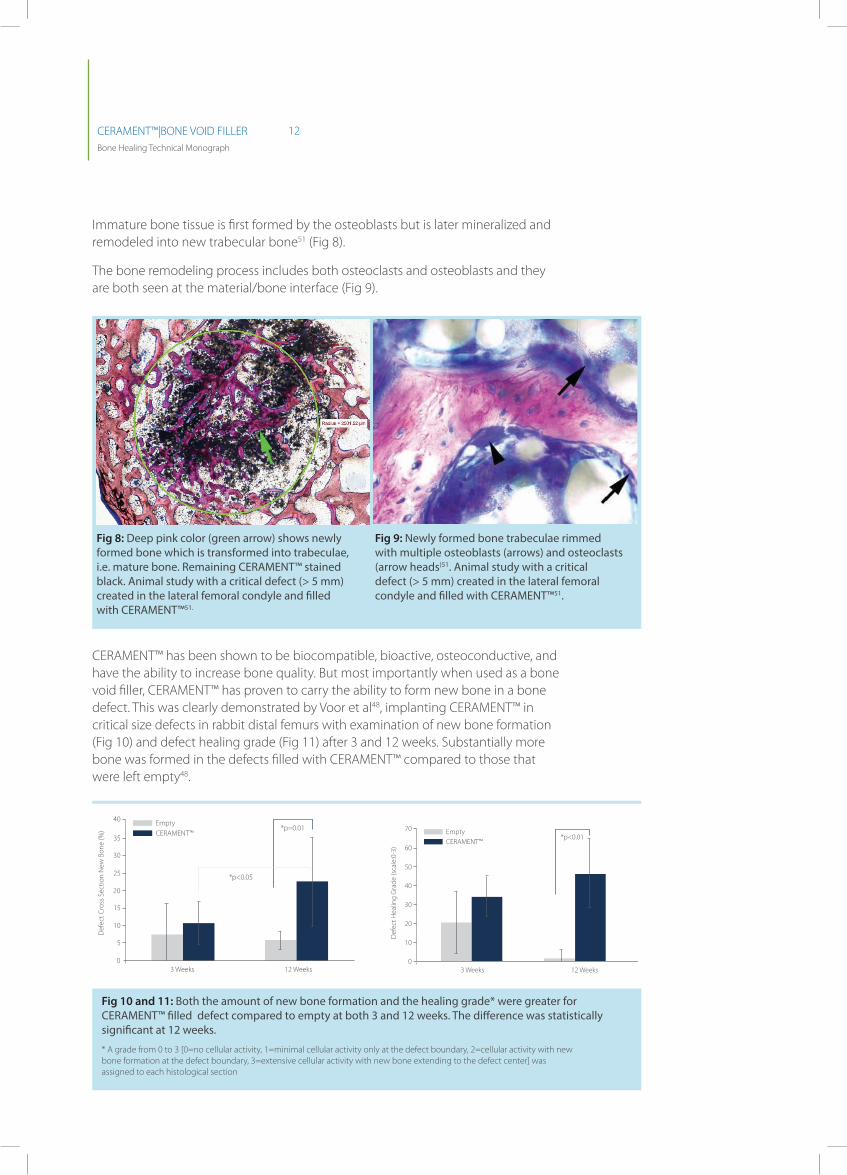
Immature bone tissue is first formed by the osteoblasts but is later mineralized and remodeled into new trabecular bone51 (Fig 8).
The bone remodeling process includes both osteoclasts and osteoblasts and they are both seen at the material/bone interface (Fig 9).
CERAMENT™ has been shown to be biocompatible, bioactive, osteoconductive, and have the ability to increase bone quality. But most importantly when used as a bone void filler, CERAMENT™ has proven to carry the ability to form new bone in a bone defect. This was clearly demonstrated by Voor et al48, implanting CERAMENT™ in critical size defects in rabbit distal femurs with examination of new bone formation (Fig 10) and defect healing grade (Fig 11) after 3 and 12 weeks. Substantially more bone was formed in the defects filled with CERAMENT™ compared to those that were left empty48.
Fig 8: Deep pink color (green arrow) shows newly formed bone which is transformed into trabeculae, i.e. mature bone. Remaining CERAMENT™ stained black. Animal study with a critical defect (> 5 mm) created in the lateral femoral condyle and filled with CERAMENT™51.
Fig 9: Newly formed bone trabeculae rimmed with multiple osteoblasts (arrows) and osteoclasts (arrow heads)51. Animal study with a critical defect (> 5 mm) created in the lateral femoral condyle and filled with CERAMENT™51.
40
30
20
35
25
15
03 Weeks
Def
ect C
ross
Sec
tion
New
Bon
e (%
)
Def
ect H
ealin
g G
rade
(sca
le:0
-3)
*p=0.01
*p<0.05
12 Weeks
EmptyCERAMENT™
EmptyCERAMENT™
10
5
60
50
70
40
30
10
03 Weeks
*p<0.01
12 Weeks
20
40
30
20
35
25
15
03 Weeks
Def
ect C
ross
Sec
tion
New
Bon
e (%
)
Def
ect H
ealin
g G
rade
(sca
le:0
-3)
*p=0.01
*p<0.05
12 Weeks
EmptyCERAMENT™
EmptyCERAMENT™
10
5
60
50
70
40
30
10
03 Weeks
*p<0.01
12 Weeks
20
Fig 10 and 11: Both the amount of new bone formation and the healing grade* were greater for CERAMENT™ filled defect compared to empty at both 3 and 12 weeks. The difference was statistically significant at 12 weeks.
* A grade from 0 to 3 [0=no cellular activity, 1=minimal cellular activity only at the defect boundary, 2=cellular activity with new bone formation at the defect boundary, 3=extensive cellular activity with new bone extending to the defect center] was assigned to each histological section

CERAMENT™|BONE VOID FILLER Bone Healing Technical Monograph
13
VII. Clinical results
Case 1
Osteotomy after distal radius fracture malunion
A man (40 years old) was included in a clinical study by Abramo et al58. He underwent osteotomy after distal radius fracture malunion.
Fixation was performed using Trimed system. CERAMENT™ was applied in the gap formed during surgery. Fig A shows the osteotomy directly post-operatively and Fig B shows the same osteotomy one year later. Complete bone healing was achieved and new trabecular and cortical bone were formed where CERAMENT™ had been implanted.
Figure a: Post-operative picture of osteotomy in distal radius.
Figure B: 1 year follow up shows complete bone healing and new trabecular and cortical bone in wrist osteotomy.
Reproduced with kind permission of Hand Unit, Department of Orthopaedics, Lund, Sweden. Ref: Images on file BONESUPPORT, Lund Sweden
Credit: Antonio Abramo (1), Mats Geijer (2), Philippe Kopylov (1), Magnus Tägil (1)
1. Department of Orthopaedics, Hand Unit, Clinical Sciences, Lund University, Lund S-221 85, Sweden
2. Department of Radiology, Lund University Hospital, Lund S-221 85, Sweden

CERAMENT™|BONE VOID FILLER Bone Healing Technical Monograph
14
Case 2
Treatment of displaced intra-articular calcaneal fracture
A female (54 years old) had open reduction and internal fixation (ORIF) Fig. A.
To avoid loss of calcaneal height of the posterior facet and reduction of Bohler’s angle when full weight bearing, surgery was augmented with CERAMENT™|BONE VOID FILLER.
Removal of the plate at 4 months due to pain (no signs of infection) facilitated a bone biopsy which showed early signs of new bone growth Fig B.
At 7 months the patient demonstrates a
good result. Fig C & D.
Figure a. X-ray immediately post surgery.
Figure B. Histology at 4 months showing new bone growth.
Figure D. Patient full weight bearing.
Figure C. X-ray after removal of the plate.
Credit: Damiano Papadia
Reparto di Ortopedia e, Traumatologia Ospedale, Santa Chiara, Trento, Italy
CERAMENT™|BONE VOID FILLER Bone Healing Technical Monograph
15
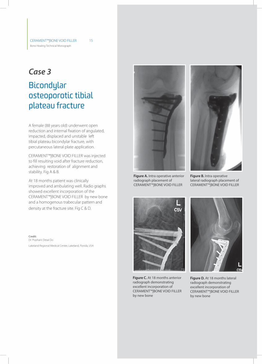
Case 3
Bicondylar osteoporotic tibial plateau fracture
A female (88 years old) underwent open reduction and internal fixation of angulated, impacted, displaced and unstable left tibial plateau bicondylar fracture, with percutaneous lateral plate application.
CERAMENT™|BONE VOID FILLER was injected to fill resulting void after fracture reduction, achieving restoration of alignment and stability. Fig A & B.
At 18 months patient was clinically improved and ambulating well. Radio graphs showed excellent incorporation of the CERAMENT™|BONE VOID FILLER by new bone and a homogenous trabecular pattern and
density at the fracture site. Fig C & D.
Figure C. At 18 months anterior radiograph demonstrating excellent incorporation of CERAMENT™|BONE VOID FILLER by new bone
Figure D. At 18 months lateral radiograph demonstrating excellent incorporation of CERAMENT™|BONE VOID FILLER by new bone
Figure a. Intra operative anterior radiograph placement of CERAMENT™|BONE VOID FILLER
Figure B. Intra operative lateral radiograph placement of CERAMENT™|BONE VOID FILLER
Credit: Dr. Prashant Desai Do
Lakeland Regional Medical Center, Lakeland, Florida, USA
CERAMENT™|BONE VOID FILLER Bone Healing Technical Monograph
16
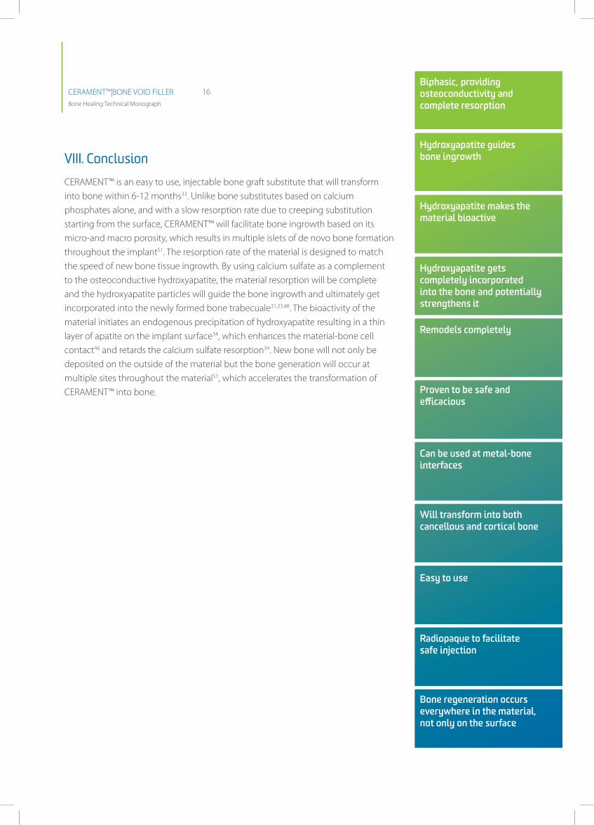
VIII. Conclusion
CERAMENT™ is an easy to use, injectable bone graft substitute that will transform
into bone within 6-12 months33. Unlike bone substitutes based on calcium
phosphates alone, and with a slow resorption rate due to creeping substitution
starting from the surface, CERAMENT™ will facilitate bone ingrowth based on its
micro-and macro porosity, which results in multiple islets of de novo bone formation
throughout the implant51. The resorption rate of the material is designed to match
the speed of new bone tissue ingrowth. By using calcium sulfate as a complement
to the osteoconductive hydroxyapatite, the material resorption will be complete
and the hydroxyapatite particles will guide the bone ingrowth and ultimately get
incorporated into the newly formed bone trabecuale31,33,48. The bioactivity of the
material initiates an endogenous precipitation of hydroxyapatite resulting in a thin
layer of apatite on the implant surface34, which enhances the material-bone cell
contact46 and retards the calcium sulfate resorption34. New bone will not only be
deposited on the outside of the material but the bone generation will occur at
multiple sites throughout the material51, which accelerates the transformation of
CERAMENT™ into bone.
Biphasic, providing osteoconductivity and complete resorption
Remodels completely
Easy to use
Hydroxyapatite guides bone ingrowth
Proven to be safe and efficacious
Radiopaque to facilitate safe injection
Hydroxyapatite makes the material bioactive
Can be used at metal-bone interfaces
Bone regeneration occurs everywhere in the material, not only on the surface
Hydroxyapatite gets completely incorporated into the bone and potentially strengthens it
Will transform into both cancellous and cortical bone

CERAMENT™|BONE VOID FILLER Bone Healing Technical Monograph
17
IX. References1. Dreesmann H: Knochenplombierung bei Hohlenforigen Defekten des Knochens. Beitr Klin Chir 9:804-810, 1892
2. Nyström G: Plugging bone cavities with rivanol-plaster porridge. Acta Chir Scand 63:296, 1928
3. Peltier LF: The Use of Plaster of Paris To Fill Defects in Bone. Clin Orthop 21:1-29, 1961
4. Nielson A: The filling of infected and sterile bone cavities by means of plaster of Paris. Acta Chir Scand 91:17-27, 1944
5. Kovacevic B: Ein Beitrag zum Problem der Hamartogenen Osteomyelitis. Arch klin Chir 276:432-443, 1953
6. Edberg E: Some experiences of filling osseous cavities with plaster. Acta Chir Scand 67:313-319, 1931
7. Blaha JD: Calcium sulfate bone-void filler. Orthopedics 21:1017- 1019, 1998
8. Tay BKB, Patel VV, Bradford DS: Calcium sulfate-and calcium phosphate-based bone substitutes - Mimicry of the mineral phase of bone. Orthopedic Clinics of North America 30:615-623, 1999
9. Gitelis S, Piasecki P, Turner T, Haggard W, Charters J, Urban R: Use of a calcium sulfate-based bone graft substitute for benign bone lesions. Orthopedics 24:162-166, 2001
10. Kelly CM, Wilkins RM, Gitelis S, Hartjen C, Watson JT, Kim PT: The use of a surgical grade calcium sulfate as a bone graft substitute: results of a multicenter trial. Clin Orthop 42-50, 2001
11. Turner TM, Urban RM, Gitelis S, Kuo KN, Andersson GBJ: Radiographic and histologic assessment of calcium sulfate in experimental animal models and clinical use as a resorbable bone-graft substitute, a bone-graft expander, and a method for local antibiotic delivery - One institution’s experience. Journal of Bone and Joint Surgery-American Volume 83A:8-18, 2001
12. Sidqui M, Collin P, Vitte C, Forest N: Osteoblast adherence and resorption activity of isolated osteoclasts on calcium- sulfate hemihydrate. Biomaterials 16:1327-1332, 1995
13. Hench LL: Bioceramics. Journal of the American Ceramic Society 81:1705-1728, 1998
14. Jarcho M: Calcium phosphate ceramics as hard tissue prosthetics. Clinical Orthopaedics and Related Research 259-278, 1981
15. Sato S, Koshino T, Saito T: Osteogenic response of rabbit tibia to hydroxyapatite particle-Plaster of Paris mixture. Biomaterials 19:1895-1900, 1998
16. Khan SN, Tomin E, Lane JM: Clinical applications of bone graft substitutes. Orthopedic Clinics of North America 31:389-398, 2000
17. Bohner M: Physical and chemical aspects of calcium phosphates used in spinal surgery. European Spine Journal 10:S114-S121, 2001
18. Bohner M: Calcium orthophosphates in medicine: from ceramics to calcium phosphate cements. Injury 31 Suppl 4:37- 47, 2000
19. LeGeros RZ: Properties of osteoconductive biomaterials: Calcium phosphates. Clinical Orthopaedics and Related Research 81-98, 2002
20. LeGeros RZ, Parsons JR, Daculsi G, Driessens FCM, Lee D, Liu ST, Metsger S, Peterson D, Walker M: Significance of the porosity and physical chemistry of calcium phosphate ceramics: Biodegradation-Bioresorption. In: Bioceramics: Material characteristics versus in vivo behaviour pp 268-271. Ed by P Ducheyne and JE Lemons. New York, Annals of the New York academy of science, vol. 523, 1988
21. Frayssinet P, Fages J, Bonel G, Rouquet N: Biotechnology, material sciences and bone repair. Eur J Orthop Surg Traumatol 8:17-25, 1998
22. Perry CR: Bone repair techniques, bone graft, and bone graft substitutes. Clin Orthop 71-86, 1999
23. Bignon A, Chouteau J, Chevalier J, Fantozzi G, Carret JP, Chavassieux P, Boivin G, Melin M, Hartmann D: Effect of micro- and macroporosity of bone substitutes on their mechanical properties and cellular response. Journal of Materials Science- Materials in Medicine 14:1089-1097, 2003
24. Eggli PSM, Moller WP, Schenk RKM: Porous Hydroxyapatite and Tricalcium Phosphate Cylinders with Two Different Pore Size Ranges Implanted in the Cancellous Bone of Rabbits: A Comparative Histomorphometric and Histologic Study of Bony Ingrowth and Implant Substitution. [Report]. Clinical Orthopaedics & Related Research 232:127-138, 1988
25. Hing KA, Annaz B, Saeed S, Revell PA, Buckland T: Microporosity enhances bioactivity of synthetic bone graft substitutes. J Mater Sci: Mater Med 16:467-475, 2005
26. Malmström, J. On bone regeneration in porous bioceramics. University of Gothenburg. 2007. ISBN: 978-91-628-7207-6
27. Rouahi M, Gallet O, Champion E, Dentzer J, Hardouin P, Anselme K: Influence of hydroxyapatite microstructure on human bone cell response. Journal of Biomedical Materials Research Part A 78A:222-235, 2006
28. Boyde A, Corsi A, Quarto R, Cancedda R, Bianco P: Osteoconduction in large macroporous hydroxyapatite ceramic implants: evidence for a complementary integration and disintegration mechanism. Bone 24:579-589, 1999
29. Hing KA, Best SM, Tanner KE, Bonfield W, Revell PA: Mediation of bone ingrowth in porous hydroxyapatite bone graft substitutes. Journal of Biomedical Materials Research Part A 68A:187-200, 2004
30. Cuneyt Tas A: A review of bone substitutes in bone remodeling: Influence of material chemistry and porosity. In: Bioceramics: Materials and Applications IV pp 15-24. Ed by Veeraraghavan Sundar, Richard P.Rusin, and Claire A.Rutiser. Nashville, 2003

CERAMENT™|BONE VOID FILLER Bone Healing Technical Monograph
18
31. Nilsson M, Wang JS, Wielanek L, Tanner KE, Lidgren L: Biodegradation and biocompatability of a calcium sulphate- hydroxyapatite bone substitute. Journal of Bone and Joint Surgery-British Volume 86B:120-125, 2004
32. Coetzee AS: Regeneration of bone in the presence of calcium- sulfate. Archives of Otolaryngology-Head & Neck Surgery 106:405- 409, 1980
33. Abramo A, Geijer M, Kopylov P, Tägil M: Osteotomy of distal radius fracture malunion using a fast remodeling bone substitute consisting of calcium sulphate and calcium phosphate. J Biomed Mater Res 92B:281-286, 2010
34. Nilsson, M. Injectable calcium sulphate and calcium phosphate bone substitutes. Lund University. 2003. ISBN: 91-628-5603-0
35. Nilsson M, Wielanek L, Wang JS, Tanner KE, Lidgren L: Factors influencing the compressive strength of an injectable calcium sulfate/hydroxyapatite cement. Journal of Materials Science: materials in medicine 14:399-404, 2003
36. Black J: Biological performance of materials: Fundamentals of biocompatibility, Boca Raton, CRC Press, 2006. ISBN:0-8493- 3959-6
37. Kalfas IH: Principles of bone healing. Neurosurg Focus 10:7-10, 2001
38. Wirsching F: Calcium Sulfate. In: Ullmann’s Encyclopedia of Industrial Chemistry, vol 4A: Benzyl Alcohol to Calcium Sulfate, pp 555-584. Ed by W Gerhartz, YS Yamamoto, FT Campbell, R Pfefferkorn, and JF Rounsaville. Weinheim, VCH Verlagsgesellschaft mbH, 1985
39. Thomas MV, Puleo DA: Calcium sulfate: Properties and clinical applications. J Biomed Mater Res Part B: Appl Biomater 88B:597- 610, 2009
40. Ishikawa K: Effects of spherical tetracalcium phosphate on injectability and basic properties of apatitic cement, pp 369-372. 2003
41. Bohner M, Baroud G: Injectability of calcium phosphate pastes. Biomaterials 26:1553-1563, 2005
42. Almen T: Development of Nonionic Contrast-Media. Investigative Radiology 20:S2-S9, 1985
43. Almen T: Visipaque - A step forward - A historical review. Acta Radiologica 36:2-18, 1995
44. Kokubo T, Miyaji F, Kim HM, Nakamura T: Spontaneous formation of bonelike apatite layer on chemically treated titanium metals. Journal of the American Ceramic Society 79:1127-1129, 1996
45. Hench LL: Bioceramics - from concept to clinic. Journal of the American Ceramic Society 74:1487-1510, 1991
46. Yan WQ, Nakamura T, Kobayashi M, Kim HM, Miyaji F, Kokubo T: Bonding of chemically treated titanium implants to bone. Journal of Biomedical Materials Research 37:267-275, 1997
47. Cabañas MV, Rodríguez-Lorenzo LM, Vallet-Regí M: Setting Behavior and in Vitro Bioactivity of Hydroxyapatite/Calcium Sulfate Cements. Chemistry of Materials 14:3550-3555, 2002
48. Voor MJ, Borden J, Burden Jr RL, Waddell SW. Cancellous bone defect healing with a novel calcium sulfate -
hydroxyapatite composite injectable bone substitute. 56th annual meeting of the Orthopaedic Research Society, New Orleans. 2010.
49. Wang JS, Nilsson M, McCarthy I, Tanner KE, Lidgren L. Resorption and bone ingrowth of injectable bone substitute: a comparative study in rabbit. European Orthopaedic Research Society annual meeting, Helsinki. 2003.
50. Wang JS, Zhang M., McCarthy I., Tanner K.E., Lidgren L. Biomechanics and bone integration on injectable calcium sulphate and hydroxyapatite in large bone defect in rat. 52nd annual meeting of the Orthopaedic Research Society, Chicago. 2006.
51. Lindberg F, Lidén E, Sandell V. Antibiotic elution and bone remodeling with a novel bone substitute impregnated with gentamicin. 31st annual meeting of the European Bone and Joint Infection Society, Montreux. 2012.
52. Wang JS, Zampelis V, Lidgren L, Isaksson H, Tägil M. The effect of a biphasic injectable bone substitute on the interface strength in a rabbit knee prosthesis model. European Orthopaedic Research Society annual meeting, Amsterdam. 2012.
53. Truedsson A, Wang JS, Lindberg P, Gordh M, Sunzel B, Warfvinge G: Bone substitute as an on-lay graft on rat tibia. Clinical Oral Implants Research 21:424-429, 2010
54. Härtter, S. Experimentelle Untersuchungen zum Einflub von Kalziumsulfat auf die Knochenheilung bei Kaninchen. 1998.
55. Hench LL: Biomaterials: a forecast for the future. Biomaterials 19:1419-1423, 1998
56. Gisep A, Wieling R, Bohner M, Matter S, Schneider E, Rahn B: Resorption patterns of calcium-phosphate cements in bone. J Biomed Mater Res A 66:532-540, 2003
57. Abramo A, T+ñgil M, Geijer M, Kopylov P: Osteotomy of dorsally displaced malunited fractures of the distal radius: No loss of radiographic correction during healing with a minimally invasive fixation technique and an injectable bone substitute. Acta Orthop 79:262-268, 2008
58. Hatten HP, Voor MJ: Bone Healing Using a Bi-Phasic Ceramic Bone Substitute Demonstrated in Human Vertebroplasty and with Histology in a Rabbit Cancellous Bone Defect Model. Interventional Neuroradiology 18:105-113, 2012

OUR MISSION is to provide an injectable radiopaque bone substitute that has been proven to rapidly remodel into bone, with the potential to be combined with other substances, and is capable of being delivered percutaneously.
BONESUPPORT ABIdeon Science Park, Scheelevägen 19 SE-223 70 Lund, Sweden
T: +46 46 286 53 70F: +46 46 286 53 71E: [email protected]
www.bonesupport.com
Bone Healing and CERAMENT™|BONE VOID FILLER iPAD App now available to download on iTunes & Google Play Store PR 0278-01 EN