
Central Nervous System Tumors in Children Dr.Mina...
86
Central Nervous System Tumors in Children . Dr.Mina Tajvidi
Transcript of Central Nervous System Tumors in Children Dr.Mina...
Central Nervous System
Tumors in Children
.
Dr.Mina Tajvidi
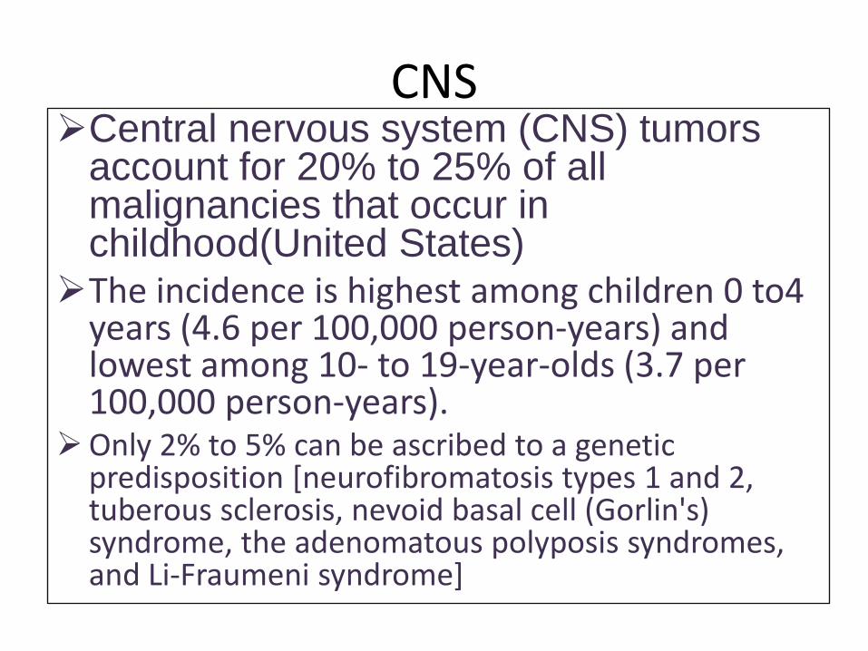
CNSCentral nervous system (CNS) tumors
account for 20% to 25% of all malignancies that occur in childhood(United States)
The incidence is highest among children 0 to4 years (4.6 per 100,000 person-years) and lowest among 10- to 19-year-olds (3.7 per 100,000 person-years).
Only 2% to 5% can be ascribed to a genetic predisposition [neurofibromatosis types 1 and 2, tuberous sclerosis, nevoid basal cell (Gorlin's) syndrome, the adenomatous polyposis syndromes, and Li-Fraumeni syndrome]
CNS
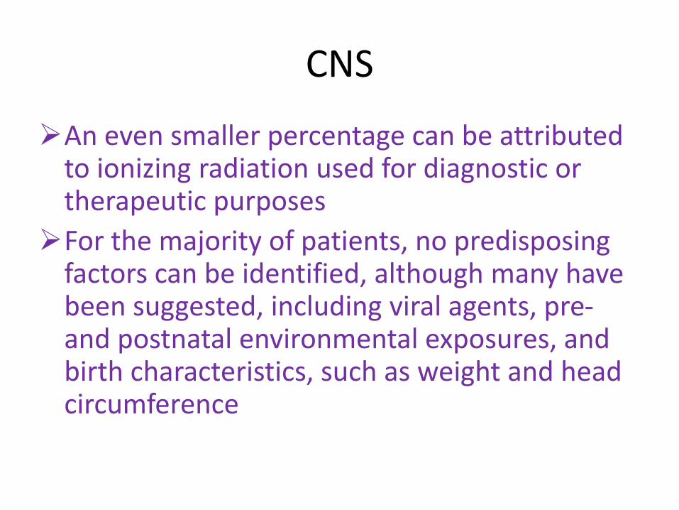
An even smaller percentage can be attributed to ionizing radiation used for diagnostic or therapeutic purposes
For the majority of patients, no predisposing factors can be identified, although many have been suggested, including viral agents, pre-and postnatal environmental exposures, and birth characteristics, such as weight and head circumference
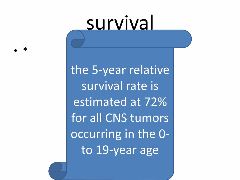
survival• *
the 5-year relative survival rate is
estimated at 72% for all CNS tumors occurring in the 0-
to 19-year age
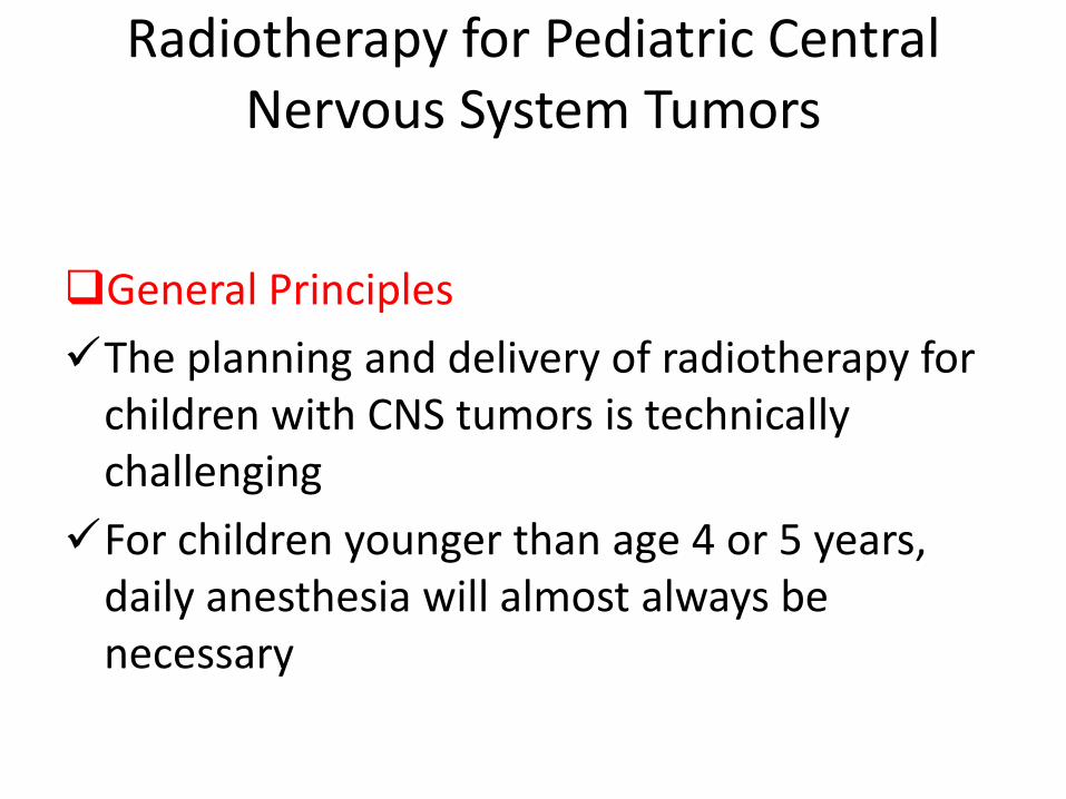
Radiotherapy for Pediatric Central Nervous System Tumors
General Principles
The planning and delivery of radiotherapy for children with CNS tumors is technically challenging
For children younger than age 4 or 5 years, daily anesthesia will almost always be necessary
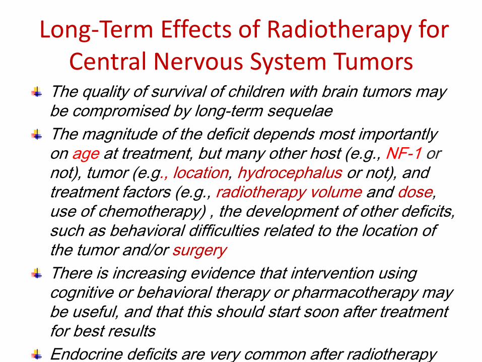
Long-Term Effects of Radiotherapy for Central Nervous System Tumors
The quality of survival of children with brain tumors may be compromised by long-term sequelae
The magnitude of the deficit depends most importantly on age at treatment, but many other host (e.g., NF-1 ornot), tumor (e.g., location, hydrocephalus or not), and treatment factors (e.g., radiotherapy volume and dose, use of chemotherapy) , the development of other deficits, such as behavioral difficulties related to the location of the tumor and/or surgery
There is increasing evidence that intervention using cognitive or behavioral therapy or pharmacotherapy may be useful, and that this should start soon after treatment for best results
Endocrine deficits are very common after radiotherapy
strategies
A number of strategies have been devised in an attempt to minimize the long-term effects of treatment for pediatric brain tumors. These include the following
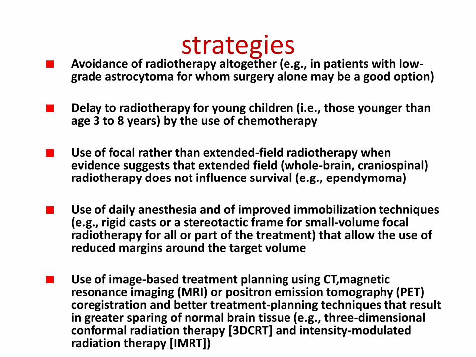
strategiesAvoidance of radiotherapy altogether (e.g., in patients with low-grade astrocytoma for whom surgery alone may be a good option)
Delay to radiotherapy for young children (i.e., those younger than age 3 to 8 years) by the use of chemotherapy
Use of focal rather than extended-field radiotherapy when evidence suggests that extended field (whole-brain, craniospinal) radiotherapy does not influence survival (e.g., ependymoma)
Use of daily anesthesia and of improved immobilization techniques (e.g., rigid casts or a stereotactic frame for small-volume focal radiotherapy for all or part of the treatment) that allow the use of reduced margins around the target volume
Use of image-based treatment planning using CT,magneticresonance imaging (MRI) or positron emission tomography (PET) coregistration and better treatment-planning techniques that result in greater sparing of normal brain tissue (e.g., three-dimensional conformal radiation therapy [3DCRT] and intensity-modulated radiation therapy [IMRT])
strategies
. Use of new radiation modalities (e.g., proton beams that allow even greater sparing of the surrounding normal brain and organs at risk)
Reduction of the dose of radiotherapy (e.g., in patients with standard risk medulloblastoma for whom in the North American studies the dose for craniospinal irradiation has been reduced progressively from 35 to 36 Gy to 23.4 Gy and, in current studies, to 18 Gy)
Use of smaller fraction sizes where appropriate (e.g., a fraction size of 1.5 Gy each day for patients with radiosensitive tumors such as germinoma)
. Use of hyperfractionated radiotherapy (HFRT) (e.g., as in the current European studies for standard risk medulloblastoma)
Radiation Dose and Dose-Fractionation Regimens
The conventional daily fraction size for the treatment of most pediatric CNS tumors is 1.8 Gy
typical dose-fractionation regimen is 54 Gy in 30 daily fractions of 1.8 Gy, which carries a very low risk of radionecrosis
When treating a primary tumor of the spinal cord it is conventional to use a lower total dose (e.g., 50.4 Gy).
It is also usual to reduce the radiotherapy dose for children younger than age 3 years to reduce the risk of neurocognitive deficits
When treating radiosensitive tumors such as intracranial germinoma, radiotherapy may be delivered using a lower dose per fraction (e.g., 1.5 Gy) and lower total doses of 30 to 50 Gy
When the target contains only a small volume of normal brain tissue, dose escalation may be feasible.
HFRT may be a useful strategy in situations where dose escalation cannot be achieved safely using conventional fractionation.
Follow-Up During and After Radiotherapy
These days nausea and vomiting secondary to treatment almost always can be prevented by the use of the 5HT-3 antagonists
Headache is not common in children and should be investigated by physical examination for signs of raised intracranial pressure and by imaging as appropriate
Steroids, if used, usually can be tapered by the third or fourth week of treatment
Fatigue is a rather common symptom and is cumulative The neurologic status of the patient, especially coordination and gait
disorders, may appear to worsen during the last weeks of treatment because of this. Children usually recover relatively quickly, however, and often can get back to their usual routine by 6 weeks to 2 months following completion of
hormonal deficits, especially growth-hormone deficit secondary to inclusion of the hypothalamic–pituitary axis and primary hypothyroidism when craniospinal irradiation (CSI) is delivered using photons
Specific Tumor Types
Specific Tumor Types There are important differences between tumors seen in
childhood and those occurring in adults In children, almost half of all tumors arise in the
infratentorial compartment
Low-grade astrocytic tumors as a group account for approximately one third to half of all CNS tumors, but medulloblastoma is the most common distinct entity
High-grade gliomas, are much less common in children.
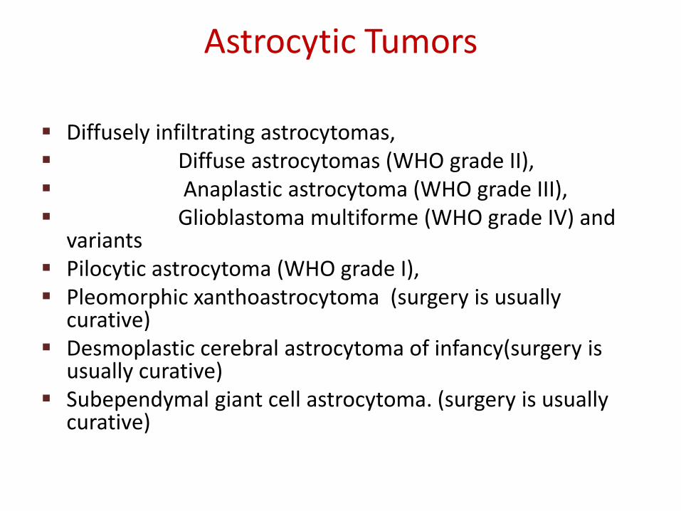
Astrocytic Tumors
Diffusely infiltrating astrocytomas, Diffuse astrocytomas (WHO grade II), Anaplastic astrocytoma (WHO grade III), Glioblastoma multiforme (WHO grade IV) and
variants Pilocytic astrocytoma (WHO grade I), Pleomorphic xanthoastrocytoma (surgery is usually
curative) Desmoplastic cerebral astrocytoma of infancy(surgery is
usually curative) Subependymal giant cell astrocytoma. (surgery is usually
curative)
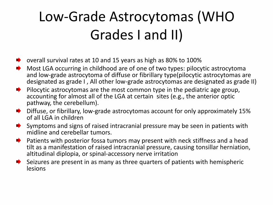
Low-Grade Astrocytomas (WHO Grades I and II)
overall survival rates at 10 and 15 years as high as 80% to 100%Most LGA occurring in childhood are of one of two types: pilocytic astrocytomaand low-grade astrocytoma of diffuse or fibrillary type(pilocytic astrocytomas are designated as grade I , All other low-grade astrocytomas are designated as grade II)Pilocytic astrocytomas are the most common type in the pediatric age group, accounting for almost all of the LGA at certain sites (e.g., the anterior optic pathway, the cerebellum).Diffuse, or fibrillary, low-grade astrocytomas account for only approximately 15% of all LGA in childrenSymptoms and signs of raised intracranial pressure may be seen in patients with midline and cerebellar tumors.Patients with posterior fossa tumors may present with neck stiffness and a head tilt as a manifestation of raised intracranial pressure, causing tonsillar herniation, altitudinal diplopia, or spinal-accessory nerve irritationSeizures are present in as many as three quarters of patients with hemispheric lesions
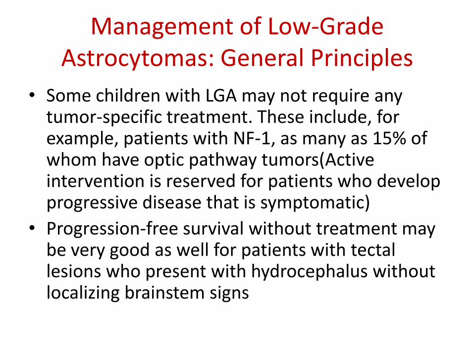
Management of Low-Grade Astrocytomas: General Principles
• Some children with LGA may not require any tumor-specific treatment. These include, for example, patients with NF-1, as many as 15% of whom have optic pathway tumors(Active intervention is reserved for patients who develop progressive disease that is symptomatic)
• Progression-free survival without treatment may be very good as well for patients with tectallesions who present with hydrocephalus without localizing brainstem signs
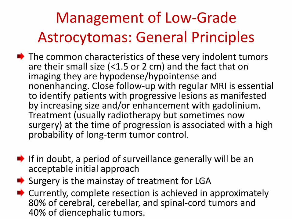
Management of Low-Grade Astrocytomas: General Principles
The common characteristics of these very indolent tumors are their small size (<1.5 or 2 cm) and the fact that on imaging they are hypodense/hypointense and nonenhancing. Close follow-up with regular MRI is essential to identify patients with progressive lesions as manifested by increasing size and/or enhancement with gadolinium. Treatment (usually radiotherapy but sometimes now surgery) at the time of progression is associated with a high probability of long-term tumor control.
If in doubt, a period of surveillance generally will be an acceptable initial approachSurgery is the mainstay of treatment for LGACurrently, complete resection is achieved in approximately 80% of cerebral, cerebellar, and spinal-cord tumors and 40% of diencephalic tumors.
Management of Low-Grade Astrocytomas: General Principles
Children with LGA who undergo complete resection fare very well, with long-term disease-free and overall survival rates of 80% to 100%,postoperative adjuvant therapy is clearly not indicated.For children who undergo less than complete resection, the progression-free survival rate after surgery alone is less satisfactory,patients who had undergone subtotal resection (<1.5 cm residual tumor) were observed without adjuvant treatment, the 5-year progression-free survival rate was only 65%, However, the majority of patients can be salvaged with a second surgical resection and/or radiotherapy, overall survival at 5 years was 95%Consequently, the usual recommendation following subtotal resection will be close follow-up.The role of postoperative radiotherapy following lesser degrees of tumor resection remains unclear. In most series, the use of radiotherapy in this situation results in improved disease-free survival without any benefit in terms of overall survival
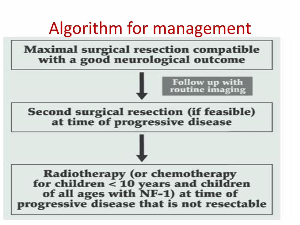
Algorithm for management
Radiotherapy in Low-Grade Astrocytomas
• Indications for Radiotherapy
Radiotherapy is not indicated after complete resection.
Radiotherapy may be indicated following incomplete resection in situations when tumor progression would compromise neurologic function (e.g., “threat to vision―).
The clearest indication for radiotherapy is in patients with progressive and/or symptomatic disease that is unresectable
Radiotherapy Target Volume
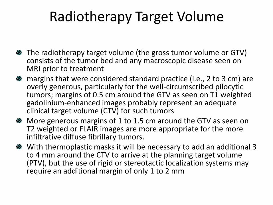
The radiotherapy target volume (the gross tumor volume or GTV) consists of the tumor bed and any macroscopic disease seen on MRI prior to treatmentmargins that were considered standard practice (i.e., 2 to 3 cm) are overly generous, particularly for the well-circumscribed pilocytictumors; margins of 0.5 cm around the GTV as seen on T1 weighted gadolinium-enhanced images probably represent an adequate clinical target volume (CTV) for such tumorsMore generous margins of 1 to 1.5 cm around the GTV as seen on T2 weighted or FLAIR images are more appropriate for the more infiltrative diffuse fibrillary tumors.With thermoplastic masks it will be necessary to add an additional 3 to 4 mm around the CTV to arrive at the planning target volume (PTV), but the use of rigid or stereotactic localization systems may require an additional margin of only 1 to 2 mm

Radiotherapy Dose
• the recommendation for children with LGA would be a “standard―dose of 50 to 54 Gydepending on the age of the child and the location of the tumor and its relationship to critical normal structures, such as the optic chiasm
• Radiotherapy Technique• The radiotherapy technique to be used is that
which provides homogeneous irradiation of the CTV and spares the greatest volume of normal tissue
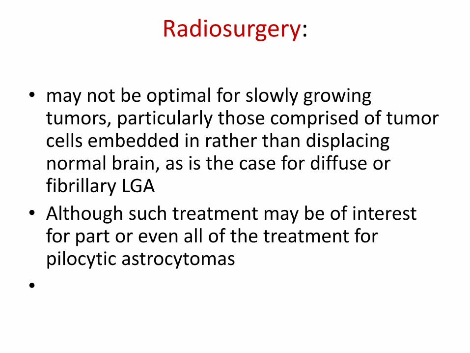
Radiosurgery:
• may not be optimal for slowly growing tumors, particularly those comprised of tumor cells embedded in rather than displacing normal brain, as is the case for diffuse or fibrillary LGA
• Although such treatment may be of interest for part or even all of the treatment for pilocytic astrocytomas
•
Other options:
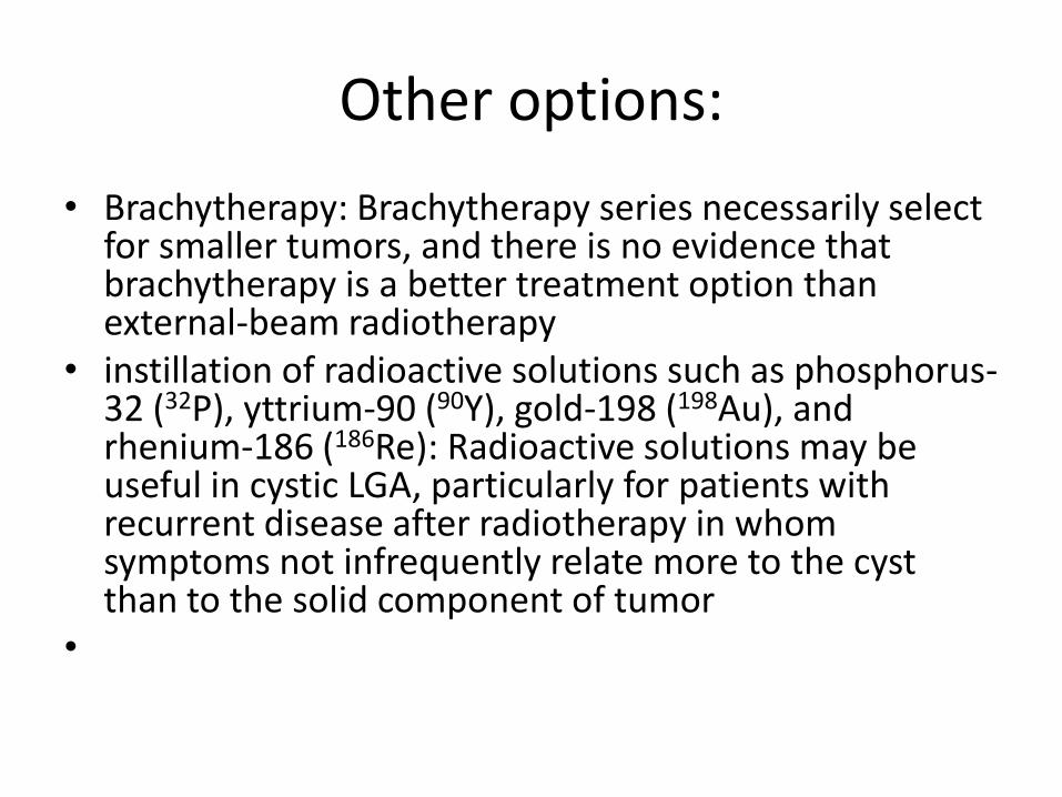
• Brachytherapy: Brachytherapy series necessarily select for smaller tumors, and there is no evidence that brachytherapy is a better treatment option than external-beam radiotherapy
• instillation of radioactive solutions such as phosphorus-32 (32P), yttrium-90 (90Y), gold-198 (198Au), and rhenium-186 (186Re): Radioactive solutions may be useful in cystic LGA, particularly for patients with recurrent disease after radiotherapy in whom symptoms not infrequently relate more to the cyst than to the solid component of tumor
•
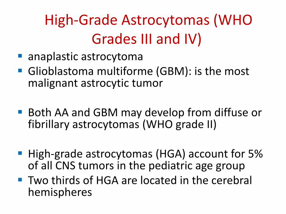
High-Grade Astrocytomas (WHO Grades III and IV)
anaplastic astrocytoma Glioblastoma multiforme (GBM): is the most
malignant astrocytic tumor
Both AA and GBM may develop from diffuse or fibrillary astrocytomas (WHO grade II)
High-grade astrocytomas (HGA) account for 5% of all CNS tumors in the pediatric age group
Two thirds of HGA are located in the cerebral hemispheres
High-Grade Astrocytomas (WHO Grades III and IV)
•
Patients with HGA usually present with symptoms of short duration (<6 months) that relate to the location of the lesion.maximal surgical resection compatible with a good neurologic outcome should be the goal for all patients, regardless of tumor location or histologyA second surgical procedure should be considered if there is significant residual tumor after the firstPostoperative radiotherapy is always indicatedradiotherapy target volume is local with a GTV that consists of the tumor bed and any macroscopic residual disease plus a margin for the CTV of 1.5 to 2 cm
High-Grade Astrocytomas (WHO Grades III and IV)
to consider a field-size reduction at 50 to 54 Gy to a CTV that consists of the tumor bed and any macroscopic residual disease plus a margin of 1 to 1.5 cmThe dose to the CTV should be at least 54 Gy given over 6 weeks, but a dose of 59 to 60 Gy is more usual if feasible.There is no evidence that higher doses, as delivered using radiosurgery or stereotactic boosts, boosts with brachytherapy, or HFRT, result in improved outcomes, but newer approaches such as IMRT, which allows accelerated treatment to a component of the target volume (such as the GTV), may be of interest if only to decrease the overall treatment time.Chemotherapy: The role of chemotherapy remains to be defined
There have been few phase III randomized trials of chemotherapy in HGA inchildren
High-Grade Astrocytomas (WHO Grades III and IV)
Off study it may be difficult to make a recommendation with respect to adjuvant chemotherapy, although the poor prognosis, particularly for patients with macroscopic residual disease following surgery, usually is given as an argument for the use of the “current best―regimenThe prognosis for children with HGA is poorPatients with lesions in the cerebral hemispheres fare better than those with tumors in other locationsThe prognosis for children with bithalamic lesions is particularly poor , and these patients are managed similarly to patients with diffuse intrinsic pontinetumors
High-Grade Astrocytomas (WHO Grades III and IV)
infants with HGA appear to fare somewhat better than older children
istologic grade (AA versus GBM) has not been shown consistently to affect outcome
p53 overexpression and a high MIB-1 labeling index appear to identify patients with a particularly adverse prognosis who might be appropriate candidates for novel therapeutic approaches
•Astrocytic Tumors . in Specific ……locations
Optic Pathway Gliomas
approximately 5% of all CNS tumors in the pediatric age group
the peak age incidence is between 2 and 6 years
One third of patients have NF-1
tumors confined to the optic nerve(s), tumors of the optic chiasm with or without optic nerve involvement , and tumors that involve the hypothalamus or adjacent structuresOptic nerve gliomas may involve one or both optic nerves. Bilateral involvement is pathognomonic of NF-1the optic nerve tumors are incidental findings on routine imaging, and patients may remain asymptomatic with nonprogressive lesions over long periods; even spontaneous regressions are well documented
Optic Pathway Gliomasmanagement of patients with optic nerve tumors will usually consist initially of close follow-up with regular ophthalmologic examinations and MRI, with active intervention, usually chemotherapy, reserved for patients with clear evidence of progression that is symptomaticPatients with unilateral optic nerve involvement may not have NF-1. They present most frequently with proptosis that may be relatively long standingOn MRI, optic nerve tumors are usually relatively small and well circumscribed, with bright enhancement typical for pilocytic astrocytoma. Biopsy is not necessary to make a diagnosisTreatment usually, although not always, will be necessary, and the approach will depend on whether there is useful visionsurgical resection will be the treatment of choiceIf useful vision is preserved, chemotherapy would be the preferred option for infants, very young children up to the age of 3 to 5 years, and for patients of all ages with NF-1Radiotherapy could be considered for older children.Long-term tumor control approaches 100% with either modality(chemotherapy&radiotherapy)
Chiasmatic gliomasChiasmatic gliomas are tumors that involve the optic chiasm and sometimes one or both optic nerves as wellPatients typically present with loss of visual acuity and temporal field defectsBiopsy is usually not necessaryA period of surveillance is appropriate initial management, particularly for patients with NF-1For patients without NF-1, especially those who present before the age of 5, Surgery is rarely an option for tumors in this locationAs for optic nerve tumors, chemotherapy usually will be the treatment of choice for infants and young children and for patients with NF-1Radiotherapy is reserved for salvage postchemotherapy and for definitive treatment of older children without NF-1Overall survival for patients with chiasmatic tumors is in the 90% to 100% range.
Posterior, or chiasmatic/hypothalamic, gliomas
Posterior, or chiasmatic/hypothalamic, gliomas account for approximately 70% of all optic pathway gliomas in childrenThey are typically rather large lesions that probably arise in the optic chiasm and extend to involve the hypothalamusThey often fill the third ventricle, eventually causing hydrocephalusEarly findings consist of nystagmus, impaired visual acuity, and visual field deficits; only later do patients present with increasing head circumference and/or symptoms and signs of raised intracranial pressureTreatment consists of CSF diversion, if necessary, and surgical resection particularly if tumor is growing exophytically into the basal cisternsIn most patients, however, resection will be incompletea period of surveillance is reasonable, although most patients will require adjuvant therapy
Posterior, or chiasmatic/hypothalamic, gliomas
The treatment of choice for children younger than 5 and those with NF-1 usually will be chemotherapy, most often now using a carboplatin-based regimenThe indications for radiotherapy are (a) progressive disease on chemotherapy for children younger than 10 years and (b) progressive disease at diagnosis or after surgery for older patientsgiven to local fields to a dose of 45 to 50 Gy for younger children and of 50 to 54 Gy for those older than 5 yearsOverall, however, the results of treatment are less satisfactory for this group of patients than for those with anterior tumorsPatients with NF-1are also at greater risk for Moyamoya syndrome, a progressive vaso-occlusive process involving the circle of Willis , This may be seen without radiotherapy when radiotherapy is used in patients with NF-1 it is important to include MR angiography as part of the regular follow-up imaging protocol and to intervene surgically if necessary to avoid a cerebrovascular accident
Brainstem Gliomas
Tumors arising in the midbrain, pons, and medulla oblongata account for 10% to 15% of all CNS tumors in the pediatric age groupimprovements in neurosurgical techniques and perioperative care have made surgery not only feasible but the treatment of choice for all except the diffuse intrinsic tumorsFocal tumors may occur at any level in the brainstem but are most frequently seen in the midbrain and medullaSigns and symptoms of raised intracranial pressure are uncommon except in patients with tumors arising in the tectal region that may cause aqueduct stenosis while still small.
Brainstem Gliomas
patients with nonenhancing focal tumors in the tectalregion who present with only hydrocephalus may do well without any treatment other than CSF diversionary procedures , Active intervention, including biopsy, is reserved for patients with clinical and radiologic evidence of progressive tumorSurgery is the treatment of choice for focal tumors at other locations that are surgically accessible (meaning either that they extend toward the surface of the brainstem laterally or at the floor of the fourth ventricle) and have imaging characteristics suggestive of low-grade histology
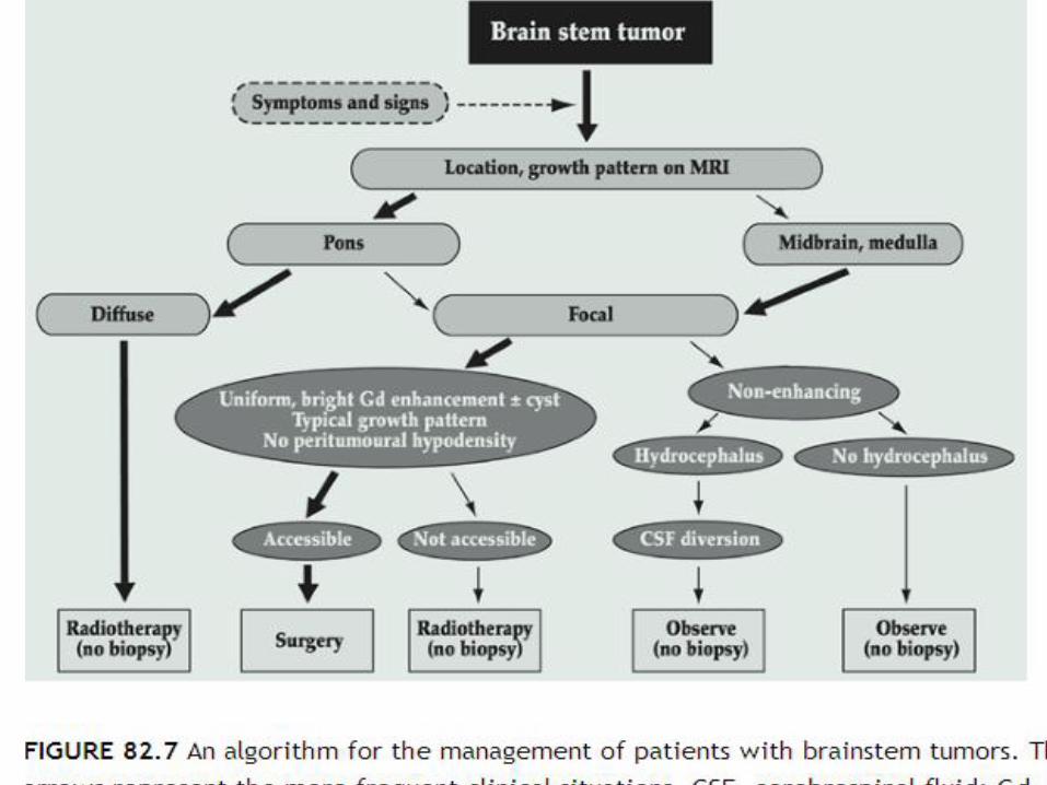
.
ith bulky tumors and for patients with tumors in the medulla with lower cranial nerve deficits who are at high risk of developing postoperative complications.There are several treatment options for patients with surgically inaccessible focal lesions(conventional radiotherapy, HFRT and with interstitial irradiation using iodine-125, radiosurgery and stereotactic irradiation)with the advent of improved treatment planning and conformal radiotherapy treatment techniques, standard treatment for these lesions should be considered to be external-beam radiotherapy, using limited treatment volumes with a margin for the CTV of 0.5 cm, to a total dose of the order of 54 Gy given over 6 weeks, as for LGA at other locations

Dorsal exophytic tumors
Dorsal exophytic tumors arise from the floor of the fourth ventricle
They are usually large, filling the fourth ventricle, but do not invade the brainstem to any significant extentSurgery is the treatment of choice for dorsal exophytic tumorsUltrasound guidance is essential to achieve a maximal degree of tumor resectio
the majority of children do well following surgery, and routine postoperative adjuvant therapy is not indicatedRadiotherapy should be considered for the rare patient who is found to have a high-grade lesion or for patients with low-grade tumors who are found to have progressive disease in the early (0 to 9 months) postoperative periodFor patients whose tumors recur later, further surgery should be considered and radiotherapy should be reserved for those with inoperable diseaseThe radiotherapy volume and dose should be similar to those used for LGA in other locations.the prognosis for patients wit
Cervicomedullary tumors
Cervicomedullary tumors arise in the upper cervical cord and grow rostrally beyond the foramen magnum.
Most are low-grade lesions
Surgery is the treatment of choice
no indication for routine postop
Diffuse intrinsic brainstem tumors
Diffuse intrinsic brainstem tumors account for 70% to 80% of all brainstem tumorsThey arise in the pons and cause diffuse enlargement of the brainstemIn contrast to the other types of brainstem tumors, the majority of diffuse intrinsic tumors are fibrillary astrocytomas with a propensity for malignant change and a very poor prognosis.Diffuse intrinsic tumors typically present with a short duration of symptoms (median 1 month) consisting of multiple, bilateral, cranial nerve deficits (especially VI and VII) as well as long tract signs and ataxiaOnly 10% of patients have hydrocephalus at diagnosisSurgery has no role in the management of patients with diffuse intrinsic lesions
Diffuse intrinsic brainstem tumors
Even biopsy, a relatively nonmorbid procedure now, no longer is considered necessary because in the context of a typical clinical presentation, the MRI findings are characteristic and histology does not influence treatment
Treated with conventional radiotherapy, the majority (75% or more) of patients with diffuse intrinsic tumors will improve clinically
the progression-free interval is short (median <6 months) and survival is poor, with median survival <1 year and survival rates at 2 years <20%.
Alternative approaches that use chemotherapy in combination with radiotherapy also have been disappointing
New agents and novel chemotherapy–radiotherapy combinations are under investigation by the pediatric cooperative groups in North America and Europe
standard treatment for diffuse intrinsic brainstem tumors consists of radiotherapy given to the GTV as demonstrated by T2-weighted or FLAIR MRI with a margin for the CTV of 1 to 1.5 cm to a dose of 54 Gy given in 30 daily fractions over 6 weeks
Low-Grade Astrocytomas of the Spinal Cord
Intramedullary spinal cord tumors account for 3% to 6% of all CNS tumors in the pediatric age groupAbout 60% are astrocytomas, the majority of which are of low histologicgradePatients typically present with symptoms of long duration, usually pain and motor deficitsResection is more difficult and less likely to be complete in patients with grade II astrocytoma because of the more infiltrative nature of these tumors and the absence of a plane of cleavage . Since outcome following complete or subtotal resection for LGA is very good, with long-term progression-free survival in the 70% to 90% range , routine postoperative adjuvant therapy is not indicatedFor children in whom complete or subtotal resection is not possible, options will be as for patients with LGA in other locations, that is, early second surgery or close follow-up with second surgery and/or radiotherapy at the time of progression
Low-Grade Astrocytomas of the Spinal Cord
As for LGA in other locations, chemotherapy may be an alternative to radiotherapy for infants and very young children in this situationImmediate treatment would be indicated for patients with a high-grade astrocytomaThe radiotherapy target volume for LGA usually will consist only of the solid portion of the tumor (including intratumoral cysts) with a margin for the CTV of 0.5 to 1 cm around the GTVThe usual dose would be 50.4 Gy given in 28 daily fractions over approximately 6 weekspatients with high-grade lesions, most of whom will have had only a biopsy; a margin beyond the entire lesion of 1.5 cm would be more appropriate , The dose prescription usually will be as for low-grade lesions because of a substantial risk of morbidity at higher doses, but often chemotherapy will be given as wel

Ependymal Tumors
Ependymomas account for 5% to 10% of all brain tumors in the pediatric age groupwith a predilection for infants and children younger than age 5 yearstwo-thirds arise in the ependymal lining of the fourth ventriclegadolinium-enhanced MRI of the whole CNS and CSF cytology are essential components of the work-up for all patients, the risk of leptomeningeal seeding at diagnosis is at most 5% to 10%.The completeness of the surgical resection is the factor that has the greatest impact on the outcome of children with ependymoma, regardless of tumor locationcomplete resection is possible in 70% to 85% of supratentorialependymomas and in a similar percentage of spinal ependymomascomplete resection is lower in patients with infratentorialependymo
Ependymal TumorsSince residual disease is associated with a much worse outcome, “second-look―surgery should be considered, if feasible, either after the realignment of structures that takes place following resection of an initially bulky tumor or after chemotherapyPostoperative radiotherapy is the standard of care for all children with ependymomaRadiotherapy for patients who have undergone complete resection: such a strategy results in worse disease-free and perhaps also overall survival and therefore can be considered acceptable only for (a) patients with ependymoma of the spinal cord who have undergone complete resection for whom disease-free survival in contemporary series approaches 100% and (b) selected patients with supratentorial ependymoma, such as those with intraventricular tumors or with extraventriculartumors that are solid and located in noneloquent areas and can be resectedwith a wider margin
Ependymal Tumors
optimal radiotherapy target volume, dose, and dose-fractionation schedule: using a GTV that is a composite of the tumor bed, based on preoperative imaging , and including any macroscopic residual disease, with a margin for the CTV of 1 cm the current standard is a dose of at least 54 to 55 Gyfor lesions in the brainbecause failure most often occurs at the site of macroscopic residual disease, even higher doses may be desirable , HFRT has been explored in ependymomafor this reason
Ependymal Tumors
•
The role of chemotherapy in ependymoma remains to be defined , Response rates to chemotherapy are quite poorIn infants and very young children chemotherapy has been used because of the desire to delay or even avoid altogether radiotherapylocal treatment consisting of the tumor bed and any macroscopic residual disease with a margin for the CTV of 1.5 cm is used for patients with localized disease; only patients with leptomeningealseeding at diagnosis receive CSIAs for ependymomas, the dose should be 54 to 55 Gy, with a boost to macroscopic residual disease, if feasible, to 59 to 60Patients with leptomeningeal dissemination at diagnosis fare extremely poorly despite aggressive treatment with craniospinalradiotherapy and systemic chemotherapy.
Myxopapillary Ependymoma
Myxopapillary ependymomas are slowly growing lesions almost always located in the conus-filum terminale region of the spinal cordThey are the most common spinal cord tumor in this locationSurgical resection is the treatment of choiceIf the tumor is not resected en bloc or if there is macroscopic residual tumor, postoperative radiotherapy should be used since outcome appears to be significantly worse in these situationsThe radiotherapy target volume is local (macroscopic disease plus a margin cephalad and caudad of 1.5 cm for the CTV) and the dose, 50.4 GyLeptomeningeal seeding: patients should be treated with curative intent with CSI followed by a boost to the primary site
Choroid Plexus Tumors
The WHO classification lists two different entities: choroid plexus papilloma (WHO grade I) and choroid plexus carcinoma (WHO grade III).
Surgery is the treatment of choice for choroid plexus papillomas both for the primary lesion and for macroscopic metastatic deposits, if feasible
Blood loss may be considerable
The role of radiotherapy following incomplete resection is unclear(choroid plexus papillomas) , close follow-up would seem
Ganglioglioma and AnaplasticGanglioglioma
Gangliogliomas are well-differentiated, slowly growing tumors composed of mature ganglion cells in combination with neoplastic glial cells WHO grade I or II.Although these tumors can arise anywhere within the CNS, most in children arise in the temporal region and typically present with seizures.Surgery is the treatment of choiceThe indications for radiotherapy would be as for patients with LGA, that is, for patients with progressive or recurrent disease that is not resectablepostoperative radiotherapy: for patients with anaplastic gangliogliomas who have undergone less than complete resection
Pineal Parenchymal Tumors
Pineal region tumors account for 2% to 8% of intracranial tumors in children.
Approximately half are germ-cell tumors, one-fourth to one-third are pineal parenchymaltumors, and most of the remainder are astrocytic tumors.
WHO classification: pineoblastoma (WHO grade IV), pineocytoma (WHO grade II), and pineal parenchymal tumor of intermediate differentiation.
Pineoblastoma
Pineoblastoma is a highly malignant primitive embryonal tumor
Leptomeningeal spread is seen in as many as 50% of patients at diagnosis
Surgery for lesions in the pineal region is particularly delicate, and complete resection is often not possible
Postoperatively, older children are treated with CSI and chemotherapy, as for medulloblastomaand supratentorial primitive neuroectodermaltumor (S-PNETs)
infants who are treated with chemotherapy without radiotherapy
PineocytomaPineocytoma is a slow-growing tumorThey account for approximately half of pineal
parenchymal tumors and in childhood most commonly occur in the teenage years
Some will have symptoms of upper mesencephalictegmental dysfunction (Parinaud's syndrome)
Treatment consists of surgical resection If complete or subtotal resection is accomplished, the
outcome is probably quite favorable even without any adjuvant treatment
Following lesser degrees of resection, postoperative radiotherapy usually is recommended using local fields consisting of macroscopic residual disease, with a margin of 1 to 1.5 cm and a dose of 50 to 55 Gy over 6 weeks

Embryonal Tumors
Embryonal tumors as a group are the second most common type of CNS tumor in the pediatric age group
Ependymoblastoma
Medulloblastoma,
Supratentorial PNET.
Medulloepithelioma,
Atypical teratoid/rhabdoid tumor
Medulloblastoma
Medulloblastoma accounts for 15% to 20% of all CNS tumors in the pediatric age groupThe median age at presentation is 6 yearsIn the majority of cases the tumor arises in the cerebellarvermis and projects into the fourth ventriclePatients typically present with symptoms and signs of raised intracranial pressure, that is, headache and morning vomitingThe frequency of spinal seeding at diagnosis is approximately 30% to 35%, and investigation at diagnosis must include a gadolinium-enhanced MRI of the spinal axis and CSF cytology.
Medulloblastoma
The former should be obtained whenever possible preoperatively or else at least 2 to 3 weeks postoperatively to avoid misinterpretation resulting from artifactual changes seen in the later postoperative period
CSF cytology: which should be obtained by lumbar puncture, often cannot be obtained preoperatively because of the presence of raised intracranial pressure and more commonly is obtained at approximately 3 weeks postoperatively
Medulloblastoma is one of the few CNS tumors to spread outside the CNS (to lymph nodes, bone), although this is an uncommon event.
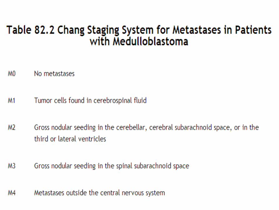
risk categories
Patients are allocated to one of two risk categories: standard and high risk
Standard risk: Those who have undergone complete or subtotal resection with <1.5 cm2 of residual tumor and no evidence of CSF dissemination (M0)
complete or near-total resection is accomplished in approximately 80% of cases
two thirds of patients will be Standard risk and one third will be high risk.
Management of Standard-Risk Medulloblastoma
Several treatment strategies designed to reduce the morbidity associated with the use of radiotherapy have been tested
Until relatively recently, the standard of care for patients older than 3 years with standard-risk disease consisted of postoperative radiotherapy to the craniospinal axis to a dose of 35 to 36 Gyfollowed by a boost to the whole posterior fossa to a total dose of 54 to 55.8 Gyreduce the radiotherapy target volume to avoid supratentorialradiation produced disastrous resultsHFRT to a CSI dose of 36 Gy without chemotherapy
reduced-dose CSI (23.4 Gy) followed by a boost to the posterior fossa to a total dose of 55.8 Gy in combination with systemic chemotherapy
Management of Standard-Risk Medulloblastoma
reduced-dose (23.4 Gy) CSI in combination with weekly vincristine, followed by adjuvant systemic chemotherapy consisting of vincristine 1.5 mg/m2, CCNU 75 mg/m2, and cisplatin 75 mg/m2
CCNU was replaced by cyclophosphamide such an approach is now considered to be the standard
of care for children with standard-risk medulloblastoma in North America.
Current studies are testing the safety of an even lower dose of CSI (18 Gy) in children aged 3 to 8 years and of a reduced-volume posterior fossa boost in children of all ages
Management of High-Risk Medulloblastoma
for patients with residual disease, M0, it would be logical to consider using a radiotherapy dose to residual disease in the posterior fossa higher than the standard 55.8 Gy, and this may be feasible using stereotactic radiotherapy or 3D conformal techniquePatients with M1 disease may do well using a standard CSI dose of 35 to 36 Gy;results for patients with M2 , 3 disease remain quite poor, although the use of a higher radiotherapy dose (40 Gy CSI plus a boost of 5 Gy to macroscopic disease) produced excellent early results in POG 9031, approaching those for patients with less advanced disease.The hyperfractionated accelerated radiotherapy (HART) regimen under investigation by the U.K. group may prove to be an equally efficacious and safer way to deliver high dose CSI.Preradiotherapy chemotherapy may also be of interest in this group of patientspreradiotherapy chemotherapy may allow identification of patients who do not respond and who might benefit from more aggressive chemotherapy, such as high-dose chemotherapy with stem cell rescue in combination with conventional radiotherapy, HFRT, or HART
Management of Medulloblastoma in Infants
Medulloblastoma accounts for 20% to 40% of all CNS tumors in infants and carries a worse prognosis than in older childrenMedulloblastoma accounts for 20% to 40% of all CNS tumors in infants and carries a worse prognosis than in older childrenInfants with M0 disease who have undergone total resection may do well with chemotherapy aloneInfants with M2â,“3 disease fare poorly and require more aggressive treatment such as high-dose chemotherapy with stem cell rescue and/or chemotherapy given using alternative routes of administration, such as intrathecal or intraventricular, with or without radiotherapy.
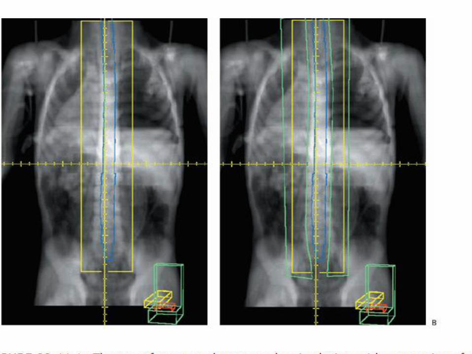
Radiotherapy for Medulloblastoma The CTV for CSI has an irregular shape that consists of the whole of the
brain and spinal cord and overlying meninges Patients have traditionally received CSI in the prone position, but modern
technology allows safe treatment in the supine position that in general is more comfortable
CT simulation is invaluable for target volume definition for patients with medulloblastoma
With current cataract surgical techniques, it is preferable to cover the target volume, keeping shielding of the lenses a secondary objective
MRI is required to determine the lower limit of CTV for the spine field
Traditionally the lower border of the spine field was placed at the lower border of the second sacral vertebra, but it is well documented that the lower border of the thecal sac can be as high as L5 or as low as S3
In the interest of both CTV coverage and normal tissue sparing it is important that the lower border be individualized according to the MRI findings.
.
Type & dose of energy
In general, photons in the 6 to 10 MV range provide satisfactory coverage of the PTV
Care is required if using electrons to treat the spinal axis
The use of electrons to treat the spinal axis most often is justified by reduced acute gastrointestinal toxicity
Because available data regarding late effects suggest that there may be little or no advantage to the use of electrons
Traditionally, the entire posterior fossa has been treated to a total dose of 54 to 55.8 Gy

.

Radiotherapy for Medulloblastoma
• The optimal CTV for a reduced-volume posterior fossa remains to be defined, although it probably consists of a composite of any macroscopic residual tumor and the surgical bed plus a margin of 1 to 1.5 cm
• A few centers have reported their results using protons for treatment of medulloblastoma
• Delay to radiotherapy may be associated with poorer outcomes and CSI ideally should start within 28 days following surgery
Supratentorial Primitive Neuroectodermal Tumor
S-PNETs account for <5% of all CNS tumors in the pediatric age group. The median age at presentation is 3 yearsLeptomeningeal seeding is present at diagnosis in up to 40% of patients, and MRI of the spinal axis and CSF cytology are mandatory prior to treatment.often using the same protocols as for high-risk medulloblastomaoften using the same protocols as for high-risk medulloblastomaFor now, the standard of care for children older than 3 years with S-PNETs without leptomeningeal spread consists of maximal surgical resection followed by postoperative radiotherapy (CSI plus a boost to doses similar to those used for high-risk medulloblastoma) followed by chemotherapyExperimental regimens such as preradiotherapy chemotherapy and high-dose chemotherapy with rescue are used in infants and young children and in patients with M2&3 disease
Atypical Teratoid/Rhabdoid Tumor
highly malignant embryonal tumor unique to childhood
outcome was very poor and the majority of patients died within 1 year of diagnosis
Radiotherapy is an important component of treatment
radiotherapy should be delivered early even in infants, and this may mean using a modified radiotherapy target volume (i.e., tumor bed and any macroscopic residual disease plus a margin
Children older than 3 years should receive early CSI.
Germ Cell Tumors
In the West, CNS germ cell tumors are relatively rare, accounting for 3% to 5% of all CNS tumors in the pediatric age group. They are more common in AsiaThe peak age incidence is 10 to 12 years. Boys are affected more frequently than girls, with a ratio of approximately 3:1CNS germ cell tumors arise from primordial germ cells in structures about the third ventricle, with the region of the pineal gland being the most common site of origin, followed by the suprasellar regionNongerminomatous germ cell tumors (NGGCT) are the most common tumor type in the former area, and germinomas in the latter.Tumors in the pineal region cause obstruction to CSF flow at the aqueduct of Sylvius,
Germ Cell Tumors
Another characteristic presentation of tumors in this region is Parinaud's syndrome as a result of dorsal midbrain compressionMeasurement of serum and CSF tumor markers is another essential part of the initial work-upIn the past, many lesions arising in or about the third ventricle were treated without histologic confirmation of diagnosisall patients should undergo biopsy unless CSF and/or serum markers confirm the presence of a NGGCT (elevated AFP and/or B-hCG >100 IU/mL) or unless a histologic diagnosis is made by other means (e.g., CSF cytology)Occasionally complete resection will be possible; this would be a reasonable strategy for patients with NGGCT, particularly for mature teratomas if it can be accomplished without major morbidity because in that situation it would obviate the need for adjuvant treatm
Germinoma In the past, standard treatment for patients with germinoma was
radiotherapy alone Whole ventricle irradiation alone may be acceptable for a patient with a
unifocal lesion with no evidence of leptomeningeal dissemination a number of reasonable options for patients with unifocal germinoma
without evidence of leptomeningeal dissemination. These include craniospinal radiotherapy, limited volume (whole-ventricle) radiotherapy alone, and chemotherapy followed by whole ventricle or local radiotherapy
regardless of the target volume so that patients at increased risk for systemic failure (e.g., those with ventriculoperitoneal shunts) may be best treated with a combined chemotherapy&radiotherapy approach
For patients with bi- or multifocal disease by imaging or by inference (for example, in a patient with a pineal region primary who has diabetes insipidus) and those with leptomeningeal spread at diagnosis, radiotherapy alone using CSI with boosts to macroscopic disease is certainly an option,
although in North America a combination chemotherapy and radiotherapy regimen consisting of two cycles of chemotherapy followed by CSI followed by a boost to all sites of involvement would be the more usual approach.
Germinoma•
Because subependymal spread is common, such treatment logically would include the lateral, third, and fourth ventricles with a margin of 1 to 1.5 cmBetter sparing can be achieved using CT planning and a four-field arrangementBetter sparing can be achieved using CT planning and a four-field arrangementResults are excellent with a CSI dose as low as 21 Gy even in patients with leptomeningeal spread.The total dose to the primary site has typically been 40 to 45 Gy but probably can be reduced to 30 Gy in patients who have undergone complete resection or those who have been treated with a combined chemotherapy and adiotherapy regimen with a complete response to chemotherapyThe total dose to the primary site has typically been 40 to 45 Gy but probably can be reduced to 30 Gy in patients who have undergone complete resection or those who have been treated with a combined chemotherapy–radiotherapy regimen with a complete response to chemotherapy
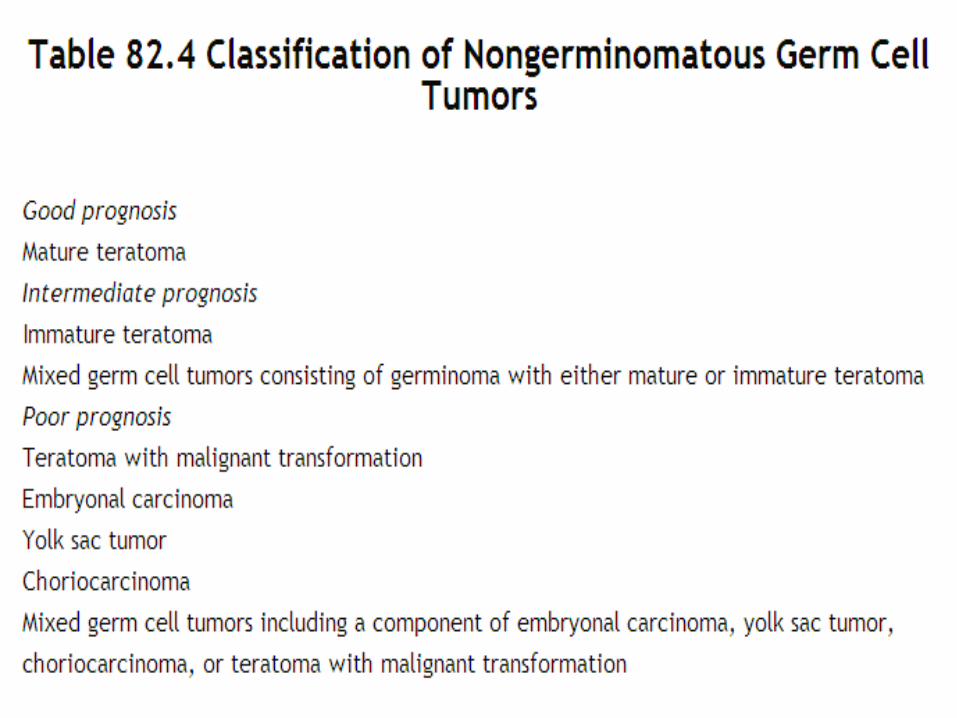
Nongerminomatous Germ Cell Tumors
• For NGGCTs the diagnosis can be made in as many as one third of all patients on the basis of imaging findings (location, appearance) plus tumor markers.
.
teratomas
Patients with mature teratomas without any associated malignant elements can be managed with surgery alone, while those with mature teratoma with germinomatous elements will be treated as germinomas
the current standard of care therefore consists of platinum-based chemotherapy followed by radiotherapy
There is controversy with respect to the radiotherapy target volume for NGGCT
NGGCT
In the current North American study, CSI is used for all patients with NGGCT. A dose of 36 Gy is used, followed by a boost to the primary site to a total dose of 54 GyPatients who have less than a complete response to chemotherapy: Second-look surgery may be useful to both exclude the possibility that the residual imaging abnormality represents mature teratoma and/or resectresidual viable tumor, If the latter, this would be followed by CSI and then more aggressive chemotherapy, such as high-dose chemotherapy with stem cell rescue
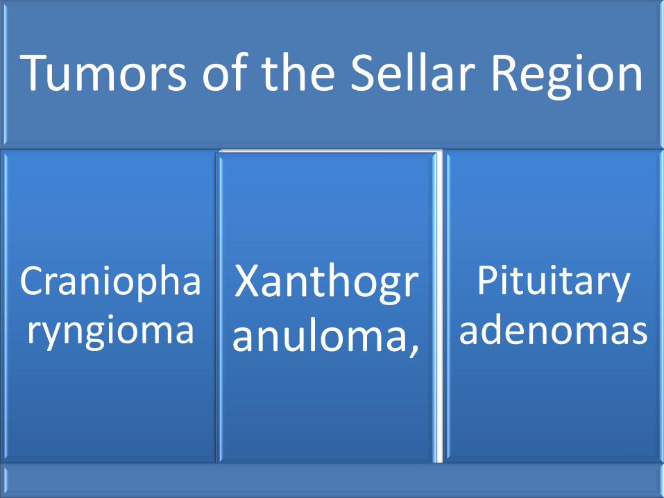
.Tumors of the Sellar Region
Craniopharyngioma
Xanthogranuloma,
Pituitary adenomas
Pituitary adenomas• craniopharyngiomas are benign partly cystic epithelial
tumors that arise in the sellar region from remnants of Rathke's pouch
• with a peak incidence between the ages of 5 and 14 years.
• In the majority of patients, craniopharyngiomas have both suprasellar and intrasellar components
• Children typically present with neuroendocrine deficits, especially diabetes insipidus and growth failure
• Visual-field deficits often go unnoticed initially• patients with tumors that are smaller and/or
subdiaphragmatic in location and without hypothalamic symptoms would be managed surgically, while other patients at higher risk for complications secondary to surgery would be managed with biopsy, cyst decompression, if necessary, and radiotherapy

Pituitary adenomas
A lesion with a small solid component and a simple cyst, for example, may be treated with intracavitary injection of liquid radioactive materialA lesion with a small solid component and a simple cyst, for example, may be treated with intracavitary injection of liquid radioactive materialconventionally fractionated external-beam irradiation using modern techniques may be a better option with a lower risk of morbidity for all except very small tumorsExternal-beam radiotherapy also will be the treatment of choice for patients with residual disease following surgery who are at high risk for progressive disease even relatively early following surgery or for those in whom surgical resection is not feasible.
Pituitary adenomas
o A margin of 0.5 cm seems reasonable using contemporary planning and delivery techniques.
o A dose of 54 to 55 Gy given in 30 daily fractions over 6 weeks appears to be necessary to achieve a high
o A margin of 0.5 cm seems reasonable using contemporary planning and delivery techniques. A dose of 54 to 55 Gy given in 30 daily fractions over 6 weeks appears to be necessary to achieve a high, usually consisting of cyst decompression, is essential
o When complete resection is not feasible, biopsy, subtotal resection, or simply cyst decompression followed by external-beam radiotherapy is a better choice
o There is equal if not better probability of tumor control in this situation with a lower risk of potentially devastating sequelae than with “heroic―surgery, particularly in less-experienced hands.
Pituitary Adenomas
Pituitary adenomas are rare in childhoodMost are functioning adenomas that present with endocrine dysfunction, most often menstrual irregularities and galactorrhea in girls and delayed puberty in boys
Transphenoidal surgery appears to be feasible and safe in children, even in those with poor pneumatization of the sphenoid sinus, using neuronavigational tools
Radiotherapy is indicated if surgical resection is not possible or if hormone levels remain elevated following surgery
Conformal fields with a margin of 0.5 cm beyond macroscopic residual disease (the GTV) are used
The usual dose, as in the adult population, will be 45 to 50 Gy over 5 to 6 weeks
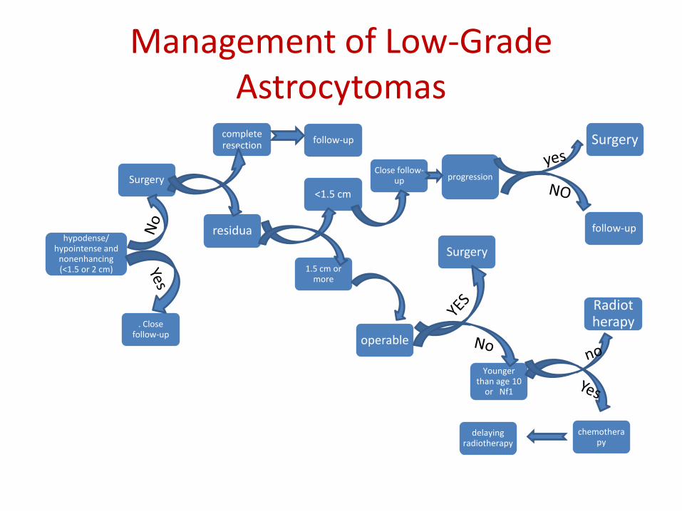
Management of Low-Grade Astrocytomas
complete resection
residua
<1.5 cm
follow-up
Close follow-up Surgery
delaying radiotherapy
1.5 cm or more
Surgery
progression
hypodense/ hypointense and
nonenhancing(<1.5 or 2 cm)
follow-up
operable
Radiotherapy
Younger than age 10
or Nf1
. Close follow-up
Surgery
chemotherapy


![Review Article Cancer: An Overview · body via the bloodstream and lymphatic systems and can affect the digestive, nervous and circulatory systems [1]. TYPES OF TUMORS: Tumors (lumps)](https://static.fdocuments.net/doc/165x107/5f0ea63d7e708231d44041dd/review-article-cancer-an-overview-body-via-the-bloodstream-and-lymphatic-systems.jpg)


