
Cellulitis 110219224340-phpapp01
97
SEMINAR ON SEMINAR ON SPREAD OF ORAL SPREAD OF ORAL INFECTIONS INFECTIONS PRESENTED BY :- PRESENTED BY :- DR. IPSHA DHALI DR. IPSHA DHALI
-
Upload
pradyumnakhairnar -
Category
Education
-
view
98 -
download
2
Transcript of Cellulitis 110219224340-phpapp01

SEMINAR ON SEMINAR ON SPREAD OF ORAL SPREAD OF ORAL
INFECTIONSINFECTIONS
PRESENTED BY :-PRESENTED BY :-DR. IPSHA DHALIDR. IPSHA DHALI
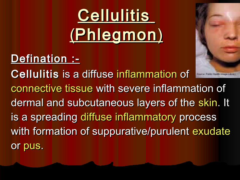
Cellulit isCellul it is ((PhlegmonPhlegmon ))
Defination :-Defination :-Cellulit isCellulit is is a diffuse is a diffuse inflammationinflammation of of connective tissueconnective tissue with severe inflammation of with severe inflammation of dermal and subcutaneous layers of the dermal and subcutaneous layers of the skinskin. It . It is a spreading is a spreading diffusediffuse inflammatoryinflammatory process process with formation of suppurative/purulent with formation of suppurative/purulent exudateexudate or or puspus. .
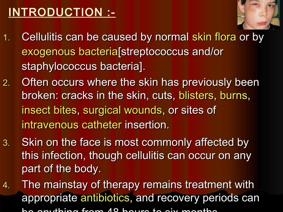
1.1. Cellulitis can be caused by normal Cellulitis can be caused by normal skin floraskin flora or by or by exogenous bacteriaexogenous bacteria[[streptococcus and/or streptococcus and/or staphylococcus bacteria]staphylococcus bacteria]..
2.2. Often occurs where the skin has previously been Often occurs where the skin has previously been broken: cracks in the skin, cuts, broken: cracks in the skin, cuts, blistersblisters, , burnsburns, , insect bitesinsect bites, , surgical woundssurgical wounds, or sites of , or sites of intravenousintravenous cathetercatheter insertion. insertion.
3.3. Skin on the face is most commonly affected by Skin on the face is most commonly affected by this infection, though cellulitis can occur on any this infection, though cellulitis can occur on any part of the body. part of the body.
4.4. The mainstay of therapy remains treatment with The mainstay of therapy remains treatment with appropriate appropriate antibioticsantibiotics, and recovery periods can , and recovery periods can be anything from 48 hours to six months.be anything from 48 hours to six months.
INTRODUCTION :-
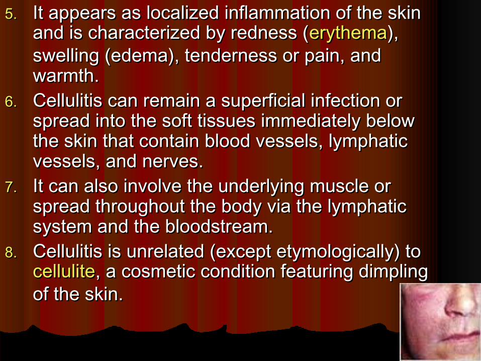
5.5. It appears as localized inflammation of the skin It appears as localized inflammation of the skin and is characterized by redness (and is characterized by redness (erythemaerythema), ), swelling (edema), tenderness or pain, and swelling (edema), tenderness or pain, and warmth. warmth.
6.6. Cellulitis can remain a superficial infection or Cellulitis can remain a superficial infection or spread into the soft tissues immediately below spread into the soft tissues immediately below the skin that contain blood vessels, lymphatic the skin that contain blood vessels, lymphatic vessels, and nerves. vessels, and nerves.
7.7. It can also involve the underlying muscle or It can also involve the underlying muscle or spread throughout the body via the lymphatic spread throughout the body via the lymphatic system and the bloodstream. system and the bloodstream.
8.8. Cellulitis is unrelated (except etymologically) to Cellulitis is unrelated (except etymologically) to cellulitecellulite, a cosmetic condition featuring dimpling , a cosmetic condition featuring dimpling of the skin.of the skin.
NOTE :NOTE : Risk: Risk: 1.1. Age is not generally considered a risk factor for cellulites, although Age is not generally considered a risk factor for cellulites, although
some studies show slightly higher incidence in individuals over age some studies show slightly higher incidence in individuals over age 45.45.
2.2. Individuals who are immunodeficient as a result of genetic Individuals who are immunodeficient as a result of genetic conditions , illness (e.g., conditions , illness (e.g., HIVHIV infection, infection, cancercancer), or ), or immunosuppressive drugs (e.g., immunosuppressive drugs (e.g., chemotherapychemotherapy, corticosteroids, , corticosteroids, antirejection drugs in transplant recipients) are at increased risk of antirejection drugs in transplant recipients) are at increased risk of infections such as cellulitis. infections such as cellulitis.
3.3. DiabetesDiabetes impairs the immune system and decreases blood impairs the immune system and decreases blood circulation, increasing risk of infection. circulation, increasing risk of infection.
4.4. Chronic skin conditions such as Chronic skin conditions such as psoriasispsoriasis, , dermatitisdermatitis, or , or eczemaeczema can can create an opportunity for entry of infectious bacteria. Individuals who create an opportunity for entry of infectious bacteria. Individuals who have recurrent fungal infections of the feet are greater risk for have recurrent fungal infections of the feet are greater risk for developing cellulitis. developing cellulitis.
5.5. Impaired peripheral circulation such as arterial insufficiency or Impaired peripheral circulation such as arterial insufficiency or venous stasis is also a risk factor. venous stasis is also a risk factor.
6.6. Subcutaneous or intravenous drug injection, body piercing, and Subcutaneous or intravenous drug injection, body piercing, and tattoos are all associated with higher risk for cellulitis. tattoos are all associated with higher risk for cellulitis.
7.7. Cellulitis may also occur as a complication of certain surgical Cellulitis may also occur as a complication of certain surgical procedures (procedures (hip replacemenhip replacement, t, liposuctionliposuction, , breast surgerybreast surgery, vein , vein surgery). surgery).
Incidence and PrevalenceIncidence and Prevalence
1.1. Cellulitis can affect anyone of any age; cellulitis Cellulitis can affect anyone of any age; cellulitis of the face is more common in children and of the face is more common in children and adults over age 50 (Cunningham). adults over age 50 (Cunningham).
2.2. The actual incidence of cellulitis is unknown The actual incidence of cellulitis is unknown because cases are seldom reported. because cases are seldom reported.
3.3. The incidence of infection by methicillin-The incidence of infection by methicillin-resistant Staphylococcus aureus (MRSA) and resistant Staphylococcus aureus (MRSA) and other antibiotic-resistant bacteria has increased other antibiotic-resistant bacteria has increased dramatically in recent years (Mayo Clinic Staff). dramatically in recent years (Mayo Clinic Staff). Infection by these organisms is much more Infection by these organisms is much more serious. serious.
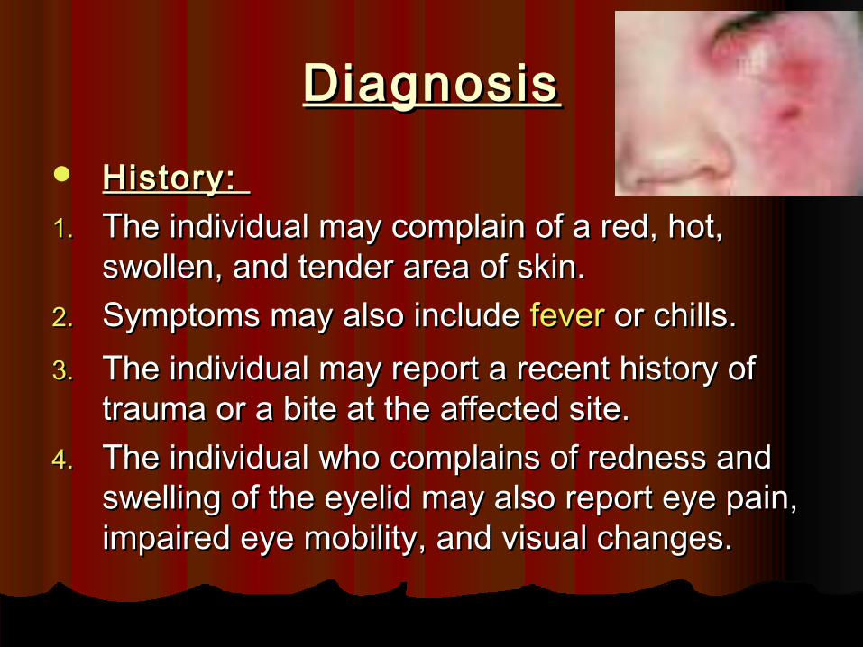
DiagnosisDiagnosis History: History: 1.1. The individual may complain of a red, hot, The individual may complain of a red, hot,
swollen, and tender area of skin. swollen, and tender area of skin. 2.2. Symptoms may also include Symptoms may also include feverfever or chills. or chills. 3.3. The individual may report a recent history of The individual may report a recent history of
trauma or a bite at the affected site. trauma or a bite at the affected site. 4.4. The individual who complains of redness and The individual who complains of redness and
swelling of the eyelid may also report eye pain, swelling of the eyelid may also report eye pain, impaired eye mobility, and visual changes. impaired eye mobility, and visual changes.
Physical exam:Physical exam: 1.1. The appearance of red, swollen, tender skin that is The appearance of red, swollen, tender skin that is
warm to the touch is usually sufficient for warm to the touch is usually sufficient for diagnosis. diagnosis.
2.2. The texture of the skin may resemble orange peel The texture of the skin may resemble orange peel (peau d'orange) and be firm to the touch. Regional (peau d'orange) and be firm to the touch. Regional lymph nodes may be inflamed and swollen. lymph nodes may be inflamed and swollen.
3.3. Adjacent skin may reveal red streaks Adjacent skin may reveal red streaks characteristic of inflamed lymphatic vessels (characteristic of inflamed lymphatic vessels (lymphangitislymphangitis). ).
4.4. If the lower leg is affected, symptoms (warmth, If the lower leg is affected, symptoms (warmth, pain, and swelling) may mimic those of clot pain, and swelling) may mimic those of clot formation in leg veins (venous formation in leg veins (venous thrombosisthrombosis). ).
5.5. Individuals with orbital cellulitis should undergo a Individuals with orbital cellulitis should undergo a thorough examination of the face, sinuses, teeth, thorough examination of the face, sinuses, teeth, mouth, and nasopharynx to identify the source of mouth, and nasopharynx to identify the source of infection. infection.
Tests:Tests: 1.1. A complete blood count may be performed to determine A complete blood count may be performed to determine
the level of white blood cells, a sensitive marker of the level of white blood cells, a sensitive marker of infection. infection.
2.2. Cultures of pus or other drainage from the area of Cultures of pus or other drainage from the area of infection and/or blood cultures may be performed to infection and/or blood cultures may be performed to identify the causative organism(s). identify the causative organism(s).
3.3. Often, the causative agent is not identified, or the report Often, the causative agent is not identified, or the report shows multiple skin organisms that may include normal shows multiple skin organisms that may include normal flora. flora.
4.4. Antibiotic sensitivity tests may be performed on the Antibiotic sensitivity tests may be performed on the cultured organisms to aid in determining the most cultured organisms to aid in determining the most appropriate antibiotic therapy. appropriate antibiotic therapy.
5.5. Individuals with orbital cellulitis may require imaging Individuals with orbital cellulitis may require imaging studies (studies (x-raysx-rays, , CTCT, or , or MRIMRI of the sinuses) to localize the of the sinuses) to localize the source of infection. source of infection.
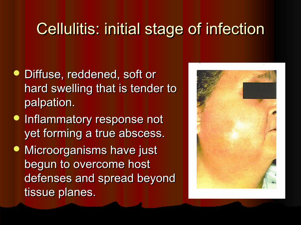
Cellulitis: initial stage of infectionCellulitis: initial stage of infection
Diffuse, reddened, soft or Diffuse, reddened, soft or hard swelling that is tender to hard swelling that is tender to palpation.palpation.
Inflammatory response not Inflammatory response not yet forming a true abscess.yet forming a true abscess.
Microorganisms have just Microorganisms have just begun to overcome host begun to overcome host defenses and spread beyond defenses and spread beyond tissue planes.tissue planes.

True abscess formationTrue abscess formation As inflammatory As inflammatory
response matures, may response matures, may develop a focal develop a focal accumulation of pus.accumulation of pus.
May have spontaneous May have spontaneous drainage intraorally or drainage intraorally or extraorally.extraorally.
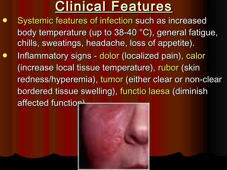
Clinical FeaturesClinical Features Systemic features of infectionSystemic features of infection such as increased such as increased
body temperature (up to 38-40 °C), general fatigue, body temperature (up to 38-40 °C), general fatigue, chills, sweatings, headache, loss of appetite). chills, sweatings, headache, loss of appetite).
Inflammatory signs - Inflammatory signs - dolordolor (localized pain), (localized pain), calorcalor (increase local tissue temperature), (increase local tissue temperature), ruborrubor (skin (skin redness/hyperemia), redness/hyperemia), tumortumor (either clear or non-clear (either clear or non-clear bordered tissue swelling), bordered tissue swelling), functio laesafunctio laesa (diminish (diminish affected function). affected function).
CausesCauses1.1. Cellulitis is caused by a type of Cellulitis is caused by a type of bacteriabacteria entering the skin, usually by way of a cut, abrasion, entering the skin, usually by way of a cut, abrasion,
or break in the skin. This break does not need to be visible. or break in the skin. This break does not need to be visible. Group AGroup A StreptococcusStreptococcus and and StaphylococcusStaphylococcus are the most common of these bacteria, which are part of the normal flora of are the most common of these bacteria, which are part of the normal flora of the skin but cause no actual infection while on the skin's outer surface.the skin but cause no actual infection while on the skin's outer surface.
2.2. Predisposing conditions for cellulitis include insect bites, Predisposing conditions for cellulitis include insect bites, blisteringblistering, animal bite, , animal bite, tattoostattoos, , pruriticpruritic (itchy) skin rash, recent (itchy) skin rash, recent surgerysurgery, , athlete's footathlete's foot, , dry skindry skin, , eczemaeczema, injecting drugs , injecting drugs (especially subcutaneous or intramuscular injection or where an attempted IV injection (especially subcutaneous or intramuscular injection or where an attempted IV injection "misses" or blows the vein), pregnancy, diabetes and obesity, which can affect circulation, as "misses" or blows the vein), pregnancy, diabetes and obesity, which can affect circulation, as well as burns and well as burns and boilsboils, though there is debate as to whether minor foot lesions contribute. , though there is debate as to whether minor foot lesions contribute. Spider bitesSpider bites are also known to cause cellulitis. are also known to cause cellulitis.
3.3. Occurrences of cellulitis may also be associated with the rare condition Occurrences of cellulitis may also be associated with the rare condition Hidradenitis SuppurativaHidradenitis Suppurativa..
4.4. The photos shown here of Cellulitis are of mild cases and are not representative of earlier The photos shown here of Cellulitis are of mild cases and are not representative of earlier stages of the disease. Usually the itch and/or rash appears a little after it vanishes leaving stages of the disease. Usually the itch and/or rash appears a little after it vanishes leaving only a small mark which is commonly ignored.only a small mark which is commonly ignored.
5.5. The appearance of the skin will assist a doctor in determining a diagnosis. A doctor may also The appearance of the skin will assist a doctor in determining a diagnosis. A doctor may also suggest blood tests, a wound culture or other tests to help rule out a blood clot deep in the suggest blood tests, a wound culture or other tests to help rule out a blood clot deep in the veins of the legs. Cellulitis in the lower leg is characterized by signs and symptoms that may veins of the legs. Cellulitis in the lower leg is characterized by signs and symptoms that may be similar to those of a clot occurring deep in the veins, such as warmth, pain and swelling be similar to those of a clot occurring deep in the veins, such as warmth, pain and swelling (inflammation).(inflammation).
6.6. This reddened skin or rash may signal a deeper, more serious infection of the inner layers of This reddened skin or rash may signal a deeper, more serious infection of the inner layers of skin. Once below the skin, the bacteria can spread rapidly, entering the lymph nodes and the skin. Once below the skin, the bacteria can spread rapidly, entering the lymph nodes and the bloodstream and spreading throughout the body.This can result in flu like symptoms with a bloodstream and spreading throughout the body.This can result in flu like symptoms with a high temperature and sweating or feeling very cold with shaking as the sufferer cannot get high temperature and sweating or feeling very cold with shaking as the sufferer cannot get warm.warm.
7.7. In rare cases, the infection can spread to the deep layer of tissue called the fascial lining. In rare cases, the infection can spread to the deep layer of tissue called the fascial lining. Necrotizing fasciitisNecrotizing fasciitis, also called by the media "flesh-eating bacteria," is an example of a , also called by the media "flesh-eating bacteria," is an example of a deep-layer infection. It represents an extreme deep-layer infection. It represents an extreme medical emergencymedical emergency..
Histological featuresHistological features
A microscopic section through an area of A microscopic section through an area of cellulitis shows a diffuse exudation of poly cellulitis shows a diffuse exudation of poly morphoneuclear leukocyte and morphoneuclear leukocyte and lymphocyte.lymphocyte.
With considerable serous fluid and fibrins With considerable serous fluid and fibrins causing separation of connective tissue causing separation of connective tissue and muscle fibres.and muscle fibres.
TreatmentTreatment1.1. Treatment consists of resting the affected area, Treatment consists of resting the affected area,
cutting away dead tissue, and antibiotics (either oral cutting away dead tissue, and antibiotics (either oral or or intravenousintravenous). ).
2.2. FlucloxacillinFlucloxacillin or or DicloxacillinDicloxacillin monotherapy (to cover monotherapy (to cover staphylococcal infection) is often sufficient in mild staphylococcal infection) is often sufficient in mild cellulitis, but in more moderate cases, or where cellulitis, but in more moderate cases, or where streptococcalstreptococcal infection is suspected, then this infection is suspected, then this course is usually combined with oral course is usually combined with oral phenoxymethylpenicillinphenoxymethylpenicillin or intravenous or intravenous benzylpenicillinbenzylpenicillin, or , or ampicillinampicillin//amoxicillinamoxicillin
3.3. Pain relief is also often prescribed, but excessive Pain relief is also often prescribed, but excessive pain should always be investigated as it is a pain should always be investigated as it is a symptom of symptom of necrotising fasciitisnecrotising fasciitis, ., .
4.4. As in other maladies characterized by wounds or As in other maladies characterized by wounds or tissue destruction, tissue destruction, hyperbaric oxygenhyperbaric oxygen treatment can treatment can be a valuable adjunctive therapy, but is not widely be a valuable adjunctive therapy, but is not widely available.available.
PrognosisPrognosis
Antibiotic therapy usually provides prompt Antibiotic therapy usually provides prompt and complete resolution of cellulitis. If left and complete resolution of cellulitis. If left untreated, cellulitis can occasionally kill untreated, cellulitis can occasionally kill the tissue (the tissue (gangrenegangrene), and/or the bacteria ), and/or the bacteria may enter the bloodstream (may enter the bloodstream (bacteremiabacteremia) ) and multiply, causing a serious, systemic, and multiply, causing a serious, systemic, life-threatening condition (life-threatening condition (sepsissepsis). ).
ComplicationsComplications1.1. Cellulitis can progress to lymphangitis, Cellulitis can progress to lymphangitis, abscessabscess formation, formation,
or sepsis. or sepsis. 2.2. Infection by additional species of bacteria (superinfection) Infection by additional species of bacteria (superinfection)
may occur, complicating treatment. Infection can also may occur, complicating treatment. Infection can also spread to the layer of tissue enveloping muscles (fascia), spread to the layer of tissue enveloping muscles (fascia), causing a serious infection (necrotizing fasciitis) Cellulitis of causing a serious infection (necrotizing fasciitis) Cellulitis of the scalp may cause scarring, leading to hair loss (the scalp may cause scarring, leading to hair loss (alopeciaalopecia). ).
3.3. Orbital cellulitis may progress to Orbital cellulitis may progress to blindnessblindness, cavernous sinus , cavernous sinus clots (clots (thrombosisthrombosis), or inflammation of all tissues of the eye ), or inflammation of all tissues of the eye (panophthalmitis). (panophthalmitis).
4.4. Infection may spread from the orbit to the brain or tissues Infection may spread from the orbit to the brain or tissues lining the brain and spinal cord (lining the brain and spinal cord (meningesmeninges). ).
5.5. Older individuals may develop a blood clot (Older individuals may develop a blood clot (thrombophlebitisthrombophlebitis) as a result of cellulitis in more superficial ) as a result of cellulitis in more superficial tissues. tissues.
Infection spread Infection spread via t issue via t issue spaces spaces
General ConsiderationsGeneral ConsiderationsCommon types of infection:Common types of infection: Periapical, peridontal, postsurgical, pericoronalPeriapical, peridontal, postsurgical, pericoronal
May begin as well-delineated, self-limiting condition May begin as well-delineated, self-limiting condition with potential to spread and result in a major fascial with potential to spread and result in a major fascial space infection.space infection.
Life-threatening sequelae can ensue:Life-threatening sequelae can ensue: Septicemia, cavernous sinus thrombosis, airway obstruction, Septicemia, cavernous sinus thrombosis, airway obstruction,
mediastinitismediastinitis
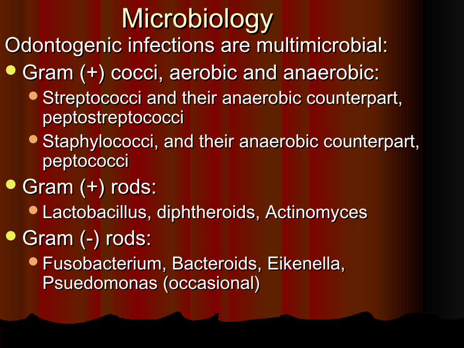
MicrobiologyMicrobiologyOdontogenic infections are multimicrobial:Odontogenic infections are multimicrobial:Gram (+) cocci, aerobic and anaerobic:Gram (+) cocci, aerobic and anaerobic:
Streptococci and their anaerobic counterpart, Streptococci and their anaerobic counterpart, peptostreptococcipeptostreptococci
Staphylococci, and their anaerobic counterpart, Staphylococci, and their anaerobic counterpart, peptococcipeptococci
Gram (+) rods:Gram (+) rods:Lactobacillus, diphtheroids, ActinomycesLactobacillus, diphtheroids, Actinomyces
Gram (-) rods:Gram (-) rods:Fusobacterium, Bacteroids, Eikenella, Fusobacterium, Bacteroids, Eikenella,
Psuedomonas (occasional)Psuedomonas (occasional)
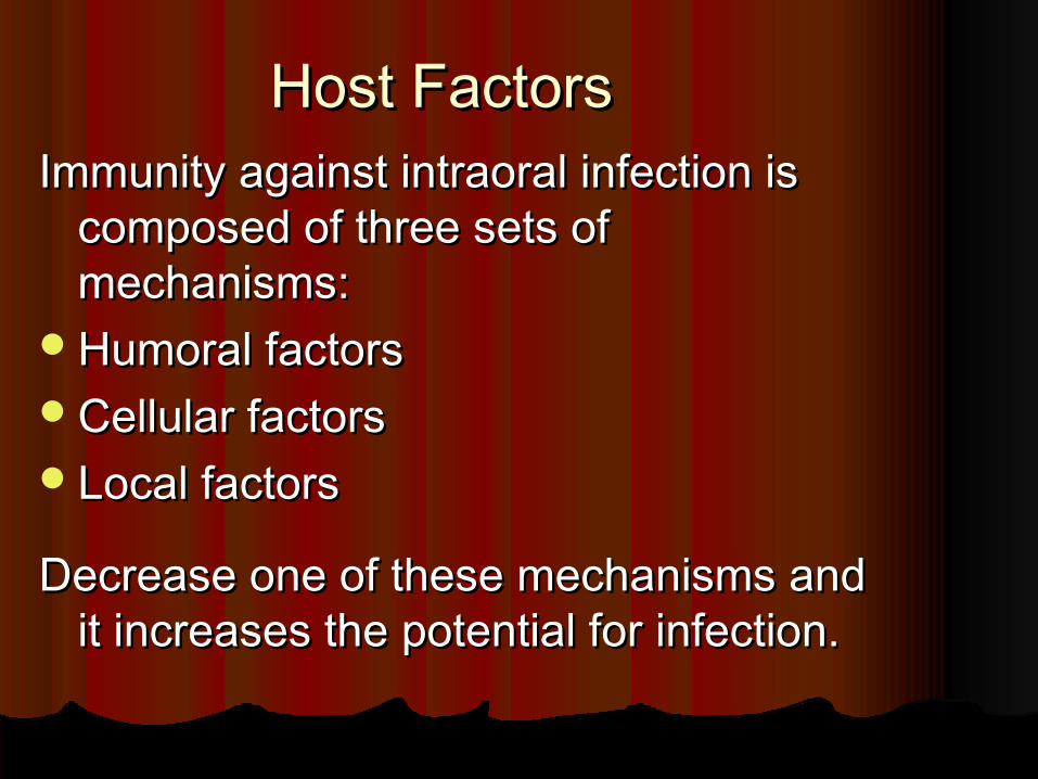
Host FactorsHost FactorsImmunity against intraoral infection is Immunity against intraoral infection is
composed of three sets of composed of three sets of mechanisms:mechanisms:
Humoral factorsHumoral factorsCellular factors Cellular factors Local factorsLocal factors
Decrease one of these mechanisms and Decrease one of these mechanisms and it increases the potential for infection.it increases the potential for infection.
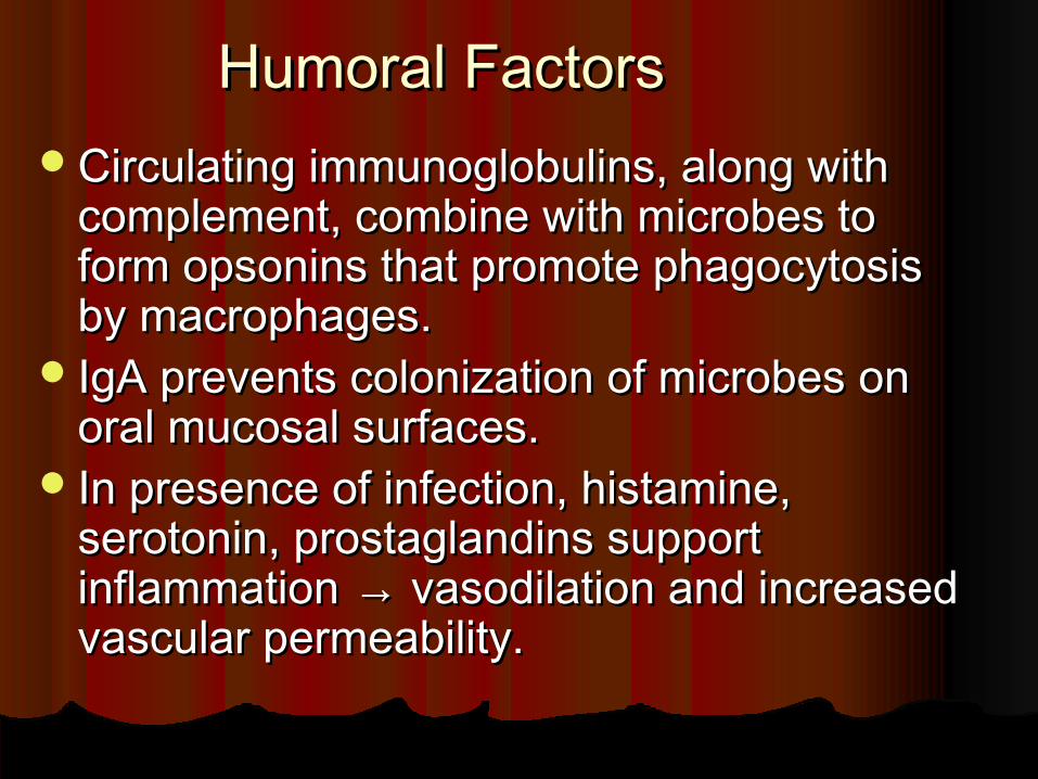
Humoral FactorsHumoral FactorsCirculating immunoglobulins, along with Circulating immunoglobulins, along with
complement, combine with microbes to complement, combine with microbes to form opsonins that promote phagocytosis form opsonins that promote phagocytosis by macrophages.by macrophages.
IgA prevents colonization of microbes on IgA prevents colonization of microbes on oral mucosal surfaces.oral mucosal surfaces.
In presence of infection, histamine, In presence of infection, histamine, serotonin, prostaglandins support serotonin, prostaglandins support inflammation inflammation →→ vasodilation and increased vasodilation and increased vascular permeability.vascular permeability.
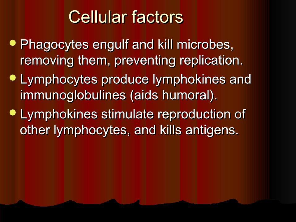
Cellular factorsCellular factorsPhagocytes engulf and kill microbes, Phagocytes engulf and kill microbes,
removing them, preventing replication.removing them, preventing replication.Lymphocytes produce lymphokines and Lymphocytes produce lymphokines and
immunoglobulines (aids humoral).immunoglobulines (aids humoral).Lymphokines stimulate reproduction of Lymphokines stimulate reproduction of
other lymphocytes, and kills antigens.other lymphocytes, and kills antigens.
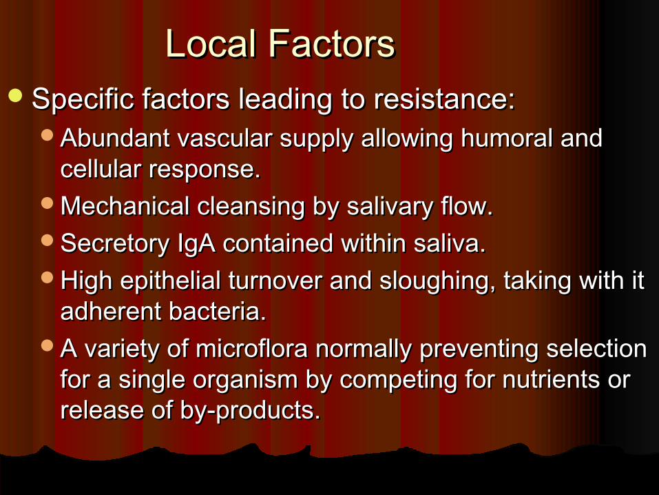
Local FactorsLocal FactorsSpecific factors leading to resistance:Specific factors leading to resistance:
Abundant vascular supply allowing humoral and Abundant vascular supply allowing humoral and cellular response.cellular response.
Mechanical cleansing by salivary flow.Mechanical cleansing by salivary flow.Secretory IgA contained within saliva.Secretory IgA contained within saliva.High epithelial turnover and sloughing, taking with it High epithelial turnover and sloughing, taking with it
adherent bacteria.adherent bacteria.A variety of microflora normally preventing selection A variety of microflora normally preventing selection
for a single organism by competing for nutrients or for a single organism by competing for nutrients or release of by-products.release of by-products.
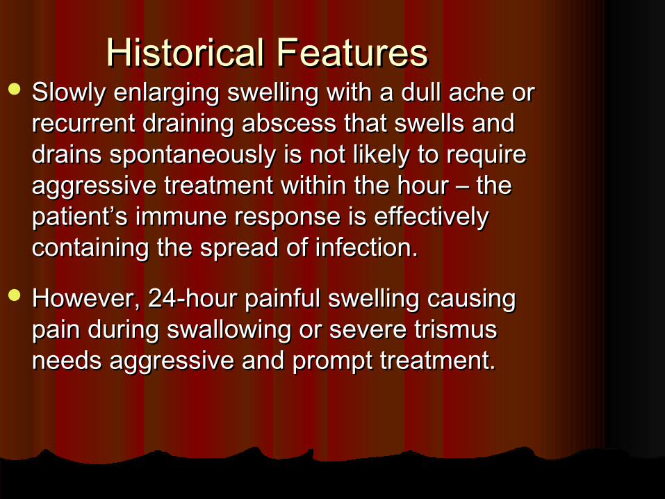
Historical FeaturesHistorical Features Slowly enlarging swelling with a dull ache or Slowly enlarging swelling with a dull ache or
recurrent draining abscess that swells and recurrent draining abscess that swells and drains spontaneously is not likely to require drains spontaneously is not likely to require aggressive treatment within the hour – the aggressive treatment within the hour – the patient’s immune response is effectively patient’s immune response is effectively containing the spread of infection.containing the spread of infection.
However, 24-hour painful swelling causing However, 24-hour painful swelling causing pain during swallowing or severe trismus pain during swallowing or severe trismus needs aggressive and prompt treatment.needs aggressive and prompt treatment.

Historical Features, con’t.Historical Features, con’t.
Immediate treatment or referral is critical Immediate treatment or referral is critical when patient’s immune system has not when patient’s immune system has not been containing the infection.been containing the infection.
Specific warning signs include:Specific warning signs include:Dyspnea (difficulty breathing)Dyspnea (difficulty breathing)Dysphagia (difficulty/pain with swallowing)Dysphagia (difficulty/pain with swallowing)Severe trismusSevere trismusRapidly progressive swellingRapidly progressive swelling

Clinical FeaturesClinical Features Inflammation is tissue response to injury or Inflammation is tissue response to injury or
invasion by microorganisms that involves invasion by microorganisms that involves vasodilation, capillary permeability, mobilization vasodilation, capillary permeability, mobilization of leukocytes, and phagocytosis.of leukocytes, and phagocytosis.
Cardinal signs of inflammation:Cardinal signs of inflammation:Red, hot, swelling, pain, with loss of functionRed, hot, swelling, pain, with loss of function
Other findings: Other findings: regional lymphadenopathyregional lymphadenopathy, , fever, fever, elevated white blood cell count, tachycardia, elevated white blood cell count, tachycardia, tachypnea, dehydration, malaise.tachypnea, dehydration, malaise.
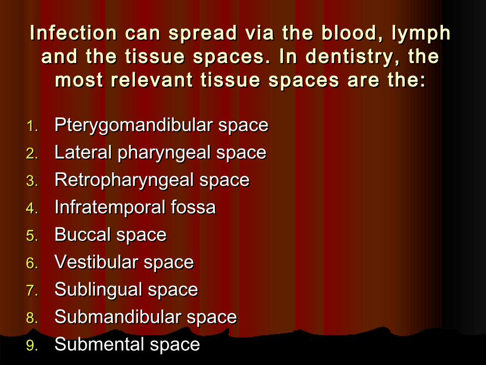
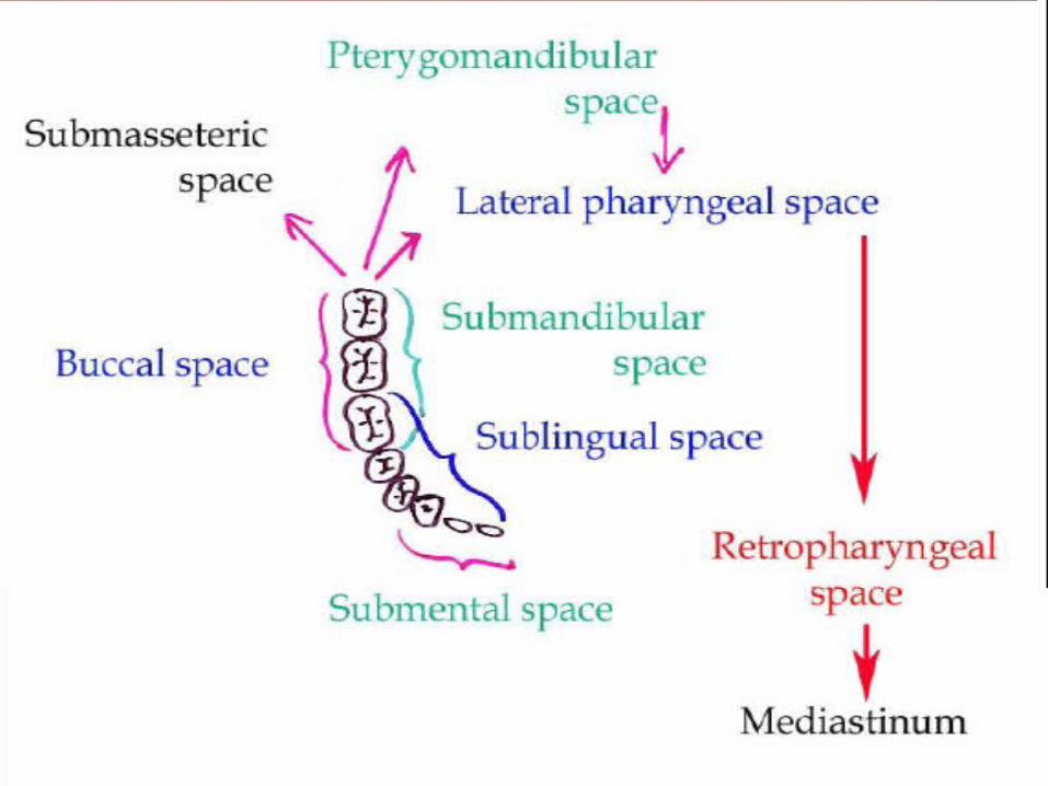
Infection can spread via the blood, lymph Infection can spread via the blood, lymph and the tissue spaces. In dentistry, the and the tissue spaces. In dentistry, the
most relevant t issue spaces are the:most relevant t issue spaces are the:
1.1. Pterygomandibular space Pterygomandibular space 2.2. Lateral pharyngeal space Lateral pharyngeal space 3.3. Retropharyngeal space Retropharyngeal space 4.4. Infratemporal fossa Infratemporal fossa 5.5. Buccal space Buccal space 6.6. Vestibular space Vestibular space 7.7. Sublingual space Sublingual space 8.8. Submandibular space Submandibular space 9.9. Submental space Submental space
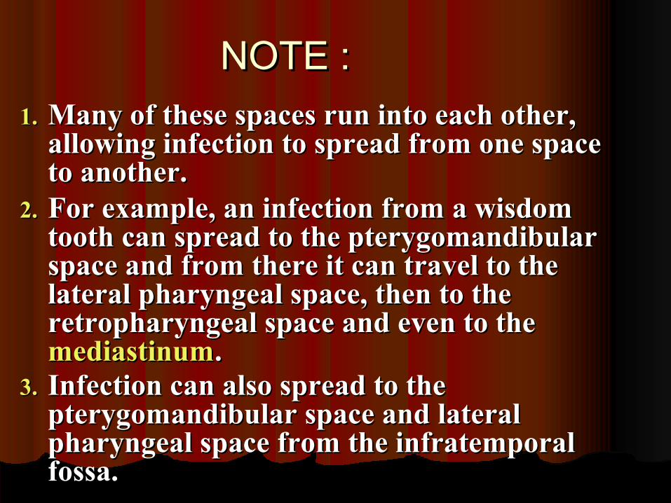
NOTE :NOTE :1.1. Many of these spaces run into each other, Many of these spaces run into each other,
allowing infection to spread from one space allowing infection to spread from one space to another. to another.
2.2. For example, an infection from a wisdom For example, an infection from a wisdom tooth can spread to the pterygomandibular tooth can spread to the pterygomandibular space and from there it can travel to the space and from there it can travel to the lateral pharyngeal space, then to the lateral pharyngeal space, then to the retropharyngeal space and even to the retropharyngeal space and even to the mediastinummediastinum..
3.3. Infection can also spread to the Infection can also spread to the pterygomandibular space and lateral pterygomandibular space and lateral pharyngeal space from the infratemporal pharyngeal space from the infratemporal fossa.fossa.
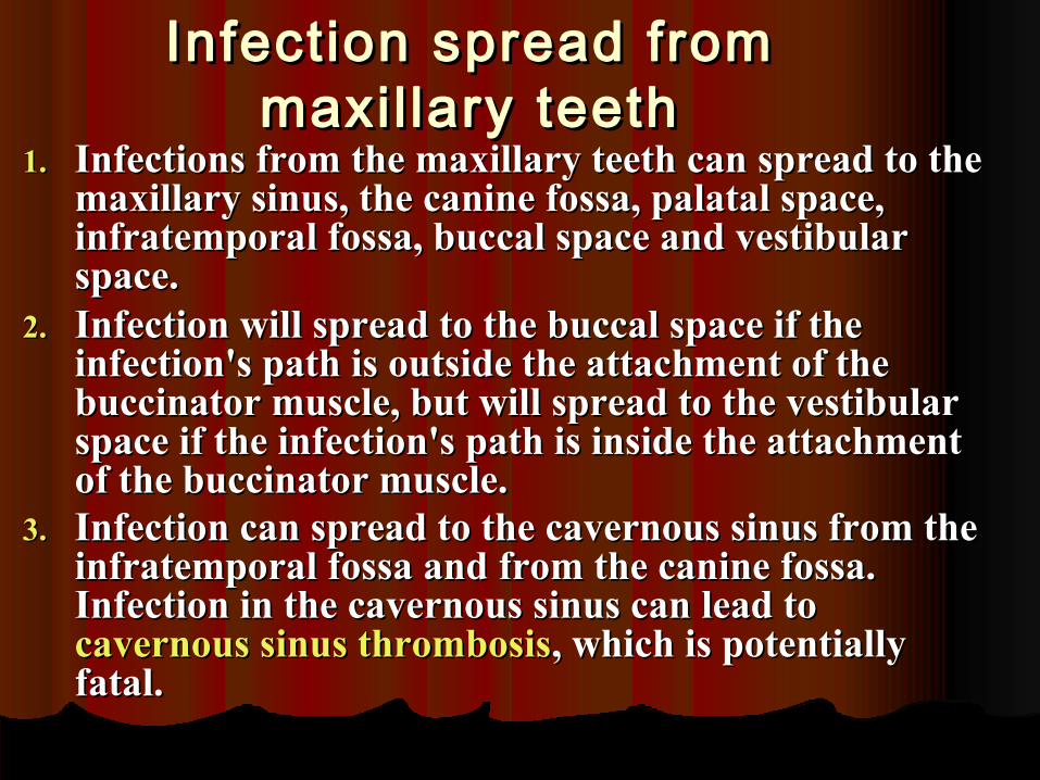
Infection spread from Infection spread from maxil lary teethmaxil lary teeth
1.1. Infections from the maxillary teeth can spread to the Infections from the maxillary teeth can spread to the maxillary sinus, the canine fossa, palatal space, maxillary sinus, the canine fossa, palatal space, infratemporal fossa, buccal space and vestibular infratemporal fossa, buccal space and vestibular space. space.
2.2. Infection will spread to the buccal space if the Infection will spread to the buccal space if the infection's path is outside the attachment of the infection's path is outside the attachment of the buccinator muscle, but will spread to the vestibular buccinator muscle, but will spread to the vestibular space if the infection's path is inside the attachment space if the infection's path is inside the attachment of the buccinator muscle.of the buccinator muscle.
3.3. Infection can spread to the cavernous sinus from the Infection can spread to the cavernous sinus from the infratemporal fossa and from the canine fossa. infratemporal fossa and from the canine fossa. Infection in the cavernous sinus can lead to Infection in the cavernous sinus can lead to cavernous sinus thrombosiscavernous sinus thrombosis, which is potentially , which is potentially fatal.fatal.

Infection spread from Infection spread from mandibular teethmandibular teeth
1.1. Infections from mandibular teeth can spread to the Infections from mandibular teeth can spread to the vestibular and buccal space in the same way as from the vestibular and buccal space in the same way as from the maxillary teeth. maxillary teeth.
2.2. Infection can also spread to the pterygomandibular space, Infection can also spread to the pterygomandibular space, sublingual space, submandibular space and submental sublingual space, submandibular space and submental space. space.
3.3. The sublingual, submental and submandibular spaces can The sublingual, submental and submandibular spaces can be referred collectively as the submandibular spaces.be referred collectively as the submandibular spaces.
4.4. Sometimes when an infection spreads to the submandibular Sometimes when an infection spreads to the submandibular spaces a life threatening condition called Ludwig's angina spaces a life threatening condition called Ludwig's angina occurs. Angina is latin for strangle therefore this angina is occurs. Angina is latin for strangle therefore this angina is referring to the sensation of being strangled caused by the referring to the sensation of being strangled caused by the swelling of the neck region. Tracheotomy is sometimes swelling of the neck region. Tracheotomy is sometimes necessary to maintain the airway. necessary to maintain the airway.
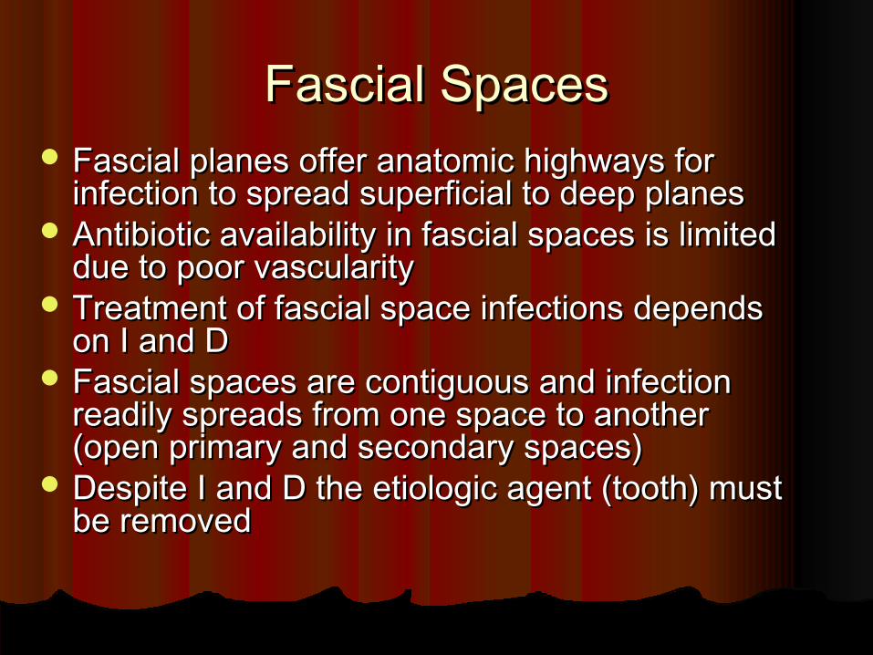
Fascial SpacesFascial Spaces Fascial planes offer anatomic highways for Fascial planes offer anatomic highways for
infection to spread superficial to deep planesinfection to spread superficial to deep planes Antibiotic availability in fascial spaces is limited Antibiotic availability in fascial spaces is limited
due to poor vascularitydue to poor vascularity Treatment of fascial space infections depends Treatment of fascial space infections depends
on I and Don I and D Fascial spaces are contiguous and infection Fascial spaces are contiguous and infection
readily spreads from one space to another readily spreads from one space to another (open primary and secondary spaces) (open primary and secondary spaces)
Despite I and D the etiologic agent (tooth) must Despite I and D the etiologic agent (tooth) must be removed be removed
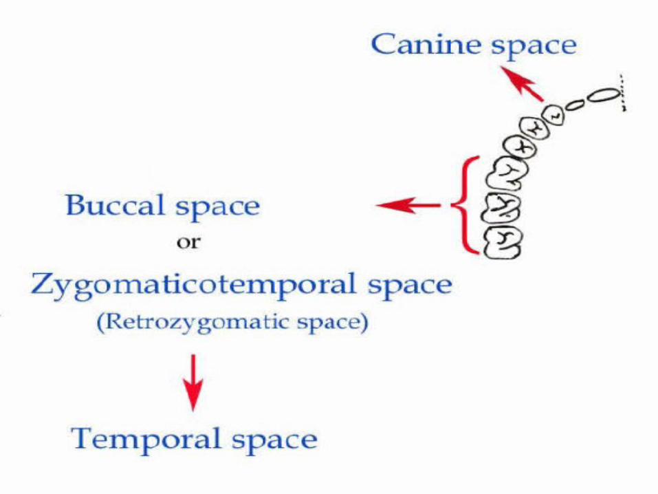
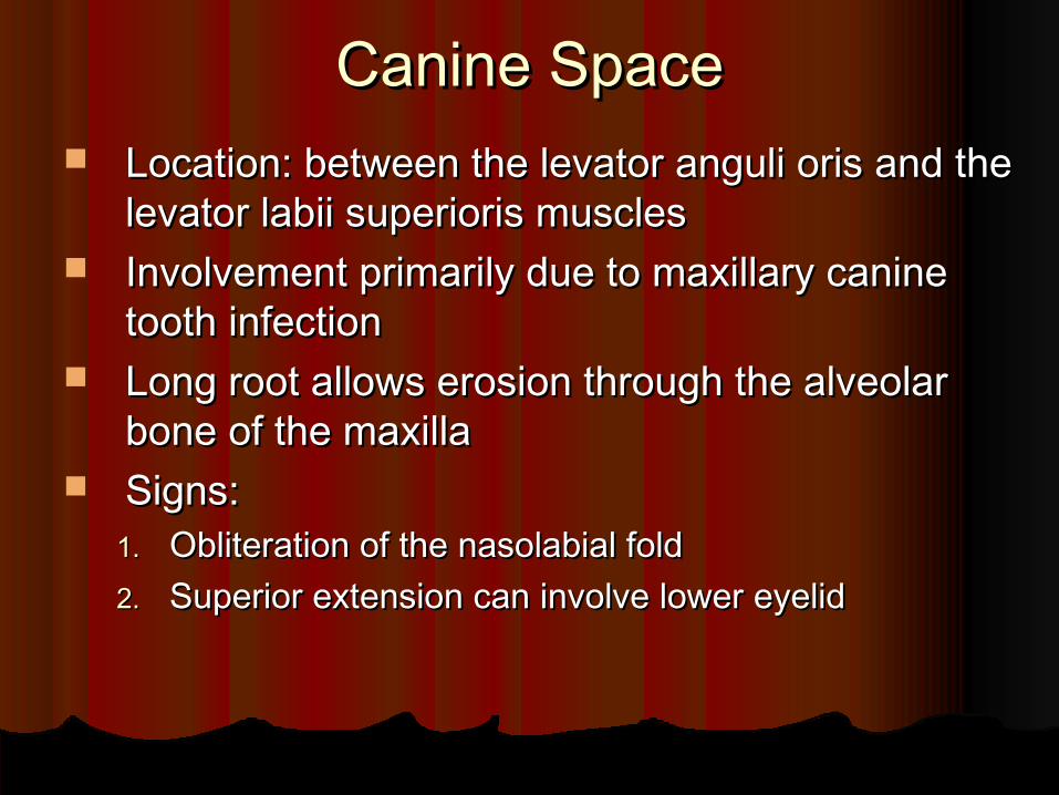
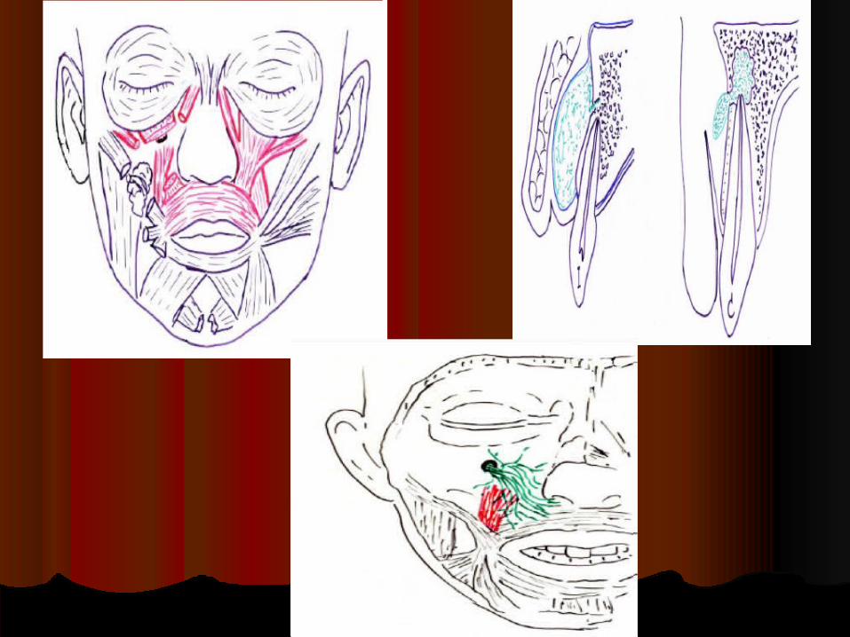
Canine SpaceCanine Space Location: between the levator anguli oris and the Location: between the levator anguli oris and the
levator labii superioris muscles levator labii superioris muscles Involvement primarily due to maxillary canine Involvement primarily due to maxillary canine
tooth infection tooth infection Long root allows erosion through the alveolar Long root allows erosion through the alveolar
bone of the maxillabone of the maxilla Signs: Signs:
1.1. Obliteration of the nasolabial fold Obliteration of the nasolabial fold 2.2. Superior extension can involve lower eyelidSuperior extension can involve lower eyelid
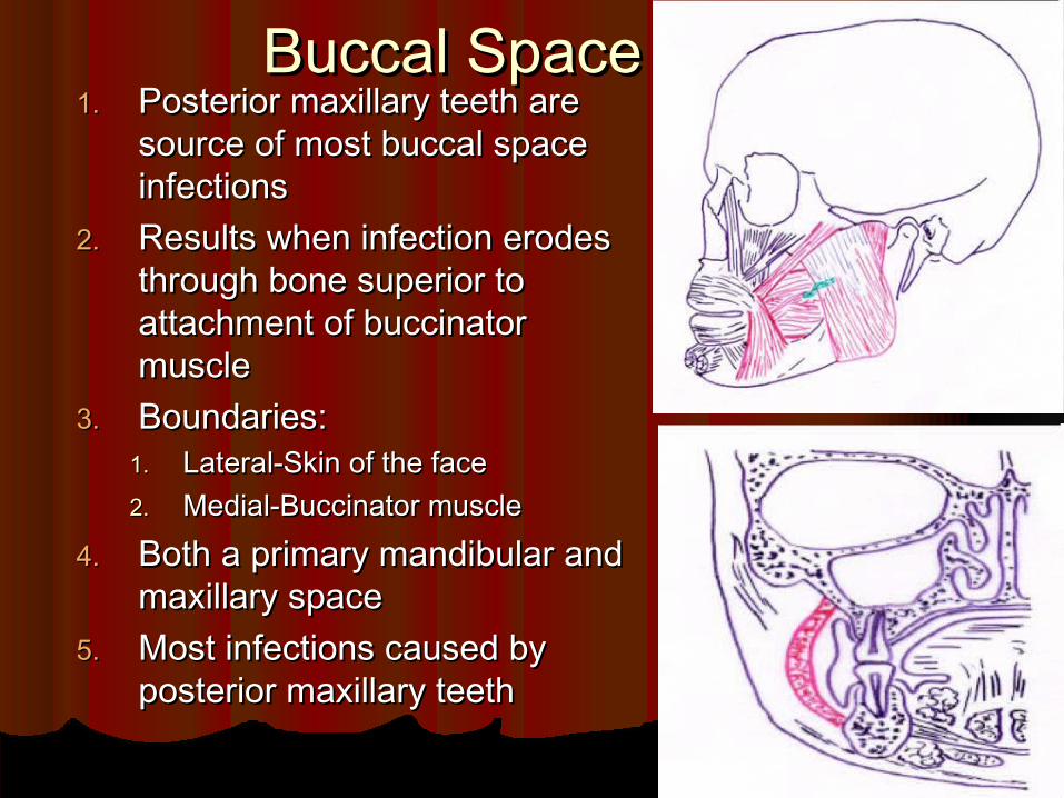
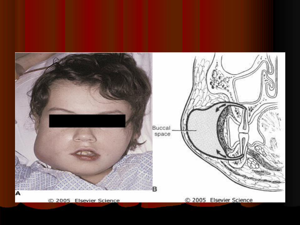
Buccal SpaceBuccal Space1.1. Posterior maxillary teeth are Posterior maxillary teeth are
source of most buccal space source of most buccal space infectionsinfections
2.2. Results when infection erodes Results when infection erodes through bone superior to through bone superior to attachment of buccinator attachment of buccinator musclemuscle
3.3. Boundaries: Boundaries: 1.1. Lateral-Skin of the face Lateral-Skin of the face 2.2. Medial-Buccinator muscle Medial-Buccinator muscle
4.4. Both a primary mandibular and Both a primary mandibular and maxillary spacemaxillary space
5.5. Most infections caused by Most infections caused by posterior maxillary teethposterior maxillary teeth
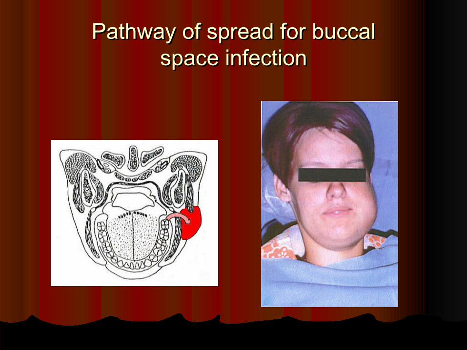
Pathway of spread for buccalPathway of spread for buccalspace infectionspace infection
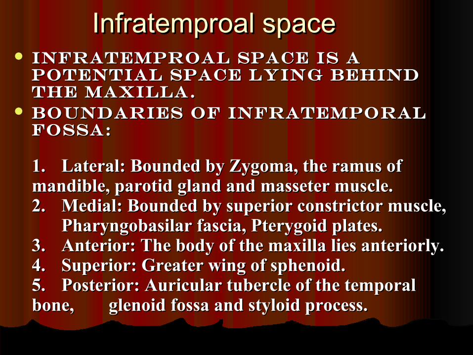
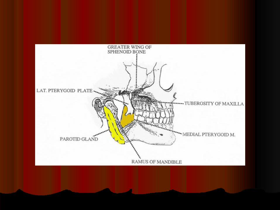
Infratemproal spaceInfratemproal space Infratemproal space is a Infratemproal space is a
potential space lying behind potential space lying behind the maxilla.the maxilla.
Boundaries of infratemporal Boundaries of infratemporal fossa:fossa:
1. 1. Lateral: Bounded by Zygoma, the ramus of Lateral: Bounded by Zygoma, the ramus of mandible, parotid gland and masseter muscle. mandible, parotid gland and masseter muscle. 2.2. Medial: Bounded by superior constrictor muscle, Medial: Bounded by superior constrictor muscle,
Pharyngobasilar fascia, Pterygoid plates.Pharyngobasilar fascia, Pterygoid plates.3.3. Anterior: The body of the maxilla lies anteriorly.Anterior: The body of the maxilla lies anteriorly.4.4. Superior: Greater wing of sphenoid.Superior: Greater wing of sphenoid.5.5. Posterior: Auricular tubercle of the temporal Posterior: Auricular tubercle of the temporal bone, bone, glenoid fossa and styloid process.glenoid fossa and styloid process.

INFECTION OF INFRATEMPORAL INFECTION OF INFRATEMPORAL SPACESPACE
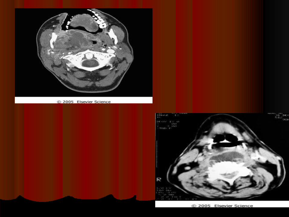
Infective complications can develop and present Infective complications can develop and present following the extraction of clinically non-infected teeth following the extraction of clinically non-infected teeth
Infratemporal space infection is a rare complication of Infratemporal space infection is a rare complication of dental extraction dental extraction
The cardinal clinical signs of maxillary and The cardinal clinical signs of maxillary and mandibular neurosensory deficit may not be mandibular neurosensory deficit may not be immediately apparent immediately apparent
The diagnosis, as in this case, can only be confirmed The diagnosis, as in this case, can only be confirmed using scanning imaging modalities (CT or MRI)using scanning imaging modalities (CT or MRI)

Clinical features :-Clinical features :-
Severe trismusSevere trismusBuldging of temporalis muscleBuldging of temporalis muscleSwelling extraorally over the region of sigmoid Swelling extraorally over the region of sigmoid
notch and intraorally in tuberosity region.notch and intraorally in tuberosity region.
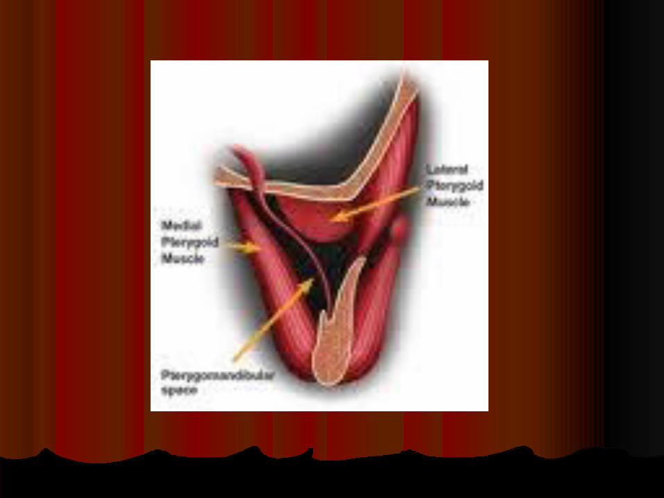
Pterygomandibular space Pterygomandibular space
The space between the medial area of the The space between the medial area of the mandible and the medial pterygoid mandible and the medial pterygoid muscle, a target area for administering muscle, a target area for administering local anesthesia to the inferior alveolar local anesthesia to the inferior alveolar nerve.nerve.
Boundries of pterygomandibular Boundries of pterygomandibular spacespace
1.1. Medially – medial pterygoid muscleMedially – medial pterygoid muscle2.2. Laterally – medial surface of ramus of mandibleLaterally – medial surface of ramus of mandible3.3. Superiorly – lateral pterygoidSuperiorly – lateral pterygoid4.4. Posteriorly – deep lobe of parotid gland Posteriorly – deep lobe of parotid gland 5.5. Inferiorly – attachment of medial pterygoid oto Inferiorly – attachment of medial pterygoid oto
the mandible the mandible 6.6. Anteriorly – pterygomandibular raphe Anteriorly – pterygomandibular raphe
infectioninfection
From lower third molar From lower third molar Clinical features :-Clinical features :-1.1. TrismusTrismus2.2. Intraoral sweeling in the medial aspect of Intraoral sweeling in the medial aspect of
ramus of mandible ramus of mandible
Retropharyngeal SpaceRetropharyngeal Space1.1. Posteromedial to lateral pharyngeal space and anterior Posteromedial to lateral pharyngeal space and anterior
to the prevertebral space to the prevertebral space 2.2. Anterior: superior pharyngeal constrictor muscle Anterior: superior pharyngeal constrictor muscle 3.3. Posterior: alar layer of prevertebral fascia Posterior: alar layer of prevertebral fascia 4.4. Extends from skull base superiorly to C7 to T1 inferiorlyExtends from skull base superiorly to C7 to T1 inferiorly5.5. Retropharyngeal space infections can spread to Retropharyngeal space infections can spread to
mediastinummediastinum6.6. Other complications of retropharyngeal space Other complications of retropharyngeal space
involvement:involvement:1.1. Airway obstruction Airway obstruction 2.2. Aspiration of pus in the event of spontaneous rupture Aspiration of pus in the event of spontaneous rupture 3.3. Rupture can occur during endotracheal intubationRupture can occur during endotracheal intubation
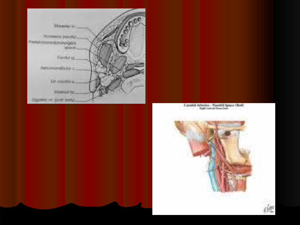
Parotid spaceParotid space A A deepdeep hollowhollow on the on the sideside at the sides of the at the sides of the faceface
flanking the flanking the posteriorposterior aspectaspect of the of the ramusramus of the mandible of the mandible with its attached with its attached musclesmuscles which is which is occupied by the occupied by the parotid glandparotid gland; it is lined with ; it is lined with fascialfascial laminae (the laminae (the parotid sheathparotid sheath) derived from ) derived from the the investing layerinvesting layer of deep of deep cervical fasciacervical fascia; the ; the structuresstructures bounding the bounding the spacespace collectively collectively constitute the constitute the parotid bedparotid bed. . SurgeonsSurgeons operating in operating in the the areaarea taketake advantage of the fact that the advantage of the fact that the anteroposterioranteroposterior dimensionsdimensions of the of the parotidparotid space space increaseincrease with with protrusionprotrusion of the of the mandiblemandible. .
Clinical featuresClinical features
1.1. Facial swelling and progressive trismus Facial swelling and progressive trismus are described. are described.
2.2. Intraoral wound drainage and prolonged Intraoral wound drainage and prolonged antibiotic therapy failed to control the antibiotic therapy failed to control the resultant chronic cellulitis. resultant chronic cellulitis.
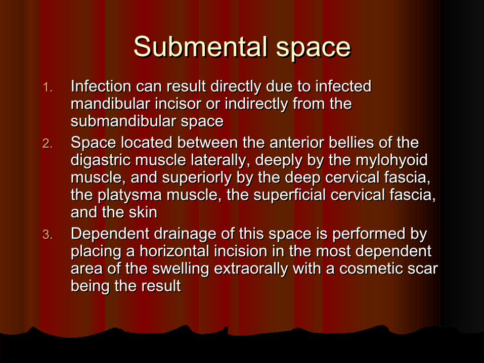
Submental spaceSubmental space1.1. Infection can result directly due to infected Infection can result directly due to infected
mandibular incisor or indirectly from the mandibular incisor or indirectly from the submandibular spacesubmandibular space
2.2. Space located between the anterior bellies of the Space located between the anterior bellies of the digastric muscle laterally, deeply by the mylohyoid digastric muscle laterally, deeply by the mylohyoid muscle, and superiorly by the deep cervical fascia, muscle, and superiorly by the deep cervical fascia, the platysma muscle, the superficial cervical fascia, the platysma muscle, the superficial cervical fascia, and the skinand the skin
3.3. Dependent drainage of this space is performed by Dependent drainage of this space is performed by placing a horizontal incision in the most dependent placing a horizontal incision in the most dependent area of the swelling extraorally with a cosmetic scar area of the swelling extraorally with a cosmetic scar being the resultbeing the result
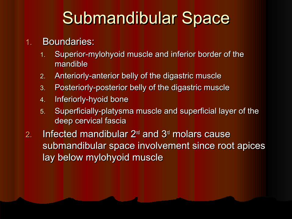
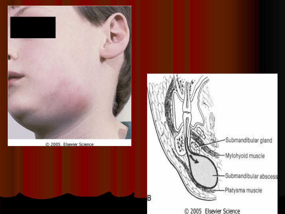
Submandibular SpaceSubmandibular Space1.1. Boundaries:Boundaries:
1.1. Superior-mylohyoid muscle and inferior border of the Superior-mylohyoid muscle and inferior border of the mandiblemandible
2.2. Anteriorly-anterior belly of the digastric muscle Anteriorly-anterior belly of the digastric muscle 3.3. Posteriorly-posterior belly of the digastric musclePosteriorly-posterior belly of the digastric muscle4.4. Inferiorly-hyoid boneInferiorly-hyoid bone5.5. Superficially-platysma muscle and superficial layer of the Superficially-platysma muscle and superficial layer of the
deep cervical fasciadeep cervical fascia
2.2. Infected mandibular 2Infected mandibular 2ndnd and 3 and 3 rdrd molars cause molars cause submandibular space involvement since root apices submandibular space involvement since root apices lay below mylohyoid muscle lay below mylohyoid muscle

Pathways of spread of submandibular space Pathways of spread of submandibular space infection from mandibular molarinfection from mandibular molar
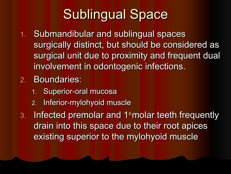
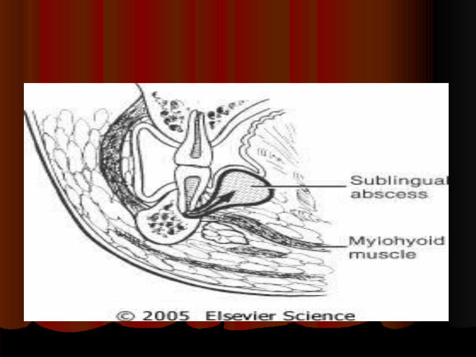
Sublingual SpaceSublingual Space1.1. Submandibular and sublingual spaces Submandibular and sublingual spaces
surgically distinct, but should be considered as surgically distinct, but should be considered as surgical unit due to proximity and frequent dual surgical unit due to proximity and frequent dual involvement in odontogenic infections. involvement in odontogenic infections.
2.2. Boundaries:Boundaries:1.1. Superior-oral mucosaSuperior-oral mucosa2.2. Inferior-mylohyoid muscleInferior-mylohyoid muscle
3.3. Infected premolar and 1Infected premolar and 1 st st molar teeth frequently molar teeth frequently drain into this space due to their root apices drain into this space due to their root apices existing superior to the mylohyoid muscle existing superior to the mylohyoid muscle
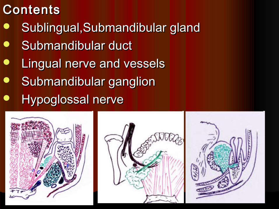
ContentsContents Sublingual,Submandibular glandSublingual,Submandibular gland Submandibular ductSubmandibular duct Lingual nerve and vesselsLingual nerve and vessels Submandibular ganglionSubmandibular ganglion Hypoglossal nerveHypoglossal nerve

Masseteric SpaceMasseteric Space
1.1. Located between lateral aspect of the Located between lateral aspect of the mandible and the masseter muscle mandible and the masseter muscle
2.2. Involvement of this space generally occurs Involvement of this space generally occurs from buccal space primary involvement from buccal space primary involvement
3.3. Signs of involvement of the masseteric space Signs of involvement of the masseteric space include trismus and posterior-inferior face include trismus and posterior-inferior face swellingswelling
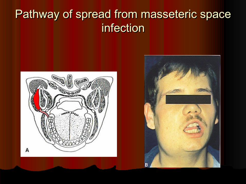
Pathway of spread from masseteric space Pathway of spread from masseteric space infectioninfection
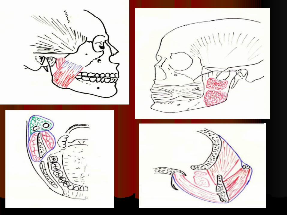
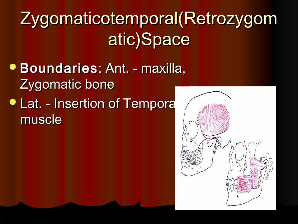
Zygomaticotemporal(RetrozygomZygomaticotemporal(Retrozygomatic)Spaceatic)Space
BoundariesBoundaries : Ant. - maxilla, : Ant. - maxilla, Zygomatic boneZygomatic bone
Lat. - Insertion of Temporalis Lat. - Insertion of Temporalis musclemuscle
Zygomaticotemporal(RetrozygomZygomaticotemporal(Retrozygomatic)Spaceatic)Space
Contents Contents : Posterior suprerior alveolar : Posterior suprerior alveolar nervenerve
and vessles buccal fat padand vessles buccal fat pad
Oral tissue examinationOral tissue examinationExamine quality and consistency:Examine quality and consistency:
Soft to fluctuant (fluid filled) to hard (indurated)Soft to fluctuant (fluid filled) to hard (indurated)Color and temperature determine the presence Color and temperature determine the presence
and extent of infectionand extent of infectionNormal v abnormal tissue architecture:Normal v abnormal tissue architecture:
Distortion of mucobuccal foldDistortion of mucobuccal foldSoft palate symmetric with uvula in midline Soft palate symmetric with uvula in midline (deviation (deviation
→ involvement of lateral pharyngeal space)→ involvement of lateral pharyngeal space)Nasal tip, nasolabial fold, circumorbital areasNasal tip, nasolabial fold, circumorbital areas
Examination, con’t.Examination, con’t.
Identify causative factors:Identify causative factors:Tooth, root tip, foreign body, etc.Tooth, root tip, foreign body, etc.
Vital signs should be taken:Vital signs should be taken:Temperatures Temperatures >> 101 to 102 101 to 102°F °F
accompanied by an elevated heart rate accompanied by an elevated heart rate indicate systemic involvement of the indicate systemic involvement of the infection and increased urgency of infection and increased urgency of treatment.treatment.

Principles in Treatment of Oral Principles in Treatment of Oral InfectionsInfections
1.1. Remove the cause.Remove the cause.
2.2. Establish drainage.Establish drainage.
3.3. Institute antibiotic therapy.Institute antibiotic therapy.
4.4. Supportive care, including proper rest Supportive care, including proper rest and nutrition.and nutrition.
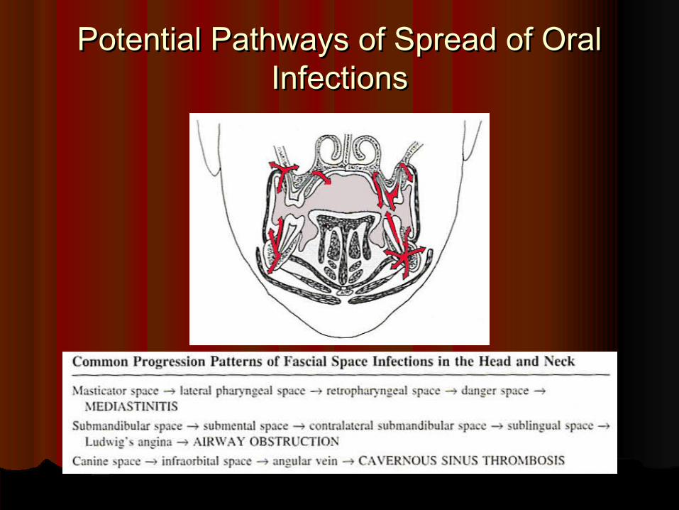
Potential Pathways of Spread of Oral Potential Pathways of Spread of Oral InfectionsInfections
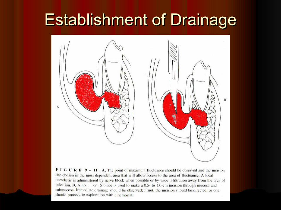
Establishment of DrainageEstablishment of Drainage
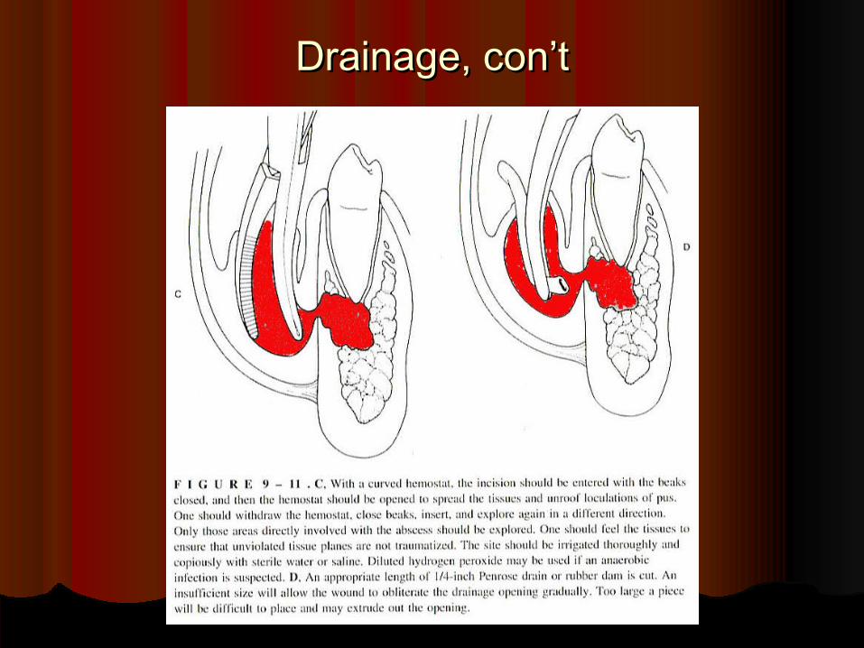
Drainage, con’tDrainage, con’t
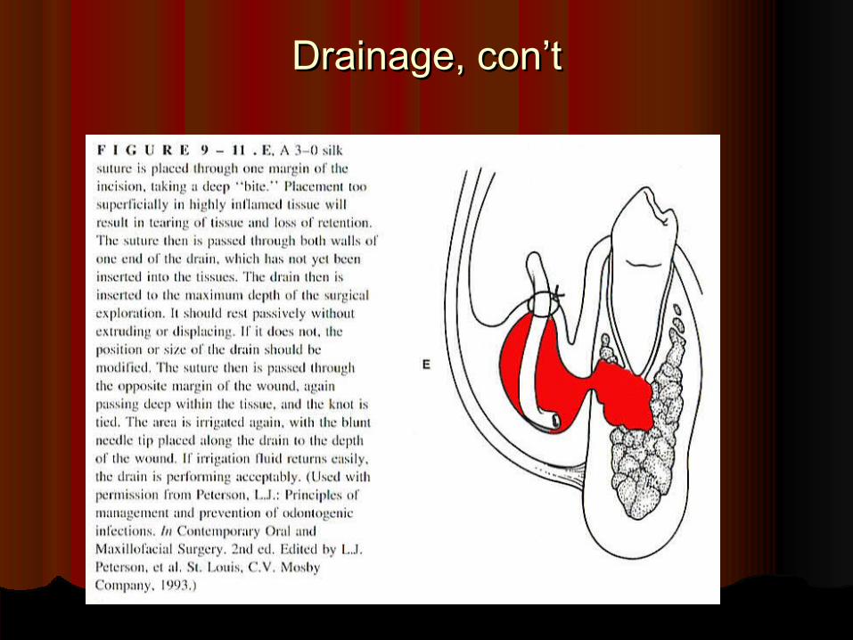
Drainage, con’tDrainage, con’t
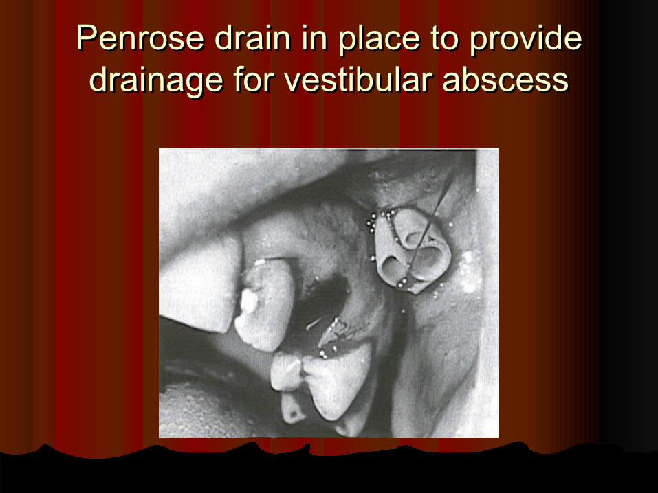
Penrose drain in place to provide Penrose drain in place to provide drainage for vestibular abscessdrainage for vestibular abscess
Antibiotic TherapyAntibiotic TherapyRemoval of the cause, drainage, and Removal of the cause, drainage, and
supportive care more important than supportive care more important than antibiotic therapy.antibiotic therapy.
Infections are cured by the patient’s Infections are cured by the patient’s defenses, defenses, notnot antibiotics. antibiotics.
Risks of allergy, toxicity, side effects, Risks of allergy, toxicity, side effects, resistance and superinfection causing resistance and superinfection causing serious or potentially fatal consequences serious or potentially fatal consequences must be considered.must be considered.
Antibiotic therapy, con’t.Antibiotic therapy, con’t.Oral infections are typically polymicrobial.Oral infections are typically polymicrobial.Antibiotic effectiveness dependent upon Antibiotic effectiveness dependent upon
adequate tissue (not serum) concentration for adequate tissue (not serum) concentration for an appropriate amount of time.an appropriate amount of time.
Antibiotics should be prescribed for at least Antibiotics should be prescribed for at least one week – adequate tissue concentration one week – adequate tissue concentration achieved in 24-48 hours, with bacteriocidal achieved in 24-48 hours, with bacteriocidal activity occurring over the next 3-5 days.activity occurring over the next 3-5 days.
Antibiotic therapy, con’t.Antibiotic therapy, con’t.Penicil l inPenicil l in (bacteriocidal) drug of choice for (bacteriocidal) drug of choice for
treatment of odontogenic infections (5% incident treatment of odontogenic infections (5% incident of allergy).of allergy).
Clindamycin Clindamycin (batericiodal) 1(batericiodal) 1stst line after line after penicillin; effective against anaerobes; stop taking penicillin; effective against anaerobes; stop taking at first sign of diarrhea.at first sign of diarrhea.
Cephalosporin Cephalosporin (slightly broader spectrum (slightly broader spectrum and bacteriocidal); cautious use in penicillin-and bacteriocidal); cautious use in penicillin-allergic patients allergic patients → cross-sensitivity; if history of → cross-sensitivity; if history of anaphylaxis to penicillin, do not use.anaphylaxis to penicillin, do not use.
Antibiotic therapy, con’t.Antibiotic therapy, con’t.
ErythromycinErythromycin (bacteriostatic) good 2(bacteriostatic) good 2ndnd line line drug after penicillin; use enteric-coated to drug after penicillin; use enteric-coated to reduce GI upset.reduce GI upset.
MetronidazoleMetronidazole (bacteriocidal) excellent (bacteriocidal) excellent against anaerobes only.against anaerobes only.
AugmentinAugmentin (amoxicillin + clavulanic acid) (amoxicillin + clavulanic acid) kills penicillinase-producing bacteria that kills penicillinase-producing bacteria that interferes with amoxicillin; expensive.interferes with amoxicillin; expensive.
Supportive CareSupportive CareTo ensure the patient’s maximum immune To ensure the patient’s maximum immune
response:response:Increase fluid intake (16 ounces/hour).Increase fluid intake (16 ounces/hour).Nutritional intake (soups, protein drinks, solids) Nutritional intake (soups, protein drinks, solids)
with three meals/day.with three meals/day.May need to see patient daily, until May need to see patient daily, until
resolution has begun.resolution has begun. If no improvement within 24- 48 hours, refer If no improvement within 24- 48 hours, refer
immediately to an oral and maxillofacial immediately to an oral and maxillofacial surgeon.surgeon.
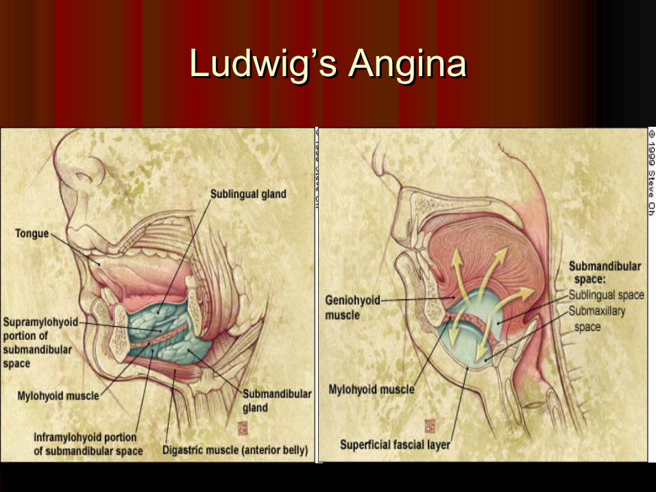
Ludwig’s AnginaLudwig’s Angina

Ludwig’s angina with bilateral involvement of Ludwig’s angina with bilateral involvement of sublingual and submandibular spacessublingual and submandibular spaces
DEFINATIONDEFINATION Ludwig's angina, otherwise known as angina ludovici, is a Ludwig's angina, otherwise known as angina ludovici, is a
serious, potentially life-threatening serious, potentially life-threatening cellulitiscellulitis[1][1], or , or connective tissue infection, of the floor of the mouth, connective tissue infection, of the floor of the mouth, usually occurring in adults with concomitant dental usually occurring in adults with concomitant dental infections. It is named after the German physician, infections. It is named after the German physician, Wilhelm Friedrich von LudwigWilhelm Friedrich von Ludwig who first described this who first described this condition in 1836.condition in 1836.[2][2][3][3] Other names include "angina Other names include "angina Maligna" and "Morbus Strangularis".Maligna" and "Morbus Strangularis".
Ludwig's angina should not be confused with Ludwig's angina should not be confused with angina pectorisangina pectoris, which is also otherwise commonly known , which is also otherwise commonly known as "as "anginaangina". The word "". The word "anginaangina" comes from the " comes from the GreekGreek word word ankhonankhon, meaning "strangling", so in this case, , meaning "strangling", so in this case, Ludwig's angina refers to the feeling of strangling, not the Ludwig's angina refers to the feeling of strangling, not the feeling of chest pain, though there may be chest pain in feeling of chest pain, though there may be chest pain in Ludwig's angina if the infection spreads into the Ludwig's angina if the infection spreads into the retrosternal space.retrosternal space.

Causes Causes The cause is usually an infection with The cause is usually an infection with StreptococcalStreptococcal bacteria, bacteria,
although other bacteria can cause the condition. Since the although other bacteria can cause the condition. Since the advent of advent of antibioticsantibiotics, Ludwig's angina has become a rare , Ludwig's angina has become a rare disease.disease.
The route of infection in most cases is from infected lower third The route of infection in most cases is from infected lower third molarsmolars or from or from pericoronitispericoronitis, which is an infection of the gums , which is an infection of the gums surrounding the partially erupted lower third molars. Although surrounding the partially erupted lower third molars. Although the widespread involvement seen in Ludwig's is usually the widespread involvement seen in Ludwig's is usually develops in develops in immunocompromisedimmunocompromised persons, it can also develop persons, it can also develop in otherwise healthy individuals. Thus, it is very important to in otherwise healthy individuals. Thus, it is very important to obtain dental consultation for lower-third molars at the first sign obtain dental consultation for lower-third molars at the first sign of any pain, bleeding from the gums, sensitivity to heat/cold or of any pain, bleeding from the gums, sensitivity to heat/cold or swelling at the angle of the jaw.swelling at the angle of the jaw.
Ludwig's angina is also associated with Ludwig's angina is also associated with piercingspiercings of the of the lingual lingual frenulumfrenulum..

Symptoms Symptoms The symptoms include swelling, pain and raising of the The symptoms include swelling, pain and raising of the
tongue, swelling of the neck and the tissues of the tongue, swelling of the neck and the tissues of the submandibular and submandibular and sublingual spacessublingual spaces, malaise, , malaise, feverfever, , dysphagiadysphagia (difficulty swallowing) and, in severe cases, (difficulty swallowing) and, in severe cases, stridor or difficulty breathing. stridor or difficulty breathing.
Swelling of the submandibular and/or sublingual spaces Swelling of the submandibular and/or sublingual spaces are distinctive in that they are hard and classically are distinctive in that they are hard and classically 'boardlike'. Important signs include the patient not being 'boardlike'. Important signs include the patient not being able to swallow his/her own saliva and the presence of able to swallow his/her own saliva and the presence of audible stridor as these strongly suggest that airway audible stridor as these strongly suggest that airway compromise is imminent.compromise is imminent.
Treatment Treatment Treatment involves appropriate antibiotic medications, Treatment involves appropriate antibiotic medications,
monitoring and protection of the airway in severe monitoring and protection of the airway in severe cases, and, where appropriate, urgent maxillo-facial cases, and, where appropriate, urgent maxillo-facial surgery and/or dental consultation to incise and drain surgery and/or dental consultation to incise and drain the collections. the collections.
A nasotracheal tube is sometimes warranted for A nasotracheal tube is sometimes warranted for ventilation if the tissues of the mouth make insertion of ventilation if the tissues of the mouth make insertion of an oral airway difficult or impossible. an oral airway difficult or impossible.
In cases where the patency of the airway is In cases where the patency of the airway is compromised, skilled airway management is compromised, skilled airway management is mandatory. mandatory.
This entails management of the airway according to the This entails management of the airway according to the American Society of Anesthesiologists' "Difficult American Society of Anesthesiologists' "Difficult Airway Algorithm" and necessitates fiberoptic Airway Algorithm" and necessitates fiberoptic intubation.intubation.
Maxillary Maxillary SinusitisSinusitis
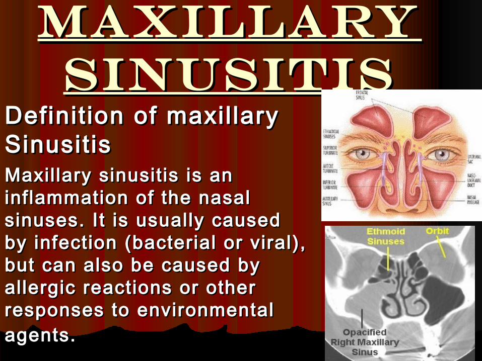
Definit ion of maxil lary Definit ion of maxil lary Sinusit isSinusit isMaxil lary sinusit is is an Maxil lary sinusit is is an inflammation of the nasal inflammation of the nasal sinuses. It is usually caused sinuses. It is usually caused by infection (bacterial or viral), by infection (bacterial or viral), but can also be caused by but can also be caused by allergic reactions or other allergic reactions or other responses to environmental responses to environmental agents.agents.
Causes and Risk Causes and Risk Factors of maxillary Factors of maxillary
SinusitisSinusitis Most maxillary sinusitis is caused by infection (such as a Most maxillary sinusitis is caused by infection (such as a
cold or an upper respiratory tract infection) spreading to cold or an upper respiratory tract infection) spreading to the sinuses from the nose along the narrow passages that the sinuses from the nose along the narrow passages that drain mucus from the sinuses into the nose. drain mucus from the sinuses into the nose.
Allergies to dust, pollen, pet dander; indoor air pollutants, Allergies to dust, pollen, pet dander; indoor air pollutants, such as cigarette smoke, rug shampoo and formaldehyde such as cigarette smoke, rug shampoo and formaldehyde (used in the manufacture of carpeting, particleboard and (used in the manufacture of carpeting, particleboard and plywood); and outdoor air pollutants all can induce plywood); and outdoor air pollutants all can induce inflammation. inflammation.
Excessive dryness in homes and offices from dry-air Excessive dryness in homes and offices from dry-air heating and air-conditioning systems can also inflame the heating and air-conditioning systems can also inflame the sinuses. sinuses.
Immunologic, as well as structural problems, such as Immunologic, as well as structural problems, such as narrow drainage passages, nasal obstruction (tumors, narrow drainage passages, nasal obstruction (tumors, polyps or a deviated septum) problems are other possible polyps or a deviated septum) problems are other possible causes of sinusitis.causes of sinusitis.

Symptoms of Symptoms of maxillary Sinusitismaxillary Sinusitis
Maxillary sinusitis manifests Maxillary sinusitis manifests as cheek or dental pain. as cheek or dental pain.
The classic symptoms of acute The classic symptoms of acute (short lasting) Maxillary (short lasting) Maxillary sinusitis are:sinusitis are:
1.1. Fever Fever 2.2. Nasal obstruction Nasal obstruction 3.3. Raspy voice Raspy voice 4.4. Pus-like (purulent) nasal discharge Pus-like (purulent) nasal discharge 5.5. Loss of sense of smell Loss of sense of smell 6.6. Facial pain or headache that is sometimes Facial pain or headache that is sometimes
aggravated by bending over (When pain is aggravated by bending over (When pain is present, this may suggest which sinus is present, this may suggest which sinus is affected.) affected.)
Symptoms of Symptoms of maxillary maxillary SinusitisSinusitis
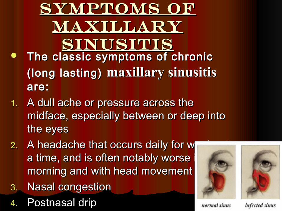
The classic symptoms of chronic The classic symptoms of chronic (long lasting) (long lasting) maxillary sinusitis maxillary sinusitis are:are:
1.1. A dull ache or pressure across the A dull ache or pressure across the midface, especially between or deep into midface, especially between or deep into the eyes the eyes
2.2. A headache that occurs daily for weeks at A headache that occurs daily for weeks at a time, and is often notably worse in the a time, and is often notably worse in the morning and with head movement morning and with head movement
3.3. Nasal congestion Nasal congestion 4.4. Postnasal drip Postnasal drip

Less common signs Less common signs of maxillary of maxillary
sinusitis include:sinusitis include: 1.1. Sore throat Sore throat 2.2. Snoring Snoring 3.3. Bad breath Bad breath 4.4. Chronic throat clearing Chronic throat clearing 5.5. Puffy eyes Puffy eyes 6.6. Coughing Coughing 7.7. Stuffy ears Stuffy ears 8.8. Fatigue, irritability and depression Fatigue, irritability and depression 9.9. A chronic cold A chronic cold 10.10. Asthma Asthma 11.11. BronchitisBronchitis

Diagnosis of Diagnosis of maxillary Sinusitismaxillary Sinusitis
The doctor will examine the mouth and throat and look up the nasal The doctor will examine the mouth and throat and look up the nasal passages to determine whether the sinus outlets are blocked. Additionally, passages to determine whether the sinus outlets are blocked. Additionally, the doctor may do a transillumintion or a CAT scan. the doctor may do a transillumintion or a CAT scan.
Transillumination is done in a dark room with a very bright flashlight that Transillumination is done in a dark room with a very bright flashlight that is pressed against the forehead or cheek. If the light shines through the is pressed against the forehead or cheek. If the light shines through the sinuses, the doctor can rule out sinusitis. If little or no light penetrates, the sinuses, the doctor can rule out sinusitis. If little or no light penetrates, the cavity is clogged and sinusitis is evident. cavity is clogged and sinusitis is evident.
CAT scan is a diagnostic technique in which the combined use of a CAT scan is a diagnostic technique in which the combined use of a computer and x-rays are passed through the body at different angles, computer and x-rays are passed through the body at different angles, producing clear, cross-sectional images of the nasal cavities. producing clear, cross-sectional images of the nasal cavities.
The doctor may also perform an endoscopic examination. This is a narrow, The doctor may also perform an endoscopic examination. This is a narrow, flexible fiber-optic scope that is placed into the nasal cavity through the flexible fiber-optic scope that is placed into the nasal cavity through the nostrils. It allows the doctor to view where the sinuses and middle ear drain nostrils. It allows the doctor to view where the sinuses and middle ear drain into the nose. into the nose.
Predisposing factors in the patient's history may help confirm the diagnosis Predisposing factors in the patient's history may help confirm the diagnosis or indicate underlying conditions that require therapy. The two most or indicate underlying conditions that require therapy. The two most common predisposing factors are a recent upper respiratory tract viral common predisposing factors are a recent upper respiratory tract viral infection (lasting more that seven to 10 days) and allergic disease. infection (lasting more that seven to 10 days) and allergic disease.
Additionally, sinusitis is especially likely if cold symptoms are unusually Additionally, sinusitis is especially likely if cold symptoms are unusually severe or accompanied by a high fever, pus-like nasal discharge or puffy severe or accompanied by a high fever, pus-like nasal discharge or puffy eyes.eyes.
Treatment of Treatment of maxillary Sinusitismaxillary Sinusitis
If a bacterial infection is present, antibiotics, If a bacterial infection is present, antibiotics, such as amoxicillin, erythromycin or sulfa such as amoxicillin, erythromycin or sulfa drugs, are usually prescribed for about 10 drugs, are usually prescribed for about 10 days. days.
Your doctor also may prescribe one or more Your doctor also may prescribe one or more of the following remedies (which can be of the following remedies (which can be useful in reducing inflammation in the useful in reducing inflammation in the sinuses and nose and speeding recovery): sinuses and nose and speeding recovery):
Decongestants.Decongestants. These temporarily relieve symptoms and These temporarily relieve symptoms and also help the healing process by draining the nose and also help the healing process by draining the nose and sinuses. sinuses.
Decongestants like pseudoephedrine, phenylpherine and Decongestants like pseudoephedrine, phenylpherine and phenylpropanolaminephenylpropanolamine constrict the blood vessels and constrict the blood vessels and shrink the sinus and nasal membranes, thus, reducing shrink the sinus and nasal membranes, thus, reducing stuffiness in the sinuses and nasal passageways. stuffiness in the sinuses and nasal passageways.
Over-the-Counter Nasal Sprays.Over-the-Counter Nasal Sprays. These products, including These products, including Afrin and Dristan, are decongestants in a spray form. They Afrin and Dristan, are decongestants in a spray form. They are effective when used for a few days, but can be addicting are effective when used for a few days, but can be addicting when used for longer periods of time. when used for longer periods of time.
After using decongestant sprays for three days, people After using decongestant sprays for three days, people usually experience a rebound effectusually experience a rebound effect - when they stop using - when they stop using the spray, they become even more congested and need more the spray, they become even more congested and need more spray for relief. People with chronic allergies or sinus spray for relief. People with chronic allergies or sinus problems should limit the use of decongestant sprays to five problems should limit the use of decongestant sprays to five treatments a week. treatments a week.

Prescription inhalersPrescription inhalers. Several types of prescription nasal . Several types of prescription nasal inhalers can help reduce sinus inflammation (these are not inhalers can help reduce sinus inflammation (these are not decongestants and are not habit-forming). Prescription inhalers decongestants and are not habit-forming). Prescription inhalers help heal sinus membranes after the bacteria have been help heal sinus membranes after the bacteria have been eliminated. These drugs include Beconase, Nasalide and eliminated. These drugs include Beconase, Nasalide and Vancenase (all cortisone derivatives) and Nasalcrom (a non-Vancenase (all cortisone derivatives) and Nasalcrom (a non-cortisone drug). cortisone drug).
When used as directed by a doctor, prescription inhalers can be When used as directed by a doctor, prescription inhalers can be taken safely for months. taken safely for months.
Expectorants.Expectorants. Medicines, such as Guaifenesin, thin the mucus so Medicines, such as Guaifenesin, thin the mucus so it drains more easily. it drains more easily.
Antihistamines.Antihistamines. These medications help relieve nasal itchiness These medications help relieve nasal itchiness and inflammation by blocking the action of histamine, however, and inflammation by blocking the action of histamine, however, they do not help mucus drain. Antihistamines include they do not help mucus drain. Antihistamines include chlorpheniramine, Hismanal, Seldane and Tavist. chlorpheniramine, Hismanal, Seldane and Tavist.
Humidifiers and salt-water sprays.Humidifiers and salt-water sprays. Dry-air heating systems and Dry-air heating systems and air-conditioning can cause sinus membranes to dry out, crack air-conditioning can cause sinus membranes to dry out, crack and become vulnerable to irritants, inflammation and infection. and become vulnerable to irritants, inflammation and infection. Keeping a humidifier running in your home and office or using Keeping a humidifier running in your home and office or using an over-the-counter salt-water spray (inhaled through the nose) an over-the-counter salt-water spray (inhaled through the nose) five or six times a day can provide dramatic relieffive or six times a day can provide dramatic relief. .
Recurring maxillary sinusitis accompanied by a Recurring maxillary sinusitis accompanied by a bacterial infection usually requires one of the bacterial infection usually requires one of the new, stronger antibiotics, such as Augmentin, new, stronger antibiotics, such as Augmentin, Ceclor or Ceftin. These drugs may be given in Ceclor or Ceftin. These drugs may be given in larger doses for a longer period of time (up to larger doses for a longer period of time (up to four weeks) than required for a brief bout of four weeks) than required for a brief bout of sinusitis. The doctor may also recommend sinusitis. The doctor may also recommend continued use of a prescription nasal inhaler for continued use of a prescription nasal inhaler for several months to keep the inflammation down several months to keep the inflammation down and prevent a recurrence. and prevent a recurrence.
Prevention of Prevention of MAXILLARY MAXILLARY SinusitisSinusitis
Reduce exposure to allergens. Reduce exposure to allergens. Improve household ventilation by opening windows Improve household ventilation by opening windows
whenever possible. whenever possible. Use a humidifier in the home or office when the person has Use a humidifier in the home or office when the person has
a cold. a cold. Sleep with the head of the bed elevated. This promotes Sleep with the head of the bed elevated. This promotes
sinus drainage. sinus drainage. Use decongestants with caution. Use decongestants with caution. Avoid air pollutants (such as smoke) that irritate the nose. Avoid air pollutants (such as smoke) that irritate the nose. Eat a balanced diet and exercise. Eat a balanced diet and exercise. Minimize exposure to persons with known infections. Minimize exposure to persons with known infections.