Cellular Physiologythe cell membrane surface and enters the cell within intracellular vesicles o...
21
CELLULAR PHYSIOLOGY
Transcript of Cellular Physiologythe cell membrane surface and enters the cell within intracellular vesicles o...

CELLULAR PHYSIOLOGY

(a) To describe the cell membrane and its properties. (b) To describe the functions of mitochondria, endoplasmic reticulum, and other
organelles. Overview of the Cell:
- The cell contains three basic components: o (1) Cell membrane o (2) Nucleus o (3) Cytoplasm, which possesses:
(a) Cytosol – Semi-gelatinous substance that has dissolved ions, nutrients, waste products and “Inclusions” (insoluble particulate materials, such as glycogen granules and lipid droplets)
(b) Organelles (either membranous or non-membranous) Cell Membrane:
- Structure of the cell membrane: o Cell membrane is a 7.5 nm (or 75 A) thick semi-permeable phospholipid bilayer
that surrounds the cell and contains proteins, cholesterol and carbohydrate moieties
o Its constituents include: (a) Phospholipids (42%)
Consists of a glycerol backbone attached to 2x FAs and a phosphate group (Eg. phosphatidylcholine)
Amphipathic molecule – (i) Polar/hydrophilic glycerol-phosphate head, and (ii) Non-polar/hydrophobic FA tail
Form a bilayer membrane with the polar glycerol-phosphate head on the outside and non-polar FA tail on the inside
(b) Cholesterol (8%) Hydrophobic molecule found within the inside of the bilayer Function – (i) Maintains cell membrane flexibility over a range of
temperatures, (ii) Makes cell membrane impermeable to small water-soluble molecules
(c) Protein (50%) Two types – (i) Integral protein (traverse the cell membrane), (ii)
Peripheral proteins (sit on intra- and extracellular surface of the cell membrane)
Function – (i) Structural (Eg. cell adhesion molecules, cell junctions), (ii) Transporter (Eg. channel and carrier proteins), (iii) Receptors (Eg. GPLR, ligand-gated ion channels, TKR), (iv) Enzymes

(d) Carbohydrates (< 1%) Attached to membrane protein (glycoprotein) or membrane lipid
(glycolipids) Role in cell-recognition by immune system (Eg. MHC)
- Functions of the cell membrane: o (a) Separates cellular contents and cytoplasm from ECF o (b) Structural support for the cell (membrane proteins hold cytoskeleton to
maintain cell shape, form junctions with other cells and ECM to stabilise tissue structure)
o (c) Allows the cell to communicate with its environment via receptors o (d) Semipermeable membrane permits regulated exchange with its environment
(selectively controls passage of ions, nutrients, and other substances via ion channels and transport proteins)
Nucleus:
- Contains genetic material that controls all cellular processes: o (a) DNA within chromatin/chromosomes – DNA is wrapped around “histones”
to form “nucleosomes”. Beads of nucleosomes and protein form “chromatin”, which is seen as randomly scattered granular clumps in non-dividing cells. During cell division, histone coils loosen and the paired “chromosome” become visible
o (b) Nucleolus (usually 1-4) – Large dark-staining granules rich in RNA, DNA and proteins that is responsible for ribosome synthesis
- Nucleus is surrounded by and separated from the cytoplasm by a two-membrane layered “Nuclear envelope” whose outer membrane is connected with the ER. This envelope contains several “Nuclear pore complexes” (protein complex with a central channel) that permits movement of small molecules only (Eg. ions, small proteins, RNA) between the nucleus and cytosol
Non-Membranous Organelles:
- (1) Ribosome: o An organelle that comprises of a large and small subunit (60S and 40S) made
from protein and rRNA, whose main function is to synthesise proteins o There are two types:
(i) Free ribosomes – Some ribosomes are free-floating in cytoplasm and can join together to form “Polyribosomes”. These ribosomes produce cytoplasmic protein (esp mitochondrial, peroxisomes, Hb)
(ii) Fixed/rER ribosomes – Most ribosomes are attached to the rER. They produce all transmembrane proteins, and most secreted proteins and stored-proteins (esp in Golgi apparatus, lysosomes and endosomes)
- (2) Cytoskeleton: o Flexible network of actin microfilaments, intermediate filaments and microtubules
within the cytoplasm o Functions include – (i) Mechanical strength to cell, (ii) Permits cells to move and
change shape, (iii) Stabilise organelle position, (iv) Transport material within the cytoplasm, and (v) Link the cell to extra-cellular support structures
- (3) Centrosomes/Centrioles: o “Centrosomes” are regions of dark-stained material close to nucleus that act as
“Microtubule Organising Centres” (where tubulins assemble to form MTs). It possesses two “Centrioles” (bundle of 27x MTs in 9x triplets) that direct DNA strand movement during cell division
- (4) Cilia/Flagella: o “Cilia” contain 9 pairs of MTs around a central pair of MT surrounded by a
surface continuous with the cell membrane. It uses energy to beat rhythmically and create currents that sweep fluid across the cell surface

o “Flagella” have a similar structure to cilia but are longer, and found on free-floating single cells (Eg. sperm) to help push them through a fluid medium
Membranous Organelles:
- (1) Mitochondria: o An elliptical sausage-shaped organelle that has originated from aerobic bacteria
and incorporated into eukaryotic cells over several millions of years o Its basic structure includes:
(i) Outer membrane – Contains enzymes that provided substrates required for mitochondrial metabolic activity
(ii) Intermembrane space – Contains high [H+] necessary for oxidative phosphorylation
(iii) Inner membrane – Folded into “Cristae” containing enzymes for oxidative phosphorylation
(iv) Central matrix – Contains DNA, enzymes, ribosomes, and granules o Functions include:
(i) Form ATP via oxidative phosphorylation (Major), via Kreb’s cycle and ETC
(ii) Regulate apoptosis (iii) Xenobiotic metabolism (esp role of MAO) (iv) Heat production (esp in brown fat) (v) Sequestration of Ca2+ ions (with swelling/damage post-ischaemia) (vi) Cholesterol and steroid synthesis
o There are several mitochondria are found within a cell – They replicate independently of the cell’s state of division (as they possess their own DNA), and they replicate in response to the metabolic demands of the cell (Ie. number of mitochondria reflects metabolic activity of the cell)
o Mitochondria DNA is unique from nuclear DNA in that it is: Contains both double-stranded circular DNA and plasmid DNA, which
are both maternally-inherited only 1% of mitochondrial proteins (esp enzymes for oxidative
phosphorylation) – Remaining 99% of proteins are encoded by nuclear DNA
- (2) Endoplasmic reticulum (ER): o ER is an interconnected series of tubules within the cell cytoplasm that is
continuous with outer nuclear membrane o It is divided into two parts:
(a) Rough (granular) ER
Note: - “Microfilaments” are thin protein fibres made of actin - “Intermediate filaments” are made of several types of protein (Eg. myosin, keratin,
neurofilament) - “Thick filaments” are formed from intermediate filaments found in muscle (myosin) - “Microtubules” are the largest protein fibre. It is made of tubulin

Contains ribosomes attached to the cytoplasmic side of the tubular membrane
Role – Protein is synthesised in the ribosomes, and then inserted into the tubule’s lumen where it is modified (Ie. folded into polypeptide chains) and packaged into transport vesicles to ship to the Golgi apparatus
(b) Smooth (agranular) ER Ribosomes are absent Role – (i) Site of steroid, FA and lipid synthesis (Ie. phospholipid
or steroid hormone synthesis), (ii) Detoxification of drugs (only in liver and renal cells), and (iii) Sequestration of Ca2+ (role as IC 2nd messenger)
Note – “Sarcoplasmic reticulum” is a modified smooth ER found in skeletal and cardiac muscles. It sequesters Ca2+ for muscle contraction
- (3) Golgi apparatus: o Consists of a collection of ~6 curved membrane-enclosed sacs (cisterns) stacked
in a continuous manner. It has a polarised structure with a (i) Cis side (convex) and (ii) Trans side (concave)
o Its role is to modify proteins that are produced by the rER: Transport vesicles containing proteins produced by the rER fuse to the
cistern on the cis-side of the apparatus Protein is than passed onto subsequent cisterns where it is modified by
several enzymes – Protein is modified by cleavage and by adding, removing or modifying carbohydrate moieties
Modified protein is passed onto the cistern on the trans-side of the apparatus, where it is then stored in either a “secretory vesicle” (Ie. for constitutive or non-constitutive secretion via exocytosis) or “storage vesicle” (Ie. in a lysosome)
- (4) Lysosome o Spherical membrane-bound storage vesicles containing several hydrolytic
enzymes (Eg. collagenase, phosphatase, ribonuclease, Etc.) whose role is to digest old organelles and endocytosed bacteria in them
o Enzymes within lysosomes are all acid hydrolases that function ONLY at acidic pH’s (pH ~ 5) – Thus, accidental leakage of lysosomal contents into the cytosol (pH ~ 7) does NOT result in autodigestion
o Formed from the Golgi apparatus with an interior pH similar to the cytosol – This gradually becomes acidic (pH ~ 5) via a H+-ATPase that actively accumulates H+ within the lysosome, thus activating the hydrolytic enzymes
- (5) Peroxisome

o Similar to lysosomes, EXCEPT they degrade long-chain FAs and toxic foreign molecules using a set of different enzymes, and are formed from the ER
o Enzymes include – (i) Oxidases (breakdown FAs into toxic H2O2), and (ii) Catalases (breakdown H2O2 into O2 and H2O)
Intercellular Connections:
- Intercellular connections are facilitated by membrane proteins known as “Cell Adhesion Molecules” (CAMs):
o There are 4 families of CAMs – (i) Integrins, (ii) Cadherins, (iii) Selectins, and (iv) Adhesion molecules of IgG superfamily – which are all integral membrane proteins that anchor to the cytoskeleton intracellularly, and bind extracellularly to like molecules on other cells (“homophilic binding”) or non-self molecules on other cells (“heterophilic binding”)
o Their roles include – (i) Attaching cells to the basal lamina, ECM and neighbouring cells, and (ii) Cell signalling (Ie. induce apoptosis if loss of contact with ECM)
- There are two types of intercellular connections: o (1) Connections that endow tissue strength and stability by attaching cells
together and to surrounding tissues (a) Tight junctions (zonula occludens)
Found at the apical cell margins of epithelia Formed by ridges of each adjoining cell that adheres tightly at the
cell junction, thus obliterating the space between them – Facilitated by transmembrane proteins (Occludins, Claudins and Junctional adhesion molecules)
Vital to endothelial barrier function – (i) Permits paracellular passage of certain ion and solutes (“leakiness” varies depending on its protein makeup), and (ii) Maintains different distribution of transporters/channels in the apical and BL membrane by preventing protein movement within the plane of the membrane
(b) Adherens junctions (zonula adherens) – Continuous with the basal side of the tight junctions. Contains “cadherins” and binds microfilament attachment intracellularly
(c) Desmosomes – Patches of apposed thickened membranes of adjacent cells that contains “cadherins”, extracellular portions of transmembrane proteins and intermediate filaments intracellularly
(d) Hemidesmosome and Focal adhesions – Attach cells to the basal lamina using “integrins”
o (2) Connections that allow transfer of molecules from one cell to another “Gap junctions” are cytoplasmic bridges between adjoining cells that
allow chemical and electrical signals to propagate rapidly between them They are formed by a narrowing of intercellular space (from 25 to 3 nm),
followed by interlocking cylindrical membrane proteins (“Connexons”)

that line up with one another to form a channel. Each connexon is formed by 6x subunits of “Connexins”
The intercellular channel is only 2 nm wide and allows small substances < 1000 Da to pass only (Eg. ions, sugars, amino acids, small solutes)

(c) To explain mechanisms of transport of substances across cell membranes including diffusion, facilitated diffusion, primary active transport and secondary active transport.
Overview of Substance Transport Across Cell Membranes: Overview of Diffusion:
- Passive process by which molecules in a gas or solution move spontaneously (as a result of their random thermal motion) along their thermodynamic activity gradient (Ie. down its [ ] gradient from ↑ [ ] to ↓ [ ]) until the [ ] is equally distributed throughout the medium and equilibrium is reached
- Rate of diffusion is determined by “Fick’s Law of diffusion”:
- “Time for diffusion” is proportionate to the square of the diffusion distance Overview of Membrane-bound Transport Proteins:
- (1) Ion channels o Membrane-spanning proteins with tightly regulated pores that are “gated” (either
opened or closed) in response to: (i) Local changes in membrane potential (“Voltage-gated”) (ii) Presence of a ligand (“Ligand-gated”) – Ligand can be external (Ie. 1st
messenger) or internal (Ie. 2nd messengers or G-proteins) (iii) Mechanical stretch (“Pressure-gated”)
- (2) Carrier-Mediated Transport o A process where a substance is moved across the cell membrane using a “Carrier
protein” – The carrier protein binds the substance and undergoes a conformational change that moves the molecule across the cell membrane
J – Net rate of diffusion D – Diffusion coefficient (which is the solubility of the substance in the boundary, divided by the square-root of the substance’s MWT A – Cross-sectional area of boundary ∆C – [ ] or partial pressure gradient across a unit area t – Thickness of boundary
J = - D x A x ∆C ; D = Solubility t √ MWT

o There are two types – (i) Facilitated diffusion and (ii) Active transport (See below) o Characterised by three features:
(i) Specificity – Ability of carrier protein to move only one molecule or group of closely related molecules
(ii) Competition – Groups of closely related molecules will compete for transport across the carrier protein, which can result in “competitive inhibition” when a competing molecule blocks transport of another
(iii) Saturation – Carrier protein transports its substrate at a maximum rate (Transport maximum) and cannot transport the substrate any faster in spite of a further rise in the substrate’s concentration. This occurs when there is either insufficient carrier protein or excessive substrate concentration present
Passive Transport: Simple Diffusion, Ion Transport and Facilitated Transport
- “Passive transport” involves movement of molecules along its chemical and electrical gradients (from high [ ] to low [ ]; from +ve to –ve areas) until a state of equilibrium is reached (Ie. via diffusion). It does NOT require energy expenditure (Ie. ATP use)
- There are three forms of passive transport: o (1) Simple diffusion – Small, non-polar molecules (O2, N2) and small uncharged
polar molecules (CO2) cross the lipid bilayer cell membrane via passive diffusion o (2) Ion transport – Charged ions (Eg. Ca2+) cross the cell membrane down its
chemical/electrical gradients via passive diffusion when “gated” ion channels are in an opened state
o (3) Facilitated transport – Larger and polar substances (Eg. glucose, a.a) bind to a carrier protein (Eg. GLUT4) that undergoes a conformational change, resulting in movement of the substances across the cell membrane down its chemical/electrical gradients via passive diffusion
- These forms of transport obeys “Fick’s Law of Diffusion” (see above) Active Transport: Carrier-Mediated Transport and Membrane-Mediated Transport
- “Active transport” is a process that moves substances against their chemical and electrical gradients, thereby creating a state of disequilibrium. This requires energy expenditure via consumption of ATP
- Two forms of active transport: o (1) Active carrier-mediated transport
Substance binds to a protein carrier that undergoes a conformational change following consumption of energy, resulting in movement of the substance across the membrane against its chemical/electrical gradients
There are two types of active carrier-mediate transport: (a) Primary active transport – Energy for protein carrier
conformational change and substance movement across the membrane comes directly from ATP usage (Eg. Na+/K+ ATPase)
(b) Secondary active transport – Potential energy stored in a [ ] gradient of one substance created by a primary active transporter is then used to transport another substance against its [ ] gradient (Eg. Ca2+ extrusion from myocardial cells uses a Na+ gradient generated by the Na+/K+ ATPase)
Protein carriers (which are usually ATPases) involved are either: (a) Uniport – Transport only one substance (Eg. Ca2+ ATPase) (b) Co-transporter – Transports more than one substance. Can be
either (i) Symport (transport substances together; Eg. SGLT-1) or (ii) Antiport (exchange one substance for another; Eg. Na+/K+ ATPase)
o (2) Membrane-mediated transport

Used to move macromolecules across the cell membrane that cannot cross via carrier-mediated transport or simple diffusion (Eg. large protein)
There are two forms of membrane-mediated transport: (a) Exocytosis
o Vesicles within the cytoplasm fuse with cell membrane (via SNARE proteins) and expel its contents extracellularly
o Requires high Ca2+ levels (to fuse vesicles with the membrane) and energy (as ATP)
o Can be “constitutive” (prompt transport of proteins to membrane for immediate release) or “nonconstitutive” (proteins stored and processed in secretory granules before exocytosis)
(b) Endocytosis o Active process (requiring ATP) where molecules indent
the cell membrane surface and enters the cell within intracellular vesicles
o Types: (i) Pinocytosis – ECF is internalised within a
vesicle (ii) Phagocytosis – Extracellular material (Eg.
bacteria, cell debris) triggers invagination of the cell membrane, thus internalising the material in a membrane-enclosed vacuole
(iii) Receptor-mediated endocytosis – Membrane receptor binds its ligand and migrates to Clathrin-coated pits on the cell membrane. This complex is internalised there into a vesicle. Clathrin molecule is then released and recycled back to the membrane, while the vesicle with the receptor-ligand is sent to a lysosome or golgi apparatus for further processing/destruction. Receptor and vesicle membrane can be returned to the membrane
(iv) Potocytosis – “Caveolae” are flask-shaped membrane invaginations with proteins (“Caveolin”) that concentrate and internalise small molecules. Receptors here are linked to the membrane by glycolipid anchor (NOT integral protein)

(d) To explain the Gibbs-Donnan Effect. Definition of the Gibbs-Donnan Effect:
- When a semipermeable membrane separates two solutions, one of which contains a non-diffusible ion (Eg. protein), the distribution of diffusions ions across the membrane is altered such that at equilibrium → (i) the concentration ratios of the diffusible ions between the two solutions will be equal and (ii) the osmolality of the solution containing the non-diffusion ion will be greater
- Mechanism of effect: Importance of the Gibbs-Donnan Effect:
- (1) Maintain stability of cell volume: o Gibbs-Donnan effect due to non-diffusible intracellular proteins and organic
phosphates is BALANCED by the Gibbs-Donnan effect due to non-diffusible Na+ in ISF, thus preserving cell volume
- (2) Maintain plasma oncotic pressure o Gibbs-Donnan effect due to non-diffusible proteins in plasma causes a small net
increase diffusible ions in plasma (due to redistribution from ISF), thus increasing plasma oncotic pressure
- (3) Contributes to resting membrane potential (minor effect) o Gibbs-Donnan effect due to non-diffusible intracellular proteins and organic
phosphates creates an asymmetric distribution of permeable ions across the cell membrane. This influences the “Nernst potential” of these ion across the membrane, thus affecting RMP

(e) To outline the role of cellular receptors and the function of secondary messengers within the cell.
(h) To describe the role of G proteins. Overview of Intercellular Communication:
- Cells communicate with each other via (i) Electrical signals (through changes in MP), or (ii) Chemical messengers (Eg. amines, amino acids, polypeptides, steroids, nucleotides)
- The route by which intercellular communication occurs is via either: o (a) Direct cytoplasmic transfer of electrical and chemical signals between two cells
via “Gap junctions” o (b) Secretion of chemical messengers into ECF to exert an effect via:
(i) Neurocrine communication – Electrical signal travels along a neuron as an AP until it reaches the nerve terminal, whereby a chemical signal is released. This signal can be a “Neurotransmitter” (released across the synapse onto a postsynaptic cell; Eg. GABA), or a “Neurohormone” (released into blood; Eg. ADH)
(ii) Endocrine communication – Hormone or growth factor (Eg. AII, IGF) circulates via blood or lymph to reach its target cells
(iii) Paracrine communication – Chemical messenger (Eg. eicosanoids, cytokines) diffuses into ECF to affect neighbouring cells
(iv) Autocrine communication – Cell secretes chemical messenger that targets itself
Intercellular Communication via Chemical Signals Follows a Common Signalling Pathway:
- (1) Extracellular ligand (“First messenger”) binds to and activates the appropriate receptor
- (2) This results in alteration of intracellular effectors that directly lead to a physiological response (Eg. opening of ion channel, alter enzyme function, alter gene transcription, Etc.)
- (3) Many effector responses involve “Signal transduction and amplification” (esp TK receptors and GPCR) to produce a physiological response. This involves:
o (a) Enzymes that are involved in cellular phosphorylation of proteins – (i) Kinases (enzymes that catalyse phosphorylation of protein) and (ii) Phosphatase (proteins that remove phosphates from proteins)
o (b) Enzymes that produce intracellular ligands (“Second messenger”) to “amplify” the primary extracellular signal
Chemical Messengers Act on Several Types of Receptors:
- (1) Nuclear/cytoplasmic receptors o Lipophilic ligand (Eg. steroids) diffuses across the cell membrane and bind to
cytoplasmic/nuclear receptors, which then undergoes a conformational change that allows it to interact with DNA and alter gene expression
- (2) Cell surface receptors o (a) Ligand-gated ion channel (Eg. nAChR)
Receptor is directly linked to an ion channel, such that receptor binding and activation by its ligand leads to channel opening
o (b) G-protein coupled receptor (GPCR) Receptor spans the cell membrane 7x (7-helix or serpentine receptor) and
is associated with a heterotrimeric G-protein intracellularly (Nb. there can be several G-proteins associated with the receptor – this permits signal amplification!)
GPCR activation by a ligand leads to a conformational change that activates its heterotrimeric G-protein – This results in (i) Opening of an

ion channel (Eg. K+ channel opening in heart via mAChR), OR (ii) Production of an IC 2nd messenger (Eg. cAMP and β-receptor)
o (c) Receptor-enzyme complexes (i) Tyrosine kinase receptor (TKR)
TKR is a single membrane-spanning domain with an IC tyrosine kinase domain
Ligand (Eg. growth factors) binding to a TKR leads to dimerisation of two similar receptors, which results in cross-phosphorylation of the IC tyrosine kinase domains. These phosphorylated domains then activate intracellular effectors (Eg. Ras, MAP kinase) that alter nuclear gene expression
(ii) Guanylyl cyclase (GC) NO activates cytosolic GC, which leads to production of cGMP
(a 2nd messenger) that is implicated in altering ion channels opening, protein kinase activity, Etc.
G-Proteins:
- “G-proteins” are a family of multi-subunit proteins that bind guanosine nucleotides and share a common biochemical process – When an activating signal reaches it, the protein exchanges GDP for GTP. The GTP-protein complex exerts the physiological effects of the G-protein until its inherent GTPase activity converts GTP back to GDP, thereby restoring the G-protein to its inactive resting state
- There are two types of G-proteins: o (1) Heterotrimeric G proteins
These are coupled to a cell surface receptor (GPCR) at its cytoplasmic tail: GPCR spans the cell membrane 7x (7-helix or serpentine
receptor) When bound to a ligand, it undergoes a conformational change
that activates the heterotrimeric G-protein Several G-proteins are associated with the receptor (~100:1 ratio)
– Facilitates signal transduction and amplification The G-protein is made of 3 subunits (α,β, γ), of which the α and γ
subunits are anchored to the cell membrane via lipid modifications
Note that these receptors can undergo modulation: - (i) “Down-regulation” – When the chemical messenger present in excess, the
number of active receptors decrease. This occurs via internalisation of the ligand-receptor complex via “Receptor-mediated endocytosis” (whereby the receptor is either recycled or replaced by de novo synthesis) OR “desensitisation” of the receptor (Ie. chemically modified so they are less responsive)
- (ii) “Up-regulation” – When there is a deficiency of chemical messenger, the number of active receptors increase

Process of G-protein activation: (i) At rest, the α subunit is bound to GDP (ii) GPCR activation by a bound ligand causes the α subunit to
exchange its GDP for a GTP, which leads to the α subunit dissociating from the combined βγ subunits
(iii) Activated α subunit exerts its biological effects (Gs, Gi or Gq), which can involve either (a) Directly altering ion channel opening, or (b) Catalysing the formation of intracellular 2nd messengers
(iv) βγ subunits remain tightly bound along the cell membrane and form a signalling molecule that can have a variety of effects
(v) α subunit possesses intrinsic GTPase activity that converts GTP to GDP, causing the α subunit to terminate its effector function and reassociate with the βγ subunit in an inactive state
o (2) Small G-proteins Includes Rab (controls vesicle traffic in cytoplasm), Rho/Rac (controls
cytoskeleton interaction with the cell membrane), Ras (regulates cell growth by signal transmission from cell membrane to nucleus)
Some are anchored to the cell membrane via lipid modifications, while others freely diffuse in cytosol
“GTPase activating proteins” (GAPs) inactivate small G-proteins by hydrolysing GTP to GDP, while “Guanine exchange factors” (GEFs) activate small G-proteins by exchanging GDP for GTP
Second Messengers:
- (1) Calcium (Ca2+) o At rest, free intracellular Ca2+ is low (~100 nmol/L), while the [Ca2+] is large in
intracellular ER/sarcoplasmic reticulum (main storage site) and in the ISF o A rise in free IC Ca2+ occurs via – (i) Release of Ca2+ from ER/SR stores
(triggered by a 2nd messenger – IP3), and (ii) Influx of Ca2+ in ECF down its electrochemical gradient due to opening of membrane Ca2+ channels (which ligand-gated, voltage-gated, or stretch-gated)
o The rise in free IC Ca2+ results in activation of Ca2+-binding proteins (esp kinases)– Calmodulin (which activates Calmodulin-dependent kinases, such as MLCK), Troponin, Calbindin – which are involved in altering enzyme activity, exocytosis, movement, Etc.
o Free IC Ca2+ is removed via – (i) Membrane Ca2+ ATPase (1° active transport that extrudes Ca2+ in ECF against its electrochemical gradient), (ii) Membrane Na+/Ca2+ antiport (2° active transport that extrudes Ca2+ using the Na+ gradient achieved by the Na+/K+ ATPase), and (iii) SERCA pump (ATPase that extrudes Ca2+ into internal stores)

- (2) cAMP o cAMP is formed from ATP via Adenylyl cylcase – This enzyme’s activity is
altered by the α-subunit of the heterotrimeric G-protein (stimulated by Gs; inhibited by Gi)
o cAMP then activates Protein kinase A, which phosphorylates proteins and alters gene transcription, kinase activity or ion channel opening
o cAMP is inactivated to 5’AMP via Phosphodiesterase
- (3) IP3 and DAG o Membrane-associated Phospholipase C is activated by the α-subunit of the
heterotrimeric G-protein (Gq) or TK receptor – PLC then hydrolyses membrane PIP2 (phosphatidyl-inositol 4,5-dipshophate) into (i) Inositol triphosphate (IP3) and (ii) Diacylglycerol (DAG)
o IP3 is water-soluble and enters the cytoplasm where it triggers Ca2+ influx from internal ER/SR stores and the ECF (via IP3 ligand-gated Ca2+ channels)
o DAG is lipid-soluble and remains in the cell membrane where it activates Protein kinase C, which phosphorylates proteins
- (4) cGMP o cGMP is formed from cytosolic Guanylyl cyclase (triggered by NO), which alters
the function of ion channels and several kinases

(f) To outline the sources of energy available to cells through metabolic processes. Overview of Cellular Energy Metabolism: Metabolism:
- Defined as sum of chemical processes in the cell → involves (i) anabolism (synthesis of macromolecules) and (ii) catabolism (breakdown of macromolecules)
Generation of cellular energy:
- Cellular energy is generated from aerobic oxidation of metabolic fuels (carbohydrates, fats, proteins) derived from digestion of meal or from breakdown of internal stores:
o These metabolic fuels are broken down into basic substrates (glucose, a.a., FFA, glycerol) → electrons removed (Ie. oxidation) at high potential from these substrates and transferred to a lower potential → release energy in doing so
o Reduced coenzymes (NAD+ and FADH) are intermediate energy storage compounds that aid electron (and energy) transfer from metabolic reactions (glycolysis and TCA cycle) to the electron transport chain (ETC)
o In the ETC, electrons are transferred through a series of carriers of lower potential → energy released by this is used to form ATP → electrons finally combine with the end electron acceptor (O2) to form H2O
- Cellular energy can also be generated anaerobically → via anaerobic glycolysis of glucose only (see below)
Cellular energy compounds:
- (1) ATP o “Energy currency” of the cell – energy stored in high-energy PO4
3- bond → loss of PO4
3- group via hydrolysis forms ADP → this releases energy required for most cellular reactions
o Body uses 100 mol ATP daily but at any time the body stores only 25 mmol ATP (sufficient for 1.5 mins of resting metabolic functions only) → thus, ATP needs to be continuously recycled from ADP (Ie. very rapid turnover)
o ATP is generated by harnessing energy from metabolic fuels via: (i) Substrate phosphorylation (glycolysis, TCA cycle) → 5% only (ii) Oxidative phosphorylation (ETC) → 95%
- (2) Creatine phosphate o Acts as a back-up energy store in brain and muscle cells → it is formed by
transfer of high-energy PO43- bond from ATP to creatine
o With ↑ cellular activity → ATP regenerated for cellular use by transfer of PO43-
from creatine phosphate back to ADP - (3) Reduced coenzymes (NAD+, FADH, NADP)
o Intermediate energy storage compounds that transfer electrons to ETC to generate ATP via oxidative phosphorylation
With aerobic metabolism → O2 consumed at the end of ETC; CO2 produced (via TCA cycle)
Aerobic metabolism (glycolysis, TCA cycle, ETC) → 1 mol glucose produces 38 mol ATPAnaerobic metabolism (anaerobic glycolysis) → 1 mol glucose produces only 2 mol ATP
Note – Production of 1 mol ATP by addition of PO4 to ADP requires ~ 7 kcal
ATP + creatine ↔ ADP + creatine phosphate

o NADH (and NADPH) donate electrons early in ETC → 3 ATP made per NADH (or NADPH) oxidised
o FADH2 donate electrons late in ETC → 2 ATP made per FADH2 oxidised Cellular Energy Metabolism: Catabolic pathways Overview of catabolic pathways:
- Metabolic fuel substrates (glucose, a.a., FFA, glycerol) undergo 3 catabolic phases: o Phase 1 reactions – Partial oxidation of metabolic fuel substrates (via glycolysis, β-
oxidation, oxidative deamination) → 33% of total energy of substrates released o Phase 2 reactions – Complete oxidation of phase 1 reaction products via TCA
cycle → remaining 66% of substrates’ total energy released o Phase 3 reaction – Oxidative phosphorylation via ETC
Phase 1 reactions:
- (1) β-oxidation of FFA o FFA derived from diet and lipolysis of fat stores (via lipoprotein transport) is
partially oxidised in mitochondrial matrix by removal of 2-C moieties (as acetyl CoA) at a time
o Lipolysis of fat stores is ↑ by GH, GC and Adr (stimulates TAG lipase)
- (2) Glycolysis of glucose o Glucose (6-C) derived from diet, GCN (liver) or glycogenolysis (liver, muscle) is
partially oxidised via a series of 10 reactions in cytoplasm to 2x pyruvate (3-C): Glucose converted to fructose diphosphate (using 1 ATP) → later cleaved
into 2x triose phosphate units → under aerobic conditions, each triose phosphate unit is converted to produce pyruvate (while forming NADH and 2x ATP)
Produces net 2x ATP (4x ATP made via substrate phosphorylation, but 2x consumed by hexokinase) and 2x NADH
o Pyruvate then enters mitochondria and irreversibly reacts with Coenzyme A to produce → (i) Acetyl-CoA (2-C), (ii) CO2, and (iii) NADH + H+
o Glycolysis is ↑ by insulin (stimulates hexokinase, phosphofructokinase, pyruvate dehydrogenase)
Note: During catabolic reactions → large energy fall forms NADH, intermediate energy fall forms FADH2, small energy fall produces ATP
- (3) Oxidative deamination of a.a. o A.a. derived from breakdown of protein in liver (↑ by glucagon/GC) and muscle
(↑ by GC) undergoes oxidative deamination as follows: (i) Transamination – Amino group transferred from C-residue of an a.a.
to an α-keto acid to form a new a.a. → this recurs repeatedly until glutamate and aspartate are formed → glutamate dehydrogenase then converts glutamate to NH3
(ii) Urea cycle – NH3 is converted by liver to urea (see “urea cycle” in liver physiology)
(iii) C-residue of a.a. (α-keto acid) is partially oxidised to form intermediates that can (a) enter TCA to produce energy , or (b) be converted to glucose and FFAs
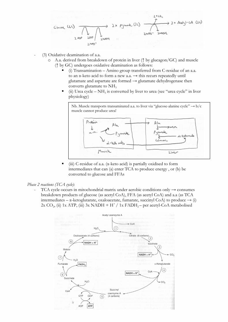
Phase 2 reactions (TCA cycle):
- TCA cycle occurs in mitochondrial matrix under aerobic conditions only → consumes breakdown products of glucose (as acetyl CoA), FFA (as acetyl CoA) and a.a (as TCA intermediates – α-ketoglutarate, oxaloacetate, fumarate, succinyl CoA) to produce → (i) 2x CO2, (ii) 1x ATP, (iii) 3x NADH + H+ / 1x FADH2 – per acetyl-CoA metabolised
Nb. Muscle transports transaminated a.a. to liver via “glucose-alanine cycle” → b/c muscle cannot produce urea!

- TCA cycle stimulated by ↓ NADH/NAD+ ratio (as NADH inhibits dehydrogenase enzymes of the cycle)
Phase 3 reactions (ETC):
- Electrons donated to ETC by NADH/FADH2 → passed along series of cytochromes (along inside surface of inner mitochondrial membrane) down its energy gradient until they are accepted by O2 at the end (via cytochrome a)
- At each cytochrome, NADH/FADH2 are re-oxidised → releases energy to extrude H+ across IMM from matrix into inner membrane space → produces a [ ] gradient of H+ → also regenerates NAD+/FADH for reuse in phase 1 and 2 reactions (see above)
- At 3 separate points in IMM, channels allow H+ to flow down its [ ] gradient back into matrix → this releases energy to produce ATP from ADP (via oxidative phosphorylation)
- At end of ETC → O2 is reduced by H+ released by re-oxidation of NADH/FADH2 along IMM → forms H2O
- ETC stimulated by ↓ NADH/NAD+ ratio (as NADH ↓ oxidation rate of ETC enzymes)
Anaerobic metabolism: - In absence of cellular O2 (anaerobic conditions), glycolysis is the ONLY catabolic
pathway that can occur: o Glucose is converted to pyrvuate as usual (and producing net 2x ATP and 2x
NADH) → BUT pyruvate is converted to lactate via LDH
o This is vital for anaerobic glycolysis to continue → b/c this allows NADH to be reoxidised to regenerate NAD+ for reuse in glycolysis
o Lactate then accumulates as anaerobic glycolysis continues → it is transported to liver where it is resynthesised as glucose (via Cori cycle)
- ETC cannot function because it requires O2 as a terminal acceptor of electrons → results in accumulation of NADH/FADH2 and loss of oxidative phosphorylation
- TCA cycle cannot function because → pyruvate is no longer converted to acetyl CoA, and there is lack of NAD+/FADH (due to ↑ NADH/FADH2 levels 2° to defunct ETC)
Remember: - NADH donates
electrons at start of ETC → 3 ATP made per NADH oxidised
- FADH2 donates electrons later in ETC → 2 ATP made per FADH2 oxidised

(g) To describe the composition of intracellular fluid and its regulation including the role of the sodium-potassium pump.
Composition of Intra- and Extracellular Fluid:
Ion Intracellular (mmol/L) Extracellular (mmol/L) K+ 150 5 Na+ 2 140 Cl- 10 105
Other anions 65 0 Regulation of Intracellular Fluid Composition:
- (1) Na+/K+ ATPase (main factor) – Primary active transporter that extrudes 3 Na+ out of the cell for 2 K+ into the cell for every ATP hydrolysed
- (2) Gibbs-Donnan effect – Presence of large non-diffusible –ve charge moieties intracellularly affects the distribution of permeable ions across the cell membrane
- (3) Membrane permeability – Cell membrane is largely permeable to K+ flow across it (due to non-gated K+ channels) but is impermeable to other ions (such as Na+, Ca2+, Etc.)
Overview of the Na+/K+ ATPase:
- Found in virtually all cells of the body – It is a heterodimer integral membrane protein that spans the entire membrane:
o α-subunit (larger subunit) – Binds Na+, ATP and PO43- intracellularly, and K+ and
ouabain/glycosides extracellularly o β-subunit (smaller subunit) – A glycoprotein
- Functions as an “electrogenic pump” as it extrudes 3 Na+ from the cell and takes in 2 K+ per ATP hydrolysed
- Process of pump function: o (i) Na+ and ATP bind to α-subunit intracellularly o (ii) ATP is hydrolysed to ADP and the phosphate is transferred to an Asp-
phosphorylation site o (iii) Conformational change of the protein causes Na+ to be extruded into ECF o (iv) K+ binds to the α-subunit extracellularly o (v) Dephosphorylation of the Asp-phosphorylation site causes the protein to
return to its resting conformation, which then transports K+ intracellularly - Pump function is inhibited by ouabain and digitalis glycosides

Voltage-gated Na+ channel: - It is an integral membrane protein consisting of 4 domains:
o Each domain has 6 α-helixes (S1-S6), with S4 having a +ve charge and S1-S3 having –ve charges
o These domains enclose a central membrane pore that is lined by –vely charged helixes and has a +vely charged “sensor”
- Channel function: o In its resting state, the channel is closed – The +ve charged sensor (S4 helix) is
attracted by the –ve intracellular charge o Membrane depolarisation causes the +vely charged sensor swings towards the
outside of the membrane (Ie. conformational change), causing the activation gate to open, thus allowing ion flow through the opened central pore
o Channel is inactivated promptly (opens for 0.7 ms only) before it can open again K+ channel:
- “Delayed rectifier” K+ channel is tetrameric with 4 subunits forming a central ion pore - Each subunit has a paddle-like extension has a +ve charge such that:
o When the channel is closed, the paddle-like extensions are near the –vely charged interior of the cell
o When MP is reduced, the paddle-like extensions bend through the cell membrane to the exterior surface, thus opening the channel pore