![Cellular Biochemistry of the Stepwise Development of ... › content › canres › ... · [CANCER RESEARCH 44, 5463-5474, December 1984] Cellular Biochemistry of the Stepwise Development](https://static.fdocuments.net/doc/165x107/5f138bb3dafc99707f18b76b/cellular-biochemistry-of-the-stepwise-development-of-a-content-a-canres.jpg)
Cellular Biochemistry: Cell Structure & Function.
163
Cellular Biochemistry: Cellular Biochemistry: Cell Structure & Function Cell Structure & Function
-
date post
22-Dec-2015 -
Category
Documents
-
view
228 -
download
1
Transcript of Cellular Biochemistry: Cell Structure & Function.
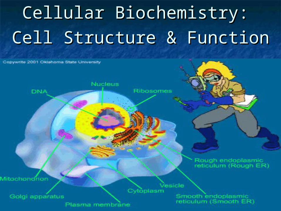
Cellular Biochemistry: Cellular Biochemistry:
Cell Structure & FunctionCell Structure & Function
A TOUR OF THE CELL
Copyright © 2002 Pearson Education, Inc., publishing as Benjamin Cummings
Section A: How We Study Cells
1. Microscopes provide windows to the world of the cell
2. Cell biologists can isolate organelles to study their function
• The discovery and early study of cells progressed with the invention and improvement of microscopes in the 17th century.
• In a light microscope (LMs) visible light passes through the specimen and then through glass lenses.
• The lenses refract light such that the image is magnified into the eye or a video screen.
1 .Microscopes provide windows to the world of the cell
Copyright © 2002 Pearson Education, Inc., publishing as Benjamin Cummings
• Microscopes vary in magnification and resolving power.
• Magnification is the ratio of an object’s image to its real size.
• Resolving power is a measure of image clarity.
• It is the minimum distance two points can be separated and still viewed as two separate points.
• Resolution is limited by the shortest wavelength of the source, in this case light.
Copyright © 2002 Pearson Education, Inc., publishing as Benjamin Cummings
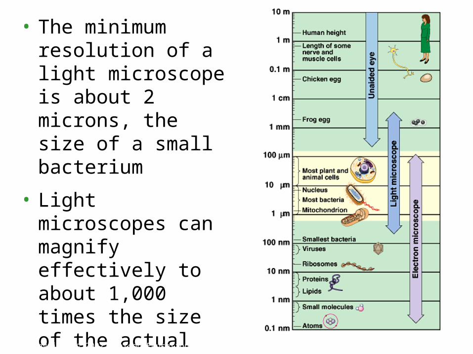
• The minimum resolution of a light microscope is about 2 microns, the size of a small bacterium
• Light microscopes can magnify effectively to about 1,000 times the size of the actual specimen.
• At higher magnifications, the image blurs.
Copyright © 2002 Pearson Education, Inc., publishing as Benjamin Cummings
Fig. 7.1
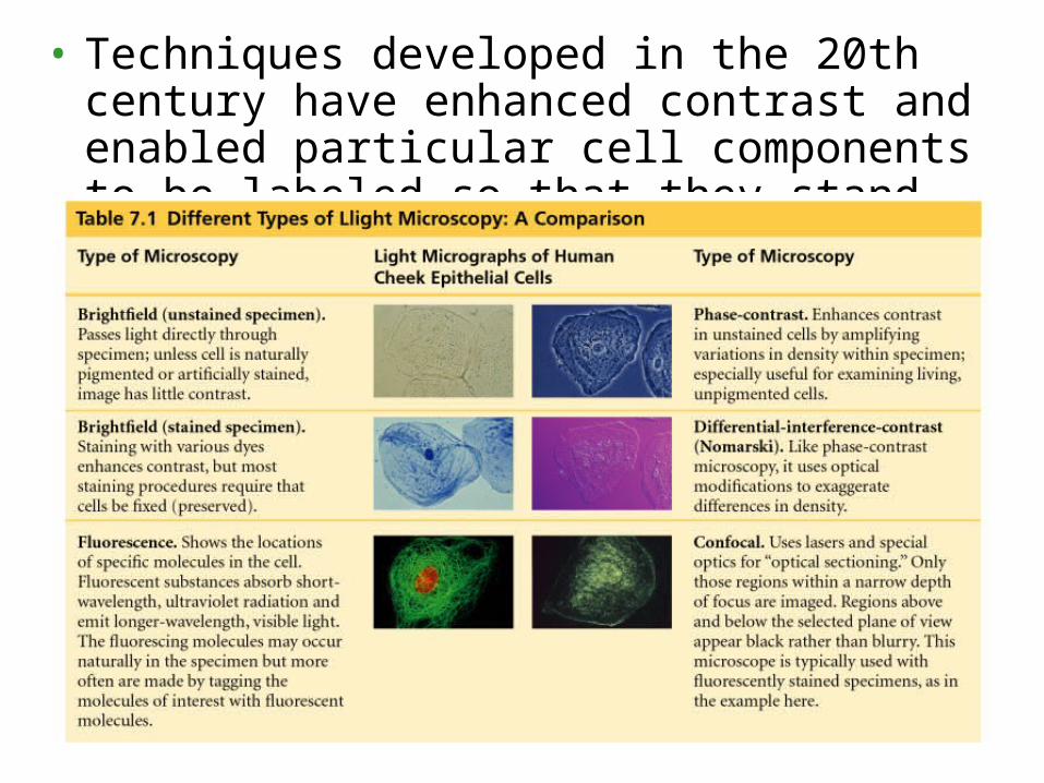
• Techniques developed in the 20th century have enhanced contrast and enabled particular cell components to be labeled so that they stand out.
Copyright © 2002 Pearson Education, Inc., publishing as Benjamin Cummings
• While a light microscope can resolve individual cells, it cannot resolve much of the internal anatomy, especially the organelles.
• To resolve smaller structures we use an electron microscope (EM), which focuses a beam of electrons through the specimen or onto its surface.
• Because resolution is inversely related to wavelength used, electron microscopes with shorter wavelengths than visible light have finer resolution.
• Theoretically, the resolution of a modern EM could reach 0.1 nanometer (nm), but the practical limit is closer to about 2 nm.
Copyright © 2002 Pearson Education, Inc., publishing as Benjamin Cummings
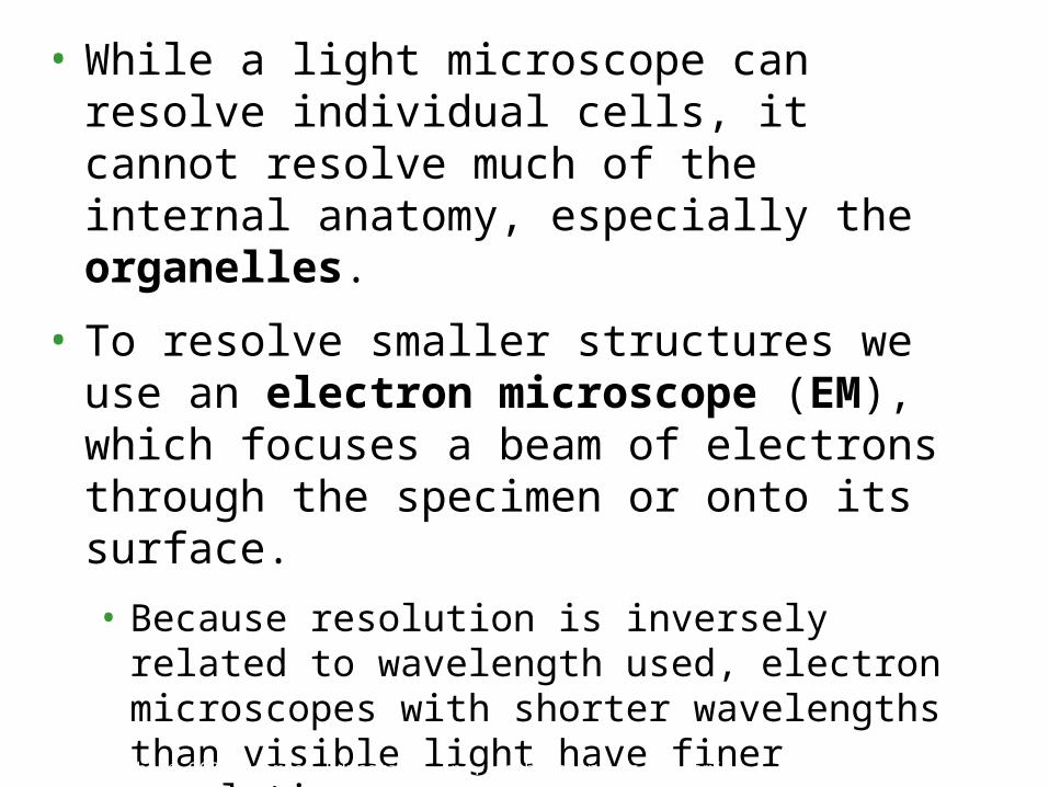
• Transmission electron microscopes (TEM) are used mainly to study the internal ultrastructure of cells.
• A TEM aims an electron beam through a thin section of the specimen.
• The image is focused and magnified by electromagnets.
• To enhance contrast, the thin sections are stained with atoms of heavy metals.
Copyright © 2002 Pearson Education, Inc., publishing as Benjamin Cummings
Fig. 7.2a
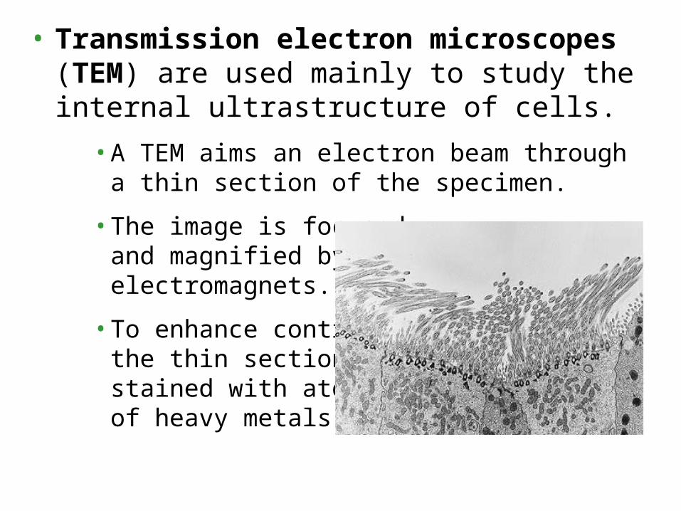
• Scanning electron microscopes (SEM) are useful for studying surface structures.
• The sample surface is covered with a thin film of gold.
• The beam excites electrons on the surface.
• These secondary electrons are collected and focused on a screen.
• The SEM has great depth of field, resulting in an image that seems three-dimensional.
Copyright © 2002 Pearson Education, Inc., publishing as Benjamin Cummings
Fig. 7.2b
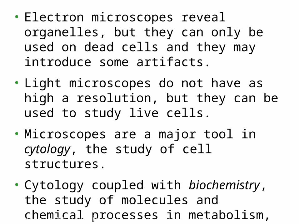
• Electron microscopes reveal organelles, but they can only be used on dead cells and they may introduce some artifacts.
• Light microscopes do not have as high a resolution, but they can be used to study live cells.
• Microscopes are a major tool in cytology, the study of cell structures.
• Cytology coupled with biochemistry, the study of molecules and chemical processes in metabolism, developed modern cell biology.
Copyright © 2002 Pearson Education, Inc., publishing as Benjamin Cummings
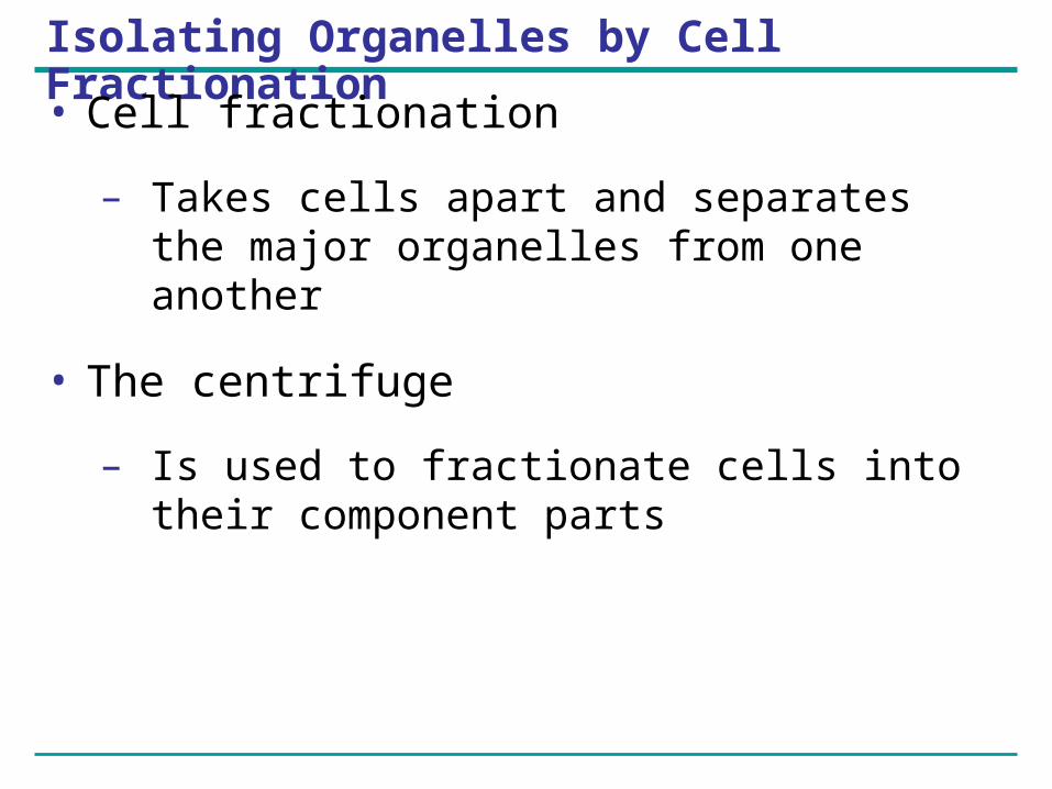
Isolating Organelles by Cell Fractionation
• Cell fractionation
– Takes cells apart and separates the major organelles from one another
• The centrifuge
– Is used to fractionate cells into their component parts
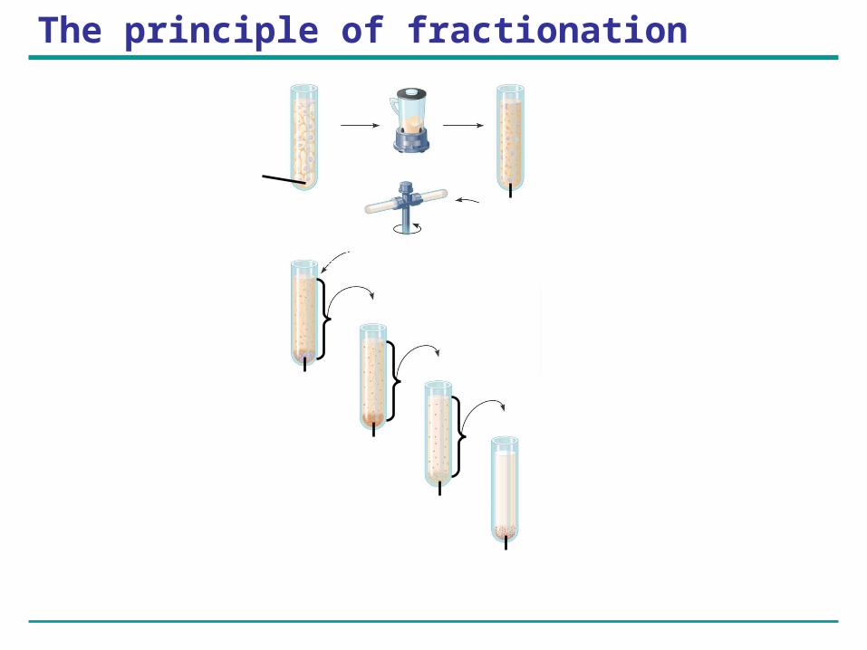
The principle of fractionation
Tissuecells
Homogenization
Homogenate1000 g(1000 times theforce of gravity)
10 min Differential centrifugationSupernatant pouredinto next tube
20,000 g20 min
Pellet rich innuclei andcellular debris
Pellet rich inmitochondria(and chloro-plasts if cellsare from a plant)
Pellet rich in“microsomes”(pieces of plasma mem-branes andcells’ internalmembranes)
Pellet rich inribosomes
150,000 g3 hr
80,000 g60 min
Figure 6.5

• The goal of cell fractionation is to separate the major organelles of the cells so that their individual functions can be studied.
2 .Cell biologists can isolate organelles to study their functions
Copyright © 2002 Pearson Education, Inc., publishing as Benjamin CummingsFig. 7.3
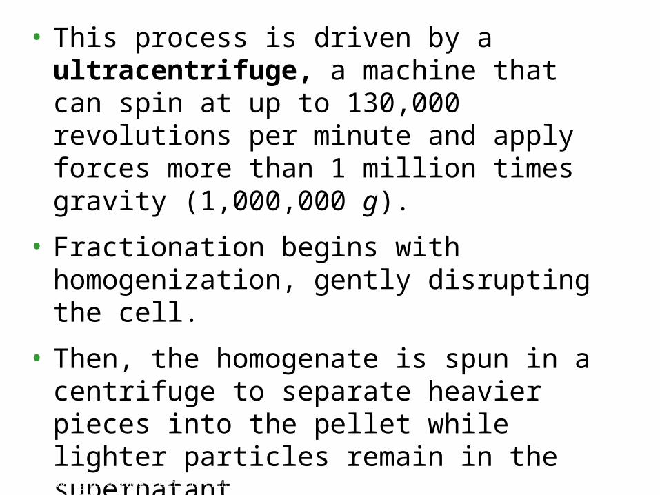
• This process is driven by a ultracentrifuge, a machine that can spin at up to 130,000 revolutions per minute and apply forces more than 1 million times gravity (1,000,000 g).
• Fractionation begins with homogenization, gently disrupting the cell.
• Then, the homogenate is spun in a centrifuge to separate heavier pieces into the pellet while lighter particles remain in the supernatant.
• As the process is repeated at higher speeds and longer durations, smaller and smaller organelles can be collected in subsequent pellets.
Copyright © 2002 Pearson Education, Inc., publishing as Benjamin Cummings
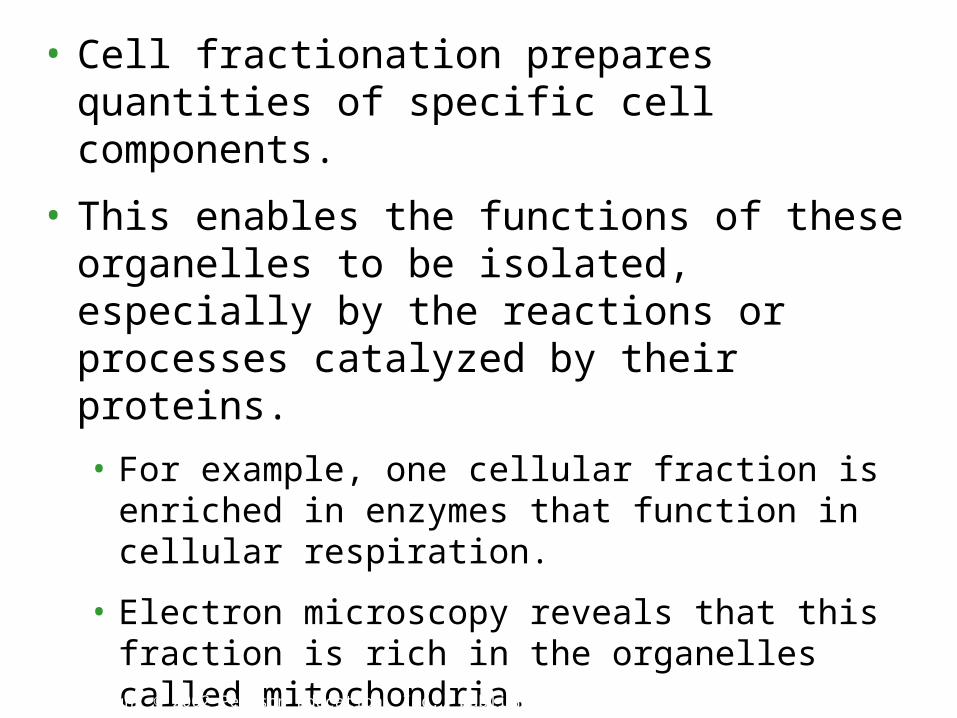
• Cell fractionation prepares quantities of specific cell components.
• This enables the functions of these organelles to be isolated, especially by the reactions or processes catalyzed by their proteins.
• For example, one cellular fraction is enriched in enzymes that function in cellular respiration.
• Electron microscopy reveals that this fraction is rich in the organelles called mitochondria.
• Cytology and biochemistry complement each other in connecting cellular structure and function.
Copyright © 2002 Pearson Education, Inc., publishing as Benjamin Cummings
The CellThe Cell
A cell is the smallest unit of A cell is the smallest unit of livingliving matter. matter.
Don’t confuse this with: atom, Don’t confuse this with: atom, element, proton, etc.element, proton, etc.
Cell TheoryCell Theory
Who?Who? Matthias Schleiden, Theodor Matthias Schleiden, Theodor Schwann, Rudolf VirchowSchwann, Rudolf Virchow
When?When? 1800s 1800s What does it say?What does it say?
All organisms are made of cells.All organisms are made of cells. A cell is the structural & function unit of A cell is the structural & function unit of
organs.organs. All cells come from pre-existing cells.All cells come from pre-existing cells. Cells are capable of self-reproduction.Cells are capable of self-reproduction.
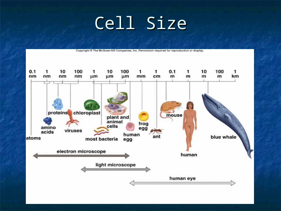
Cell SizeCell Size
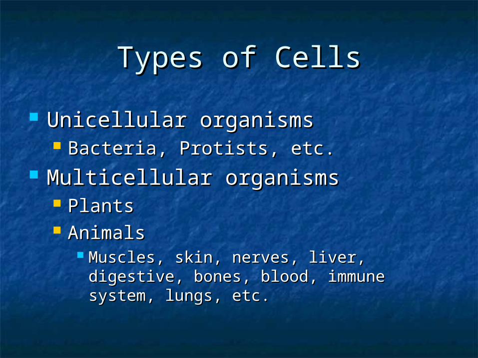
Types of CellsTypes of Cells
Unicellular organismsUnicellular organisms Bacteria, Protists, etc.Bacteria, Protists, etc.
Multicellular organismsMulticellular organisms PlantsPlants AnimalsAnimals
Muscles, skin, nerves, liver, digestive, bones, Muscles, skin, nerves, liver, digestive, bones, blood, immune system, lungs, etc.blood, immune system, lungs, etc.
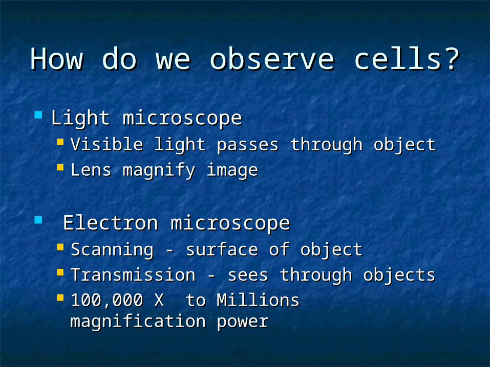
How do we observe cells?How do we observe cells?
Light microscopeLight microscope Visible light passes through objectVisible light passes through object Lens magnify imageLens magnify image
Electron microscopeElectron microscope Scanning - surface of objectScanning - surface of object Transmission - sees through objectsTransmission - sees through objects 100,000 X to Millions magnification 100,000 X to Millions magnification
powerpower
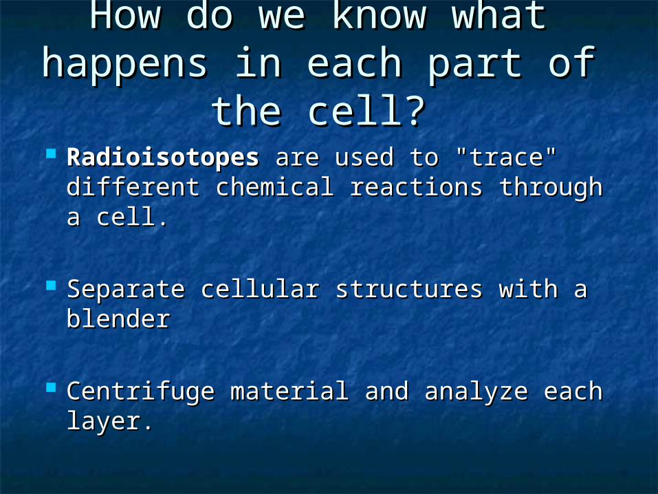
How do we know what How do we know what happens in each part of the happens in each part of the
cell?cell? RadioisotopesRadioisotopes are used to "trace" are used to "trace"
different chemical reactions through a different chemical reactions through a cell.cell.
Separate cellular structures with a blenderSeparate cellular structures with a blender
Centrifuge material and analyze each Centrifuge material and analyze each layer.layer.
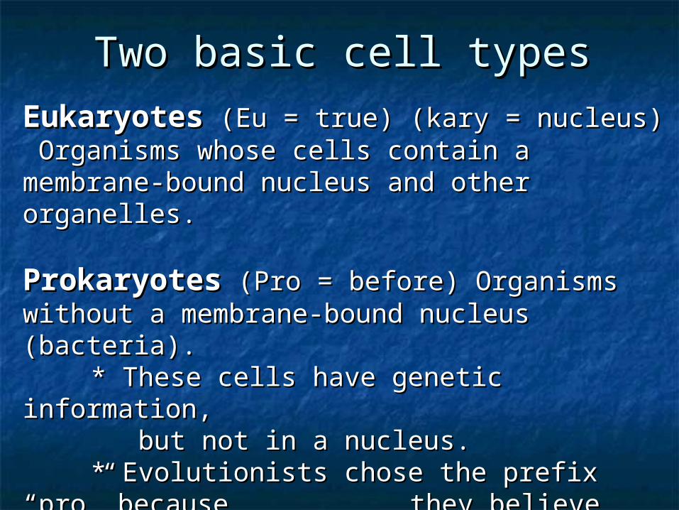
Two basic cell typesTwo basic cell types
EukaryotesEukaryotes (Eu = true) (kary = nucleus) (Eu = true) (kary = nucleus) Organisms whose cells contain a membrane-Organisms whose cells contain a membrane-bound nucleus and other organelles.bound nucleus and other organelles.
ProkaryotesProkaryotes (Pro = before) Organisms (Pro = before) Organisms without a membrane-bound nucleus (bacteria).without a membrane-bound nucleus (bacteria).
* These cells have genetic information, * These cells have genetic information, but not in a nucleus.but not in a nucleus.* Evolutionists chose the prefix “pro” * Evolutionists chose the prefix “pro”
because because they believe these evolved they believe these evolved before others.before others.
Prokaryotic CellsProkaryotic Cells
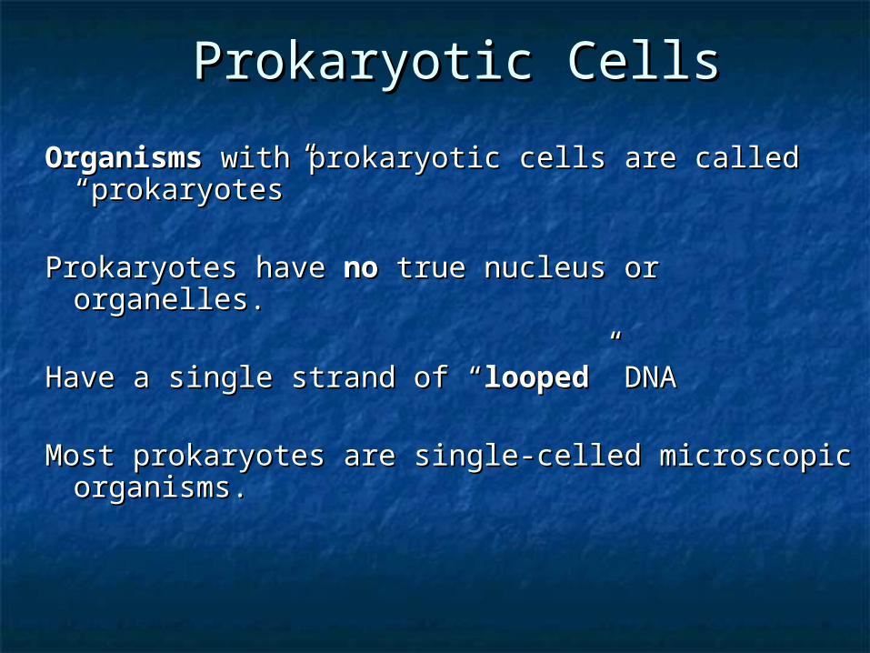
OrganismsOrganisms with prokaryotic cells are called with prokaryotic cells are called “prokaryotes”“prokaryotes”
Prokaryotes have Prokaryotes have nono true nucleus or organelles. true nucleus or organelles.
Have a single strand of “Have a single strand of “loopedlooped” DNA” DNA
Most prokaryotes are single-celled microscopic Most prokaryotes are single-celled microscopic organisms.organisms.

Eukaryotic CellsEukaryotic Cells Organisms composed of eukaryotic Organisms composed of eukaryotic
cells are called “eukaryotes”cells are called “eukaryotes” Have a membrane bound nucleus which Have a membrane bound nucleus which
contains the cell’s DNAcontains the cell’s DNA Some eukaryotes are one-celled Some eukaryotes are one-celled
organismsorganisms All multicellular organisms are All multicellular organisms are
eukaryoteseukaryotes Have Have organellesorganelles, each of which is , each of which is
surrounded by (or bound in) a “plasma surrounded by (or bound in) a “plasma membrane”membrane”
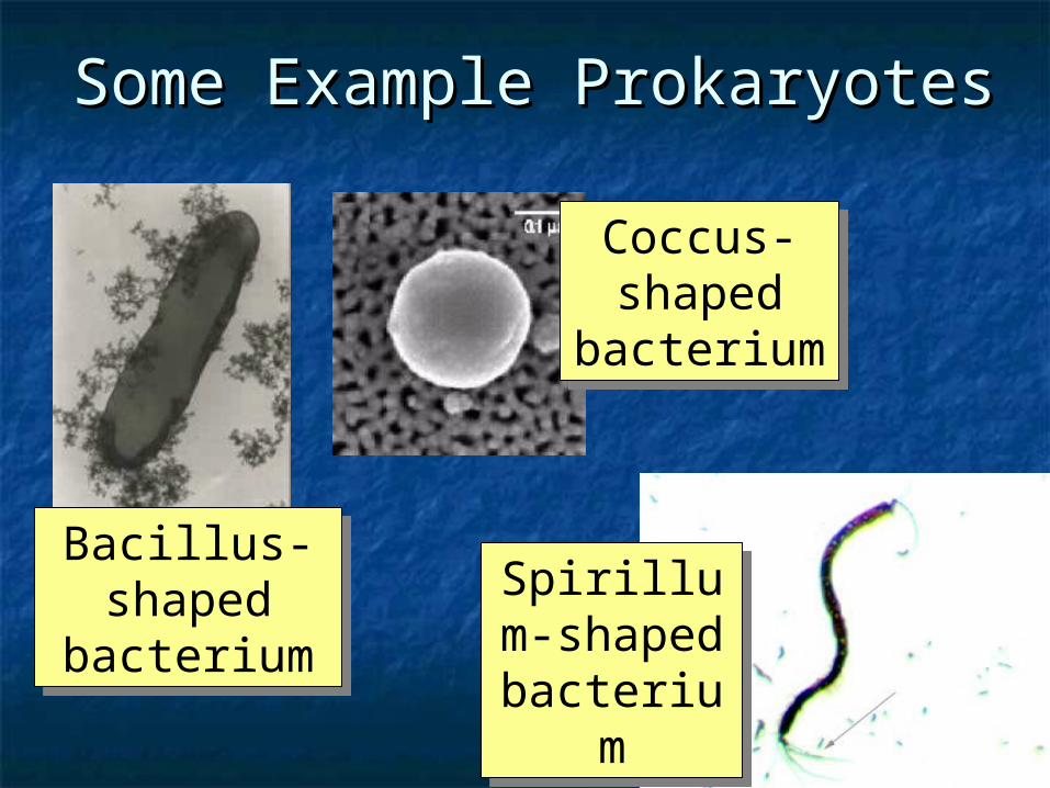
Some Example ProkaryotesSome Example Prokaryotes
Bacillus-shaped
bacterium
Bacillus-shaped
bacterium
Coccus-shaped
bacterium
Coccus-shaped
bacterium
Spirillum-shaped
bacterium
Spirillum-shaped
bacterium
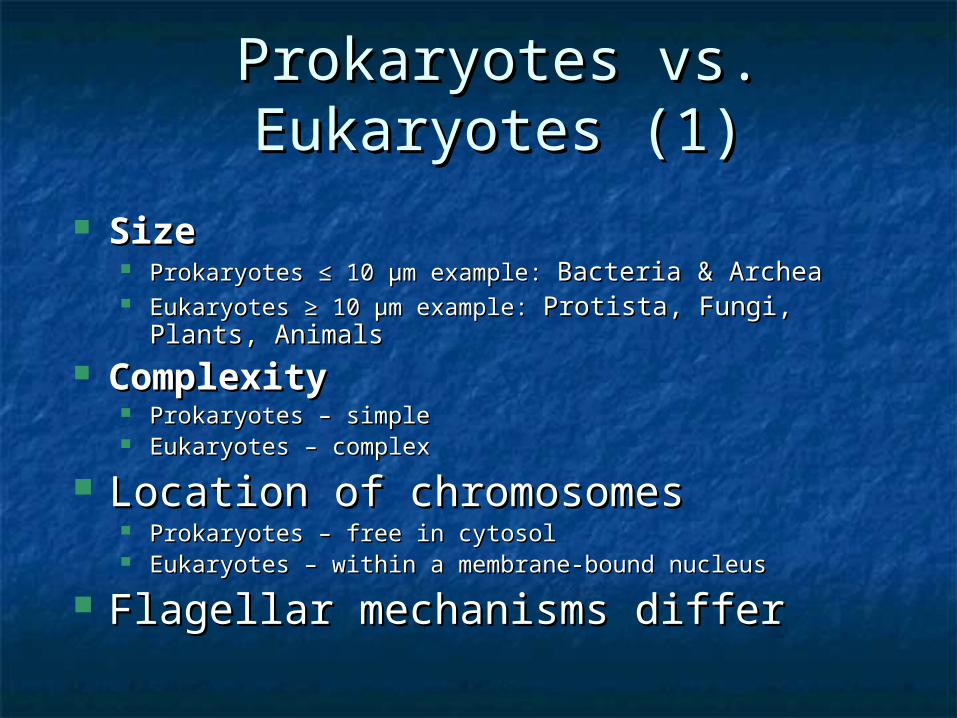
Prokaryotes vs. Eukaryotes Prokaryotes vs. Eukaryotes (1)(1)
SizeSize Prokaryotes Prokaryotes ≤≤ 10 10 µm example: µm example: BacteriaBacteria & Archea& Archea Eukaryotes Eukaryotes ≥≥ 10 µm example: 10 µm example: Protista, Fungi, Plants, Protista, Fungi, Plants,
AnimalsAnimals ComplexityComplexity
Prokaryotes – simpleProkaryotes – simple Eukaryotes Eukaryotes –– complex complex
Location of chromosomesLocation of chromosomes Prokaryotes – free in cytosolProkaryotes – free in cytosol Eukaryotes – within a membrane-bound nucleusEukaryotes – within a membrane-bound nucleus
Flagellar mechanisms differFlagellar mechanisms differ
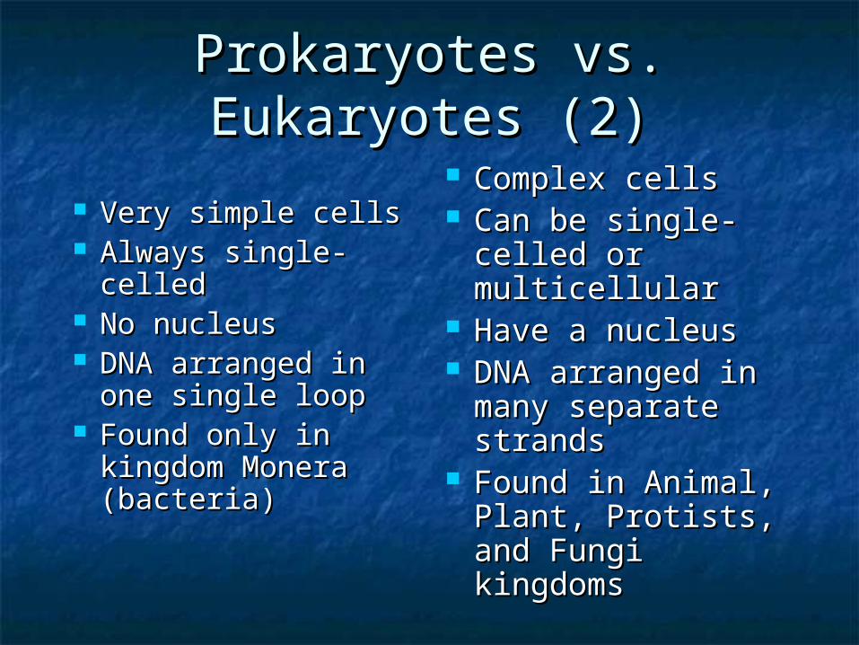
Prokaryotes vs. Prokaryotes vs. Eukaryotes (2)Eukaryotes (2)
Very simple cellsVery simple cells Always single-Always single-
celledcelled No nucleusNo nucleus DNA arranged in DNA arranged in
one single loopone single loop Found only in Found only in
kingdom Monera kingdom Monera (bacteria)(bacteria)
Complex cellsComplex cells Can be single-Can be single-
celled or celled or multicellularmulticellular
Have a nucleusHave a nucleus DNA arranged in DNA arranged in
many separate many separate strands strands
Found in Animal, Found in Animal, Plant, Protists, and Plant, Protists, and Fungi kingdomsFungi kingdoms
Prokaryotic CellsProkaryotic Cells
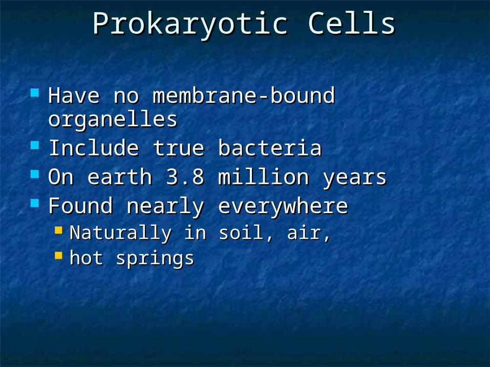
Have no membrane-bound Have no membrane-bound organellesorganelles
Include true bacteriaInclude true bacteria On earth 3.8 million yearsOn earth 3.8 million years Found nearly everywhereFound nearly everywhere
Naturally in soil, air, Naturally in soil, air, hot springshot springs

ribosomes
cell wall
plasma membrane
food granule
prokaryoticflagellum
cytoplasm
nucleoid (DNA)Prokaryotic Cells
VirusesViruses Viruses contain DNA or RNA & a protein Viruses contain DNA or RNA & a protein
coatcoat Some are enclosed by an envelopeSome are enclosed by an envelope Most viruses infect only specific types Most viruses infect only specific types
of cells in one hostof cells in one host Host range is determined by specific Host range is determined by specific
host attachment sites and cellular host attachment sites and cellular factorsfactors
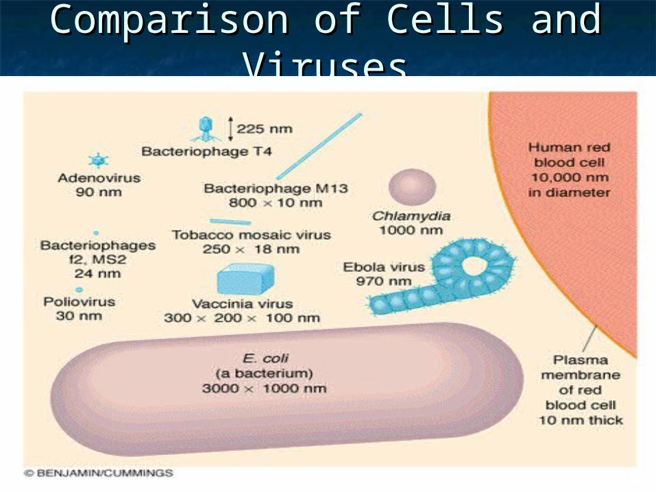
Comparison of Cells and Comparison of Cells and VirusesViruses

Bacterium (prokaryote)
Animal (eukaryote)
Plant (eukaryote)
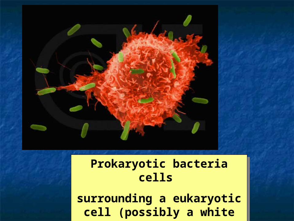
Prokaryotic bacteria cells
surrounding a eukaryotic cell (possibly a white
blood cell?)
Prokaryotic bacteria cells
surrounding a eukaryotic cell (possibly a white
blood cell?)
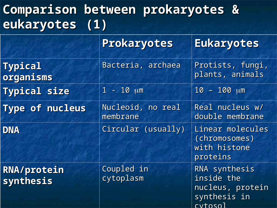
ProkaryotesProkaryotes EukaryotesEukaryotesTypical Typical organismsorganisms
Bacteria, archaeaBacteria, archaea Protists, fungi, Protists, fungi, plants, animalsplants, animals
Typical sizeTypical size 1 - 10 1 - 10 mm 10 – 100 10 – 100 mm
Type of nucleusType of nucleus Nucleoid, no real Nucleoid, no real membrane membrane
Real nucleus w/ Real nucleus w/ double membranedouble membrane
DNADNA Circular (usually)Circular (usually) Linear molecules Linear molecules (chromosomes) with (chromosomes) with histone proteinshistone proteins
RNA/protein RNA/protein synthesissynthesis
Coupled in cytoplasmCoupled in cytoplasm RNA synthesis RNA synthesis inside the nucleus, inside the nucleus, protein synthesis in protein synthesis in cytosolcytosol
Comparison between prokaryotes & Comparison between prokaryotes & eukaryoteseukaryotes (1) (1)
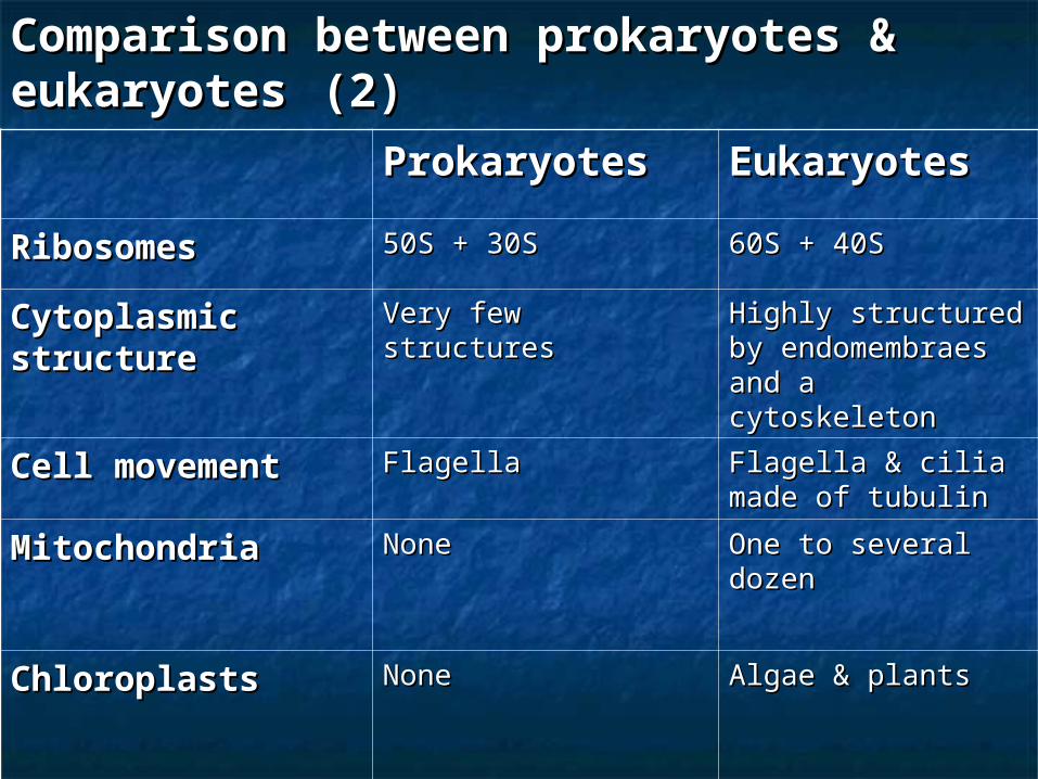
ProkaryotesProkaryotes EukaryotesEukaryotes
RibosomesRibosomes 50S + 30S50S + 30S 60S + 40S60S + 40S
Cytoplasmic Cytoplasmic structurestructure
Very few structuresVery few structures Highly structured by Highly structured by endomembraes and endomembraes and a cytoskeletona cytoskeleton
Cell movementCell movement FlagellaFlagella Flagella & cilia Flagella & cilia made of tubulinmade of tubulin
MitochondriaMitochondria NoneNone One to several One to several dozendozen
ChloroplastsChloroplasts NoneNone Algae & plantsAlgae & plants
Comparison between prokaryotes & Comparison between prokaryotes & eukaryoteseukaryotes (2) (2)
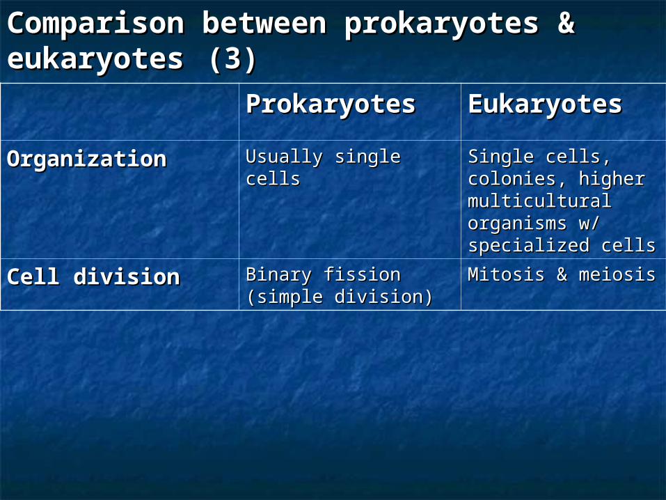
ProkaryotesProkaryotes EukaryotesEukaryotesOrganizationOrganization Usually single cellsUsually single cells Single cells, Single cells,
colonies, higher colonies, higher multicultural multicultural organisms w/ organisms w/ specialized cells specialized cells
Cell divisionCell division Binary fission (simple Binary fission (simple division)division)
Mitosis & meiosisMitosis & meiosis
Comparison between prokaryotes & Comparison between prokaryotes & eukaryoteseukaryotes (3) (3)
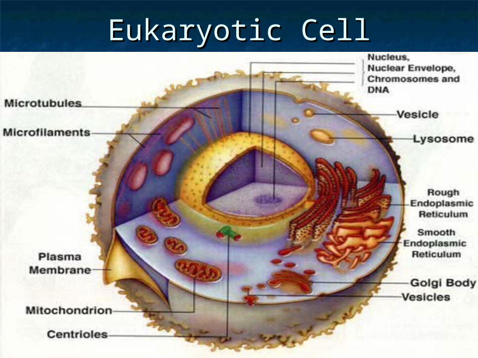
Eukaryotic CellEukaryotic Cell
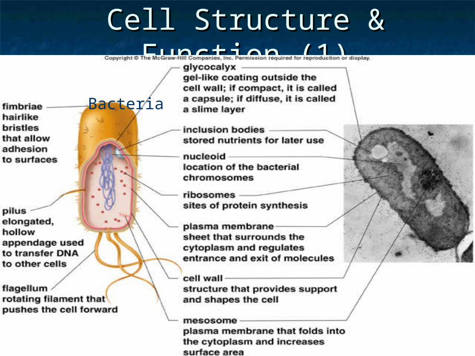
Cell Structure & Function (1)Cell Structure & Function (1)
Bacteria
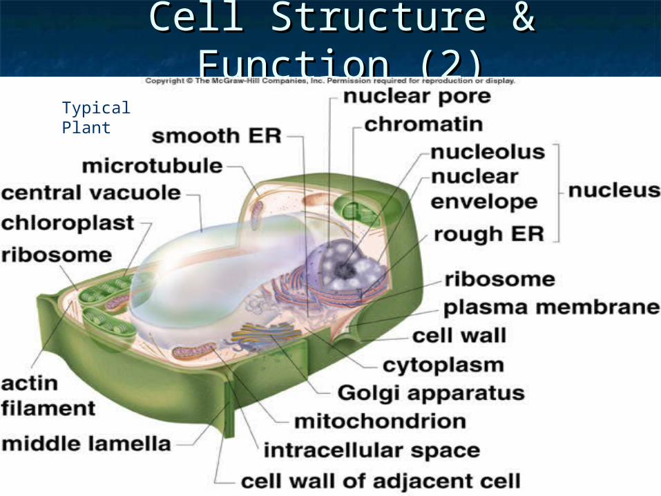
Cell Structure & Function (2)Cell Structure & Function (2)
Typical Plant
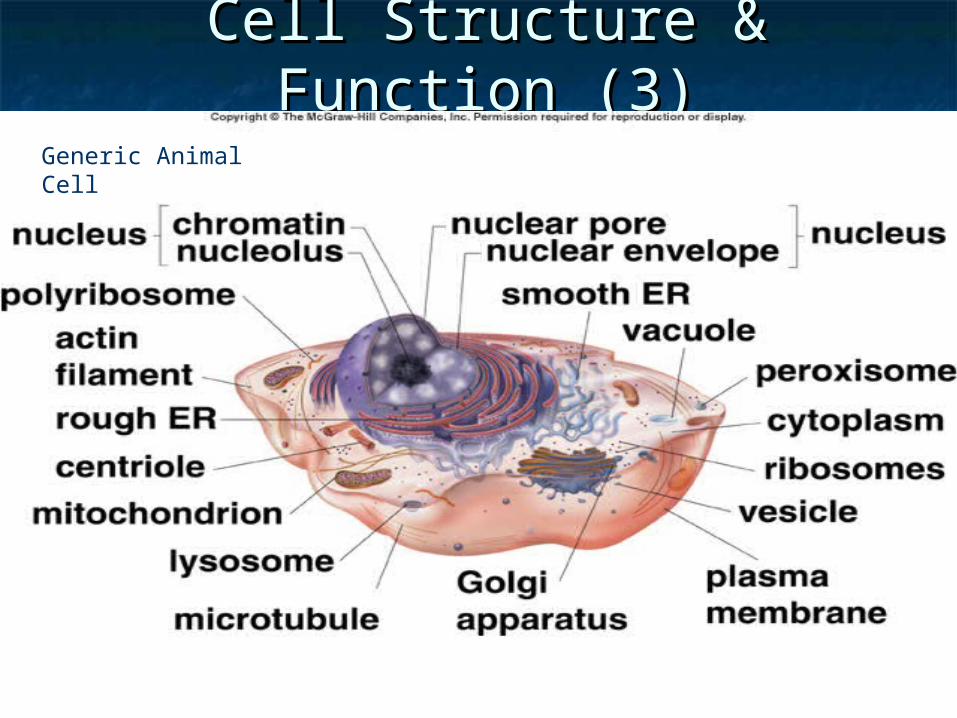
Cell Structure & Function (3)Cell Structure & Function (3)
Generic Animal Cell
Eukaryotic Cells Structure Eukaryotic Cells Structure (1)(1)
Have numerous internal Have numerous internal structuresstructures
Various types & formsVarious types & forms Plants, animals, fungi, protistsPlants, animals, fungi, protists
Multicellular organismsMulticellular organisms
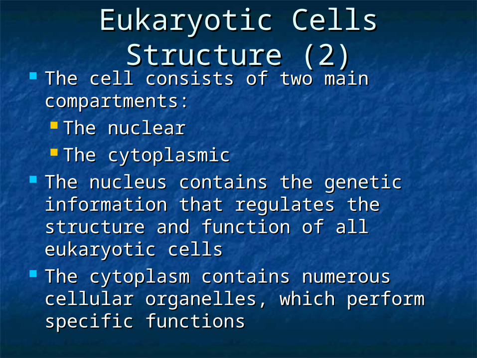
Eukaryotic Cells Structure Eukaryotic Cells Structure (2)(2)
The cell consists of two main The cell consists of two main compartments: compartments: The nuclearThe nuclear The cytoplasmicThe cytoplasmic
The nucleus contains the genetic The nucleus contains the genetic information that regulates the structure information that regulates the structure and function of all eukaryotic cellsand function of all eukaryotic cells
The cytoplasm contains numerous The cytoplasm contains numerous cellular organelles, which perform cellular organelles, which perform specific functionsspecific functions
Plant & Animal Cells (1)Plant & Animal Cells (1)
SimilaritiesSimilarities
Both constructed from eukaryotic cellsBoth constructed from eukaryotic cells
Both contain similar organellesBoth contain similar organelles
Both surrounded by cell membraneBoth surrounded by cell membrane
Plant & Animal Cells (2)Plant & Animal Cells (2) DifferencesDifferences
Plants havePlants have Cell wall – provides strength & rigidityCell wall – provides strength & rigidity Have chloroplasts, photosynthetic siteHave chloroplasts, photosynthetic site Large vacuolesLarge vacuoles
Animals haveAnimals haveOther organelle not found in plants Other organelle not found in plants (lysosomes formed from Golgi)(lysosomes formed from Golgi)
Centrioles, important in cell divisionCentrioles, important in cell division
Cellular OrganellesCellular Organelles CytoplasmCytoplasm NucleusNucleus
Chromosomes, Chromosomes, nuclear envelope, nuclear envelope, nuclear pores, nuclear pores, nucleolusnucleolus
RibosomesRibosomes Endoplasmic Endoplasmic
reticulum (smooth & reticulum (smooth & rough)rough)
Golgi ApparatusGolgi Apparatus
LysosomesLysosomes VesiclesVesicles PeroxisomesPeroxisomes VacuolesVacuoles ChloroplastChloroplast MitochondriaMitochondria CytoskeletonCytoskeleton CentriolesCentrioles Cilia, FlagellaCilia, Flagella Plasma MembranePlasma Membrane
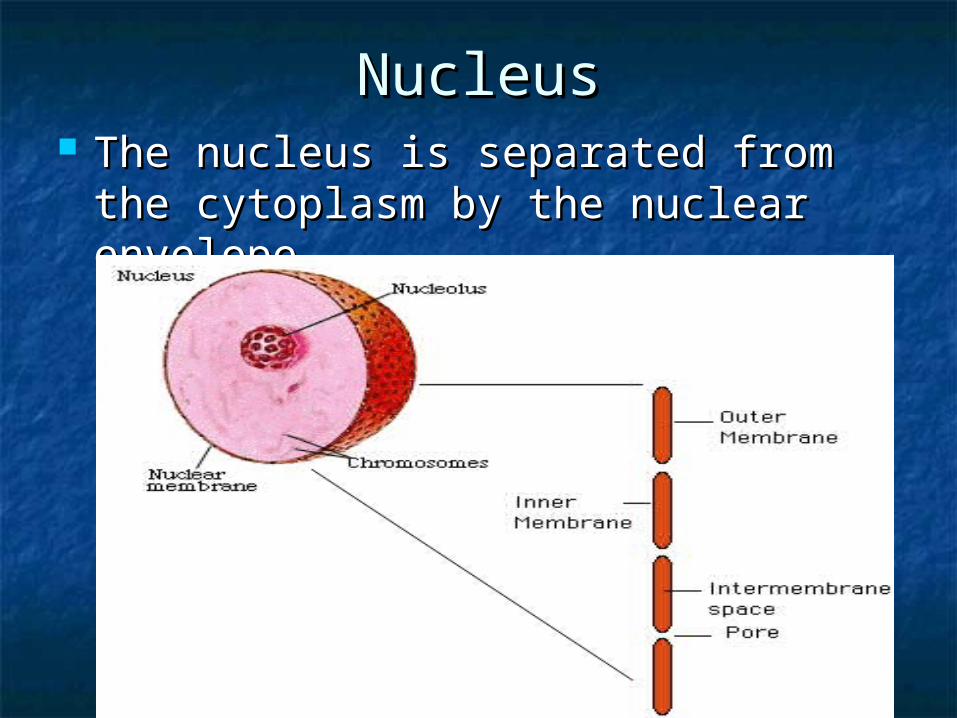
NucleusNucleus The nucleus is separated from the The nucleus is separated from the
cytoplasm by the nuclear envelope cytoplasm by the nuclear envelope
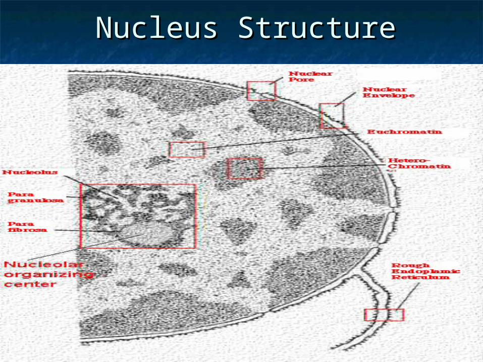
Nucleus StructureNucleus Structure
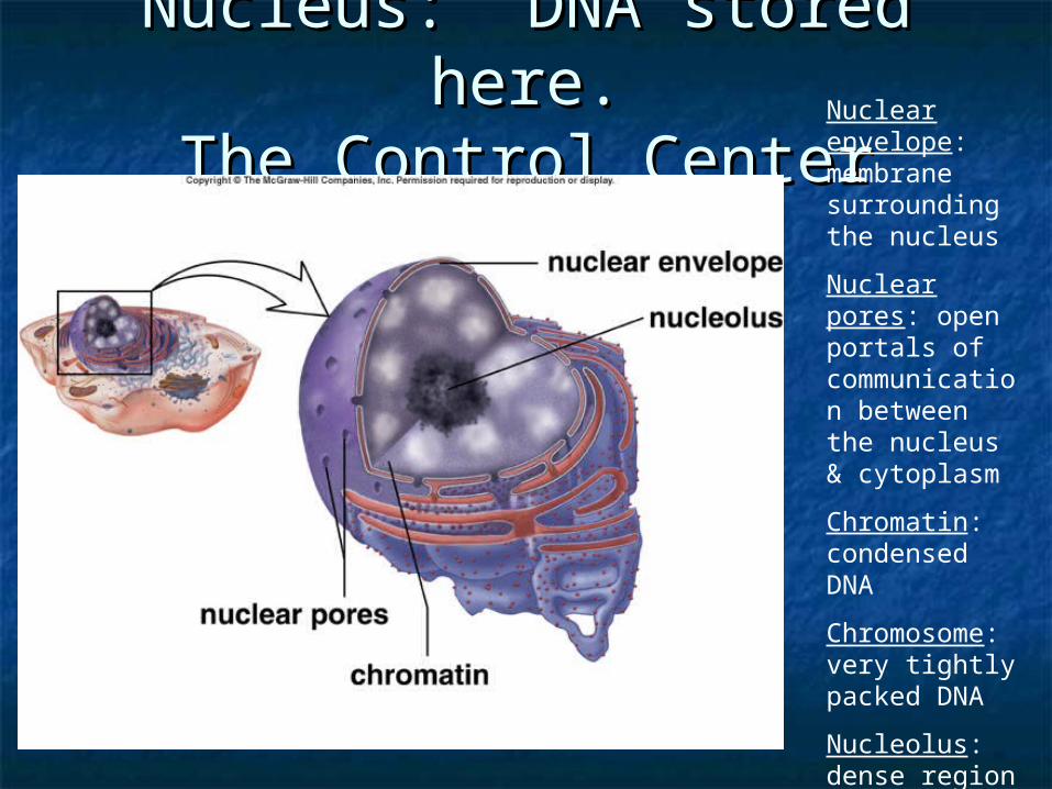
Nucleus: DNA stored here.Nucleus: DNA stored here.The Control CenterThe Control CenterNuclear
envelope: membrane surrounding the nucleus
Nuclear pores: open portals of communication between the nucleus & cytoplasm
Chromatin: condensed DNA
Chromosome: very tightly packed DNA
Nucleolus: dense region of chromatin
DNA proteinsDNA proteins
DNA is associated with two major types of DNA is associated with two major types of proteins: proteins:
The histone and nonhistone chromosomal The histone and nonhistone chromosomal proteins proteins
The histones are primarily structure The histones are primarily structure molecules that pack DNA into chromatin molecules that pack DNA into chromatin fibersfibers
The nonhistones include proteins that carry The nonhistones include proteins that carry out one of the most important cellular out one of the most important cellular functions, the regulation of gene activityfunctions, the regulation of gene activity
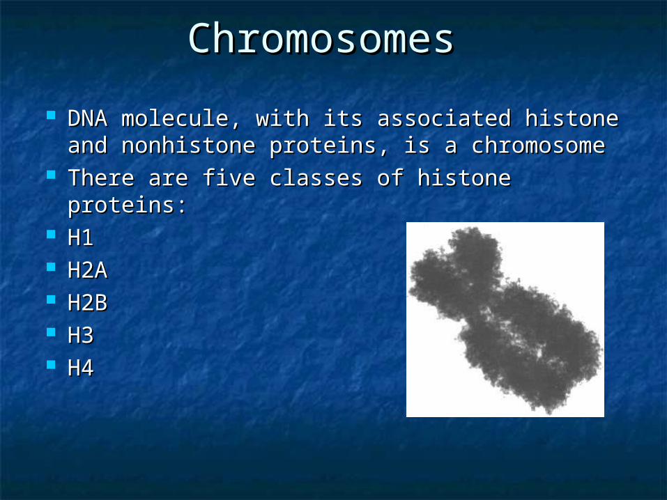
Chromosomes Chromosomes
DNA molecule, with its associated histone DNA molecule, with its associated histone and nonhistone proteins, is a chromosome and nonhistone proteins, is a chromosome
There are five classes of histone proteins: There are five classes of histone proteins: H1 H1 H2A H2A H2B H2B H3 H3 H4 H4
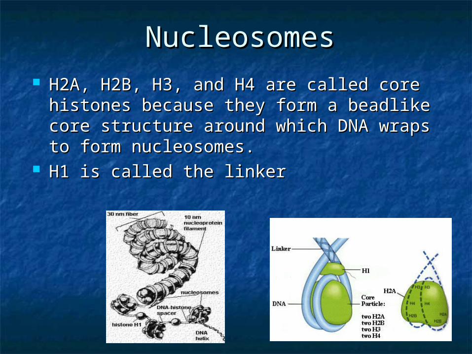
NucleosomesNucleosomes H2A, H2B, H3, and H4 are called core H2A, H2B, H3, and H4 are called core
histones because they form a beadlike histones because they form a beadlike core structure around which DNA wraps core structure around which DNA wraps to form nucleosomes. to form nucleosomes.
H1 is called the linker H1 is called the linker
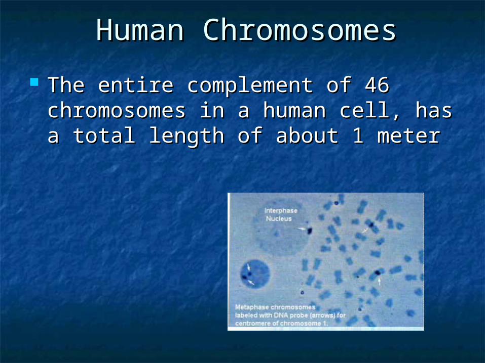
Human ChromosomesHuman Chromosomes
The entire complement of 46 The entire complement of 46 chromosomes in a human cell, has a chromosomes in a human cell, has a total length of about 1 metertotal length of about 1 meter
Nucleolus (Nucleoli) Nucleolus (Nucleoli)
The RNA of ribosomes is synthesized The RNA of ribosomes is synthesized from genes in the nucleolus from genes in the nucleolus
No membranes separate nucleoli from No membranes separate nucleoli from the surrounding chromatin in the the surrounding chromatin in the nucleusnucleus

Protein-encoding geneProtein-encoding gene
Each DNA segment containing the information in Each DNA segment containing the information in a protein constitutes a genea protein constitutes a gene
The information in a Protein-encoding gene is The information in a Protein-encoding gene is copied into a messenger RNA (mRNA) molecules copied into a messenger RNA (mRNA) molecules that moves to the cytoplasm through the pores of that moves to the cytoplasm through the pores of the nuclear envelopthe nuclear envelop
In the cytoplasm, mRNA molecules are used by In the cytoplasm, mRNA molecules are used by ribosomes as directions for the assembly of ribosomes as directions for the assembly of proteinsproteins
DNA -----------> mRNA -----------> Protein (enzymes) DNA -----------> mRNA -----------> Protein (enzymes)
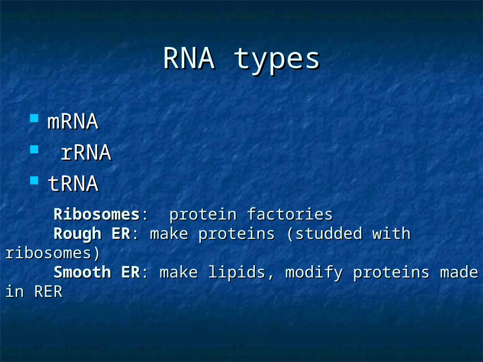
RNA typesRNA types
mRNAmRNA rRNA rRNA tRNAtRNA
RibosomesRibosomes: protein factories: protein factoriesRough ERRough ER: make proteins (studded with ribosomes): make proteins (studded with ribosomes)Smooth ERSmooth ER: make lipids, modify proteins made in : make lipids, modify proteins made in
RERRER
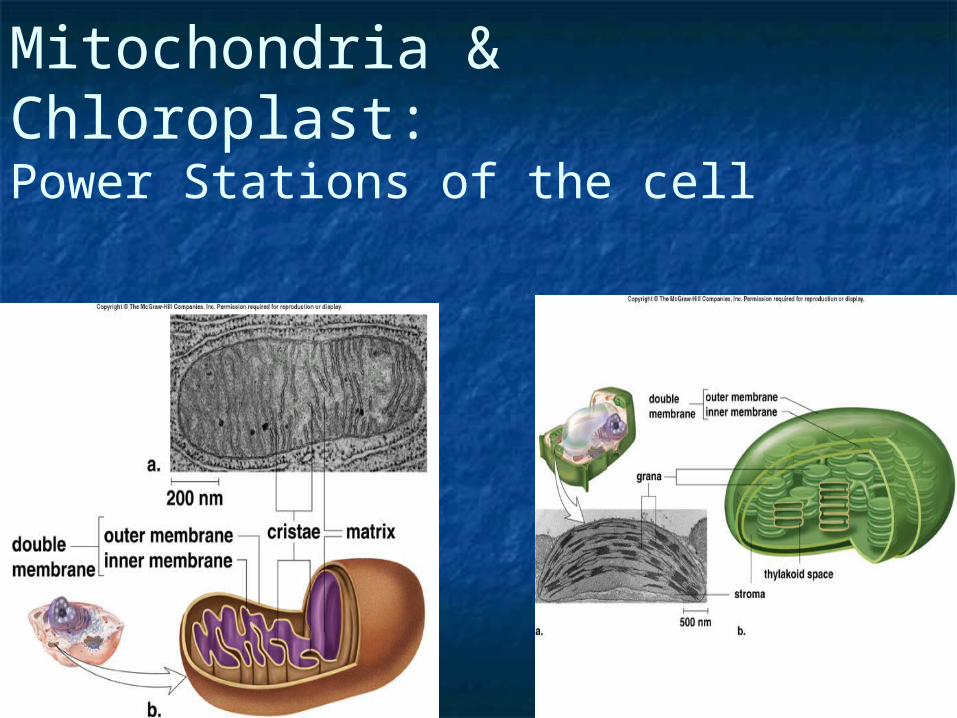
Mitochondria & Chloroplast:Power Stations of the cell
Mitochondria (1)Mitochondria (1) The mitochondria major role is ATP The mitochondria major role is ATP
production in the eukaryotic cellproduction in the eukaryotic cell These are mobile and flexible These are mobile and flexible
organelles,organelles, although in some cells they tend to although in some cells they tend to
stay in a fixed positionstay in a fixed position Mitochondria are also self-reproducing, Mitochondria are also self-reproducing,
they have their own circular DNAthey have their own circular DNA
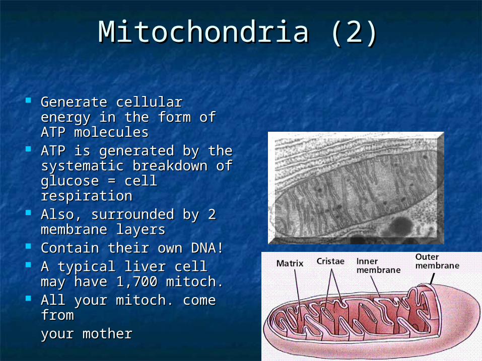
Mitochondria (2)Mitochondria (2)
Generate cellular energy Generate cellular energy in the form of ATP in the form of ATP moleculesmolecules
ATP is generated by the ATP is generated by the systematic breakdown of systematic breakdown of glucose = cell respirationglucose = cell respiration
Also, surrounded by 2 Also, surrounded by 2 membrane layersmembrane layers
Contain their own DNA!Contain their own DNA! A typical liver cell may A typical liver cell may
have 1,700 mitoch.have 1,700 mitoch. All your mitoch. come All your mitoch. come
from from your motheryour mother
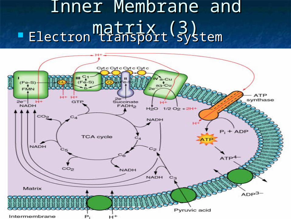
Inner Membrane and matrix Inner Membrane and matrix (3)(3) Electron transport systemElectron transport system
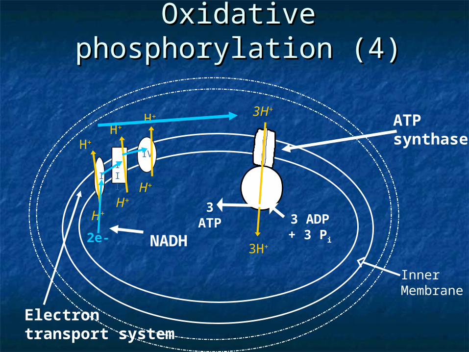
Oxidative phosphorylation Oxidative phosphorylation (4)(4)
Inner Membrane
ATPsynthase
Electrontransport system
IIII
IV
H+
H+
H+
H+ 3H+
3H+
3 ADP + 3 Pi
3ATP
NADH
H+
2e-
H+
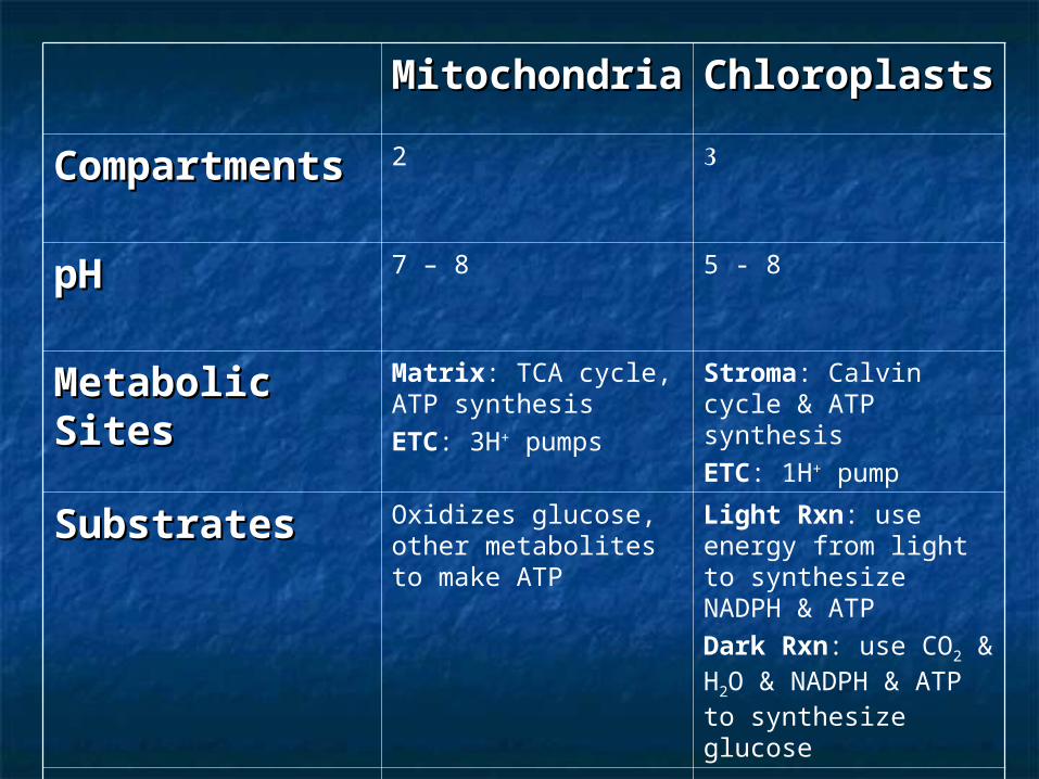
MitochondriMitochondriaa
ChloroplastsChloroplasts
CompartmentCompartmentss
2 3
pHpH 7 – 8 5 - 8
Metabolic Metabolic SitesSites
Matrix: TCA cycle, ATP synthesisETC: 3H+ pumps
Stroma: Calvin cycle & ATP synthesisETC: 1H+ pump
SubstratesSubstrates Oxidizes glucose, other metabolites to make ATP
Light Rxn: use energy from light to synthesize NADPH & ATPDark Rxn: use CO2 & H2O & NADPH & ATP to synthesize glucose
WastesWastes CO2 & H2O O2
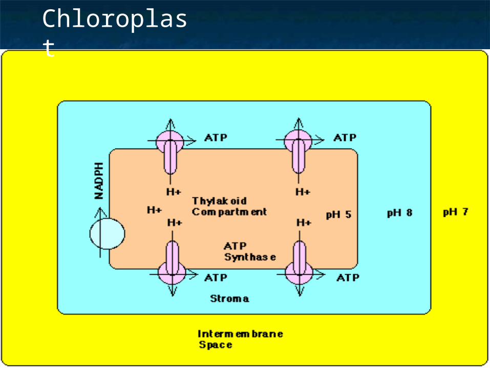
Chloroplast
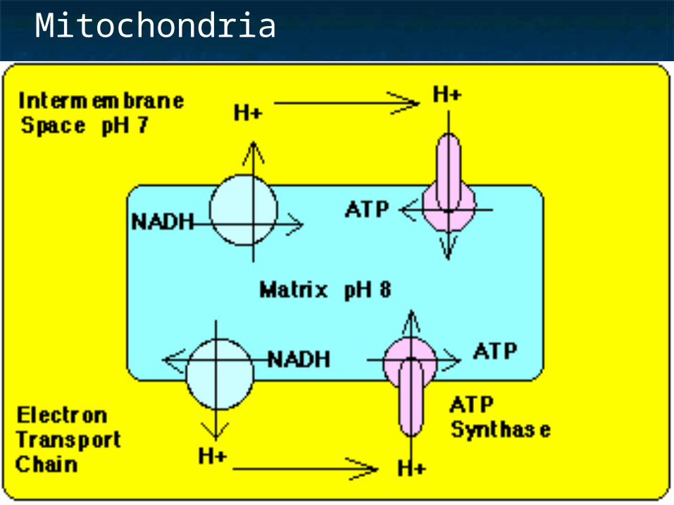
Mitochondria
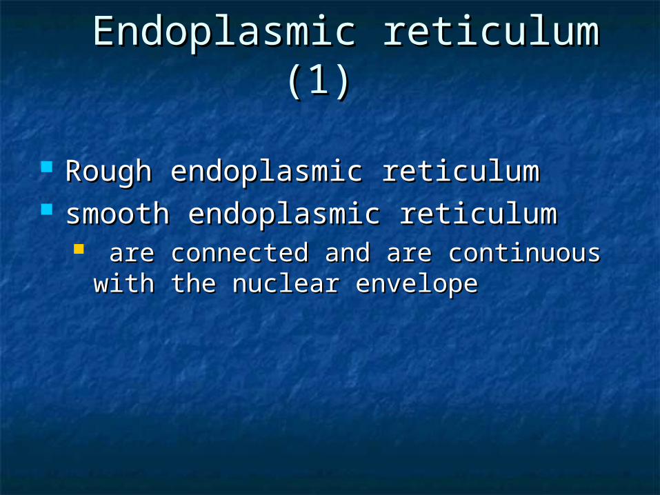
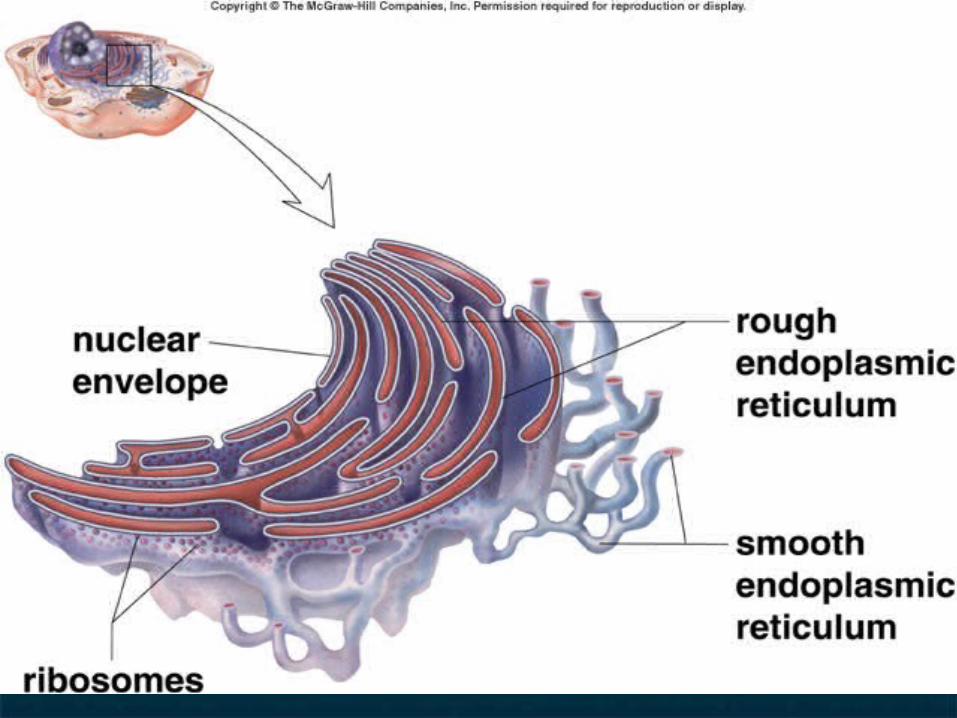
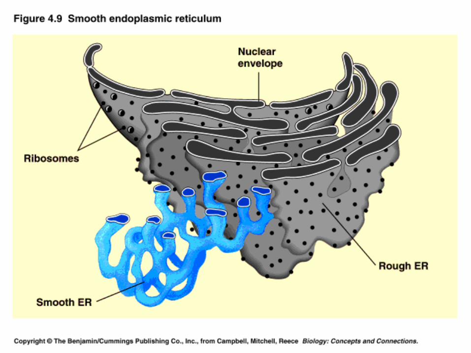
Endoplasmic reticulum (1)Endoplasmic reticulum (1)
Rough endoplasmic reticulum Rough endoplasmic reticulum smooth endoplasmic reticulumsmooth endoplasmic reticulum
are connected and are continuous with are connected and are continuous with the nuclear envelopethe nuclear envelope
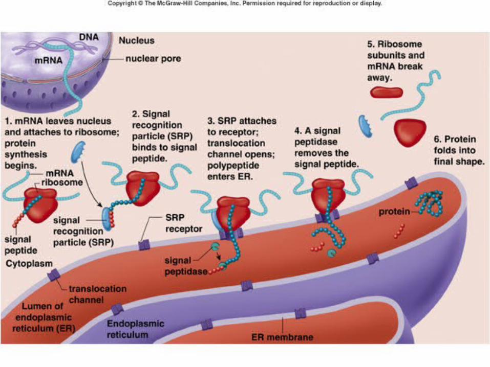
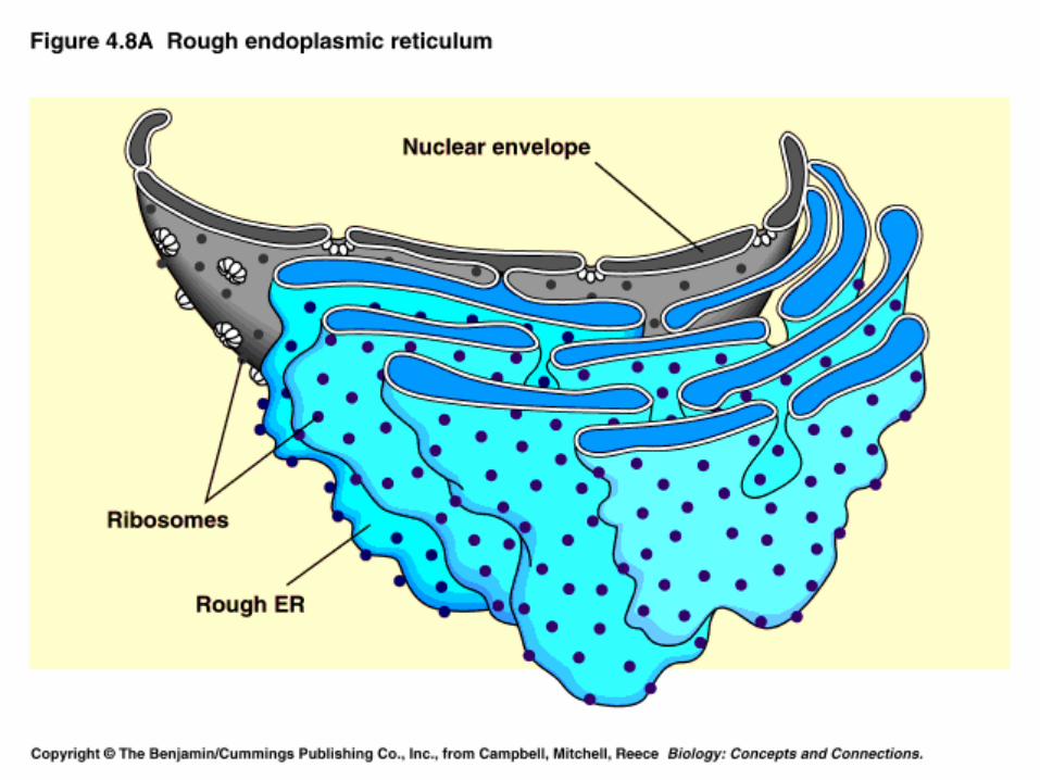
Rough endoplasmic Rough endoplasmic reticulum (2)reticulum (2)
It is rough because imbedded in the It is rough because imbedded in the membrane are ribosomesmembrane are ribosomes
the site of the synthesis of secretory proteinsthe site of the synthesis of secretory proteins The rough ER is also the site for the synthesis The rough ER is also the site for the synthesis
of membraneof membrane Enzymes synthesize phospholipid that forms Enzymes synthesize phospholipid that forms
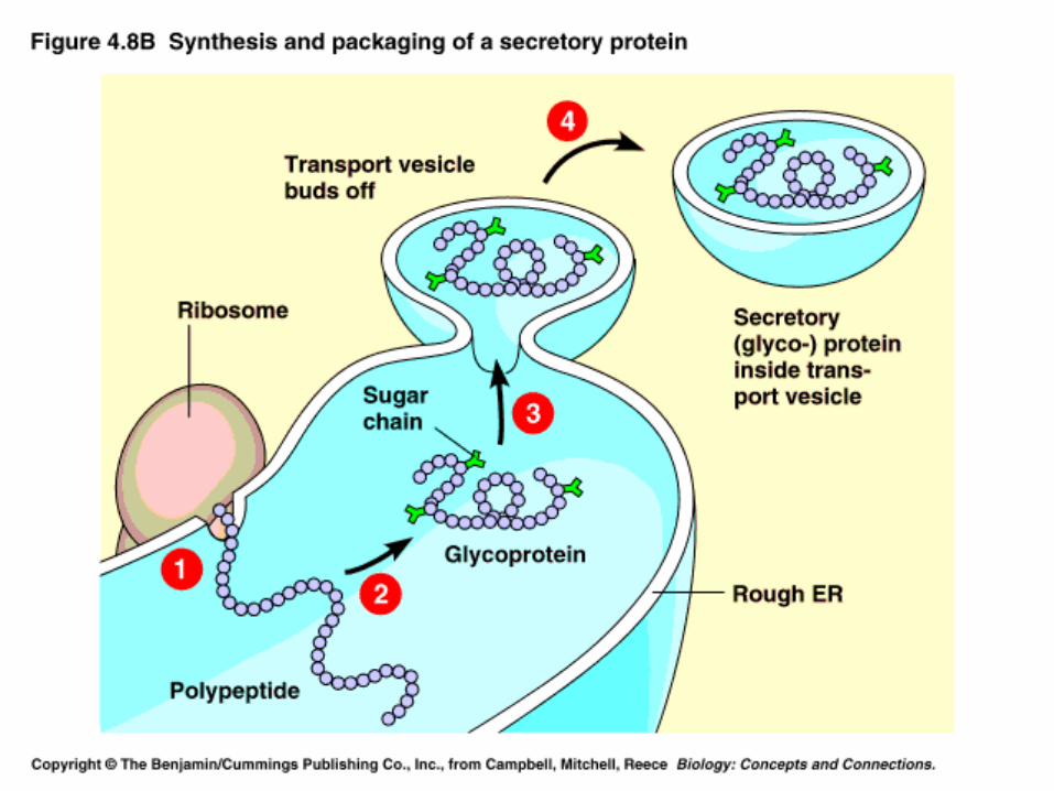
all the membranes of the cellall the membranes of the cell Ribosomes in the rough ER synthesize protein Ribosomes in the rough ER synthesize protein
that then are converted to glycoprotein and that then are converted to glycoprotein and packaged in transport vesicles for secretionpackaged in transport vesicles for secretion
Smooth endoplasmic reticulum Smooth endoplasmic reticulum (3) (3)
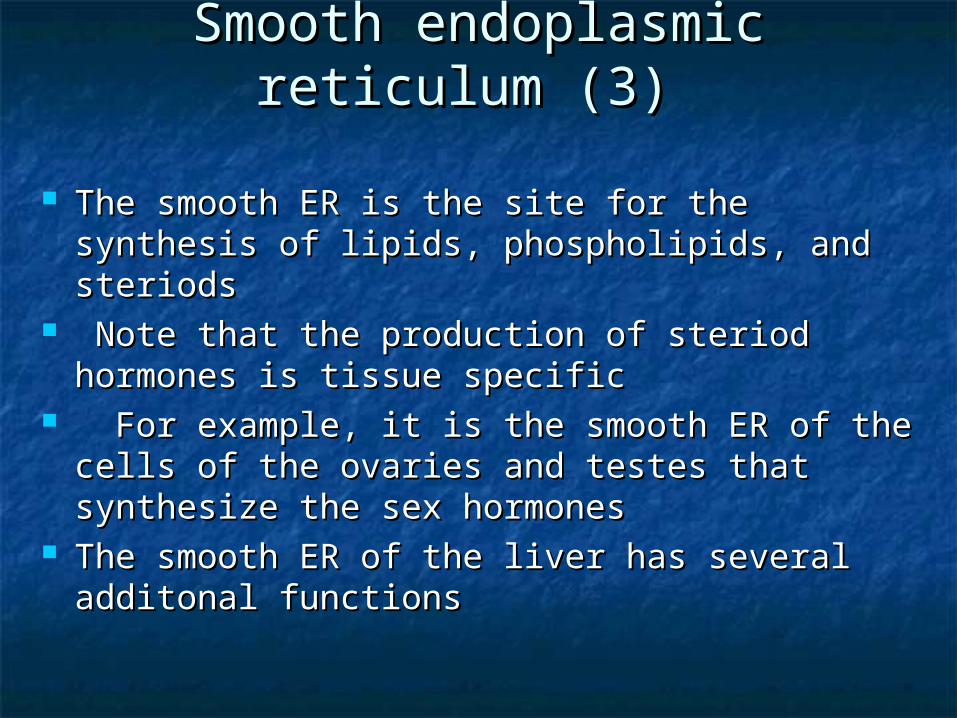
The smooth ER is the site for the synthesis of The smooth ER is the site for the synthesis of lipids, phospholipids, and steriodslipids, phospholipids, and steriods
Note that the production of steriod hormones Note that the production of steriod hormones is tissue specificis tissue specific
For example, it is the smooth ER of the cells For example, it is the smooth ER of the cells of the ovaries and testes that synthesize the of the ovaries and testes that synthesize the sex hormonessex hormones
The smooth ER of the liver has several The smooth ER of the liver has several additonal functionsadditonal functions
Smooth endoplasmic reticulum Smooth endoplasmic reticulum (4)(4)
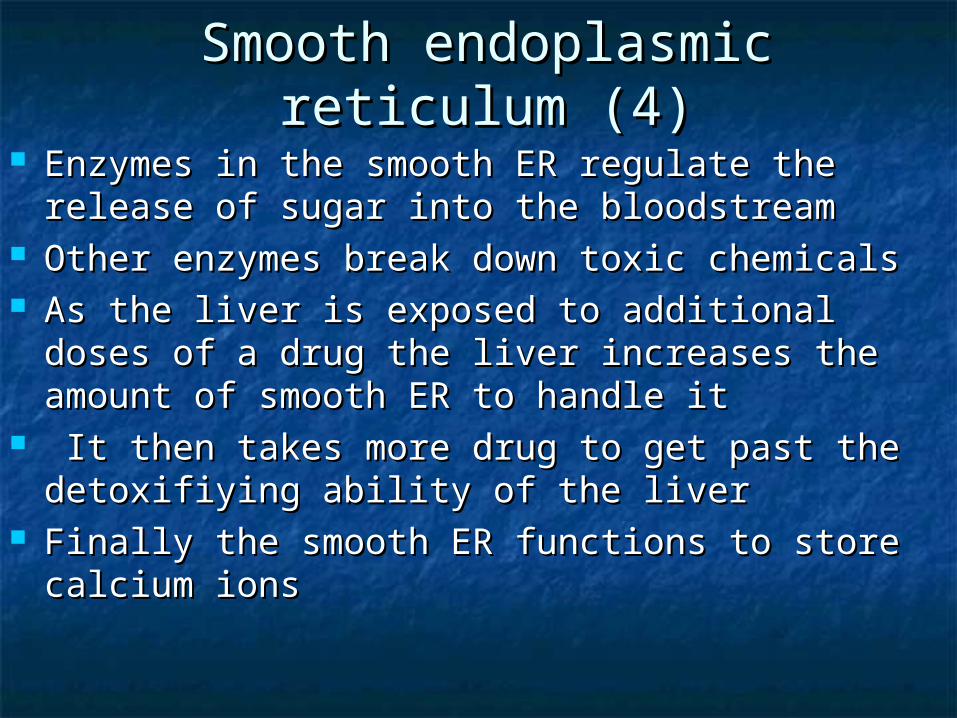
Enzymes in the smooth ER regulate the release Enzymes in the smooth ER regulate the release of sugar into the bloodstreamof sugar into the bloodstream
Other enzymes break down toxic chemicalsOther enzymes break down toxic chemicals As the liver is exposed to additional doses of a As the liver is exposed to additional doses of a
drug the liver increases the amount of smooth drug the liver increases the amount of smooth ER to handle itER to handle it
It then takes more drug to get past the It then takes more drug to get past the detoxifiyingdetoxifiying ability of the liverability of the liver
Finally the smooth ER functions to store calcium Finally the smooth ER functions to store calcium ionsions

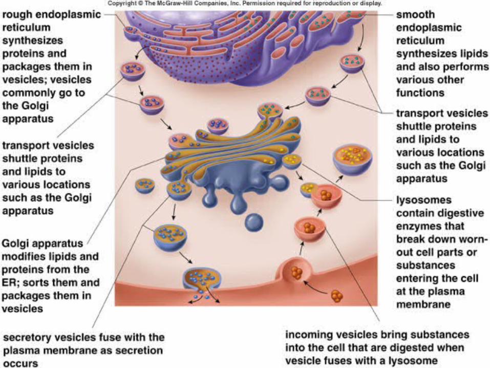
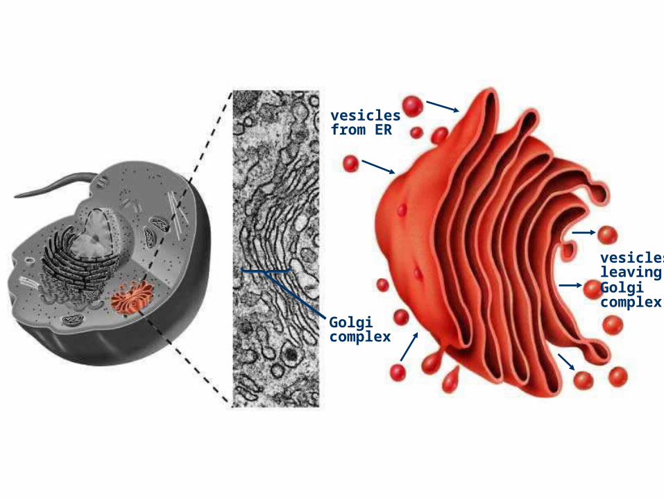
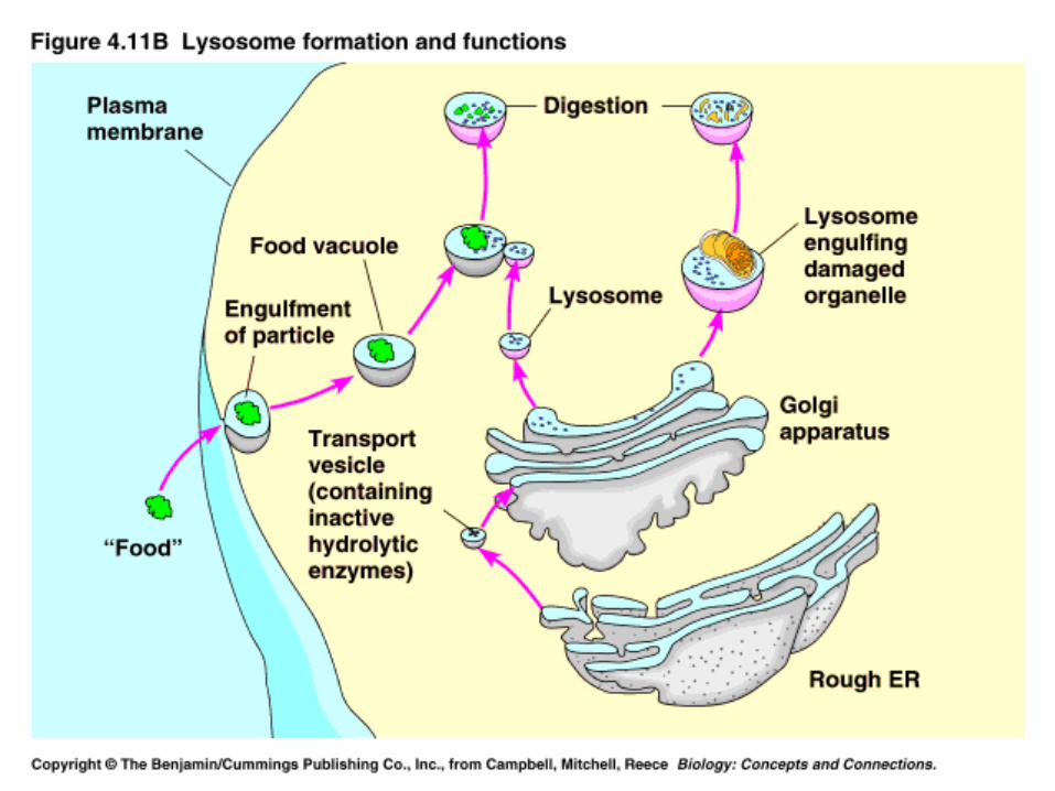
Golgi apparatus (1)Golgi apparatus (1) The Golgi apparatus, like the ER, is a series of folded The Golgi apparatus, like the ER, is a series of folded
membranesmembranes It functions in processing enzymes and other products of the It functions in processing enzymes and other products of the
ER to a finished productER to a finished product It is the source of the production of lysosomesIt is the source of the production of lysosomes Receives proteins & lipids in membrane-bound vesicles from ERReceives proteins & lipids in membrane-bound vesicles from ER Modifies those proteins & lipidsModifies those proteins & lipids Sorts and ships the proteins & lipids away in membrane-bound Sorts and ships the proteins & lipids away in membrane-bound
vesiclesvesicles

Golgi complex
vesiclesfrom ER
vesiclesleavingGolgi complex
LysosomesLysosomes These are membrane bound vesicles These are membrane bound vesicles
that harbor digestive enzymesthat harbor digestive enzymes The membrane of a lysosome will The membrane of a lysosome will
fuse with the membrane of vacuoles fuse with the membrane of vacuoles releases these digestive enzymes to releases these digestive enzymes to the interior of the vacuole to digest the interior of the vacuole to digest the material inside the vacuolethe material inside the vacuole
VacuolesVacuoles These are membrane-bound sacs that These are membrane-bound sacs that
have many different functionshave many different functions The central vacuole of a plant cell The central vacuole of a plant cell
serves as a large lysosomeserves as a large lysosome It may also function in absorbing It may also function in absorbing
water. water. The central vacuoles of flower petal The central vacuoles of flower petal
cells may hold the pigments that give cells may hold the pigments that give the flower its colorthe flower its color
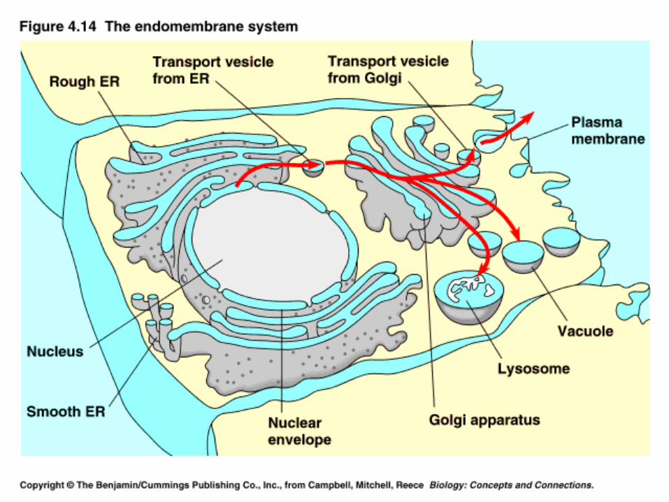
Endomembrane systemEndomembrane system
This section reviews the This section reviews the endomembrane systemendomembrane system
which encludes the nuclear envelope,which encludes the nuclear envelope, the rough and smooth ER,the rough and smooth ER, the Golgi apparatus, the Golgi apparatus, lysosomes andlysosomes and vacuolesvacuoles
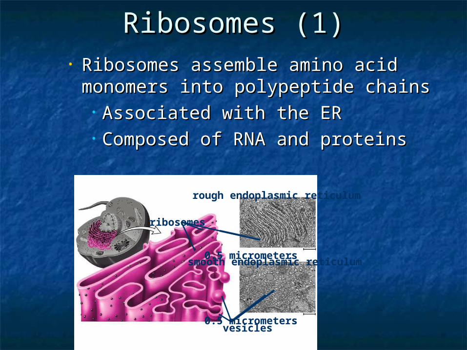
Ribosomes (1)Ribosomes (1)• Ribosomes assemble amino acid Ribosomes assemble amino acid
monomers into polypeptide chainsmonomers into polypeptide chains• Associated with the ERAssociated with the ER• Composed of RNA and proteinsComposed of RNA and proteins
0.5 micrometers
smooth endoplasmic reticulum
vesicles
ribosomes
rough endoplasmic reticulum
0.5 micrometers
Ribosome Assembly/Structure Ribosome Assembly/Structure (2)(2)
If individual proteins and rRNAs are If individual proteins and rRNAs are mixed, functional ribosomes will mixed, functional ribosomes will assemble assemble
Structures of large and small Structures of large and small subunits have been determined in subunits have been determined in 2000/20012000/2001
Growing peptide chain is thought to Growing peptide chain is thought to thread through the tunnel during thread through the tunnel during protein synthesis protein synthesis
Eukaryotic ribosomes (3)Eukaryotic ribosomes (3) Mitochondrial and chloroplast ribosomes Mitochondrial and chloroplast ribosomes
are quite similar to prokaryotic ribosomes, are quite similar to prokaryotic ribosomes, reflecting their supposed prokaryotic origin reflecting their supposed prokaryotic origin
Cytoplasmic ribosomes are larger and Cytoplasmic ribosomes are larger and more complex, but many of the structural more complex, but many of the structural and functional properties are similarand functional properties are similar
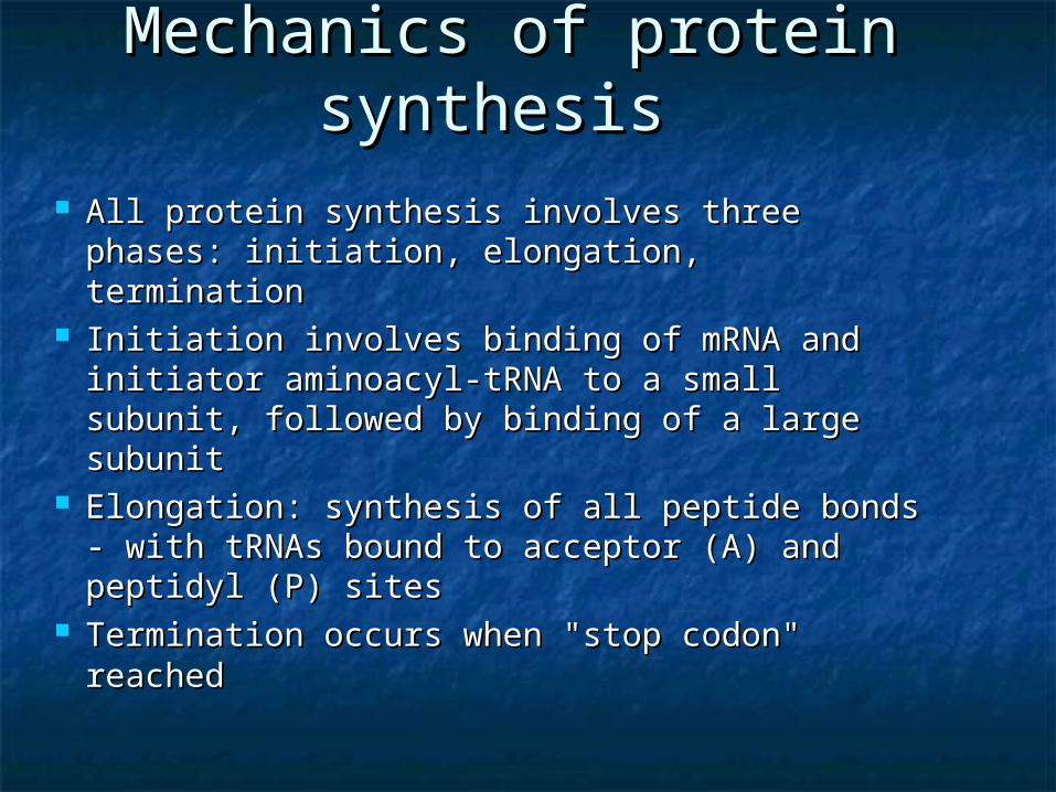
Mechanics of protein Mechanics of protein synthesissynthesis
All protein synthesis involves three phases: All protein synthesis involves three phases: initiation, elongation, termination initiation, elongation, termination
Initiation involves binding of mRNA and Initiation involves binding of mRNA and initiator aminoacyl-tRNA to a small subunit, initiator aminoacyl-tRNA to a small subunit, followed by binding of a large subunit followed by binding of a large subunit
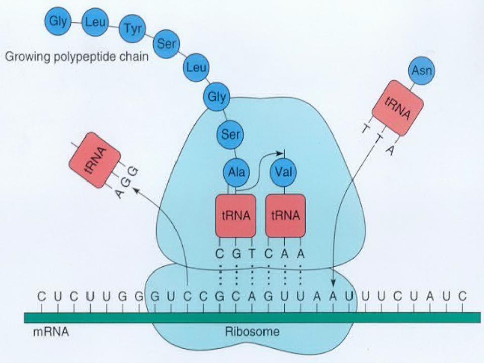
Elongation: synthesis of all peptide bonds - Elongation: synthesis of all peptide bonds - with tRNAs bound to acceptor (A) and with tRNAs bound to acceptor (A) and peptidyl (P) sitespeptidyl (P) sites
Termination occurs when "stop codon" Termination occurs when "stop codon" reachedreached

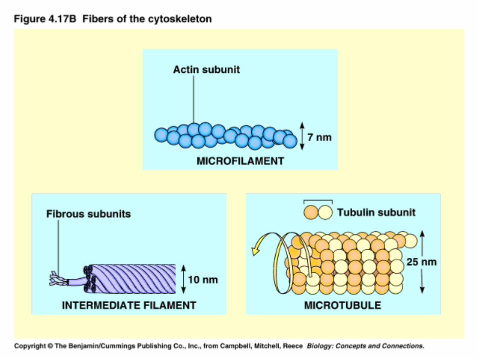
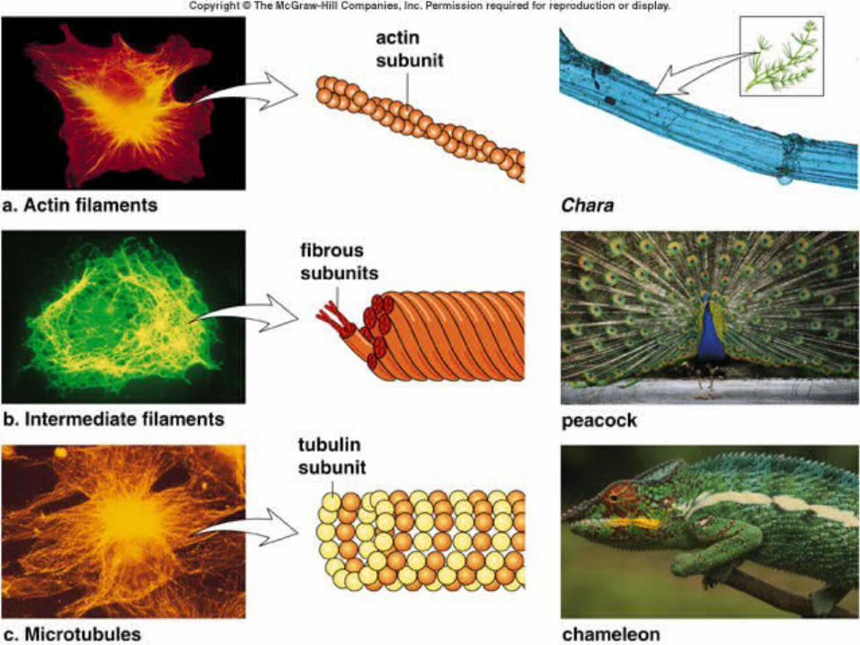
Eukaryotic cells has a meshwork of tiny fibers that Eukaryotic cells has a meshwork of tiny fibers that support the structure. This network is the cytoskeleton. support the structure. This network is the cytoskeleton. Three types of fibers exist. Microfilaments are solid Three types of fibers exist. Microfilaments are solid helical rods composed of the protein actin. There is a helical rods composed of the protein actin. There is a twist double chain of actin molecules that make up twist double chain of actin molecules that make up microfilaments. These are found in cells that must microfilaments. These are found in cells that must contract such as muscle cells. Intermediate filaments contract such as muscle cells. Intermediate filaments are variable but in general are ropelike structures made are variable but in general are ropelike structures made of twisted filaments of fibrous proteins. These function of twisted filaments of fibrous proteins. These function in bearing tension and anchoring organelles. in bearing tension and anchoring organelles. Microtubles are straight, hollow tubes composed of Microtubles are straight, hollow tubes composed of proteins called tubulins. These anchor organelles and proteins called tubulins. These anchor organelles and provide tract along which organelles may move. They provide tract along which organelles may move. They also make up flagella and cilia.also make up flagella and cilia.
CytoskeletoCytoskeletonn
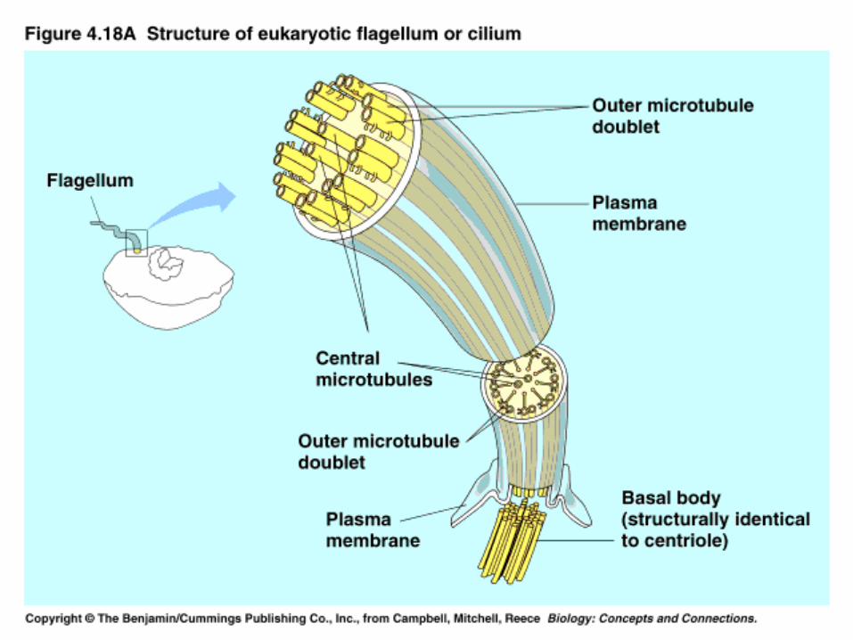
These are found on cells, such as protists, that are These are found on cells, such as protists, that are motile. Cilia are short and numerous. Longer less motile. Cilia are short and numerous. Longer less numerous appendages are flagella. These are numerous appendages are flagella. These are composed of a core of microtubules wrapped in an composed of a core of microtubules wrapped in an extension of the plasma membrane. It is sufficient to extension of the plasma membrane. It is sufficient to know that Energy is required to move the cilia or flagella know that Energy is required to move the cilia or flagella in a whiplike motion to propel the cell.in a whiplike motion to propel the cell.
Cilia and flagellaCilia and flagella
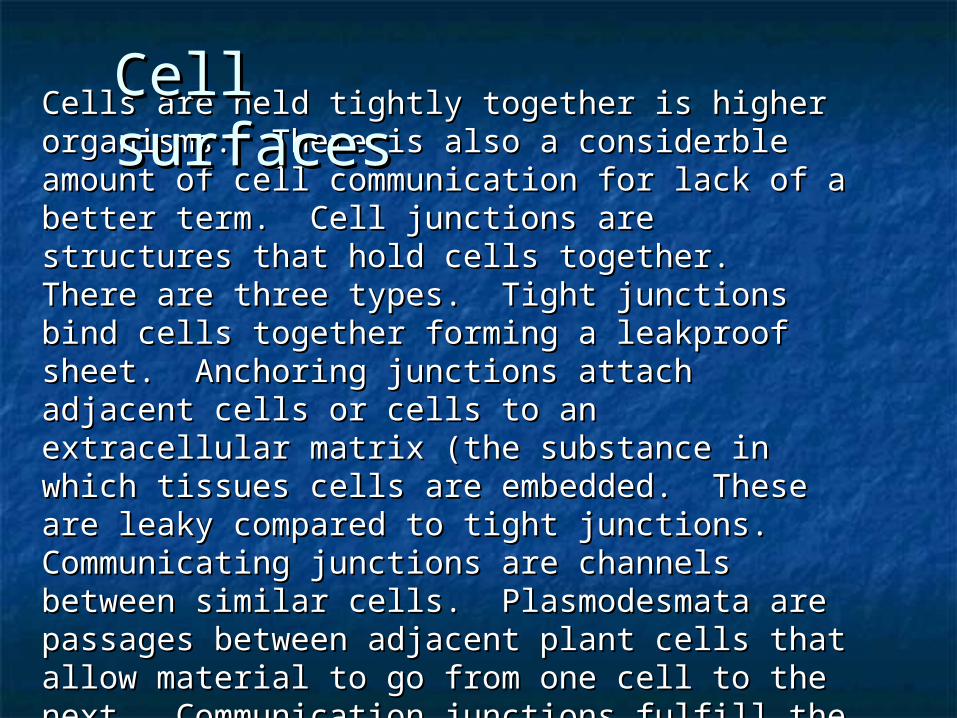
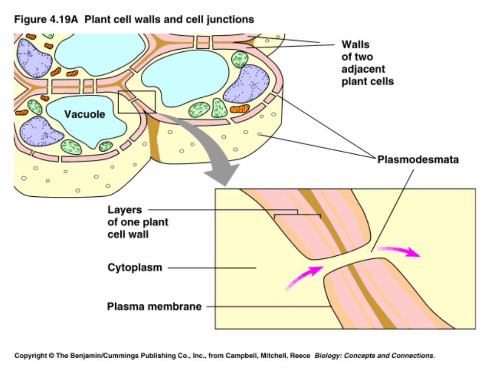
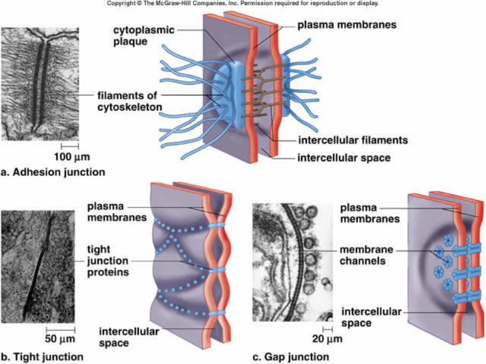
Cells are held tightly together is higher organisms. There Cells are held tightly together is higher organisms. There is also a considerble amount of cell communication for is also a considerble amount of cell communication for lack of a better term. Cell junctions are structures that lack of a better term. Cell junctions are structures that hold cells together. There are three types. Tight hold cells together. There are three types. Tight junctions bind cells together forming a leakproof sheet. junctions bind cells together forming a leakproof sheet. Anchoring junctions attach adjacent cells or cells to an Anchoring junctions attach adjacent cells or cells to an extracellular matrix (the substance in which tissues cells extracellular matrix (the substance in which tissues cells are embedded. These are leaky compared to tight are embedded. These are leaky compared to tight junctions. Communicating junctions are channels junctions. Communicating junctions are channels between similar cells. Plasmodesmata are passages between similar cells. Plasmodesmata are passages between adjacent plant cells that allow material to go between adjacent plant cells that allow material to go from one cell to the next. Communication junctions fulfill from one cell to the next. Communication junctions fulfill the same role between animal cells. the same role between animal cells.
Cell Cell surfacessurfaces

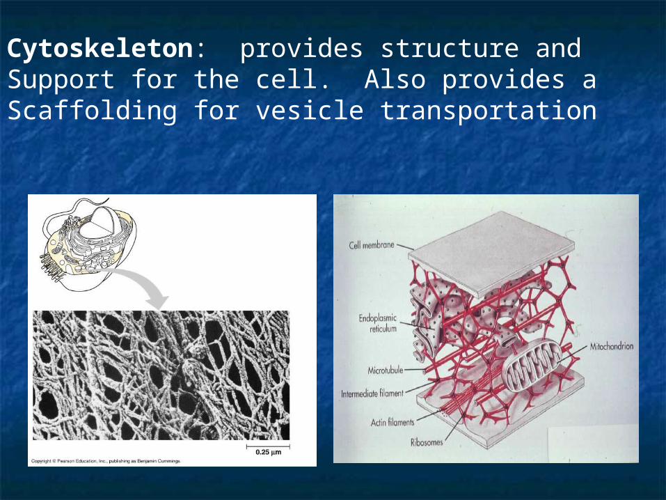
Cytoskeleton: provides structure and Support for the cell. Also provides a Scaffolding for vesicle transportation
OrganellOrganellee
ProkaryotProkaryoteses
EukaryotesEukaryotes FunctionsFunctions
ProkaryotProkaryoteses
Animal Animal cellscells
PlanPlant t cellscells
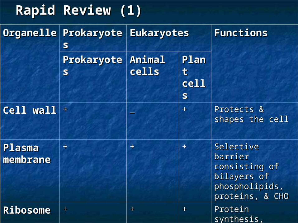
Cell wallCell wall ++ __ ++ Protects & shapes Protects & shapes the cellthe cell
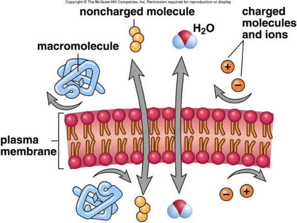
Plasma Plasma membranmembranee
++ ++ ++ Selective barrier Selective barrier consisting of consisting of bilayers of bilayers of phospholipids, phospholipids, proteins, & CHOproteins, & CHO
RibosomRibosomee
++ ++ ++ Protein synthesis, Protein synthesis, formed in formed in nucleolusnucleolus
Rapid Review (1)Rapid Review (1)
OrganellOrganellee
ProkaryotProkaryoteses
EukaryotesEukaryotes FunctionsFunctions
ProkaryotProkaryoteses
Animal Animal cellscells
PlanPlant t cellscells
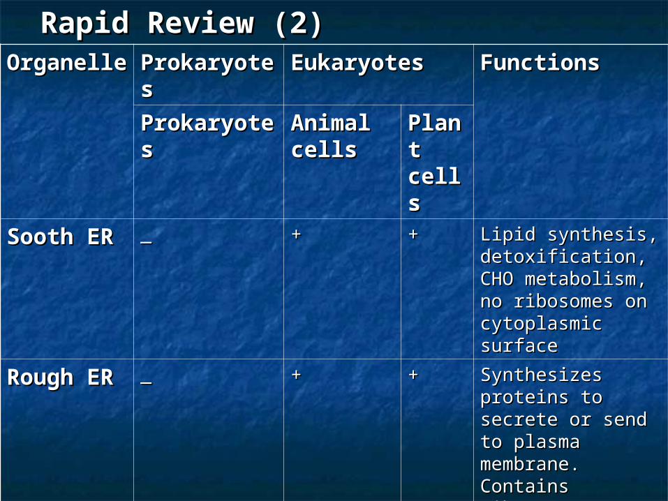
Sooth ERSooth ER __ ++ ++ Lipid synthesis, Lipid synthesis, detoxification, CHO detoxification, CHO metabolism, no metabolism, no ribosomes on ribosomes on cytoplasmic cytoplasmic surfacesurface
Rough ERRough ER __ ++ ++ Synthesizes Synthesizes proteins to secrete proteins to secrete or send to plasma or send to plasma membrane. membrane. Contains Contains ribosomes on ribosomes on cytoplasmic cytoplasmic surfacesurface
GogliGogli __ ++ ++ Modifies lipids, Modifies lipids, proteins, etc & proteins, etc & sends them to sends them to other sites in the other sites in the cellcell
Rapid Review (2)Rapid Review (2)
OrganelleOrganelle ProkaryotProkaryoteses
EukaryotesEukaryotes FunctionsFunctions
ProkaryotProkaryoteses
Animal Animal cellscells
Plant Plant cellscells
MitochondrMitochondriaia
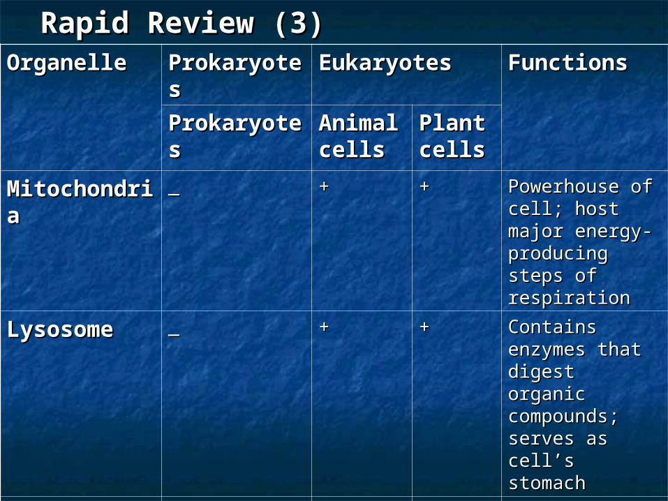
__ ++ ++ Powerhouse of Powerhouse of cell; host major cell; host major energy-energy-producing steps producing steps of respirationof respiration
LysosomeLysosome __ ++ ++ Contains Contains enzymes that enzymes that digest organic digest organic compounds; compounds; serves as cell’s serves as cell’s stomachstomach
NucleusNucleus __ ++ ++ Control center Control center of cell. Host for of cell. Host for transcription, transcription, replication & replication & DNADNA
Rapid Review (3)Rapid Review (3)
OrganelleOrganelle ProkaryoteProkaryotess
EukaryotesEukaryotes FunctionsFunctions
ProkaryoteProkaryotess
AnimaAnimal cellsl cells
PlanPlant t cellscells
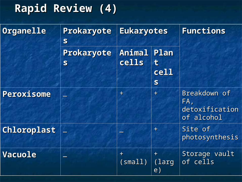
PeroxisomePeroxisome __ ++ ++ Breakdown of Breakdown of FA, FA, detoxification of detoxification of alcoholalcohol
ChloroplastChloroplast __ __ ++ Site of Site of photosynthesisphotosynthesis
VacuoleVacuole __ + + (small)(small)
+ + (large)(large)
Storage vault of Storage vault of cellscells
Rapid Review (4)Rapid Review (4)
OrganelleOrganelle ProkaryoteProkaryotess
EukaryotesEukaryotes FunctionsFunctions
ProkaryoteProkaryotess
AnimaAnimal cellsl cells
PlanPlant t cellscells
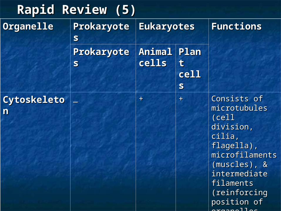
CytoskeletoCytoskeletonn
__ ++ ++ Consists of Consists of microtubules microtubules (cell division, (cell division, cilia, flagella), cilia, flagella), microfilaments microfilaments (muscles), & (muscles), & intermediate intermediate filaments filaments (reinforcing (reinforcing position of position of organellesorganelles
CentriolesCentrioles __ ++ __ Part of Part of microtubule microtubule separation separation apparatus that apparatus that assists cell assists cell divisiondivision
Rapid Review (5)Rapid Review (5)
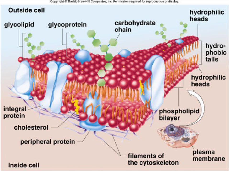
CELL MEMBRANESCELL MEMBRANESCELL MEMBRANESCELL MEMBRANES The Fluid-Mosaic ModelThe Fluid-Mosaic Model
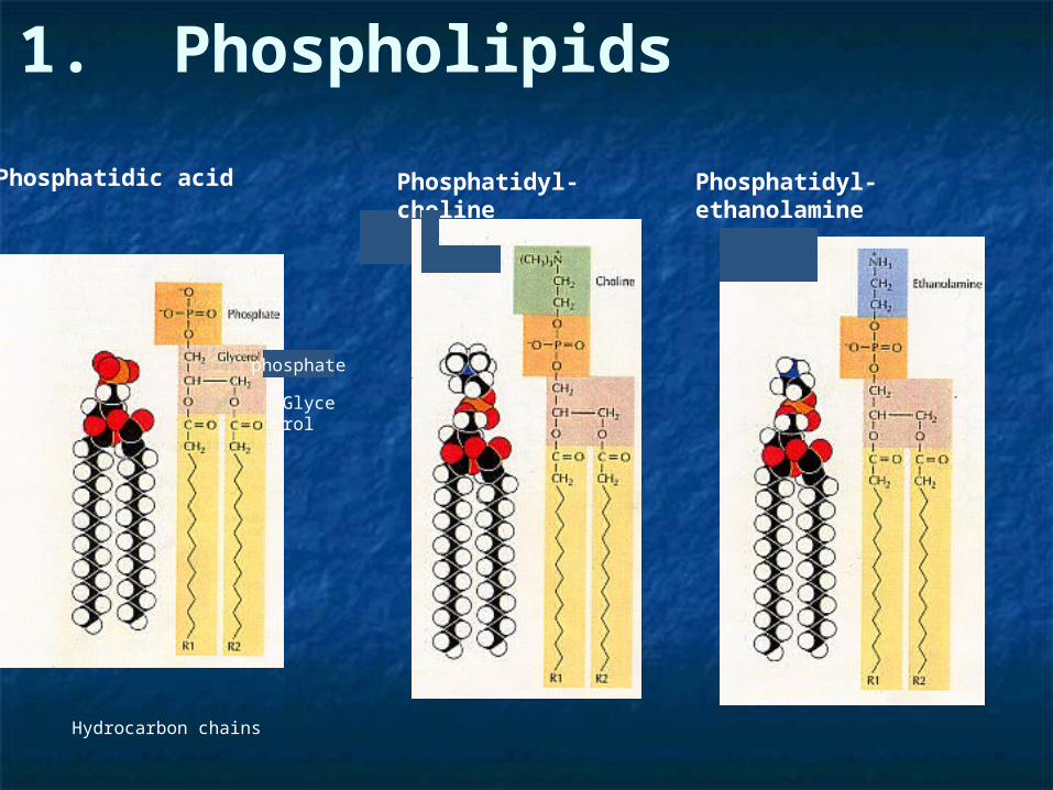
Phosphatidic acid Phosphatidyl-ethanolamine
phosphate
Glycerol
Hydrocarbon chains
Phosphatidyl-choline
1. Phospholipids
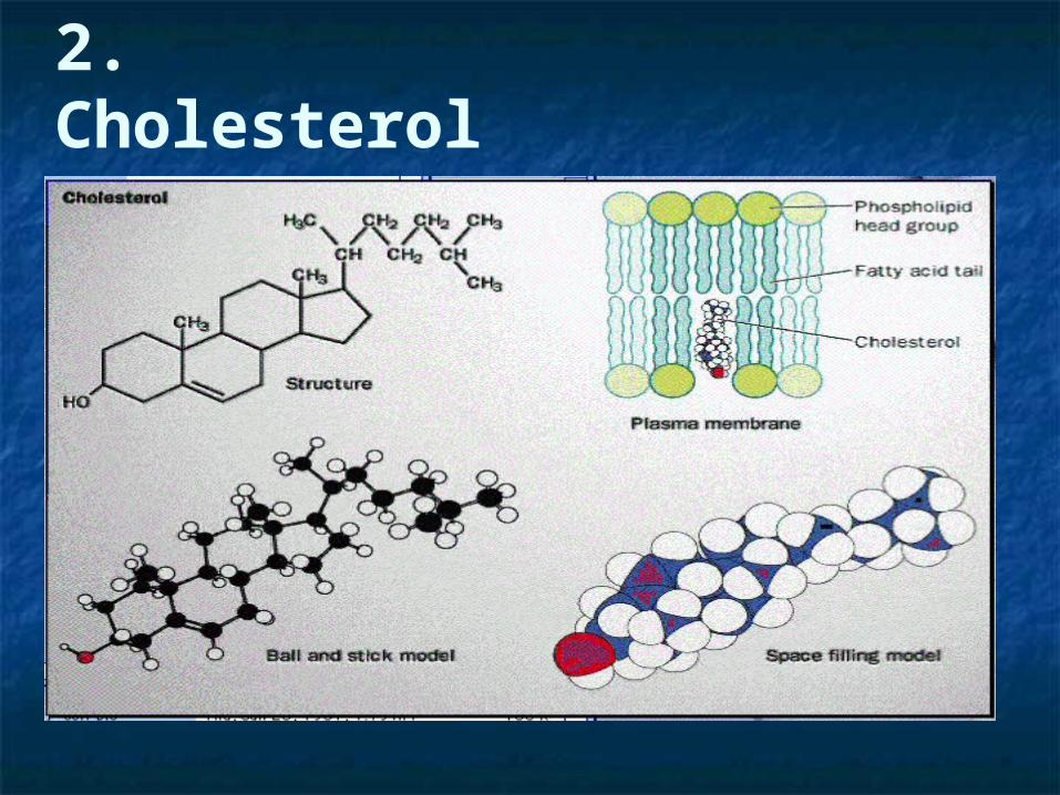
2. Cholesterol
Function of cell membranesFunction of cell membranesFunction of cell membranesFunction of cell membranes
Compartmentalization of tissuesCompartmentalization of tissues Regulation of cell contentsRegulation of cell contents Provides surface for enzymes, Provides surface for enzymes,
receptors, recognition, etc.receptors, recognition, etc.
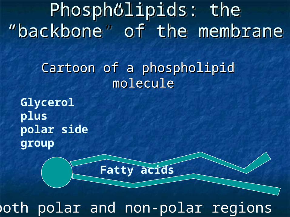
Phospholipids: the “backbone” Phospholipids: the “backbone” of the membraneof the membrane
Phospholipids: the “backbone” Phospholipids: the “backbone” of the membraneof the membrane
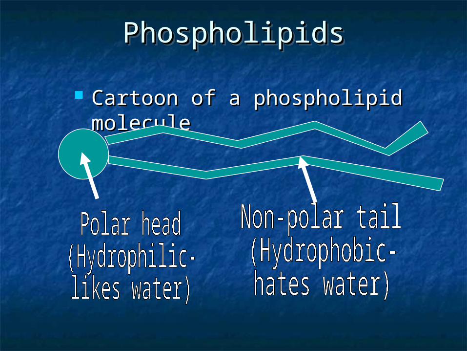
Cartoon of a phospholipid Cartoon of a phospholipid moleculemolecule
*both polar and non-polar regions
Fatty acids
Glycerol pluspolar side group
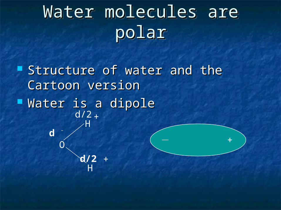
Water molecules are polarWater molecules are polarWater molecules are polarWater molecules are polar
Structure of water and the Cartoon Structure of water and the Cartoon versionversion
Water is a dipoleWater is a dipole
O
H
H
+
+
d
d/2
d/2
+
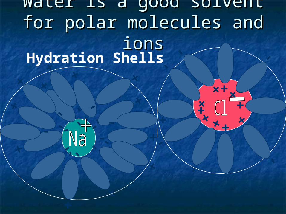
Water is a good solvent for Water is a good solvent for polar molecules and ionspolar molecules and ions
- +- +- +
- + - +
- +
-
+
Hydration Shells - +
- +
- +
-
+
-
+
- +
- +-
+
-
+
- +
- +
- +
- +
- +
- +
- + -
+
- +
- +
PhospholipidsPhospholipidsPhospholipidsPhospholipids
Cartoon of a phospholipid moleculeCartoon of a phospholipid molecule
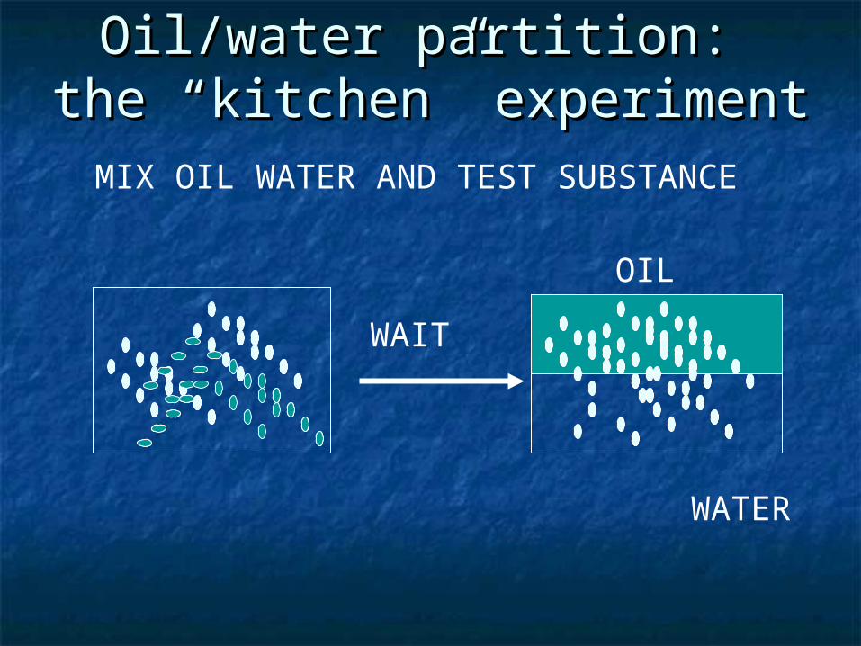
Oil/water partition: the Oil/water partition: the “kitchen” experiment“kitchen” experiment
MIX OIL WATER AND TEST SUBSTANCE
WAIT
OIL
WATER
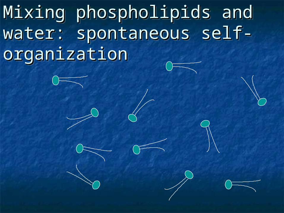
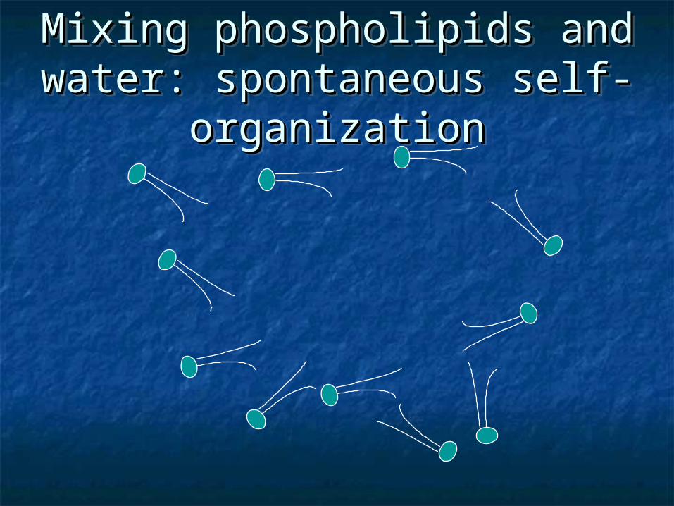
Mixing phospholipids and Mixing phospholipids and water: spontaneous self-water: spontaneous self-organizationorganization
Mixing phospholipids and Mixing phospholipids and water: spontaneous self-water: spontaneous self-organizationorganization
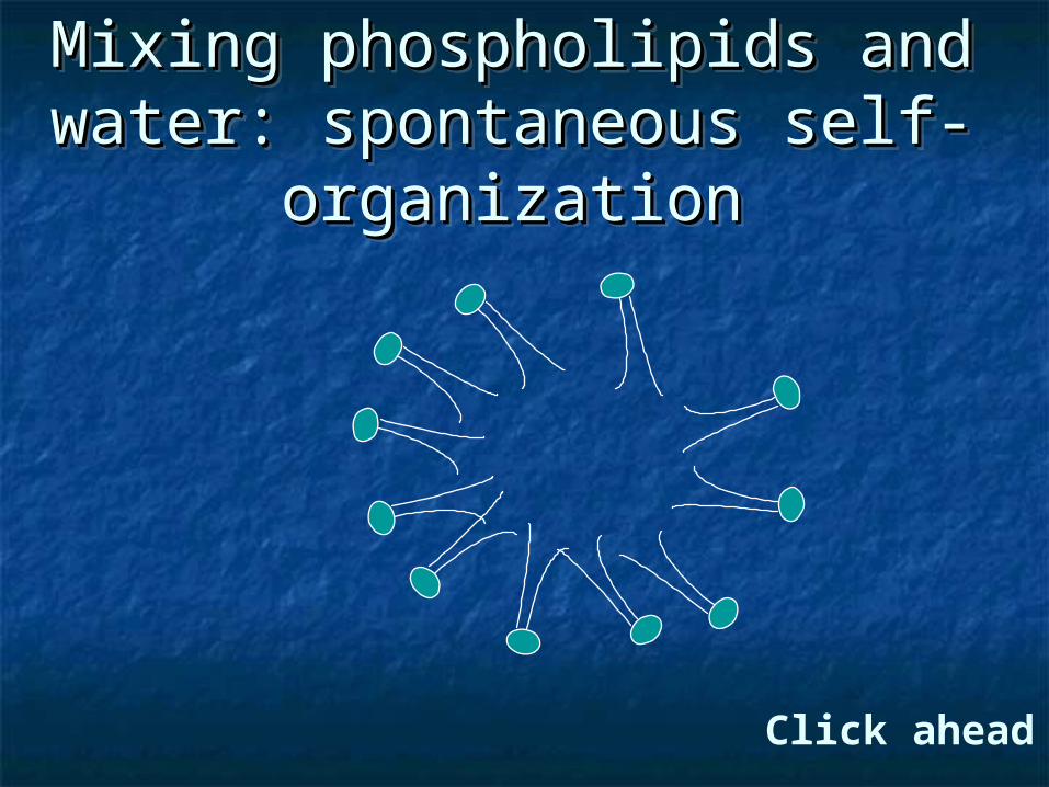
Mixing phospholipids and Mixing phospholipids and water: spontaneous self-water: spontaneous self-
organizationorganization
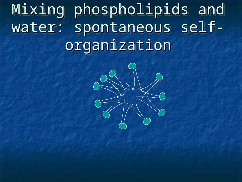
Mixing phospholipids and Mixing phospholipids and water: spontaneous self-water: spontaneous self-
organizationorganization
Mixing phospholipids and Mixing phospholipids and water: spontaneous self-water: spontaneous self-
organizationorganization
Mixing phospholipids and Mixing phospholipids and water: spontaneous self-water: spontaneous self-
organizationorganization
Click ahead
Mixing phospholipids and Mixing phospholipids and water: spontaneous self-water: spontaneous self-
organizationorganization
Mixing phospholipids and Mixing phospholipids and water: spontaneous self-water: spontaneous self-
organizationorganization
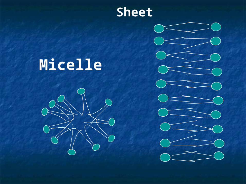
Micelle
Sheet
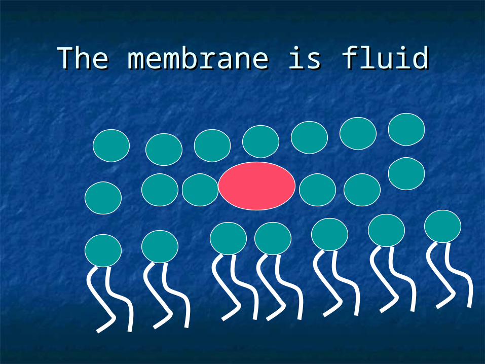
The membrane is fluidThe membrane is fluid
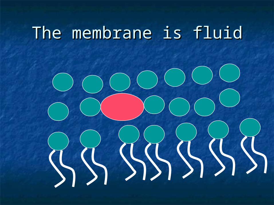
The membrane is fluidThe membrane is fluid
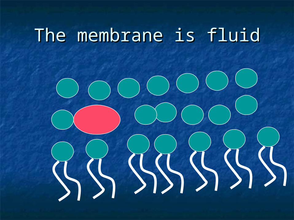
The membrane is fluidThe membrane is fluid
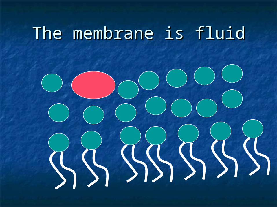
The membrane is fluidThe membrane is fluid

Cholesterol sits between fatty Cholesterol sits between fatty tailstails
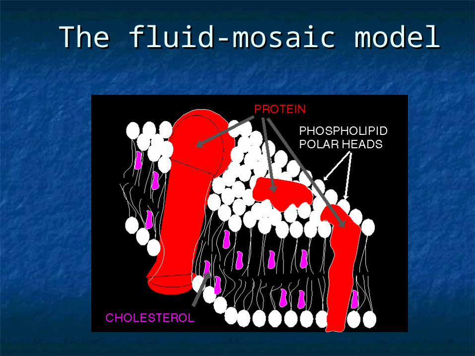
The fluid-mosaic modelThe fluid-mosaic model
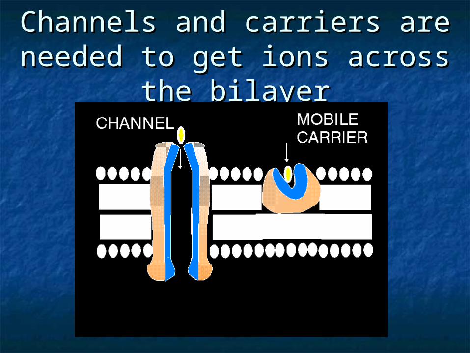
Channels and carriers are Channels and carriers are needed to get ions across the needed to get ions across the
bilayerbilayer
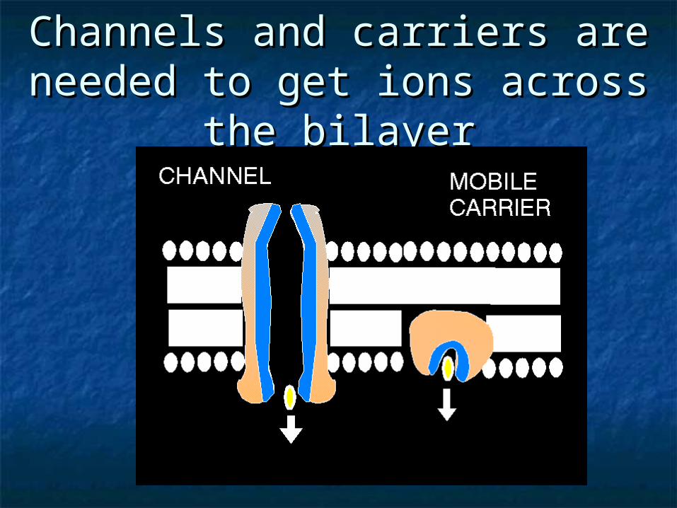
Channels and carriers are Channels and carriers are needed to get ions across the needed to get ions across the
bilayerbilayer
Types eucaryotic cells (1)Types eucaryotic cells (1)
Stem CellsStem Cells Hemopoietic cellsHemopoietic cells MonocytesMonocytes MacrophagesMacrophages PhagocytesPhagocytes
Stem Cells (2)Stem Cells (2)
Research on stem cells is advancing Research on stem cells is advancing knowledge about:knowledge about:
how an organism develops from a single cell how an organism develops from a single cell and how healthy cells replace damaged and how healthy cells replace damaged
cells in adult organismscells in adult organisms This promising area of science is also This promising area of science is also
leading scientists to investigate the leading scientists to investigate the possibility of cell-based therapies to treat possibility of cell-based therapies to treat disease, which is often referred to as disease, which is often referred to as regenerative or reparative medicineregenerative or reparative medicine
What are stem cells and why What are stem cells and why are they important?are they important? (3) (3)
Stem cells have two important characteristics Stem cells have two important characteristics that distinguish them from other types of cells:that distinguish them from other types of cells:
First, they are unspecialized cells that renew First, they are unspecialized cells that renew themselves for long periods through cell divisionthemselves for long periods through cell division
The second is that under certain physiologic or The second is that under certain physiologic or experimental conditions, they can be induced to experimental conditions, they can be induced to become cells with special functions such as: become cells with special functions such as:
the beating cells of the heart muscle the beating cells of the heart muscle or the insulin-producing cells of the pancreasor the insulin-producing cells of the pancreas
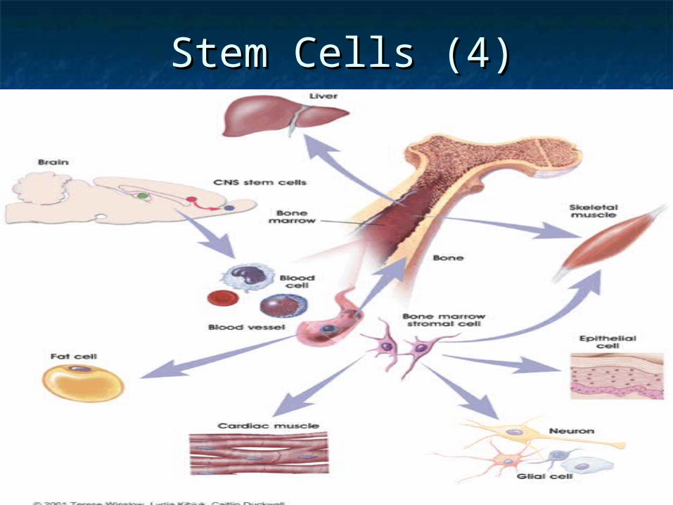
Stem Cells (4)Stem Cells (4)
Kinds of stem cells (5)Kinds of stem cells (5)
Scientists primarily work with two Scientists primarily work with two kinds of stem cells from animals and kinds of stem cells from animals and humans:humans:
embryonic stem cells embryonic stem cells and adult stem cells, and adult stem cells, which have different functions and which have different functions and
characteristics characteristics
Stem cells are important for Stem cells are important for living organisms (5)living organisms (5)
Stem cells are important for living organisms for many Stem cells are important for living organisms for many reasons. reasons.
In the 3 to 5 day old embryo, called a blastocyst, In the 3 to 5 day old embryo, called a blastocyst, a small group of about 30 cells called the inner cell a small group of about 30 cells called the inner cell
mass gives rise to the hundreds of highly specialized mass gives rise to the hundreds of highly specialized cellscells
needed to make up an adult organism. needed to make up an adult organism. In the developing fetus, stem cells in developing In the developing fetus, stem cells in developing
tissues give rise to the multiple specialized cell types tissues give rise to the multiple specialized cell types that make up that make up
the heart, the heart, lung, lung, skin, skin, and other tissuesand other tissues
In some adult tissues (6)In some adult tissues (6) In some adult tissues, such as:In some adult tissues, such as: bone marrow, bone marrow, muscle, muscle, and brain, and brain, discrete populations of adult stem discrete populations of adult stem
cells generate replacements for cells generate replacements for cells that are lost: cells that are lost:
through normal wear and tear,through normal wear and tear, injury, injury, or diseaseor disease
Hemopoietic cells (7)Hemopoietic cells (7) The basis of haemopoiesis is a small The basis of haemopoiesis is a small
population of population of self-replicating self-replicating stem stem cells, cells,
which ultimately can generate all which ultimately can generate all types of blood cells.types of blood cells.
The process of haematopoiesis is The process of haematopoiesis is controlled by a group of at least 11 controlled by a group of at least 11 growth factors.growth factors.
Three of these glycoproteins initiate Three of these glycoproteins initiate the differentiation of macrophages the differentiation of macrophages from uni- and bipotential progenitor from uni- and bipotential progenitor cells in the bone marrow. cells in the bone marrow.
Macrophages and monocytes Macrophages and monocytes (8)(8)
Their development takes in the bone Their development takes in the bone marrow and passes through the marrow and passes through the following steps:following steps:
stem cellstem cell committed stem cellcommitted stem cell monoblastmonoblast promonocytepromonocyte monocyte (bone marrow)monocyte (bone marrow) monocyte (peripheral blood)monocyte (peripheral blood) macrophage (tissues) macrophage (tissues)
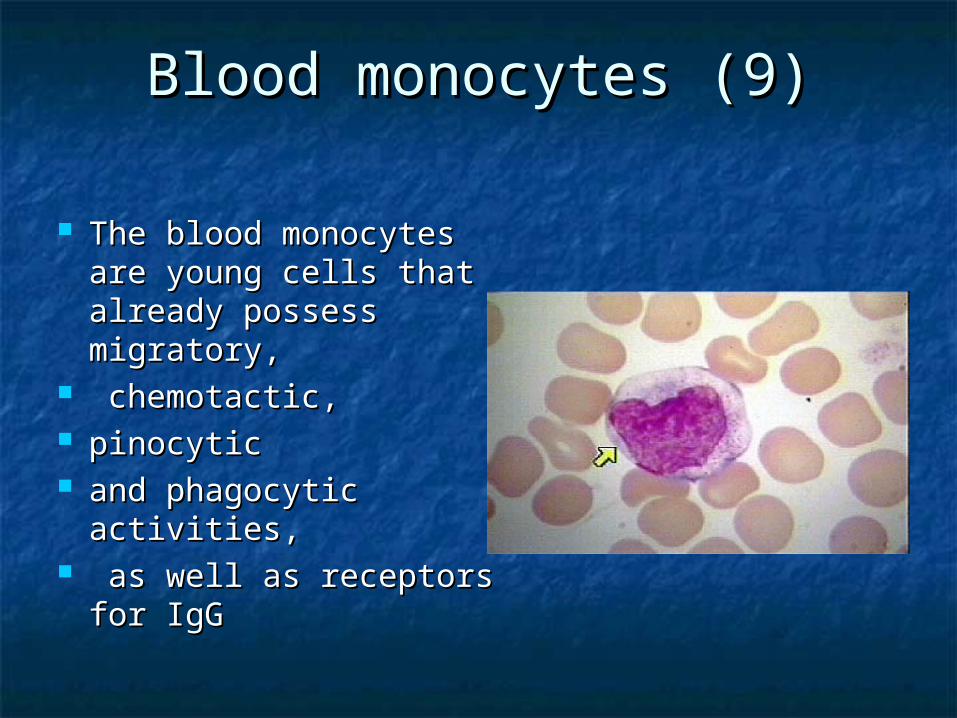
Blood monocytes (9)Blood monocytes (9)
The blood monocytes are The blood monocytes are young cells that already young cells that already possess migratory,possess migratory,
chemotactic, chemotactic, pinocytic pinocytic and phagocytic activities,and phagocytic activities, as well as receptors for as well as receptors for
IgG IgG
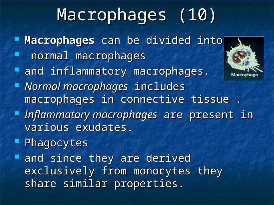
Macrophages (10)Macrophages (10) MacrophagesMacrophages can be divided into can be divided into normal macrophagesnormal macrophages and inflammatory macrophages. and inflammatory macrophages. Normal macrophagesNormal macrophages includes includes
macrophages in connective tissue .macrophages in connective tissue . Inflammatory macrophagesInflammatory macrophages are present in are present in
various exudates. various exudates. PhagocytesPhagocytes and since they are derived exclusively from and since they are derived exclusively from
monocytes they share similar properties. monocytes they share similar properties.
Phagocytes (11)Phagocytes (11)
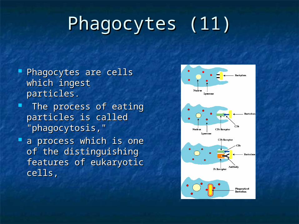
Phagocytes are cells Phagocytes are cells which ingest particles.which ingest particles.
The process of eating The process of eating particles is called particles is called "phagocytosis," "phagocytosis,"
a process which is one a process which is one of the distinguishing of the distinguishing features of eukaryotic features of eukaryotic cells, cells,

Phagocytes (12)Phagocytes (12)
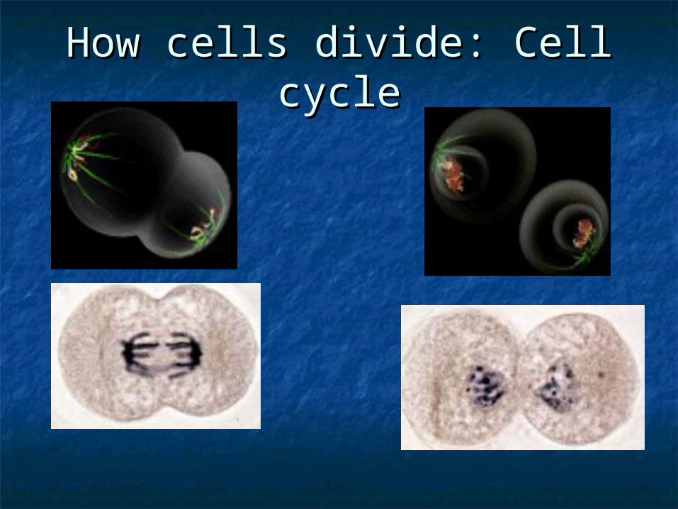
How cells divide: Cell cycleHow cells divide: Cell cycle
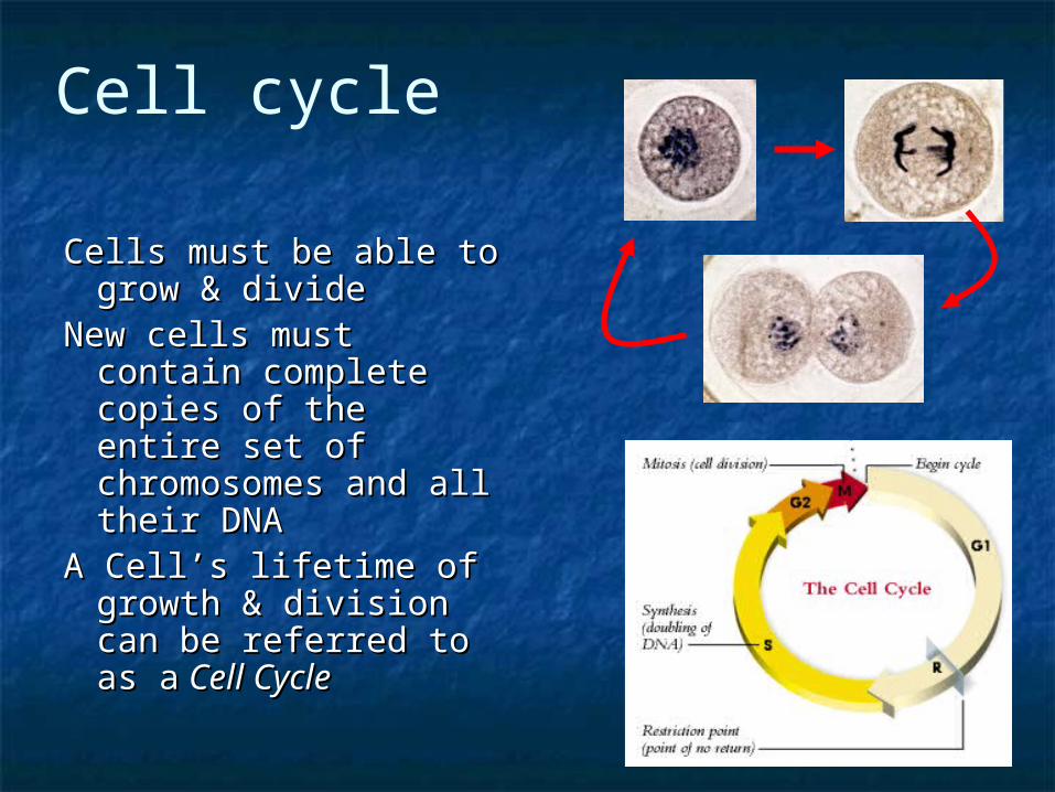
Cells must be able to Cells must be able to grow & dividegrow & divide
New cells must contain New cells must contain complete copies of complete copies of the entire set of the entire set of chromosomes and all chromosomes and all their DNA their DNA
A Cell’s lifetime of A Cell’s lifetime of growth & division can growth & division can be referred to as abe referred to as a Cell CycleCell Cycle
Cell cycle
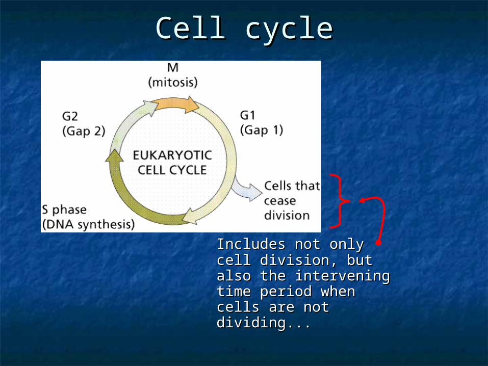
Cell cycleCell cycle
Includes not only cell Includes not only cell division, but also the division, but also the intervening time intervening time period when cells are period when cells are not dividing... not dividing...
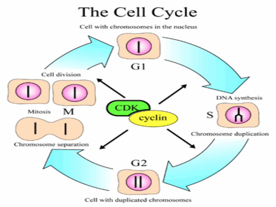
Cell cycle phasesCell cycle phases
Interphase: cell growth & DNA Interphase: cell growth & DNA replicationreplication
Mitosis: nuclear & cell DivisionMitosis: nuclear & cell Division

InterphasInterphasee
Composed of G1, S & G2 phases Composed of G1, S & G2 phases
Interphase includes everything except Mitosis
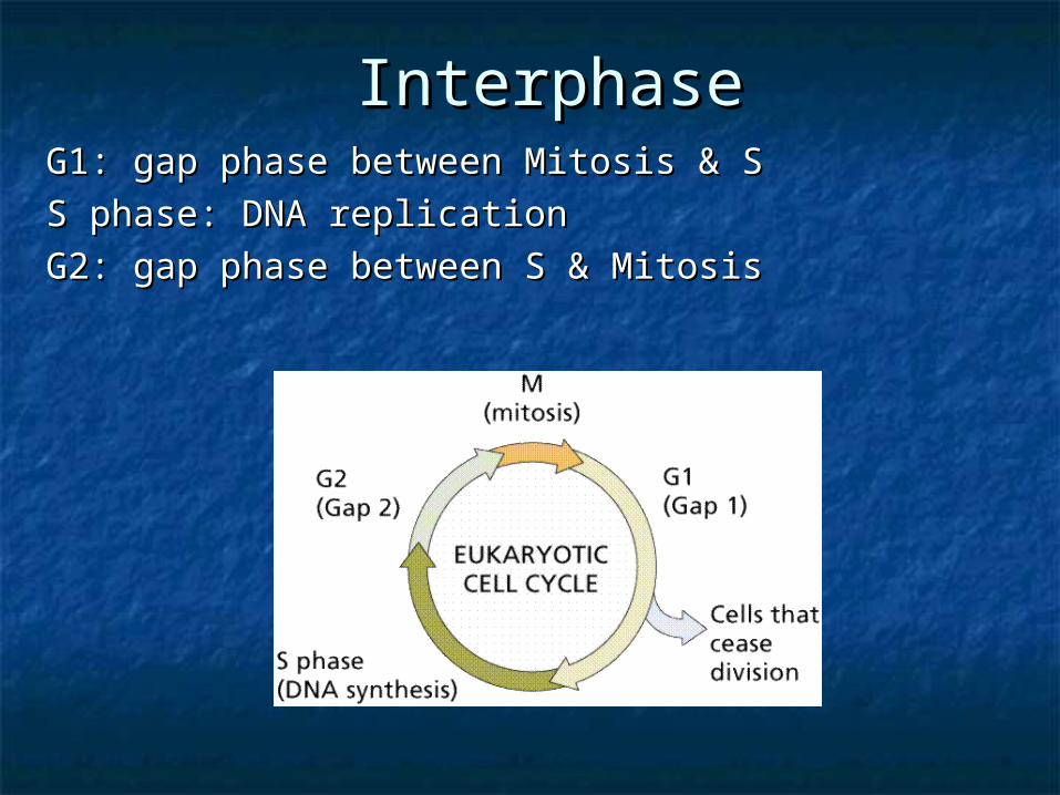
InterphaseInterphaseG1: gap phase between Mitosis & SG1: gap phase between Mitosis & S
S phase: DNA replicationS phase: DNA replication
G2: gap phase between S & MitosisG2: gap phase between S & Mitosis
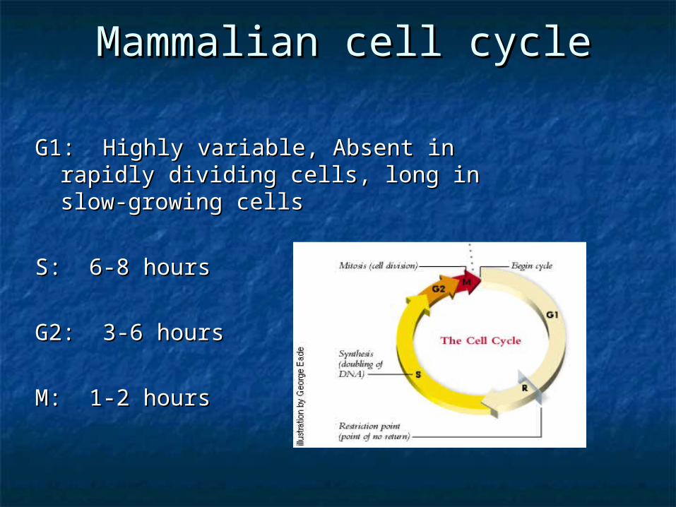
Mammalian cell cycleMammalian cell cycle
G1: Highly variable, Absent in rapidly G1: Highly variable, Absent in rapidly dividing cells, long in slow-growing cellsdividing cells, long in slow-growing cells
S: 6-8 hoursS: 6-8 hours
G2: 3-6 hoursG2: 3-6 hours
M: 1-2 hours M: 1-2 hours
G1 arrested cellsG1 arrested cells
An important control point in cell cycle An important control point in cell cycle holds cells in G1holds cells in G1
Cells can remain indefinitely in G1Cells can remain indefinitely in G1 Such cells are said to reside in a GSuch cells are said to reside in a G00 state, state,
a cell cycle holding pointa cell cycle holding point GG00 Cells may re-enter the normal cell Cells may re-enter the normal cell
cycle if given conditions suitable for cycle if given conditions suitable for growthgrowth

The S phaseThe S phaseEach Chromosome replicates to form Each Chromosome replicates to form
2 2 ChromatidsChromatids. .
Replicated chromatids are joined Replicated chromatids are joined
together at their together at their centromerescentromeres
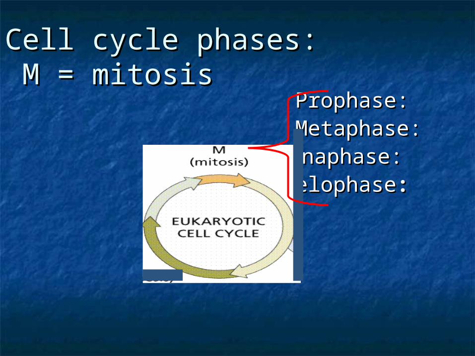
Cell cycle phases:Cell cycle phases: M = mitosis M = mitosis
Prophase: Prophase: Metaphase: Metaphase: Anaphase: Anaphase: TelophaseTelophase::
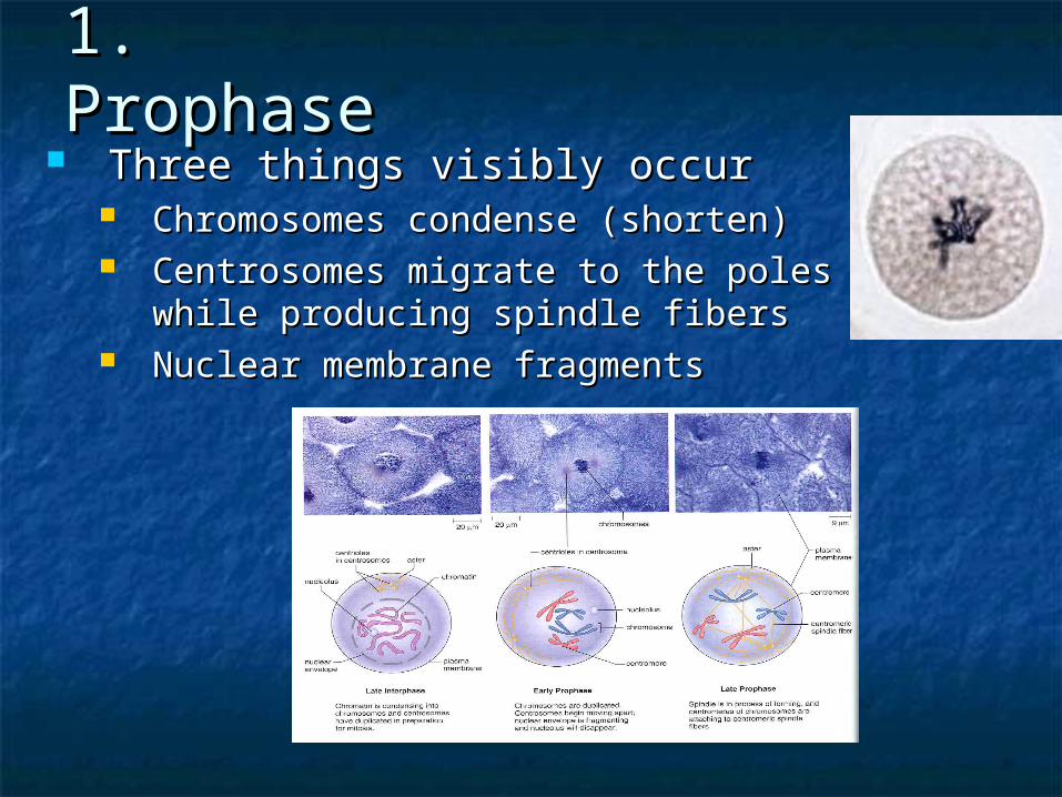
1. 1. ProphaseProphase
Three things visibly occurThree things visibly occur Chromosomes condense (shorten)Chromosomes condense (shorten) Centrosomes migrate to the poles Centrosomes migrate to the poles
while producing spindle fiberswhile producing spindle fibers Nuclear membrane fragmentsNuclear membrane fragments
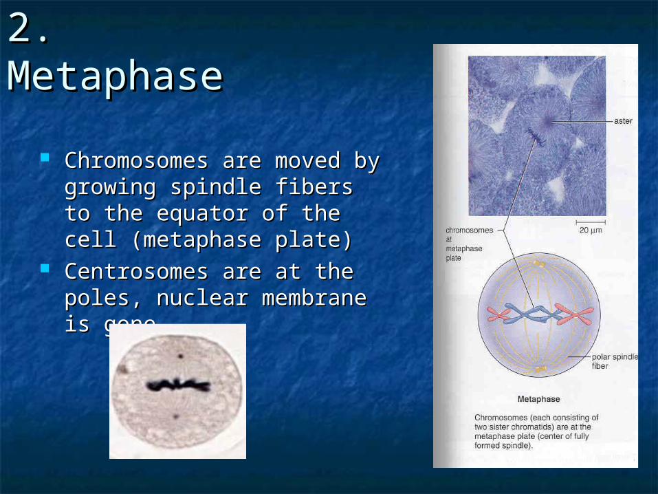
2. 2. MetaphaseMetaphase
Chromosomes are moved by Chromosomes are moved by growing spindle fibers to the growing spindle fibers to the equator of the cell equator of the cell (metaphase plate)(metaphase plate)
Centrosomes are at the Centrosomes are at the poles, nuclear membrane is poles, nuclear membrane is gonegone
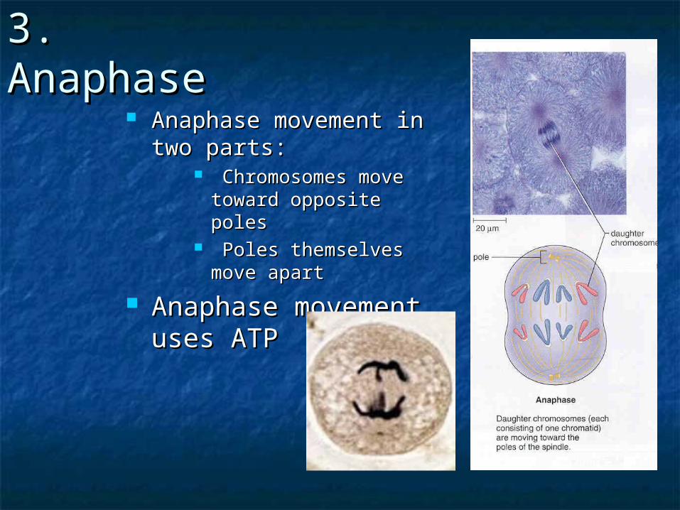
3. Anaphase3. Anaphase Anaphase movement in Anaphase movement in
two partstwo parts:: Chromosomes move Chromosomes move
toward opposite polestoward opposite poles Poles themselves Poles themselves
move apart move apart
Anaphase movement Anaphase movement uses ATPuses ATP
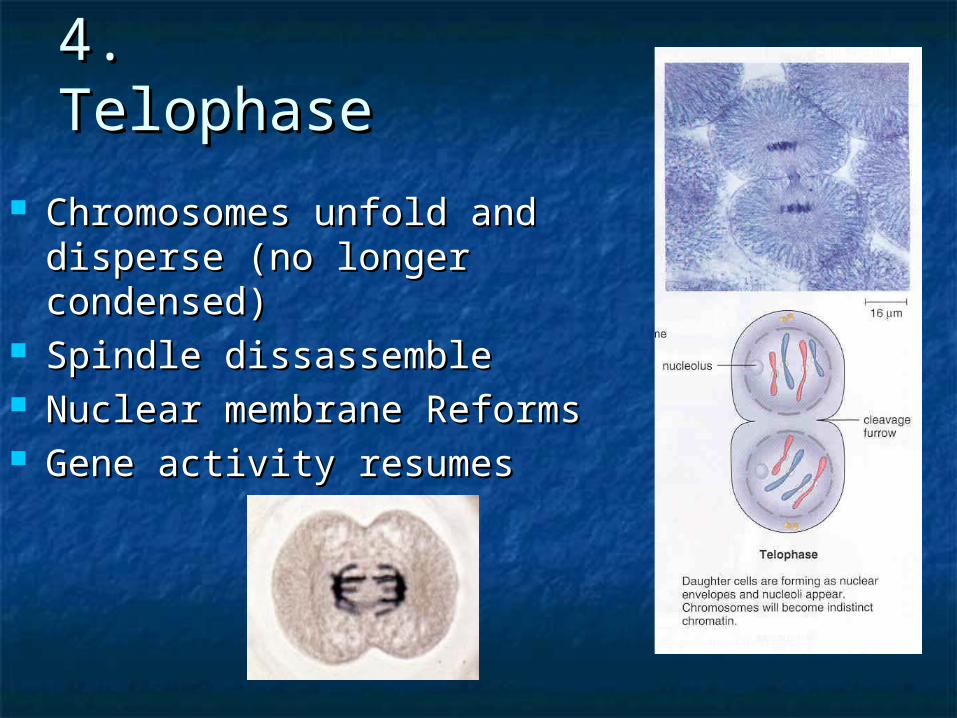
4. Telophase4. Telophase
Chromosomes unfold and Chromosomes unfold and disperse (no longer disperse (no longer condensed)condensed)
Spindle dissassembleSpindle dissassemble Nuclear membrane ReformsNuclear membrane Reforms Gene activity resumesGene activity resumes
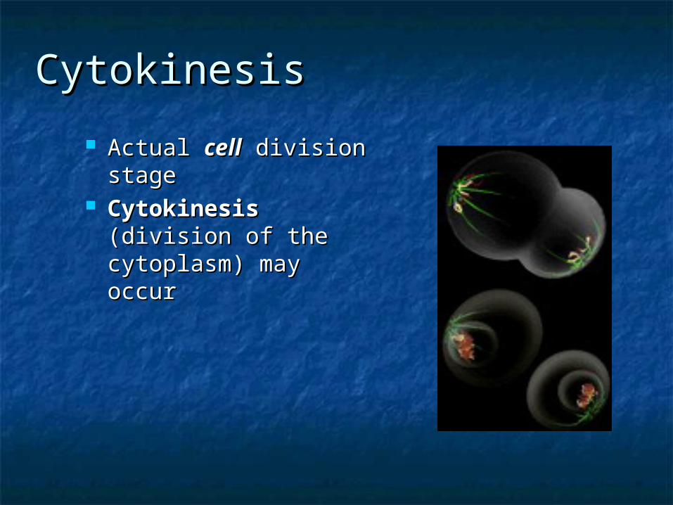
CytokinesisCytokinesis
Actual Actual cellcell division division stagestage
CytokinesisCytokinesis (division of the (division of the cytoplasm) may cytoplasm) may occuroccur

SEXUAL(MEIOSIS)
Figure 14.32. Comparison of meiosis and mitosis. Both meiosis and mitosis initiate after DNA replication, so each chromosome consists of two sister chromatids. In meiosis I, homologous chromosomes pair and then segregate to different cells. Sister chromatids then separate during meiosis II, which resembles a normal mitosis. Meiosis thus gives rise to four haploid daughter cells. Fuente: Cooper, 2000
In Conclusion
A parent cell provides each daughter cell with hereditary instructions
Eukaryotes divide by mitosis or meiosis
Each chromosome is one DNA molecule with proteins attached
Cells with a diploid number (2n) contain two of each kind of chromosome
In ConclusionMitosis maintains the chromosome number
from one cell generation to the next
Mitosis is the basis of growth and tissue repair, and asexual reproduction in some eukaryotes
The cell cycle includes interphase and mitosis
The phases of mitosis are prophase, metaphase, anaphase, and telophase
developed by M. Roig
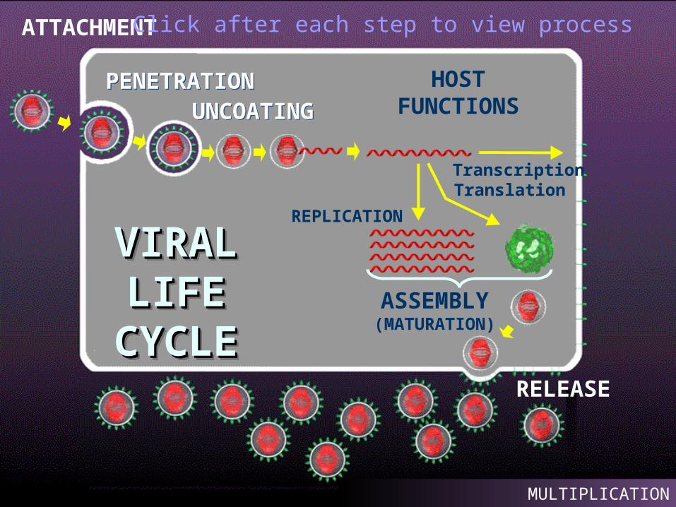
VIRAL VIRAL LIFE LIFE CYCLCYCL
EE
VIRAL VIRAL LIFE LIFE CYCLCYCL
EE
ATTACHMENT
PENETRATIONPENETRATION HOSTFUNCTIONS
ASSEMBLY(MATURATION)
Transcription
REPLICATION
RELEASE
UNCOATINGUNCOATING
Translation
MULTIPLICATION
Click after each step to view process