Cellular and physiological changes during secretion in ...
191
Retrospective eses and Dissertations Iowa State University Capstones, eses and Dissertations 1966 Cellular and physiological changes during secretion in crayfish hepatopancreas Robert Francis Loizzi Iowa State University Follow this and additional works at: hps://lib.dr.iastate.edu/rtd Part of the Zoology Commons is Dissertation is brought to you for free and open access by the Iowa State University Capstones, eses and Dissertations at Iowa State University Digital Repository. It has been accepted for inclusion in Retrospective eses and Dissertations by an authorized administrator of Iowa State University Digital Repository. For more information, please contact [email protected]. Recommended Citation Loizzi, Robert Francis, "Cellular and physiological changes during secretion in crayfish hepatopancreas " (1966). Retrospective eses and Dissertations. 2905. hps://lib.dr.iastate.edu/rtd/2905
Transcript of Cellular and physiological changes during secretion in ...

Retrospective Theses and Dissertations Iowa State University Capstones, Theses andDissertations
1966
Cellular and physiological changes during secretionin crayfish hepatopancreasRobert Francis LoizziIowa State University
Follow this and additional works at: https://lib.dr.iastate.edu/rtd
Part of the Zoology Commons
This Dissertation is brought to you for free and open access by the Iowa State University Capstones, Theses and Dissertations at Iowa State UniversityDigital Repository. It has been accepted for inclusion in Retrospective Theses and Dissertations by an authorized administrator of Iowa State UniversityDigital Repository. For more information, please contact [email protected].
Recommended CitationLoizzi, Robert Francis, "Cellular and physiological changes during secretion in crayfish hepatopancreas " (1966). Retrospective Thesesand Dissertations. 2905.https://lib.dr.iastate.edu/rtd/2905

This dissertation has been
microfilmed exactly as received 66—10,428
LOIZZI, Robert Francis, 1935— CELLULAR AND PHYSIOLOGICAL CHANGES DURING SECRETION IN CRAYFISH HEPATO-PANCREAS.
Iowa State.University of Science and Technology Ph.D., 1966 Zoology
University Microfilms, Inc., Ann Arbor, Michigan

CELLULAR AND PHYSIOLOGICAL CHANGES DURING
SECRETION IN CRAYFISH HEPATOPANCREAS
by
A Dissertation Submitted to the
Graduate Faculty in Partial Fulfillment of
The Requirements for the Degree of
DOCTOR OF PHILOSOPHY
Major Subject: Cell Biology
Robert Francis Loizzi
Approved:
KÎ Major Work
Chaf lrman Advisory Comml tC ll Biology Program
Dean bf Graduée College
Chairman of Major Department
Iowa State University Of Science and Technology
Ames, Iowa
1966
Signature was redacted for privacy.
Signature was redacted for privacy.
Signature was redacted for privacy.
Signature was redacted for privacy.
il
TABLE OP CONTENTS
Page
INTRODUCTION 1
REVIEW OP LITERATURE 10
PART I. MICROSCOPIC ANATOMY OF HEPATOPANCREAS TUBULE 34
PART II. RHYTHMIC CHANGES IN PROTEASE ACTIVITY AFTER FEEDING 99
PART III. OBSERVATIONS OF TUBULE CONTRACTIONS 157
GENERAL DISCUSSION 16?
SUMMARY AND CONCLUSIONS 178
LITERATURE CITED 180
ACKNOWLEDGEMENTS 186

1
INTRODUCTION
The hepatopancreas as a research tool
Twentieth century biology is typified by a willingness
to assimilate concepts and techniques from other natural
sciences in order to synthesize an integrated understanding
of life processes. If the multidisciplined effort is the
currently favored approach toward biological problems, then
the crustacean hepatopancreas is, simultaneously, one of
the more useful and neglected organs in contemporary re
search.
First, let us consider the following hypothetical sys
tem; a monolayer of epithelial cells on a basement membrane
in the form of a long, very slender triangle. The monolayer
is more or less divided into regions such that mitotic divi
sions are restricted to the apex of the triangle and large
secretory cells are located in a broad zone which takes up
approximately 50 of the triangle between apex and base.
In addition to undifferentiated cells, at least three spe
cialized cell types (absorptive, synthetic, and secretory)
are fairly well localized in the various regions. The posi
tioning of the cells, however, does not imply a static con
dition in the monolayer; there is continued migration and
differentiation of cells both from the region of mitosis to
the other zones and also within each zone. Thus, the sys-

tern would be useful in studying cell secretion, differenti
ation and migration, with the added advantage that cell
types are easily located in their respective regions.
In addition to secretion, the monolayer carries out a
wide variety of interesting functions at the cellular level.
The region adjacent to the apex of the triangle is special
ized for absorption of foodstuffs and the cells in this
region resemble those found in the vertebrate small intes
tine. In fact, the monolayer does carry on nearly all of
the absorptive functions normally associated with the gut.
The monolayer is highly specialized for nutrient storage
and specific cells respond to hormonal influences by either
retaining or releasing massive amounts of glycogen and
lipids and by controlling the level of blood sugar. Be
sides metabolite storage, curious, metal-containing granules
are located in specific cells. Granules containing salts of
copper, iron or calcium may also be stored in, or mobilized
from, the monolayer as in the case of the metabolites. The
functions of these granules have not been clearly estab
lished,
Metabolically the cells of the monolayer are very
active. They exhibit a high Q02 and possess complete enzyme
systems for nucleic acid degradation and for the biosynthe
sis of bile salts, cholesterol, urea and uric acid.
However, the monolayer' s most striking biosynthetic

3
capabilities are related, to its secretory functions. Cells
in the monolayer synthesize and secrete all of the catego
ries of digestive enzymes found in vertebrates with the
exception of pepsin and thus are capable of effecting com
plete digestion of proteins, lipids and carbohydrates. In
addition, experiments suggest that a few non-vertebrate
enzymes such as cellulase and chltlnase are also secreted.
Finally, the monolayer can absorb foreign materials and
waste products through its basement membrane, concentrate
them, and extrude them via the cells' free surfaces, thus
indicating a possible excretory role.
Therefore, the monolayer may be used to study a vari
ety of cellular functions which normally are not carried
out in the same tissue. And, because of the geometric
arrangement of cells into regions, cytologlcal and cyto-
chemlcal techniques are aided by the monolayer' s natural
orientation.
If the hypothetical monolayer is formed into a' cylin
der, (more properly, a long, slender cone) and then encased
in a delicate network of circular and longitudinal muscle
fibers, an entirely new level of Investigation is possible,
that of physiological control of the cellular functions
discussed above. What is the relationship between muscle
contraction and each of the cellular functions? By what
mechanisms are the functions and the muscles controlled?
4
Finally, since the entire network does not contract equally,
what coordinates Its selective contractions?
The system described Is, of course, not hypothetical
but a single tubule from the crayfish hepatopancreas. More
over, the description did not exhaust Its useful properties.
Its size, for Instance, Is about 2-4 mm, long and 0.2-0.4
mm. In diameter which Is large enough for ease of Isolation
and study under the dissecting microscope but small enough
for the compound microscope or for cutting complete cross
sections for electron microscopy. The system can be studied
In vitro for several days In a drop of crayfish blood at.
slightly lowered temperatures. Moreover, because of the
hepatopancreas' location Inside the crayfish and the trans
parency of Its connective covering, little skill Is required
to observe Individual tubules vivo while the crayfish
eats or carries on other normal activities.
Finally, the Integrity of the tubule Is hormonally de
pendent. It Is sensitive to changes In the animal's molt
cycle and certain cells In the tubule which are normally
rich In HNA will lose this compound In the absence of hor
mones from glands In the eyestalk and will regain It upon
addition of eyestalk extracts. I
Many of the functions discussed above have never been
thoroughly Investigated or else were studied long before
current concepts of protein synthesis and molecular biology
5
came into being. Most of the previous research on the
hepatopancreas dealt with anatomy and histology of the gland
and its various secretions but these studies can be grouped
into sporadic periods over the last 125 years. For example,
only one brief paper has been published on the fine struc
ture of the crayfish hepatopancreas and this was limited to
a study of the metal-containing cells.
In general, therefore, this remarkable, polyvalent sys
tem has gone largely unnoticed in spite of the current trend
toward multidisciplined approaches and synthesis of biolog
ical concepts from all levels of organization.
Problem to be investigated
The versatility of the hepatopancreas allowed several
choices of single processes which could be studied at many
levels of biological organization. The particular process
chosen for this research was the secretion of digestive
enzymes.
The term secretion usually gives to the secretory act
an aura of circumscription or narrowness of scope, as though
it involved a simple transfer of the secretory product. In
reality, secretion is Influenced at many levels of biolog
ical organization. Modern concepts of secretion, such as
those expounded by Junquieira and Hirsch (2?) and by Gabe
and Arvy (17), reveal that secretion involves at least three

6
general activities; (1) ingestion of raw materials by the
cell from the surrounding medium; (2) synthesis and con
centration of the secretory product within the cell; and
(3) extrusion of the secretory product into the extracellu
lar environment. Each activity, of course, is reflected by
biochemical and morphological changes which may be studied
by a variety of means. Even these three elements, broad as
they are, confine the concept of secretion within the
limits of the secretory cell though it is evident that se
cretion involves a fourth activity, transfer of the secre
tory product from the secretory cell to the site of action.
To illustrate this, Richardson (49) has described fourteen I
separate factors (anatomical, neural and hormonal) which
operate in mammary gland secretion after extrusion of the
milk by the alveolar cells. Secretion, therefore, is the
end result of factors at many levels acting in concert.
These include; (1) the secretory cell's genetically deter
mined "program" of differentiation; (2) the maze of bio-
synthetic pathways and their controls; (3) the dynamic
activities and relationships of intracellular organelles;
(4) the anatomical relationships between the secretory cell
and neighboring structures which might influence it; and
(5) extracellular influences such as drugs, hormones and
nerve impulses, including their reception and integration
as well as the mechanisms by which they influence secretion.
7
To limit the problem still further it was decided spe
cifically to investigate the reported presence of secretory
rhythms in the hepatopancreas. The details of previous
studies on cyclic secretion of digestive enzymes will be
covered in later sections but, suffice it to say, such
rhythmicity has been established and it is a reflection of
the cell's secretory cycle. Beyond this, no two authors
agree on the exact source of the rhythmicity. It can be
explained by the mode of secretion, but holocrine, apocrine
and merocrine secretion have all been described in the
gland. And, although sequence of secretory cell differen
tiation is definitely a determining factor, at least four
different sequences have been described. Prom a practical
standpoint rhythmicity is often a very desirable phenomenon.
In discussing secretion of digestive enzymes, Harrington (3)
mentions certain intertidal molluscs in which perfect cor
relation has been found between the secretory rhythm and the
tidal rhythm, demonstrating how adequate digestive enzymes
are economically supplied just prior to the arrival of food
via the incoming tide.
Essentially, therefore, this dissertation is involved
with confirming the presence of rhythmic, digestive enzyme
secretion in a particular species of crayfish not previous
ly studied, and investigating its possible sources.

8
Approach to the problem
Some of the work carried out in previous investigations
had to be repeated with this species of crayfish, although
newer techniques were utilized, in order to provide a basis
for comparison. In addition, certain aspects not previous
ly studied helped to correlate the older observations and
these too were examined.
A three-fold approach was used in which data and con
clusions were obtained independently in each part and then
combined in a general, integrating discussion.
In Part I the morphology of hepatopancreas tubules was
studied using a variety of cytological and cytochemical
techniques including electron microscopy. The purpose was
to gather information about the various cell types, their
location in the tubule, cytochemistry, fine structure and
appearance during secretion in order to clarify the mode of
secretion and the sequence of cell differentiation.
In Part II the activity of one digestive enzyme, a
trypsin-like protease, was assayed in hepatopancreas ho-
mogenates at close intervals for several hours after feeding
and a duplicate study carried out using animals treated with
colchicine to block mitosis In the tubules. The same ho-
mogenates were then assayed for protein content to study its
changes and also to calculate specific activities of the
protease. Prom the results of these studies it was possible

9
to determine if a secretory rhythm was present and whether
or not secretion was exclusively holocrine.
In Part III it was originally intended to carry out a
variety of stimulation experiments using drug's, electric
shock and extracts of tissues from fed animals to study
tubule contraction with regard to timing and mechanism of
control. However, difficulties arose which necessitated
reducing the goals of this part to simple observations in
tubules under various conditions without formal experi
mentation.
The results of these three parts were then analyzed
for either confirming or contradictory findings and then
were used in conjunction with previous work in an effort
to answer the central problem of rhythmic secretion and its
causes.

10
REVIEW OP LITERATURE - •
General Trends in Hepatopancreas Research
Although observations on the crustacean hepatopancreas
have been described in the literature for at least a centu
ry and a quarter, the total information accumulated is
meager when compared to other areas of biology for several
reasons: First, crustacean physiology, unlike that of the
vertebrates, is a young science. For example, in the area
of crustacean endocrinology, Carlisle and Knowles (4) point
out that the endocrine glands of vertebrates had been de
scribed in detail many years (even centuries in some cases)
before experiments on function were performed and that this
knowledge facilitated the ablation and other physiological
techniques used in endocrine research. In crustacean
endocrinology, however, evidence for blood-bome hormones
was originally based on simple physiological experiments
and preceded detailed anatomical and histological descrip
tions by several years. Even the morphology of the sinus
gland was not described until 19 7. Although endocrinology
is perhaps one of the physiological areas most affected by
lack of precise anatomical information, other areas have
been similarly delayed.
The second reason is an apparent lack of communication
among scientists who were interested in the hepatopancreas,

11
both in related disciplines and in different countries. A
literature search reveals that many times gross anatomists
and histologists working on the hepatopancreas were unaware
of each other's findings and that a definite information
barrier existed between the earlier works of American and
German investigators and, more recently, between those of
German and Japanese workers. Later, it will be shown how
some 70 years of valuable research has become buried in the
old German literature,
A third probable factor is that digestion in the cray
fish has its most dramatic aspects in the complex mechanical
system for mastication, made up of chitinous ossicles, bars,
plates and teeth, filters of setae and several pairs of
powerful muscles, collectively referred to as the gastric
mill and compared to which the greenish, soft hepatopancreas
seemed uninteresting to the anatomically orientated biolo
gists of the day.
Finally, as is the case in many fields, general inter
est in the hepatopancreas and its secretory functions could
only occur after two prerequisites had been met. First,
some imaginative and respected scientist would have to dem
onstrate the gland' s broad potential value in acquiring new
biological knowledge; and, second, a more quantitative
method than those generally available to zoologists had to
be developed by which secretion could be studied. The two

12
factors appeared almost simultaneously in the 1880's and
the bulk of hepatopancreas research, sporadic though it has
been, appeared after this date.
With regard to meeting the first prerequisite, this is
usually attributed to the publication in 1880 of a small
but unique zoology text by Thomas H. Huxley (25) which was
widely read and enjoyed in several languages for its com-
pletness and attention to detail, its literary prose which
made life processes understandable, and its beautiful wood
cuts and line drawings. It was a unique text because Huxley
demonstrated the universality and integration of life proc
esses by presenting all the many facets of zoology in a
single animal; the latter was, of course, "the commonest
and most insignificant of animals", the lowly crayfish. In
chapter II, there is a poetic description of chemical diges
tion after which Huxley describes the sources of the diges
tive fluid; the "livers". This included the anatomy of the
hepatopancreas, the relationship of the ducts and the
"innumerable caeca", and, in one of the earliest descrip
tions of secretory cell differentiation in the hepato
pancreas tubule, he explains that It is the cells of the
tubule epithelium "which are the seat of the manufacturing
process which results in the formation of the secretion"
and that; "To this end they are constantly being reformed
at the summits of the caeca. As they grow, they pass down

13
towards the duct and, at the same time, separate into their
interior certain special products, among which globules of
yellow fatty matter are very conspicuous." He goes on to
describe the intracellular growth of these globules from a
minute size in young cells near the summit of the caeca to
their state in the more proximal cells where the globules
are larger and more numerous and from which the globules are
released and eventually removed from the gland via the duct
system. He concludes, "In fact, few glands are better
fitted for the study of the manner in which secretion is
effected than the crayfish's liver."
In 1883, at about the same time zoologists around the
world were reading Huxley' s book, E. Heidenhain published a
handbook of physiology which included the results of fifteen
years of investigations on secretion in gastric and salivary
glands, Heidenhain' s work is described in detail by Babkin
(2) who credits Heidenhain with having laid the foundation
of our knowledge of the secretory process and of being the
first to appreciate the internal changes of gland cells
which are collectively referred to as the secretory cycle.
Heidenhain's "method" was simply to correlate histological
observations and biochemical assays of digestive enzymes in
the same tissue, on the same time scale, after stimulation.
This approach, although an obvious one for today' s biolo
gist, was an innovation then because It required combining

14
the "artistry" of the histologist and the more quantitative
skills of the organic chemist, two demanding and usually
exclusive abilities, in a single individual. Although simi
lar efforts had been made before him, Heidenhain success
fully brought the two fields together in studying the se
cretory cell and used the combination to reveal a wealth of
information about many cellular processes as well as secre
tion.
After the appearances of Huxley' s exposition of the
role of the hepatopanoreas and its research potential and
Heidenhain' s interesting approach to the study of secretion,
many zoologists, especially in Germany, began to turn their
attention to this organ, first examining its histology and
secretions and then combining the two in the manner of
Heidenhain, all of which resulted in a large mass of liter
ature on the subject in the 50-year period from 1880 to
1930. Then, almost as suddenly as it arose, interest in the
hepatopanoreas waned and the gland slipped into obscurity.
The latter is reflected today by the brief coverage the
hepatopanoreas and its multiple functions receive in modem
zoology texts in spite of past interest. During the last
few years, however, the gland has been studied with in
creasing frequency, possibly because of a revitalization of
the comparative and invertebrate aspects of biology and be
cause it appears to be an interesting system for use in

15
modern biological research.
Thus, research on the hepatopancreas appears to fall
into five periods:
1. prior to 1880; a few isolated studies concerned
mainly with anatomical considerations, especially
nervous innervation and tubule musculature.
2. 1880 to 1905: accelerated research, possibly stim
ulated by Huxley' s work, characterizing the di
gestive juice and its enzymes, making general I
histological observations of the tubule, and per
forming basic physiological experiments to eluci
date hepatopancreatlc functions (e.g. feeding and
Injection of dyes).
3. 1905 to 1930 : increasingly sophisticated correla
tions between histology and physiology in the
manner of Heidenhain, culminating in the work of
Hirsch and Jacobs in 1928 - 1930,
4. 1930 to 1955: a blank period characterized by
nearly complete lack of interest in the hepato
pancreas; many of the former Investigators had
turned their attention to secretion in vertebrates,
5. 1955 to present: sporadic but increasingly frequent
studies in which the hepatopancreas has been used
to investigate a variety of topics including cyto-
dlfferentiation, the role of mitochondria in osmo-
l6
regulation, unusual glycolytic pathways and the
cytochemical effects of molting hormones.
Studies Related to Protease Secretion
A review of specific investigations relating to the
cytology and physiology of protease secretion will now be
presented, beginning with the digestive juice Itself and
then tracing its origin to the dynamic activities of the
tubule epithelium.
Nature of the digestive .juice
A general review of how digestive juice is collected
for analysis, its enzymes, bile salts, Ions and pH may be
found in Vonk (5 ). A complete list of crustacean digestive
enzymes investigated prior to 195 was prepared by Mansour-
Bek for "Tabulae Blologlcae" (37).
According to Yonge (6o), it was Hoppe-Selyer in 1876
who first demonstrated that the crustacean hepatopancreas
was a digestive organ and, by 19IO, considerable evidence
had accumulated for the presence of amylolytlc, proteolytic
and lipolytic enzymes in the digestive juice. In the same
paper, Yonge reported the presence of enzymes in the Norway
lobster, Nephrops norvegicus. for the digestion of starch,
glycogen, sucrose, maltose and lactose with optimum diges
tion at 57°C and neutral pH; for the splitting of fats and
a wide range of fatty acid esters at an alkaline pH; and for

17
the digestion of fibrin to amino acids, also with an
alkaline pH optimum and believed to be tryptic rather than
peptic in nature. In 1928, Kriiger and Graetz (30) found
optimal protease activity at pH 7,5 and 50°C for the diges
tive juice of the crayfish Astacus: however, in the same
year, Shinoda (51), using the same species, reported pH
optima of 6,2 - 6,7 for peptones and proteoses and 8-9 for
fibrin and gelatin. In 1932, Mansour-Bek (38), using the
crab Ma.1a squinado and a variety of substrates, compared the
activities of digestive enzjànes in gastric juice and crude
hepatopancreas extracts with the activities of enzymes puri
fied by column absorption techniques. Casein-splitting by
the juice and crude extract was most active at pH 6,2 and,
for gelatin, at pH 6,0, The purified proteinases had optima
for casein and gelatin at pH 8,1 and 7,4 respectively and
the purified enzyme is activated by pig enterokinase but
HON and H2S had no effect. The study indicates a strong
functional similarity between this protease and vertebrate
trypsin. According to Vonk (55)1 no protease similar to
vertebrate pepsin has been found in crustaceans or other
invertebrates and, apparently, none of the crustacean diges
tive enzymes have been isolated or crystallized for further '
analysis. There may be several as yet undiscovered diges
tive enzymes in the hepatopancreas. Hi 1964, Koolman (29)

18
raised the number of digestive carbohydrases in the lobster
Homarus and in Astacus to 13 Including chitinase and cellu-
lase.
Transport of digestive .juice
The factors involved in transporting digestive juice
from its source in the hepatopancreas to its initial site of
action in the alimentary canal are (1) anatomical route, (2)
mechanical factors effecting a flow of the Juice, and (3)
physiological control of these factors.
Both Yonge (6o) in 1924 and Lochhead (35) in 1950 have
described a system of grooves and channels in the wall of
the alimentary canal between cardiac stomach and midgut at
the junctions of the hepatopancreatic ducts. The system is
beset with numerous setae which act as filters and allow the
passage of only very small particles. It thus forms a path
way through the complex cardiac and pyloric stomachs, in
cluding the gastric mill. The main route for partially di
gested food particles entering the hepatopancreas (or, in
the reverse direction, for transfer of digestive juice to
the alimentary canal) probably follows the ventral groove
which begins at the esophogeal portion of the cardiac stom
ach and then divides.into two forks, passes on either side
of the cardiopyloric valve, and proceeds through a gland
filter with setae spaced 10 micra apart into channels lead

19
ing to the openings of right and left divisions of the
hepatopancreas. All along the route there are muscular
valves which regulate flow by closing the grooves.
In regard to mechanical factors which effect a flow of
digestive juice, most of the modem literature (that of the
last 4o-plus years) is practically silent on the subject,
Ramsay (46), in 1952, published a monograph on invertebrate
physiology in which he explains that molluscs and crusta
ceans use different mechanisms to remove digestive juice
from the tubules. In molluscs, transport occurs through
ciliary movement along the tubule epithelium but, in crusta
ceans, the epithelium is not ciliated and secretions are
transported by contractions of a "thin coat of muscle" sur
rounding the hepatopancreas tubule. The monograph, how
ever, does not include a bibliography and so the statement
cannot be traced in the literature. In 1959, Ogura (43)
included some hepatopancreas morphology in his paper on
metal granules in the tubule and shows a line drawing of a
tubule encased by circular and longitudinal muscle fibers.
However, the muscles are not discussed in the paper and,
again, references are not given. By chance, this author
came across an unpublished report prepared by Smith (52) in
1965 for an undergraduate research course. The paper was
concerned with the system of muscle fibers in the hepato
pancreas tubule and included an excellent bibliography.
20
Prom the latter, this author commenced a literature search
which revealed that a mass of research had "been carried out
on the muscle network during the period 18 5 to 1914. The
last and most complete work was by Pump (45) in 1914 who
surveyed the literature of this 70-year period In addition
to making his own observations. The muscle net consists of
two types of fibers; thick, striated, circular fibers, con
taining one nucleus per fiber, and evenly spaced along the
length of the tubule except at the distal tip where they
are spaced at smaller intervals; and very fine, usually non-
striated longitudinal fibers which appear to connect two
successive circular fibers, and which come off the latter at
all angles and sometimes divide into two or three branches.
The network appeared to lie on the surface of a tunica
propria which enclosed the tubule itself. Apparently all of
the work done during this period was descriptive histology
and morphology with no physiological Investigations, In
regard to the striations, Pump noted only A and Z bands in
the circular muscles but Smith (52), using Biebrich' s
scarlet, was able to distinguish A, I, H, and Z regions in
whole mounts.
In spite of the apparent lack of physiological experi
ments, the obvious anatomical relationship of the-muscle
network to the tubule epithelium led Yonge (6o) to write;
"The forcing out of the secretion and taking in of '•-he dis-
/
21
solved matter is brought about by a rhythmical contraction
and expansion of the hepatopancréatic tubules. The circular
muscle fibers cause contraction, and the relaxation of these
together with the contraction of the striped longitudinal
fibers cause expansion." The latter mechanism is difficult
to conceive. •
In regard to physiological control of the tubule con
tractions, neither hormonal nor neural mechanisms have been
demonstrated in crayfish. Several studies have been carried
out on the neural control of crayfish intestinal movements-
but nothing pertaining to the hepatopancreas. Even the
question of innervation has not been answered, Neuro-
physiologists cite only three basic references which even
mention innervation of the hepatopancreas, the most detailed
of which is by Keim (28) in 1915. In the crayfish, the two
large circumesophogeal commissures of the ventral nerve cord
each give off a delicate, medial branch and these unite
anterior to the esophogus forming the stomatogastric nerve.
T is nerve is then closely applied to the anteroventral
surface of the cardiac stomach and follows its curvature
first forward, then upward and, finally, backward on the
stomach's dorsal surface. At the point where the nerve
passes between the anterior gastrics, two large muscle bun
dles which attach the cardiac stomach to the dorsal cara
pace, it forms the ventricular ganglion and then continues
22
posteriorly as the dorsal ventricular nerve, passing off
paired right and left branches along the way. Near the
region of Junction between cardiac and pyloric portions of
the stomach, the nerve divides into, right and left branches,
each of which turns outward and downward abruptly, giving
off more branches as it follows the curvature of the
alimentary canal ventrally. One of these small branches,
which Keim calls the liver nerve, can be traced to the junc
tion of midgut and hepatopancreatic duct. At this point,
however, the nerve ramifies to such an extent that it cannot
be traced beyond the jtmction, thus leaving open the ques
tion of innervation. Keim quotes an earlier German inves
tigator, Schlemm who, writing in Latin, described the same
nerve (and the same difficulty) in 1844.
Cytology of the hepatopancreas tubule
In 1880, Huxley (25) described the individual tubules
as having an outer wall lined by a single layer of epithe
lium. In the same year, Weber (57), who also described the
muscle network, identified two cell types in the epithelium
of tubules from Astacus fluvlatllis. the enzyme-producing
Fermentzellen and the fat-storing Leberzellen. The same
two types were observed by Frenzel (15) in 1893 who called
Fermentzellen. those which possessed a single, large vacu
ole, and Fettezellen. those which had many lipid droplets.

23
Cytoplasm of the enzyme cells had a fibrillar appearance
when stained with either light green and safranin or hema
toxylin and safranin. In addition, he observed a third cell
type, the enzyme-replacement cells, which also had a fibril
lar cytoplasm but whose vacuoles had a range of sizes down
to the limits of resolution. Mitosis of enzyme- and fat-
mother cells was observed in the blind tip of the tubule
but the two cell types could also form amitotically from
pre-existing cells of the same type. A striated boarder was
present on the enzyme, fat and replacement cells.
In the United States in 1928, Dorman (7), in his stud
ies on the comparative physiology of digestion, described
the morphology and histology of the hepatopancreas. He,
too, described two principle cell types which he names he
patic and pancreatic cells. The first was a columnar epi
thelial cell possessing one or two round and basally situ
ated nuclei and with granular cytoplasm containing pigmented
inclusions, fat globules and glycogen deposits; pancreatic
cells stained much more darkly than hepatic cells with most
cytoplasmic stains, had a deeply granular appearance, and
their long axes were perpendicular to the basement membrane.
The basal portion was broad and rested flat against the
basement membrane but the apical end was attenuated and
drawn out to form a duct and was closely applied to a crypt
in the lumen. Occasionally the ducts of several pancreatic
24
cells coalesced before meeting a crypt.
The most extensive cytological description of the tu
bule epithelium was made by Jacobs (26) in 1928 who studied
the formation of secretory granules in the hepatopancreas
of Astacus leptodactylus. Jacobs described four cell types
which he called Embr.vonale zellen (E-cells), Restzellen (R-
cells), Pibrillenzellen (P-cells) and Blasenzellen (B-
cells). A fifth type, Wanderzellen. was not involved in the
secretory function of the gland, E-cells were undifferenti
ated columnar epithelial cells which, together with mitotic
divisions, were restricted to the blind, distal tip of the
tubule. More proximally other E-cells were found with evi
dences of beginning differentiation such as a basophilic
vacuole, the parasome, closely applied to the apical surface
of a large elongated nucleus, and a few, discrete Golgi
apparati. R-cells and P-cells were found throughout the
length of the tubule except for the tip. R-cells were ab
sorptive in appearance and distinguished by the presence of
several fat vacuoles and a small, rounded nucleus in the
basal cytoplasm. P-cells had a larger nucleus which was
more centrally located. Most of their cytoplasm was richly
basophilic and had a fibrillar appearance except for a
region in the basal portion which was acidophilic and non-
fibrillar. Both cell types possessed a striated border.
B-cells were the most striking of the four due to the
25
presence of a single, large, blister-like, secretory vacuole
which left only a small amount of cytoplasm at the apical
and basal ends of the cell. The nucleus was pressed flat
against the basal membrane by the vacuole. B-cells were
restricted to the middle 50-6o^ of the tubule and their
striking presence allowed Jacobs to divide the tubule into
three regions: Region I was the most distal portion in
cluding the tip and contained E-cells followed by R- and P-
cells; Region II stood out due to the presence of B-cells,
although E- and P-cells were located here also; Region III,
the most proximal portion, resembled the epithelium of the
collecting ducts and contained, in addition to R- and F-
cells, some degenerating cells with pycnotic nuclei. B-
cells also possessed a definite, striated boarder.
Jacobs also described intermediate cell types, espe
cially in Region I; for example, E-cells with increasing
amounts of lipid or increasing basophilic and fibrillar
cytoplasm, and P-cells with a single vacuole of increasing
, size.
More recent authors have described similar cell types.
In 1957, Travis (53) found that in the spiny lobster,
Panulirus. the absorption cells, under certain conditions,
had large concentrations of alkaline phosphatase in the
striated border, calcium phosphate deposits or calco-
spherltes in the apical cytoplasm, and large amounts of gly-

26
cogen and fats. In the crayfish Procambarus clarkll Ogura
(43), in 1959, observed the presence of iron granules in
the fibrillar cells and copper granules in the absorptive
cells and thus designated the two types Pe-cells and Cu-
cells respectively. Miyawakl al. (4o), in 19^1, confirmed
these results in the same species and added the following
observations. The Pe-cells were rich in RNA with the excep
tion of the Pe-containing vacuole Itself. The latter was
rich in mucopolysaccharides or mucoproteins bound with iron.
The cytoplasm of Pe-cells was also rich in phosphatases and
calcium. However, the Cu-cells contained copper only when
this metal was in excessive concentrations in the media. In
another study, in 19^2, Miyawakl and Tanoue (42) presented /
what is probably the only previous electron micrographie
study of cell types in the crayfish hepatopancreas albeit
quite brief and concentrating on the metal-containing cells.
The Pe-cells possessed an extensive endoplasmic reticulum
and a very large, electron-dense Iron granule. The Cu-cells
possessed a large, complex organelle which was identified as
the Cu-granule. The organelle was bounded by a double mem
brane, with many internal vesicles, membranes and electron-
dense particles, Irregular in shape, size and distribution.
Both cell types possessed a border of microvilli.

27
Secretion and cell differentiation
In addition to the morphological descriptions given
above, Jacobs (26) also observed the growth and maturation
of Golgl-assoclated secretion granules In F-cells with grad
ual coalescence of the granules until a secretory or B-cell
resulted. On this basis, and because of the location and
Intermediate forms of the four basic cell types, he postu
lated the following sequence of cell differentiation:
mitosis, E-cell, B-cell, P-cell, B-cell, secretion; how
ever, differences in morphology cast some doubt about in
cluding E-cells-in direct sequence with the other cell
types. This sequence would account for the various loca
tions of the cell types and it provided a cytological con
firmation of Huxley' s original observation that the secre
tory vacuoles grew in size as cells migrated from the dis
tal end of the tubule to the more proximal region where
secretion took place.
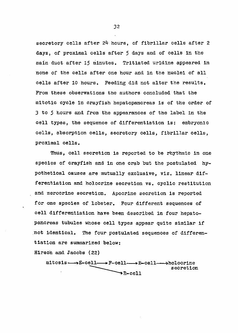
In the period 1928-1930, Hirsch and Jacobs (21, 22) and
Hirsch and Buchmann (20) published a classical series of
investigations in which the subjects of digestive enzyme
secretion and cell differentiation were studied in the cray
fish Astacus leptodactylus. The studies included enzyme
assays and cell counts (21), correlation and analysis of the
results (22), and cytochemical studies of the various cell
types (20). Activities of amylase, casein-protease, and

28
peroxidase were assayed in the stomach Juice and in glycerin
extracts of the hepatopancreas in starved crayfish and in
animals sacrificed every half-hour after feeding for 6J
hours. Histological preparations were made from the same
glands and counts were made of mitotic figures and each of
the four cell types in the tubule. The results indicated
that all of the enzyme activities fluctuated with time
according to a regular rhythm. The rhythms of amylase and
protease were in agreement while that of peroxidase was dif
ferent. The curve for each digestive enzyme activity pos
sessed two maxima in the 6§ hour period. Moreover, the
cycling in the stomach juice, although similar to that of
the extract, followed it by ^ to 2 hours depending on the
enzyme and thus confirmed the source of the digestive en
zymes in the stomach juice to be the hepatopancreas. These
results were reproducible in all seasons of the year al
though the over-all values and maxima were highest in
Spring. When curves of cell counts were compared with those
of enzyme activities, the former also varied rhythmically,
but only the B-cells were in phase with enzyme activity in
the gland extract. This was taken as evidence that the
source of the enzyme was the B-cell, a conclusion previously
made on purely morphological grounds.
When the other curves were carefully analyzed, it was
apparent that the cycles for the various cell types, in-

29
eluding dividing cells, were all slightly out of phase so
that a sequence of maxima was repeated nearly three times
in a 6§ hour period. This sequence, it was concluded, rep
resented the sequence of cell differentiation leading to
secretion. The sequence was; mitosis, E-cells, P-cells,
B^-cells, Bg-cells, B-cells, holocrine secretion. E-cells
were differentiated separately from E-cells, and and B2
were stages in the maturation of the B-cell, The time re
quired fof differentiation of E-cell to F-cell was 2 hours,
for F-cell to B-cell differentiation 1 hour, and for move
ment of the enzyme to the gastric juice 0,5 to 1,5 hours.
The first complete cycle (utilizing cells which divided
prior to feeding and thus called the hunger phase) took
place during the period 0 to 4 hours post-feeding; the sec
ond cycle (restitution phase #1) in the period 1,5 to 6
hours; and the third cycle (restitution phase #2) began at
3.5 hours but its completion was not covered in the time
period studied. Each succeeding phase required a slightly
longer period to complete as the lapse of time since stimu
lation increased.
To explain Region III, Hirsch and Jacobs postulated a
secondary flow of cells from the duct which also contributed
to the secretory cells in Region II,
The cell count method (but not the enzyme assays) was
also used by van Weel (54) in 1955 in studying the hepato-

30
pancreas of the freshwater crab, At.va splnlpes, over in
creasingly longer intervals of time (30 minutes to 6 hours)
for 36 hours after feeding. He, too, observed cycling of
the cell numbers but the mode of secretion observed in sec
tioned material was merocrine rather than holocrine as re
ported for Astacus. This was confirmed by the observation
that; in living cells, a slight pressure on the coverslip
resulted in extrusion of secretory material without ruptur
ing the cell membrane. The author postulated cyclic cell
secretion followed by restitution of secretory material in
the same cells. The cell types he described are similar to
those in Astacus but different names are used to emphasize
the cyclic nature of their differentiation. The sequence of
differentiation he reported was: embryonic cells, transi
tional cells (T.C.), light cells (L.C.) which were similar
to R-cells, extrusion cells (E.G.) which were secretory
cells, merocrine secretion, empty cells, dark cells (D,C,)
which were similar to P-cells, and reformation of the light
cells. Van Weel considered the fibrillar appearance of the
dark cells an artifact caused by protein precipitating
fixatives. In his scheme, replacement light cells are
formed from embryonic via transitional cells when the mature
cells were lost or worn out,
Travis (53) reported apocrine secretion in the hepato-
pancreas of Panulirus but no attempt was made to interpret

31
this mode of secretion in terms of cell differentiation or
the secretory cycle.
In studying the various metal granules in the hepato-
pancreas tubule cells, Ogura (^3) in 1959 was able to trace
all three cell types (Pe-cell, Cu-cell and B-cell) back to
the distal tip of the tubule on the basis of their specific
inclusions. Pe-cells are labelled by their rich basophilia
and by the presence of iron, either as the granule or, in
immature cells, as diffuse iron in the cytoplasm. Cu-cells
(i.e. absorption cells) accumulate fat droplets very early
in their development and B-cells have neither the fat drop
lets nor the iron granules. Since he believed exchange or
ready release of the metal granules was unlikely, the three
types must each arise independently from E-cells. He also
concluded that the secretion in Region II is holocrine and
that Hegion III does not result from a secondary flow of
cells from the duct but a continuation of the flow from the
distal end minus the secretory cells lost in holocrine se
cretion.
Finally, in 1964, Davis and Burnett (6) combined cyto-
chemical staining and autoradiography to investigate cell
differentiation in the hepatopancreas tubule. Tritiated
thymidine was injected into specimens of Frocambarus
blandingli and the label appeared in the nuclei of embryonic
cells after 5 minutes, of absorptive cells after 2 hours, of
32
secretory cells after 24 hours, of fibrillar cells after 2
days, of proximal cells after 5 days and of cells in the
main duct after 15 minutes, Tritiated uridine appeared in
none of the cells after one hour and in the nuclei of all
cells after 10 hours. Feeding did not alter the results.
Prom these observations the authors concluded that the
mitotic cycle in crayfish hepatopancreas is of the order of
3 to 5 hours and from the appearances of the label in the
cell types, the sequence of differentiation is: embryonic
cells, absorption cells, secretory cells, fibrillar cells,
proximal cells.
Thus, cell secretion is reported to be rhythmic in one
species of crayfish and in one crab but the postulated hy
pothetical causes are mutually exclusive, viz. linear dif
ferentiation and holocrine secretion vs. cyclic restitution
, and merocrlne secretion. Apocrine secretion is reported
for one species of lobster. Pour different sequences of
cell differentiation have been described in four hepato
pancreas tubules whose cell types appear quite similar if
not identical. The four postulated sequences of differen
tiation are summarized below:
Hlrsch and Jacobs (22)
mitosis E-cell *• P-cell »B-cell ^holocrine
E-cell secretion

33
van Weel (5^)
merocrlne secretion
E.C.
Ogura (43)
empty cell
D.C. embryonic
T.C,*-" cell
,Cu-cell
E-ce'll »Pe-cell
'B-cell •holocrine secretion
Davis and Burnett (6)
E-cell »E-cell- -»B-cell »P-cell-
holocrine secretion
-»pycnotlc cell

34
PART I. MICROSCOPIC ANATOMY OP HEPATOPANCREAS TUBULE
Introduction
In order to investigate rhythmic digestive enzyme se
cretion in Orconectes virilis. a crayfish not previously
used for this purpose, it was first necessary to study the
cytology of its hepatopancreas tubule epithelium for the
following reasons: first, to compare the cell types of
Orconectes with Astacus and Procambarus which have already
been described; second, to apply some of the newer cyto-
chemical techniques to the tubules at intervals after feed-I
ing in the manner of Hirsch and Jacobs (21) but which were
not yet available when these authors studied the problem;
third, to fully characterize E-, R-, P- and B-cells in
Orconectes using light microscopy and cytochemistry and then
to study the same cells with electron microscopy and corre
late the results; and, finally, to study the relationships
between the network of muscle fibers surrounding the tubule
and the epithelial cells within the tubule.
Methods of Procedure
Intermolt specimens of Orconectes virilis averaging
about four inches in length were obtained in late winter
from the E, G. Steinhilber Co., Oshkosh, Wisconsin. The
crayfish were maintained in shallow, artificial spring water
made up according to the formula of Hopkins and Pace (23)

35
but omitting PeCl^ due to its precipitating action. They
were kept at room temperature, 20-25°C, and fed either raw
liver or hamburger twice a week. For most experiments the
animals were fasted two days prior to being sacrificed.
The general procedure for collecting tissues is as
follows, with some modification for particular techniques.
Hepatopancreas tubules were obtained by first removing the
entire dorsal portion of the carapace with sharp scissors
and probe, exposing internal organs in the céphalothorax.
Anterior and posterior lobes of right and left glands were
then gently pulled from their respective spaces between
other organs using a dull probe. The digestive tract was
then severed at the esophagus and at the beginning of the
hindgut thus freeing the hepatopancreas, stomach and midgut
from the other viscera. The entire hepatopancreas was re
moved by picking up the stomach and midgut, to which the
glands were attached. The organs were transferred to a
petri dish with van Harreveld's solution, a balanced phys
iological saline solution for crayfish, which was made up
according to the formula given by Welsch and Smith (58),
Under a dissecting microscope, the connective tissue cover
ing was removed with forceps allowing the tubules to float
free in the media although still attached at their proximal
ends to the ducts. The glands were then divided into small
pieces, each containing 20 to 30 tubules, and washed in two
36
changes of ringers. For some studies, individual tubules
were isolated using watchmaker' s forceps and microdissection
instruments made from insect pins and finely drawn glass rod.
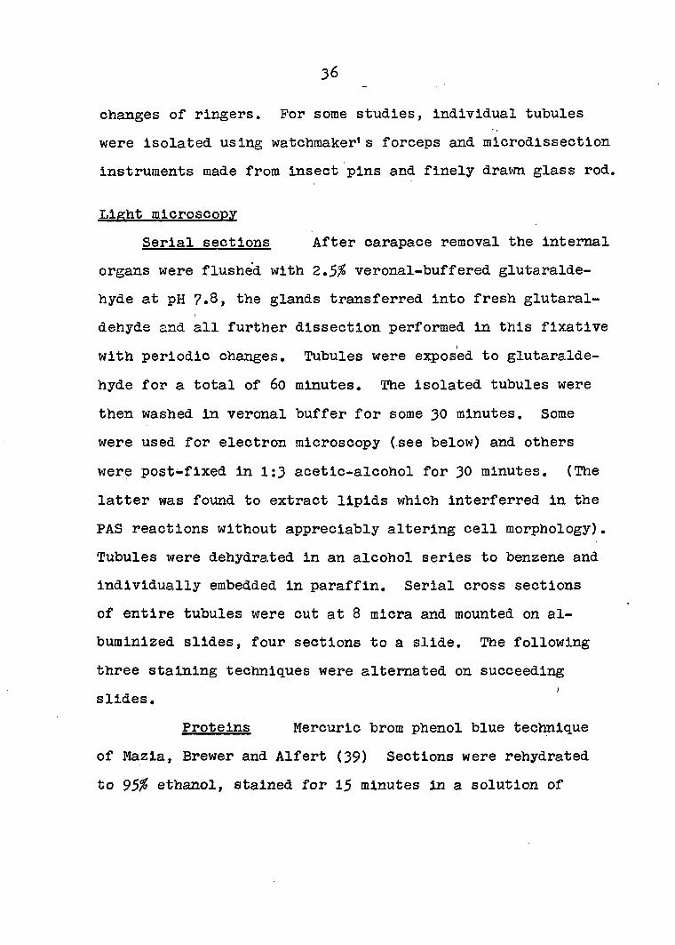
Light microscopy
Serial sections After carapace removal the internal
organs were flushed with veronal-buffered glutaralde-
hyde at pH 7.8, the glands transferred into fresh glutaral-
dehyde and all further dissection performed in this fixative
with periodic changes. Tubules were exposed to glutaralde-
hyde for a total of 60 minutes. The isolated tubules were
then washed in veronal buffer for some 30 minutes. Some
were used for electron microscopy (see below) and others
were post-fixed in 1:3 acetic-alcohol for 30 minutes, (The
latter was found to extract lipids which interferred in the
PAS reactions without appreciably altering cell morphology).
Tubules were dehydrated in an alcohol series to benzene and
individually embedded in paraffin. Serial cross sections
of entire tubules were cut at 8 micra and mounted on al
buminized slides, four sections to a slide. The following
three staining techniques were alternated on succeeding
slides.
Proteins Mercuric brom phenol blue technique
of Mazia, Brewer and Alfert (39) Sections were rehydrated
to 95% ethanol, stained for 15 minutes in a solution of

37
Brom phenol blue and mercuric chloride In 95% ethanol,
rinsed for 20 minutes In 0,5% acetic acid, and differenti
ated In acidified water (pK 6-7 for 3 minutes to bring out
the blue color). Sections were then dehydrated and mounted
In HSR media.
DNA polysaccharides and protein Triple stain
of Hlmes and Morlber (19) Sections were rehydrated to
water, hydrolyzed 12 minutes In 1 N HCl at 6o°C, rinsed In
water, stained 5 minutes In Azure A-Schlff for DNA, rinsed,
bleached and rinsed twice, oxidized In periodic acid for 2
minutes, rinsed and treated with basic fuchsln-Schlff for
polysaccharides, bleached and rinsed twice, rinsed and
stained In Naphthol yellow for proteins, rinsed and dehy
drated In tertiary butyl alcohol, and mounted in HSR,
DNA and RNA Azure B bromide method of Flax
and Hlmes (14) Dehydrated sections were stained three hours
in buffered Azure B bromide, pH 4.0, at 4o°C, rinsed in ice
water, blotted, rinsed, differentiated in tertiary butyl
alcohol overnight, cleared in xylene and mounted in HSR,
Changes in osmiophilia after feeding Crayfish were
starved two weeks at the end of which time some were fed
raw liver. Glands were collected from the starved animals
and from fed animals at half-hour intervals after feeding.
Pieces of hepatopancreas containing 3-6 tubules were treated
with the Ludford osmium impregnation method for Golgi appa

38
ratus as described in Humason (24). Tubules were fixed in
Mann's osmic sublimate (0,5^ osmium tetroxide, mercuric
chloride and sodium chloride in water) for 18 hours, washed
30 minutes in distilled water, and impregnated with osmium
tetroxide according to the following schedule: 2% for three
days at 30°C, 2% for 1 day at 35°C, Ifa for 1 day at 35°C,
and Q»5% for 1 day at 35Tissues were then washed in
distilled water 1 day, dehydrated, cleared in benzene, em
bedded in paraffin and sectioned at 10 micra. The original
procedure stated 6-7 micra for Golgi observations but the
osmium impregnation yielded very "gristly" sections due to
the high lipid concentration in the hepatopancreas and sec
tions thinner than 10 micra could not be cut. Sections were
mounted on albuminized slides, deparaffinized and mounted in
HSR.
Muscle fibers Richardson' s myoepithelium stain
(49); designed for mammary gland, was modified slightly for
use with hepatopancreas. Pieces of gland containing 3-6
tubules were fixed four to six weeks in Weber' s fixative
(dioxane, isopropyl alcohol, formalin, formic acid, cobalt
nitrate, chloral hydrate and glacial acetic acid), washed
in running water overnight, washed several hours in sodium
acetate buffer at pH 5.3 and then treated with a silver
reagent (silver nitrate, ethanol, pyridine and water) for
10 minutes at 55°C. Tissues were then rinsed for five sec

39
onds In absolute ethanol and reduced in a solution of hydro-
quinone and formalin in water until a dark brown color ap
peared. They were then washed in distilled water, fixed in
5^ aqueous hypo and dehydrated in tertiary butyl alcohol.
Some tissues were paraffin embedded and sectioned at 20
micra but the majority were prepared as whole mounts. For
the latter, broken cover slips were used as spacers and the
tubules were mounted in HSR.
Nerve fibers Two techniques were utilized to look
for nerve fibers in hepatopancreas tubules.
In situ staining with leuco-methylene blue The
method of Larimer and Ashby (34) called for 0.4^ methylene
blue, reduced to the leuco-form with 0.01 M sodium hydro-
sulfite, both solutions in van Harreveld' s solution. Tissue
was immersed in the colorless stain for twenty minutes,
transferred to fresh media for oxidation to the colored
state, and the color enhanced with 1% hydrogen peroxide in
the crayfish perfusion fluid. Some tissues were prepared
as whole mounts after dehydration in tertiary butyl alcohol.
Double-imprégnâtion silver staining Fitz
gerald' s technique (13) required fixation in a mixture of
alcohol-picric acid, formaldehyde and glacial acetic acid
for three days, followed by treatment in absolute alcohol
for 24 hours, clearing in benzene, embedding in paraffin
and sectioning at 15 micra. Sections were mounted on albu
40
minized slides and stored overnight at 37°C. Sections were
then deparaffinized and brought to water within 30 minutes,
treated with 10^ silver nitrate foi^ 2 hours at 56°C, washed
in three changes of distilled water in 1,5 minutes, and
stored in 0,2^ protargol-S solution for 18 hours at 37°C.
They were then rinsed, developed in hydroquinone-sulfite
for 5 minutes, washed in running tap water, rinsed in dis
tilled water, treated with 0.1^ acidified gold chloride
solution for 10 minutes, washed for 0.5 minute, developed in
aniline-alcohol for 30 seconds, washed for two minutes,
fixed in sodium thiosulfate for 10 minutes, dehydrated,
cleared and mounted.
Living tubules In addition to the above techniques,
phase microscopy was used to observe tubules in a variety
of states including both fresh specimens and tubules which
had been maintained in vitro in a hanging drop of crayfish
blood for 1 to 5 days.
Electron microscopy
Tubules were obtained by first flushing the internal
organs with glutaraldehyde and then dissecting in this fix
ative in the same manner as described under "Light micros
copy, Serial sections". After being washed in veronal
buffer, tubules were postfixed in veronal-buffered 1^ osmium
tetroxide at pH 7.8. This was followed by dehydration In an

4l
acetone series and flat-embed.d.ing in Karaglas using aluminum
boats. Alternate embedding methods such as empty capsules
and. capsules with drilled, holes in polymerized plastic were
not satisfactory. The above method, allowed, optimum orienta
tion during block trimming, and ease in choosing particular
regions along the tubule for sectioning.
Cross sections were obtained from several locations
along the length of the tubule such as the distal tip, 0.1-
0.5 mm. and 0,75 mm. from the tip, and the middle of the
tubule to insure sections through a variety of cell types.
Sections were cut on either a Porter-Blum microtome or LKB
ultratome using a glass or diamond knife respectively. They
were mounted on parlodion-coated grids and either left un
stained, stained with Reynold's lead citrate (4?) alone, or
lead citrate followed by uranyl acetate. Specimens were
examined on an RCA EKU-2A or ÈMU-3F instrument.
Results
The anatomy of the hepatopancreas in 0, virilis (Pigs,
1 and 2) is similar to that described for P. clarkil (^3) /
and P. blandingii (6). A short, main duct arises ventrally
from each side of the digestive tract at the junction of
stomach and midgut, and divides into three collecting ducts.
Each of these ramifies further and gives rise to hundreds of
blind tubules grouped into three lobes of different size and

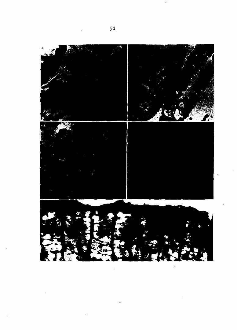
Pig. 1. (top) Orconectes virilis Species of crayfish used for most of the studies presented in this dissertation. (xO.5)
Pig. 2. (center, left) Partial dissection of Orconectes virilis Carapace has been removed exposing internal organs. Middle lobe of right hepatopancreas (arrow) is visible between gills (right) and heart and gonads (left), (xo.75)
Pig. 3. (center, right) Isolated hepatopancreas Dorsal view of Intact gland shows anterior (A), middle (M), and posterior (P) lobes, (xl.5)
Pig. 4. (bottom, left) Isolated hepatopancreas Connective tissue covering has been removed from three lobes, (xl.5)
Pig. 5. (bottom, right) Left posterior lobe of isolated gland Removal of connective tissue covering allows visualization of single tubules (arrow), (x3)

43
appearance. The anterior lobe is elongated with a blunt
anterior end and makes up about 30^ of the length of the
entire gland, the middle lobe is small and rounded, while
the posterior lobe is long, about $0^ or more of the gland,
and has its posterior end pointed. When the entire gland
is removed intact it has the appearance of a butterfly with
long, narrow wings (Pigs. 3 and 4). Each gland is enclosed
in a connective tissue sac (Pigs. 4 and 5) to which the
distal ends of the individual tubules are attached. This
covering is continuous between right and left glands under
the region of the main ducts and this bridge, rather than
the fragile ducts, supports the connection of gland to mid
gut.
Light microscopy
Serial cross sections through a typical 2 mm. hepato-
pancreas tubule yielded 25^, 8-micra sections. The first
25 sections from the distal end contained undifferentiated
cells and transitional columnar epithelium. The following
30 to 4o sections showed a nearly simultaneous appearance
of absorption and fibrillar cells. Sections 75 through 200
contained a mixture of absorption, fibrillar and secretory
cells, the last type possessing a large secretory vacuole
and granules from about sections 100 to 175. In the most
proximal end of the tubule the secretory cells become flat
45
tened and their nuclei pycnotic. They finally disappear al
together leaving mainly fibrillar and absorption cells.
The Azure A triple stain (Pig. 6) yielded blue to blue-
green nuclei and yellow to orange cytoplasm. Nuclei of
connective tissue cells and muscle cells were blue through
out the tubule as were the epithelial cells in the distal
portion, while nuclei of the secretory cells were a definite
green. The cytoplasm of absorption and fibrillar cells also
differed; fibrillar cells were darker but less reddish, ex
cept for a central vacuole, while the absorption cells were
granular and reddish. The most striking sections were those
containing secretory cells with large granules in the secre
tory vacuole. These granules were strongly PAS-positive,
staining a deep red (Pig, 7). In the same cells, a short
segment of the apical cytoplasm separating secretory vacuole
from lumen was also PAS-positive but the portion to either
side of this segment was less reddish and matched the orange
of the fibrillar cells. A brush border is present on the
secretory cells and, in some cases, there are faint, radiat
ing lines of PAS-positive material extending for a short
distance from brush border into the lumen (Pig. 7). Masses
of material moving into the lumen from ruptured secretory
cells and other evidenbes of classical holocrine secretion
were negligible compared to that for merocrine secretion.
Azure B bromide stained the nuclei and cytoplasm of

Pig. é. (top, left) Azure A triple stain, distal region Section no, 32. Fibrillar cells are darker showing increased protein. Absorption cells are granular; reddish color indicates PAS-positive material is contained in these cells. (x683)
Fig, 7. (top, right) Azure A triple stain, middle region Section no. 104. PAS-positive,granules in secretory vacuoles. One secretory cell is seen releasing material through a break in the membrane while another has faint, radiating lines of material extending outward from the apical surface with no break in the membrane, (x3^2)
Fig, 8, (center, left) Azure B bromide stain, distal region Section no, 3^. Fibrillar cells stain more darkly than absorption cells. (x3^2)
Pig, 9. (center, right) Azure E bromide stain, middle region ' Section no, 108, Material in secretory vacuole is refractile to stain, Para-vacuolar cytoplasm stains similar to fibrillar cells, (x3^2)
Fig, 10, (bottom, left) Higher magnification of section in Pig. 8 Green granules appear blue in copies of original 35 mm, transparencies. (x683)
Pig, 11. (bottom, right) Mercuric brom phenol blue stain, middle region Granules in secretory vacuole are protein-positive, (x5^6)

47

48
all cells blue; however, the intensity and hue varied great
ly. The cytoplasm of fibrillar cells (Fig. 8) stained a
very intense blue, especially toward the middle of the
tubule, and stood out markedly from the other cells, while
the nuclei of these cells stained blue-green. The cytoplasm
of absorption cells throughout the tubule and of fibrillar
cells near the distal tip stained a lighter blue or purple.
In secretory cells (Fig, 9) the contents of the vacuole did
not stain but the cytoplasm on either side of the vacuole
stained an Intense blue; however, in most cases it was dif
ficult to differentiate between para-vacuolar cytoplasm and
adjacent fibrillar cells. Many of the absorption cells,
especially those in the most distal third of the tubule
(Pig. 10); possessed a peculiar vacuole not seen elsewhere.
Inside the vacuole was a worm-like granule, resembling a
thick, twisted strand, which always stained green and ap-i
peared to be solid or crystalline in consistency.
Mercuric brom phenol blue staining also yielded blue
cells; however, the intensity was lighter and the color
more uniform and less bright than in the Azure B bromide
sections. In spite of this uniformity, there were differ
ences in intensity, fibrillar cells appearing more dense
than the other cells and the large granules in the secre
tory cell vacuoles very darkly stained (Fig. 11). Embry
onic and transitional cells at the blind tip and cells in
49
the most proximal portion of the tubule appeared less dense
than the other cells in the tubule giving them a "washed
out* appearance. The green structures appeared in cells
prepared with this stain also.
Osmium impregnation was not successful in regard to
localization of Golgi apparati due to the large lipid con
centrations in the hepatopancreas and the thickness of the
sections. However, the following information was obtained
from this study: First, a coiled, membranous structure was
seen Inside the lumen in the distal region of some tubules.
In one tubule, examined three hours after feeding, the
structure is continuous with the apical cell membranes and
is actually being torn from the cells (Pig, 12), Second,
although a few instances of what appears to be holocrine
secretion are evident, there are many more examples of
membrane-enclosed globules of cytoplasm either being pushed
into the lumen or appearing free in the lumen indicating
apocrine secretion (Pig. 13). Third, some fibrillar cells
possess a vacuole located apipal to the nucleus and which
is non-osmiophilic and is delimited by the surrounding
osmiophilic cytoplasm. Inside the vacuole is a smaller
structure which did take up osmium. Pourth, at hours
after feeding, minute spheres appear in the secretory cells,
Pinally, there is a gradation of lipid deposit along the
length of the tubule and a change in the manner of deposi-

Pig, 12. (top, left) Intralumenal membrane Membrane (arrow) appears to be tearing away from apical portion of cells. In other sections the structure is tightly coiled and lies in center of lumen. Osmium impregnation. (x667)
Fig. 13. (top, right) Apocrine secretion 1§ hours after feeding lumen is filled with membrane-bound droplets of cytoplasm (arrow). Osmium Impregnation. (xl33)
Pig, l4, (center, left) Lipid deposits in distal region Heavy lipid deposits in apical and lateral portions of absorption cells. Osmium impregnation. (XI67)
Fig. 15. (center, right) Lipid deposits in proximal region Lipid deposits are in droplet form. Size decreases toward proximal end of tubule. Osmium impregnation. (xl67)
Pig, 16, (bottom) Muscle network Large, evenly-spaced circular muscle fibers (cm) are connected by fine, almost randomly-arranged, longitudinal fibers (Im), Myoepithelium stain. (x521)
51

52
tlon. At the distal end, the absorption cells are densely
stained with osmium at their absorbing surfaces and along
their intercellular surfaces, while the inner cytoplasm and
basal end of the cells is relatively free of osmium (Fig.
14). More proximally the lipids take the form of large
droplets which gradually become distributed throughout the
cells (Fig. 15). Finally, in the most proximal portion of
the tubule, the lipid droplets become very small in size
and number,
Richardson' s myoepithelium stain yielded dark brown to
black tubules which, in most cases, were too dense to use
as whole mounts even after treatment with weak ammonium
hydroxide to dissolve excess silver or clearing in tertiary
butyl alcohol. In a few tubules, however, discrete regions
were found which were relatively free of lipids and provided
a clear view of the muscle fibers under high power magnifi
cation (Pig. l6). Large circular fibers occurred regularly
along the length of the tubule and appeared to be connected
by very fine longitudinal fibers. These fibers stained
intensely black against a brown background, the intensity
of the latter depending on the number and proximity of lipid
droplets in the region. Both circular and longitudinal
fibers were covered with very fine, short projections or
extensions to the underlying cells of the epithelium. It
could not be ascertained whether these represented real

53
structures or silver deposits along the basal, intercellular
junctions of the epithelial cells. Thick sections gave
about the same picture except that striations could be ob
served in the circular fibers. The two fiber types could
also be seen in freshly stained whole tubules, with difficul
ty, using reflected light and a dissecting microscope. The
silver impregnated muscle fibers appeared bright gold
against the brownish tubule surface.
Both nerve stains yielded negative results. In situ
staining with leuco-methylene blue did not visualize nerve
fibers but the cytoplasm of the epithelial cells stained
nicely. Double impregnation silver staining gave positive
results with large nerves around the stomach but within the
hepatopancreas only reticular connective tissue located in
some of the inter-tubule spaces were stained. It should be
mentioned that this technique gave beautiful cell preserva
tion and morphology although lack of contrast prevented
localization of discrete organelles.
Phase observation of living cells revealed the presence
of structures in the secretory cells, nearly at the resolu
tion limit of the microscope, which underwent what appeared
to be Brownian movement in the cytoplasm of these cells.
Nearly all of the secretory cells burst during the course
of one or two hours observation and, upon doing so, released
the above particles. In ,this state, the speed of the par-
54
tides was reduced and a dark ring could be seen to encircle
each particle. It is doubtful that the rings were arti
facts caused by movements of the particles between focal
planes since orientation was provided by a single thickened
region on the ring and the ring could be seen at all angles,
like an equator, as the particles revolved and moved through
the media. A second observation, at lower magnification,
concerned movements within the tubule lumen as pressure was
applied to the tubule. Masses of free cells or membrane-
enclosed cytoplasm appeared to be packed inside the tubule
and these droplets, inside of which the more refractive
lipid droplets could be observed, could be made to slip past
each other with slight pressure on the cover slip. If the
distal tip of the tubule was broken with a needle, the
droplets moved in both directions. Upon reaching the media
outside the tubule, the droplets appeared to rupture, thus
releasing their contents. Lipid droplets then rose to the
surface of the media.
Electron microscopy
Cross sections through the blind tip of the tubule con
tained undifferentiated cells with large, indented nuclei
and little cytoplasm (Pig. 17). The nuclei were smooth-
surfaced and each contained a single nucleolus, sharply
delimited by its high electron opacity. Developing endo-

Fig. 17. (top, left) Undifferentiated cells from blind tip Electron micrograph. (xl910)
Pig. 18. (top, right) Young absorption and fibrillar cells Note continuity of striated border on both cell types lining lumen (L). Electron micrograph. (33170)
Fig, 1 9 . (bottom) Young absorption and fibrillar cells Fibrillar (F) and absorption (A) cells are enclosed by a basement membrane (bm), A very fine longitudinal muscle fiber (Im) lies in the membrane. Electron micrograph. (x6450)

56
%

57
plasmlc reticulum, both smooth and rough, appears in short
lengths with either open or collapsed intracistemal spaces.
About 0.1 mm. from the tip of the tubule two other cell
types appear (Pigs. 18 and 19). One is a typical absorption
cell with a brush border of blunt, closely packed micro
villi, averaging about 0.6 micron long and 0.1 micron in
diameter (Pig. 20), and an area nearly free of organelles,
the terminal web, just below the microvilli. A layer of
fine, particulate matter lies in the lumen on the surface
of the brush border. Filamentous structures originating in
each microvillus extend down into the apical cytoplasm for
a distance of one micron or more. Mitochondria are con
centrated just under the terminal web with many of their
long axes orientated perpendicular to the cell's free sur
face. The cytoplasm contains large, irregularly shaped
vacuoles (Pig. 18) which are electron transparent and prob
ably contained fat prior to acetone dehydration. The most
distinguishing characteristic of these cells is the heavy
concentration of dense, asterisk-shaped granules, 18-36
millimicra in diameter (Pig, 21). (The cytoplasm of these
cells gave a positive PAS reaction above.) Copper, and
calcium granules seen by other investigators were not ob
served in this study. However, located near the junction
of an absorption cell with its neighbor, are large, double-
membrane enclosed areas (Pig. 22) whose cytoplasm is denser

Fig. 20, (top) Absorption cell, apical portion Striated border is made up of microvilli. Note filamentous structures (arrows) extending from each microvillus to cell interior. Electron micrograph. (x33,700)
Fig. 21. (bottom, left) Absorption cell, granules Cytoplasm is rich in asterisk-shaped granules (g) resembling liver glycogen. Electron micrograph, (z46,600)
Pig, 22. (bottom, right) Absorption cell, inclusion Double membrane-bound inclusion (i) contains a denser cytoplasm than rest of cell and one or more crystalloids (c). Electron micrograph. (33,480)
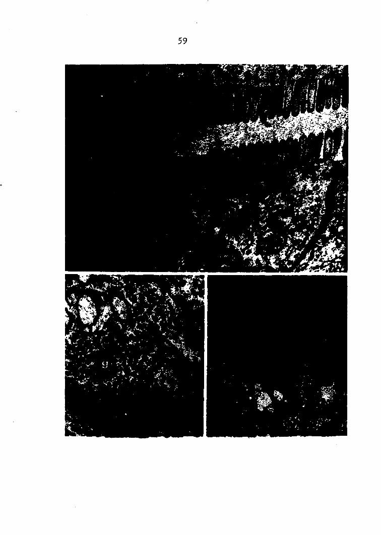
59

6o
than that of the absorption cell Itself. Within the en
closed area are irregular vacuoles, large rough granules
similar in size and shape to those in the absorption cell
proper but fewer in number, and large discoidal crystalloids
with a dense core. Abundant in the absorption cells are
lysosomes and smooth endoplasmic reticulum with amorphous
material in the intracistemal spaces.
The second cell type in this region contains an abun
dance of rough endoplasmic reticulum with large intracis
temal spaces and heavy concentrations of free ribosomes
in the ground cytoplasm (Pigs. 18, 19, 23 and 24). Near
the blind end of the tubule the intracistemal spaces are
round to oval and vacuolar in appearance (Fig, 23) while
toward the middle of the tubule the spaces appear elongated.,
usually perpendicular to the cell surface (Pig. 24), These
cells possess a brush border with microvilli of the same
dimensions as that of the absorption cells but whose fila
mentous extensions into the apical cytoplasm are fewer and
less distinct (Pig. 18). No terminal web is seen and the
mitochondria are dispersed throughout the cytoplasm. A
large number of Golgi apparati may occur in a single cell.
In cells near the distal end of the tubule (Fig. 23) the
Golgi apparatus occurs as a compact group of tubules with
a few lateral vesicles; but, in cells of the middle portion
of the tubule (Pig. 25), the Golgi region is characterized
Pig. 23. (top) Young fibrillar cell Intracisternal spaces are rounded and Golgi region (G) has few vesicles. Electron micrograph. (xlo,i6o)
Pig. 24. (bottom) Mature fibrillar cell Cell contains elongated intracisternal spaces and highly vesiculated Golgi region (G). Electron micrograph. (xll,910)

Pig. 25. (top) Fibrillar cell Many contacts are present between nucleus and cytoplasm (arrows), Golgi region (G) is vesicu-lated. Electron micrograph. (xl4,000)
Pig. 26. (bottom, left) Mature fibrillar cell Nuclear surface is extremely wrinkled. Electron micrograph. (x9210)
Pig. 2 7 . (bottom, right) Pibrillar cell Cell possesses a vacuole (V) filled with whorls of myelin figures and material resembling colloid. Electron micrograph, (x56lO)
I
1
I

64

65
by a multitude of different sized vesicles with smaller ones
in the center. Numerous multivesicular bodies and micro
tubules are also seen. Nuclei appear highly wrinkled in
cells located in the middle of the tubule (Pig. 26) with
suggestions of many contacts between cytoplasm and nucleo
plasm (Pigs. 25 and 26). (Nuclei in this region of the
tubule showed an increased protein staining in the above
cytochemical studies.) 'Occasionally a large vacuole is
present (Pig. 27) and filled with numerous myelin figures
characterized by whorls of membranes in a large, electron
transparent vacuole. No evidence was found for the large,
dense iron granules reported by other authors.
About 0,5 mm. from the blind end of the tubule, se
cretory cells (Pig. 28) make their'appearance and continue
for about 60^ of the length of the tubule. The cells con
tain a single, large vacuole which is separated from the
lumen by a narrow apical structure (Pigs. 28 and 29) con
sisting of brush border, dense cytoplasm, pinocytotic
vesicles of all sizes, mitochondria elongated toward the
cell surface and bundles of microtubules (Fig. 30) also
pointing toward the surface. The interior of the vacuole
is, for the most part, electron transparent except for some
scattered remnants of rough EE and a few large secretory
granules (Pig. 31). (The latter gave positive results with
cytochemical tests for protein and polysaccharides in the

Pig. 2 8 . (top, inset) Secretory cell Phase micrograph of thick section prepared for electron microscopy. Note definite apical structure, (x2,190)
Pig. 2 9 . (top) Secretory cell, apical structure Active pinocytosis is present in this region. Mitochondria are elongated and oriented toward apical surface. Electron micrograph. (x5,000)
Pig. 3 0 . (center) Secretory cell, apical structure Bundles of microtubules (arrows) are also oriented toward lumenal surface. Closer examination revealed that entire apical structure was filled with microtubules. Electron micrograph. (x21,200)
Pig, 3Î-. (bottom, left) Secretory cell, vacuole Secretory vacuole contains mainly amorphous material with scattered remnants of rough endoplasmic reticulum and some secretory granules. Electron micrograph. (xl0,760)
Pig. 3 2 , (bottom, right) Secretory cell, vacuole Occasionally, a vacuole of myelin figures is present similar to that in the fibrillar cell shown in Pig. 27. Electron micrograph. (X6,300)
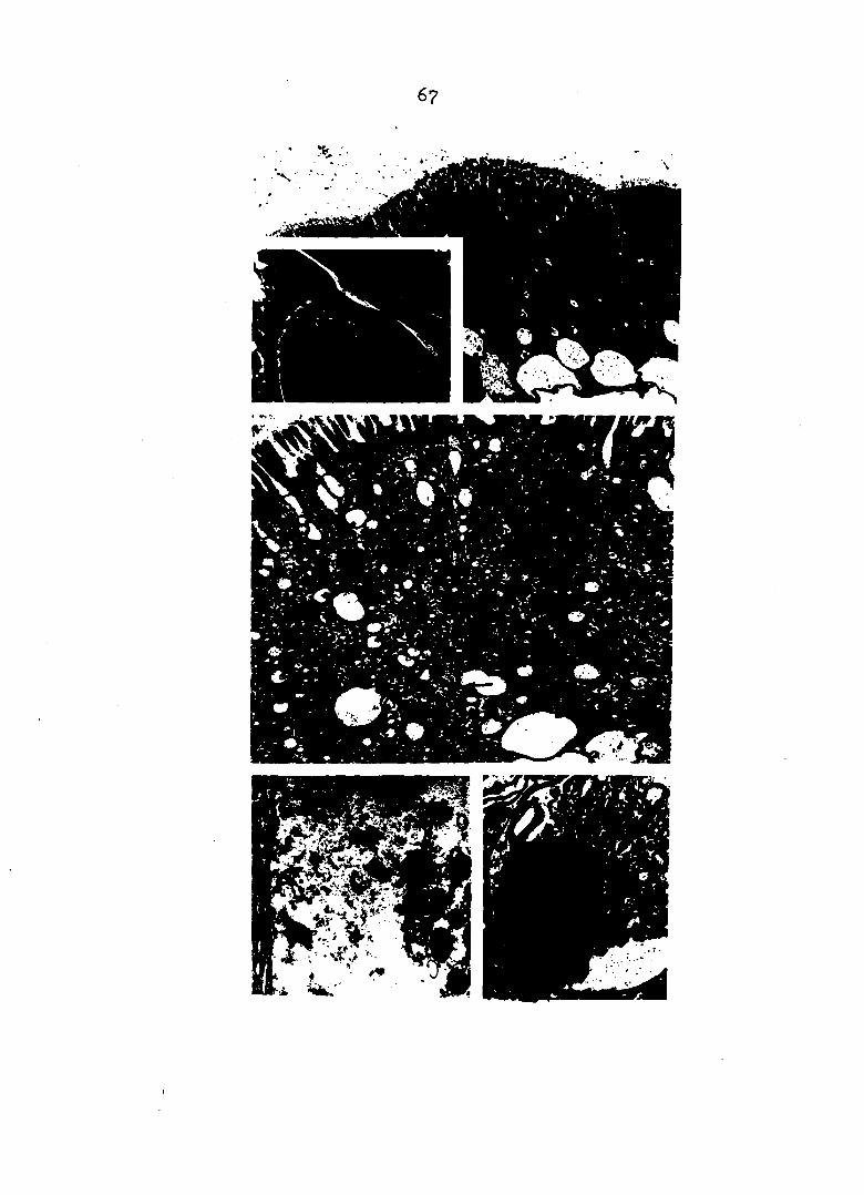
67
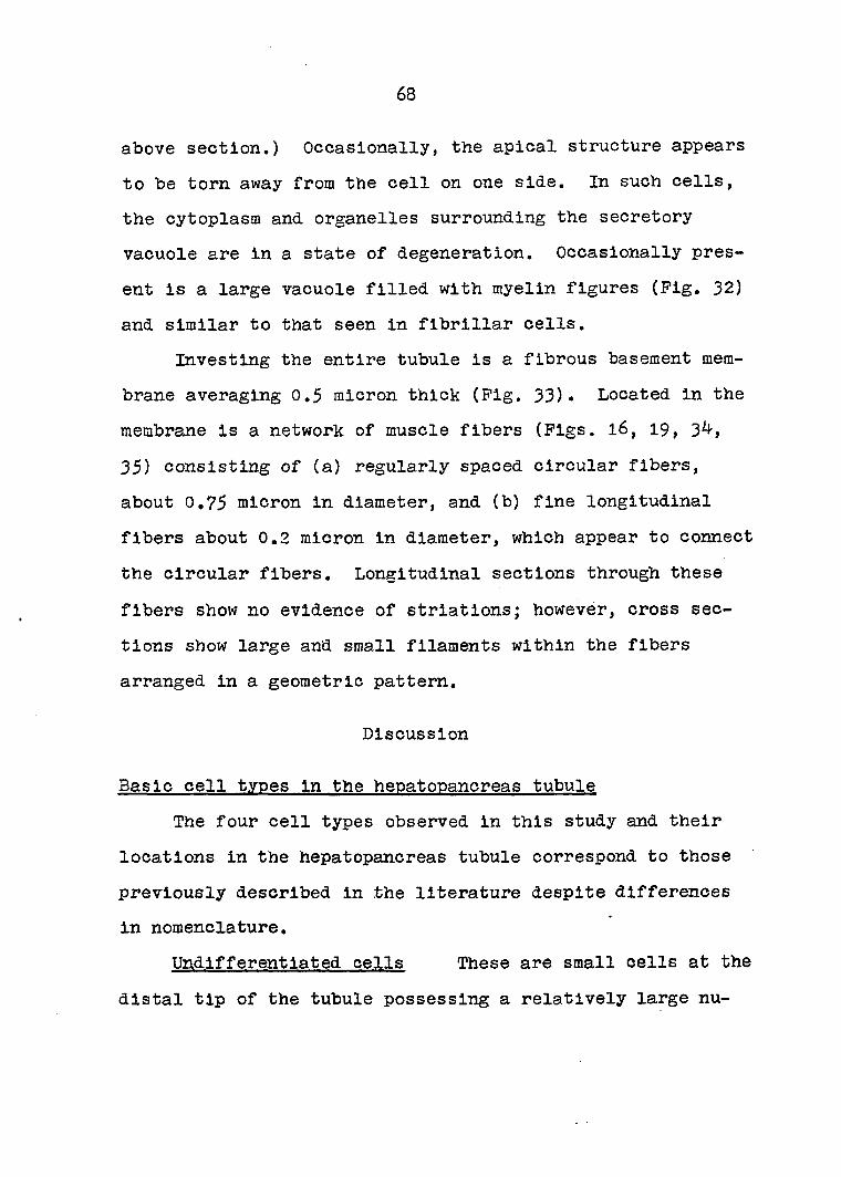
68
above section.) Occasionally, the apical structure appears
to be torn away from the cell on one side. In such cells,
the cytoplasm and organelles surrounding the secretory
vacuole are in a state of degeneration. Occasionally pres
ent is a large vacuole filled with myelin figures (Pig. 32)
and similar to that seen in fibrillar cells.
Investing the entire tubule is a fibrous basement mem
brane averaging 0.5 micron thick (Pig. 33). Located in the
membrane is a network of muscle fibers (Pigs. l6, 19, 34,
35) consisting of (a) regularly spaced circular fibers,
about 0.75 micron in diameter, and (b) fine longitudinal
fibers about 0,2 micron in diameter, which appear to connect
the circular fibers. Longitudinal sections through these
fibers show no evidence of striations; however, cross sec
tions show large and small filaments within the fibers
arranged in a geometric pattern.
Discussion
Basic cell types in the hepatopancreas tubule
The four cell types observed in this study and their
locations in the hepatopancreas tubule correspond to those
previously described in the literature despite differences
in nomenclature.
Undifferentiated cells These are small cells at the
distal tip of the tubule possessing a relatively large nu-

Pig. 33. (top) Basement membrane (bm) Electron micrograph, (xl3,710)
Pig. 3^. (center) Muscle fibers Oblique section through two muscle fibers shows geometric pattern of myofilaments within fibers. Electron micrograph. (x32,200)
Pig. 35. (bottom) Muscle fibers Unusual section shows both longitudinal muscle fiber (Im) cut lengthwise and circular muscle fiber (cm) in cross section. Electron micrograph. (xl5,400)

70

71
cleus, developing endoplasmic reticulum (both smooth and
rough) and other organelles, typical of undifferentiated
cells. They correspond to the E-cells of Jacobs (26), the
embryonic cells of Davis and Burnett (6), the mother cells
of Frenzel (15) and the undifferentiated cells of Ogura
Absorption cells Appearing just proximal to the
blind end of the tubule, these cells contain large lipid
deposits and their cytoplasm is rich in polysaccharide gran
ules. Similar granules are seen in mammalian liver cells
and are usually identified by investigators, e.g. Rhodin
(48), as glycogen granules. The striated border is strik
ingly geometric and composed of blunt, even microvilli.
Prom the border, filamentous extensions pass through a ter
minal web into the deeper cytoplasm. Thus, these cells are
similar in fine structure to absorption cells in the mamma
lian intestine (48). Light microscopy reveals the presence
of green, irregularly shaped crystals which may be copper
granules. E.M. sections revealed large, double membrane- '
enclosed Inclusions resembling the copper granules of
MiyaWaki and Tanoue (42) in Procambarus. These cells cor
respond to the fat cells of Frenzel (15)> the hepatic cells
of Dorman (7), the R-cells of Jacobs (26), the light cells
of van Weel (54), the Cu-cells of Ogura (43) and the absorp
tion cells of Davis and Burnett (6).

. 7 2
Fibrillar cells This term refers to the fibrillar
appearance of the cells' cytoplasm under light miscroscopy.
However, the appearance is not due to artifact but to the
coarse and elongated rough endoplasmic reticulum and swollen
intracistemal spaces in these cells, particularly in mature
fibrillar cells of Region II. A similar EE system was ob
served by Ekholm (9) in mouse thyroid follicle cells which
elaborate colloid.
The intense basophilia seen in light microscopic prep
arations is a result of the high rlbosome concentration,
both membrane-bound and free, in these cells. Yamamoto (59)
in i960 reported the degeneration of these cells after
eyestalk ablation and stated this could be prevented by im
planting eyestalk extracts. Miyawakl et al. (4o) later
demonstrated that the basophilia of the fibrillar cells was
due to RNA and Miyawakl and Tancue (42) reported degenera
tion of the endoplasmic reticulum after eyestalk ablation,
Pingerman (11) reported a decrease in the BNA content
of the hepatopancreas after eyestalk ablation using both
hlstochemical techniques to study the Pe-cells and spectro-
photometric assays of BNA extracts from gland homogenates.
The decrease was Inhibited by injections of eyestalk ex
tracts. In 1965 Pingerman and Yamamoto (12) correlated an
increased pH and decreased amylase activity in digestive
juice with this loss of ENA.

73
The cytoplasm of these cells also shows a high protein
content but it is difficult to determine the exact reason.
The protein may be in the cytoplasm, the intracistemal
spaces or the ribosomes or it may simply be an artifact due
to the packed membranes in the cell.
Irregularities in the nuclear surface and the several
points of contact between nucleus and cytoplasm suggest some
interchange of material between these two regions, Kurosumi
(32) mentions that certain types of secretory cells have
characteristic, irregularly-surfaced nuclei, especially
those cells involved with mucous or mucoid secretion. He
suggests that this type of nuclear surface makes possible
an active nucleocytoplasmic interaction by providing much
greater surface areas on the nuclei.
The role of the fibrillar cell, as evidenced by the
elaborate ER system, the ribosome content, the vesiculation
of the Golgi, and the swollen intracistemal spaces, is
obviously synthetic. However, the material it synthesizes
has not been established and this problem is related to the
sequence of cell differentiation in the tubule. The large
vacuole seen in some cells and filled with whorls of myelin
figures and spiral lamellae is similar to the system of
membranes described by Schulz and de Paola (50) which they
called the delta-cytomembranes and which, according to
Kurosumi (32), have since been observed in a variety of cell

74
types. Schulz and de Paola postulated that the structure
has a role in the synthesis of mucopolysaccharides and pos
sibly in mucous secretion. Miyawaki aX, (4o) reported
that the Pe-containing vacuole was the only region in these
cells rich in mucopolysaccharides or mucoprotein bound with
iron. It appears therefore, that the fibrillar cells are
capable of synthesizing an iron-associated polysaccharide
or mucoprotein. However, until the exact nature and role
of the iron-polysaccharide is elucidated, these observations
remain a mystery. The iron may be a waste product, perhaps
accumulated from the degradation of porphyrin groups from
digested tissues, or it may serve some metabolic purpose
for the crayfish, for example, as an enzyme activator in
the same manner it activates peptidases in certain micro
organisms (16). In regard to the latter function, it is
interesting to note that the powerful trypsin Inhibitor
present in egg white is a mucoprotein ( I 6 ) ,
In addition, there appears to be a relationship between
the calcium- and iron-storing functions of this cell. Yonge
(60) and van Weel (54) observed active iron absorption by
fibrillar cells and Ogura (43) found that eyestalk ablation,
which is a molt-Inducing operation, stimulates Pe-granule
discharge. Miyawaki and Sasaki (4l) reported that calcium
is actively taken up after removal of the eyestalks and
Miyawaki al. (4o) observed that the hepatopancreas is

75
richer in this metal during molting presumably because the
gland is storing calcium to be used in forming the new
exoskeleton. Thus, the same stimulus which causes a general
decrease in normal metabolic functions and RNA concentration
and, in general, prepares the animal for molting, simulta
neously effects iron discharge and calcium resorption.
Fibrillar cells correspond to the P-cells of Jacobs
(26), the dark cells of van Weel (5^)i the Pe-cells of Ogura
(^3) and the fibrillar cells of Davis and Burnett (6). Pos
sibly, they are also the same as Prenzel' s enzyme-
replacement cells (15).
Secretory cells These cells do not resemble mamma
lian pancreatic, liver or gastric secretory cells. Because
, of its large secretory vacuole with protein and PAS-positive
granules, the hepatopancreatic secretory cell, to some
extent, resembles the mucous cell of the sublingual gland
described by Kurtz (33). The similarity is also seen when
the fine structure of these two cells are compared. In
both, there is a single large vacuole which is mainly elec-I
tron transparent except for an assortment of granules and
remnants of rough ER, Also, the lateral basal portions of
both cells are filled with a very dense cytoplasm and endo
plasmic reticulum which seem to be in a state of degenera
tion, The similarity, however, ends here because the
hepatopancreatic secretory cell possesses an apical struc
76
ture unlike that of the raucous cell. The apical complex of
the former is PAS-positive, has a striated border similar
to the absorption and fibrillar cells, and shows evidence
of very active pinocytosis. The apical portion of the
sublingual gland mucous cell does not possess a striated
border and is free of visible pinocytotic activity. In
regard to its apical portion, the secretory cell of the
hepatopancreas resembles the intestinal goblet cell since
the latter possesses a striated border, Palay (44) has
studied the fine structure of the latter and, interesting
ly, the "resting" goblet cell does not look like the text
book descriptions of a goblet cell and can only be distin
guished from adjacent absorption cells by the density of its
cytoplasm, a situation not unlike that of the hepatopancre-
atic secretory cell. Secretion in the goblet cell is both
holocrine and apocrine; that is, as secretory material is
synthesized it begins to fill the cell in the form of ves
icles which gradually enlarge and coalesce into a single
vacuole. Extrusion occurs when the cell membrane ruptures
and material is released freely into the exterior rather
than pinched off within a membrane as in typical apocrine
secretion. Nevertheless, the basal portion of the cell
remains intact, with the nucleus, and forms another resting
cell. The latter, of course, does not occur in classical
holocrine secretion. The results of this study, using
I

77
osmium impregnation, showed the presence of apocrine secre
tion after feeding. Travis (53) similarly observed apocrine
secretion in Homarus.
Another interesting observation by Palay (44) was that
the increased density of the goblet cell cytoplasm is due
to the presence of "densely matted, fine fibrils" especial
ly in the apical cytoplasm. In the present study, masses
of oriented microtubules were found in the apical structure
directed toward the striated border. Their presence is
interesting in view of current concepts (1) associating
several diverse forms of cell movement, including mitosis,
muscle contraction and cytoplasmic flow, with the presence
of filaments and microtubules. There is no evidence, how
ever, to show that the microtubules in the secretory cell
are not passively oriented by the obviously active flow of
materials across the striated border. Conversely, the
microtubules may be involved in the extrusion phase of the
secretory process. Similar structures are present in elec
tron micrographs of the secretory cell in snail hepato-
pancreas in an article by David and Gotze (5).
The secretory cells correspond to the enzyme cells of
Prenzel (15), the pancreatic cells of Dorman (7), the B-
cells of Jacobs (26), the extrusion cells of van Weel (54),
and the secretory cells of Davis and Burnett (6),
78
Mode of secretion In the hepatopancreas
Confusion In the nomenclature of the various cell types
resulted from the fact that some Investigators chose names
on the basis of morphology and others according to supposed
function and each system contained many opposing views.
However, most of the differences can first be grouped into
one of two theories regarding mode of secretion.
The theories proposed by Hlrsch and Jacobs (21, 22)
and by van Weel (5^) on the mode of digestive enzyme secre
tion in the hepatopancreas differ in that the former is
based on linear secretory cell differentiation followed by
holocrine secretion while the latter depends on cyclic
differentiation and merocrine secretion. The difference
might be resolved simply by assuming that the mode of se
cretion in crayfish is different from that in crabs since
these were the two animals used. This assumption is not
entirely warranted, however, for many reasons. (1) The
anatomy and morphology of the hepatopancreas in the two
animals ,is quite similar; (2) also similar are the basic
cell types in the tubule and their locations and arrange
ments into three zones in the epithelial layer; (3) the two
studies utilized the same technique of stimulation by star
vation-feeding and then counting cells and plotting their
numbers against time after feeding; (4) both theories pro
pose that all cells in the tubule epithelium, including

79
the secretory cells, have their origin in the embryonic
cells at the distal end of the tubule; and (5) the baso
philic, fibrillar cell (or dark cell) is an intermediate
stage in secretory cell formation in both theories. Aside
from terminology, therefore, the main difference is that
Hirsch and Jacobs postulate that secretion is an endpoint,
that the cells are lost and must be replaced via another
wave of linear cell differentiation while van Weel concluded
that, in secretion, the secretory cell is not lost but is
reformed and more secretory material is synthesized in
cyclic restitution.
In spite of the similarities described above, there is
' one important difference in the procedures employed. Hirsch
and Jacobs made observations every half-hour for 6* hours
after feeding while van Weel studied the gland for a total
of 36 hours after feeding, with time intervals between ob
servations ranging from an initial 30 minutes up to six hours
at the end of the period. Thus, in spite of the apparent
conflict between the conclusions of these two studies, a com
parison is almost impossible because of differences in the
time scales, Hirsch and Jacob's work gives a precise picture
of events during a relatively short period following feeding
while van Weel's work presents an over-all picture of gross
changes occurring over a very long period of time after
feeding, Coincidently, 36 hours is the approximate length
80
of time that food Is retained in the crayfish alimentary
tract. Therefore, the two studies complement rather than
contradict each other and, since their conclusions are not '
mutually exclusive, they might be reconciled by stating
that, in crustaceans, the immediate response to starvation-
feeding is holocrine secretion but afterwards secretion is
merocrine.
There are several factors which support this hypoth
esis; (1) Although Hirsch and Jacobs' data show good cor
relation among the cycling of numbers of various cell types,
of mitotic indices and of enzyme activities, there is little
agreement between the absolute number of mitoses and that of
the various cell types. In all cases, the peak number of
cell divisions was far lower than the B-cell maxima which,
according to these authors' theory, resulted from it. The
autoradiography studies of Davis and Burnett (6) led them
to conclude that the mitotic cycle in the crayfish hepato-
pancreas is 3 to 5 hours in length. This value is about
the same as that derived from Hirsch and Jacobs' studies
but; more Importantly, the rate Is too slow to account for
the discrepancy between cell number and number of cell di
visions by assuming that the low mitotic frequency is over
come by a very short mitotic cycle. Therefore, since each
B-cell is not derived from a separate mitotic division,
some cyclic restitution of B-cells must take place which.

81
reveals the presence of either merocrlne or apocrine secre
tion.
(2) In Davis and Burnett's study, the H^-thymidine
label appeared in the nuclei of embryonic cells within 15
minutes after injection into the crayfish circulation but
was not observed in the nuclei of secretory cells until 24
hours. That is, the label appeared sometime between the
observation at 10 hours and the next one at 2^ hours.
Hirsch and Jacobs found that approximately three hours were
required for the differentiation of embryonic cells to se
cretory cells. Since the autoradiography study did not
utilize a starvation-feeding stimulus but was conducted
using non-starved animals, we can assume that its results
more closely reflect the situation in the normal animal and
that between 10 and 24 hours are required to replace lost
secretory cells from embryonic cells. Prom Hirsch and
Jacobs' work it would appear that starvation-feeding results
in the loss of vary large numbers of secretory cells and
that, immediately after such stimulation, the replacement
process is accelerated and requires only about 3 hours to
complete. The picture is made more complete by van Weel' s
results showing peak numbers of secretory cells at 1, 12
and 24-27 hours after starvation-feeding, indicating that
restitution requires 11-13 hours when an over-all view of
both immediate and long-range effects is examined. This

82
figure is in agreement with that obtained by Davis and
Burnett.
(3) In all of these studies, the physiology of the
hepatopancreas has been largely ignored above the level of
the cell. The present study has confirmed the presence of
a system of muscle fibers in the basement membrane of the
tubule and this system is capable of contracting the tubule
and changing its shape from a long, slender tube with a
relatively smooth surface to a shortened series of bulbous
expansions separated by sphincter-like constrictions but
having a common lumen and resembling, in miniature, a seg
ment of mammalian intestine in extreme peristalsis. The
physiological control of this system has not been investi
gated but, from observations of contracted tubules under
the dissecting microscope, one can easily assume that the
contractions exert great pressures both within the tubule
lumen and also within the cells. It is conceivable that
such pressures, in addition to their effecting a flow of
material within the lumen, might also play a role in aiding
the flow of material either into or out of the cells de
pending on the relative pressures on the two sides of the
cell membrane. Of even greater probability is the possible
role of these pressures in either rupturing the secretory
cell membrane or squeezing out whole cells as in classical
holocrine secretion. If the assumption is made that long
83
starvation followed by feeding is an extreme stimulus and
that the tubule contractions which it produces are of
greater strength than normal, then the logical conclusion
is that starvation-feeding will result in a greater number
of secretory cells undergoing holocrine secretion than in
the animal which has been fed without prior starvation, hi
the latter, the weaker contractions would result in a lower
incidence of holocrine secretion. Since Hlrsch and Jacobs
used starvation-feeding, this may explain why they found
three-hour replacement periods while Davis and Burnett,
using non-starved animals, found a 10-24 hour period neces
sary to replace secretory cells, van Weel also used
starvation-feeding as a stimulus but his time Intervals in
creased almost geometrically from 30 minutes to six hours
so that only a general picture of the cellular changes
could be provided. In spite of this, van Weel's findings
show both an initial peak at one hour and a second peak 12
hours later thus reflecting the immediate effects (those
studied by Hlrsch and Jacobs) and the long term affects
(studied by Davis and Burnett). That is, the one hour peak
may be the same as the first peak of Hlrsch and Jacobs and
due to holocrine secretion while the 12 hour peak (measured
long after the violent reaction to the starvation-feeding
stimulus subsided) represents the resumption of normal se
cretory mechanisms, i.e. merocrlne secretion, to continue

84
digestion of nutrients in the alimentary tract.
Prom these observations, the original point of conflict
regarding mode of secretion, viz, holocrine vs. merocrine,
may be resolved by assuming that both types may be present.
A strong stimulus such as starvation-feeding, might result
in massive holocrine secretion within an hour or so, due
possibly to extreme contractions of the muscle network sur
rounding the hepatopancreas tubule (although extreme stimu
lation may also act on the cells themselves as in pilocar
pine treatment). If this were the case, then the following
speculation is suggested: In the normal state when feeding
is regular and not interrupted by long periods of fasting,
secretion is not holocrine but merocrine or apocrine and se
cretory cell replacement is mainly via cyclic restitution.
Sequence of cell differentiation
It villi be recalled that four different theories have
been postulated concerning the sequence of cell differen
tiation in the hepatopancreas. These are;
E.G. -»empty cell »D.C.;
( 1 ) ;
(2) embryonic >T.C. »L.C,-t
Cu-cell
(3) E;^^^Pe-cell;
^B-cell

85
and (4) E >-P; proposed, respectively, by Hlrsch
and Jacobs (22), van Weel (5^), Ogura (^3) and Davis and
Burnett (6), Once again, It should be emphasized that all
of these may be correct since different species of crayfish
and one crab were used while, at the same time, similarities
in the anatomy and cytology of the hepatopancreas of the
four animals utilized is striking. Nevertheless, the ques
tion should be asked whether it is feasible that actually a
single system of differentiation exists here which when
studied by different methods presents the characteristics
of a variety of systems.
Separate origins of P-cell and R-cell Prom the
present study, where serial sections through an entire
tubule were examined, undifferentiated cells were restrict
ed to the distal tip of the tubule, absorption and fibrillar
cells both began to appear about 0,25 mm. from the tip of
the tubule, secretory cells occurred in the middle 50^ of
the tubule, and the most proximal portion of the tubule con
tained a few pycnotic secretory cells but mainly absorption
and fibrillar cells. The most obvious conclusion from this
localization of cell types is that absorption and fibrillar
cells either have separate origins or one is capable of
making a very rapid transformation into the other while
migrating only a fevj micra in the tubule. The latter alter
native is unlikely for the following reasons: (1) Both
86
light and electron microscopy reveal vast morphological dif
ferences between the two cell types including the presence
of large glycogen and lipid deposits, "green crystals",
rich concentrations of ribosomes and an elaborate rough
endoplasmic reticulum to name some of the differences. In
fact, the only similarity is the presence of a striated
border in both cells, (2) Both types undergo maturation
along the length of the tubule. In the absorption cell the
lipid droplets become altered in size and location while
in the fibrillar cell there is an elongation and orientation
of the ES and a vesiculation of the Golgi apparatus. Prom
these observations it may be concluded that absorption and
fibrillar cells have separate origins as proposed by Hirsch
and Jacobs (22) and Ogura (^3).
Differentiation of P-cells into B-cells It was
also observed in the serial sections of this study that
fibrillar cells appear prior to secretory cells in the
tubule, that is, closer to the region of cell division in
the distal tip. This indicates that fibrillar cells are
correctly positioned for differentiation into secretory
cells but that the reverse could not occur unless cyclic
restitution takes place. That is, the same picture would
result if secretory cells were first differentiated from
fibrillar cells and, after extrusion, appear again as fi
brillar cells to carry on the cycle.

87
Before examining this last point, the main question
must be resolved: Do either absorption or fibrillar cells
give rise to secretory cells or do the latter arise inde
pendently? Hlrsch and Jacobs, among others, have shown that
a transition of the basophilic cells occurs along the tu
bule. This transition includes a gradual size increase and
coalescence of their vacuoles terminating in the typical
secretory vacuole of the B-cell. This was taken as strong
evidence for differentiation of P-cells into B-cells. The
present study showed other similarities in morphology be
tween fibrillar and secretory cells including a high ribo-
some concentration and the presence of delta-cytomembranes
whereas several differences exist between absorption and
secretory cells which need not be reiterated here. Thus,
on the basis of the prior appearance of P-cells in the tu
bule and the similarities between P-cells and B-cells, it
is concluded that P-cells differentiate into B-cells.
Pate of the secretory cell In view of the above
conclusions, the following scheme may be presented showing
how, at least initially, the various cells types are dif
ferentiated;
This, of course, is the scheme of Hlrsch and Jacobs (22).
However, the latter investigators postulated that only
88
holocrine secretion took place in the hepatopancreas. In
the present study there are several indications that holo
crine secretion is not the major extrusion mechanism in
this gland. The Azure-A triple stain showed PAS-Positive
material being extruded from a secretory cell in the clas
sical merocrlne fashion and the osmium-impregnation tech
nique revealed the presence of apocrine secretion after
feeding. Electron microscopy of the secretory cell revealed
very active pinocytosis in the apical structure which might
indicate that some form of merocrlne secretion is utilized,
possibly what Kurosumi (32) refers to as eccrine secretion:
Either merocrlne or apocrine secretion implies cyclic res
titution and, if this is present in the hepatopancreas tu
bule, there should be some evidence for van Weel's empty
cells and dark cells. In the present study, the middle
region of the tubule, although prédominatly a secretory
zone, possessed both absorption and fibrillar cells inter
spersed among the secretory cells. Thus, either of these
could be the "post-secretory" cell. However, as described
above, there are great morphological similarities between
fibrillar and secretory cells while absorption and secre
tory cells have little in common. Thus, if cyclic resti
tution is present, the most logical intermediate form of
the secretory cell is the fibrillar cell and cycling would
include the repeated transformations of fibrillar cells

89
into secretory cells and, after extrusion, back again into
fibrillar cells. This scheme is similar to that proposed
by van Weel in which cyclic restitution involved a dark cell
precursor of the secretory cell, van Weel' s light cell,
which he placed between the dark cell and the extrusion
cell; cannot be accounted for unless one supposes either a
mistaken inclusion of the absorption cell into the scheme or
that the light cell is a young secretory cell (e.g. the
and B2 cells of Hirs'ch and Jacobs) neither of which is a
satisfactory explanation in view of van Weel' s careful and
detailed cytological descriptions.
Davis and Burnett's autoradiographic studies offer some
obstacles to the above scheme. Their results showed that
the label appeared in embryonic cells in 10-15 minutes, ab
sorptive cells in ^--2 hours, secretory cells in 10-24 hours,
and fibrillar cells after 1-2 days; i.e. these times rep
resent two successive observations with the label absent in
the first and present in the second. Since secretory cells
picked up the label at least 24 hours prior to the fibril
lar cells, this was interpreted by the authors to mean that
secretory cells differentiate into rather than from fibril
lar cells. This absence of labelling in the fibrillar cells
prior to secretory cells appears to conflict with the above
scheme that secretory cells originate from fibrillar cells
even if the presence of cyclic restitution is accepted.

90
However, they do not disprove the scheme if one carefully
examines the time scale upon which observations were made.
In their experiments, Davis and Burnett have several long
periods of time during which no observations were made and
this point is brought out in their Discussion (6). Most
relevant to the present discussion is the l4-hour period
between 10 and 24 hours after injection of the radioactive
sample. The possibility must be considered whether, during
this l4-hour period, fibrillar cells could have been formed
from embryonic cells and then differentiated into secretory
cells thus giving the impression that fibrillar cells were
never present en masse during this period. A corollary
question is why fibrillar cells are not labeled until 1-2
days post injection.
It is impossible to answer these problems fully on the
basis of available information, either from the literature
or this study, since too many unknowns are involved. How
ever, some speculations can be made. Hirsch and Jacobs
concluded from their studies that the transformation from
E-cell to F-cell took 2 hours and from F-cell to B-cell, 1
hour. Since we have assumed that these times are accurate
only after extreme stimulation, the absolute values cannot
be applied to Davis and Burnett' s work. However, the met
abolic processes,effecting these differentiations are prob
ably the same in both cases with only a change in rate. If

91
It Is assumed that the ratios of the required times are the
same, no matter what type of stimulus Is applied or what
the absolute times are, then It requires only half the time
to convert P-cells to B-cells as It does E-cells to F-cells.
Davis and Burnett' s results show that label appears In the
secretory cells 10-24 hours after Injection thus Indicating
that differentiation of secretory cells from embryonic cells
in non-starved crayfish takes between 10 and 24 hours.
Using the 2:1 ratio from Hirsch and Jacobs' work and the .
maximum time, 24 hours, for complete secretory cell forma
tion, it would require l6 hours to convert E-cells to P-
cells and 8 hours to form P-cells into B-cells, The crucial
time, l6 hours, thus falls into the 10 to 24-hour period
during which Davis and Burnett made no autoradiographic ob
servations, In fact, using these criteria, the time re
quired for complete differentiation of secretory cells from
embryonic cells could have varied anywhere from a little
more than 15 to 24 hours without producing a label in the
fibrillar cells at either 10 hours or 24 hours post-
injection, And, from such results, one would receive the
impression that secretory cell formation does not occur via
fibrillar cells.
However, label did appear in the fibrillar cells after
1-2 days. This observation does not conflict with the sug-I
gested scheme of differentiation since the latter included
92
reformation of fibrillar cells after extrusion of secretory
material as part of the cyclic restitution process. In
regard to timing^ much of the synchrony of differentiation,
which began with the simultaneous uptake of tritiated
thymidine by several undifferentiated cells in the blind
tip, would be expected to disappear after one or two days
especially if cyclic secretion were in operation. Thus,
the time of appearance of label in the fibrillar cells is
not pertinent to the proposed scheme of differentiation nor I
is there any available evidence which might confirm or con
tradict this time.
Admittedly, the above speculation does not prove which
is the correct sequence of differentiation; however, it
does show that the data of Hirsch and Jacobs and that of
Davis and Burnett, although opposite in their respective
conclusions, do not really conflict when placed on the same
time scale before comparison. Because of differences in
method and timing, a critical point such as P-cell formation
in one study has no means of -confirmation or disproof in the
other.
Evidence against independent B-cell formation
Ogura' s suggestion that Cu-cells, Fe-cells and B-cells each
arise independently of the others from E-cells is based on
several factors including the spatial locations of the
cells in the tubules. But the crucial factor was the asso-
93
dation of fibrillar cells with iron granules, absorption
cells with copper granules, and. the absence of these metals
in secretory cells. Ogura was able to follow the maturation
of metal granules (or their precursors) and secretory ves
icles in their respective cell types along the length of the
tubule and. used these inclusions as a marker in specific
cell types. In the case of Pe-cells, although iron-granules
themselves d.id. not appear until Region II, minute, ferric-
positive particles were observed in cells of Region I.
Ogura concluded that these particles were precursors of iron
granules. Because he was able to observe the markers as far
back as the region of embryonic and transitional cells, he
concluded that the three cell types had separate origins
beginning at the blind end of the tubule which meant the
existence of three separate lines of differentiation for
Cu-cells, Pe-cells and B-cells.
Since the present study and that of Hirsch and Jacobs
indicates independent formation of absorption and fibrillar
cells, the only conflict concerns B-cell formation. Ogura
indicates in his Discussion (43) that it "may not reason
ably be supposed that either iron or copper is so readily
replaced by the other in the same granule or so suddenly
lost 3^ situ without the granule being lost from the
cell;" that is, the hypothesis that the three cell types
arise Independently is itself dependent on the stability of

94
the metal granules for if, as an example, iron granules were
discharged from Pe-cells, the latter would then be difficult
to distinguish from a young secretory cell thus leaving open
the possibility that Pe-cells are precursors of B-cells and
conflict with the concept of independent differentiation.
In the same paper, however, Ogura reports that the metal-
containing cells undergo changes in various conditions of
the animal. During starvation, there is a decrease in
ferric-positive granules in the Pe-cells; during breeding,
Pe- and Cu-granules are found in the feces indicating dis
charge by the cells; and finally, eyestalk extirpation
causes a marked enlargement of both Pe- and Cu-containing
vacuoles in their respective cells and an Increased excre
tion of these metals in the feces. Miyawaki et (4o,
42) have studied the cytochemistry and electron microscopy
of the metal-containing cells and found that a correlation
exists between the metal concentration in the hepatopancreas
and its concentration in the animal's external aquatic en
vironment. In the present study, only structures resembling
other authors' descriptions of Gu-granules were observed in
both light and electron microscopy preparations; no evidence
was seen for the Pe-granules, It would seem from all of
these observations that Pe-granules are not as stable or as
universally present as the Cu-granules nor do the former
seem to be essential for normal functioning of the hepato-

95
pancreas. Miyawaki suggests that, at least for the Cu-
granule, if the metal is in too high a concentration in the
crayfish's environment, it simply becomes concentrated in
hepatopancreas as a detoxification measure. He also
presents some evidence for Pe-granule discharge. In addi
tion, van Weel (5^) fed iron-saccharate to crayfish and
found that practically all cells in the tubule had ferric-
positive particles in their cytoplasm within a short period
of time. Thus, it would appear that neither Pe- nor Cu-
granules have the stability required to serve as a cell
marker in following differentiation. The point is academic
in the case of Cu-granules since the absorption cells also
possess the more characteristic lipid and glycogen deposits.
However, since only the possession of an iron granule or
rather, ferric-positive particles assumed to be precursors
of iron granules, conflicts with a mass of evidence linking
fibrillar cells and secretory cells, and since the presence
of the iron is subject to a variety of hormonal and envi
ronmental conditions, the probability of independent forma
tion of secretory cells seems far less likely than their
differentiation from fibrillar cells.
Thus, from a critical examination of supposedly con
flicting theories, and a comparison of previous investi
gations with the present study, it would appear that cell
differentiation in the crayfish hepatopancreas is neither

96
exclusively linear nor cyclic but a combination of the two.
The various cell types in the tubule epithelium are formed,
initially, in the lianner described by Hirsch and Jacobs but
secretory cells may be reconstituted while passing through
a fibrillar cell stage. In addition, absorption cells are
apparently formed independently from the fibrillar-secretory
cell line. In all probability, the main path of differen
tiation is cyclic, the linear portion being used to produce
replacement cells.
Conclusions
1, The hepatopancreas of 0. virilis has the same anatomy,
basic cell types and tubule regions as that of previous
ly studied crayfish and the crab Atya spinlpes.
2. Electron microscopic observations of the basic cell
types revealed that the PAS-positive reaction in the
cytoplasm of absorption cells is due to the presence of
large masses of granules identical in size and form to
the glycogen granules in liver cells; that the baso
philia of the fibrillar cell is due to rich concentra
tions of free and membrane-bound ribosomes and the light
microscopic fibrillar appearance of this cell originates
from a coarse system of rough ER ajid swollen intracis-
temal spaces orientated in an apicobasal direction;
and, that secretory cells possess a complex apical

97
structure which actively carries on pinocytosis and in
cludes orientated mitochondria and microtubules.
Secretion by the hepatopancreas secretory cells in the
normal crayfish is primarily either merocrine (eccrine)
and/or apocrine with no evidence of holocrine secre
tion.
Extreme stimuli, such as prolonged starvation followed
by feeding, may initially produce massive holocrine
secretion which is probably due to intense tubule con
tractions. As the cell population is gradually re
turned to normal levels, there is resumption of pri
marily merocrine or apocrine mechanisms.
Secretory cells are differentiated from embryonic cells
in the distal tip of the tubule via fibrillar cells.
Absorption cells are differentiated independently.
These pathways are used in replacing cells lost either
in small numbers, due to age and wear, or in massive I
quantities, due to abnormally stimulated holocrine se
cretion,
Merocrine secretion is followed by restitution of the
secretory material and the sécretory cell via the
fibrillar cell stage.
Previous, conflicting theories regarding mode of se
cretion and sequence of cell differentiation in the
hepatopancreas tubule can be explained by the fact that

98
no two investigators used the same methods and time
scale and, thus, were probably observing different
facets of the same process. Analysis of these investi
gations and comparison with the present study shows that
the apparently conflicting results were based, in most
cases, on complementary and even confirming data.
99
PART II. RHYTHMIC CHANGES IN PROTEASE
ACTIVITY AFTER FEEDING
Introduction
Cytological observations made in Part I were necessar
ily restricted by the static nature of fixed tissue sec
tions; therefore, any conclusions regarding dynamic proc
esses acting within the hepatopancreas must be considered
more or less speculative in the absence of non-histological
confirmation. In Part II, such confirmation is sought from
studying fluctuations of enzyme activity and protein content I
after feeding.
The basis for these studies can best be understood in
terms of Hirsch and Jacobs' work (21, 22). These investi
gators found rhythmic changes of several histological param
eters in the crayfish hepatopancreas tubule after
starvation-feeding. V/hen these fluctuations were placed on
a single time scale, an ordered and reproducible sequence
of maximal values of the parameters was observed. Accord
ing to the authors, this sequence enabled them to follow
the complete differentiation of secretory cells, from their
origin in mitoses at the distal tip of the tubule to their
maturation and extrusion of secretory products in Region
II of the tubule. The most important conclusions from these
studies, in relation to the experiments in this section were
100
that (1) secretion was exclusively holocrine and, (2) the
rate-limiting factor of rhythmic secretion, as well as the
source of all the above rhythmicities, was the cycling of
mitotic divisions in the blind end of the tubule.
In Part I, however, it was postulated that secretion
in this gland may be a combination of both holocrine and
merocrine secretion, the extent of each depending on the
type of stimulus provided. To test this hypothesis, it was
decided to repeat a portion of Hirsch and Jacobs' enzyme
analyses after feeding to determine (1) if rhythmic activity
could be observed in 0. virilis. and (2) how such cycling
might be affected by a mitotic blocking agent.
Only one enzyme, a trypsin-like, casein-protease, would
be studied but assays would be made every 30 minutes for 6f-
hours as in the original work.
Hirsch and Jacobs found that the first mitotic rate
maximum occurred 1^ hours after feeding and this was suc
ceeded by maxima of E-, P-, and B-cells, the B-cell peak
occurring at 4.5 hours, and by a tissue enzyme maximum at
5-5*5 hours. During the 6-|- hour period, evidence of three
overlapping sets of peaks were observed; (1) the first set
does not include a mitotic peak since the cells divided
during starvation but the cell-type peaks are all present
followed by a tissue enzyme maximum at 2.5 hours after
feeding; (2) the second set, extending from 1.5 to 5 * 5
. 101
hours, was the only complete sequence; and (3) the last set
of maxima began with a mitotic peak at about 3 hours but
does not include the enzyme peak which occurred, supposedly,
beyond the.6^ hour period of observation. Thus, tissue
enzyme peaks for two sequences may be found in the 6-|T hour
period, the first at 2.5 hours and the second at about 5.5
hours. In the authors' view, each set of maxima represented
the synchronized (due to starvation-feeding) transformation
of a mass of cells from one stage to the next in their dif
ferentiation from cell division to holocrine secretion;
i.e., each set is a separate wave of such synchronized
cells.
If, in the present study, rhythmic protease activity is
observed in 0. virilis and if Hirsch and Jacobs' hypothesis
is correct, prior colchicine treatment should in some way
retard the cycling. However, what form should this retarda
tion take? Theoretically, it should only affect rhythmic
cycling related to, or occurring after, the first synchro
nous mitotic division in the presence of colchicine. If
this division occurs 1.5 hours after feeding, the enzyme
peak to be affected by the drug is the one normally occur
ring at 5-5.5 hours. Therefore, the results expected in
this study, if exclusively holocrine secretion occurs in
the tubule, is that, in the first half of the 6^ hour period
(locus of first enzyme peak) there will be no difference in

102
protease activity between controls and colchiclne-treated
animals but, in the second half of the period (locus of
second enzyme peak), the activity rhythm will be retarded,
either in time, amplitude or both.
Before beginning this investigation, pilot studies had
to be run to determine effective colchicine dosage and
method of exposure to block cell division in the hepato-
pancreas. In addition, assays of protein concentrations
were carried out on the same tissues to determine whether
post-feeding rhythms could also be detected in either the
protein concentration or in the calculated specific activ
ity of the protease.
Methods of Procedure
Colchicine pilot studies
Several dozen small (1^ to 2 inches) crayfish of mixed
species seined from streams around Lake Okoboji, Iowa, were
placed on a feeding schedule in the laboratory and adapted
to a tap water media. Colchicine treatment was carried out
by adding known amounts of colchicine to the media using
final concentrations of 0, 10~9, 10~®, 10"?, 10"^, and
10-5m. Solutions were mixed from a 10-3M stock solution of
colchicine, which was kept in a dark bottle and refriger
ated, Three separate experiments were carried out to de
termine correct exposure time and dosage while, at the same

103
time, simulating the starvation-feeding stimulus to be used
in the enzyme studies. Group A was fed briefly, placed in
colchicine 24 hours, end examined for mitotic figures in the
hepatopancreas tubules. Group E was exposed to colchicine
24 hours after feeding, starved 9 days, fed briefly and ex
amined after six hours. Group C was fed, starved 9 days,
treated with colchicine for 24 hours, fed, and examined
after six hours. Since Groups A and B produced a high mor
tality and mixed results, these methods of exposure were
not used, A more detailed description of the method used
with Group C will now be presented.
Specimens were maintained in 500 ml of media in plastic
mouse cages, one cage for each colchicine concentration.
Pour control specimens were maintained in the colchicine-
free water and three crayfish were exposed to each of the
five drug concentrations. Media was changed every other
day during the starvation period. Colchicine exposure began
at 4:45 on the day prior to examination of the tubules for
mitoses and the media was protected from strong light by
covering the containers with paper and turning off the room
lights. After 24 hours, each specimen was hand-fed a small
piece of raw hamburger to insure eating. After six hours,
the glands were removed as described in Part I and trans
ferred to van Harreveld's solution. The connective tissue
covering was removed and each lobe of the gland was divided

104
into 6-10 pieces and transferred to fresh solution in vials.
The van Karreveld's solution was then slowly diluted to Z5%
normal by adding distilled water in several steps over a
30-minute period in order to initiate chromosome separation
and facilitate identification of metaphase figures. All
solution changes from this point were carried on in the
original vial using a finely drawn suction pipette with a
curved tip to avoid tubule loss or injury. Tissues were
fixed 15 minutes in 3:1 acetic-alcohol and washed twice in
distilled water. Most of the tissue samples were then
stored for future use, leaving 2-3 pieces of hepatopancreas
per crayfish for the remainder of the study. PeuIgen-
squashes were then prepared as follows; Vials containing
the fixed tissues in distilled water were transferred to a
constant temperature water bath at 6o°C and time allowed
for the tissues to reach this temperature. The distilled
water was then replaced with pre-warmed 1 N HCl and hydrol
ysis carried out for 12 minutes. Operations werq staggered
by one minute to facilitate continuous operatioh. After
hydrolysis, tissues were washed in three changes of distilled
water and stained for two hours in the colorless, Peulgen
reagent mixed according to Humason (24). Here, they grad
ually acquired the typical violet color which was most in
tense at the distal tips. Staining was followed by three,
two-minute periods in bleach solution (24) and tubules were

105
I
then transferred to a watch glass of distilled water for a
lO-minute washing. During this step, individual tubules
were freed from the gland pieces utilizing a dissecting
microscope and fine needles. Five tubules from a single
crayfish were transferred to a microscope slide, excess
water was drawn off, and a drop of ^5% acetic acid was added
to soften the tubules. After two or three minutes, the tu
bules were examined for adequate separation on the slide, a
cover slip was added, excess acetic acid was blotted, and
the squash was made by inverting the slide on paper towel
ling and tapping it with a wooden probe. Tubules were
located under low power and metaphase figures were identi
fied and counted under oil immersion. Five tubules were
examined from each specimen yielding 15 counts per colchi
cine concentration.
Enzyme assays
Approximately 100 crayfish were used in pilot studies
and assays to measure the activities of pH 7.6 casein-
protease and the concentrations of biuret protein in the
hepatopancreas of starved and fed animals every 30 minutes
after feeding. Data was collected from 84 animals including
68 specimens of Orconectes virilis and l6 of Orconectes
immunis. The former were approximately 4" long and ob
tained from the E. G, Steinhilber Co., Oshkosh, Wise, during

lo6
late summer after the major molting season. 0. Immunis was
smaller in size and obtained during July from the Carolina
Biological Supply House, Elon, N. C. 0. virilis appeared
to be in a weakened condition, possibly due to the proximity
of the molting season. Two previous shipments were lost,
either iji toto following shipment or gradually, after a few
week's time in the laboratory. The third shipment was
allowed to equilibrate in the laboratory for about two weeks
and time allowed for the dying specimens to be weeded out. I
During this time the crayfish were maintained in shallow,
artificial spring water, 12 to a large plastic tub. It was
found that the mortality rate decreased if a layer of coarse
pebbles was placed on the bottom of the tub. The room was
air-conditioned with a temperature range of about 18-21®C.
On the day after their arrival, the animals were placed on
a feeding regime which provided a small amount of hamburger
mixed with oatmeal every three days. Those animals either
still in molt or fatally weakened by it refused food and
moved about sluggishly; and, each day more of their number J
died or were killed and partially consumed by the healthier
s p e c i m e n s . B e c a u s e o f t h e l o s s o f s o m a n y s p e c i m e n s o f 0 ,
virilis. l6 out of 84 experimental points had to be filled
with the second species, 0. immunis. After two weeks,
practically all of the remaining crayfish appeared to be in
good health. Later, during the experiments, even some of

107
these refused the stimulus meal or required several minutes
of coaxing and a few were found to have a second shell
growing under the old and were thus in pre-molt.
In preparation for the enzyme studies, a schedule was
followed which allowed 9-12 days of starvation. Colchicine
treatment was carried out during the final day by isolating
the animals in individual containers of pond water, with or
without colchicine. This was always done at noon on the day
preceding the assays to avoid any differences in diurnal
cycling of the mitotic rate as suggested by Eigsti and
Dustin on p. 375 (8). The hour time period under in
vestigation consisted of half-hour intervals from point
0 (starved animals) to point l4 (6-| hours post-feeding).
At each interval a total of six animals, one control-treated
pair for each of three trials, were studied. Members of a
pair were matched for species and size but not sex. Forty-
two such pairs were numbered lA through l4c to designate
time interval and trial. The numbers were then randomized
before scheduling the enzyme assays to minimize similarities
between particular time intervals examined on any two days
of the study.
On the day of the enzyme assay, controls were fed at
9:00 a.m., treated specimens at 9:15» and tissues collected
at appropriate times after feeding. Thus, treated specimens
were exposed to colchicine for 21 hours prior to feeding and
108
for 21-27 hours before being sacrificed. The 15-minute
feeding differential allowed nearly simultaneous collection
of glands from the members of a pair but yet allowed suffi
cient time for tissue preparation of each.
Since "time after feeding" was a crucial variable, it
will be defined as the interval between the crayfish's
acceptance of food and the cessation of enzymatic activities
in the hepatopancreas by cold temperature, homogenization
and dilution.
A few minutes prior to the scheduled time, tissue col
lection was initiated by cutting away the rostral portion of
the crayfish's carapace and all its appendages to facilitate
exsanguination as well as handling. The entire dorsal exo-
skeleton of the céphalothorax was then removed, and the
stomach, midgut and both sides of the hepatopancreas were
freed and transferred to crayfish perfusion fluid as de
scribed in Part I. The entire right hepatopancreas was then
separated from the midgut, rinsed in fresh perfusion fluid,
quickly blotted on filter paper, and weighed on a torsion
balance to the nearest 2 mg. A 4o-ml glass homogenizer
containing 1 ml lO' N HCl had been placed in crushed ice
and water beforehand to achieve rapid cooling. HCl' was used
as the homogenizing medium to retard autoactlvation and
digestion of the protease. After being weighed, the tissue
was transferred to the bottom of the homogenizer and crudely
109
broken up with the pestle to facilitate cooling. The amount
of 10"% HCl necessary to yield a 5 w/v homogenate was
quickly calculated, 1 ml was subtracted, and the resulting
volume added to the homogenizer, Homogenization was then
carried out in the ice bath with 15 strokes of the pestle.
Approximately 30 seconds elapsed between transfer of the
gland to the homogenizer tube and commencement of homogeni
zation; thus, "time after feeding" is probably accurate to
within one minute on graphs in the Results section.
The homogenate was then transferred to a 15-ml centri
fuge tube, centrifuged in a table model centrifuge at half
speed (about 2500 rpm) for 10 minutes and the centrlfugate
transferred to a pre-chilled test tube and stored at 5-9°C
for 1 to 6 hours until the remainder of the samples had been
collected.
Assay of the casein-protease was carried out according
to a modification of Kunitz' method for assaying trypsin
activity (31). Pilot studies showed a linear Increase in
optical density with increased volume of homogenate up to
0.3 ml. The procedure was carried out in 15-ml centrifuge
tubes at 35°C in the following steps:
1. Add 0.9 ml 0.1 M Sorensen' s phosphate buffer,
pH 7.6.
2. Add 0.1 ml, well-shaken, homogenate centrlfu
gate.

110
3. Timing this step to the second, add 1 ml substrate
(1/3 casein in buffer).
4. After exactly 20 minutes, precipitate remaining
protein with 3 ml 10 TCA and mix well with glass
stirrer.
5. Allow to stand one hour.
6. Filter through Whatman #1 filter paper into clean
test tube.
7. Read- O.D. at 280 milliraicra against blank.
For each sample, three assay tubes were prepared including
two experimental and one blank. Thus, each sample was used
to make its own blank by adding TCA prior to the substrate.
Optical densities were read in a Beckman DB spectrophoto
meter using the narrow slit and a hydrogen UV light source.
Samples were placed in quartz cuvettes, 100 transmission
was adjusted for the blank, the two experimental tubes were
read and their average was taken as the O.D. for the speci
men.
Modifications of Kunitz' procedure were as follows:
(1) TCA precipitate was removed by filtration rather than
centrifugation because of the presence of floating debris
from the precipitate which could not be spun down even with
higher speeds, (2) A standard curve was not constructed
because relative values were adequate to observe rhythmic
fluctuations in activity and, since neither the nature of

I l l
the protease nor that of its casein hydrolysate is defined,
even a standard curve would yield relative values. Enzyme
activity was expressed directly in terms of optical density,
i.e. the difference in O.D. at 280 millimlcra between exper
imental and blank samples per 0,1 ml homogenate per 20 min
utes (AO.D.280/0 1 ml/20 mln). (3) Blanks were prepared for
each sample. They were especially critical because of the
variation in pigment and coloration among the glands and the
high U.V. absorption of TCA.
The remainder of the homogenate was shaken to resuspend
its contents and about 5 ml were stored in a vial at -30®C
for the protein assays.
Protein assays
The high concentration and variety of lipids in the
hepatopancreas yielded a turbid and colored homogenate cen-
trifugate with suspended lipid droplets and pigments which
would interfere with colorlmetric protein assays. TCA
precipitation of the homogenate yielded a cream to greenish
precipitate and a supernatant with yellow-green oil droplets
and other debris, either floating on the surface or sticking
to the sides of the tube. These conditions persist in the
solution through the various steps preparing it for reading
in the colorimeter and thus introduce substantial errors in
the absorptions. The method finally employed was a modlfi-

112
cation of the technique of Hiller, Mcintosh and Van Slyke
for assaying urine protein using a NaOH-CuSOij. biuret re
action and to which lipid extraction steps were added for
the present study. The original method is described in
Hawk, Oser and Summerson (18),
Frozen samples were thawed and vigorously shaken to
resuspend lipid droplets and other debris. The homogenate
was drawn up and down in the pipette several times before
measuring a sample. Details of the modified method are as
follows ;
1. 1 ml of homogenate centrifugate is transferred to
a 15 ml centrifuge tube.
2. 1 ml of 10 TCA is added to homogenate with vig
orous stirring; allowed to stand 15 minutes,
3. Mixture is centrifuged 10 minutes at #6o (about
3000 rpm) in table model centrifuge.
TCA is decanted and discarded; tube is drained by
inverting on paper towel for 5 min.
5. Pellet is extracted with 3 ml 2:1 ether:ethanol
(all oily debris adhering to sides of tube is re
moved with this step) ; drain on paper towel,.
6. Extraction is repeated twice for 20 min each time.
7. Pellet is broken up in 1.00 ml yfo NaOH using a
thin glass rod and yields a turbid, beige-colored
fluid.
113
8. Gradually add 2.00 ml of yfo NaOH In steps with I
mixing and standing between additions to facilitate
dissolving of the protein.
9. Add 0,10 ml of 20 CuSO and mix vigorously; allow
to stand exactly 15 min. A turbid, blue to violet
solution with a blue, crystalline ppt. will appear.
10. Centrifuge exactly 15 min at #6o. A clear solution
with some debris floating in a very compact layer
on the surface should result.
11. Clean surface with a finely-drawn glass suction
pipette with curved tip. Even extensive cleaning
resulted in less than 0.5 ml fluid loss out of 3
ml.
12. Read absorption of the supernatant against a blank
of 3 ml of 3# NaOH and 0.1 ml CuSO at 5 0 mllli-
micra. Samples were read quickly in separate
cuvettes since a crusty film is left by the NaOH
which isn't removed by simple rinsing.
For the standard curve a stock, 0,5 solution of puri
fied casein in the above buffer was prepared and used to
make up a range of % casein solutions including 0.10, 0.25,
0,30, 0.35, and 0.40 \ The above assay procedure was
carried out on these protein solutions and yielded à stand
ard curve which was linear over the range 0 to 0.5 protein.
This corresponded to an O.D. range of 0 to 0.288. The

114
O.D.'s of the homogenates were in the range 0.10 to 0.20
which corresponded to homogenate protein concentrations of
0.15 to 0.5 . Since the amount of protein lost during
centrifugation of the homogenates was not measured a com
parison with normal protein values in the hepatopancreas
can only point out similarities in range. The above meas
urements indicate gland protein concentrations from 3 to
10 (plus the amount lost in centrifugation). Literature
values for one crab, Cancer pagurus. range from 3.00 to
6.22# (56).
Results
Colchicine studies
It was mentioned above, that Groups A and B showed a
high mortality rate and did not yield reproducible results
while, in Group C, no specimens died although one animal
appeared to be diseased and three glands did not appear
healthy.
The results obtained with Group C are shown in Pig.
36. In the squash preparations, the five tubules per slide
were well spaced and easy to locate. Various regions of
the tubule were easily differentiated because cells remained
orientated though spread and because the distal portion was
distinguishable by a freedom from fat droplets and the pres
ence of small, compact cells. These aids made counterstain-
I
Pig. 36. Histogram showing effects of colchicine dosage on mitotic arrest Maximum number of metaphase figures per tubule occurred with lO- M colchicine. Drug was administered by exposing intact animal to colchicine in water for 24 hours.
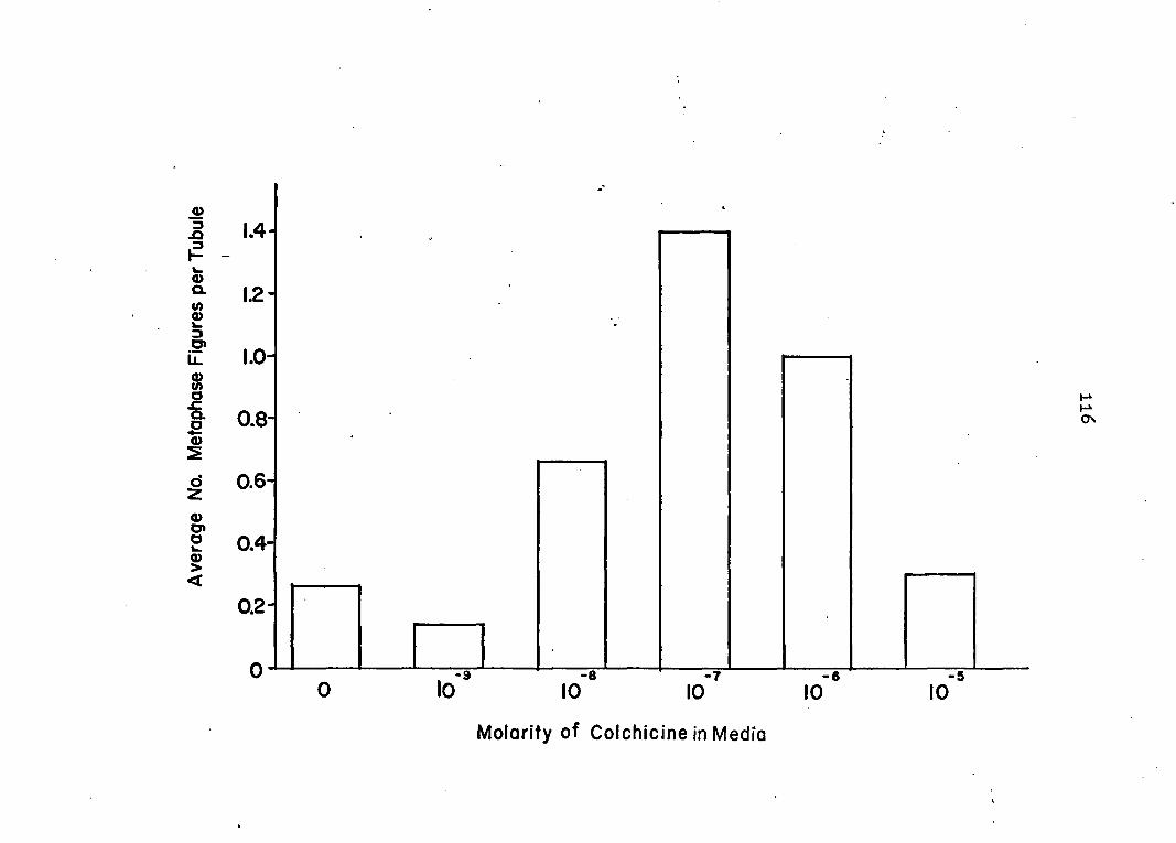
1.4-
1.2-
1.0-
0.8-
0.6-
0.4-
0.2-
0 - -7 10 10 10 10
Molarity of Colchicine in Media
-6 10

117
ing unnecessary. All nuclei stood out as bright, violet-
colored structures against the light, yellow green back
ground of the tubule cells. Mature tubule cells possessed
very large, ellipsoidal nuclei with wrinkled surfaces; con
nective tissue and muscle cells had very small, dense and
flat nuclei; and the cells in the blind tip were of inter
mediate size and density and were spherical in shape. Mi
totic figures were easily identified under oil immersion and
appeared to possess the very high chromosome number (circa
200 for 2n) reported for several crustaceans by Makino (36)
although no attempt was made to count the chromosomes. Be
cause of the high number and minute size of the latter, it
was difficult to identify a few figures as being either late
prophase or metaphase and, in these instances, the figure
was not included.
The number of definite metaphase figures ranged from 0
to 5 per tubule yielding average counts from O.13 to 1.4.
Pig. 36 demonstrates that a normal curve results when aver
age counts are graphed against colchicine concentrations
between 0 and lO'- M. The highest number of figures was
obtained with lO' M, while 10' and 10~ M colchicine yielded
counts about the same as control animals which were not ex
posed to the drug.
In addition to these quantitative results, qualitative
changes were observed with the higher colchicine concentra-
118
tiens. With lO' M the chromosomes in a small number of
cells appeared as a clumped mass. The number of such fig
ures rose sharply with 10" M and remained high with lO' M.
In addition, the latter two concentrations seemed to alter
the general appearance of cells in the distal end of the
tubule; the nuclei appeared more angular, as though packed
together more tightly, the tip itself appeared larger and
more bulbous than normal and, although cell counts were not
made, there appeared to be more cells in this region com
pared to the situation with lower colchicine concentrations.
For these reasons, it was decided to use lO M colchi
cine in the enzyme studies.
Casein-protease assays
The activities of pH 7.6 casein-protease for all 84
specimens are given in Table 1 and Pig, 37. l en the data
are examined without regard to species or molting state,
the differences in 0,D. per 0,1 ml per 20 minutes ranged
from 0.048 to 0.204 for control specimens and from 0.048 to
0.290 for treated specimens. The results for both groups
display a great deal of scatter and the probable reasons for
this will be analyzed in the Discussion. In general, both
groups show signs of a bimodal distribution of activities
which is more evident in the controls; that is, the values
appear to be grouped into two maxima which are separated by

Table 1, Activities of casein-protease at intervals after feeding
AO.D. ^ /0,1 ml/20 mln Controls Colchlcine-•treated
Hours A B C Average A B C Average
Starved .118 .094 .184 .132 .100 .056 .103 .086 0.5 .050 .150 .095a .098 .201 .124 .093® .139 1.0 .082 .137% .079a .099 .132 .082 .078a .097 1.5 .201 .089. .0688 .119 .086 .120 .060a .089 2.0 .080 .168* .138 .129 .088 .221 .132 .147 2.5 .102 .134° .084® .107 .096 .090 .048® .078 3.0 .108 .060 .048® .072 .114b .281 .062® .152 3.5 • . .081 .128 .110 .106 .100 .184 .120 .135 4.0 ' .086 .204 .170* .153 .084 .226 .136» .149 4.5 .072 .114° .080® .089 .151 .268 .076® .165 5.0 .180 .243 .085® .169 .126 , .080 .072® .093 5.5 .142 .102 .098 .114 .092 .211. .290 .198 6.0 .070 .132 .165 .122 .088 .150b .093 - .110 6.5 . .116 .214 .086® .139 .072% .172 .048® .097
®0. immunis.
Dld not eat stimulus meal.
Molting.
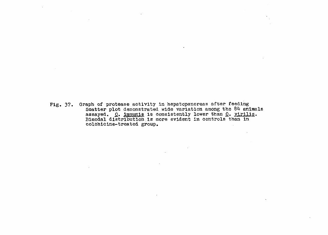
Pig. 37. Graph of protease activity in hepatopancreas after feeding Scatter plot demonstrated wide variation among the 84 animals assayed. 0. immunis is consistently lower than 0. virilis. Bimodal distribution.is more evident in controls than in colchicine-treated group.

0.30 H
0.26-
10.22-o CVi
E 0.18-
O
§ 0.14-o
CONTROLS
O 0. virilis ® molting or did not eat
O 0. immunîs O
O
O ® O
a o 0.10-
o
o
o
o o o ®
o o o
2: o ® o o o
0.06-o
o ~T I I I 1 I - I I î 1 i i I r S I 2 3 4 5 6
COLCHICINE TREATED
O
O O
O O
O O
° o ° ® o °
j o o c . , , . 8
o i I i i I î ! î i i I i r
S 12 3 4 5 6
M
HOURS AFTER FEEDING
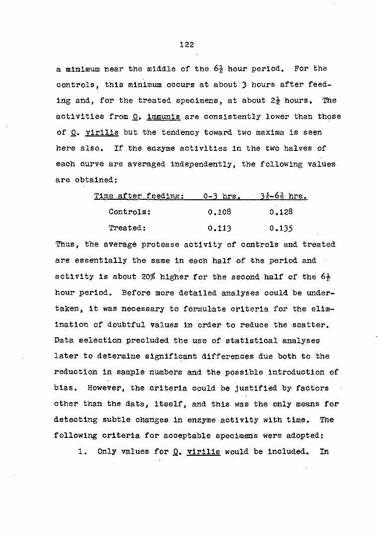
122
a minimum near the middle of the 6-| hour period. For the
controls, this minimum occurs at about 3 hours after feed
ing and, for the treated specimens, at about 2f hours. The
activities from 0, immunis are consistently lower than those
of 0. virilis but the tendency toward two maxima is seen
here also. If the enzyme activities in the two halves of
each curve are averaged independently, the following values
are obtained;
Time after feeding; 0-3 hrs. 34-6 hrs.
Controls; 0.108 0.128
Treated; 0.113 0.135
Thus, the average protease activity of controls and treated
are essentially the same in each half of the period and I
activity is about 20 higher for the second half of the 6
hour period. Before more detailed analyses could be under-
taken,, it was necessary to formulate criteria for the elim
ination of doubtful values in order to reduce the scatter.
Data selection precluded the use of statistical analyses
later to determine significant differences due both to the
reduction in sample numbers and the possible introduction of
bias. However, the criteria could be Justified by factors
other than the data, itself, and this was the only means for
detecting subtle changes in enzyme activity with time. The
following criteria for acceptable specimens were adopted;
1. Only values for 0. virilis would be Included. In

123
addition to being another species, 0. immunis had consist
ently lower protease activities indicating it could not be
used to represent the same populations as _0. virilis.
2. Specimens obviously in molt were eliminated. Be
cause of the central role of the hepatopancreas in molting
and the degenerative effects of molting on the gland's me
tabolism, animals in this state could not be considered
normal. No attempt was made to identify more subtle signs
of molt stage,
3. Specimens which refused food or required several
minutes of coaxing before eating were excluded. Feeding was
the stimulus for the entire experiment and these conditions
either made the results worthless or introduced errors in
"time after feeding." Also, crayfish starved for 9-12 days
usually do not refuse food unless they are diseased or in
molt.
4. A time interval was left blank unless values from
at least two specimens were available. The normal range of
values was considered too wide to allow comparison of a
single activity with averages of two or three.
The following is a breakdown of the number of animals elim
inated by these criteria:

124
Controls Treated
Original number 42 42
Number eliminated by criteria #1 criteria #2 criteria #3 criteria #4
8 2 2 3
_2 13
8 0
Total number eliminated 15
Pinal number included 27 29
In all further analyses, data from animals in the last group
will be referred to as the "selected values" as opposed to
the 84, original values.
When the averages of the selected values of casein-
protease activity are plotted against time after feeding the
general trends observed in the scatter plots of the original
values become much more apparent. In Pig. 38 the control
enzyme activities are seen to decrease during the first half
hour and then increase, yielding a maximum at 1 hours.
Activity then decreases to its lowest point at 3 hours. A
second maximum, nearly twice the value of the first, appears
at 5 hours, after which time activity decreases and then
begins to rise again at the end of the period studied.
Glands from colchicine-treated animals start at low activity
which immediately increases to a first maximum at hour,
then decreases to a minimum at 1-1 hours. A second maximum
appears at 2 hours and then a drop similar to that of the
controls with a minimum at 2 hours. The activity then

Pig. 38. (top) Rhythmic protease activity after feeding Each dot or circle represents an average of two or three animals. Activities of controls and-colchicine-treated animals are similar in values but appear out of phase.
Pig. 39. (bottom) Phase-check of protease activity curves Each dot or circle represents the average of a control average and a treated average in the above graph. See text for explanation.
I

126
-o- controls treated
.SO
IS -
14 -O
ci.12 -
10-
08
HOURS AFTER FEEDING
O— control and treated In phase # treated advanced 30 min.
.22-
..20-
.14
.12- Z.0
S 5 6 2 3 4
HOURS AFTER FEEDING

127
rises to a high maximum at 4-% hours, again similar to the
controls, followed by a minimum at 5 hours, a maximum at
hours and a dro& to a very low value at 6 hours.
Because the two curves displayed similarities in values
but differences in timing, efforts were made to examine the
curves for phase differences in their rhythmic fluctuations.
By averaging the control and treated values at each time
interval, it could be ascertained whether maximum and mini
mum values were increased, decreased or unaltered by the
addition and whether or not the general smoothness of the
curves was improved. This information could give indica
tions as to the general phase agreement between control and
treated curves in a manner analogous to constructive and
destructive interference of light waves.
Pig. 39 shows the results of such a phase check on the
above enzyme activity data, VJhen control and treated values
from the same time interval are simply averaged together ajid
the results plotted against time after feeding, most of the
rhythmic variation seen in the separate curves is lost.
There is an initial increase in activity at hour, a nearly
stable activity for the next 3 hours and a broad maximum
during the second half of the curve with a final drop at 6
hours. However, if the treated values are first advanced
30 minutes and then averaged with the controls, a smooth
rhythmicity results which enhances that in the separate
128
curves; there is an initial decrease in activity to a mini
mum at hour, a maximum at 1 hours, a minimum at 2-3
hours, a high broad maximum reaching its peak at 5 hours and
a final drop at 5-1 hours.
Protein assays
Results of the biuret-protein assays in hepatopancreas
homogenates from 83 specimens are given in Table 2. Acci
dentally, one sample was used up in standardizing the pro
tein assay technique. The protein concentrations varied
from 1. 5 to 3. 5 mg/ml for the controls and 1,1? to 3.52
mg/ml for treated specimens. Pig. 40 shows a plot of se
lected values against time after feeding. In general, the
control curve reaches higher values and undergoes greater
changes in amplitude than that of the treated. In spite of
these differences, there is a striking correlation between
the maxima and minima of the two curves; that is, the con
trol and treated values differ in quantity but their fluc
tuations are synchronized. Another observation is that
values in the second half of the curve do not increase over
those in the first as seen in the enzyme curves.
These relationships are made more apparent in Pig. 4l
which shows a phase check of this data. The "in-phase"
averages yield a smooth curve with time beginning with an
initial decrease in protein during the first hour followed

Table 2, Concentrations of biuret protein at intervals after feeding
mg proteln/ml Controls Colchlclne--treated
Hours A B C Average A B C Average
Starved 1.68 2.70 2.50. 2.29 2.42 3.52 _ a 2.97 0.5 2.18 2.52 2.47% 2.39 1.17 2.52 2.25% 1.98 1.0 3.18 2.48C 1.98% 2.55 2.75 2.58 1.30% 2.21 1.5 3.40 3.35 2.20% 2.98 2.98 2.43 2.12% 2.51 2.0 1.58 2.17° 3.0,7 2.27 2.38 2.68 2.35 2.47 2.5 2.50 2.86* 1.77b 2.38 2.30 3.42 1.77% 2.50 3.0 3.05 1.88 1.80% 2.24 2.70° 2.75 2.23% 2.56 3.5 2.84 3.49 2.33 2.89 3.17 2.33 2.50 2.67 4.0 2.34 2.72, 1.45. 2.17 1.87 2.62 1.82, 2.10 4.5 2.03 1.95* 1.88% 1.95 2.34 2.74 1.98% 2.35 5.0 3.27 3.28 2.07% 2.87 2.72 2.62 1.72% 2.35 5.5 1.90 1.97 1.84 1.90 1.97 2.42 2.78 2.39 6.0 2.34 2.36 2.32. 2.34 2.00 3.10° 2.02 2.37 6.5 3.16 2.90 1.58% 2.55 2.58° 2.08 1.90% 2.19
•Sample used up before assay.
JO, Immunls.
®Dld not eat stimulus meal.
• Molting.
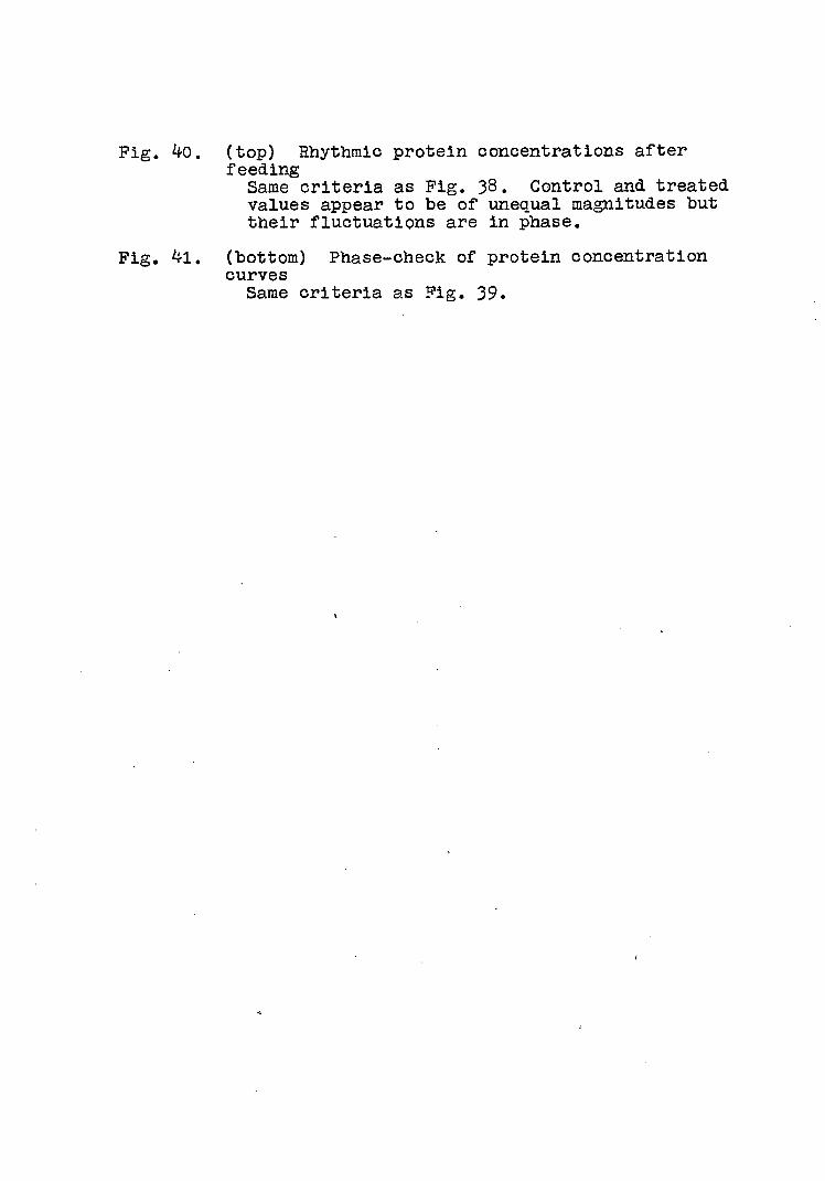
Pig. 4o. (top) Rhythmic protein concentrations after feeding Same criteria as Fig. 38. Control and treated values appear to be of unequal magnitudes but their fluctuations are in phase.
Pig. 4l. (bottom) Phase-check of protein concentration curves Same criteria as Pig. 39.
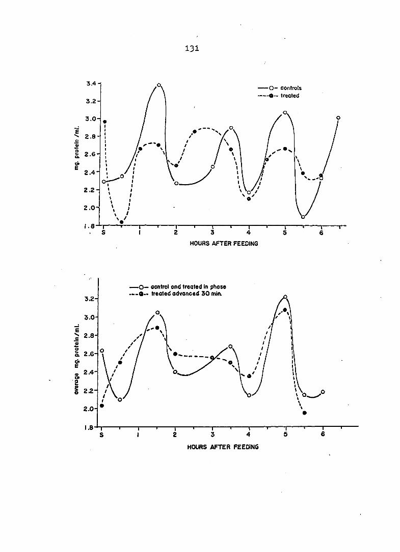
131
3.4-1 —O- controls —O— treated
3.2-
3.0-
.= « S o. 2.6-
E 2.4-
2.2-
2.0-
HOURS AFTER FEEDING
O— control ond treated In phose O— treated advanced 30 min.
3.2-
3.0-
> 2.8-
Q. 2.6
2.4-
2.0
HOURS AFTER FEEDING

132
by maxima at If, 3è and 5 hours after feeding. When the
treated values are advanced 30 minutes before averaging,
there Is a decrease In both the absolute values and the
amplitude of the fluctuations. The protein concentration
Increases to a maximum at If hours, remains more or less
steady between 2 and 4 hours with a slight decrease at 4
hours. This Is followed by a second maximum at 5 hours and
a decrease at hours to about the same value as the con
trols. /
Specific enzyme activity
Specific activity was calculated by dividing each en
zyme activity by Its respective protein concentration and
was expressed as the difference In optical density per mg
protein per 20 minutes C*0.D.280/mg/20 mln), Table 3 gives
the specific activity for 83 specimens. The values ranged
from 0.229 to 1.172 for controls and from 0.159 to 1.718 for
treated specimens.
Pig. 42 shows the curves for selected values. Specific
activities in control specimens showed little variation
during the first half of the curve beginning with an initial
decrease to a minimum at %-l hour, a low maximum at 2 hours
and a minimum at 3 hours. The second half of the curve
shows an activity about twice that of the first half begin
ning with a steep rise to a maximum at 4 hours followed by
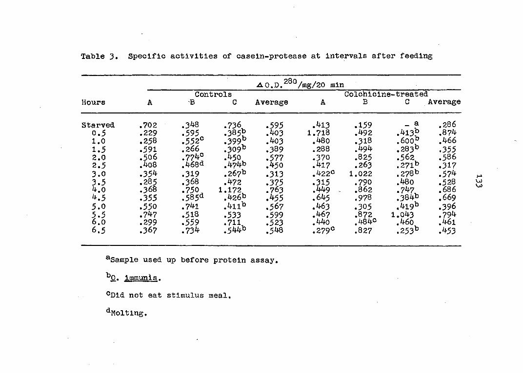
Table 3. Specific activities of casein-protease at intervals after feeding
vl0.D.2G0/mg/20 min Controls Colchicine -treated
Hours A -B C Average A B C Average
Starved .702 .348 .736 .595 .413 .159 _ a .286 0.5 .229 .595 .385% .403 1.718 .492 .413% .874 1.0 .258 .552° .399° .403 .480 .318 .600% .466 1.5 .591 .266 .309° .389 .288 .494 .283% .355 2.0 .506 .7740 .450 .577 .370 .825 .562 .586 2.5 .408 .4683 .474b .450 .417 .263 .271% .317 3.0 .354 .319 .267b .313 .422® 1.022 .278% .574 3.5 .285 .368 .472 .375 .315 .790 .480 .528 4.0 .368 .750 1.172 .763 .449 - .862 .747 .686 4.5 .355 .585* .426% .455 .645 .978 .384% .669 5.0 .550 .741 .411% .567 .463 .305 .419% .396 5.5 .747 .518 .533 .599 .467 .872 1.043 .794 6.0 .299 .559 .711 .523 .440 .4840 .460 .461 6.5 .367 .734 .544b .548 .279° .827 .253b .453
^Sample used up before protein assay.
^0. itnmunis.
®Dld not eat stimulus meal.
^Molting.

Fig. 42. (top) Rhythmic protease specific activities after feeding Same criteria as Fig. 38.
Fig, 43. (bottom) Phase check of protease specific activities curves Same criteria as Pig. 39.

135
O— controls •O— treated
1.0-
0.9-
0.8
C 0.7-
a 0.6 / a> / TO'
0.5-
03
S 2 3 4 5 6
HOURS AFTER FEEDING
O— control and treated in phase O" treated advanced 30 min.
cj 0j8
0.7
3 . OJ6
S . OS S
0.4
HOURS AFTER FEEDING

136
a slow descent from 4 to 6% hours. In general, the entire
curve is smooth and possesses broad maxima and minima. In
contrast, the treated curve has four maxima, at -I, 2, and
5-| hours and undergoes rapid fluctuations beginning with a •
very steep rise during the first 30 minutes.
A phase check of the specific enzyme activity data,
shown in Pig. 43, gives the following results: "in-phase"
averaging of control and treated values yields a curve which
initially rises, contains four maxima, and then drops. The
points appear randomly distributed, simply alternating as
maxima and minima of various heights indicating very fre
quent and rapid changes in activity. Advancing the treated
values by 30 minutes before averaging with control values
yields a smooth curve which drops sharply during the first
30 minutes, rises to a maximum at 1^ hours, undergoes a min
imum at 2-3 hours followed by a broad, second maximum at
4-5 hours which is higher than the first, and finally drops
down at 5^ hours.
Discussion
Colchicine dosage
A concentration of lO'^M colchicine yielded the highest
number of mitotic figures of the five doses tested. It was
assumed, therefore, that this dosage effectively blocked
mitotic division in the distal tip of the hepatopancreas tu-
137
bule when administered in the manner described. It must be
pointed out, however, that this assumption was not based on
reported values in the literature but had to be made on the
basis of general information on colchicine treatment which
makes the effectiveness of this concentration seem likely.
Most of the basic information on colchicine action was ob
tained from Eigsti and Dustin (8). (1) A dosage between —8 <
10" and 10~^M appears effective for most species and tis
sues. Therefore, the experimental range 10"9 to IQ-^M
covered in this study should have included the most probable
optimum dosage for crayfish hepatopancreas. (2) Dosages
above that required to simply arrest mitosis usually produce
a variety of toxic side effects within the cell. These may
include, in addition to inhibition, a clumping of the chro
mosomes and such cells may recover but many terminate in
death. The presence of clumped chromosomes seen in the
present study and their increase in number above lO'^M leads
one to suspect that the higher dosages of colchicine prob
ably exert effects beyond typical metaphase arrest which are
probably toxic to the cell. (3) Observations of changes in
the morphology of the distal tip reinforces this suspicion
of extra-mitotlc effects at the higher dosages. (4) Cor
rect timing of colchicine exposure Is undefined except for
a few systems and must usually be determined empirically.

138
There may be a period of latency before which the drug acts,
thus increasing the required exposure time; whereas, if too
long a treatment is employed, recovery of the cells may al
ready be underway before mitotic figures are counted. Many
systems have an optimum six hour exposure time but this is
far too short for most cold-blooded animals and even some of
the homoiotherms; e.g. 24 hours is the recommended exposure
time for bone marrow culture. An exposure time of 24 hours
was chosen in this study because (1) the crayfish is
poikilothermic and (2) colchicine was administered via the
pond water which meant that considerable time probably
elapsed before the drug came into contact with the hepato-
pancreas cells, and its concentration was probably reduced
by selectively permeable membranes such as the epithelium of
the gill branchiae through which it probably entered the
animal. Both factors would result in a far lower exposure
than the apparent 24 hours in Therefore, the peak
number of metaphase figures at 10~7m colchicine was inter
preted as being the most effective mitotic arresting of all
the concentrations tested.
The number of metaphase figures per tubule was small,
ranging from 0 to 5 with an average of 1.4 per tubule in
10-7n colchicine. The results are in agreement with values
of 0 to 6 per tubule reported by Hirsch and Jacobs <21).
When the data of these authors is examined at each time in

139
terval after feeding, the ranges were 0 to 0.5 per tubule
In the summer and winter, and 0 to 2,2 in the spring.
Suggestions of extra-raitotic effects in the tubules
deserves further consideration. The appearance of greater
numbers of cells, more closely packed together, in the dis
tal tip of the tubule at higher concentrations indicates
either increased cell division and/or inhibition of cell
migration. Prom the above argument's concerning dosage, in
creased cell division was probably not a factor in these
deformities. Eigsti and Dustin (8) have discussed the abil
ity of colchicine to produce tumors in plants; The growth
of these abnormalities is not due to colchicine-induced
polyploidy nor is it related to any of the mitotic effects
of the drug. C-tumors, as they are called, possess the same
volume as analogous tissue in the normal plant but there is
a complete loss of cell polarization during tissue growth,
the cells simply moving haphazardly in any direction after
division. Apparently the phenomena has been studied almost
exclusively in plants; however, the possibility exists that
colchicine treatment may have affected cell migration in the
hepatopancreas tubule. Related to this subject is the cur
rent interest in microtubules, filaments and similar linear
macromolecular complexes found in many cells but most fre
quently in various types of movement systems possessed by
cells (1), Since colchicine acts by interfering with
l4o
spindle fiber synthesis (8), an interesting approach might
be to study the effects of this and other spindle poisons
on various types of movement systems including those having
a role in cell migration.
Enzyme assays
Sources of scatter Results of the 84 assays of
protease activity were disappointing in view of the high
scatter obtained. Several causes of this wide variation are
now realized and most are related to the crustacean molting
cycle and its widespread effects on the animal' s over-all
physiology.
Metabolic Instability Although crayfish are
always in some stage of the molt cycle, the most dramatic
effects occur in pre-molt, ecdysis, and post-molt. This
entire period is marked by a lack of metabolic stability.
For example, the concentrations of glycogen, lipids and
proteins in the hepatopancreas rapidly rise or fall and the
animals eat little or no food. The specimens of 0. virilis
used in this experiment passed through a principal molt
during early summer and were assayed in mid-August. The
high mortality rate of these crayfish during and after ship
ment, their reluctance to eat, and the presence of a few
specimens still in pre-molt, all testified to their weakened
condition. Even though only healthy animals which had sur-
l4l
vived this early period in the laboratory and ate well were
used in the experiments, their metabolic parameters were
probably still far from stable and various activities and
metabolic concentrations were probably still declining or
ascending as the case may be. Thus, animals which normally
would have displayed equal protease activities but were in
slightly different molt stages would exhibit dissimilar
activities with a resultant increased scatter of values.
Scatter due to lower values Hirsch and Jacobs
(21) showed that digestive enzyme activities in crayfish
were much lower in summer and winter compared to values in
the spring, both in quantity and in amplitude of fluctua
tion. When all of these values are graphed together, the
scatter of the summer values does not appear excessive.
However, when the summer values are graphed alone and the
scales expanded to create amplitudes similar to spring
value, the scatter appears much greater than that of the
spring values. That is, the per cent variation increases
as the average level of activity decreases.
In general, therefore, the scattered values reflect a
highly unstable metabolic condition of the animals during
the period in which they were studied. Presumably, if the
same experiments were performed in the spring, the values
would be higher and the scatter lower.

142
Effects of colchicine
In spite of the" scatter, there are striking similari
ties in the original data between control and colchicine-. )
treated protease activities including the averages in each
half of the 6^ hour period and the 20^ rise in the second
half. Thus, colchicine did not effect any apparent de
crease in protease activity from the original values.
Examination of the selected value curves confirms this
similarity and yields the following information: (1) Pro
tease activities in both control and treated specimens
undergo a definite cycling for 6|- hours after feeding. (2)
In general, neither the peak enzyme activities nor the in
tervals between peaks were appreciably altered by colchi
cine, (3) There appears to be a 30 minute retardation of
cycling over the entire treated curve compared to the con
trols, That is, summing the control and treated curves
causes reinforcement of maxima and minima only if the
treated curve is advanced 30 minutes. The fact that this
phase disagreement is present but not a decrease in activ
ity is interesting in view of the null hypothesis of this
experiment; i.e. holocrine secretion would be indicated by
a reduction in protease activity during the second half of
the curve, I
Causes of retardation Two possibilities might ex
plain the inducement of retardation by colchicine: (1)

14]
secretion in the tubule is holocrine, but mitotic blocking
had a different effect than simple inhibition of protease
activity. Instead, the blocking merely resulted in fewer,
mature secretory cells available at the time of feeding
and, by some mechanism, secretion was delayed until the
appropriate number of secretory cells had completed differ
entiation. This process required the extra 30 minutes,
(2) The second possibility is that secretion is not exclu
sively holocrine and retardation was due to the action of
colchicine on some extra-mitotic factor(s) related to the
secretion of active protease. These factors might include
cell differentiation, protease synthesis, and protease
extrusion among others.
The first alternative is difficult to accept because
the curve indicates a true inhibition of secretion for 30
minutes. Some process had been held up but was then re
leased by the feeding stimulus or something related to it.
If the enzyme peak were delayed several hours, it could be
correlated with the beginning of colchicine treatment, A
3-5 hour delay would relate it to duration of the mitotic
cycle (6) or E-cell to B-cell differentiation (22), In
short, if a mechanism is postulated in which colchicine
delays secretion via mitotic arrest, then this delay should
be in terms of hours rather than minutes. A second objec
tion is the unlikelihood of a physiological stimulus, feed

1#
ing, overcoming colchicine arrest within 30 minutes and then
yielding a normal amount of enzyme. Finally, the treated
animals had 8 to 11 days to slowly build up a full comple
ment of secretory cells prior to colchicine treatment which
means that mitotic arrest would not have affected their
number nor would 30 extra minutes in a colchicine-rich en
vironment significantly add to it.
The second alternative can better explain the 30 minute
retardation because it places the site(s) of colchicine in
hibition more proximal to production of the active protease
which is what the delayed peak actually represents. The
suggested sites are more logical choices for effecting only
a 30 minute delay, also, because the possible mechanisms of
action would permit normal secretory cell differentiation
in large numbers up to some step within 30 minutes of the
"ripe" stage. Colchicine inhibition might then act via
depression of protein synthesis or through other of its
knovm actions (8). Finally, the feeding stimulus might then
effect release of the inhibition by acting on the function
ing of the secretory cell, a system more amenable to rapid,
physiological control mechanisms than is the mitotically
dividing cell.
Since retardation is present from the start, the ini
tial portions of the two curves are especially important.
Enzyme activity in the controls decreases during the first
1^5
30 minutes while, In the treated, it increases. This in
dicates that the feeding stimulus normally causes a re
moval of active protease from the hepatopancreas within the
first half hour but, in the treated specimens, it stimulates
a rapid production of the active enzyme which was in a very
low concentration in the gland at time 0, prior to feeding.
Protein assays
Protein concentrations possess the same scatter as the
protease activities and this is probably due to the same
reasons proposed above, i.e. factors related to the molting
cycle. Curves of the selected values show rhythmic changes,
with some differences from the protease curves, but still
provide confirmation of an activity cycle within the hepato
pancreas after feeding.
Effects of colchicine In spite of a similar rhyth-
micity, the effects of colchicine on protein concentration
show two important differences from its effects on protease
activity. (1) In the original data there is no apparent
difference between the two halves of the 6-| hour period and,
therefore, the protein concentrations do not reflect the
20^ rise observed with protease activity. Moreover, when
curves of the selected values are examined, the m8,xima of
control values are nearly always higher than those of the
treated, indicating that colchicine reduced the protein con

146
centrations in the treated hepatopancreases. (2) In con
trast to the protease fluctuations, the protein contents of
control and treated animals display a nearly synchronous,
in-phase rhythm.
The effects of colchicine on protein concentration are,
therefore, opposite to those on protease activity. The drug
apparently reduces the amount of protein in the hepatopan-
creas but it does not alter the phase of the rhythmic
changes in protein concentration from that of the controls.
Before analyzing these differences in response to
colchicine, the natures of the two parameters must them
selves be examined. Protease activity and protein concen
tration are related to the extent that the enzyme is itself
a protein and that both represent an algebraic sum of
amounts present after formation and removal; i,e, enzyme
activation and enzyme removal, protein synthesis and protein
removal. In the hepatopancreas, as in other gland tissues,
removal of enzyme and protein are probably cyclic physio
logical events which agree in rhythm although the amplitudes
may differ. Active protease may fluctuate between some max
imum and close to zero while the protein would have as its
lower limit the mass of structural protein making up the
tissue. Differences in the two parameters are much greater
in regard to formation. The appearance of new, biuret pro
tein is registered as soon as the enzyme presursors have

14?
reached a stage of synthesis (peptides?) which is measurable
by the biuret reaction and will appear as a gradual Increase
I in amount whether the change is in the size of the molecules
or their number. It is not restricted to "active protein",
it will be increased by synthesis of non-enzymatic protein
(e.g. in cell growth), and decreased by degradation of any
protein in the system to provide amino acids. Hence,
biuret-indicated fluctuations in "new protein" is apt to be
influenced by many other changing factors besides active
protease molecules, the changes may or may not be cyclic,
and the amplitude is expected to be quite low because of the
many biuret-positive molecules in the system. Protease
activity, conversely, measures only active enzymes and,
thus, changes are expected to occur very rapidly as the en
zyme molecule passes from the inactive to the active stage.
Protease activation is expected to be cyclic as it is the
product of a cyclic process, synchronized differentiation
of secretory cells. Finally, the amplitude should be great
since the concentration of active protease does not have a
minimal limit and may even approach zero.
Therefore, although the two parameters are related, and
each is composed of a formation and a removal component,
bluret-protein concentration is dependent on cyclic re
moval and, relative to protease activation, a nearly stable
synthesis; while casein-protease activity is dependent on

148
cyclic removal and cyclic activation, and the rhythmicity
of each ma^ or may not agree in phase.
Some information about the interaction of the two
parameters is obtained by comparing the protease activity
curve with the protein concentration curve for each group
of animals. For the controls, the two maxima of the pro
tease curve occur simultaneously as the first and third
peaks in protein concentration and the minima of the two
curves show good correspondence. There is no middle.peak
in the protease curve corresponding to that of protein but,
otherwise, most of the peaks and valleys in the two curves
appear to be in phase. In the case of the treated animals,
however, the curves are more similar in shape but events in
the protein curve precede those in the enzyme curve by about
30 minutes.
The probable reasons for these differences in agree
ment are as follows; (1) enzyme removal and protein re
moval are probably the same in all four curves; (2) enzyme
activation and protein synthesis may take place independ
ently; thus there need not be agreement between a rising
protein concentration and a stationary enzyme activity; (3)
enzyme synthesis and enzyme removal may take place simul
taneously and thus the resulting curve of enzyme activity
may Increase, decrease or remain the same depending on the
contribution and extent of each factor.
149
Therefore, in order to determine the true effects of
colchicine on protease activity, enzyme activation must be
separated.from enzyme removal. This was done by calculating
specific activities of the protease which largely removed
the influence of protein concentration and the results will
be discussed shortly.
Tubule contractions Similar rhythms between the
protein concentrations in control and treated specimens in
dicated that colchicine did not retard the cycling although
the amount of protein appears to have been decreased. , In
the above discussion, the protein concentration curves were
said to be dependent on two factors but only one of these,
protein removal, underwent periodic fluctuations of large
magnitude while the other, protein synthesis, was compara
tively stable. Thus, the minima of the protein concentra
tion curves are more accurate indicators of the time of pro
tein removal than are the activity curve minima for enzyme
removal. Hence the rhythm of the concentration curves is
equated to the timing of some gross, physiological event(s)
which cause the removal of protein. On the basis of the
morphology of the muscle network (described both in the
literature and in Part I) and because of statements by
Yonge (60) and Ramsay (46) discussed earlier, these rhyth
mic, physiological changes are identified as tubule con
tractions. Fluctuations in the protein concentration curves

150
may therefore be used to follow rhythmic tubule contractions
and minimal values of protein concentration indicate times
when the collective, contractile state of the hepatopancreas
is at its maximum. On this basis, both the control and
colchicine-treated glands are maximally contracted for the
first time, at 30 minutes, and again, about every 1| hours
for the duration of the 6^ hour period.
Tubule contractions and protease activity Some type
of coordination should be expected to exist between enzyme
activity and tubule contraction in the normal animal, e.g.
production of the enzyme followed by its removal, since most
physiological systems follow a logical sequence. In the
control animals this coordination is seen in operation. The
similarities in rhythm between the protease activity and the
protein concentration curves was pointed out above. An ex
planation for this similarity can now be postulated. The
first peak in both curves is at hours Indicating maximum
activity and maximum protein at this point in time. This
is followed by a sharp drop in protein, identified now as
the second, maximum tubule contraction, coincident with a
drop in enzyme activity thus indicating the removal of
active enzyme from the gland by contraction of the tubules.
The middle peak in the protein curve, which has no counter
part in the activity curve, is explained by presence of the
next (third) tubule contraction from 3^ to 4 hours while
I
151
enzyme formation continues to rise toward its very high
value in the second half of the curve. As explained above,,
if enzyme formation is being carried on at an accelerated
rate, even during enzyme removal, the activity curve might
not reflect the loss of enzyme. Evidence for only two en
zyme activity peaks is obtained from Hirsch and Jacobs'
results (22) in which a three hour interval exists, both
between the two secretory cell maxima in this region of the
curve and between the two associated enzyme activity peaks.
In the present study, therefore, one would expect the next
activity maximum after the one at hours to occur at 4^
hours; in this experiment, it occurred at 5 hours which
obeys the prediction within reasonable limits. Coincident
with this activity peak is a protein peak which drops sharp
ly between 5 and hours indicating the next (fourth) tu
bule contraction. This time, the drop is accurately re
flected in the protease curve showing that here, in contrast
to the previous situation, the cycle of enzyme activation is
at its lowest ebb.
In summary, the protease activity curve is composed of
two fluctuating parameters whose rhythmicities are super
imposed. In the controls, peak enzyme activities occur at
1^ and 5 hours, while maximum, concerted tubule contraction
takes place at 30 minutes, 2, 4 and hours. Since the tu
bules possess a high concentration of active enzyme at time

152
0, the superlmposltlon of these two rhythms yields a highly
coordinated effort in which active enzyme is formed and re
moved in proper sequence for efficient and economic transfer
of enzymes to the midgut.
Paradox in treated specimens Colchicine treatment
does not appear to affect the rhythmlclty of tubule con
tractions; however, the protease activity rhythm is markedly
altered from that of the controls. Thus, at 30 minutes and
at 2 hours a paradoxical situation is seen in which the ac
tivity has maximum values while, from the protein curve, tu
bule contractions are also at their maximum at the same two
points. In the latter half of the curve the situation is
less extreme since the opposing maxima are not coincident,
but there is still lack of coordination as seen in the con
trols, Also, this may be an explanation for the "suspi
cious" double peak in the enzyme curve between 4* and 5i
hours.
An explanation for this lack of coordination must be
found within the first 30 minutes because differences are
first noted at time 0, It was postulated above that col
chicine Interfered with one or more steps in the formation
of active enzyme with three likely loci mentioned. If this
were true, the treated specimens possessed a very low con
centration of active protease at time 0 compared to the con
trols, Feeding stimulated tubule contractions in both

153
groups reaching a maximum at 30 minutes. However, because
only the controls possessed a large amount of active en
zyme, only this group shows a decrease in activity coinci
dent with contraction. Treated specimens, having little
activated enzyme to start with, would not reflect the con
traction in their activity curve. Moreover, the feeding
stimulus in some manner effects completion of the enzyme
activation delayed by colchicine. This completion takes 30
minutes and, so, peak activity is seen at 30 minutes. The
question now remains, if the tubules were being maximally
contracted, why was the newly activated enzyme not removed
as it was being formed? One possibility is that the acti
vated enzyme was removed from the tubules but was still
present in the ducts and thus included in assays of the en
tire gland, A second possible answer is that, in the
absence of a large mass of active enzyme in the lumen at
time 0, the synchronized contraction of the tubules took
place on schedule but the force of the contraction was di
minished by a lack of intralumenal pressure. In many sys
tems such pressure acts as a stretch-stimulus for greater
contraction. If this were true, the amount of material re
moved from the treated glands during this tubule contrac
tions should be less than in the controls. Examination of
the protein concentration curve shows that, in general,
there is a lower amplitude of variation in protein in the

15^
treated animals than in the controls, i.e. less protein is
being moved out of the gland with each tubule contraction
maximum.
In general, therefore, weaker contractions allowed a
build-up of active enzyme, thus maintaining lack of coordi
nation between enzyme activation and tubule contraction.
The same situation occurs at 2 hours but the two processes
tend to approach synchrony toward the end of the period.
Specific enzyme activity
• As previously stated, the main advantage in basing ac
tivity on protein content is that fluctuations in protein
are, to some extent, removed and the picture more closely
resembles true enzyme activity. In this study, where fluc
tuations in both activation and protein concentration occur,
the specific activities better represent the variations in
enzyme activation separated from changes in protein levels.
Effects of colchicine Except for the first 30 min
utes and the extra minimum in the curve for the treated
specimens, the two specific activity curves are remarkably
similar in that peak enzyme activity occurs at 2 hours and
at 4.5 to 5.5 hours.
During the first 30 minutes, enzyme activation decreases
In the controls but is increased over threefold in the
treated, a difference also suggested in the previous sec-
155
tlons. This difTerence confirms the previous speculations
that (1) colchiciine delayed enzyme activation by 30 min
utes, (2) this al_tered the entire protease activity cycle
of the treated aiuimals as well as the coordination normally
existing between enzyme activation and tubule contraction,
and (3) feeding TOS a stimulus for resumption of the process
of enzyme activatoion which had been retarded at some stage
by colchicine. TShe smooth, "out-of-phase" curve for spe
cific activity coonfirms both the 30 minute retardation in
the treated animaflls' activity curve and the existence of
only two time intcervals at which enzyme activation is maxi
mum. • •
Conclusions
1. Exposure of ccrayfish to lO'^M colchicine for 24 hours
effectively blocked cell division in the hepatopancreas
tubule.
2. The activity of casein-protease in hepatopancreas ho-
mogenates underwent rhythmic fluctuations or cycling
after starvat:l.on-feeding in the manner described by
others,
3. Colchicine diid not reduce protease activity even though
production of? replacement cells was blocked indicating
that secretloa in the hepatopancreas is not exclusively
holocrine.
156
Colchicine retarded rhythmic secretion so that the
entire protease activity cycle in treated animals was
30 minutes behind that In the controls.
Protein concentration was also cyclic and probably re
flects rhythmic tubule contractions. These occur every
1|- hours with the first maximum at 30 minutes after
feeding.
Fluctuations in enzyme activity reflected two different
rhythmic parameters: changes in enzyme activation and
tubule contractions. These two are normally coordinated
for maximum efficiency in digestive enzyme transport.
Colchicine retarded enzyme activity but did not affect
tubule contractions thus removing this coordination.
Colchicine retardation was probably not related to its
mitotic effects but acted on some step(s) in the acti
vation of the casein-protease. After feeding, about
30 minutes were required for the enzyme to complete its
activation compared to zero time for the controls'
enzyme.

157
PAST III. OBSERVATIONS OF TUBULE CONTRACTIONS
It had been intended, in this section, to study in
vitro contractile responses of hepatopancreas tubules to a
variety of agents including extracts of nervous tissues,
blood from fed animals, various types and degrees of elec
trical shock, and a variety of drugs with known stimulatory
or inhibitory effects in other invertebrate tissues. Thus,
the purpose of Part III was to define the physiological re
sponses of the tubules with regard to contraction and to
seek relationships between tubule contraction and the cyto-
logical and enzymatic changes observed in the previous sec
tions. However, attempts to stimulate or control tubule
contractions jji vitro were largely unsuccessful. Contrac
tion could not be initiated consistently by any of the
methods tried, in spite of the fact that (1) such contrac
tions are always observed in vivo in the glands of fed ani
mals, and (2) isolated tubules often underwent contractions
in van Harreveld' s solution without obvious external stimu
lation.
It soon was apparent that several unknowns and vari
ables were involved, the most essential of these being a
defined perfusion medium to maintain the tubules jji vitro
for several hours. Crayfish balanced salt solution was not
adequate to prevent cell death and the use of crayfish blood

158
serum, although capable of maintaining the tubules in vitro
for several days, would require sacrificing a large number
of animals to obtain sufficient perfusion fluid for each
study. Other factors which seemed to be important were the
temperature of the system (tubules appeared to degenerate
" rapidly at room temperature and above) and the incidental,
mechanical stimulations produced during dissection and iso
lation of the tubules.
Because of these problems, experimentation was post
poned until a standardized perfusion media and technique
could be worked out. In Part III, therefore, in lieu of
formal investigations, some general observations of tubule
contractions will be presented which were recorded while
performing various experiments on the hepatopancreas over
a two-year period; and, from these, some conclusions will
be drawn."
Appearance during dissection
liJhen the hepatopancreas is removed from a fasted cray
fish and its connective tissue covering removed, the indi
vidual tubules float free in the perfusion fluid giving the
gland a feathery appearance. At this time, the tubules are
nearly all in a relaxed state and resemble long, thin fin
gers extending out from the ducts and having rounded points
at their distal ends. The lumen appears as a light streak
159
running the length of the tubule and the circular muscles
provide a regular pattern of faint indentations on the tu
bules' surfaces.
About one hour after removal, many of the tubules are
in a contracted state, but the event occurs too slowly to
"see" contractile movements. Along the length of a single
tubule various types of constrictions are apparent. Some
of these are narrow, as though caused by a single fiber and I
others are wide, resembling a tight collar. Several con
strictions may be closely grouped together or a single con
striction may be remote from others. The unconstricted,
bulbous portions may be spherical or cylindrical and their
length varies considerably. Many times constrictions occur
within a few tenths of a millimeter from the blind end but
leaving the tip free, thus giving it the appearance of a
child's top. The lumen can be observed in the bulbous por
tions where its diameter increases with that of the tubule.
In vivo appearance after feeding
Several attempts were made to follow materials into
the hepatopancreas. The most encouraging of these exper
iments included feeding the specimen dye-impregnated food
(such as liver soaked in the lipid soluble Nile Blue A) and
observing movement of the dye in the alimentary tract.
The crayfish is anesthetized in crushed ice for 10 or

l6o
15 minutes, its dorsal carapace is removed and the specimen'
is placed in cold van Harreveld's solution. The hepatopan-
creas is exposed with a minimum of probing and the individ
ual tubules are observed with a dissecting microscope
through the transparent, connective tissue covering of the
gland. As the media warms to room temperature, the cray
fish begins to recover, its heart rate returns to normal,
and blood is gradually replaced by media due to the heart's
action.
As the animal recovers, its movements appear normal, in
spite of the dorsal opening, and it will accept and ingest
food, if starved beforehand. Thus, feeding may take place
either before or after exposure of the hepatopancreas tu
bules. Within a short time after feeding, small globules
may be seen entering the main ducts of the hepatopancreas.
Pat dyes remain in the globules while water-soluble dyes
tend to diffuse into the tissues and cells. After another
period of time, tiny droplets of dye are seen to enter the
tubules themselves.
The state of tubule contraction is not uniform through
out the gland. Tubules on the dorsal surface of the gland
are usually relaxed while those on the inside medial sur
faces, especially near the hilum, are observed mainly in
the contracted state. Peristalsis or similar waves of con
traction were not in evidence in any of the observed glands,
l6i
although the rates of movement may have been too slow to
follow. In one experiment the animal was fed liver without
dye. The dark brown color of the liver was seen first in
the ducts and then, progressively, in more distal portions
of tubule in the bulbous, non-constricted regions. Although
the actual movement was too slow to observe, the dark brown
coloration gradually filled the lumen of most of the tu
bules.
Stimulation experiments
Both electrical and chemical stimulation of the tu
bules were attempted. For the first, crayfish were dis
sected to expose the hepatopancreas as described above and
electrodes were placed in various regions, including the
anterior portion of the stomach where Keim (28) located the
ventricular ganglion, various regions of the alimentary
tract, and on the gland itself. Repetitive stimuli of 30
volts, when applied to the gland at 5-10 per second for 10
seconds, sometimes resulted in massive contractions of tu
bules after 30 to 6o minutes, however, the usual result was
negative.
Several drug experiments were attempted in which iso
lated tubules were exposed to blood from fed or starved
animals, nicotine, atropine, ATP, adrenaline, eserine,
gamma-amino butyric acid, 5-hydroxytryptamine, and

162
tubocurarlne. All were applied at 10~3m strength and all
yielded negative results. However, Pig. 44 shows a group
of tubules which had been placed in perfusion fluid contain
ing equal concentrations of acetylcholine and ^-hydroxy-
tryptamine. Erspamer (10) reports that this combination
initiated rhythmic contractions in several invertebrate
muscles which are relaxed by 5-HT alone. Although rhythmic
contractions were not observed, this was the only treatment
that caused extensive tubule contraction. In addition,
while tubules in the other solutions became flaccid and
underwent degeneration after a short time, the tubules shown
in Pig, 44 had been in the combined drug solution at room ;
temperature for about four hours and showed no signs of de
generation.
Observations during protease studies
In about half the animals used in the casein-protease
studies, after removal and preparation of the right gland
for homogenization, the left gland was examined for tubule
contractions. Notes were taken on the extent of tubule
contraction such as "no contraction", "minor contraction
near h Hum", "50^ of tubules in maximum contraction" "ex
tensive contractions over entire gland", etc. Later, these
subjective descriptions were assigned a number from an ar
bitrary scale on which 0 = no contraction, 5 = moderate con-

Pig. 44. Isolated group of tubules in contracted state After four hours' exposure to 10~3h 5-hydroxy-tryptamine and acetylcholine at room temperature, tubules do not appear to be degenerating as they do in media without these drugs. (x20)


165
traction, 10 = maximum contraction. Porty-six observations
were recorded with n = 1 to 6 animals per time interval.
The number of observations and scale average for each in
terval are as follows;
Time
starved 1 hr. 1 li 2 2i 3
I*
5
I* 6i
n
1 2 3 4 4 4 6 4 3 3 4 2 3 3
Average contraction
7.5* 3.3
8.8* 3.8
s'J*
6 . 2 7.5.
10.0* 6.7
As indicated by the asterisks (*), the averages show a
maximum at 2, 3&, 4§ and 6 hours; that is, maximal con
tractions appear at regular intervals of approximately 1-g
hours beginning at hour after feeding. The animals are a
combination of colchicine-treated and control crayfish.
Conclusions
1. Crayfish perfusion fluid at room temperature is not
aii adequate system for maintaining isolated hepatopancreas
tubules, especially for studies on tubule contraction.
2. Tubule contraction is involved with the movement of
food particles into the tubule lumen but the rate of move
ment is too slow for direct observation.
l66
3. There is some indication that tubule contraction
is selectively controlled in different regions of the gland.
4. The tubules are sensitive to electrical and chem
ical stimulation but the procedures have not been standard
ized to allow investigations into the mechanism of stimu
lation or its role in physiological control of the gland.
5. Observations of a limited number of glands after
starvation-feeding indicate rhythmic tubule contractions at
hour intervals with the first appearing at ^ hour.
l 6 ?
GENERAL DISCUSSION
The original goal of this research was to carry out a
series of experiments on a remarkably versatile gland, the
crayfish hepatopancreas, which were designed to provide
complementary information concerning secretion at many bio
logical levels of organization. When examined realistical
ly, such a goal is more proper to a long-term research plan
rather than a dissertation problem. It was therefore modi
fied so that three different areas (general cytology, pro
tease activity and tubule contractions) related by one prob
lem (rhythmic secretion) would be investigated. The wide
variety and nature of the experiments and the reduction in
their specific objectives require that two questions be
asked here: (1) How do the results and conclusions of Parts
I, II, and III compare with each other and fit into a gen
eral scheme? (2) What are the essential experiments which
must be carried out to pursue or enlarge this work and what
will be the direction of this future research?
Correlations within the Present Studies
The present investigations have dealt with rhythmic se
cretion in the hepatopancreas. A general statement of the
problem investigated is as follows: Does cyclic secretion
of digestive enzymes actually occur in crayfish hepatopan
creas as previously described and, if so, what is the basis

163
of this rhythmicity?
Answers to this problem, in the literature, have been
discussed above. Common to all is the dependence of rhyth
micity on the mode secretion and the sequence of cell dif
ferentiation in the hepatopancreas tubule. In general,
these theories fall into two main categories: (1) Secretion
is holocrine and is the final step in the linear differenti
ation of the secretory cell. Therefore, rhythmic secretion
is ultimately dependent on rhythmic mitotic divisions. (2)
Secretion is merocrine and the rhythmicity is based on cy
clic secretion and restitution of the secretory cell.
Rhythmic secretion in 0. virilis The rhythmic pro
tease activity after feeding observed in Part II of these
studies was nearly identical to that found by Hirsch and
Jacobs, proponents of holocrine secretion. The cycling was
confirmed by rhythmic protein concentrations in the same
tissue samples although specific differences existed between
the two activity curves.
Sequence of cell differentiation The present
studies have demonstrated, in Part I, the presence of the
same four basic cell types in the tubules of 0, virilis
which were described in other crustaceans, regardless of
differences in nomenclature. Evidence was presented, in
cluding location in the tubule and morphological similari
ties between certain of these cell types, which indicated
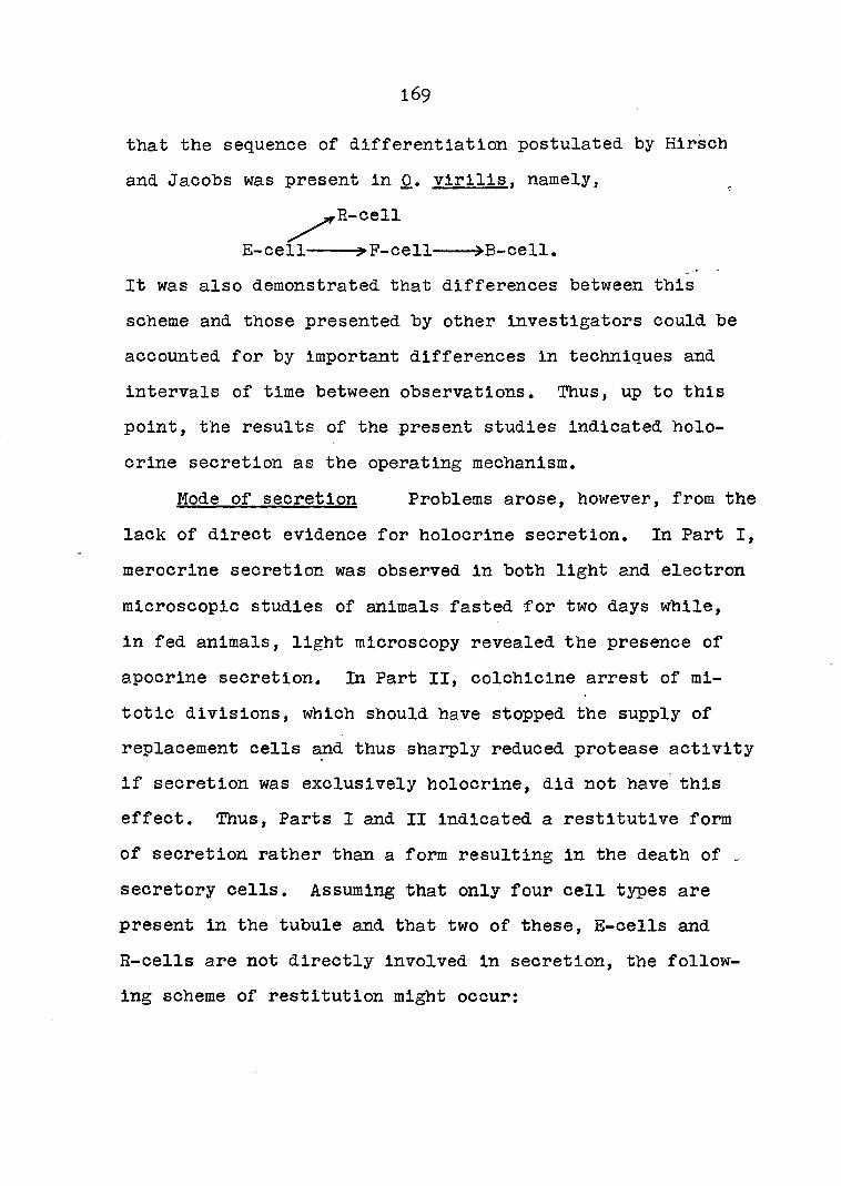
169
that the sequence of differentiation postulated by Hirsch
and Jacobs was present in 0. virllis. namely,
R-cell
It was also demonstrated that differences between this
scheme and those presented by other investigators could be
accounted for by important differences in techniques and
intervals of time between observations. Thus, up to this
point, the results of the present studies Indicated holo
crine secretion as the operating mechanism.
Mode of secretion Problems arose, however, from the
lack of direct evidence for holocrine secretion. In Part I,
merocrlne secretion was observed in both light and electron
microscopic studies of animals fasted for two days while,
in fed animals, light microscopy revealed the presence of
apocrine secretion. In Part II, colchicine arrest of mi
totic divisions, which should have stopped the supply of
replacement cells and thus sharply reduced protease activity
If secretion was exclusively holocrine, did not have this
effect. Thus, Parts I and II indicated a restitutlve form
of secretion rather than a form resulting in the death of .
secretory cells. Assuming that only four cell types are
present in the tubule and that two of these, E-cells and
R-cells are not directly involved in secretion, the follow
ing scheme of restitution might occur:
E-cell ^-P-cell »B-oell
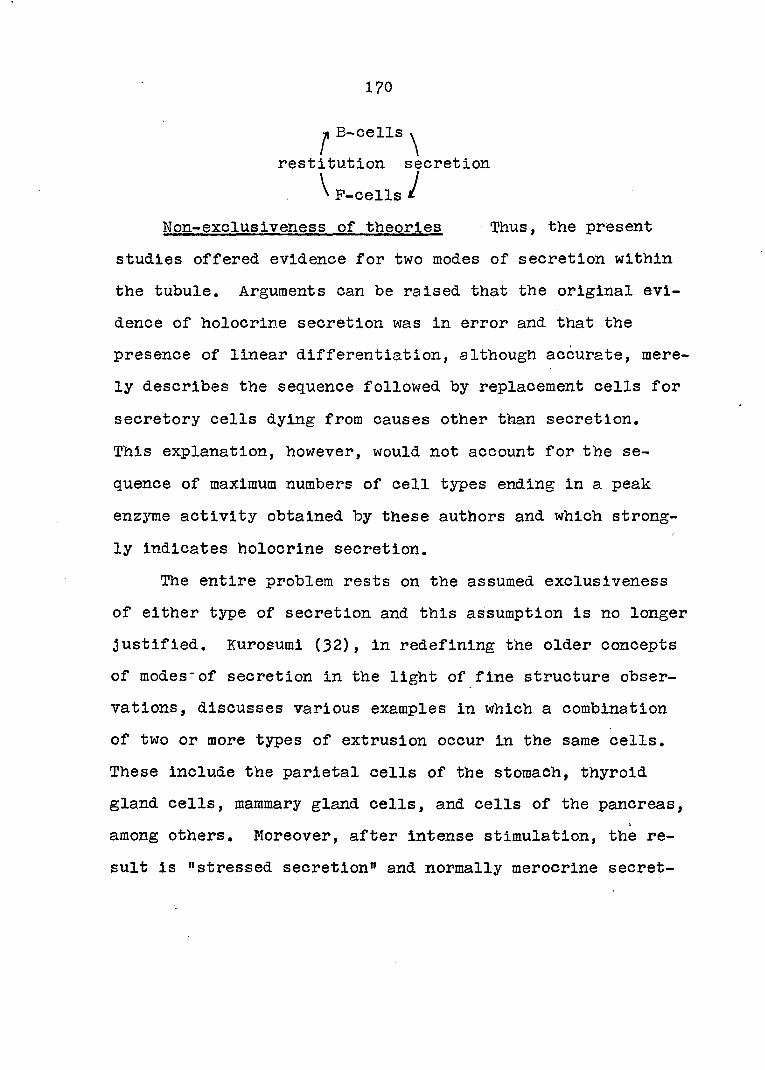
170
H E-cells
restitution secretion
^ P-cells i
Non-excluslveness of theories Thus, the present
studies offered evidence for two modes of secretion within
the tubule. Arguments can be raised that the original evi
dence of holocrine secretion was in error and that the
presence of linear differentiation, although accurate, mere
ly describes the sequence followed by replacement cells for
secretory cells dying from causes other than secretion.
This explanation, however, would not account for the se
quence of maximum numbers of cell types ending in a peak
enzyme activity obtained by these authors and which strong
ly indicates holocrine secretion.
The entire problem rests on the assumed exclusiveness
of either type of secretion and this assumption is no longer
justified. Kurosumi (32), in redefining the older concepts
of modes*of secretion in the light of fine structure obser
vations, discusses various examples in which a combination
of two or more types of extrusion occur in the same cells.
These include the parietal cells of the stomach, thyroid
gland cells, mammary gland cells, and cells of the pancreas,
among others. Moreover, after intense stimulation, the re
sult is "stressed secretion" and normally merocrine secret

171
ing cells will undergo apocrine secretion or even complete
loss of their cytoplasmic contents which appears identical
to holocrine secretion.
Tubule contractions Does the hepatopancreas tubule
possess any mechanism which might result in "stressed se
cretion"? In Part I, the presence of the muscle network
described in the older literature was observed in both light
and electron microscopy, Yonge (6o) stated that this system
was responsible for movements of materials into and out of
the tubule. In Part II, evidence was presented for the
rhythmic removal of both protease and protein from the tu
bule, An argument was presented that, of the two, the cy
clic protein changes best reflected the timing of this
process, that removal was probably due to maximum, concerted
contractions by the tubules, and that the latter occurred
about every Ij hours beginning at 30 minutes after feeding.
In Part III, simple observations of glands after feeding
indicated that tubules actually did follow a rhythmic pat
tern of maximum contraction and the timing agreed with that
indicated by protein removal. Thus, if such contractions
were intensely stimulated, as Kurosumi observed, they could
result in "stressed secretion" and rupturing or releasing
of secretory cells giving the appearance of holocrine se
cretion, It is suggested that starvation for a long period
of time followed by feeding is, indeed, a stressed stimulus.

172
Vonk (56) criticized the work of Hirsch and Jacobs on this
basis, claiming that their results probably did not repre-
sent mechanisms in the normal animal.
An integrated scheme A mechanism which would ac
count for both the rhythmicity of secretion and the above
observations is now offered. Secretion may be both holo
crine and merocrine, the latter term used in the older sense
implying restitution of the secretory cell. After moderate
stimulation and during short periods between feedings, mero
crine secretion is the major mode of extrusion. However,
after extreme stimulation, such as starvation-feeding, a
large number of secretory cells, packed with secretory
material due to lack of stimuli, are ruptured or released
by intense contractions of the tubules brought about by
feeding, i.e. holocrine secretion. Therefore, secretion is
rhythmic because the secretory cells have been synchronized
by prolonged starvation followed by massive holocrine se
cretion and the synchrony is maintained in the waves of re
placement cells undergoing differentiation. As the time
after feeding increases, the synchrony diminishes, the in
tensity of tubule contractions returns to normal, and the
merocrine mechanisms again become the major mode of secre
tion, This scheme may be summarized as follows:

173
R-cells intense stimulation
E-cells >P-cells B-cells », holocrine secretion
normal stimulation
raerocrine secretion
Future Work
Even before the above research was carried out, a mul
titude of interesting experiments were suggested by the
unique combination of simple morphology and variety of func
tions present in the hepatopancreas tubule. Furthermore,
the present work indicated several new problems including;
from Part I, the effects of eyestalk hormones on the dif
ferentiation and fine structure of the fibrillar cell, and
fluorescent antibody experiments to localize zymogens in
the tubule cells or to compare known substances, such as
trypsin inhibitor, with the polysaccharide of the iron gran
ules; from Part II, the effects of colchicine and other mi
totic inhibitors on cell migration and on extrusion of se
cretory materials; and, from Part III, time-lapse cinema
tography of tubule contractions.
Standard conditions However, the present research
revealed that several unknowns were present, both in basic
knowledge and technique, which madg some desirable ap
proaches untenable. With benefit of hindsight, the neces
174
sary preliminary experiments, as well as a long-range plan,
can now be outlined which would still utilize the advantages
of the system but proceed in a more step-wise fashion.
The first step will be to establish proper conditions
for the vitro maintenance of isolated hepatopancreas tu
bules for long periods of time. This will be an empirical
study possibly involving additions to the crayfish physio
logical saline solution such as energy sources and buffers,
and determining correct concentrations. Another approach
is to start with a defined culture media, bring it to the
proper osmolality and pH, and then enrich it with crayfish
serum. Temperature appears to be critical since isolated
tubules usually degenerated quickly at room temperature but
were maintained for five days at 14°C until bacterial in
vasion occurred. Lower temperatures might be obtained by
circulating cold perfusion fluid over the specimen on the
microscope stage. Finally, proper specimen preparation
procedures and dissection techniques will have to be devised
to avoid injury or unwanted stimulation of the tubules.
Isolated tubules Once the standard conditions have
been defined, the next step would be to incorporate them
into a micro-perfusion system for observing Isolated tubules
with either dissecting or compound microscope. A commercial
chamber is now available which holds protozoans or other
small specimens between two horizontal coversllps for micro-

175
scoplc observation and through which media may be perfused,
A simpler perfusion chamber might also be fashioned from a
capillary tube.
This technique would be the key to a wide variety of
experiments. (1) The contraction process could be studied
by adding drugs to the perfusion fluid and recording the
state of contraction at intervals of time or by the use of
time-lapse cinematography. Thus, the rate and state of con
traction could be placed on a time scale for comparison with
other parameters. (2) The perfusion system would facilitate
light and electron microscopy studies by permitting almost
immediate fixation by perfusing fixatives at the precise
moment desired. Thus, contraction, secretion, and cell dif
ferentiation could be controlled and studied with far
greater precision than previously. (3) If the system were
provided with fine, wire leads, some interesting electro
physiological experiments could be carried out. Stimulating
electrodes could be used to stimulate the tubules and pick
up electrodes to detect electrical changes during secre
tion, In either case, the perfusate would be available to
analyze for digestive enzymes and other secretory products.
The above techniques have been used with micro
organisms and should be readily adaptable to the hepato-
pancreas. The advantage of the system is that it would
allow the simultaneous collection of precise data at many

176
levels of organization in the hepatopancreas tubule.
Intact animal The results of these experiments on
isolated tubules must be confirmed in the intact animal.
In the present studies, crayfish were used in late summer
when the metabolic and nutritional state of the hepatopan
creas was most unstable. Future studies would be carried
out in the spring to obtain maximum values of digestive en
zyme secretion. Specimens would be maintained at lower tem
peratures, probably l4°C to l8°0, in running water. Final
ly, more data would be recorded regarding sex, size and molt
state. The same parameters as above would be studied but,
in addition to simple stimulation, the role of photoperiods,
diurnal cycles and molting might be investigated.
To complete the cytological and physiological data ob
tained from the above experiments, studies would be carried
out on the effects of hormones on tubule cultures. Using
such organ cultures, the long-term effects of purified
eyestalk extracts on RNA maintenance in the fibrillar cells
and the mechanisms involved could be investigated.
The over-all goal of this.plan is to correlate data
from cytology, physiology and biochemistry in order to
present a detailed concept of secretion including mechanisms
at all levels of organization in the animal. Secretion is
an example of a life process in which all levels, from genes

177
t o muscles, participate and the crayfish hepatopancreas is
a system especially suitable for studying life processes at
a variety of levels.
I

178
SUMMARY AND CONCLUSIONS
Secretion in the crayfish hepatopancreas was investi
gated in three ways. (1) The•morphology and cytochemistry
of the hepatopancreas tubule and its epithelium was studied
in the living state and using light and electron microscopy.
(2) Variations in the production of a trypsin-like protease
by this tissue as well as protein concentrations were meas
ured at 30-minute intervals for 6^ hours after feeding and
specific enzyme activities were calculated. The effects of
colchicine treatment on these parameters were investigated.'
(3) Observations were made of contractions in individual tu
bules after various treatments and at intervals after feed
ing.
, The following conclusions were made;
1. The four cell types described by previous investi
gators are present in 0. virilis.
2. The fibrillar cell observed with light microscopy
owes its appearance to a coarse system of rough endoplasmic
reticulum with distended intracistemal spaces which are
elongated toward the apical surface in the mature cell.
3. Secretion of digestive enzymes by the hepatopan
creas is probably merocrine or apocrine in the normal animal
but extensive holocrine secretion occurs immediately after
intense stimulation.

179
4. The secretory cycle includes both linear cell dif
ferentiation to replace lost cells and cyclic restitution
during merocrine secretion.
5. Rhythmic secretion of digestive enzymes does occur
in the hepatopancreas but its source is not entirely due to
a secretory cycle within the cells. It is probably a com
bination of the latter plus rhythmic emptying of the tubules
by contractions of the muscle network. In the normal ani
mal, the rhythms of these two processes are coordinated for
maximum efficiency of digestive juice transport.
6. Exposure of the intact crayfish to lO'^M colchi
cine for 24 hours arrested mitosis in the distal end of the
tubules,
7. Colchicine treatment retarded the entire secretory
rhythm by 30 minutes, probably by interfering with some
phase of active enzyme formation. It did not affect rhyth
mic emptying of the tubule.

180
LITERATURE CITED
1. Allen, R. D, and Kamlya, N., eds. Primitive motile systems in cell biology. New York, N.Y., Academic Press, Inc. 1964.
2. Babkin, B. P. Secretory mechanism of the digestive glands. New York, N.Y., Paul B. Hoeber, Inc. 1944.
3. Barrington, E. J. W. Digestive enzymes. Advances in Comparative Physiology and Biochemistry 1:1-6?. 1962.
4. Carlisle, David E. and Knowles, Bart. Endocrine control in crustaceans. London, England, Cambridge University Press. 1959.
5. David, H. and Gotze, J. Elektronenmikroskopische Befunde an der Mitteldarmdruse von Schencken. Ztschr. mikro. Anat. 70:252-272. 1963.
6. Davis, L. S. and Burnett, A. L. A study of growth and cell differentiation in the hepatopancreas of the crayfish. Devel. Biol. 10:122-153. 1964. .
7. Dorman, H. P.- The morphology and physiology of the hepatopancreas of Cambarus virilis. Jour, of Morphology 45:505-535. 1928.
8. Eigsti, 0. J. and Dustin, P. Colchicine - in agriculture, medicine, biology and chemistry. Ames, Iowa, Iowa State College Press. 1955.
9. Ekholm, Ragnar. Thyroid gland. In Kurtz, Stanley M., ed. Electron microscopic anatomy, pp. 221-237. New York, N.Y., Academic Press, Inc. 1964.
10. Erspamer, V. 5-Hydroxytryptamine. In von Euler, U. S., ed. Comparative endocrinology. Vol. 2. pp. 159-181. New York, N.Y., Academic Press, Inc. 1963.
11. Pingerman, M., Miyawaki, M., Oguro, C., and Dominiczak, T. Neurosecretory control of ribonucleic acid in crayfish hepatopancreas. (Abstract) Proc. Internat'1. Congr. Zool. 2:75. 1963.
12. Fingerman, M. and Yamamoto, Y. Neuroendocrine control of the crustacean hepatopancreas. Biol. Bull, 129:389-390, 1965,

181
13. Fitzgerald, M. J. T. The double-imprégnâtion silver technique for nerve fibres in paraffin sections. Quart. J. Hier. Soi. 105:359-361. 1964.
14. Flax, M. H, and Himes, M. H. Hicrospectrophotometric analysis of metachromatic staining of nucleic acid. Physiol. Zool. 25:297-311. 1952.
15. Frenzel, J. Die Mitteldarmdruse des Flusskrebses und die amitotische Zelltheilung. Arch, mikro. Anat, 41:389-451. 1893.
16. Fruton, J. S, and Simmonds, S. General biochemistry. 2nd ed. New York, N.Y., John Wiley and Sons. I961.
17. Gabe, M. and Arvy, L. Gland cells. In Brachet, J. and Mirsky, A. E., eds. The cell. Vol. 5. PP. 1-8. New York, N.Y., Academic Press, Inc. 1961.
18. Hawk, P. B., Oser, B. L. and Summerson, W. H. Practical physiological chemistry. 13th ed. New York, N.Y. McGraw-Hill Book Co., Inc. 1954.
19. Himes, M. and Moriber, L. A triple stain for desoxyribonucleic acid, polysaccharides and proteins. Stain Technol. 31:67-70. 1956.
20. Hirsch, G. C. and Buchmann, W. Beitràge zur Analyse der Rongalitweissreaktion. Nachweis einer intrazeïlulâren Oxydo-Redukase LM. Ztschr. Zellforsch. mikro. Anat. 11:255-315. 1930.
21. Hirsch, G. C. and Jacobs, W. Der Arbeitsrhythmus der Mitteldarmdruse von Astacus leptodactylus. I. Methodik und Technik: Der Beweis der Periodizitat. Ztschr. wiss. Biol. 8:102-144. 1928.
22. Hirsch, G. C. and Jacobs, W. Der Arbeitsrhythmus der Mitteldarmdruse von Astacus leptodactvlus. II. Wachstum als primârer Paktor des Hhythmus eines polyphasischen organigen Sekretionssystems. Ztschr, wiss, Biol. 12:524-558. 1930,
23. Hopkins, D. L. and Pace, D. M. The culture of Amoeba proteus. In Needham, J. G., ed. Culture methods for invertebrate animais, pp, 76-80, New York, N.Y., Dover Publications, Inc. 1959.

182
24. Humason, G. L. Animal tissue techniques. San Francisco, Cal., W. H. Freeman, and Co, 1962.
25. Huxley, T. H. An introduction to the study of zoology illustrated by the crayfish. New York, N.Y., D. Appleton and Co. 1910.
26. Jacobs, ¥. Untersuchungen uber die Cytologie der Sekretbildung in der Mitteldarmdruse von Astacus leptodactylus. Ztschr. Zellforsch. mikro, Anat. 8:1-T2, 1928.
27. Junquieira, L. C. U. and Hirsch, G. C. Cell secretion: A study of pancreas and salivary glands. Internat, Rev. Cyt. 5:323-364. 1956.
28. Keim, W. Das Nervensystem von Astacus fluviatllis. Bin Beitrag zur Morphologie der Decapoden. Ztschr. wiss. Zool. 113:485-545. 1915.
29. Kooiman, P. The occurrence of carbohydrases in digestive juice and in hepatopancreas of Astacus fluviatilis Fabr. and of Homarus vulgaris M-E. J. Cell. Cotnp, Physiol. 63:197-201. 1964.
30. Kruger, P. and Graetz, E; Die Fermente des Flusskrebs-Magensaftes. Zool. Jahrbucher Zool. Physiol. 45:463-514. 1928.
31. Kunitz, M, Crystalline soybean trypsin inhibitor. II. General properties. J. Gen. Physiol. 30:291-310. 1947.
32. Kurosumi, K. Electron microscopic analysis of the secretion mechanism. Internat. Rev. Cyt. 11:1-117. 1961.
33. Kurtz, S. M. The salivary glands. In Kurtz, S. H., ed. Electron microscopic anatomy, pp. 97-122. New York, N.Y., Academic Press, Inc. 1964.
34. Lai^imer, J. L. and Ashby, E. Reduced methylene blue as a stain for crustacean nerves. Stain Technol. 39: 369-371. 1964.
35. Lochhead, J, H. Crayfishes (and Homarus). In Brown, Frank A., ed. Selected invertebrate types, pp. 422-447. New York, N.Y., John Wiley and Sons, Inc. 1950.

183
36. Makino, S. An atlas of the chromosome numbers in animals, 2nd ed, Ames, Iowa, The Iowa State College Press. 1951.
37. Mansour-Bek, J. J. The digestive enzymes in inverte-brata and protochordata. Tabulae Eiologicae 21:75-367. 1954.
38. Mansour-Bek, J. J. Die proteolytischen Enzyme von Ma.ia squinado latr, Ztschr. wiss. Biol. 17:153-208. 1932.
39. Mazia, D., Brewer, P. A., and Alfert, M. The cyto-chemical staining and measurement of protein with mercuric bromphenol blue. Biol. Bull. 104:57-67. 1953.
40. Miyawaki, M., Matsuzaki, M., and Sasaki, N. Histo-cheraical studies on the hepatopancreas of the crayfish, Procambarus clarkii. Kumamoto Jour. Sci. Series B, . 5:161-169. 19^
41. Miyawaki, M. and Sasaki, N. A preliminary report on uptake of Ca^5 by the hepatopancreas of crayfish, Procambarus clarkii. Kumamoto Jour. Sci. Series B, 5:170-172. 1961.
42. Miyawaki, M. and Tanoue, S. Electron microscopy of the hepatopancreas in the crayfish, Procambarus clarkii. Kumamoto Jour. Sci. Series B, 6:1-12. 1962.
43. Ogura, K. Midgut gland cells accumulating iron or copper in the crayfish, Procambarus clarkii. Annot. Zool. Jap. 32:133-142. 1959.
44. Palay, S. L. The morphology of secretion. In Palay, S. L,, ed. Frontiers in cytology, pp. 305-342. New Haven, Conn., Yale University Press, 1958,
45. Pump, W. Uber die Muskelnetze der Mitteldarradruse von Crustaceen. Arch, mikro. Anat, 85:167-218. 1914.
46. Ramsay, J. A. A physiological approach to the lower animals. London, England, Cambridge University Press. 1952.
47. Reynolds, E. S. The use of lead citrate at high pH as an electron opaque stain in electron microscopy. J. Cell Biol. 17:203-212. I963.

184
48. Hhodin, Johannes A. G. An atlas of ultrastructure. Philadelphia, Penn., W. E. Saunders Co. I963.
49. Richardson, K. C. Contractile tissues in the mammary gland with special reference to the myoepithelium in the goat. Proc. Roy. Soc. (London) Series B, 136:30-45. 1949.
50. Schulz, H. and de Paola, D. Delta-Cytomembranen und lamellare Cytosomen: Ultrastruktur, Histochemie und ihre Beziehungen zur Schleim-sekretion. Ztschr. Zellforsch. mikro. Anat. 49;125-141. 1958.
51. Shinoda, 0. Uber die eiweisslosenden Enzyme im Magensafte von Asatcus. Ztschr. wiss. Biol. 7:323-364. 1928.
52. Smith, Cynthia L. A study of the digestive gland in crayfish and the structure of its muscle net. Unpublished senior independent study paper. Wooster, Ohio, Department of Biology, The College of Wooster, 1965.
53. Travis, D. P. The molting cycle of the spiny lobster, Panulirus argus latralle. IV. Post-ecdysial histo-chemical changes in the hepatopancreas and integumental tissues. Biol. Bull. 113:451-479. 1957.
54. van Weel, P. B. Processes of secretion, restitution and resorption in gland of midgut of Atya spinipes nevjport. Physiol. Zool. 28.;40-54. 1955.
55. Vonk, H. J. Comparative biochemistry of digestive enzymes. In Plorkin, M. and Mason, H. S.. eds. Comparative biochemistry. Vol. 6. pp. 347-401. New York, N.Y., Academic Press, Inc. 1964.
56. Vonk, H. J. Digestion and metabolism. In Waterman, T. H., ed. Physiology of crustacea. Vol. 1. Metabolism and growth, pp. 291-316. New York, N.Y., Academic Press, Inc. I960.
57. Weber, Max. Ueber den Bau und die Thatigkeit der sog. Leber der Crustaceen. Arch, mikro. AnatT 17:385-^57. 1880.
58. Welsch, J. H. and Smith, R. I. Laboratory exercises in invertebrate zoology. Rev. ed. Minneapolis, Minn., Burgess Publishing Co. i960.
135
59. Yamamoto, Y. Effect of eyestalk hormones on the midgut gland in the crayfish, Procambarus clarkii. (English summary). Dobutsugaki Zasshi 69;195-201. i960.
60. Yonge, G. M. Studies on the comparative physiology of digestion. II. The mechanism of feeding, digestion, and assimilation in Nephrons norvegicus. Erit. J. Exper. Biol. 1:3^3-389. 1924.

186
ACKNOWLEDGEMENTS
The author is greatly indebted to his major professor,
Dr. James B. Redmond, for guidance, support and indulgence
of time. Of great influence were Dr. Redmond' s contagious
fascination with living things and his philosophy that divi
sions of biology are not exclusive domains, but are special
tools which the biologist should select to uncover life's
secrets. He did not make the author a specialist; he did
make him more curious and reminded him to think broadly.
Completion of this work was favored by fruitful dis
cussions with Doctors John H. D. Bryan and John Arnold of
Zoology concerning cytology, cytochemistry, cell differen
tiation and photomicrography; with Dr. Donald J. Graves of
Biochemistry and Biophysics concerning enzyme assay tech
niques; and with various staff members of the Microscopy
Laboratory, Biochemistry and Biophysics concerning electron
microscopy. Special thanks in this last group are due
Mr. Bobert Thomas, who cut some of the E. M. sections for
this study.
The author is also grateful to his typist, Mrs, A. J.
Terrenes, whose sharp eye caught a large number of unusual
spellings and other special renditions of English usage.
Finally, the author affectionately thanks and dedicates

- 187
this work to his wife, Charlotte Marie, whose patience and
encouragement have made it as much her accomplishment as
his.
I