Cell, Vol. 79, 669-678, November 18, 1994, Copyright 0 ...
10
Cell, Vol. 79, 669-678, November 18, 1994, Copyright 0 1994 by Cell Press Isolation and Characterization of the Faciogenital Dysplasia (Aarskog-Scott Syndrome) Gene: A Putative Rho/Rat Guanine Nucleotide Exchange Factor N. German Pasteris,” Amy Cadle,” Lindsay J. Logie,t Mary E. M. Potteous,t Charles E. Schwartz,* Roger E. Stevenson,* Thomas W. Glover,‘§ R. Ski Wilroy,II and Jerome L. Gorski’l *Department of Human Genetics §Department of Pediatrics University of Michigan Ann Arbor, Michigan 48109-0688 fDepartment of Human Genetics University of Edinburgh Edinburgh EH4 2XU Scotland *Greenwood Genetic Center Greenwood, South Carolina 29646 IlDepartment of Pediatrics University of Tennessee Memphis Health Science Center Memphis, Tennessee 38105 Summary Faciogenital dysplasia (FGDY), also known as Aar- skog-Scott syndrome, is an X-linked developmental disorder characterized by disproportionately short stature and by facial, skeletal, and urogenital anoma- lies. Molecular genetic analyses mapped FGDY to chromosome Xpll.21. To clone this gene, YAC clones spanning an FGDY-specific translocation breakpoint were isolated. An isolated cDNA, FGDT, is disrupted by the breakpoint, and FGD7 mutations cosegregate with the disease. FGD7 codes for a 961 amino acid protein that has strong homology to RholRac guanine nucleotide exchange factors (GEFs), contains a cyste- ine-rich zinc finger-like region, and, like the RasGEF mSos, contains two potential SH&binding sites. These results provide compelling evidence that FGD7 is re- sponsible for FGDY and suggest that FGDl is a Rho/ RacGEF involved in mammalian development. Introduction Faciogenital dysplasia (FGDY), which is also known as Aarskog-Scott syndrome, is a rare inherited multisystemic developmental disorder described in the early 1970s by Aarskog (1970) and Scott (1971). The disease phenotype consists of short stature and characteristic facial, skeletal, and urogenital anomalies. Impaired growth is a major man- ifestation of the disease, and affected males rarely exceed 160 cm in height. Stature is disproportionate and the distal extremities are most severely shortened (Duncan et al., 1977). Radiographic abnormalities include hypoplastic phalanges and retarded bone maturation (Grier et al., 1983). A variety of vertebral anomalies, including cervical spina bifida occulta, odontoid hypoplasia, and segmenta- tion anomalies, occur in about half of affected males (Fryns, 1992). Facial features typically consist of widely spaced eyes, external ear anomalies, and maxillary hypo- plasia (Melnick and Shields, 1976). Urogenital malforma- tions include scrotal anomalies, hypospadius, and kidney hypoplasia (Gorlin et al., 1990). Based on these pheno- types, it has been suggested that FGDY be considered a multiple congenital anomaly syndrome involving at least two developmental field defects, the skeletal and urogeni- tal systems (Gorlin et al., 1990). Pedigree analyses of families segregating FGDY strongly suggested an X-linked recessive pattern of inheritance (Gorlin et al., 1990). Typically, the phenotype of obligate female heterozygotes was limited to relatively short stat- ure and subtler craniofacial anomalies. Although the ob- servation of male-to-male transmission of an FGDY-like syndrome suggested that FGDY may be genetically het- erogeneous(Grier et al., 1983; van deVooren et al., 1983) genetic linkage studies confirmed that, in most affected f&hilies, FGDY wasxlinked and mapped tothe pericentric region of the X chromosome (Porteous et al., 1992; Ste- venson et al., 1994). FGDY was also mapped to this region based on the observation of a mother and son who both displayed all of the major characteristics of FGDY in asso- ciation with a reciprocal X;8 chromosome translocation (Bawle et al., 1984). Since the overwhelming majority of disease-associated reciprocal translocations have been found to have breakpoints within a candidate gene locus (Tommerup, 1993) it was reasonable to hypothesize that the FGDY-specific breakpoint directly disrupted the dis- ease locus. Glover et al. (1993) recently sublocalized the FGDY X chromosome breakpoint to a region within Xpl 1.21 that was flanked by loci ALAS2 and DXS323. Ear- lier radiation hybrid mapping experiments indicated that these loci were separated by approximately 350 kb (Gorski et al., 1992). Together, these results suggested that it would be possible to use a positional cloning strategy to isolate the FGDY gene. We report here the isolation and characterization of a candidate gene, termed FGD7, for X-linked FGDY. The FGD7 gene maps to the appropriate physical interval and is disrupted by the disease-specific t(X;8) breakpoint. An FGD7 mutation detected by single strand conformation polymorphism (SSCP) analysis cosegregates with the dis- ease in a second affected family. FGDl shows strong ho- mology to Rho/Rat guanine nucleotide exchange factors (GEFs), proteins involved in growth regulation and signal transduction. These results suggest that molecular de- fects in this gene cause the developmental growth anoma- lies underlying FGDY. Results Isolation of Yeast Artificial Chromosomes Spanning an FGDY Breakpoint Markers that detect the closest known loci flanking the FGDY X chromosomal breakpoint, ALAS2 and DXS323 (Figure 1A), were used to screen a human Xchromosome- specific yeast artificial chromosome (YAC) library. Three clones, including 21G3 and 2904, were found to contain
Transcript of Cell, Vol. 79, 669-678, November 18, 1994, Copyright 0 ...

Cell, Vol. 79, 669-678, November 18, 1994, Copyright 0 1994 by Cell Press
Isolation and Characterization of the Faciogenital Dysplasia (Aarskog-Scott Syndrome) Gene: A Putative Rho/Rat Guanine Nucleotide Exchange Factor
N. German Pasteris,” Amy Cadle,” Lindsay J. Logie,t Mary E. M. Potteous,t Charles E. Schwartz,* Roger E. Stevenson,* Thomas W. Glover,‘§ R. Ski Wilroy,II and Jerome L. Gorski’l *Department of Human Genetics §Department of Pediatrics University of Michigan Ann Arbor, Michigan 48109-0688 fDepartment of Human Genetics University of Edinburgh Edinburgh EH4 2XU Scotland *Greenwood Genetic Center Greenwood, South Carolina 29646 IlDepartment of Pediatrics University of Tennessee Memphis Health Science Center Memphis, Tennessee 38105
Summary
Faciogenital dysplasia (FGDY), also known as Aar- skog-Scott syndrome, is an X-linked developmental disorder characterized by disproportionately short stature and by facial, skeletal, and urogenital anoma- lies. Molecular genetic analyses mapped FGDY to chromosome Xpll.21. To clone this gene, YAC clones spanning an FGDY-specific translocation breakpoint were isolated. An isolated cDNA, FGDT, is disrupted by the breakpoint, and FGD7 mutations cosegregate with the disease. FGD7 codes for a 961 amino acid protein that has strong homology to RholRac guanine nucleotide exchange factors (GEFs), contains a cyste- ine-rich zinc finger-like region, and, like the RasGEF mSos, contains two potential SH&binding sites. These results provide compelling evidence that FGD7 is re- sponsible for FGDY and suggest that FGDl is a Rho/ RacGEF involved in mammalian development.
Introduction
Faciogenital dysplasia (FGDY), which is also known as Aarskog-Scott syndrome, is a rare inherited multisystemic developmental disorder described in the early 1970s by Aarskog (1970) and Scott (1971). The disease phenotype consists of short stature and characteristic facial, skeletal, and urogenital anomalies. Impaired growth is a major man- ifestation of the disease, and affected males rarely exceed 160 cm in height. Stature is disproportionate and the distal extremities are most severely shortened (Duncan et al., 1977). Radiographic abnormalities include hypoplastic phalanges and retarded bone maturation (Grier et al., 1983). A variety of vertebral anomalies, including cervical spina bifida occulta, odontoid hypoplasia, and segmenta- tion anomalies, occur in about half of affected males (Fryns, 1992). Facial features typically consist of widely spaced eyes, external ear anomalies, and maxillary hypo-
plasia (Melnick and Shields, 1976). Urogenital malforma- tions include scrotal anomalies, hypospadius, and kidney hypoplasia (Gorlin et al., 1990). Based on these pheno- types, it has been suggested that FGDY be considered a multiple congenital anomaly syndrome involving at least two developmental field defects, the skeletal and urogeni- tal systems (Gorlin et al., 1990).
Pedigree analyses of families segregating FGDY strongly suggested an X-linked recessive pattern of inheritance (Gorlin et al., 1990). Typically, the phenotype of obligate female heterozygotes was limited to relatively short stat- ure and subtler craniofacial anomalies. Although the ob- servation of male-to-male transmission of an FGDY-like syndrome suggested that FGDY may be genetically het- erogeneous(Grier et al., 1983; van deVooren et al., 1983) genetic linkage studies confirmed that, in most affected f&hilies, FGDY wasxlinked and mapped tothe pericentric region of the X chromosome (Porteous et al., 1992; Ste- venson et al., 1994). FGDY was also mapped to this region based on the observation of a mother and son who both displayed all of the major characteristics of FGDY in asso- ciation with a reciprocal X;8 chromosome translocation (Bawle et al., 1984). Since the overwhelming majority of disease-associated reciprocal translocations have been found to have breakpoints within a candidate gene locus (Tommerup, 1993) it was reasonable to hypothesize that the FGDY-specific breakpoint directly disrupted the dis- ease locus. Glover et al. (1993) recently sublocalized the FGDY X chromosome breakpoint to a region within Xpl 1.21 that was flanked by loci ALAS2 and DXS323. Ear- lier radiation hybrid mapping experiments indicated that these loci were separated by approximately 350 kb (Gorski et al., 1992). Together, these results suggested that it would be possible to use a positional cloning strategy to isolate the FGDY gene.
We report here the isolation and characterization of a candidate gene, termed FGD7, for X-linked FGDY. The FGD7 gene maps to the appropriate physical interval and is disrupted by the disease-specific t(X;8) breakpoint. An FGD7 mutation detected by single strand conformation polymorphism (SSCP) analysis cosegregates with the dis- ease in a second affected family. FGDl shows strong ho- mology to Rho/Rat guanine nucleotide exchange factors (GEFs), proteins involved in growth regulation and signal transduction. These results suggest that molecular de- fects in this gene cause the developmental growth anoma- lies underlying FGDY.
Results
Isolation of Yeast Artificial Chromosomes Spanning an FGDY Breakpoint Markers that detect the closest known loci flanking the FGDY X chromosomal breakpoint, ALAS2 and DXS323 (Figure 1 A), were used to screen a human Xchromosome- specific yeast artificial chromosome (YAC) library. Three clones, including 21G3 and 2904, were found to contain

Cell 670
A xp11.21
FGDY t(X;B)
LCe5.5
pFBte1 .O . .
Figure 1. Molecular Map of the FGDY Region and the FGD7 cDNA Contig
(A) A schematic representation of the FGDY region within band Xpli .21. Locus order was determined previously (Gorski et al., 1992); the FGDY X;6 translocation breakpoint was mapped to the region between loci ALAS2 and OX5323 (Glover et al., 1993). Bars indicate the relative X chromosomal content of somatic cell hybrid cell lines C9-5, HPPI, and GM10501 as previously reported (Lafreniere et al., 1991; Gorski et al., 1992); all bars extend to Xqter. A detailed composite long-range restriction map of the FGDY breakpoint region derived from YAC clones 27G3 and 2904 is shown be- low; bars indicate relative clone content. LE and RE indicate the left and right ends of clone 27G3, respectively; the interrupted bar indi- cates a chimeric clone segment. A composite restriction map of the FGD7 cDNA is shown below the YAC diagram; bars indicate individ- ual cDNA clone content. (6) Mapping of genomic and cDNA clones to a region spanning the FGDY t(X;6) breakpoint. Probe LCe5.5 and cDNA clones pFCFI.1 and pFBte1 .O were sequentially hybridized to a fil- ter containing EcoACdigested hybrid and cell line DNA. Relative probe location is shown schematically in (A). In the pedigree, affected FGDY family members with the t(X;6) translo- cation are indicated with closed symbols (PP and CP), and unaffected members are indi- cated with open symbols (GP and GM).
distal marker DXS323. To map YAC inserts relative to the tal to the t(X;8) breakpoint and was not contained in a t(X;8) breakpoint, we recovered YAC insert end clones hybrid cell line containing the FGDY-specific derivative X and mapped them by using a regional Xpl 1.21 somatic chromosome, hybrid HPPI (Glover et al., 1993), the left cell hybrid mapping panel. Although DXS323 mapped dis- end of clone 27G3 was present in HPPI (data not shown).

FGDI Is a RholRac Guanine Nucleotide Exchange Factor 671
A
4.4 -
2.4 -
1.35-
FETAL
Kb
9.5 - 7.5 -
4.4 -
2.4 -
1.35-
Jr; 4
ADULT Fluorescent in situ hybridization studiesshowed that clone 27G3 was nonchimeric and hybridized to the rearranged X and I3 chromosomes and the normal X homolog (data not shown). Together, these results confirmed that clone 27G3 spanned the FGDY t(X;8) breakpoint. The con- structed restriction maps of clones 27G3 and 2904 were generally in agreement, suggesting that the two conform- ing YAC insert segments were colinear with genomic DNA (Figure 1A).
Long-range restriction analyses tentatively localized the FGDY t(X;8) breakpoint to a 10 kb BssHll fragment (data not shown); this fragment was contained in clone 27G3 and was flanked by two potential CpG islands (Figure 1A). A h phage subclone contig of clone 21G3 was constructed (Pasteris et al., 1993) and phage DNA inserts were exam- ined to identify low copy (LC) and evolutionarily conserved sequences. One such fragment was identified from the 10 kb interval, a 5.5 kb EcoRl fragment, LCe5.5 (Figure 1A). When hybridized to a filter containing DNA derived from FGDY t(X;8) family members, probe LCe5.5 detected the normal 5.5 kb EcoRl fragment in the unaffected family members GP and GM and in affected member PP and detected two unique fragments, 8.0 and 3.0 kb in size, in affected members PP and CP (Figure 16). Only the 3.0 kb fragment was detected in cell line HPPI. The 8.0 and 3.0 kb fragments were detected only in the DNA of the affected family members. The 5.5 kb fragment alone was observed in the DNA of 16 different unrelated individuals; similar results were obtained by using other restriction en- zymes (data not shown). Together, these results indicated that probe LCe5.5 spanned the FGDY translocation breakpoint and detected the junctional EcoRl fragments
Figure 2. FGD7 Is Widely Expressed in Hu- man Tissues
The expression of FGD7 mRNA in selected fe- tal (A) and adult (6) tissues was investigated by Northern blot analysis. The position of RNA size markers in kilobases is shown.
of both the derivative X and the reciprocal derivative 8 chromosome. These results also showed that the 3.0 kb fragment was derived from the FGDY derivativeXchromo- some and that, at this level of resolution, no apparent dele- tion was associated with the translocation.
Isolation of Candidate cDNA Clones ProbeLCe5.5 was used to screen human 16-week-old fetal brain (FB) and fetal craniofacial (FCF) cDNA libraries. We isolated eight independent FCF clones and two distinct FB clones from 2 x 1 O6 clones screened. Of the positive FCF clones, three were found to be identical by restriction mapping and were designated clone pFCF1 .l . This clone was used to rescreen the FCF library and isolate 30 addi- tional clones; these clones were analyzed to construct a cDNA contig (Figure 1A). To verify that the isolated cDNA clones spanned the FGDY breakpoint, cDNA subclones derived from the extreme portions of the constructed con- tig were hybridized to FGDY t(X;8) family member DNA. As shown in Figure 1 B, pFCF1 .l, a probe containing the 5’ end of the cDNA, detected a pattern of 5.5 and 3.0 kb fragments that was superimposable to that detected by LCe5.5, including the 3.0 kb fragment detected in HPPI DNA. In contrast, pFBte1 .O, a probe containing the 3’ end of the cDNA, detected a 2.5 kb EcoRl fragment that was present in the DNA of FGDY t(X;8) family members and in hybrid cell lines GM10501 and GM06318, but absent in HPPI DNA. These results mapped pFCF1.1 proximal to (and pFBte1 .O distal to) the FGDY breakpoint and con- firmed that the cDNA, termed FGD7, was directly disrupted by the FGDY t(X;8) breakpoint.

Cell 672
Prorich hdER:EF
Cys-rich region region
111 I I
5’ Open Reading Frame 3’ Untranslated
4 w
(0.73kb) (2883kb) (0.653kb)
Figure 3. Nucleotideand Deduced Protein Se- quence of FGDI
(A) DNA and deduced amino acid sequence of the gene encoding FGDI. The ATG codon representing the putative translation start site is underlined. An upstream and putatively non- functional ATG initiation codon and two in- frame TGA termination codons are boxed. Within the predicted FGDl protein sequence, six potential N-linked glycosylation sites are underlined. (B) A schematic representation of the FGD7 gene. The ORF coding region is shown as a boxed segment; three closed blocks represent the Rho/RacGEF SCRs, the hatched area rep- resents the proline-rich 5’ region, and the 3’ closed block represents the cysteine-rich zinc finger-like domain,
Expression of the FGDl Gene Hybridization of the FGD7 cDNA to a Northern blot con- taining poly(A)f RNA detected a 4.4 kb transcript in fetal heart, brain, lung, kidney, placenta, and (to alesserextent) liver (Figure 2A). A similarly sized transcript was detected in adult heart, brain, lung, skeletal muscle, and (to a lesser extent) pancreas and liver (Figure 2B). Several other less- prominent transcripts that may represent alternatively spliced or cross-hybridizing transcripts were observed in placenta and adult brain, lung, and skeletal muscle.
FGD7 cDNA Sequence Analysis Both strands of the composite FGD7 cDNA were se- quenced in at least two independent clones over the entire
length of the contig. In addition, 90% of the sequence was also confirmed by sequencing cloned genomic DNA (N. G. P. et al., unpublished data). The cDNA was 4266 bp in length and contained a 2883 nt open reading frame (ORF) that was predicted to encode a protein of 961 amino acids with a predicted mass of 107 kDa (Figure 3). The putative initiation codon of this ORF began at nucleotide 731, and the flanking sequences conformed to a transla- tion initiation consensus sequence (Kozak, 1987). Prelimi- nary data indicated that the first 936 bp of the cDNA was contained in a single exon that mapped proximal to the FGDY t(X;8) breakpoint (data not shown), indicating that the predicted ORF was directly disrupted by the breakpoint.
The sequence 5’ of the proposed initiating methionine
FGDl Is a Rho/Rat Guanine Nucleotide Exchange Factor 673
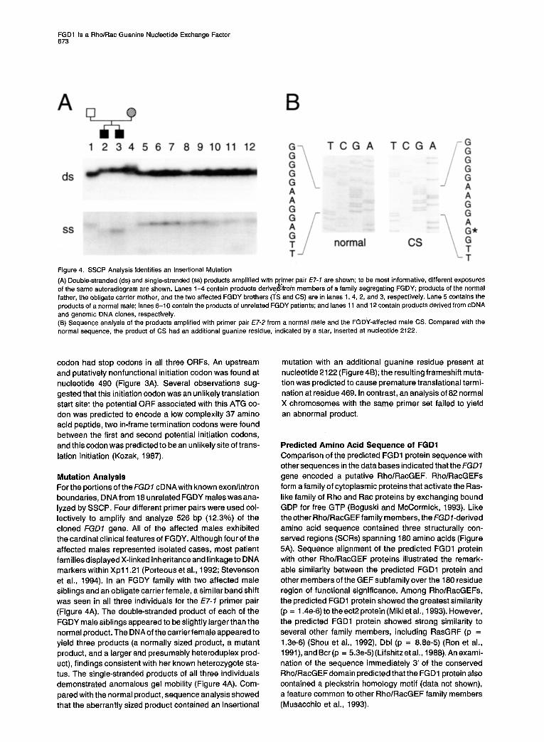
Figure 4. SSCP Analysis Identifies an Insertional Mutation
(A) Double-stranded (ds) and single-stranded (ss) products amplified with p imer pair E7-7 are shown; to be most informative, different exposures of the same autoradiogram are shown. Lanes 1-4 contain products h derive, ,from members of a family segregating FGDY; products of the normal father, the obligate carrier mother, and the two affected FGDY brothers (TS and CS) are in lanes 1, 4, 2, and 3, respectively. Lane 5 contains the products of a normal male; lanes 6-10 contain the products of unrelated FGDY patients; and lanes 11 and 12 contain products derived from cDNA and genomic DNA clones, respectively. (6) Sequence analysis of the products amplified with primer pair E7-2 from a normal male and the FGDY-affected male CS. Compared with the normal sequence, the product of CS had an additional guanine residue, indicated by a star, inserted at nucleotide 2122.
codon had stop codons in all three ORFs. An upstream and putatively nonfunctional initiation codon was found at nucleotide 490 (Figure 3A). Several observations sug- gested that this initiation codon was an unlikely translation start site: the potential ORF associated with this ATG co- don was predicted to encode a low complexity 37 amino acid peptide, two in-frame termination codons were found between the first and second potential initiation codons, and this codon was predicted to be an unlikely site of trans- lation initiation (Kozak, 1987).
Mutation Analysis For the portions of theFGD7 cDNAwith known exonlintron boundaries, DNA from 18 unrelated FGDY males was ana- lyzed by SSCP. Four different primer pairs were used col- lectively to amplify and analyze 526 bp (12.3%) of the cloned FGD7 gene. All of the affected males exhibited the cardinal clinical features of FGDY. Although four of the affected males represented isolated cases, most patient families displayed X-linked inheritance and linkage to DNA markers within Xpll.21 (Porteous et al., 1992; Stevenson et al., 1994). In an FGDY family with two affected male siblings and an obligate carrier female, asimilar band shift was seen in all three individuals for the ET-7 primer pair (Figure 4A). The double-stranded product of each of the FGDY male siblings appeared to be slightly larger than the normal product. The DNAof the carrier female appeared to yield three products (a normally sized product, a mutant product, and a larger and presumably heteroduplex prod- uct), findings consistent with her known heterozygote sta- tus. The single-stranded products of all three individuals demonstrated anomalous gel mobility (Figure 4A). Com- pared with the normal product, sequence analysis showed that the aberrantly sized product contained an insertional
mutation with an additional guanine residue present at nucleotide 2122 (Figure4B); the resultingframeshih muta- tion was predicted to cause premature translational termi- nation at residue 469. In contrast, an analysis of 82 normal X chromosomes with the same primer set failed to yield an abnormal product.
Predicted Amino Acid Sequence of FGDl Comparison of the predicted FGDl protein sequence with other sequences in the data bases indicated that the FGD7 gene encoded a putative FiholRacGEF. RholRacGEFs form afamilyof cytoplasmic proteins that activate the Ras- like family of Rho and Rat proteins by exchanging bound GDP for free GTP (Boguski and McCormick, 1993). Like theother RholRacGEFfamily members, the FGD7-derived amino acid sequence contained three structurally con- served regions (SC&) spanning 180 amino acids (Figure 5A). Sequence alignment of the predicted FGDl protein with other RholRacGEF proteins illustrated the remark- able similarity between the predicted FGDl protein and other members of the GEF subfamily over the 180 residue region of functional significance. Among RholRacGEFs, the predicted FGDl protein showed the greatest similarity (p = 1.4e-6) to the ect2 protein (Miki et al., 1993). However, the predicted FGDl protein showed strong similarity to several other family members, including RasGRF (p = 1.3e-6) (Shou et al., 1992) Dbl (p = 8.8e-5) (Ron et al., 1991), and Bcr(p = 5.3e-5) (Lifshitz et al., 1988). An exami- nation of the sequence immediately 3’ of the conserved RholRacGEFdomain predicted thatthe FGDl protein also contained a pleckstrin homology motif (data not shown), a feature common to other RholRacGEF family members (Musacchio et al., 1993).

Cell 674
B
Figure 5. Comparative Analysis of Potential FGDl Structural Domains
(A) Amino acid alignment between FGDl and the SC& of RholRacGEF proteins. Amino acids comprising previously identified SCRs are boxed (Boguski and McCormick, 1993). Residues common to four or more proteins are indicated as consensus amino acids and shown in bold. Dbl and Bcr are human sequences, proto-Vav and ect2 are mouse sequences, and RasGRF and Cdc24 are from the rat and S. cerevisiae, respectively; corresponding GenBank accession numbers are Pi091 1, L11316, YOO661, 523669, P28818, and PI 1433. (B) Amino acid alignment between FGDl and proteins containing cysteine-rich zinc finger-like domains, Residues common to three or more proteins are indicated as consensus amino acids and shown in bold. Vacl and Fabl are from S. cerevisiae, and ZK632.12 and PKCy are C. elegans and human sequences, respectively; corresponding GenBank accession numbers are P32809, P34756, P34657, and M13977. (C) Amino acid alignment between FGDl and proteins containing SH3-binding sites (Ren et al., 1993; Rozakis-Adcock et al., 1993). Residues common to all of the aligned amino acids are shown in bold and indicated as a consensus sequence. The m4nAChR sequence is from the rat (Bonner et al., 1987); 3BPl and 3BP2 (Cicchetti et al., 1992) mSos2 (Bowtell et al., 1992) and formin (Woychik et al., 1990) are mouse sequences.
Comparison of the derived FGDl protein sequence with other sequences in the data bases suggested that it con- tained two additional structural motifs (see Figure 38). The 3’ region of the sequence was predicted to encode a 50 amino acid cysteine-rich region that contained a zinc fin- ger-like structural motif. Although this region showed the greatest similarity with two proteins of unknown function, the Caenorhabditis elegans protein ZK632.12 (p = 7.le- 12) and the Saccharomyces cerevisiae protein Fabl (p = 1.9e-5) this region showed some degree of similarity to the zinc finger-like regions of both the Vacl protein (Weisman and Wickner, 1992) and the diacylglycerollphorbol dies- ter-binding regulatory domain of protein kinase CT (PKCy) (Quest et al., 1994). The sequence alignment of the pre- dicted FGDl protein with the other zinc finger-like proteins is shown in Figure 5B. This alignment illustrated that the predicted FGDl zinc finger-like structural motif, C-X&- X1&-X,-C-X&-X&XI&-X~-C (single letter code; X de- noting variable residues), was identical to that found in the ZK632.12 and Fabl protein sequences and similar to the PKCr motif. The remarkably strong conservation of this region among proteins derived from organisms as dis- tantly related as humans, nematodes, and yeast inferred that this region was functionally significant and suggested
that this region contained a cysteine-rich zinc finger-like domain thatwas similarto, but distinct from, thatcontained in PKCy.
An analysis of the predicted FGDl protein showed that the 5’ region was remarkably proline-rich and that proline constituted 22% of the first 250 amino acid residues. Since proline-rich regions have been shown to contain Src ho- mology 3 (SHS)-binding domains (Egan et al., 1993; Ro- zakis-Adcock et al., 1993), the 5’ portion of the derived FGDl protein sequence was compared with other se- quences known to contain SH3-binding sites. As shown in Figure 5C, this comparison identified two partially over- lapping segments of the predicted FGDl sequence that exhibited strong similarity to the functionally significant regionsof several proteins with demonstrated SH3 binding activity, including the Abl SH8binding proteins 3BPl and 3BP2 (Ren et al., 1993) and the RasGEF mSos2 (Egan et al., 1993; Rozakis-Adcock et al., 1993). The derived consensus sequence for the alignment was identical to that proposed by Ren et al. (1993) who showed that mu- tations within this consensus sequence abolished SH3 domain binding. Short peptides containing similar se- quences have been shown to be necessary and sufficient for SH3 binding (Li et al., 1993; Rozakis-Adcock et al.,

FGDI Is a RholRac Guanine Nucleotide Exchange Factor 675
1993). These results suggested, fhat, like 3BP1, 3BP2, and mSos2, the FGDl protein contained an SH3-binding domain.
Discussion
A number of lines of evidence strongly suggest that FGD7 is the gene responsible for X-linked FGDY. First, it maps to Xpl1.21, the region known to contain a gene responsi- ble for FGDY. Second, the fGD7 locus is directly disrupted by a translocation breakpoint in a family in which the dis- ease is segregating. Third, an insertional mutation, pre- dicted to result in a severely abbreviated and nonfunc- tional FGDI protein, segregates with the FGDY phenotype in an affected family. Fourth, fGD7 mRNA is expressed in most tissues, including FCF bones, a finding consistent with the disease phenotype. Finally, the derived FGDl amino acid sequence predicts a protein with RholRacGEF activity. In addition, this predicted sequence contains two other potential structural motifs: a cysteine-rich zinc fin- ger-like structural domain that is related to the regulatory domain of PKCy and two putative SH9binding domains. The remarkable similarity of FGD7 to several proto- oncogenes involved in the regulation of growth-related sig- nal transduction (i.e., Dbl, Bcr, ect2, and VW), further sug- gests that the FGDl protein is responsible for the altered pattern of embryonic development observed in FGDY.
Our assertion that FGDl is a putative RholRacGEF is based upon the results of our comparative sequence anal- ysis. The predicted FGDl protein sequence contains a 180 amino acid structural motif that is significantly similar to other RholRacGEF family members, including Cdc24, Dbl, and ect2. Several lines of evidence indicate that mem- bers of this family do indeed represent RholRacGEFs. In yeast, CDC24 has been shown to regulate CDC42Sc (Bender and Pringle, 1989), whose human homolog, CDC42Hs, encodes a Rho-related GTP-binding protein involved in the regulation of cytoskeletal organization (Shinjo et al., 1990). Dbl has been shown specifically to stimulate guanine exchange activity for human CDC42Hs and RhoA (Hart et al., 1991, 1994). Although ect2 has not been found to stimulate exchange activity for CDC42Mm, it has been shown specifically to bind to several Rho- related proteins, including Racl, RhoA, and RhoC (Miki et al., 1993). Furthermore, the structurally conserved 180 amino acid motif identified in FGDl represents the only common sequence contained in all RholRacGEF protein family members (Boguski and McCormick, 1993). It has recently been shown that the transforming and GEF activi- ties of the Dbl protein are carried in a 238 amino acid peptide that contains this 180 residue domain (Hart et al., 1994). These results indicate that this domain is sufficient and specific for RholRacGEF activity and suggest that FGDl is a family member. However, the recent demon- stration that Vav, a putative RholRacGEF family member, stimulates the exchange of GTP for ~21”” but not for Rho- related proteins (Gulbins et al., 1993), suggests that this motif may not be an absolute predictor of GEF specificity (Boguski and McCormick, 1993).
The results of our comparative sequence analysis also
indicate that, like other RholRacGEFs, the predicted FGDI sequence contains two additional structural do- mains that may either modify or regulate its activity or do both: a putative SHS-binding domain and a cysteine-rich zinc finger-like domain. SH3 domains have been shown specifically to bind to short (9-l 0 amino acid) proline-rich structurally conserved motifs contained in a variety of pro- teins, including the RasGEF SOS (Li et al., 1993; Ren et al., 1993; Rozakis-Adcock et al., 1993). Like the putative FGDl SHS-binding domains, the SOS-binding motifs are quitesimilartothosecontained in 3BP1,3BP2, andformin, domains with demonstrated Abl SH3 domain specificity (Egan et al., 1993; Li et al., 1993; Rozakis-Adcock et al., 1993). Recently, it has been shown that a protein com- posed entirely of an SH2 domain flanked by two SH3 do- mains, GrbP (homologous to Sem-5 in C. elegans and Drk in Drosophila), selectively binds the proline-rich motifs of the SOS protein to form a link in a signal transduction path- yJay that functionally ties tyrosine kinase receptors to Ras (B&day and Downward, 1993; Egan et al., 1993; Li et al., 1993; Rozakis-Adcock et al., 1993). Among the identified Ras and RholRacGEF family members, SOS is unique in containing an SH3- or GrbP-binding domain (Boguski and McCormick, 1993). The identification of a putative proline- rich SHB-binding domain in the predicted FGDl protein implies that, like SOS, the location, activity, or both of the FGDl protein may be modified by Abl- and GrbPlike pro- teins.
Like the product of the proto-oncogene RholRacGEF member Vav (Coppola et al., 1991), the predicted FGDl sequence also contains a putative cysteine-rich zinc fin- ger-like domain that is related to but distinct from the dia- cylglycerol and phorbol diester-binding regulatory domain of PKCy (Quest et al., 1994). The zinc finger-like domain of theVav protein has been shown to be functionallysignifi- cant; mutations of the conserved cysteine residues were found to -abolish the transforming activity of the proteins (Coppola et al., 1991). Similar putative regulatory domains have been identified in a variety of Ras-associated pro- teins, including the product of the raf proto-oncogene, the RholRac GTPase-activating protein n-chimerin, and dia- cylglycerol kinase (Liscovitch and Cantley, 1994). The ob- servation that the predicted FGDl sequence contains a putative zinc finger-like domain suggests that the FGDl protein may interact with lipid second messenger mole- cules. Therefore, it is possible that the FGDl protein may interact with the components of multiple signal transduc- tion pathways.
Since both of the identified FGDY mutations predict the total absence of FGDl protein in affected individuals, our findings predict that the constitutional loss of FGDl Rho/ RacGEF activity in humans results in the clinical FGDY phenotype of disproportionately short stature and cranio- facial, skeletal, and urogenital anomalies. However, our results do not suggest a specific mechanism as to how the absence of the FGDl protein would result in the observed developmental anomalies. In S. cerevisiae, Schizosac- charomyces pombe, Drosophila melanogaster, and C. elegans, the genetic loss of GEF function has effects similar to a loss of the associated Ras protein (see

Cell 878
Boguski and McCormick, 1993). In mammals, Rho protein family members consist of at least eight distinct proteins (RhoA, RhoB, RhoC, Racl, Race, TCl 0, CDC42Hs1, and CDC42Hs2) (Downward, 1992; Boguski and McCormick, 1993). Rho-like proteins have been shown to be involved in the regulation of the architecture of the actin cytoskeleton (Ridley and Hall, 1992; Ridley et al., 1992), yeast bud site assembly and cell polarity(Drubin, 1991), and the NADPH oxidase system in phagocytes (Abo et al., 1991). There- fore, the study of FGDl may provide important information regarding the roles that Rho- and Rat-like proteins play in signal transduction and mammalian development. Fur- thermore, if, as we expect, FGDl interacts with other sig- nal transduction components, genes that encode FGDl- associated proteins may be responsible for other similar inherited developmental disorders. Such anticipated inter- actions may provide an explanation for the previous re- ports of FGDY genetic heterogeneity.
Several Ras-associated proteins have been found to be responsible for human genetic diseases. Neurofibromin (NFl), the human protein defective in neurofibromatosis type 1, contains a RasGAP domain homologous to the catalytic domains of pl20-GAP, IRA, and IRA2 (Buchberg et al., 1990). As recently reported by the European Chro- mosome 16 Tuberous Sclerosis Consortium (1993) tu- berin, the gene responsible for the form of tuberous sclerosis mapped to chromosome 16 (TSC2), has some homology to the GAP3 protein. The gene responsible for choroderemia, a retinal degeneration syndrome, is similar to a Rab geranylgeranyl transferase (Seabra et al., 1993). However, because FGDl is a member of the Rho/RacGEF family directly implicated in causing an inherited human disease, the study of FGDl , in the context of its normal function and regulation, will provide important information regarding the roles that Ras-like GEFs play in signal trans- duction and mammalian development. The demonstrated strong homology of other Ras-associated family members implies that it may be possible and valuable to study homo- logs of the FGDl protein in other animal systems. Further- more, studies of the features of FGDl that resemble those of the transforming RholRacGEF family members may aid in the understanding of oncogene regulation and the events leading to transformation. Indeed, it will be interest- ing to determine whether FGDl is involved in human neo- plastic disease.
Experimental Procedures
Somatic Cell Hybrids and Lymphoblastoid Cell Lines Epstein-Barr virus-transformed lymphoblastoid cell lines were de- rived from a female patient with FGDY and an X;8 translocation (PP), her similarly affected son (CP), and her normal parents (GM and GP) (Bawle et al., 1984). HPPI is a human-hamster hybrid cell line that contains the translocated X chromosome t(X;8)(pll.21;11.21) derived from PP (Glover et al., 1993). C9-5 contains the translocated X chromo- some t(X;S)(pl1.21;q34.3) derived from a female with incontinentia pigmenti type 1 (Gorski et al., 1989, 1992). GM10501 contains the translocation chromosome t(X;17)(17qter-I 7ql1.2::Xpli .Pl-Xqter) (La- freniere et al., 1991). GM08318 contains only a structurally intact hu- man X chromosome (Coriell Medical Institute). This panel of hybrid cell lines has been previously described (Gorski et al., 1992).
YAC Clone Analyses YAC clones were isolated from a human Xpter-Xq27.3~specific library by colony hybridization (Lee et al., 1992). DNA markers that detect ALA.92 and DXS323 have been described (Gorski et al., 1992). High molecular weight yeast DNA was prepared and analyzed as described (Gorski et al., 1992). Restriction maps were obtained by hybridizing YAC vector-specific and insert-specific probes to blots of fully and partially digested PFGE-fractionated YAC DNA. YAC insert end clones were recovered by inverse PCR (Arveiler and Porteous, 1991) con- firmed by restriction analysis, and mapped to Xpll.21 by using frag- ments as probe to perform somatic cell hybrid and YAC DNA hybridiza- tions.
Standard and PFGE Hybridization Analysis DNA was isolated, digested, transferred to Hybond-N nylon mem- branes (Amersham), and hybridized as described (Gorski et al., 1992). Hybridizations were performed for 18-24 hr at 85’C, and membranes were washed to a final stringency of 0.1 x SSC, 0.1% SDS at 85OC for at least IO min (Gorski et al., 1992). Plasmid DNA was prepared by standard techniques; prior to use as probe, DNA fragments were recovered from agarose gel by electroelution (Sambrook et al., 1989) or by adhesion to a silica matrix (810 101).
cDNA Isolation and Characterization ZAPII human 1 g-week-old FB and FCF cDNA libraries were screened by standard methods (Sambrook et al., 1989). Bluescript KS(+) plas- mid clones containing cDNA inserts were isolated by in vivo excision as per recommendations of the supplier (Stratagene). Fetal and adult tissue poly(A)+ RNA Northern blots were obtained from Clonetech and hybridizations were performed as described (Sambrook et al., 1989). Double-stranded cDNA and genomic subclones were sequenced by using Sequenase version 2.0 as per the recommendations of the manufacturer (United States Biochemicals). Oligonucleotides for se- quencing and PCR amplification were synthesized by standard phos- phoramidite chemistry on an Applied Biosystems model 394 DNA syn- thesizer.
Mutatlon Analysis Portions of the FGDI cDNA for which exonlintron boundaries were known were PCR amplified and analyzed by SSCP (Orita et al., 1989). Primer pairs were used as follows: E7-7, GGACCGCTATCCACGCATTGGAGA and TCAAAGTTCTTCACATACTCACC, which amplify cDNA sequences from bases 2072 to 2154; E4-7, ATACCCTGCTTCCATGTGTGTCC and GAAACAGGCAGCGGGAGGCCTCA, which amplify cDNA sequences from bases 1388 to 1529; E4-2, TGAGGCCTCCCGCTGCCTGTTTC and GGCAACAGGCACACTAGCCAGGG, which amplify cDNA sequences from bases 1507 to 1899; E4-3, CCCTGGCTAGTGTGCCTGTTGCC and CTCTCGGTGAACACAGGTCAGT, which amplify cDNA sequences from bases 1877 to 1830. Exon amplifications were performed in 50 ul volumes containing 50 mM KCI, 10 mM Tris (pH 8.0), 10 mglml BSA, 1.5 mM MgCl*, 200 pM each of dATP, dGTP, and dlTP, 20 uM dCTP, 1 uCi of [a-3*P]dCTP, 100 ng of genomic DNA, 0.5 uM primers, and 2.5 U of AmpliTaq (Perkin-Elmer Cetus). Amplifications were per- formed in a DNA Thermal Cycler 480 (Perkin-Elmer Cetus) with 35 cycles of 1 min of denaturation at 94OC, 1 min of annealing at 55’C- 59°C and 2-4 min of extension at 72OC.
For SSCP analysis, 2 PI of a PCR was mixed with an equal volume of stop solution (United States Biochemicals), boiled for 5 min, cooled on ice for 2 min, and loaded on a 8% polyacrylamide gel containing 5% glycerol and 0.5 x TBE buffer. Samples were electrophoresed at both 4OC for 4-8 hr at 20 W and overnight at room temperature at 1-5 W of constant power. PCR products were sequenced as described (Casanova et al., 1990) with slight modifications. Amplification was performed by substituting primer pair E7-2 (GGACCGCTATCCACG- CATTGGAGAand ATGCGCTGCACAGGCTCCAGCAT), which ampli- fies cDNA sequences from bases 2072 to 2289, and 180 uM dCTP for the 1 nCi of [aJ2P]dCTP. Product was purified by adhesion to silica matrix (610 101) and resuspended in 21 nl of water: 7 91 of the product (approximately 200 ng) was sequenced as above by using primer CTGCACCTCATGGATGATGACTT (bases 2227-2205), added in 20 M excess. Prior to sequencing, the reaction was boiled for 2 min and

FGDl Is a RholRac Guanine Nucleotide Exchange Factor 677
snap frozen in dry ice and ethanol,?The reaction was performed for 45 s at room temperature and terminated for 5 min at 37%.
Analysis of Derived Amino Acid Sequences Homologysearcheswerecarried out at the National Center for Biotech- nology Information by using the BLAST network service (Altschul et al., 1994). Protein alignments and consensus determinations were performed by using the PILEUP program of the Genetics Computer Group software package (Devereux et al., 1984) with minimal manual modification.
Acknowledgments
Correpondence and requests for reprints should be addressed to J. L. G. We wish to thank the members of the Aarskog Syndrome Support Group and Michael Schneider for their contributions of time and patient materials; Robert Nussbaum for the human YAC library; Jeff Murray for the FCF cDNA library; David Kurnit for the brain cDNA library: Richard Jove, Michael Long, and Diane Robins for critically reviewing the manuscript; and Diane Vossfor assistance in preparing this manuscript. The work was supported in part by the General Clin- ical Research Center at the University of Michigan and was funded by National Center for Research Resources grant RR-00042 and by March of Dimes-Birth Defects Foundation basic science grant l-93-0326 and National Institutesof Health grant NS-30771 to J. L. G.
Received August 9, 1994; revised September 1, 1994.
References
Aarskog, D. (1970). A familial syndrome of short stature associated with facial dysplasia and genital anomalies. J. Pediatr. 77, 856-861.
Abo, A., Pick, E., Hall, A., Totty, N., Teahan, C., and Segal, A. (1991). Activation of the NADPH oxidase involves the small GTP-binding pro- tein p21’~c’. Nature 353, 668-670.
Altschul, S. F., Boguski, M. S., Gish, W., and Wooton, J. C. (1994). Issues in searching molecular sequence databases. Nature Genet. 6, 119-129.
Arveiler, B., and Porteous, D. J. (1991). Amplification of end fragments of YAC recombinants by inverse-polymerase chain reaction. Tech- nique 3, 24-28.
Bawle, E., Tyrkus, M., Lipman, S., and Bozimowski, D. (1984). Aarskog syndrome: full male and female expression associated with an X-autosome translocation. Am. J. Med. Genet. 77, 595-602.
Bender, A., and Pringle, J. R. (1989). Multicopy suppression of the cdc24 budding defect in yeast by CDC42 and three newly identified genes including the ras-related gene RSR7. Proc. Natl. Acad. Sci. USA 86, 9976-9980.
Boguski, M. S., and McCormick, F. (1993). Proteins regulating Ras and its relatives. Nature 366, 643-653.
Bonner, T. I., Buckley, N. J., Young, A. C., and Brann, M. R. (1987). Identification of a family of muscarinic acetylcholine receptor genes. Science 237,527-531.
Bowtell, D., Fu, P., Simon, M., and Senior, P. (1992). Identification of murine homologues of the Drosophila son of sevenless gene: potential activators of ras. Proc. Natl. Acad. Sci. USA 89, 6511-6515.
Buchberg, A. M., Cleveland, L. S., Jenkins, N. A., and Copeland, N. G. (1990). Sequence homology shared by neurofibromatosis type-l gene and IRA and IRA2 negative regulators of the Ras cyclic AMP pathway. Nature 347, 291-294.
Buday, L., and Downward, J. (1993). Epidermal growth factor regulates ~21”’ through the formation of a complex of receptor, Grb2 adapter protein, and SOS nucleotide exchange factor. Cell 73, 61 l-620.
Casanova, J. L., Pannetier, C., Jaulin, C., and Kourilsky, P. (1990). Optimal conditions for directly sequencing double stranded PCR prod- ucts with sequenase. Nucl. Acids Res. 78, 4028.
Cicchetti, P., Mayer, 6. J., Thiel, G., and Baltimore, D. (1992). Identifi- cation of a protein that binds to the SH3 region of Abl and is similar
to Bcr and GAP-rho. Science 257, 803-806.
Coppola, J., Bryant, S., Koda, T., Conway, D., and Barbacid, M. (1991). Mechanism of activation of the Vav protooncogene. Cell Growth Differ. 2, 95-105.
Devereux, J., Haeberli, P., and Smithies, 0. (1984). A comprehensive set of sequence analysis programs for the VAX. Nucl. Acids Res. 72, 387-395.
Downward, J. (1992). Rat and Rho in tune. Nature 359, 273-274.
Drubin, D. G. (1991). Development of cell polarity in budding yeast, Cell 65, 1093-1096.
Duncan, P. A., Klein, R. M., Wilmot, P. L., and Shapiro, L. R. (1977). Additional features of the Aarskog syndrome. J. Pediatr. 97,769-770.
Egan, S. E., Giddings, B. W., Brooks, M. W., Buday, L., Sizeland, A. M., and Weinberg, R. A. (1993). Association of SOS Ras exchange protein with Grb2 is implicated in tyrosine kinase signal transduction and transformation. Nature 363, 45-51.
The European Chromosome 16 Tuberous Sclerosis Consortium (1993). Identification and characterization of the tuberous sclerosis gene on chromosome 16. Cell 75, 1305-1315.
Fryns, J. P. (1992). Aarskog syndrome: the changing phenotype with age. Am. J. Med. Genet. 43,420-427.
&over, T. W., Verga, V., Rafael, J., Gorski, J. L., Bawle, E., and Hig- gins, J. V. (1993). Translocation breakpoint in Aarskog syndrome maps to Xpll.21 between ALAS2 and DXS323. Hum. Mol. Genet. 70,1717- 1718.
Gorlin, R. J., Cohen, M. M., and Levin, L. S. (1990). Syndromes of the Head and Neck, Third Edition (New York: Oxford University Press).
Gorski, J. L., Stein, C. K., and Glover, T. W. (1989). A somatic cell hybrid panel to facilitate identification of DNA sequences in the vicinity of the incontinentia pigmenti locus (/P/j. Cytogenet. Cell Genet. 52, 90-92.
Gorski, J. L., Boehnke, M., Reyner, E. L., and Burright, E. N. (1992). A radiation hybrid map of the proximal short arm of the human X chromosome spanning incontinentia pigmenti 1 (IP7) translocation breakpoints. Genomics 74, 657-665.
Grier, R. E., Farrington, F. H., Kendig, R., and Mamunes, P. (1983). Autosomal dominant inheritance of the Aarskog syndrome. Am. J. Med. Genet. 75, 39-46.
Gulbins, E., Coggeshall, K. M., Baier, G., Katzav, S., Burn, P., and Altman, A. (1993). Tyrosine kinase-stimulated guanine nucleotide ex- change activity of Vav in T cell activation, Science 260, 822-825.
Hart, M. J., Eva, A., Evans, T., Aaronson, S. A., and Cerione, Ft. A. (1991). Catalysis of guanine nucleotide exchange on the CDC42Hs protein by the dbl oncogene product. Nature 354, 311-314.
Hart, M. J., Eva, A., Zangrill, D., Aaronson, S. A., Evans, T., Cerione, R. A., and Zheng, Y. (1994). Cellular transformation and guanine nucle- otide exchange activity are catalyzed by a common domain on the dbl oncogene product. J. Biol. Chem. 269, 62-65.
Kozak, M. (1987). An analysis of 5’nonencoding sequences from 699 vertebrate messenger RNAs. Nucl. Acids Res. 75, 8125-8148.
Lafreniere, R. G., Brown, C. J., Powers, V. E., Carrel, L., Davies, K. E., Barker, D. F., and Willard, H. F. (1991). Physical mapping of 60 DNA markers in the p21 .l-q21.3 region of the human X chromosome. Genomics 7 7, 352-363.
Lee, J. T., Murgia, A., Sosnoski, D. M., Olivos, I. M., and Nussbaum, R. L. (1992). Construction and characterization of a yeast artificial chromosome library for Xpter-Xq27.3: a systematic determination of cocloning rate and X-chromosome representation. Genomics 72,526- 533.
Li, N., Batzer, A., Daly, R.,Yajnik,V., Skolnik, E.,Chardin, P., Bar-Sagi, D., Margolis, B., and Schlessinger, J. (1993). Guanine-nucleo tide-releasing factor hSos1 binds to GrbP and links receptor tyrosine kinases to Ras signaling. Nature 363, 85-88.
Lifshitz, B., Fainstein, E., Marcelle, C., Shtivelman, E., Amson, R., Gale, R. P., and Canaani, E. (1988). Bcr genes and transcripts. Onco- gene 2, 113-I 17.

Cell 676
Liscovitch, M., and Cantley, L. C. (1994). Lipid second messengers. Cell 77, 329-334.
Melnick, M., and Shields, E. D. (1976). Aarskog syndrome: new oral- facial findings. Clin. Genet. 9, 20-24.
Miki, T., Smith, C. L., Long, J. E., Eva, A., and Fleming, T. P. (1993). Oncogene ect2 is related to regulators of small GTP-binding proteins. Nature 362, 462-465.
Musacchio, A., Gibson, T., Rice, P., Thompson, J., and Saraste, M. (1993). The PH domain: a common piece in the structural patchwork of signalling proteins. Trends Biochem. Sci. 78, 343-348.
Orita, M., Suzuki, Y., Sekiya, T., and Hayashi, K. (1989). Rapid and sensitive detection of point mutations and DNA polymorphisms using the polymerase chain reaction. Genomics 5, 874-879.
Pasteris, N. G., Bialecki, M. D., and Gorski, J. L. (1993). YAC subclone contig assembly by serial interspersed repetitive sequence (IRS)-PCR product hybridizations. Nucl. Acids Res. 27, 5275-5276.
Porteous, M. E. M., Curtis, A., Lindsay, S., Williams, O., Goudie, D., Kamakari, S., and Bhattacharya, S. S. (1992). The gene for Aarskog syndrome is located between DXS255 and DXS566 (Xpll.2-Xq13). Genomics 74, 298-301.
Quest, A. F. G., Bardes, E. S. G., and Bell, R. M. (1994). A phorbol ester binding domain of protein kinase Cy: deletion analysis of the CysP domain defines a minimal 43-amino acid peptide. J. Biol. Chem. 269, 2961-2970.
Ren, R., Mayer, B. J., Cicchetti, P., and Baltimore, D. (1993). Identifica- tion of a ten-amino acid proline-rich SH3 binding site. Science 259, 1157-1161.
Ridley, A. J., and Hall, A. (1992). The small GTP-binding protein rho regulates the assembly of focal adhesions and actin stress fibers in response to growth factors. Cell 70, 389-399.
Ridley, A. J., Paterson, H. F., Johnston, C. L., Diekmann, D., and Hall, A. (1992). The small GTP-binding protein rat regulates growth factor- induced membrane ruffling. Cell 70, 401-410.
Ron, D., Zannini, M., Lewis, M., Wickner, R. B., Hunt, L. T., Graziani, G., Tronick, S. R., Aaronson, S. A., and Eva, A. (1991). A region of proto-dblessential for its transforming activity shows sequence similar- ity to a yeast cell cycle gene, CDC24, and the human breakpoint cluster gene, bcr. New Biol. 3, 372-379.
RozakisAdcock, M., Fernley, R., Wade, J., Pawson, T., and Bowtell, D. (1993). The SH2 and SH3 domains of mammalian Grb2 couple the EGF receptor to the Ras activator mSos1. Nature 363, 83-85.
Sambrook, J., Fritsch, E. F., and Maniatis, T. (1989). Molecular Clon- ing: A Laboratory Manual, Second Edition (Cold Spring Harbor, New York: Cold Spring Harbor Laboratory Press).
Scott, C. I., Jr. (1971). Unusual facie& joint hypermobility, genital anomaly and short stature: a new dysmorphic syndrome. In The Clini- cal Delineation of Birth Defects, Volume 10: The Endocrine System (Baltimore: Williams and Wilkins), pp. 2401246.
Seabra, M. C., Brown, M. S., and Goldstein, J. L. (1993). Retinal degen- eration in choroideremia: deficiency of Rab geranylgeranyl trans- ferase. Science 259, 377-381.
Shinjo, K., Koland, J. G., Hart, M. J., Narasimhan, V., Johnson, D. I., Evans, T., and Cerione, R. A. (1990). Molecular cloning of the gene for the human placental GTP-binding protein Gp (G25K): identification of this GTP-binding protein as the human homolog of the yeast cell- division-cycle protein CDC42. Proc. Natl. Acad. Sci. USA 87, 9853- 9857.
Shou, C., Farusworth, C. L., Neel, B. G., and Feig, L. A. (1992). Molecu- lar cloning of cDNAs encoding a guanine nucleotide releasing factor for Ras ~21. Nature 358, 351-354.
Stevenson, R. E., May, M., Arena, J. F., Millar, E. A., Scott, C. I., Jr., Schroer, R. J., Simensen, R. J., Lubs, H. A., and Schwartz, C. E. (1994). Aarskog-Scott syndrome: confirmation of linkage to the peri- centric region of the X chromosome. Am. J. Med. Genet., in press.
Tommerup, N. (1993). Mendelian cytogenetics: chromosome re- arrangements associated with Mendelian disorders. J. Med. Genet. 30, 713-727.
van de Vooren, M. J., Niermeijer, M. F., and Hoogeboom, A. J. M. (1983). The Aarskog syndrome in a large family, suggestive for autoso-
mal dominant inheritance. Clin. Genet. 24, 439-445.
Weisman, L. S., and Wickner, W. (1992). Molecular characterization of VAC7, a gene required for vacuole inheritance and vacuole protein sorting. J. Biol. Chem. 267, 618-623.
Woychik, R. P., Maas, R. L., Zeller, R., Vogt, T. F., and Leder, P. (1990). Formins: proteins deduced from the alternative transcripts of the limb deformity gene. Nature 346, 850-853.
GenBank Accession Number
The accession number for the sequence reported in this paper is U11690.