Cell, Vol. 34, 525-533, September 1983, Copyright 0 1983 ...
9
Cell, Vol. 34, 525-533, September 1983, Copyright 0 1983 by MIT 0092-6674/83/090525-09 $02.00/O Disassembly and Reconstitution of Signal Recognition Particle Peter Walter* and Gunter Blobel The Rockefeller ‘University New York, New York 10021 Summary Signal recognition particle (SRP) is a ribonucleopro- tein consisting of six distinct polypeptides and one molecule of small cytoplasmic 7SL-RNA. The particle was previously shown to function in protein trans- location across, and protein integration into, the endoplasmic reticulum membrane. A rapid proce- dure was developed to disassemble SRP into native protein and RNA components. The method utilizes unfolding of SRP with EDTA and dissociation on polycationic matrixes. SRP proteins prepared this way sediment below 7S and are inactive in activity assays. When recombined with 7SL-RNA in the pres- ence of magnesium, the proteins are shown to reas- sociate stoichiometrically with 7SL-RNA to form fully active 11s SRP. Introduction The signal recognition particle (SRP) functions as an adapter between the cy-toplasmic protein translation ap- paratus and the protein translocation machinery in the membrane of the endoplasmic reticulum. In the cytoplasm, SRP recognizes signal sequences on nascent polypep tides chains of secretory (Walter et al., 1981; Stoffel et al., 1981; Meyer et al., 1982a), lysosomal (Erickson et al., submitted), and certain classes of integral membrane pro- teins (Anderson et al., 1982). By virtue of a tight binding of SRP to the ribosomes that are in the process of synthe- sizing these proteins (Wafter et al., 1981) SRP causes a site-specific arrest of the biosynthesis of the nascent pro- tein (Walter and Blobel, 1981b). When these elongation- arrested polysomes interact with specific components of the membrane of the endoplasmic reticulum, their binding and the formation of a functional ribosome-membrane junction are observed (Walter and Blobel, 1981a). The direct interaction of SRP with the SRP receptor (Gilmore et al., 1982a, 1982b) (also termed docking protein; Meyer et al., 1982a), an integral membrane protein of the endoplas- mic reticulum (Meyer et al., 1982b), was shown to be responsible for the release of elongation arrest. As trans- lation resumes, the nascent polypeptide chain is now vectorially translocated into the lumen of the endoplasmic reticulum (Blobel and Dobberstein, 1975a, 1975b). Structurally, SRP can be defined as a ribonucleoprotein (RNP) with a sedimentation coefficient of 1 lS, consisting of six different polypeptide chains (Walter and Blobel, 1980) and one molecule of the small cytoplasmic 7SL-RNA (Walter and Blobel, 1982) of known sequence (Ullu et al., ’ Present address: Department of Biochemistry and Biophysics, University of California, San Francisco, California 94143. 1982; Li et al., 1982). Both RNA and protein are required for SRP’s activity, since it could be shown that nuclease treatment as well as alkylation with N-ethylmaleimide (Wal- ter and Blobel, 1980) inactivates the particle. At present, little is known about the function of the individual building blocks of SRP. We describe a procedure for a rapid and nondestructive disassembly of SRP into individual components, which can be reversed to reconstitute an active particle. These stud- ies provide insight into the structure of SRP and open the way for detailed structure-function analyses. Results EDTA Causes Partial Unfolding of SRP The integrity of many RNPs is dependent on the presence of divalent cations. Thus chelating agents could be suc- cessfully employed to unfold partially or to disassemble ribosomal subunits (Spirin, 1974; Newton et al., 1975; Blobel, 1971) RNAase P (Guthrie and Atchison, 1980) and small cytoplasmic RNPs (Mukherjee and Sarkar, 1981). The data in Figure 1 demonstrate that SRP undergoes structural changes in the absence of Mg2+ as well. We observed a time course of the hyperchromicity effect during nuclease digestion as a measure for the nuclease sensitivity of the particle. As the Mg2+ concentration was decreased from 10 mM to 1 mM and finally to 0 mM by the addition of EDTA, SRP was rendered increasingly susceptible to nucleolytic attack. Interestingly, the kinetics of nuclease digestion of SRP in 10 mM Mg2+ appeared to have a biphasic character: an initial rapid digestion of apparently exposed and accessible regions of 7SL-RNA, followed by a rather slow digestion of RNA regions that were probably protected by protein. The latter regions were rendered more nuclease sensitive as the Mg2+ con- centration was decreased. The nuclease digestion of na- ked 7SL-RNA (Figure 2, lanes d) was rapid and independ- ent of the presence or absence of Mg2’ (data not shown). When a Mg2+-depleted SRP preparation was analyzed for its integrity by sedimentation in sucrose gradients containing EDTA and 500 mM KOAc, SRP still sedimented at 11 S and no polypeptide chains were dissociated (data not shown). Thus, although EDTA appears to unfold SRP, it is not sufficient to dissociate the particle under these conditions. Unfolded SRP Can Be Dissociated with Polycationic Substances SRP binds to DEAE-ion exchange resins and can be eluted as an intact particle if all buffers contain Mg2+. However, if SRP was incubated with DEAE-cellulose in the presence of EDTA, we observed differential elution only of SRP polypeptides-with the 7SL-RNA remaining bound to the column. Under the premise that this result was due to a dissociation of SRP, we optimized the conditions to obtain a maximum yield of dissociated SRP proteins. The introduction of a brief incubation at elevated temperature of SRP in the presence of both EDTA and DEAE-cellulose
Transcript of Cell, Vol. 34, 525-533, September 1983, Copyright 0 1983 ...
Cell, Vol. 34, 525-533, September 1983, Copyright 0 1983 by MIT 0092-6674/83/090525-09 $02.00/O
Disassembly and Reconstitution of Signal Recognition Particle
Peter Walter* and Gunter Blobel The Rockefeller ‘University New York, New York 10021
Summary
Signal recognition particle (SRP) is a ribonucleopro- tein consisting of six distinct polypeptides and one molecule of small cytoplasmic 7SL-RNA. The particle was previously shown to function in protein trans- location across, and protein integration into, the endoplasmic reticulum membrane. A rapid proce- dure was developed to disassemble SRP into native protein and RNA components. The method utilizes unfolding of SRP with EDTA and dissociation on polycationic matrixes. SRP proteins prepared this way sediment below 7S and are inactive in activity assays. When recombined with 7SL-RNA in the pres- ence of magnesium, the proteins are shown to reas- sociate stoichiometrically with 7SL-RNA to form fully active 11s SRP.
Introduction
The signal recognition particle (SRP) functions as an adapter between the cy-toplasmic protein translation ap- paratus and the protein translocation machinery in the membrane of the endoplasmic reticulum. In the cytoplasm, SRP recognizes signal sequences on nascent polypep tides chains of secretory (Walter et al., 1981; Stoffel et al., 1981; Meyer et al., 1982a), lysosomal (Erickson et al., submitted), and certain classes of integral membrane pro- teins (Anderson et al., 1982). By virtue of a tight binding of SRP to the ribosomes that are in the process of synthe- sizing these proteins (Wafter et al., 1981) SRP causes a site-specific arrest of the biosynthesis of the nascent pro- tein (Walter and Blobel, 1981b). When these elongation- arrested polysomes interact with specific components of the membrane of the endoplasmic reticulum, their binding and the formation of a functional ribosome-membrane junction are observed (Walter and Blobel, 1981a). The direct interaction of SRP with the SRP receptor (Gilmore et al., 1982a, 1982b) (also termed docking protein; Meyer et al., 1982a), an integral membrane protein of the endoplas- mic reticulum (Meyer et al., 1982b), was shown to be responsible for the release of elongation arrest. As trans- lation resumes, the nascent polypeptide chain is now vectorially translocated into the lumen of the endoplasmic reticulum (Blobel and Dobberstein, 1975a, 1975b).
Structurally, SRP can be defined as a ribonucleoprotein (RNP) with a sedimentation coefficient of 1 lS, consisting of six different polypeptide chains (Walter and Blobel, 1980) and one molecule of the small cytoplasmic 7SL-RNA (Walter and Blobel, 1982) of known sequence (Ullu et al.,
’ Present address: Department of Biochemistry and Biophysics, University of California, San Francisco, California 94143.
1982; Li et al., 1982). Both RNA and protein are required for SRP’s activity, since it could be shown that nuclease treatment as well as alkylation with N-ethylmaleimide (Wal- ter and Blobel, 1980) inactivates the particle. At present, little is known about the function of the individual building blocks of SRP.
We describe a procedure for a rapid and nondestructive disassembly of SRP into individual components, which can be reversed to reconstitute an active particle. These stud- ies provide insight into the structure of SRP and open the way for detailed structure-function analyses.
Results
EDTA Causes Partial Unfolding of SRP The integrity of many RNPs is dependent on the presence of divalent cations. Thus chelating agents could be suc- cessfully employed to unfold partially or to disassemble ribosomal subunits (Spirin, 1974; Newton et al., 1975; Blobel, 1971) RNAase P (Guthrie and Atchison, 1980) and small cytoplasmic RNPs (Mukherjee and Sarkar, 1981).
The data in Figure 1 demonstrate that SRP undergoes structural changes in the absence of Mg2+ as well. We observed a time course of the hyperchromicity effect during nuclease digestion as a measure for the nuclease sensitivity of the particle. As the Mg2+ concentration was decreased from 10 mM to 1 mM and finally to 0 mM by the addition of EDTA, SRP was rendered increasingly susceptible to nucleolytic attack. Interestingly, the kinetics of nuclease digestion of SRP in 10 mM Mg2+ appeared to have a biphasic character: an initial rapid digestion of apparently exposed and accessible regions of 7SL-RNA, followed by a rather slow digestion of RNA regions that were probably protected by protein. The latter regions were rendered more nuclease sensitive as the Mg2+ con- centration was decreased. The nuclease digestion of na- ked 7SL-RNA (Figure 2, lanes d) was rapid and independ- ent of the presence or absence of Mg2’ (data not shown).
When a Mg2+-depleted SRP preparation was analyzed for its integrity by sedimentation in sucrose gradients containing EDTA and 500 mM KOAc, SRP still sedimented at 11 S and no polypeptide chains were dissociated (data not shown). Thus, although EDTA appears to unfold SRP, it is not sufficient to dissociate the particle under these conditions.
Unfolded SRP Can Be Dissociated with Polycationic Substances SRP binds to DEAE-ion exchange resins and can be eluted as an intact particle if all buffers contain Mg2+. However, if SRP was incubated with DEAE-cellulose in the presence of EDTA, we observed differential elution only of SRP polypeptides-with the 7SL-RNA remaining bound to the column. Under the premise that this result was due to a dissociation of SRP, we optimized the conditions to obtain a maximum yield of dissociated SRP proteins. The introduction of a brief incubation at elevated temperature of SRP in the presence of both EDTA and DEAE-cellulose
Cell 526
5mM
I I I I 1 I I 1 I I 0
time ~~~nuclease Fzatment 7%“)
Figure 1. Nuclease Sensitivity of SRP
Gradient-purtfied SRP (see Experimental Procedures) was diluted to 0.2 b units/ml, and the ionic concentration was adjusted to yield a final concentration of 50 mM TEA, 100 mM KOAc, 1 mM DlT, 0.01% Nikkol. containing 10 mM Mg(OAck (0). 1 mM Mg(OAck (A), or 5 mM EDTA (W), respectively. The solutions were warmed to 30°C in a heated cuvette hdder (Beckman DU6 spectrophotometer), and at time 0 min a solution of RNAase A (Sigma, 600 *g/ml) was added to yield a final concentration of 1 pg/ml. The absorbance at 260 nm was recorded as a function of time (Sarkar et al., 1961).
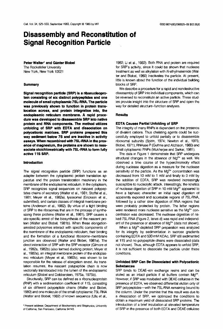
and the use of a DEAE-cellulose resin with a high surface density of charged groups (Whatman DE53) led to an almost quantitative disassembly of SRP (Figure 2, compare lanes b with lanes a). 7SLRNA could subsequently be eluted with an increase in ionic strength from the DEAE- cellulose in intact form (Figure 2, lanes c), demonstrating that in the incubation steps no nucleolytic breakdown of the EDTA-unfolded SRP occurred. This 7SLRNA fraction was subsequently treated with proteases and extracted with phenol to provide a 7SL-RNA fraction (Figure 2, lanes d) that was free of residual SRP proteins, to be used in the reconstitution studies. The dissociation reaction was independent of the time for which SRP was incubated with EDTA-i.e., the unfolding appeared to occur instantane- ously. Therefore no long incubation steps, as described for a similar disassembly of RNAase P (Guthrie and Atchi- son, 1980) were necessary. Furthermore, prolonged in- cubation with DEAE-cellulose did not further increase the yield of SRP proteins.
Among the polycationic substances tested to dissociate SRP were polyetheleneimine (PEI) and various other prep- arations of DEAE-ion exchange resins (Whatman DE52; Pharmacia DEAE-Sephadex, DEAE-Sepharose, and DEAE- Sephacel). Although all of these substances caused SRP to dissociate, they were unpractical for the following rea- sons. PEI had to be precisely titrated into the dissociation reaction, as an excess of PEI prevented 7SL-RNA from precipitating. It only dissociated the 54, 68, and 72 kd SRP polypeptides, whereas the smaller polypeptides would precipitate together with 7SL-RNA and PEI. Furthermore, we were unsuccessful in attempts to recover protein or RNA in a form free of PEI from these pellet fractions. The other DEAE-ion exchange resins (listed above) produced
I-
A ---1. B htr a b c d abed NT
x10-J
19- 14-
9-
Figure 2. Dissociation of SRP
SRP was unfolded with EDTA and dissociated on DEAE-cellulose as described in Experimental Procedures. Equivalent atiquots of the various fractions (corresponding to 20 ~1 of the starting SRP preparation) were TCA-precipitated, and the polypeptides analyzed by PAGE in SDS on 1 O%- 15% gradient gels followed by staining in Coomassie blue (A). Separate aliquots were treated with proteinase K (see Experimental Procedures), RNA was isolated by the perchlorate procedure (Lizardi and Engelbsrg. 1979) and analyzed by PAGE in 7 M urea on 7% gels followed by staining in ethidium bromide(B). The m&c&r weights (M,) of the SRP polypeptides and the size of 7SL-RNA (number of nucleotides, Nf) are indicated.
Lanes a show the SRP preparation used as starting material in this study. It was dissociated on DE53 in EDTA as described in Experimental Procedures to yiild the SPR protein fraction (lanes b). 7SL-RNA was eluted from the DE53 resin by raising the salt to 1 M KOAc (lanes c) and freed of residual SRP potypeptfdes by protefnase K treatment and repeated phenol extractions (lanes d).
The wet Coomassie-blue-stained polyacrytamide gel (A; lanes a, b. and c) was scanned at 630 nm in the Beckman DUS spectrophotometer. From the integrated absorbances the yields of the individual dissociated SRP polypeptides (lane b) were calculated to be 90% (72 kd), 75% (69 kd), 1000/o (54 kd), 100% (19 kd), 45% (14 kd), 55% (9 kd) wtth respect to the starting SRP preparation (lane a).
Note that some contaminating polypeptides in the original SRP prepa- ration (A, lane a) bind to the DEAE-cetlutose under dissociation conditions. They are thus absent in the SRP protein fraction (lane b), and all subsequent analyses in which this fraction was employed (Figures 3 and 5).
SRP proteins only at a reduced yield, and in each case these SRP protein fractions were contaminated with resid- ual 7SL-RNA. Thus it appears that the charge density on the surface of the ion-exchange resins is an important factor to obtain clear-cut fractionation of SRP in the dis- sociation reaction.
RNAase P has been successfully reconstituted after dissociation in urea (Kole and Altman, 1979). SRP could also be completely dissociated in 7 M urea in the presence of Mg2+ or in 4 M urea in the presence of EDTA (data not shown). We have, however, not been able to reconstitute active SRP after removal of the urea (by gel filtration) and thus have not pursued this experimental approach.

Reconstiiution of Signal Recognition Particle 527
Characterization of the SRP Protein Fraction When the SRP protein fraction was subjected to sedimen- tation analysis in sucrose gradients, all polypeptides sedi- mented below 7S (Figures 3A and 36). This sedimentation behavior was independent of the presence or absence of Mg2+ (data not shown). No material sedimented in the position of authentic SRP, and no distinct peak in the UV absorbance profile was observed-i.e., no detectable con- tamination with 7SLRNA was present. SRP proteins were also fractionated by chromatography on CM-Sepharose columns (Figures 3C and 30). The unusually high ionic strength required to elute five of the six SRP polypeptides from this resin is indicative of their very basic character and is consistent with their behavior on denaturing isoelec- tric focusing gels (pi >8.0, data not shown).
Interestingly, in both the sucrose gradient and ion ex- change chromatography fractionation schemes two poly- peptides behave as monomers (19 and 54 kd), whereas the 9 and the 14 kd polypeptides as well as the 88 and
the 72 kd polypeptides appeared to be still bound to each other. These polypeptides sedimented consistently with the interpretation of being heterodimers (Figure 38) and they also cochromatographed on CM-Sepharose columns (Figure 3D).
When subjected to activity assays, the SRP protein fraction (Figure 2, lanes b) and 7SL-RNA (Figure 2, lanes d) were completely inactive (Figure 48, open symbols). SRP proteins did not compete with intact SRP in these assays, even when added in a fivefold molar excess (data not shown).
Reconstitution of SRP Having established that SRP proteins were no longer in a complex, but behaved as inactive monomeric and heter- odimeric proteins, we attempted to reconstitute a func- tional particle with 7SL-RNA. As a starting point for these reconstitution experiments, we chose ionic conditions close to those reported by Traub and Nomura (1988,1989)
%
0.c
(
0.8
T 0.6”
0.4 Y 0
0.2 *
0
Sedimentation - Fraction
Figure 3. Fractionation of SRP Proteins
(A and 6) Fractionation by sucrose gradient cenkifugatf. An afiquot (250 ~1) of the SRP protein fraction (Figure 2, lanes b) was adjusted to 500 mM KCAc, 5 mM Mg(OAck. layered on top of a 5%-20% sucrose gradient in 50 mM T5A, 500 mM KOAc, 5 mM Mg(OAch, 1 mM DTT, 0.01% Nikkof, and centrifuged for 20 hr at 4OC at 40,ooO rpm in a Beckman SW40 rotor. The gradient was fractionated using an ISCC gradient fractionator, and the absorbance at 230 nm was recorded (A). Fractions of 1.0 ml were collected. The top nine fractions were TCA-precipitated, and the pofy ptfdes analyzed (B) by PAGE in SDS as described in Figure 2. The position of size markers (bovine serum albumin, BSA [4S], rabbit IgG [7S], bovine
$ tafase [l 1 S]. and undissociated SRP) in parallel gradients was determined by recording the UV absorbance at 280 nm (A).
b) was diluted with 4.0 mf of a 4°C. The flowthrough fra&ffn
KOAc concentratffns (50 mM to 1100 fraction was TCA.precipitated,
and the polypspttdes were analyzed by PAGE in SDS as described in Figure 2. The faint bands of the lower mot with an arrowhead.

Cell 528
IA , SRP -1 B
ng RNA per
5%2 I;$*. rotein
0 20 40 60 25 PI translation
80 r 1
t * O; 0.1 0.2 d.3 ‘W
pg RNA in reconstitution
Figure 4. Activity Assay of Reconstituted SRP
SRP activity (expressed as % processing + % inhibition) was determined in a wheat germ cell-free translation system, programmed wfth bovine pituitary RNA, and supplemented with SRP-depleted microsofnal vesicles as described in Experimental Procedures. (A) shows a titration of undisso- ciated SRP (0) (Figure 2, lanes a) into the assay system. (B) shows titrations of either SRP proteins (0) (Figure 2, lanes b), 7SL-RNA (A), (Figure 2, lanes d), or SRP proteins plus 7SL-RNA (W) into the assay system after incubation under reconstiiution conditions (see Experimental Procedures for concen- tration of components in the reconstitution reaction). The amount of SRP (0). reconstituted SRP 0, or ‘ISL-RNA (A) is expressed as the amount of RNA contained in these samples; the amount of SRP proteins (0) is the same as the amount of SRP protein present in the reconstituted SRP sample (B) at each given data point.
(C and D) shows activity assays of reconstituted SRP preparations where the amount of RNA in the reconstitution reaction (total volume 6 cl. see Experimental Procedures) was varii, while the amount of SRP proteins in the reconstitution reaction was kept constant as given in Experimental Procedures. A 4 pl aliquot of each reconstituted SRP preparation was assayed in a 25 pl in vitro translation system. The following RNA prepara- tions were used in these experiments. (C) canine 7SL-RNA (O), canine ribosomal5S RNA (0); (Cl) D. melanogaster 7S RNA (A), X. laevis 7S RNA (B), E. coli 6s RNA (II).
for the reconstitution of functional 30s ribosomal subunits (370 mM K+, 20 mM Mg”). SRP activity could be recon- stituted (Figure 48) by simply recombining SRP proteins with a stoichiometric amount of 7SL-RNA in the presence of Mg*+. A short incubation step at elevated temperature (10 min at 37°C) was required to drive the reconstitution to completion. If this incubation step was omitted, func-
tional SRP was obtained nevertheless, albeit at a reduced yield (about 30% of the activity shown in Figure 48). In contrast to the reconstitutions of ribosomal subunits, we observed that the ionic conditions could be varied over a wide range (150-500 mM K+, 4-20 mM Mg*+) without affecting the efficiency of the reaction.
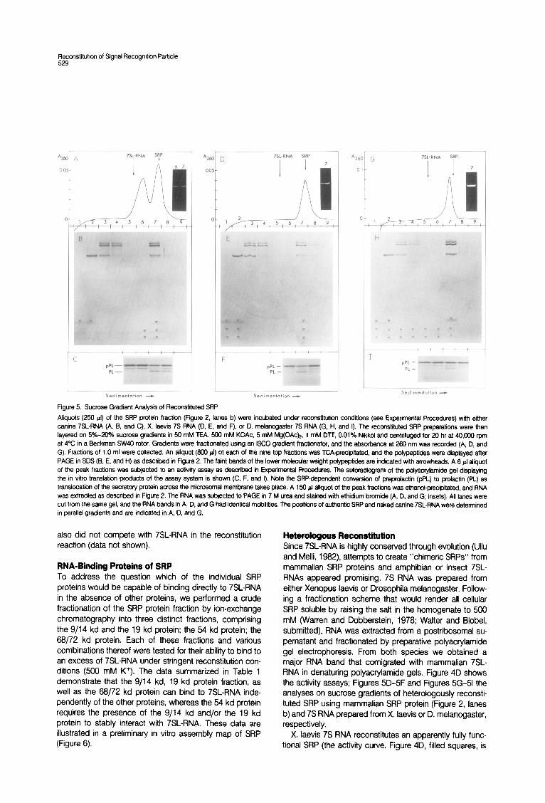
When reconstituted SRP was analyzed by sucrose gra- dient centrifugation, we observed that all six SRP polypep- tides had reassembled in about stoichiometric amounts with the 7SL-RNA and formed an active 11s particle (Figures 5A, 58, and 5C). 7SL-RNA was added in stoichi- ometric amounts with respect to the 9/14 kd SRP protein, which is the limiting component in the SRP protein fraction (see Figure 2). Thus all other SRP proteins were in molar excess and remained in part on top of the gradient (Figure 5B). A second, more slowly sedimenting peak, comprising about half of the absorbance of the reconstitution reaction, was observed (Figure 5A), which consisted of 7SL-RNA together with the 9/14 kd SRP protein (Figures 5A, 58, fraction 6). 7SL-RNA in both peaks was not degraded (Figure 5A, inset), and only the 11s peak (Figures 5A, 5B, fraction 7) was active in the translocation assay (Figure 5C). We cannot distinguish whether the slower sediment- ing peak constitutes an assembly intermediate of SRP or whether it is an incorrectly assembled form acting as a dead-end in the assembly process.
From the activity assay shown in Figure 4 it is apparent that the reconstituted SRP fraction was about half as active (Figure 48) as the control SRP preparation (Figure 4A). However, because the amount of SRP and reconstituted SRP is given in Figure 4 as the amount of RNA contained in the fraction, and because from the data in Figures 5A, 58, and 5C it follows that only half of the 7SL-RNA in the reconstitution mixture assembled into active 11 S SRP, we conclude that the fully reconstituted SRP is as active as the undissociated control preparation. Thus the assembly process occurred with about 50% yield with respect to the limiting components. To test for cooperativity in the recon- stitution process, we varied the concentration of 7SL-RNA in the standard reconstitution reaction (Figure 4C). We calculated that 70 ng 7SL-RNA would be approximately equimolar with respect to the limiting amount of the 9/14 kd SRP protein in the reconstitution mixture. As shown by the data in Figure 4C (filled circles), we indeed saturated the reconstitution reaction around 50-l 00 ng ‘ISL-RNA. At higher RNA conconcentrations the activity of reconstituted SRP remained on a plateau-i.e., in contrast to the recon- stitution of ribosomal subunits, a molar excess of 7SL-RNA did not compete in the in vitro assembly process of SRP.
We have also attempted to use unrelated RNA species in the reconstitution reaction to demonstrate specificity for 7SL-RNA. When ribosomal 5s RNA was used, no SRP activity could be detected (Figure 4C, open circles). When the products of such a reconstitution reaction were ana- lyzed on sucrose gradients, none of the SRP proteins was found to associate with the RNA (data looked identical to Figure 38). A tenfold molar excess of ribosomal 5s RNA
A YRNA S!P
C pPL-
PL-
r U.i’
Sedim.nt.tion - L
Sedimentation -
Figure 5. Sucrose Gradient Analysis of Reconstiiut&d SRP
Aliquots (250 ~1) of the SRP protein fraction (Figure 2, lanes b) were incubated under reconstftutfon conditions (see Experimental Procedures) with ekher canine 7SLRNA (A, B, and C), X. laevis 7s RNA (D, E, and F), or D. metancgaster 7s RNA (G, H, and I), The reconstiiuted SRP preparations were then layered on 5%-20% sucrose gradients in 50 mM TEA, 500 mM KOAc, 5 mM Mg(OAc)*. 1 mM DTT, 0.01% Nikkd and centrifuged for 20 hr at 40,OOiJ rpm at 4°C in a Beckman SW40 rotor. Gradients were fractionated using an ISCO gradient fractionator, and the absorbance at 260 nm was recorded (A, D, and G). Fractions of 1 .O ml were collected. An aliquot (SO0 ~1) of each of the nine top fractions was TCA-precipitated, and the polypeptides were displayed after PAGE in SDS (B, E, and H) as described in Figure 2. The faint bands of the lower molecular weight polypeptides are indicated with arrowheads, A 5 IJ aliquot of the peak fractions was subjected to an activity assay as described in Experimental Procedures. The autoradicgram of the polyacryfamide gel displaying the in vitro translation products of the assay system is shown (C, F. and I). Note the SRP-dependent conversion of preprolactin (pPL) to prolactin (PL) as translocation of the secretory protein across the microsomal membrane takes place. A 150 pl atiquot of the peak fractions was ethand-precipitated, and RNA was extracted as described in Figure 2. The RNA was subjected to PAGE in 7 M urea and stained with ethidium bromide (A, D, and G; insets). All lanes were cut from the same gel, and the RNA bands in A, D, and G had identical mobiliiies. The positions of authentic SRP and naked canine 7SL.RNA were detenined
7SL-RNA SRP
I I 7
pPL - PL -
A2b( G T-St-RNA =P
0.i I I7
2 /- 1 ,/--y-a, 4,5, 6,7,8,9
Y ’ ’ ’ c ’ c +
I pPL -
PL- “’ In
I^” “’ I..
in parallel gradients and are indicated in A, D, and G.
also did not compete with 7SLRNA in the reconstitution reaction (data not shown).
RNA-Binding Proteins of SRP To address the question which of the individual SRP proteins would be capable of binding directly to 7SLRNA in the absence of other proteins, we performed a crude fractionation of the SRP protein fraction by ion-exchange chromatography into three distinct fractions, comprising the 9/14 kd and the 19 kd protein; the 54 kd protein; the 68/72 kd protein. Each of these fractions and various combinations thereof were tested for their ability to bind to an excess of 7SLRNA under stringent reconstitution con- ditions (500 mM K’). The data summarized in Table 1 demonstrate that the 9/14 kd, 19 kd protein fraction, as well as the 68/72 kd protein can bind to 7SL-RNA inde- pendently of the other proteins, whereas the 54 kd protein requires the presence of the 9114 kd and/or the 19 kd protein to stably interact with 7SL-RNA. These data are illustrated in a preliminary in vitro assembly map of SRP (Figure 6).
Heterologous Reconstitution Since 7SL-RNA is highly conserved through evolution (Ullu and Melli, 1982) attempts to create “chimeric SRPs” from mammalian SRP proteins and amphibian or insect 7SL- RNAs appeared promising. 7S RNA was prepared from either Xenopus laevis or Drosophila melanogaster. Follow- ing a fractionation scheme that would render all cellular SRP soluble by raising the salt in the homogenate to 500 mM (Warren and Dobberstein, 1978; Walter and Blobel, submitted), RNA was extracted from a postribosomal su- pernatant and fractionated by preparative polyacrylamide gel electrophoresis. From both species we obtained a major RNA band that comigrated with mammalian 7SL- RNA in denaturing polyacrylamide gels. Figure 4D shows the activity assays; Figures 5D-5F and Figures 5G-51 the analyses on sucrose gradients of heterologously reconsti- tuted SRP using mammalian SRP protein (Figure 2, lanes b) and 7S RNA prepared from X. laevis or D. melanogaster, respectively.
X. laevis 7S RNA reconstitutes an apparently fully func- tional SRP (the activity curve, Figure 4D, filled squares, is
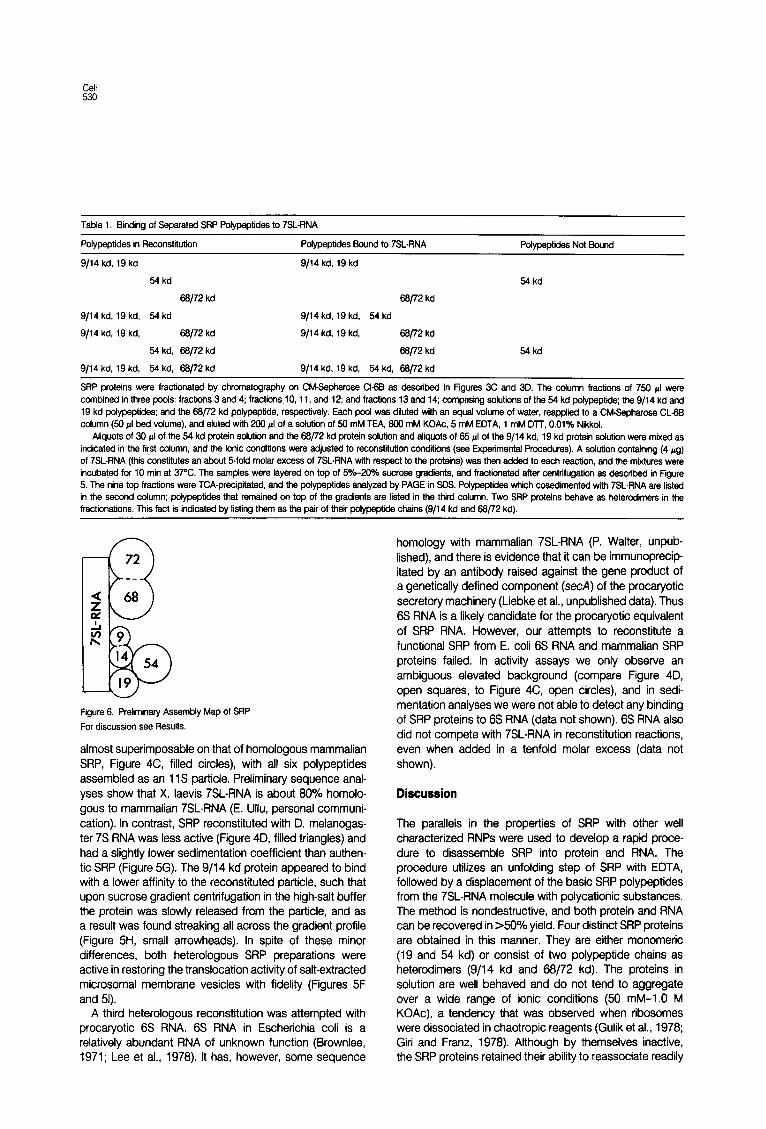
Table 1. Binding of Separated SRP Pofypsptides to 7SLRNA
Polypeptides in Reconstiiutiin Pdypeptides Sound to 7SLRNA Pdypeptk-Jes Not Sound
9/14 kd. 19 ko 9114 kd, 19 kd
54 kd 54 kd
66/72 kd 66/72 kd
9114 kd, 19 kd, 64 kd 9/14 kd, 19 kd, 64 kd
9/14 kd, 19 kd, Se/72 kd 9/14 kd, 19 kd, 66/72 kd
54 kd, 66172 kd 68/72 kd 54 kd
9114 kd, 19 kd. 64 kd, 66/72 kd 9/14 kd, 19 kd, 64 kd. 66/72 kd
SRP proteins were fractionated by chromatography on CM-Sepharose Ct6B as described in Figures 3C and 3D. The column fractions of 7% pl were combined in three pods: fractions 3 and 4; fractionslO, 11, and 12; and fractions 13 and 14; comprising solutiis of the 54 kd polypeptide; the 9/14 kd and 19 kd polypeptides; and the 66j72 kd pofypeptide, respectively. Each pool was diluted wfth an equal votume of water, reapplied to a CM-Sepharose CL- column (50 4 bed vdume), and eluted wfth 200 cl of a sotution of 66 mM TEA, 800 mM KOAc, 5 mM EDTA, 1 mM Dll, 0.01% Nikkot.
Afiquots of 30 pl of the 64 kd protein sdution and the 66/72 kd protein solution and afiquots of 66 ~1 of the 9/14 kd, 19 kd protein sotutiin were mixed as indicated in the first cotumn, and the ionic conditions were adjusted to reccnstituticn conditions (see Experimental Procedures). A sdution containing (4 pg) of 7SL-RNA (this constitutes an about dfold molar excess of 7SL-RNA with respect to the proteins) was then added to each reaction, and the mixtures were incubated for IO min at 37’C. The samples were layered on top of F&-20% sucrose gradients, and fractiinated after centrifugatiin as described in Figure 5. The nine top fractions were TCA-precipitated, and the pofypeptides analyzed by PAGE in SDS. Polypeptides which cosedimented with 7SL-RNA are listed in the second cdumn; pofypeptides that remained on top of the gradients are listed in the third cdumn. Two SRP proteins behave as heterodimers in the fractionations. This fact is indicated by listing them as the pair of their potypeptide chains (9/14 kd and 66772 kd).
Figure 6. Preliminary Assembly Map of SRP
For discussion see Resufts.
almost superimposable on that of homologous mammalian SRP, Figure 4C, filled circles), with all six polypeptides assembled as an 11 S particle. Preliminary sequence anal- yses show that X. laevis 7SLRNA is about 80% homolo- gous to mammalian 7SLRNA (E. Ullu, personal communi- cation). In contrast, SRP reconstituted with D. melanogas- ter 7s RNA was less active (Figure 4D, filled triangles) and had a slightly lower sedimentation coefficient than authen- tic SRP (Figure 5G). The 9/l 4 kd protein appeared to bind with a lower affinity to the reconstituted particle, such that upon sucrose gradient centrifugation in the high-salt buffer the protein was slowly released from the particle, and as a result was found streaking all across the gradient profile (Figure 5H, small arrowheads). In spite of these minor differences, both heterologous SRP preparations were active in restoring the translocation activity of salt-extracted microsomal membrane vesicles with fidelity (Figures 5F and 51).
A third heterologous reconstitution was attempted with procaryotic 6s RNA. 6s RNA in Escherichia coli is a relatively abundant RNA of unknown function (Brownlee, 1971; Lee et al., 1978). It has, however, some sequence
homology with mammalian 7SL-RNA (P. Walter, unpub- lished), and there is evidence that it can be immunoprecip- itated by an antibody raised against the gene product of a genetically defined component (secA) of the procaryotic secretory machinery (Liebke et al., unpublished data). Thus 6s RNA is a likely candidate for the procaryotic equivalent of SRP RNA. However, our attempts to reconstitute a functional SRP from E. coli 6s RNA and mammalian SRP proteins failed. In activity assays we only observe an ambiguous elevated background (compare Figure 4D, open squares, to Figure 4C, open circles), and in sedi- mentation analyses we were not able to detect any binding of SRP proteins to 6s RNA (data not shown). 6s RNA also did not compete with 7SL-RNA in reconstitution reactions, even when added in a tenfold molar excess (data not shown).
Discussion
The parallels in the properties of SRP with other well characterized RNPs were used to develop a rapid proce- dure to disassemble SRP into protein and RNA. The procedure utilizes an unfolding step of SRP with EDTA, followed by a displacement of the basic SRP polypeptides from the 7SL-RNA molecule with polycationic substances. The method is nondestructive, and both protein and RNA can be recovered in >50% yield. Four distinct SRP proteins are obtained in this manner. They are either monomeric (19 and 54 kd) or consist of two polypeptide chains as heterodimers (9/14 kd and 68/72 kd). The proteins in solution are well behaved and do not tend to aggregate over a wide range of ionic conditions (50 mM-1.0 M KOAc), a tendency that was observed when ribosomes were dissociated in chaotropic reagents (Gulik et al., 1978; Giri and Franz, 1978). Although by themselves inactive, the SRP proteins retained their ability to reassociate readily

F3ytitution of Signal Recognition Particle
and stoichiometrically with 7SLRNA to form active 11 S SRP. Because of the mild dissociation conditions, we feel confident that the structure of SRP proteins in solution retains features of the structure of the proteins in the particle. For example, it is likely that the SRP polypeptides that exist in heterodimeric form in solution also exist in a nearest neighbor relationship with each other in SRP.
Since 7SL-RNA is required for the reconstitution of SRP proteins into an 11 S particle, we conclude that at least one function of the RNA molecule in SRP is structural. The RNA is providing a matrix for the coordinate assembly of the SRP proteins. This effect is clearly RNA sequence specific, since unrelated RNA molecules are not able to replace 7SL-RNA in this function, nor do any of the SRP proteins interact with measurable affinity to unrelated RNAs.
Our data on the in vitro reassembly of SRP suggest that this process is ordered and cooperative. When the sepa- rated SRP proteins were assayed for their ability to bind independently to 7SL-RNA (Table l), we found that two of the three fractions that we had generated were able to bind specifically to 7SL-RNA in the absence of the other SRP proteins. Thus, whereas at least two, SRP proteins (monomers or heterodimers), namely 68/72 kd and 19 and/or 9/14 kd, act as RNA-binding proteins in SRP, only one of them must bind initially to 7SL-RNA to nucleate the in vitro SRP assembly. If two or more SRP proteins would initially bind to 7SL-RNA independently, then an excess of 7SL-RNA would compete in the assembly process, be- cause different SRP proteins would become tied-up on different RNA molecules, and the observed cooperativity could not be explained. This situation is similar to the ribosome, where in the small subunit 12 of 21 proteins (Mizushima and Nomura, 1970; Hochkeppel et al., 1976) and in the large subunit 17 of 32 proteins (Garrett et al., 1974; Marquardt et al., 1979) were shown to be RNA- binding proteins, but in each case only 2 or 3 proteins could be accounted for (Nomura et al., 1969) or identified (Nowotny and Nierhaus, 1982) to be such “assembly initiator proteins” in in vitro reconstitution reactions.
The fact that amphibian and insect 7SL-RNAs are ca- pable of reconstituting active SRP with mammalian SRP proteins reinforces the notion that both 7SL-RNA and SRP-and thus the mechanism of cotranslational protein translocation across membranes-are highly conserved through evolution (Muller et al., 1982). It also demonstrates how interspecies reconstitutions of SRP can be taken advantage of to introduce perturbations into SRP structure. For example, when SRP was reconstituted with insect 7SL- RNA, the 9/l 4 kd SRP protein bound with a lower apparent affinity and readily dissociated on sucrose gradients, whereas the other SRP proteins remained bound. In a different experiment we demonstrated that the binding of the 54 kd protein to 7SL-RNA was dependent on the presence of the mixture of the 9/l 4 kd and the 19 kd SRP proteins, which we did not have available in separate fractions. We could therefore not distinguish whether the 9114 kd, the 19 kd, or both proteins were required for the
binding of the 54 kd protein. Together with the data discussed above, however, these data enable us to con- clude that the binding of the 54 kd protein must be mediated through the 19 kd protein, since as the 9/14 kd protein was gradually displaced from the chimeric SRP on the sucrose gradient, the 54 kd protein (and the 19 kd protein) remained tightly bound.
As it was summarized in the Introduction, the function of SRP in the process of protein translocation across the membrane of the endoplasmic reticulum is well established and can be readily assayed in vitro. Thus we anticipate that the relatively simple molecular composition of SRP and the herein demonstrated ease with which the particle can be manipulated, fractionated, and specifically per- turbed will allow us in future studies to relate structural features of SRP to its intriguing multiple functions in the cellular translation and translocation machinery.
Experimental Procedums
Matertals A stock solution of 1 .O M triethanolamine (Sigma) was adjusted to pH 7.5 at room temperature with acetic acid and, as such, is referred to as TEA. A stock solution of 4.0 M KOAc was adjusted to pH 7.5 at room temperature with acetic acid.
The nonionic detergent Nikko1 (octaethyleneglycoldodecylether) was purchased directly from Nikko Chemii Corp., Tokyo, Japan. Nikko1 was previously shown to stabiiize SRP activity (Walter and Blobel, 1990). We therefore also included the detergent at low concentratiin in all buffers of the disassembly and reassembly reactions of SRP. So far, however, we have no evidence that this precaution is necessary.
Fbpamtbn of M kfonomal Membranes and SRP salt-extracted (SRPdepleted) microsomal membranes were prepared as described elsewhere (Walter and Blobel, 19B3a), except that the column- washing step (chrcmatcgraphy on Sepharose CUB) was replaced by three consecutive washes of the membranes by pelleting (30 min. 109,ooO x g) and resuspending of the membranes in 50 mM TEA, 1.5 mM Mg(OAc)*, 1 mM EDTA, 1 mM DTT, 0.5 mM PMSF.
SRP was prepared from these membranes as described (Waker and Blobel, 1993b). It was purified on an waminopentyi agarose column, concentrated on DEAE-Sepharose CI-BB, and subjected to sucrose gradient centrifugation.
ActMty A-Y Bovine pituitary and rabbi reticulocyte RNA were translated together in a wheat germ cell-free system (25 4 final volume) in the presence of 1 equivalent of saltextracted (SRP-depleted) microscmal membranes as described elsewhere (Wafter and Blobel, 1993a). Translatlcn products were displayed by PAGE in SDS, and bands were localized by autoradicgraphy and quantllated by scintillation counting of the excised gel pieces (Waker et at., 1979).
The two effects of SRP in this assay system-namely, to catalyze the translocation of secretory protein (preproiactin) across the microsomal membrane (and concomitantly converting it from preprolactin to prdactin) and to cause some arrest of preprolactin synthesis (but not of globin synthesis. the control mRNA in this assay)-were quantiited as described previously (Gilmore et al.. 1992a). Their sum expresses total SRP activity (% processing plus % inhibition) as the percentage of the total nascent preprdactin chains affected by SRP. The ionic conditions of the assay system were kept constant at 150 mM KOAc and 2 mM Mg(OAch in all cases.
DlssocIatlon of SRP An aliquot (1 ml) of gradient-purified SRP (3.0 & units/ml, 1.9 FM) in 350 mM sucrose, 50 mM TEA, 500 mM KOAc. 5 mM Mg(OAck, 1 mM DlT,

Cell 532
0.01% Nikkd was diluted with an equal vdume (1 ml) of a solution of 50 mM TEA, 10 mM EDTA, 1 mM DlT. 0.01% Nikko1 on ice. MAE-cellulose (DE 53, Whatman) was prepared by resuspending the resin in 4 M KOAc, followed by two washes in about IO volumes of water, followed by two washes in about 10 volumes of dissociation buffer (50 mM TEA, 250 mM KOAc, 5 mM EDTA. 1 mM DTT. 0.01% Nikkol). The SRP solution prepared above was added to the pelleted DEAE-ceflulose (1 ml vofume of pellet) and incubated for IO min on ice, fallowed by an incubation at 37’C for IO min. The tube was mixed by inversion every minute. The supernatant was then removed after pelleting the DEAE-ceflulose. The DEAE-ceflulose was resuspended in 2 ml of dissociation buffer (see above) and incubated for 10 min at 37OC. The supematant was removed, combined with the first supematant, and is referred to as SRP protein fraction.
The remaining DEAE-cellulose pellet was efuted twice in batch with a total of 4 ml dissociation buffer containing 1 M KOAc at 37’C. The high- salt eluates were combined. Ice-cold ethanol, 10 ml, was added, and the mixture was incubated for 30 min at -8ooC. It was then centrifuged for IO min at 10,ooO x g. The resulting pellet was resuspended in 0.5 ml of a solution of 1% SDS, 150 mM NaOAc, 20 mM Na phosphate, pH 6.5, containing proteinase K (Boehringer) at 200 pg/ml following incubation for 1 hr at 37°C. This suspension was then extracted three times with an equal volume of buffer-saturated phenol:chloroform:isoamyl alcohol (50:50:1), extracted once with an equal volume of diethyl ether, and reprecipitated with 2.5 volumes of ethanol after the salt was raised to 300 mM NaOAc. The ethanol precipitation was repeated twice more; the final pellet was washed with ice&d 60% ethanol, dried in vacuum, and resuspended in sterile water. The concentration was estimated by absorbance determina- tion (assuming 20 & units equal 1 mg RNA) and adjusted to 2 mg/ml. From 500 r.rg SRP we obtained 60 rg RNA, which is about 60% of the theoretical yield. The RNA fraction, referred to as 7SL-RNA, was protein- free as determined by protein assay (Schaffner and Weissman, 1973) and PAGE in SDS and did not inhibit in vitro protein synthesis.
Preparation of Other RNAs Canine rlbosomal 5s RNA was prepared by extractions of pancreatic microsomal membranes with EDTA and 0.5 M KOAc. The resulting salt extract was fractionated on o-aminopentyf agarose cdumns, and the eluate was further fractionated on sucrose gradients as described (Walter and Blobel, 1963b). The 7s peak was cdlected, comprising the known complex of ribosomal protein L5 together with ribosomal 5s RNA. The RNA was extracted by proteinase K treatment followed by phenol extractions as described above.
Drosophila melanogaster 7s RNA and Xenopus laevis 7s RNA were prepared from 3 g of embryos (6-12 hr harvest) and 3 g of liver tissue, respectively. The material was homogenized in 12 ml of a solution of 250 mM sucrose, 50 mM TEA, 50 mM KOAc, 5 mM Mg(OAc~, 1 mM DTT, 0.5 mM PMSF in a Potter-Elvehjem homogenizer. Ice-cold 4 M KOAc was added to the homogenate to a final concentration of 0.5 M KOAc to extract SRP from ribosomes and endoplasmic reticulum (Walter and Blobel, sub- mitted). The homogenates were then centrifuged for 2.5 hr at 140,900 x g. RNA was extracted from the supernatant fractions after ethanol precipi- tation and proteinase K treatment as described above. The RNA was electrophoresed on a 7% preparative polyacrylamide slab gel in 7 M urea. Side strips were cut from the gel and stained in ethidium bromide. In the 7s region of the gel a major sharp band was detected in both cases which comigrated with canine 7SL-RNA. Stained side strips were used as markers, and this band was excised from the main unstained portions of the gel. The gel pieces were incubated for 1 hr in sterile water and minced with a sterile scalpel blade, and the RNA was eluted by shaking overnight into 12 ml of a solution of 0.5 M NH,OAc, 10 mM Mg(OAck, 1 mM EGTA. The eluates were diluted with an equal volume of water and passed over 100 AI DE53 columns. The RNAs were eluted with 250 ~1 1 M NaOAc and ethanol precipitated as described above. The final yields were about 20 rg each.
Escherichia coli 6s RNA was prepared from a 1 M saft wash of E. coli ribosomes (which was a generous gift of Dr. Matthias Mtifler) by preparative PAGE and DEAE chromatography as described above.
Reconsttlution of SRP SRP was routinely reconstituted at 500 mM KOAc and 5 mM Mg(OAc)z, although different salt conditions (see Results) were equally successful. A
4 al afiquot of SRP protein fraction was mixed with 1 j~l of a solution of 7SL-RNA at 0.07 rg/d on ice. This concentration made 7SL-RNA about stoichfometric with the limiting amounts of the 9/14 kd SRP protein (see Figure 2 and Results). A 1 pl aliquot of a sdution of 100 mM TEA, 2M KOAc, 50 mM Mg(OAc),, 2 mM DTT, 0.02% Nikkd was then added to the mixture to yield the ionic conditions of tha raconstftutfon reaction of 50 mM TEA, 500 mM KOAc, 5 mM Mg(OAcb, 1 mM DlT, 0.01% Nikkol. The mixture was incubated for IO min on ice, foflowed by 10 min at 37OC. Reconstituted SRP could be rapidly frozen in liquid N2 and stored at -8ooC before analysis by activity assay or sucrose gradient centrffugation.
Acknowledgments
We wish to thank Drs. Reid Gilmore, Christine Guthrie, Paul Fisher, Matthias Muller, and Elizabetta Ulla for many helpful discussions and suggestions. We thank Ms. Gisele Nimic for the preparation of this manuscript. This work was supported by National Institutes of Health grant GM-27155.
The costs of pubfication of this article were defrayed in part by the payment of page charges. This article must therefore be hereby marked “advertisement” in accordance with 16 USC. Section 1734 solely to indite this fact.
Received July 1,1963
References
Anderson, D. J.. Waiter, P., and Blcbef, G. (1962). Signal recognition protein is required for integration of acetylcholine receptw defta subunit, a trans- membrane glycoprotein, into the endoplasmic reticulum membrane. J. Ceil Bid. 93,501~506.
Blobel, G. (1971). lsdation of a 5s RNA-protein complex from mammalian ribosomes. Proc. Nat. Acad. Sci. USA 68, 1661-1685.
Blobel, G., and Dobberstein, 8. (1975a). Transfer of proteins across mem- branes. I. Presence of proteoiytically processed and unprocessed nascent immunoglobulin light chains on membrane-bound rfbosomes of murfne myeloma. J. Cell Bid. 67, 635-651.
Blobel. G., and Dobberstein. B. (1975b). Transfer of proteins across membranes. II, Reconstitution of functional rough mfcrosornes from heter- ologous components. J. Cell Biol. 67, 852-662.
Brownlee, G. G. (1971). Sequence of 6s RNA of E. co/i. Nature New Biol. 229,147-149.
Garrett, R. A., Muller, S., Spierer, P., and Zimmerman, R. A. (1974). Binding of 50s ribosomsf subunl proteins to 23s RNA of Escherichia co/i. J. Mol. Biol. 88, 553-557.
Gilmore, R., Blobel, G., and Wafter, P. (1962a). Protein translocatiin across the endopfasmic reticulum. I. Detection in the microsomai membrane of a receptor for the signal recognition particle. J. Cell Viol. 95, 483-369.
Gilmore, R., Walter, P., and Blobel, G. (1962b). Protein translocation across the endoplasmic reticulum. II, Isolation and characterization of the signal recognition particle receptor. J. Cell Biol. 95, 470-477.
Giri, F.. and Franz, A. (1978). The shape of proteins S15 and Sl6 from the small subunit of the Escherichia co/i nbosome. FEBS Lett. 87,31-36.
Gulik. A., Freund, A. M., and Vachette, P. (1978). Small-angle x-ray scatter- ing study of rfbosomaf proteins 53, S4. S7 and S20. J. Mol. Biol. 779. 391- 397.
Guthrie, C., and Atchison, R. (1960). Biochemical characterization of RNase P: a tRNA processing activity with protein and RNA components. In Transfer RNA: Biological Aspects, J. Abelson, P. Schimmel, and D. Soefl, eds. Cold Spring Harbor, New York: Cold Spring Harbor Laboratory, pp. 63-97.
Hcchkeppel, H. K., Spicer. E., and Craven, G. R. (1976). A method of preparing Escherichia co/i 16s RNA possessing previously unobserved 305 ribosomal protein binding sites. J. Mol. Biol. 107. 155-170.
Kale. R., and Altman, S. (1979). Reconstitution of RNase P activity from inactive RNA and protein. Proc. Nat. Acad. Sci. USA. 76, 3795-3799.
Lee, S. Y., Bailey, S. C., and Apirion, D. (1978). Small stabfe RNAs from Escherichia co/i: evidence for the existence of new molecules and for a new rfbonucleoprotein particle containing 6s RNA. J. Bacterial. 733, 1015- 1023.
FIFtitution of Signal Recognition Particle
Li, W. Y., Reddy, R.. Henning, D.. Epstein, P., and Bush, H. (1982). Nucleotide sequence of 7s RNA. Homdogy to Alu DNA and LA 4.5s RNA. J. Biol. Chem. 257, 5136-5142.
Lizardi, P. M., and Engelberg, A. (1979). Rapid isolation of RNA using proteinase K and sodium perchlorate. Anal. Biochem. 98, 116-122.
Marquardt, O., Roth, H. E., Wystub, G.. and Nierhaus, K. H. (1979). Binding of Escberichia co/i ribosomal proteins to 23s RNA under reconstitution conditions for the 50s subunit. Nucl. Acids Res. 6, 3641-3650.
Meyer, D. I., Krause, E., and Dobberstein, B. (1982a). Secretory proteins translocation across membranes. Nature 297, 647-650.
Meyer, D. I., Louvard. D., and Dobberstein, B. (1982b). Characterization of molecules involved in protein translocation using a specific antibody. J. Cell Biol. 92, 579-583.
Mizushima. S.. and Nomura, M. (1970). Assembly mapping of 30s ribo- somal proteins from E. coli. Nature 226, 1214-1218.
Mtiller, M.. Ibrahimi, L., Chang, C. N.. Walter, P., and Blobel, G. (1982). A bacterial secretory protein requires signal recognition particle for translo- cation across mammalian endoplasmic reticulum. J. Bid. Chem. 257, 11860-11863.
Mukherjee, A. K., and Sarkar, S. (1981). The translational inhibitor 10s cytoplasmic ribonucleoprotein of chick embryonic muscle. J. Biol. Chem. 256, 11301-11306.
Newton, I., Rinke. J., and Brimacombe, A. (1975). Random exchange of ribosomal proteins in EDTA subparticles. FEBS Lett. 57, 215-218.
Nomura, M., Traub, P., Guthrie, C., and Nashimoto, H. (1969). The assembly of ribosomes. J. Cell Physiol. 74, Suppl. 1, 241-252. Nowotny. V., and Nierhaus, K. H. (1982). Initiator proteins for the assembly of the 50s subunit from Escberichia co/i rfbosomes. Proc. Nat. Acad. Sci. USA 79, 72387242.
Sarkar. S., Mukherjee, A. K., and Guha, C. (1981). A ribonuclease-resistant cytcplasmic 10s ribonucleoprotein of chick embryonic muscle. J. Biol. Chem. 256, 50775086.
Schaffner, W., and Weissman, C. (1973). A rapid, sensitive and specific method for the determination of protein in dilute solution. Anal. B&hem. 56,502~504.
Spirin. A. S. (1974). Structural transformations of ribosomes (dissociation, unfolding and disassembly). FEBS Lett. 40, Suppl. 1, 38-47.
Stoffel, W., Blobel, G.. and Wafter, P. (1981). Synthesis in vitro and translocation of apolipoprotein Al across microsomal vesicles. Eur. J. Biochem. 120.519-522.
Traub, P., and Nomura, M. (1966). Structure and function of E. co/i ribosomes. V. Reconstitution of functionally active 30s ribosomal particles from RNA and proteins. Proc. Nat. Acad. Sci. USA 59, 777-784.
Traub, P., and Nomura. M. (1969). Structure and function of E. co/i ribosomes. VI. Mechanism of assembly of 30s rtbosomes studied in vitro. J. Mol. Biol. 40. 391-413.
Ullu, E., and Melli, M. (1982). Cloning and characterization of cDNA copies of the 7S RNAs of HeLa cells. Nucl. Acids Res. 10, 2209-2223.
Ullu. E.. Murphy, S., and Melli. M. (1982). Human 7SL RNA consists of a 140 nucleotide middle-repetitive sequence inserted in an Alu sequence. Cell 29, 195-201.
Walter, P., and Blobel, G. (1980). Purification of a membrane-associated protein complex required for protein translocation across the endoptasmic retculum. Proc. Nat. Acad. Sci. USA 77, 7112-7116.
Walter, P., and Blobel. G. (1981a). Translocation of proteins across the endoplasmic reticulum. Il. Signal recognition protein (SRP) mediates the selective binding to microsomal membranes of in vitro assembled poly- somes synthesizing secretory protein. J. Cell Biol. 97, 551-558.
Walter, P.. and Blobel, G. (1981 b). Translocation of proteins across the endoplasmic reticulum. Ill. Signal recognition protein (SRP) causes signal sequence dependent and site-specific arrest of chain elongation that is released by microsomal membranes. J. Cell Biol. 91, 557-561.
Walter, P., and Blobel, G. (1982). Signal recognition particle contains a 7S RNA essential for protern translocation across the endopiasmic reticulum, Nature 299, 691-698.
Walter, P.. and Blobel, G. (1983a). Preparation of microsomal membranes for cotranslational protein translocation. Meth. Enzymol. 96, 84-93.
Wafter, P., and Bfobel. G. (1983b). Signal recognition particle. A ribonucleo- protein required for cotranslational translocation of proteins. Isolation and properties. Meth. Enzymol. 96, 662-691.
Walter, P., Jackson, R. C., Marcus, M. M., Lingappa. V. R.. and Blobel, G. (1979). Tryptic dissection and reconstitution of translocation activity for nascent presecretory proteins across microsomal membranes, Proc. Nat. Acad. Sci. USA 76, 1795-l 799.
Wafter, P., Ibrahimi. I., and Blobel, G. (1981). Translocation of proteins across the endcpfasmic reticulum. I. Signal recognition particle (SRP) binds to in vitro assembled polysomes synthesizing secretory proteins. J. Cell Biol. 97, 545-550.
Warren, G., and Dobberstein, B. (1978). Protein transfer across microsomai membranes reassembled from separated membrane components. Nature 273, 569-571.